Genetic Differences in Transcript Responses to Low-Dose Ionizing Radiation Identify Tissue Functions Associated with Breast Cancer Susceptibility Antoine M. Snijders, Francesco Marchetti ¤a , Sandhya Bhatnagar, Nadire Duru, Ju Han, Zhi Hu ¤b , Jian-Hua Mao, Joe W. Gray ¤b , Andrew J. Wyrobek* Life Sciences Division, Lawrence Berkeley National Laboratory, Berkeley, California, United States of America Abstract High dose ionizing radiation (IR) is a well-known risk factor for breast cancer but the health effects after low-dose (LD, ,10 cGy) exposures remain highly uncertain. We explored a systems approach that compared LD-induced chromosome damage and transcriptional responses in strains of mice with genetic differences in their sensitivity to radiation-induced mammary cancer (BALB/c and C57BL/6) for the purpose of identifying mechanisms of mammary cancer susceptibility. Unirradiated mammary and blood tissues of these strains differed significantly in baseline expressions of DNA repair, tumor suppressor, and stress response genes. LD exposures of 7.5 cGy (weekly for 4 weeks) did not induce detectable genomic instability in either strain. However, the mammary glands of the sensitive strain but not the resistant strain showed early transcriptional responses involving: (a) diminished immune response, (b) increased cellular stress, (c) altered TGFb-signaling, and (d) inappropriate expression of developmental genes. One month after LD exposure, the two strains showed opposing responses in transcriptional signatures linked to proliferation, senescence, and microenvironment functions. We also discovered a pre-exposure expression signature in both blood and mammary tissues that is predictive for poor survival among human cancer patients (p = 0.0001), and a post-LD-exposure signature also predictive for poor patient survival (p,0.0001). There is concordant direction of expression in the LD-exposed sensitive mouse strain, in biomarkers of human DCIS and in biomarkers of human breast tumors. Our findings support the hypothesis that genetic mechanisms that determine susceptibility to LD radiation induced mammary cancer in mice are similar to the tissue mechanisms that determine poor-survival in breast cancer patients. We observed non-linearity of the LD responses providing molecular evidence against the LNT risk model and obtained new evidence that LD responses are strongly influenced by genotype. Our findings suggest that the biological assumptions concerning the mechanisms by which LD radiation is translated into breast cancer risk should be reexamined and suggest a new strategy to identify genetic features that predispose or protect individuals from LD-induced breast cancer. Citation: Snijders AM, Marchetti F, Bhatnagar S, Duru N, Han J, et al. (2012) Genetic Differences in Transcript Responses to Low-Dose Ionizing Radiation Identify Tissue Functions Associated with Breast Cancer Susceptibility. PLoS ONE 7(10): e45394. doi:10.1371/journal.pone.0045394 Editor: Yi Li, Baylor College of Medicine, United States of America Received May 30, 2012; Accepted August 22, 2012; Published October 15, 2012 This is an open-access article, free of all copyright, and may be freely reproduced, distributed, transmitted, modified, built upon, or otherwise used by anyone for any lawful purpose. The work is made available under the Creative Commons CC0 public domain dedication. Funding: This work was supported by the Director, Office of Science, Office of Biological and Environmental Research, of the U.S. Department of Energy under Contract No. DE-AC02-05CH11231, with additional support from the Lawrence Berkeley National Laboratory Directed Research and Development (LDRD) program funding to AJW and with separate funding to AMS under Contract No. DE-AC02-05CH11231. The funders had no role in study design, data collection and analysis, decision to publish, or preparation of the manuscript. Competing Interests: The authors have declared that no competing interests exist. * E-mail: [email protected]¤a Current address: Mechanistic Studies Division, Environmental Health Centre, Health Canada, Ottawa, Canada ¤b Current address: Biomedical Engineering Department, Oregon Health and Science University, Portland, Oregon, United States of America Introduction Human population exposures to low-dose ionizing radiation (LD, ,10 cGy) are a growing medical and public health concern due to the increasing use in medical diagnostics, therapies, security screening, and exposure to emissions from nuclear power generation and unexpected events. The human breast is sensitive to radiation-induced cancer after higher doses [1] with risks depending on exposure regimen, age at exposure, and genetic background [2,3]. However, we know remarkably little of the molecular tissue responses after LD exposures, of response mechanisms that may be protective or risky for cancer, and how individuals may vary in their tissue repair and cancer risks. The consequences of these gaps in knowledge are not trivial and there can be serious public misconceptions and fears as dramatically illustrated in Japan and the rest of the world after the radiation releases from the Fukushima reactor complex after the Great East Japan Earthquake and tsunami of 2011. Advanced genomic technologies have demonstrated the rich molecular responses in cells and tissues exposed to LD radiation in transcriptome, metabolome, epigenome, proteome and other omics. We have learned that LD responses can vary dramatically with genetic backgrounds and that there is very little overlap between LD and HD responses at the level of genes, pathways, networks, and functions [4,5,6,7]. Although the linear-no-thresh- old (LNT) model remains the regulatory standard for estimating LD risks [8], it is under increasing scientific challenge because of the mounting evidence that many, and maybe most, cellular and PLOS ONE | www.plosone.org 1 October 2012 | Volume 7 | Issue 10 | e45394
Transcript
Genetic Differences in Transcript Responses to Low-DoseIonizing Radiation Identify Tissue Functions Associatedwith Breast Cancer SusceptibilityAntoine M. Snijders, Francesco Marchetti¤a, Sandhya Bhatnagar, Nadire Duru, Ju Han, Zhi Hu¤b,
Jian-Hua Mao, Joe W. Gray¤b, Andrew J. Wyrobek*
Life Sciences Division, Lawrence Berkeley National Laboratory, Berkeley, California, United States of America
Abstract
High dose ionizing radiation (IR) is a well-known risk factor for breast cancer but the health effects after low-dose (LD,,10 cGy) exposures remain highly uncertain. We explored a systems approach that compared LD-induced chromosomedamage and transcriptional responses in strains of mice with genetic differences in their sensitivity to radiation-inducedmammary cancer (BALB/c and C57BL/6) for the purpose of identifying mechanisms of mammary cancer susceptibility.Unirradiated mammary and blood tissues of these strains differed significantly in baseline expressions of DNA repair, tumorsuppressor, and stress response genes. LD exposures of 7.5 cGy (weekly for 4 weeks) did not induce detectable genomicinstability in either strain. However, the mammary glands of the sensitive strain but not the resistant strain showed earlytranscriptional responses involving: (a) diminished immune response, (b) increased cellular stress, (c) altered TGFb-signaling,and (d) inappropriate expression of developmental genes. One month after LD exposure, the two strains showed opposingresponses in transcriptional signatures linked to proliferation, senescence, and microenvironment functions. We alsodiscovered a pre-exposure expression signature in both blood and mammary tissues that is predictive for poor survivalamong human cancer patients (p = 0.0001), and a post-LD-exposure signature also predictive for poor patient survival(p,0.0001). There is concordant direction of expression in the LD-exposed sensitive mouse strain, in biomarkers of humanDCIS and in biomarkers of human breast tumors. Our findings support the hypothesis that genetic mechanisms thatdetermine susceptibility to LD radiation induced mammary cancer in mice are similar to the tissue mechanisms thatdetermine poor-survival in breast cancer patients. We observed non-linearity of the LD responses providing molecularevidence against the LNT risk model and obtained new evidence that LD responses are strongly influenced by genotype.Our findings suggest that the biological assumptions concerning the mechanisms by which LD radiation is translated intobreast cancer risk should be reexamined and suggest a new strategy to identify genetic features that predispose or protectindividuals from LD-induced breast cancer.
Citation: Snijders AM, Marchetti F, Bhatnagar S, Duru N, Han J, et al. (2012) Genetic Differences in Transcript Responses to Low-Dose Ionizing Radiation IdentifyTissue Functions Associated with Breast Cancer Susceptibility. PLoS ONE 7(10): e45394. doi:10.1371/journal.pone.0045394
Editor: Yi Li, Baylor College of Medicine, United States of America
Received May 30, 2012; Accepted August 22, 2012; Published October 15, 2012
This is an open-access article, free of all copyright, and may be freely reproduced, distributed, transmitted, modified, built upon, or otherwise used by anyone forany lawful purpose. The work is made available under the Creative Commons CC0 public domain dedication.
Funding: This work was supported by the Director, Office of Science, Office of Biological and Environmental Research, of the U.S. Department of Energy underContract No. DE-AC02-05CH11231, with additional support from the Lawrence Berkeley National Laboratory Directed Research and Development (LDRD) programfunding to AJW and with separate funding to AMS under Contract No. DE-AC02-05CH11231. The funders had no role in study design, data collection and analysis,decision to publish, or preparation of the manuscript.
Competing Interests: The authors have declared that no competing interests exist.
¤a Current address: Mechanistic Studies Division, Environmental Health Centre, Health Canada, Ottawa, Canada¤b Current address: Biomedical Engineering Department, Oregon Health and Science University, Portland, Oregon, United States of America
Introduction
Human population exposures to low-dose ionizing radiation
(LD, ,10 cGy) are a growing medical and public health concern
due to the increasing use in medical diagnostics, therapies, security
screening, and exposure to emissions from nuclear power
generation and unexpected events. The human breast is sensitive
to radiation-induced cancer after higher doses [1] with risks
depending on exposure regimen, age at exposure, and genetic
background [2,3]. However, we know remarkably little of the
molecular tissue responses after LD exposures, of response
mechanisms that may be protective or risky for cancer, and how
individuals may vary in their tissue repair and cancer risks. The
consequences of these gaps in knowledge are not trivial and there
can be serious public misconceptions and fears as dramatically
illustrated in Japan and the rest of the world after the radiation
releases from the Fukushima reactor complex after the Great East
Japan Earthquake and tsunami of 2011.
Advanced genomic technologies have demonstrated the rich
molecular responses in cells and tissues exposed to LD radiation in
transcriptome, metabolome, epigenome, proteome and other
omics. We have learned that LD responses can vary dramatically
with genetic backgrounds and that there is very little overlap
between LD and HD responses at the level of genes, pathways,
networks, and functions [4,5,6,7]. Although the linear-no-thresh-
old (LNT) model remains the regulatory standard for estimating
LD risks [8], it is under increasing scientific challenge because of
the mounting evidence that many, and maybe most, cellular and
PLOS ONE | www.plosone.org 1 October 2012 | Volume 7 | Issue 10 | e45394
tissue responses are not linear into the LD range [7,9]. Dose rate is
also an important variable for risk, with fractionated LD and
adaptive response regimens providing protection against radiation-
induced cell damage, genomic damage, and cancer endpoints
[10,11,12,13]. In the mammary glands (MG) of mice, lifetime
tumor incidence was associated with how the exposure was
fractionated, ranging from full protection to additivity of risk [12].
In the mouse p53-null chimera model, 10 cGy LD exposure to the
MG stroma reduced tumor latency, suggesting that LD altered the
tissue microenvironment [6], although 50 cGy did not show this
effect, warranting further inquiry. While LD expression studies
have provided evidence for conserved as well as cell-type specific
low-dose responses [4,5,6,7], the roles of genetic background on
resulting tissue damage and down-stream cancer risks remain
poorly understood.
Mouse models facilitate exploration of the biological and genetic
features that influence risk of developing MG cancer as a result of
LD exposure. The risk estimates for radiation-induced breast
cancer, lung cancer and leukemia do not vary significantly
between humans and mice, supporting the mouse as a reasonable
surrogate model [14]. We selected two inbred strains of mice that
differ in their genetic susceptibility to radiation-induced MG
cancer: BALB/c as more sensitive, and C57BL/6 as more resistant
[14]. BALB/c mice carry two DNA-PKcs polymorphisms with
reduced protein expression, reduced catalytic activity and defec-
tive non-homologous-end-joining (NHEJ) of double strand breaks
[15]. But, as we will show in this report, BALB/c and C57BL/6
also vary in RNA processing and stress response functions
(including other DNA repair genes) that may contribute to their
genetic differences in radiation sensitivity.
Our research strategy employs a system biology approach to
examine LD-induced genomic instability and expression responses
(transcriptome with in situ protein analyses) in radiation sensitive
and resistant strains, with the purpose of identifying candidate
mechanisms of genetic susceptibility for LD tissue damage and
cancer risks. Radiation-induced genomic instability is a hallmark
of cancer, with strong evidence that it can be induced by high dose
exposures. Using a sensitive flow method for detecting chromo-
somal damage in white blood cells, we demonstrate that high-dose
exposure induces persistent genomic instability, but only in the
cancer-sensitive BALB/c mice (not in C57BL/6). In contrast, LD
exposure does not induce persistent genomic instability in either
strain, even though BALB/c mice are more susceptible to LD-
induced cancer.
We then launched a system search for molecular mechanisms
that might explain the strain differences in breast cancer
susceptibility to LD exposure using transcript profiling [5,6], since
previously we found recurrent expression changes in cell lines from
unrelated individuals after doses as low as 1 cGy [7]. We
investigated three exposure scenarios (Figure 1A): (1) low dose
(LD) group – four weekly doses of 7.5 cGy, (2) high dose (HD)
group – four weekly doses of 1.8 Gy, (3) unexposed group – four
weekly sham exposures. We analyzed expression profiles to
identify expression signatures associated with biological functions
that might explain the differential LD cancer susceptibility
between these strains. We then tested LD susceptibility associated
signatures in other murine and human knowledgebases (TGFb-
responsive, pubertal mammary development, human DCIS and
breast cancer biomarkers, and disease free survival in human
breast cancer patients) to understand their relevance to breast
cancer [16,17,18,19,20]. We test the hypothesis that genetic
variation in baseline expression (i.e., expression levels before
radiation exposure) and in responses to LD exposures can be used
to identify tissue functions that determine susceptibility to LD-
induced MG cancer and tissue functions that determine individual
variation for better or poorer survival among breast cancer
patients. We identified several tissue functions and two transcrip-
tional signatures that are associated with susceptibility to LD-
induced cancer in mice and with poor survival in breast cancer
patients. This research lays the foundation for a new systems-
biology approach for identifying the mechanisms of LD radiation-
induced breast cancer, and suggests a new strategy to identify
genetic features that predispose/protect individuals from risk of
LD radiation-induced breast cancer.
Results
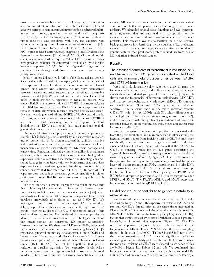
The baseline frequencies of micronuclei in red blood cellsand transcription of 131 genes in nucleated white bloodcells and mammary gland tissues differ between BALB/cand C57BL/6 female mice
We used a highly sensitive flow-cytometric assay to assess the
frequency of micronucleated red cells as a measure of genome
instability in unirradiated young adult female mice [21]. Figure 1B
shows that the frequencies of immature reticulocytes (MN-RET)
and mature normochromatic erythrocytes (MN-NCE) carrying
micronuclei were ,36% and ,57% higher in the radiation-
sensitive BALB/c strain than in the more radiation-resistant
C57BL/6 strain [14] (p,0.0001; Table S1). These differences are
at the high end of baseline variation among mouse strains [22],
and are consistent with the significant associations that have been
reported between blood micronuclei frequencies and cancer risks
in human studies [23].
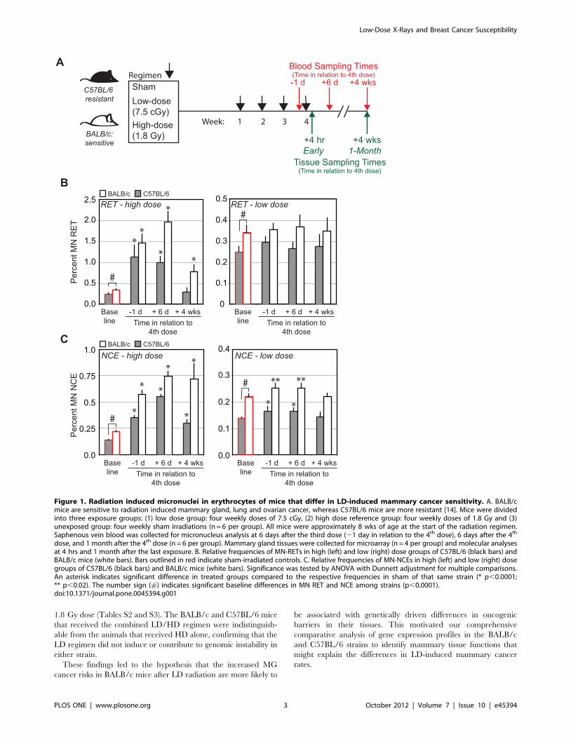
We also compared the transcript profiles for nucleated cells
from the peripheral blood and mammary glands (after excising the
inguinal lymph nodes) from BALB/c and C57BL/6 female mice
to identify common variations in gene expression and their
associated tissue functions. Figure 2A shows that the BALB/c to
C57BL/6 transcript ratios for the 131 genes comprising the
‘‘systemic baseline signature’’ are strongly correlated in blood and
levels than C57BL/5 for the DNA repair genes PARP3 and
RAD23A (not reported previously), and higher transcript levels for
MSH5 and SMC6. The PARP3, MSH5, and SMC6 expression
findings were confirmed by qPCR (Table S7).
LD did not induce or contribute to genomic instability ineither strain
We measured the frequencies of micronucleated red blood cells
after whole-body LD and HD exposures in sensitive BALB/c and
resistant C57BL/6 female mice at the three times indicated in
Figure 1A. The LD exposures induced small transient increases in
MN-NCE in both strains at the two early sampling times (p,0.02),
but neither strain showed evidence of radiation-induced genomic
instability at 1 month after exposure (Figure 1C). The HD
reference exposures (Figures 1B and 1C) increased in the
frequencies of MN-RET and MN-NCE at the early sampling
times in both strains (p,0.0001, Tables S2 and S3). Interestingly,
the radiation-sensitive BALB/c showed significant radiation-
induced genomic instability at 1 month after HD exposure while
the radiation-resistant C57BL/6 mice showed no evidence of this
(p,0.0001; Figure 1B, Tables S2 and S3). We confirmed this
finding in a separate study of mice treated with a combined LD/
HD regimen where each 7.5 cGy dose was followed 6 hr later by a
Low-Dose X-Rays and Breast Cancer Susceptibility
PLOS ONE | www.plosone.org 2 October 2012 | Volume 7 | Issue 10 | e45394
1.8 Gy dose (Tables S2 and S3). The BALB/c and C57BL/6 mice
that received the combined LD/HD regimen were indistinguish-
able from the animals that received HD alone, confirming that the
LD regimen did not induce or contribute to genomic instability in
either strain.
These findings led to the hypothesis that the increased MG
cancer risks in BALB/c mice after LD radiation are more likely to
be associated with genetically driven differences in oncogenic
barriers in their tissues. This motivated our comprehensive
comparative analysis of gene expression profiles in the BALB/c
and C57BL/6 strains to identify mammary tissue functions that
might explain the differences in LD-induced mammary cancer
rates.
Figure 1. Radiation induced micronuclei in erythrocytes of mice that differ in LD-induced mammary cancer sensitivity. A. BALB/cmice are sensitive to radiation induced mammary gland, lung and ovarian cancer, whereas C57BL/6 mice are more resistant [14]. Mice were dividedinto three exposure groups: (1) low dose group: four weekly doses of 7.5 cGy, (2) high dose reference group: four weekly doses of 1.8 Gy and (3)unexposed group: four weekly sham irradiations (n = 6 per group). All mice were approximately 8 wks of age at the start of the radiation regimen.Saphenous vein blood was collected for micronucleus analysis at 6 days after the third dose (21 day in relation to the 4th dose), 6 days after the 4th
dose, and 1 month after the 4th dose (n = 6 per group). Mammary gland tissues were collected for microarray (n = 4 per group) and molecular analysesat 4 hrs and 1 month after the last exposure. B. Relative frequencies of MN-RETs in high (left) and low (right) dose groups of C57BL/6 (black bars) andBALB/c mice (white bars). Bars outlined in red indicate sham-irradiated controls. C. Relative frequencies of MN-NCEs in high (left) and low (right) dosegroups of C57BL/6 (black bars) and BALB/c mice (white bars). Significance was tested by ANOVA with Dunnett adjustment for multiple comparisons.An asterisk indicates significant difference in treated groups compared to the respective frequencies in sham of that same strain (* p,0.0001;** p,0.02). The number sign (#) indicates significant baseline differences in MN RET and NCE among strains (p,0.0001).doi:10.1371/journal.pone.0045394.g001
Low-Dose X-Rays and Breast Cancer Susceptibility
PLOS ONE | www.plosone.org 3 October 2012 | Volume 7 | Issue 10 | e45394
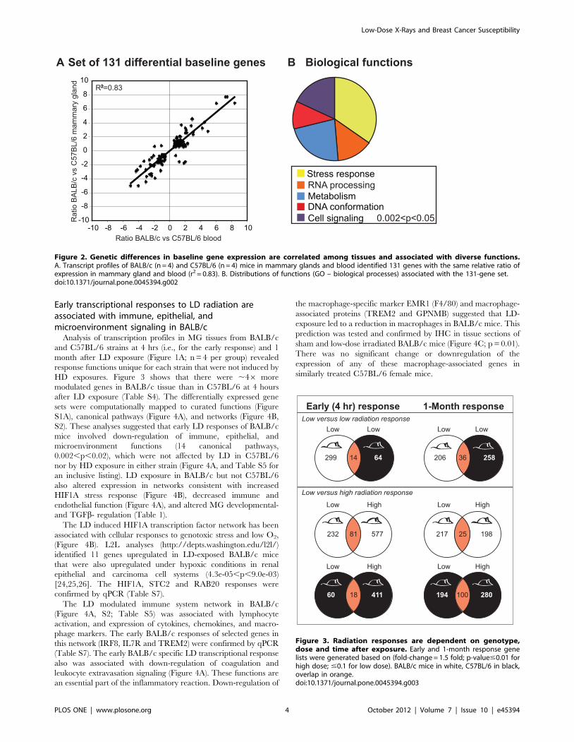
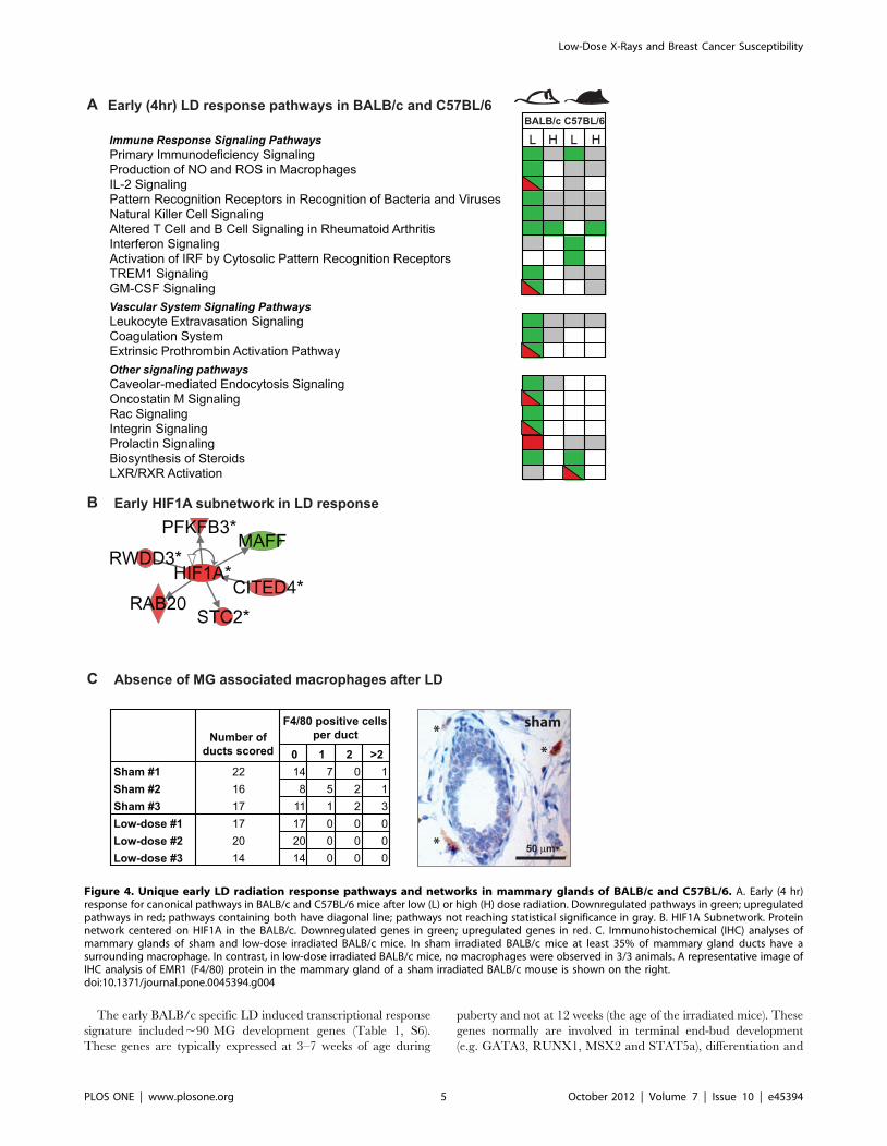
Early transcriptional responses to LD radiation areassociated with immune, epithelial, andmicroenvironment signaling in BALB/c
Analysis of transcription profiles in MG tissues from BALB/c
and C57BL/6 strains at 4 hrs (i.e., for the early response) and 1
month after LD exposure (Figure 1A; n = 4 per group) revealed
response functions unique for each strain that were not induced by
HD exposures. Figure 3 shows that there were ,46 more
modulated genes in BALB/c tissue than in C57BL/6 at 4 hours
after LD exposure (Table S4). The differentially expressed gene
sets were computationally mapped to curated functions (Figure
S1A), canonical pathways (Figure 4A), and networks (Figure 4B,
S2). These analyses suggested that early LD responses of BALB/c
mice involved down-regulation of immune, epithelial, and
identified 11 genes upregulated in LD-exposed BALB/c mice
that were also upregulated under hypoxic conditions in renal
epithelial and carcinoma cell systems (4.3e-05,p,9.0e-03)
[24,25,26]. The HIF1A, STC2 and RAB20 responses were
confirmed by qPCR (Table S7).
The LD modulated immune system network in BALB/c
(Figure 4A, S2; Table S5) was associated with lymphocyte
activation, and expression of cytokines, chemokines, and macro-
phage markers. The early BALB/c responses of selected genes in
this network (IRF8, IL7R and TREM2) were confirmed by qPCR
(Table S7). The early BALB/c specific LD transcriptional response
also was associated with down-regulation of coagulation and
leukocyte extravasation signaling (Figure 4A). These functions are
an essential part of the inflammatory reaction. Down-regulation of
the macrophage-specific marker EMR1 (F4/80) and macrophage-
associated proteins (TREM2 and GPNMB) suggested that LD-
exposure led to a reduction in macrophages in BALB/c mice. This
prediction was tested and confirmed by IHC in tissue sections of
sham and low-dose irradiated BALB/c mice (Figure 4C; p = 0.01).
There was no significant change or downregulation of the
expression of any of these macrophage-associated genes in
similarly treated C57BL/6 female mice.
Figure 2. Genetic differences in baseline gene expression are correlated among tissues and associated with diverse functions.A. Transcript profiles of BALB/c (n = 4) and C57BL/6 (n = 4) mice in mammary glands and blood identified 131 genes with the same relative ratio ofexpression in mammary gland and blood (r2 = 0.83). B. Distributions of functions (GO – biological processes) associated with the 131-gene set.doi:10.1371/journal.pone.0045394.g002
Figure 3. Radiation responses are dependent on genotype,dose and time after exposure. Early and 1-month response genelists were generated based on (fold-change = 1.5 fold; p-value#0.01 forhigh dose; #0.1 for low dose). BALB/c mice in white, C57BL/6 in black,overlap in orange.doi:10.1371/journal.pone.0045394.g003
Low-Dose X-Rays and Breast Cancer Susceptibility
PLOS ONE | www.plosone.org 4 October 2012 | Volume 7 | Issue 10 | e45394
The early BALB/c specific LD induced transcriptional response
signature included,90 MG development genes (Table 1, S6).
These genes are typically expressed at 3–7 weeks of age during
puberty and not at 12 weeks (the age of the irradiated mice). These
genes normally are involved in terminal end-bud development
(e.g. GATA3, RUNX1, MSX2 and STAT5a), differentiation and
Figure 4. Unique early LD radiation response pathways and networks in mammary glands of BALB/c and C57BL/6. A. Early (4 hr)response for canonical pathways in BALB/c and C57BL/6 mice after low (L) or high (H) dose radiation. Downregulated pathways in green; upregulatedpathways in red; pathways containing both have diagonal line; pathways not reaching statistical significance in gray. B. HIF1A Subnetwork. Proteinnetwork centered on HIF1A in the BALB/c. Downregulated genes in green; upregulated genes in red. C. Immunohistochemical (IHC) analyses ofmammary glands of sham and low-dose irradiated BALB/c mice. In sham irradiated BALB/c mice at least 35% of mammary gland ducts have asurrounding macrophage. In contrast, in low-dose irradiated BALB/c mice, no macrophages were observed in 3/3 animals. A representative image ofIHC analysis of EMR1 (F4/80) protein in the mammary gland of a sham irradiated BALB/c mouse is shown on the right.doi:10.1371/journal.pone.0045394.g004
Low-Dose X-Rays and Breast Cancer Susceptibility
PLOS ONE | www.plosone.org 5 October 2012 | Volume 7 | Issue 10 | e45394
ductal branching and morphogenesis. L2L analyses also showed
that the set of LD upregulated genes in BALB/c were highly
associated with the developing MG of pubertal mice in other
studies (p = 5.14e-30) [19]. CD24, KRT19, WNT4, AREG and
IDO1 responses in BALB/c mice were confirmed by qPCR (Table
S7). Importantly, ,50% of these early BALB/c LD response
genes (144/313) were TGFb responsive (Table 1, S6; http://actin.
ucd.ie/tgfbeta/, [20]). Extracellular TGFb activation occurs in
response to the generation of ROS [27] and regulates broad
epithelial and stromal radiation damage response functions of the
BALB/c LD genes. The differential activation of TGFb responsive
genes that were activated in BALB/c were not activated in
C57BL/6 mice, indicating that there is a major genetic difference
in TGFb response to LD radiation in the MGs in these two strains.
This is consistent with the increasing evidence of the regulatory
role of the TGFb response in radiation carcinogenesis of the
mammary gland [6].
Late MG transcriptional responses to LD radiation areassociated with proliferation, senescence, andmicroenvironment function
We saw a dramatic transition in the transcript profiles between
the early and 1-month responses in MG tissues in radiation-
sensitive BALB/c and radiation-resistant C57BL/6 strains of mice.
While, similar numbers of genes were modulated in these two
strains 1 month after LD exposures (Figure 3, Table S4), only a
few functions significantly modulated at 4 hours in BALB/c
remained so 1 month after LD exposure (Figure 5A, S1 and Table
S5). One month after exposure, 5 canonical pathways were
uniquely associated with C57BL/6 and a different 11 pathways
were unique to BALB/c (Figure 5A). BALB/c mice acquired an
enhanced proliferation phenotype (referenced to sham) while
C57BL/6 acquired a diminished proliferation phenotype, consis-
tent with senescence (Figure 5; S3). The BALB/c 1-month LD
response showed upregulation of a MYC-centric network consist-
ing of mitosis genes (Figure 5B) plus a subnetwork associated with
minichromosome maintenance, (Figure S3A). In contrast, the 1-
month response of C57BL/6 mice showed a highly saturated
protein interaction network with the network node, CDKN1A, a
negative regulator of cell cycle progression, (Figure 5B) and with
down-regulation of many genes associated with DNA replication,
cell-cycle progression and development. L2L analyses of the genes
in the C57BL/6-specific senescence signature identified significant
associations with expression signatures of cell cycle arrest and
senescence (1.13E-11,p,1.15E-04; [28,29,30]) including down-
regulation of SOX9, SKP2, CCNA2 and CDKN1A (confirmed by
qPCR; Table S7 and Figure 5B). SOX9 is a mark for adult human
progenitor cells, mediates deposition of ECM component and is a
major transcriptional regulator of mitotic activity in breast cancer
[31,32]. Consistent with its role in the control of expression of
ECM, we observed down-regulation of genes associated with
ECM remodeling and epithelial differentiation (not seen in BALB/
c mice) suggesting a reduced turn-over of the ECM in C57BL/6
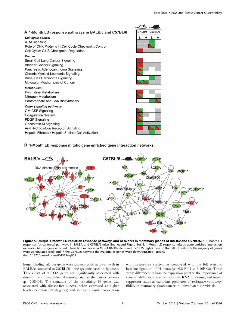
mice (Figure S3B). Figure 6 shows that expression of SOX9
protein in the MG was limited to the nuclei of luminal and
myoepithelial cells and that the fraction of SOX9-positive cells was
significantly reduced after LD exposure in C57BL/6 mice
(p,0.0001), consistent with reduced mitotic activity in MG of
C57BL/6 mice at 1 month after LD. Transcript levels of SOX9
were unaffected in similarly treated BALB/c mice.
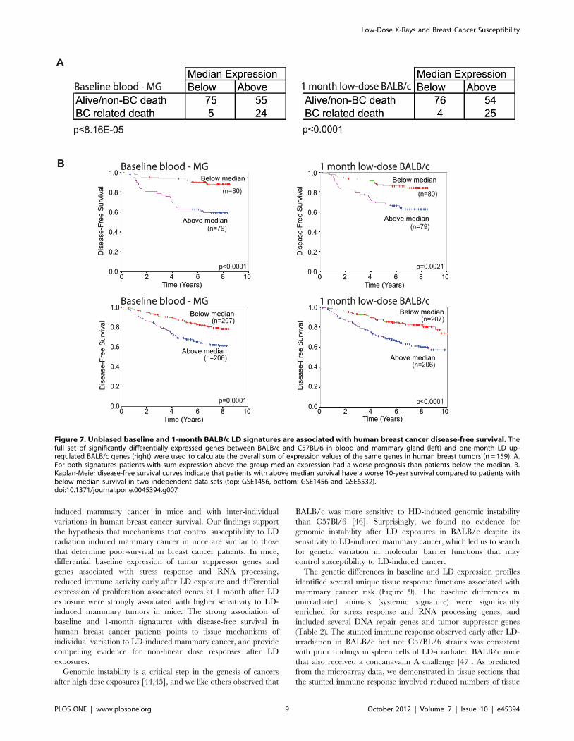
LD regulated genes in MG are associated with humanbreast cancer survival duration
We asked whether any of the strain specific baseline transcripts
or LD modulated transcripts were associated with human breast
cancer by using human knowledgebases that link expression
profiles with breast cancer outcomes.
We began by testing the hypothesis that expression levels of the
131 genes that are differentially expressed between non-irradiated
BALB/c and C57BL6 mice in blood and MG tissues are
associated with breast cancer outcomes. We tested the association
of transcript levels for the 94 human orthologs that we were able to
associate with the mouse baseline signature with outcome in 156
breast cancer patients for which information on disease-free
survival was available [17]. We accomplished this by calculating
for each patient, the sum of the normalized expression intensities
of the human orthologs. As shown in Figure 7A, patients with
above-median expression had significantly reduced survival
duration compared to patients with below-median expression
(p,8.16 E-05) and had significantly worse prognosis (Figure 7B;
p,0.0001) [16,17]. Interestingly, murine genes that showed
significant strain differences only in MG or only in blood cells
did not show significant associations with cancer survival,
underscoring the importance of selecting genes that show
‘‘systemic differences’’ across tissues. As a negative control,
expression levels of a set of 131 mouse genes that showed the
‘‘least’’ differential expression in both MG and blood between the
two strains were not significantly associated with breast cancer
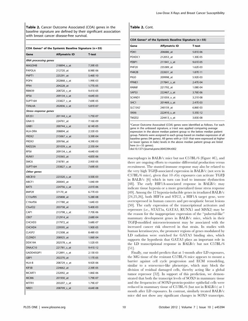
survival (p = 0.4). Among the 94 human orthologs in the murine
baseline signature, we identified 55 cancer outcome associated
(COA) genes (Table 2) that individually showed differential
expression between the above-median and below-median patient
groups (5.3E-12,p,8.4E-03). This set of COA genes was
enriched for stress response (11 genes) and RNA processing (9
genes) (Figure 2B and Table 2). Interestingly, this COA gene set
contained a small number of genes (n = 9), which showed a
significant association with breast cancer survival when expressed
at lower levels in the above-median patient group. Among these
are a number of previously proposed tumor suppressor genes:
RUNX1, CBX7, PRDX2 and PRDX3. Concordant with this
Table 1. The early LD radiation response in BALB/c mice is mediated by TGFb, involves inappropriate expression of mammarydevelopment genes, and involves breast cancer associated genes.
Gene Function Early (4-hr) LD responsive genes in BALB/c mice (total = 313)
TGFb responsivea 144 (46%)
MG development [19] 89b (28%)
Breast-cancer associated [18] 41 (13%)
aTGFb signaling and interaction database (http://actin.ucd.ie/tgfbeta/) and [20].b42 genes overlap with TGFb responsive genes.doi:10.1371/journal.pone.0045394.t001
Low-Dose X-Rays and Breast Cancer Susceptibility
PLOS ONE | www.plosone.org 6 October 2012 | Volume 7 | Issue 10 | e45394
human finding, all four genes were also expressed at lower levels in
BALB/c compared to C57BL/6 in the systemic baseline signature.
This subset of 9 COA genes was significantly associated with
disease free survival when down-regulated in the cancer patients
(p = 5.5E-04). The signature of the remaining 46 genes was
associated with disease-free survival when expressed at higher
levels (55 minus 9 = 46 genes) and showed a similar association
with disease-free survival as compared with the full systemic
baseline signature of 94 genes (p = 6.8 E-03 vs 8.16E-05). These
strain differences in baseline expression point to the importance of
systemic differences in stress response, RNA processing and tumor
suppressor status as candidate predictors of resistance vs suscep-
tibility to mammary gland cancer in unirradiated individuals.
Figure 5. Unique 1-month LD radiation response pathways and networks in mammary glands of BALB/c and C57BL/6. A. 1-Month LDresponses for canonical pathways in BALB/c and C57BL/6 mice (See legend Figure 4A). B. 1-Month LD response mitotic gene enriched interactionnetworks. Mitosis gene enriched interaction networks in MG of BALB/c (left) and C57BL/6 (right) mice. In the BALB/c network the majority of geneswere upregulated (red) and in the C57BL/6 network the majority of genes were downregulated (green).doi:10.1371/journal.pone.0045394.g005
Low-Dose X-Rays and Breast Cancer Susceptibility
PLOS ONE | www.plosone.org 7 October 2012 | Volume 7 | Issue 10 | e45394
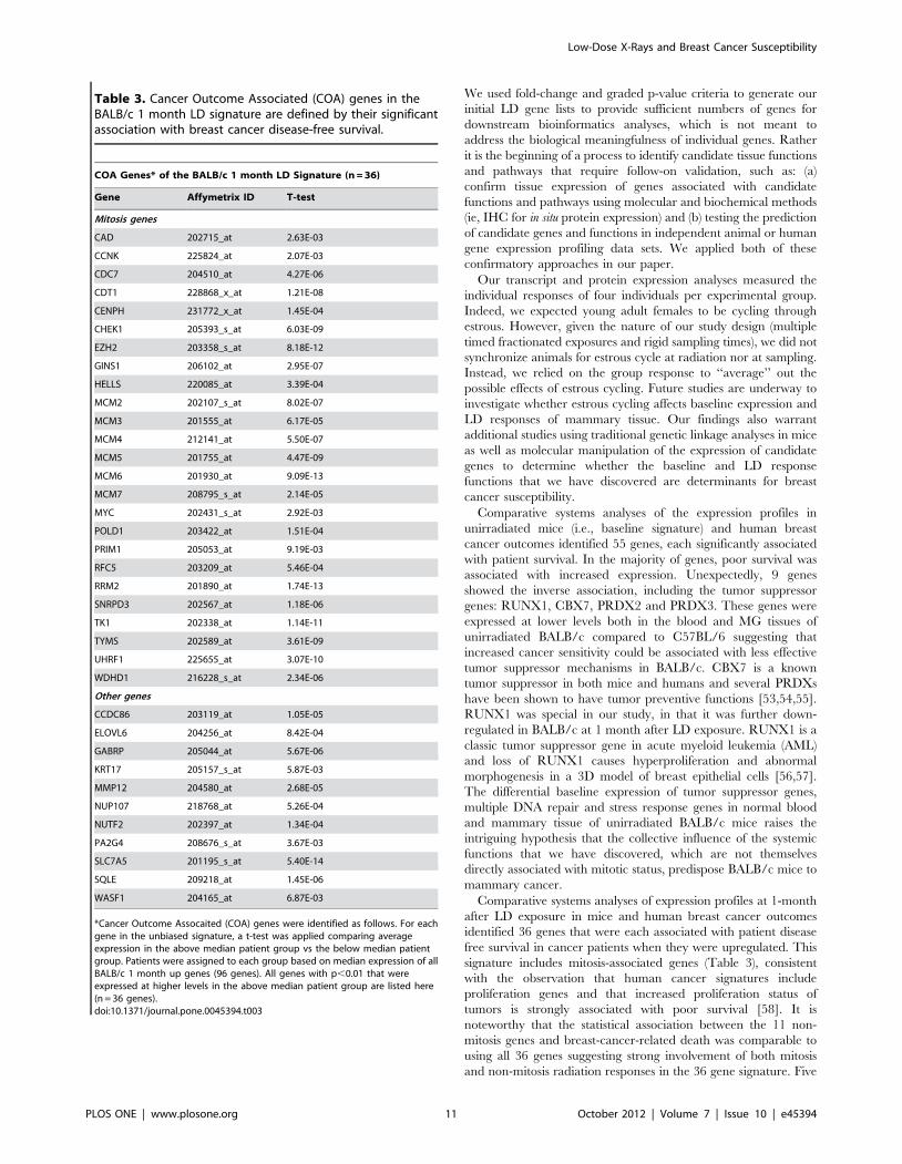
We then tested the hypothesis that the 1-month BALB/c
signature (i.e., the genes that are significantly upregulated at 1
month after LD exposure in relation to sham) was associated with
disease-free survival among breast cancer patients. We selected the
full and unbiased set of 105 BALB/c genes with significantly
increased expression at 1 month after LD exposure. We examined
the association of this signature with disease-free survival in breast
cancer patients using two human knowledgebases that contain
tumor expression profiles obtained at surgery linked to patient
survival [16,17]. Similar to our analyses of the baseline signature,
we summed the expression intensities of all corresponding human
orthologs (n = 96) from tumor samples and divided the patients
into two groups by the median. The patients with ‘‘above median’’
expression values experienced higher rates of breast-cancer related
deaths than ‘‘below median’’ patients, (Figure 7A, p,0.0001) and
had significantly worse prognosis (Figure 7B; p = ,0.0021)
[16,17]. As a negative control, we selected 105 of the 1-month
BALB/c genes with the ‘‘least’’ differential expression between
irradiated and sham mice, and showed that the corresponding set
of human orthologs was not significantly associated with breast
cancer survival (p = 0.2). Among the 96 human orthologs of the 1-
month BALB/c signature, we identified 36 additional COA genes
(Table 3) that individually showed differential expression between
the above-median and below-median patient groups (5.4E-
14,p,9.2E-03). Of these, 25 were related to mitosis, many of
which have individually been associated with breast cancer
survival in prior studies. Six of the 11 non-mitosis related genes
were previously associated with poor survival in breast cancer
patients: KRT17, MMP12, SLC7A5, SQLE, GABRP and PA2G4
[33,34,35,36,37,38], but the remaining 5 had not been previously
associated with cancer risk: CCDC86, NUP107, NUTF2, WASF1
and ELOVL6. The association between the 11 non-mitosis genes
and breast-cancer-related death was comparable to using the full
set of 96 human orthologs or the refined set of 36 genes suggesting
strong involvement of both mitosis and non-mitosis radiation
responses in defining the poor prognosis for breast cancer patients.
These strain differences in 1-month post LD expression responses
point to the importance of both mitosis and non-mitosis genes as
candidate predictors of resistance vs susceptibility to mammary
gland cancer in individuals exposed to LD ionizing radiation.
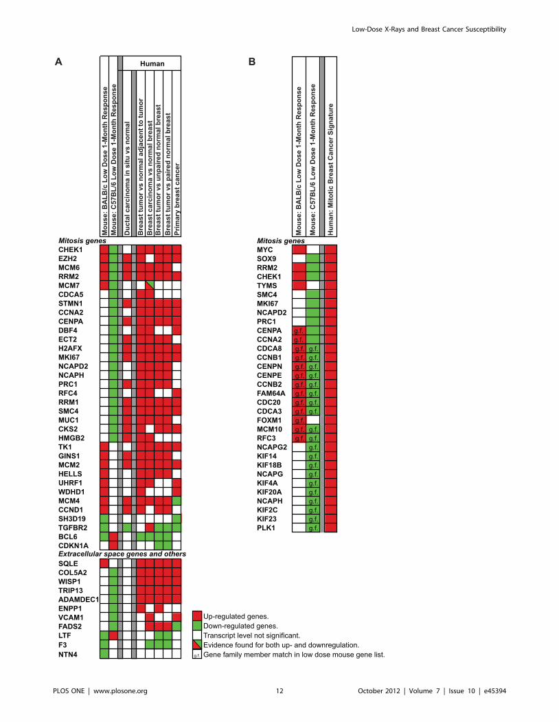
We then addressed the hypothesis that the direction of
expression changes in the resistant and sensitive strains at 1-
month after LD exposures are concordant with the differences in
gene expression of human DCIS and invasive breast cancer, and
poor prognosis. (Concordance is defined as up-regulated in
BALB/c, no change or down-regulated in C57BL/6 and up in
human DCIS or human breast cancer; or vice versa). The BALB/
c 1 month COA genes that were associated with poor prognosis
when upregulated were enriched for mitosis associated genes
(Table 3), whereas in C57BL/6 many of the same genes were
down-regulated. As a further test of the importance of concor-
dance in direction of expression, we compared the full set of genes
modulated at 1-month after LD exposure in BALB/c and C57BL/
6 mice against a meta-gene signature of 946 human breast cancer
biomarkers [18], and the direction of expression for overlapping
genes was compared against expression in human DCIS and
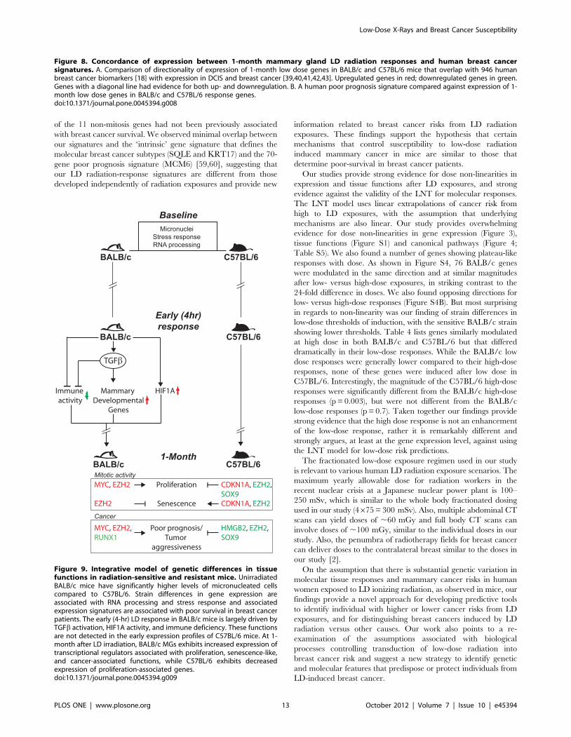
breast cancer [39,40,41,42,43]. This analyses (Figure 8A) identi-
fied 45 concordant genes (34 mitosis and 11 stromal genes) with
opposing responses in BALB/c and C57BL/6 where the direction
of the BALB/c response matched the direction of response in
independent studies of human breast cancers. Furthermore, a
subset of 19 genes (Figure 8A) showed concordant responses
between the mouse strains and human DCIS.
Lastly, we tested the association between the 1-month LD
BALB/c signature and poor prognosis for human breast cancer.
Figure 8B shows strong concordance in the direction of LD-
induced expression in mammary tissue in the resistant and
sensitive strains of mice and the direction of expression in patients
with poor prognosis (Figure 8B). The human poor prognosis
signature is under transcriptional control of SOX9, which we
demonstrated to be down-regulated at both the transcript level
and protein level in the resistant C57BL/6 strain (Figure 5B and
6).
Discussion
We employed in vivo systems analyses to identify genetic
differences in baseline (i.e., before radiation exposure) and LD-
induced mammary gland gene expression in radiation-sensitive
and resistant strains of mice that are at the far ends of the
mammary cancer sensitivity spectrum [12], and then used in situ
protein validation and multi-species bioinformatic resources to
identify distinct tissue functions and signatures associated with
cancer risks and with properties of breast cancer behavior in
humans. Strain variations in baseline and LD-response expression
signatures were associated with differential susceptibility to LD-
Figure 6. Reduction in SOX9-labelled luminal and myoepithelial cells after LD exposure in C57BL/6 mammary glands. A. The percentSOX9 positive luminal and myoepithelial cells are significantly reduced in low-dose irradiated mammary glands 1-month after low-dose exposure(p,0.0001). B. Representative images of immunohistochemical analysis of SOX9 protein in mammary glands of sham (left) or LD (right) irradiatedC57BL/6 mice. Myoepithelial cells were defined as those cells directly surrounding the luminal cell layer. Counterstaining was performed usinghematoxylin. Note positive staining is limited to the nucleus in cells of luminal or myoepithelial origin.doi:10.1371/journal.pone.0045394.g006
Low-Dose X-Rays and Breast Cancer Susceptibility
PLOS ONE | www.plosone.org 8 October 2012 | Volume 7 | Issue 10 | e45394
induced mammary cancer in mice and with inter-individual
variations in human breast cancer survival. Our findings support
the hypothesis that mechanisms that control susceptibility to LD
radiation induced mammary cancer in mice are similar to those
that determine poor-survival in breast cancer patients. In mice,
differential baseline expression of tumor suppressor genes and
genes associated with stress response and RNA processing,
reduced immune activity early after LD exposure and differential
expression of proliferation associated genes at 1 month after LD
exposure were strongly associated with higher sensitivity to LD-
induced mammary tumors in mice. The strong association of
baseline and 1-month signatures with disease-free survival in
human breast cancer patients points to tissue mechanisms of
individual variation to LD-induced mammary cancer, and provide
compelling evidence for non-linear dose responses after LD
exposures.
Genomic instability is a critical step in the genesis of cancers
after high dose exposures [44,45], and we like others observed that
BALB/c was more sensitive to HD-induced genomic instability
than C57Bl/6 [46]. Surprisingly, we found no evidence for
genomic instability after LD exposures in BALB/c despite its
sensitivity to LD-induced mammary cancer, which led us to search
for genetic variation in molecular barrier functions that may
control susceptibility to LD-induced cancer.
The genetic differences in baseline and LD expression profiles
identified several unique tissue response functions associated with
mammary cancer risk (Figure 9). The baseline differences in
unirradiated animals (systemic signature) were significantly
enriched for stress response and RNA processing genes, and
included several DNA repair genes and tumor suppressor genes
(Table 2). The stunted immune response observed early after LD-
irradiation in BALB/c but not C57BL/6 strains was consistent
with prior findings in spleen cells of LD-irradiated BALB/c mice
that also received a concanavalin A challenge [47]. As predicted
from the microarray data, we demonstrated in tissue sections that
the stunted immune response involved reduced numbers of tissue
Figure 7. Unbiased baseline and 1-month BALB/c LD signatures are associated with human breast cancer disease-free survival. Thefull set of significantly differentially expressed genes between BALB/c and C57BL/6 in blood and mammary gland (left) and one-month LD up-regulated BALB/c genes (right) were used to calculate the overall sum of expression values of the same genes in human breast tumors (n = 159). A.For both signatures patients with sum expression above the group median expression had a worse prognosis than patients below the median. B.Kaplan-Meier disease-free survival curves indicate that patients with above median survival have a worse 10-year survival compared to patients withbelow median survival in two independent data-sets (top: GSE1456, bottom: GSE1456 and GSE6532).doi:10.1371/journal.pone.0045394.g007
Low-Dose X-Rays and Breast Cancer Susceptibility
PLOS ONE | www.plosone.org 9 October 2012 | Volume 7 | Issue 10 | e45394
macrophages in BALB/c mice but not C57BL/6 (Figure 4C), and
there are ongoing efforts to examine differential production versus
recruitment. The stunted immune response may also be related to
the very high TGFb-associated expression in BALB/c (not seen in
C57BL/6 mice), given that 10 cGy exposures can activate TGFbin BALB/c [6] which in turn can lead to immune dysfunction
[48]. The early HIF1A-associated response in BALB/c may
indicate tissue hypoxia or a more generalized tissue stress response
[49]. Among the 12 hypoxia-inducible genes in irradiated BALB/c
[24,25,26], both HIF1a and STC2, a HIF1A target gene, were
overexpressed in human cancers and pre-neoplastic breast lesions
[50]. The early expression of the transcriptional activators and
repressors (i.e., STAT5a, GATA3, RUNX1 and MSX2) may be
the reason for the inappropriate expression of the ‘‘pubertal-like’’
mammary development genes in BALB/c mice, which in their
TGFb-modified microenvironment may be associated with the
increased cancer risk observed in that strain. In studies with
human keratinocytes, the promoter regions of genes modulated by
LD radiation were enriched for GATA3 binding sites, which
supports the hypothesis that GATA3 plays an important role in
the LD transcriptional response in BALB/c but not C57BL/6
[51].
Finally, our model predicts that at 1-month after LD exposures,
the MG tissue of the resistant C57BL/6 mice appears to mount a
barrier against cell cycle progression and ECM remodeling,
similar to a senescence-like phenotype, which may block the
division of residual damaged cells, thereby acting like a global
tumor repressor [52]. In support of this prediction, we demon-
strated that both the transcript levels of SOX9 in mammary tissue
and the frequencies of SOX9-protein-positive epithelial cells were
reduced in mammary tissue of C57BL/6 (but not in BALB/c) at 1
month after LD exposures. In contrast, similarly treated BALB/c
mice did not show any significant changes in SOX9 transcripts.
Table 2. Cancer Outcome Associated (COA) genes in thebaseline signature are defined by their significant associationwith breast cancer disease-free survival.
COA Genes* of the Systemic Baseline Signature (n = 55)
Gene Affymetrix ID T-test
RNA processing genes
MAGOHB 218894_s_at 7.39E-05
PAPOLA 212720_at 8.98E-06
PNPT1 225291_at 5.46E-10
POP4 202868_s_at 1.99E-03
PPIH 204228_at 1.77E-05
RBM39 208720_s_at 9.41E-05
RPS6 209134_s_at 4.64E-03
SUPT16H 233827_s_at 7.69E-05
TXNL4A 202836_s_at 5.81E-07
Stress response genes
EIF2S1 201144_s_at 1.75E-07
GNA13 224761_at 7.16E-09
GNB1 200744_s_at 8.14E-05
HLA-DRA 208894_at 2.33E-05
PRDX2 215067_x_at 3.39E-04
PRDX3 209766_at 4.39E-03
RAD23A 201039_s_at 2.33E-04
RPS6 209134_s_at 4.64E-03
RUNX1 210365_at 4.85E-06
SMC6 218781_at 2.95E-05
SUPT16H 233827_s_at 7.69E-05
Other genes
ABCB10 223320_s_at 3.50E-03
ABCF1 200045_at 1.04E-09
BAT5 224756_s_at 2.01E-06
BMP2K 37170_at 6.77E-03
C17orf95 225808_at 5.28E-08
C19orf56 217780_at 1.64E-03
C5orf22 203738_at 5.40E-03
CAP1 213798_s_at 7.70E-06
CBX7 212914_at 2.68E-04
CHCHD3 217972_at 5.68E-07
CHCHD4 229595_at 1.90E-05
CLASP2 212308_at 8.48E-03
CLDND1 208925_at 1.06E-04
DDX19A 202578_s_at 1.52E-05
DNAJC10 221781_s_at 9.41E-12
GADD45GIP1 212891_s_at 2.13E-03
GBP1 202270_at 1.17E-05
HLA-B 208729_x_at 9.92E-06
KIF5B 224662_at 2.39E-05
MCART1 232092_at 1.86E-06
MCM6 201930_at 1.79E-07
MTFR1 203207_s_at 1.79E-07
NRD1 208709_s_at 4.64E-08
Table 2. Cont.
COA Genes* of the Systemic Baseline Signature (n = 55)
Gene Affymetrix ID T-test
PDK1 206686_at 5.81E-06
PDXDC1 212053_at 1.30E-05
PEBP1 211941_s_at 9.61E-05
PHF20 235389_at 1.62E-03
PI4K2B 222631_at 1.87E-11
PIGO 209998_at 5.92E-03
PPME1 217841_s_at 2.47E-04
RAB6B 221792_at 1.08E-04
SAPS3 222467_s_at 3.76E-06
SCAND1 231059_x_at 3.21E-08
SHC1 201469_s_at 2.47E-03
SLC15A2 240159_at 6.86E-03
SNX6 222410_s_at 5.35E-12
TM2D2 224413_s_at 3.83E-08
*Cancer Outcome Assocaited (COA) genes were identified as follows. For eachgene in the unbiased signature, a t-test was applied comparing averageexpression in the above median patient group vs the below median patientgroup. Patients were assigned to each group based on median expression of allbaseline genes (94 genes). All genes with p,0.01 that were expressed at higheror lower (genes in italic) levels in the above median patient group are listedhere (n = 55 genes).doi:10.1371/journal.pone.0045394.t002
Low-Dose X-Rays and Breast Cancer Susceptibility
PLOS ONE | www.plosone.org 10 October 2012 | Volume 7 | Issue 10 | e45394
We used fold-change and graded p-value criteria to generate our
initial LD gene lists to provide sufficient numbers of genes for
downstream bioinformatics analyses, which is not meant to
address the biological meaningfulness of individual genes. Rather
it is the beginning of a process to identify candidate tissue functions
and pathways that require follow-on validation, such as: (a)
confirm tissue expression of genes associated with candidate
functions and pathways using molecular and biochemical methods
(ie, IHC for in situ protein expression) and (b) testing the prediction
of candidate genes and functions in independent animal or human
gene expression profiling data sets. We applied both of these
confirmatory approaches in our paper.
Our transcript and protein expression analyses measured the
individual responses of four individuals per experimental group.
Indeed, we expected young adult females to be cycling through
estrous. However, given the nature of our study design (multiple
timed fractionated exposures and rigid sampling times), we did not
synchronize animals for estrous cycle at radiation nor at sampling.
Instead, we relied on the group response to ‘‘average’’ out the
possible effects of estrous cycling. Future studies are underway to
investigate whether estrous cycling affects baseline expression and
LD responses of mammary tissue. Our findings also warrant
additional studies using traditional genetic linkage analyses in mice
as well as molecular manipulation of the expression of candidate
genes to determine whether the baseline and LD response
functions that we have discovered are determinants for breast
cancer susceptibility.
Comparative systems analyses of the expression profiles in
unirradiated mice (i.e., baseline signature) and human breast
cancer outcomes identified 55 genes, each significantly associated
with patient survival. In the majority of genes, poor survival was
associated with increased expression. Unexpectedly, 9 genes
showed the inverse association, including the tumor suppressor
genes: RUNX1, CBX7, PRDX2 and PRDX3. These genes were
expressed at lower levels both in the blood and MG tissues of
unirradiated BALB/c compared to C57BL/6 suggesting that
increased cancer sensitivity could be associated with less effective
tumor suppressor mechanisms in BALB/c. CBX7 is a known
tumor suppressor in both mice and humans and several PRDXs
have been shown to have tumor preventive functions [53,54,55].
RUNX1 was special in our study, in that it was further down-
regulated in BALB/c at 1 month after LD exposure. RUNX1 is a
classic tumor suppressor gene in acute myeloid leukemia (AML)
and loss of RUNX1 causes hyperproliferation and abnormal
morphogenesis in a 3D model of breast epithelial cells [56,57].
The differential baseline expression of tumor suppressor genes,
multiple DNA repair and stress response genes in normal blood
and mammary tissue of unirradiated BALB/c mice raises the
intriguing hypothesis that the collective influence of the systemic
functions that we have discovered, which are not themselves
directly associated with mitotic status, predispose BALB/c mice to
mammary cancer.
Comparative systems analyses of expression profiles at 1-month
after LD exposure in mice and human breast cancer outcomes
identified 36 genes that were each associated with patient disease
free survival in cancer patients when they were upregulated. This
signature includes mitosis-associated genes (Table 3), consistent
with the observation that human cancer signatures include
proliferation genes and that increased proliferation status of
tumors is strongly associated with poor survival [58]. It is
noteworthy that the statistical association between the 11 non-
mitosis genes and breast-cancer-related death was comparable to
using all 36 genes suggesting strong involvement of both mitosis
and non-mitosis radiation responses in the 36 gene signature. Five
Table 3. Cancer Outcome Associated (COA) genes in theBALB/c 1 month LD signature are defined by their significantassociation with breast cancer disease-free survival.
COA Genes* of the BALB/c 1 month LD Signature (n = 36)
Gene Affymetrix ID T-test
Mitosis genes
CAD 202715_at 2.63E-03
CCNK 225824_at 2.07E-03
CDC7 204510_at 4.27E-06
CDT1 228868_x_at 1.21E-08
CENPH 231772_x_at 1.45E-04
CHEK1 205393_s_at 6.03E-09
EZH2 203358_s_at 8.18E-12
GINS1 206102_at 2.95E-07
HELLS 220085_at 3.39E-04
MCM2 202107_s_at 8.02E-07
MCM3 201555_at 6.17E-05
MCM4 212141_at 5.50E-07
MCM5 201755_at 4.47E-09
MCM6 201930_at 9.09E-13
MCM7 208795_s_at 2.14E-05
MYC 202431_s_at 2.92E-03
POLD1 203422_at 1.51E-04
PRIM1 205053_at 9.19E-03
RFC5 203209_at 5.46E-04
RRM2 201890_at 1.74E-13
SNRPD3 202567_at 1.18E-06
TK1 202338_at 1.14E-11
TYMS 202589_at 3.61E-09
UHRF1 225655_at 3.07E-10
WDHD1 216228_s_at 2.34E-06
Other genes
CCDC86 203119_at 1.05E-05
ELOVL6 204256_at 8.42E-04
GABRP 205044_at 5.67E-06
KRT17 205157_s_at 5.87E-03
MMP12 204580_at 2.68E-05
NUP107 218768_at 5.26E-04
NUTF2 202397_at 1.34E-04
PA2G4 208676_s_at 3.67E-03
SLC7A5 201195_s_at 5.40E-14
SQLE 209218_at 1.45E-06
WASF1 204165_at 6.87E-03
*Cancer Outcome Assocaited (COA) genes were identified as follows. For eachgene in the unbiased signature, a t-test was applied comparing averageexpression in the above median patient group vs the below median patientgroup. Patients were assigned to each group based on median expression of allBALB/c 1 month up genes (96 genes). All genes with p,0.01 that wereexpressed at higher levels in the above median patient group are listed here(n = 36 genes).doi:10.1371/journal.pone.0045394.t003
Low-Dose X-Rays and Breast Cancer Susceptibility
PLOS ONE | www.plosone.org 11 October 2012 | Volume 7 | Issue 10 | e45394
Low-Dose X-Rays and Breast Cancer Susceptibility
PLOS ONE | www.plosone.org 12 October 2012 | Volume 7 | Issue 10 | e45394
of the 11 non-mitosis genes had not been previously associated
with breast cancer survival. We observed minimal overlap between
our signatures and the ‘intrinsic’ gene signature that defines the
molecular breast cancer subtypes (SQLE and KRT17) and the 70-
gene poor prognosis signature (MCM6) [59,60], suggesting that
our LD radiation-response signatures are different from those
developed independently of radiation exposures and provide new
information related to breast cancer risks from LD radiation
exposures. These findings support the hypothesis that certain
mechanisms that control susceptibility to low-dose radiation
induced mammary cancer in mice are similar to those that
determine poor-survival in breast cancer patients.
Our studies provide strong evidence for dose non-linearities in
expression and tissue functions after LD exposures, and strong
evidence against the validity of the LNT for molecular responses.
The LNT model uses linear extrapolations of cancer risk from
high to LD exposures, with the assumption that underlying
mechanisms are also linear. Our study provides overwhelming
evidence for dose non-linearities in gene expression (Figure 3),
tissue functions (Figure S1) and canonical pathways (Figure 4;
Table S5). We also found a number of genes showing plateau-like
responses with dose. As shown in Figure S4, 76 BALB/c genes
were modulated in the same direction and at similar magnitudes
after low- versus high-dose exposures, in striking contrast to the
24-fold difference in doses. We also found opposing directions for
low- versus high-dose responses (Figure S4B). But most surprising
in regards to non-linearity was our finding of strain differences in
low-dose thresholds of induction, with the sensitive BALB/c strain
at high dose in both BALB/c and C57BL/6 but that differed
dramatically in their low-dose responses. While the BALB/c low
dose responses were generally lower compared to their high-dose
responses, none of these genes were induced after low dose in
C57BL/6. Interestingly, the magnitude of the C57BL/6 high-dose
responses were significantly different from the BALB/c high-dose
responses (p = 0.003), but were not different from the BALB/c
low-dose responses (p = 0.7). Taken together our findings provide
strong evidence that the high dose response is not an enhancement
of the low-dose response, rather it is remarkably different and
strongly argues, at least at the gene expression level, against using
the LNT model for low-dose risk predictions.
The fractionated low-dose exposure regimen used in our study
is relevant to various human LD radiation exposure scenarios. The
maximum yearly allowable dose for radiation workers in the
recent nuclear crisis at a Japanese nuclear power plant is 100–
250 mSv, which is similar to the whole body fractionated dosing
used in our study (4675 = 300 mSv). Also, multiple abdominal CT
scans can yield doses of ,60 mGy and full body CT scans can
involve doses of ,100 mGy, similar to the individual doses in our
study. Also, the penumbra of radiotherapy fields for breast cancer
can deliver doses to the contralateral breast similar to the doses in
our study [2].
On the assumption that there is substantial genetic variation in
molecular tissue responses and mammary cancer risks in human
women exposed to LD ionizing radiation, as observed in mice, our
findings provide a novel approach for developing predictive tools
to identify individual with higher or lower cancer risks from LD
exposures, and for distinguishing breast cancers induced by LD
radiation versus other causes. Our work also points to a re-
examination of the assumptions associated with biological
processes controlling transduction of low-dose radiation into
breast cancer risk and suggest a new strategy to identify genetic
and molecular features that predispose or protect individuals from
LD-induced breast cancer.
Figure 8. Concordance of expression between 1-month mammary gland LD radiation responses and human breast cancersignatures. A. Comparison of directionality of expression of 1-month low dose genes in BALB/c and C57BL/6 mice that overlap with 946 humanbreast cancer biomarkers [18] with expression in DCIS and breast cancer [39,40,41,42,43]. Upregulated genes in red; downregulated genes in green.Genes with a diagonal line had evidence for both up- and downregulation. B. A human poor prognosis signature compared against expression of 1-month low dose genes in BALB/c and C57BL/6 response genes.doi:10.1371/journal.pone.0045394.g008
Figure 9. Integrative model of genetic differences in tissuefunctions in radiation-sensitive and resistant mice. UnirradiatedBALB/c mice have significantly higher levels of micronucleated cellscompared to C57BL/6. Strain differences in gene expression areassociated with RNA processing and stress response and associatedexpression signatures are associated with poor survival in breast cancerpatients. The early (4-hr) LD response in BALB/c mice is largely driven byTGFb activation, HIF1A activity, and immune deficiency. These functionsare not detected in the early expression profiles of C57BL/6 mice. At 1-month after LD irradiation, BALB/c MGs exhibits increased expression oftranscriptional regulators associated with proliferation, senescence-like,and cancer-associated functions, while C57BL/6 exhibits decreasedexpression of proliferation-associated genes.doi:10.1371/journal.pone.0045394.g009
Low-Dose X-Rays and Breast Cancer Susceptibility
PLOS ONE | www.plosone.org 13 October 2012 | Volume 7 | Issue 10 | e45394
Materials and Methods
Ethics StatementFemale, virgin C57BL/6 and BALB/c mice (,6 weeks old;
Harlan Laboratories, Livermore, CA) were acclimatized for 2
weeks, and the study was carried out in strict accordance with the
Guide for the Care and Use of Laboratory Animals of the National
Institutes of Health. The protocol was approved by the Committee
on the Ethics of Animal Experiments of the Lawrence Berkeley
National Laboratory (Approval number: 25001). At 8 weeks of
age, mice (n = 6 per group) were exposed to 4 weekly doses of (a)
7.5 cGy, (b) 1.8 Gy, or (c) sham, using a Pantak 320 kVp X-ray
machine, operated at 300 kV (2 mA and 196 mGy/min for low
dose, 10 mA and 783 mGy/min for high dose).
Micronucleus AnalysesFor the analyses of micronucleated red blood cells, peripheral
blood was collected from each mouse (n = 6 per group) at 6 days
after each weekly irradiation and at 28 days after the fourth
irradiation. Approximately 100 ml of blood was collected per time
point from the saphenous vein [61] and processed with the
MicroFlowBASIC kit for the mouse (Litron Laboratories, Roches-
ter, NY) according to the manufacturer’s instructions. Samples
were kept at 280uC until shipment to Litron Laboratories where
they were analyzed by flow cytometry for the frequencies of
micronucleated reticulocytes (MN-RET) and micronucleated
normochromatic erythrocytes (MN-NCE) [21]. Frequencies of
MN-RET and MN-NCE of exposed mice were compared against
the respective frequencies in sham of that same strain by ANOVA
with Dunnett adjustment for multiple comparisons. A p-value less
than 0.05 was considered significant. Differences in baseline MN
frequencies were compared using two-tailed T-test with unequal
variance.
RNA isolation, expression profiling and bioinformaticsAt 4 hours and 1-month after the last exposure we harvested the
4th pair of mammary glands and removed their inguinal lymph
nodes; mice were randomized and individually processed for RNA
isolations (See Text S1, for details). Microarray hybridizations
were carried out on each of four independent mice per dose group
(n = 4) using Affymetrix’s HT Mouse Genome 430A 96-Array
Plate. The data has been deposited at NCBI GEO with accession
number GSE40066 (http://www.ncbi.nlm.nih.gov/geo/). RMA
was used to create an expression matrix and NUSE was used to
assess array quality. The following bioinformatics software tools
and databases were used (see Text S1, for details.): L2L (http://
edu/webgestalt/) DAVID (http://david.abcc.ncifcrf.gov/), puber-
tal mammary gland development genes [19], TGFb-responsive
genes (http://actin.ucd.ie/tgfbeta/ and [20]), 942 biomarkers of
breast cancer [18] and gene expression in human DCIS and breast
cancers (http://www.nextbio.com/).
Human breast cancer datasets of disease-free survivalExpression levels of human orthologs of overlapping genes in
blood and mammary gland tissue of unirradiated BALB/c and
C57BL/6 mice and an unbiased set of 105 up-regulated 1-month
BALB/c-specific low dose genes (i.e., genes not up-regulated in
C57BL/6) were summed in breast cancer samples of patients from
two independent curated breast cancer data sets (GSE1456 and
GSE6532) [16,17]. The median expression value was used as a
cut-point to assess group survival outcomes. A Kaplan-Meier
disease-free survival curve was generated for patients with above
median and below median expression. Log-rank tests were
performed to compare the difference in disease-free survival
between patients in the two clusters. See Text S1 for details.
Supporting Information
Figure S1 Tissue functions associated with the earlyand 1-month LD response in BALB/c and C57BL/6mammary glands. Distributions of predicted functions (based
on GO, KEGG, IPA Canonical Pathways, Genes of Interest) at
early (4 hr) and 1-month sampling times in both strains. Numbers
of genes used to generate charts are listed (unique genes in
parentheses).
(EPS)
Figure S2 Early LD radiation response networks in themammary glands of BALB/c mice show broad down-regulation of immune response genes. The top two protein
interaction networks for the BALB/c low dose genes include the
functions of Inflammatory Disease/Cell Mediated Immune
Response/Cellular Movement (top) and Cellular Growth, Prolif-
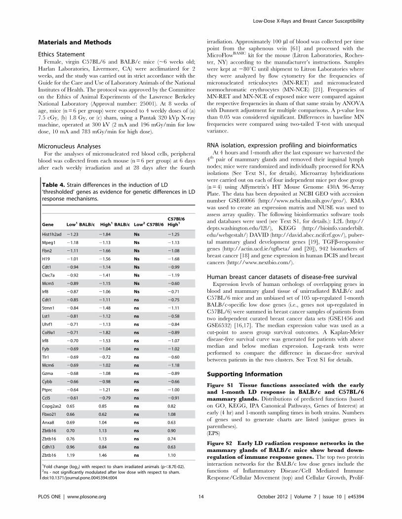
Table 4. Strain differences in the induction of LD‘thresholded’ genes as evidence for genetic differences in LDresponse mechanisms.
1Fold change (log2) with respect to sham irradiated animals (p,8.7E-02).2ns - not significantly modulated after low dose with respect to sham.doi:10.1371/journal.pone.0045394.t004
Low-Dose X-Rays and Breast Cancer Susceptibility
PLOS ONE | www.plosone.org 14 October 2012 | Volume 7 | Issue 10 | e45394
eration/Hematological System Development, and Function/
Humoral Immune Response (bottom). Upregulated and down-
regulated genes are represented in red and green, respectively.
(EPS)
Figure S3 Genetic differences in protein-interactionnetworks in mammary glands of BALB/c and C57BL/6mice at 1-month after LD exposures. A. Gene interaction
network enriched for genes involved in DNA replication
containing mostly MCM family genes were found to contain
mostly upregulated genes (highlighted in red) in mammary gland
of BALB/c mice at 1 month after low dose radiation exposure. B.
Protein interaction analyses of the mammary gland of C57BL/6
mice at 1-month after low dose radiation show an extracellular-
matrix network and a keratin-enriched network. Genes upregu-
lated after low dose radiation are shown in red and downregulated
genes in green. In each of these networks, the majority of genes
were downregulated.
(EPS)
Figure S4 Early expression non-linearities in the mam-mary glands of BALB/c mice exposed to LD radiation. A.
Fold changes in responses were calculated with respect to sham-
irradiated mammary glands for genes modulated after low dose and
high dose radiation exposures, represented in blue and orange,
respectively. Genes were filtered on fold change (+/21.5) and p-
value (,0.1 low dose and ,0.05 high dose). 76 BALB/c genes were
modulated in the same direction and at similar magnitudes after
low- versus high-dose exposures (left two panels). These genes are
enriched for DNA metabolism (p = 0.002), DNA replication
(p = 0.009) and immune responses (p,0.05) as well as mitosis. Note
the large cluster of 35 genes that was differentially modulated, i.e.,
upregulated after low dose and downregulated after high dose (far
right panel). B. Mammary epithelial markers with opposite direction
of response after low and high dose exposures in BALB/c. The
markers that are associated with mammary epithelium are
upregulated after low dose radiation, whereas after high dose
radiation the same genes were found to be downregulated.
(EPS)
Table S1 Baseline levels of micronucleated reticulocytes(RET) and normochromatic erythrocytes (NCE) are sig-nificantly higher in BALB/c compared to C57BL/6 mice.(PDF)
Table S2 Time-course of micronuclei (MN) induction inblood reticulocytes (RET) and normochromatic eryth-rocytes (NCE) of C57BL/6 female mice after fractionat-ed exposures to low and high doses of ionizing radiation.
(PDF)
Table S3 Time-course of micronucleus (MN) inductionin blood reticulocytes (RET) and normochromaticerythrocytes (NCE) of BALB/c female mice after frac-tionated exposures to low and high doses of ionizingradiation.
(PDF)
Table S4 Number of genes modulated after low andhigh dose exposures in the mammary glands of BALB/cand C57BL/6 at early (top) and late (bottom) timepointsafter exposure.
(PDF)
Table S5 Canonical pathways significantly modulatedafter fractionated low or high dose exposures in themammary gland of BALB/c or C57Bl/6.
(PDF)
Table S6 BALB/c early response genes associated withTGFb response, MG development, and breast cancer.
(PDF)
Table S7 Summary of quantitative RT PCR confirma-tions of microarray findings.
(PDF)
Text S1 Supplementary methods.
(PDF)
Acknowledgments
The authors thank Mina Bissell for discussions on mammary gland biology
and this manuscript.
Author Contributions
Conceived and designed the experiments: AMS FM JWG AJW. Performed
the experiments: AMS FM SB. Analyzed the data: AMS JH AJW ND.
Contributed reagents/materials/analysis tools: ZH JHM JWG. Wrote the
paper: AMS AJW. Advised on presentation: JWG.
References
1. Mole RH (1978) The sensitivity of the human breast to cancer induction by
ionizing radiation. Br J Radiol 51: 401–405.
2. Boice JD Jr, Harvey EB, Blettner M, Stovall M, Flannery JT (1992) Cancer in
the contralateral breast after radiotherapy for breast cancer. N Engl J Med 326:
781–785.
3. Hancock SL, Tucker MA, Hoppe RT (1993) Breast cancer after treatment of
Hodgkin’s disease. J Natl Cancer Inst 85: 25–31.
4. Coleman MA, Yin E, Peterson LE, Nelson D, Sorensen K, et al. (2005) Low-
dose irradiation alters the transcript profiles of human lymphoblastoid cells
including genes associated with cytogenetic radioadaptive response. Radiat Res
164: 369–382.
5. Lowe XR, Bhattacharya S, Marchetti F, Wyrobek AJ (2009) Early brain
response to low-dose radiation exposure involves molecular networks and
pathways associated with cognitive functions, advanced aging and Alzheimer’s
disease. Radiat Res 171: 53–65.
6. Nguyen DH, Oketch-Rabah HA, Illa-Bochaca I, Geyer FC, Reis-Filho JS, et al.
(2011) Radiation Acts on the Microenvironment to Affect Breast Carcinogenesis
by Distinct Mechanisms that Decrease Cancer Latency and Affect Tumor Type.
Cancer Cell 19: 640–651.
7. Wyrobek AJ, Manohar CF, Krishnan VV, Nelson DO, Furtado MR, et al.
(2011) Low dose radiation response curves, networks and pathways in human
lymphoblastoid cells exposed from 1 to 10 cGy of acute gamma radiation. Mutat
Res 722: 119–130.
8. (2006) Health Risks From Exposure to Low Levels of Ionizing Radiation BEIR
VII Phase 2. Washington, DC: The National Academies Press.
9. Neumaier T, Swenson J, Pham C, Polyzos A, Lo AT, et al. (2011) Evidence for
formation of DNA repair centers and dose-response nonlinearity in human cells.
Proc Natl Acad Sci U S A 109: 443–448.
10. Ina Y, Tanooka H, Yamada T, Sakai K (2005) Suppression of thymic lymphoma
induction by life-long low-dose-rate irradiation accompanied by immune
activation in C57BL/6 mice. Radiat Res 163: 153–158.
11. Olivieri G, Bodycote J, Wolff S (1984) Adaptive response of human lymphocytes
to low concentrations of radioactive thymidine. Science 223: 594–597.
carcinogenesis: time-dose relationships. Radiat Res 111: 179–184.
13. Wolff S (1998) The adaptive response in radiobiology: evolving insights and
implications. Environ Health Perspect 106 Suppl 1: 277–283.
14. Storer JB, Mitchell TJ, Fry RJ (1988) Extrapolation of the relative risk of
radiogenic neoplasms across mouse strains and to man. Radiat Res 114: 331–
353.
15. Yu Y, Okayasu R, Weil MM, Silver A, McCarthy M, et al. (2001) Elevated
breast cancer risk in irradiated BALB/c mice associates with unique functional
polymorphism of the Prkdc (DNA-dependent protein kinase catalytic subunit)
gene. Cancer Res 61: 1820–1824.
16. Loi S, Haibe-Kains B, Desmedt C, Lallemand F, Tutt AM, et al. (2007)
Definition of clinically distinct molecular subtypes in estrogen receptor-positive
breast carcinomas through genomic grade. J Clin Oncol 25: 1239–1246.
17. Pawitan Y, Bjohle J, Amler L, Borg AL, Egyhazi S, et al. (2005) Gene expression
profiling spares early breast cancer patients from adjuvant therapy: derived and
validated in two population-based cohorts. Breast Cancer Res 7: R953–964.
Low-Dose X-Rays and Breast Cancer Susceptibility
PLOS ONE | www.plosone.org 15 October 2012 | Volume 7 | Issue 10 | e45394
18. Abba MC, Lacunza E, Butti M, Aldaz CM (2010) Breast cancer biomarker
discovery in the functional genomic age: a systematic review of 42 geneexpression signatures. Biomark Insights 5: 103–118.
19. McBryan J, Howlin J, Kenny PA, Shioda T, Martin F (2007) ERalpha-CITED1
co-regulated genes expressed during pubertal mammary gland development:implications for breast cancer prognosis. Oncogene 26: 6406–6419.
20. Xu XL, Kapoun AM (2009) Heterogeneous activation of the TGFbeta pathwayin glioblastomas identified by gene expression-based classification using
TGFbeta-responsive genes. J Transl Med 7: 12.
21. Dertinger SD, Torous DK, Tometsko KR (1997) Flow cytometric analysis ofmicronucleated reticulocytes in mouse bone marrow. Mutat Res 390: 257–262.
22. Bhilwade HN, Chaubey RC, Chauhan PS (2004) Gamma ray induced bonemarrow micronucleated erythrocytes in seven strains of mouse. Mutat Res 560:
19–26.23. Bonassi S, Znaor A, Ceppi M, Lando C, Chang WP, et al. (2007) An increased
micronucleus frequency in peripheral blood lymphocytes predicts the risk of
cancer in humans. Carcinogenesis 28: 625–631.24. Harris AL (2002) Hypoxia–a key regulatory factor in tumour growth. Nat Rev
Cancer 2: 38–47.25. Leonard MO, Cottell DC, Godson C, Brady HR, Taylor CT (2003) The role of
HIF-1 alpha in transcriptional regulation of the proximal tubular epithelial cell
response to hypoxia. J Biol Chem 278: 40296–40304.26. Jiang Y, Zhang W, Kondo K, Klco JM, St Martin TB, et al. (2003) Gene
expression profiling in a renal cell carcinoma cell line: dissecting VHL andhypoxia-dependent pathways. Mol Cancer Res 1: 453–462.
27. Barcellos-Hoff MH, Dix TA (1996) Redox-mediated activation of latenttransforming growth factor-beta 1. Mol Endocrinol 10: 1077–1083.
28. Chang HY, Sneddon JB, Alizadeh AA, Sood R, West RB, et al. (2004) Gene
expression signature of fibroblast serum response predicts human cancerprogression: similarities between tumors and wounds. PLoS Biol 2: E7.
29. Johung K, Goodwin EC, DiMaio D (2007) Human papillomavirus E7 repressionin cervical carcinoma cells initiates a transcriptional cascade driven by the
retinoblastoma family, resulting in senescence. J Virol 81: 2102–2116.
30. Wu Q, Kirschmeier P, Hockenberry T, Yang TY, Brassard DL, et al. (2002)Transcriptional regulation during p21WAF1/CIP1-induced apoptosis in human
ovarian cancer cells. J Biol Chem 277: 36329–36337.31. Hanley KP, Oakley F, Sugden S, Wilson DI, Mann DA, et al. (2008) Ectopic
SOX9 mediates extracellular matrix deposition characteristic of organ fibrosis.J Biol Chem 283: 14063–14071.
32. Furuyama K, Kawaguchi Y, Akiyama H, Horiguchi M, Kodama S, et al. (2011)
Continuous cell supply from a Sox9-expressing progenitor zone in adult liver,exocrine pancreas and intestine. Nat Genet 43: 34–41.
33. Furuya M, Horiguchi J, Nakajima H, Kanai Y, Oyama T (2012) Correlation ofL-type amino acid transporter 1 and CD98 expression with triple negative breast
cancer prognosis. Cancer Sci.
34. Helms MW, Kemming D, Pospisil H, Vogt U, Buerger H, et al. (2008) Squaleneepoxidase, located on chromosome 8q24.1, is upregulated in 8q+ breast cancer
and indicates poor clinical outcome in stage I and II disease. Br J Cancer 99:774–780.
35. Liu ZB, Wu J, Ping B, Feng LQ, Di GH, et al. (2009) Basal cytokeratinexpression in relation to immunohistochemical and clinical characterization in
breast cancer patients with triple negative phenotype. Tumori 95: 53–62.
36. McGowan PM, Duffy MJ (2008) Matrix metalloproteinase expression andoutcome in patients with breast cancer: analysis of a published database. Ann
Oncol 19: 1566–1572.37. Ou K, Kesuma D, Ganesan K, Yu K, Soon SY, et al. (2006) Quantitative
profiling of drug-associated proteomic alterations by combined 2-nitrobenzene-
38. Symmans WF, Fiterman DJ, Anderson SK, Ayers M, Rouzier R, et al. (2005) Asingle-gene biomarker identifies breast cancers associated with immature cell
type and short duration of prior breastfeeding. Endocr Relat Cancer 12: 1059–
1069.
39. Cheng AS, Culhane AC, Chan MW, Venkataramu CR, Ehrich M, et al. (2008)
Epithelial progeny of estrogen-exposed breast progenitor cells display a cancer-
like methylome. Cancer Res 68: 1786–1796.
40. Emery LA, Tripathi A, King C, Kavanah M, Mendez J, et al. (2009) Early
dysregulation of cell adhesion and extracellular matrix pathways in breast cancer
progression. Am J Pathol 175: 1292–1302.
41. Hu Z, Fan C, Livasy C, He X, Oh DS, et al. (2009) A compact VEGF signature
associated with distant metastases and poor outcomes. BMC Med 7: 9.