Glutamate is an inhibitory neurotransmitter in the Drosophila olfactory system Wendy W. Liu and Rachel I. Wilson 1 Department of Neurobiology, Harvard Medical School, Boston, MA 02115 Edited by Liqun Luo, Stanford University, Stanford, CA, and approved March 28, 2013 (received for review November 30, 2012) Glutamatergic neurons are abundant in the Drosophila central ner- vous system, but their physiological effects are largely unknown. In this study, we investigated the effects of glutamate in the Drosoph- ila antennal lobe, the first relay in the olfactory system and a model circuit for understanding olfactory processing. In the antennal lobe, one-third of local neurons are glutamatergic. Using in vivo whole-cell patch clamp recordings, we found that many glutamatergic local neurons are broadly tuned to odors. Iontophoresed glutamate hyperpolarizes all major cell types in the antennal lobe, and this effect is blocked by picrotoxin or by transgenic RNAi-mediated knockdown of the GluClα gene, which encodes a glutamate-gated chloride channel. Moreover, antennal lobe neurons are inhibited by selective activation of glutamatergic local neurons using a nonna- tive genetically encoded cation channel. Finally, transgenic knockdown of GluClα in principal neurons disinhibits the odor responses of these neurons. Thus, glutamate acts as an inhibitory neurotransmitter in the antennal lobe, broadly similar to the role of GABA in this circuit. However, because glutamate release is con- centrated between glomeruli, whereas GABA release is concen- trated within glomeruli, these neurotransmitters may act on different spatial and temporal scales. Thus, the existence of two parallel inhibitory transmitter systems may increase the range and flexibility of synaptic inhibition. interneuron | olfaction | glomerulus | VGlut | volume transmission I dentifying the physiological effects of neurotransmitters is critical to deciphering neural circuit function. In the vertebrate central nervous system (CNS), glutamate serves as the major excitatory neurotransmitter, whereas GABA and glycine serve as the major inhibitory neurotransmitters. Like the vertebrate CNS, the Drosophila CNS uses several major neurotransmitters: Acetylcholine is the major fast excitatory neurotransmitter, and GABA is the major fast inhibitory neurotransmitter. Recent studies have demonstrated that glutamatergic neurons are widespread in the Drosophila CNS (1, 2), but its effects are poorly understood. Much attention has been focused on the idea that the effects of glutamate in the Drosophila CNS are excit- atory (3–8). However, this idea has remained largely untested. There are 30 putative ionotropic glutamate receptor subunits in the Drosophila genome. Most are homologous to mammalian AMPA/kainate and NMDA receptors (9), but the genome also contains a metabotropic glutamate receptor (10) and a gluta- mate-gated chloride channel (11), suggesting that glutamate can have a variety of physiological effects. Much of what we know about synaptic physiology in the Drosophila CNS comes from studies of the antennal lobe. The antennal lobe is one of the most well-studied regions of the fly brain, and because it bears some homology to the vertebrate ol- factory bulb, it has been a model for understanding olfactory processing (12, 13). Roughly one-third of antennal lobe local neurons (LNs) are immunopositive for the vesicular glutamate transporter (60–70 of ∼200 total LNs); these cells are also immu- nonegative for GABA, unlike most LNs (8, 14). These observa- tions imply a major role for glutamate in this neural circuit. There is evidence for several glutamate receptors in the antennal lobe, including NMDA receptors (3–5) and metabotropic glutamate receptors (15, 16). Knocking down NMDA receptor expression specifically in antennal lobe projection neurons interferes with olfactory habituation (3, 4). However, the effects of glutamate have not been characterized in this circuit. In this study, we investigated the effect of glutamate on antennal lobe neurons and also the functional role of glutamatergic neurons in olfactory processing. Results Glutamate Release Is Concentrated in the Interglomerular Space. The antennal lobe is divided into ∼50 glomeruli (Fig. 1A), with each glomerulus corresponding to a different type of olfactory receptor neuron (ORN). Antennal lobe LNs interconnect glomeruli via dendrodendritic synapses onto projection neurons (PNs), and/or dendroaxonic synapses onto ORNs. Previous studies have shown that some antennal lobe LNs are immunopositive for the vesicular glutamate transporter (VGlut) and immunonegative for GABA (8, 14). These neurons have somata that are ventral to the antennal lobe and are labeled by the OK371-Gal4 line (Fig. 1 B and C). In the neuropil, we noticed that VGlut is concentrated pri- marily in the spaces between glomeruli and is only sparsely present inside glomeruli (Fig. 1D). This pattern contrasts with that of the vesicular GABA transporter, which is densely and fairly uniformly expressed throughout the antennal lobe neuropil (Fig. 1E). This observation suggests that glutamate and GABA act differently within the antennal lobe. Glutamatergic LNs Have Diverse Morphologies and Odor Responses. Next, we performed in vivo whole-cell recordings to charac- terize glutamatergic LNs (Glu-LNs). We used GFP to target our electrodes to Glu-LNs, and we filled cells with biocytin via the patch pipette. We observed that these neurons have diverse morphologies, consistent with previous reports (8, 14), and also diverse physiological properties. One morphological class of Glu-LNs innervated many glomeruli (Fig. 2A). These neurons were broadly tuned to odors (Fig. 2 B and C). A second class of Glu-LNs had more selective innervation patterns, generally projecting to one ventral glomerulus (Fig. 2D). Some of the ORNs innervating this region are narrowly tuned to organic acids (17). Accordingly, some Glu-LNs with this innervation pattern responded preferentially to the organic acid in our test set (butyric acid), although most were broadly tuned (Fig. 2 E and F). A third class of Glu-LNs sent only sparse projections to olfactory glomeruli and, instead, densely innervated the region just posterior to olfactory glomeruli (Fig. 2G). This region contains several glo- meruli that receive input from hygrosensitive and thermosensitive neurons in the arista (18). These Glu-LNs typically responded more strongly to water vapor than to odors (Fig. 2 H and I). These data indicate that Glu-LNs constitute a diverse pop- ulation of neurons. Nonetheless, most Glu-LNs are broadly tuned, and so most stimuli will recruit many Glu-LNs, raising the issue of how glutamate affects other neurons in the antennal lobe. Author contributions: W.W.L. and R.I.W. designed research; W.W.L. performed research; W.W.L. analyzed data; and W.W.L. and R.I.W. wrote the paper. The authors declare no conflict of interest. This article is a PNAS Direct Submission. Freely available online through the PNAS open access option. 1 To whom correspondence should be addressed. E-mail: [email protected]. This article contains supporting information online at www.pnas.org/lookup/suppl/doi:10. 1073/pnas.1220560110/-/DCSupplemental. 10294–10299 | PNAS | June 18, 2013 | vol. 110 | no. 25 www.pnas.org/cgi/doi/10.1073/pnas.1220560110 Downloaded by guest on March 9, 2020
Transcript
Glutamate is an inhibitory neurotransmitterin the Drosophila olfactory systemWendy W. Liu and Rachel I. Wilson1
Department of Neurobiology, Harvard Medical School, Boston, MA 02115
Edited by Liqun Luo, Stanford University, Stanford, CA, and approved March 28, 2013 (received for review November 30, 2012)
Glutamatergic neurons are abundant in the Drosophila central ner-vous system, but their physiological effects are largely unknown. Inthis study, we investigated the effects of glutamate in the Drosoph-ila antennal lobe, the first relay in the olfactory system and a modelcircuit for understanding olfactory processing. In the antennal lobe,one-third of local neurons are glutamatergic. Using in vivowhole-cellpatch clamp recordings, we found that many glutamatergic localneurons are broadly tuned to odors. Iontophoresed glutamatehyperpolarizes all major cell types in the antennal lobe, and thiseffect is blocked by picrotoxin or by transgenic RNAi-mediatedknockdown of the GluClα gene, which encodes a glutamate-gatedchloride channel. Moreover, antennal lobe neurons are inhibited byselective activation of glutamatergic local neurons using a nonna-tive genetically encoded cation channel. Finally, transgenicknockdown of GluClα in principal neurons disinhibits the odorresponses of these neurons. Thus, glutamate acts as an inhibitoryneurotransmitter in the antennal lobe, broadly similar to the role ofGABA in this circuit. However, because glutamate release is con-centrated between glomeruli, whereas GABA release is concen-trated within glomeruli, these neurotransmitters may act ondifferent spatial and temporal scales. Thus, the existence of twoparallel inhibitory transmitter systems may increase the range andflexibility of synaptic inhibition.
Identifying the physiological effects of neurotransmitters iscritical to deciphering neural circuit function. In the vertebrate
central nervous system (CNS), glutamate serves as the majorexcitatory neurotransmitter, whereas GABA and glycine serveas the major inhibitory neurotransmitters. Like the vertebrateCNS, the Drosophila CNS uses several major neurotransmitters:Acetylcholine is the major fast excitatory neurotransmitter, andGABA is the major fast inhibitory neurotransmitter. Recentstudies have demonstrated that glutamatergic neurons arewidespread in the Drosophila CNS (1, 2), but its effects arepoorly understood. Much attention has been focused on the ideathat the effects of glutamate in the Drosophila CNS are excit-atory (3–8). However, this idea has remained largely untested.There are 30 putative ionotropic glutamate receptor subunitsin the Drosophila genome. Most are homologous to mammalianAMPA/kainate and NMDA receptors (9), but the genome alsocontains a metabotropic glutamate receptor (10) and a gluta-mate-gated chloride channel (11), suggesting that glutamate canhave a variety of physiological effects.Much of what we know about synaptic physiology in the
Drosophila CNS comes from studies of the antennal lobe. Theantennal lobe is one of the most well-studied regions of the flybrain, and because it bears some homology to the vertebrate ol-factory bulb, it has been a model for understanding olfactoryprocessing (12, 13). Roughly one-third of antennal lobe localneurons (LNs) are immunopositive for the vesicular glutamatetransporter (60–70 of ∼200 total LNs); these cells are also immu-nonegative for GABA, unlike most LNs (8, 14). These observa-tions imply a major role for glutamate in this neural circuit. Thereis evidence for several glutamate receptors in the antennal lobe,including NMDA receptors (3–5) and metabotropic glutamatereceptors (15, 16). Knocking down NMDA receptor expressionspecifically in antennal lobe projection neurons interferes with
olfactory habituation (3, 4). However, the effects of glutamate havenot been characterized in this circuit. In this study, we investigatedthe effect of glutamate on antennal lobe neurons and also thefunctional role of glutamatergic neurons in olfactory processing.
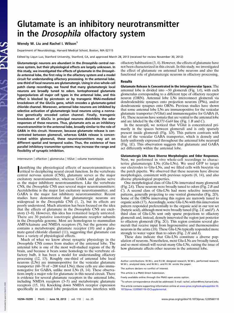
ResultsGlutamate Release Is Concentrated in the Interglomerular Space. Theantennal lobe is divided into ∼50 glomeruli (Fig. 1A), with eachglomerulus corresponding to a different type of olfactory receptorneuron (ORN). Antennal lobe LNs interconnect glomeruli viadendrodendritic synapses onto projection neurons (PNs), and/ordendroaxonic synapses onto ORNs. Previous studies have shownthat some antennal lobe LNs are immunopositive for the vesicularglutamate transporter (VGlut) and immunonegative for GABA (8,14). These neurons have somata that are ventral to the antennal lobeand are labeled by the OK371-Gal4 line (Fig. 1 B and C).In the neuropil, we noticed that VGlut is concentrated pri-
marily in the spaces between glomeruli and is only sparselypresent inside glomeruli (Fig. 1D). This pattern contrasts withthat of the vesicular GABA transporter, which is densely andfairly uniformly expressed throughout the antennal lobe neuropil(Fig. 1E). This observation suggests that glutamate and GABAact differently within the antennal lobe.
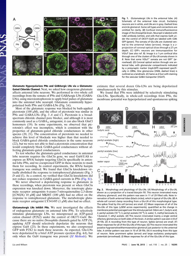
Glutamatergic LNs Have Diverse Morphologies and Odor Responses.Next, we performed in vivo whole-cell recordings to charac-terize glutamatergic LNs (Glu-LNs). We used GFP to targetour electrodes to Glu-LNs, and we filled cells with biocytin viathe patch pipette. We observed that these neurons have diversemorphologies, consistent with previous reports (8, 14), and alsodiverse physiological properties.One morphological class of Glu-LNs innervated many glomeruli
(Fig. 2A). These neurons were broadly tuned to odors (Fig. 2 B andC). A second class of Glu-LNs had more selective innervationpatterns, generally projecting to one ventral glomerulus (Fig. 2D).Some of the ORNs innervating this region are narrowly tuned toorganic acids (17). Accordingly, someGlu-LNs with this innervationpattern responded preferentially to the organic acid in our test set(butyric acid), althoughmost were broadly tuned (Fig. 2E and F). Athird class of Glu-LNs sent only sparse projections to olfactoryglomeruli and, instead, densely innervated the region just posteriorto olfactory glomeruli (Fig. 2G). This region contains several glo-meruli that receive input from hygrosensitive and thermosensitiveneurons in the arista (18). These Glu-LNs typically respondedmorestrongly to water vapor than to odors (Fig. 2 H and I).These data indicate that Glu-LNs constitute a diverse pop-
ulation of neurons. Nonetheless, most Glu-LNs are broadly tuned,and so most stimuli will recruit many Glu-LNs, raising the issue ofhow glutamate affects other neurons in the antennal lobe.
Author contributions: W.W.L. and R.I.W. designed research; W.W.L. performed research;W.W.L. analyzed data; and W.W.L. and R.I.W. wrote the paper.
The authors declare no conflict of interest.
This article is a PNAS Direct Submission.
Freely available online through the PNAS open access option.1To whom correspondence should be addressed. E-mail: [email protected].
This article contains supporting information online at www.pnas.org/lookup/suppl/doi:10.1073/pnas.1220560110/-/DCSupplemental.
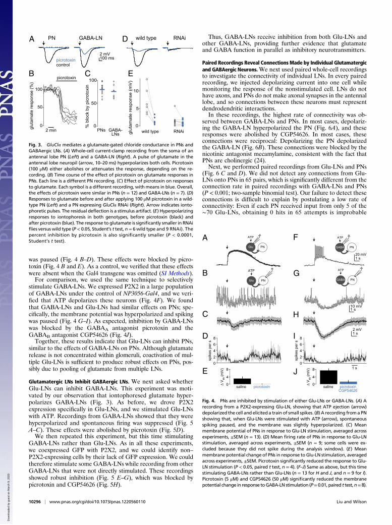
Glutamate Hyperpolarizes PNs and GABAergic LNs via a Glutamate-Gated Chloride Channel. Next, we asked how exogenous glutamateaffects antennal lobe neurons. We performed in vivo whole cellrecordings from the somata of PNs and GABAergic LNs (GABA-LNs), using microiontophoresis to apply brief pulses of glutamateinto the antennal lobe neuropil. Glutamate consistently hyper-polarized both PNs and GABA-LNs (Fig. 3A).Most of the glutamate response was blocked by bath-applied
picrotoxin (100 μM), and the effect of picrotoxin was similar inPNs and GABA-LNs (Fig. 3 A and C). Picrotoxin is a broad-spectrum chloride channel pore blocker, and although it is mostcommonly used as a GABAA antagonist, it can also block GluClhomomers (19). In some experiments, we observed that pic-rotoxin’s effect was incomplete, which is consistent with theproperties of glutamate-gated chloride conductances in otherspecies (20, 21). The concentration of picrotoxin we needed toachieve this level of blockade was higher than that needed toblock GABA-gated chloride conductances in the same neurons(22), but we were not able to find a picrotoxin concentration thatwould completely block GABA-gated conductances without af-fecting glutamate-gated conductances.To test whether the glutamate-gated conductance in antennal
lobe neurons requires the GluClα gene, we used Gal4/UAS toexpress an RNAi hairpin targeting GluClα specifically in anten-nal lobe PNs, and we coexpressed GFP in these neurons to markthem for recording. In control experiments, the RNAi hairpintransgene was omitted. We found that GluClα knockdown vir-tually abolished the response to iontophoresed glutamate (Fig. 3D and E). As a control, we verified that GluClα knockdown didnot reduce responses to GABA-gated currents in PNs (Fig. S1).We never observed a depolarizing response to glutamate in
these recordings, when picrotoxin was present or when GluClαexpression was knocked down. Moreover, the ionotropic gluta-mate receptor antagonists 6-cyano-7-nitroquinoxaline-2,3-dione[(CNQX) 10 μM] and MK801 (100 μM) had no effect on theresponse to iontophoresed glutamate. The metabotropic gluta-mate receptor antagonist LY341495 (1 μM) also had no effect.
Glutamatergic LNs Inhibit PNs. We next investigated the effectsof endogenous glutamate on antennal lobe PNs. To selectivelystimulate glutamatergic LNs, we misexpressed an ATP-gatedcation channel (P2X2) under the control of OK371-Gal4. Be-cause there are no native Drosophila channels gated by ATP (9),applying ATP should selectively depolarize the neurons thatexpress Gal4 (23). In these experiments, we also coexpressedGFP with P2X2 to mark these neurons. As expected, Glu-LNswere depolarized by a brief ATP pressure ejection (Fig. 4A), butonly when the Gal4 transgene was present (SI Methods). We
estimate that several dozen Glu-LNs are being depolarizedsimultaneously by this stimulus.We found that PNs were inhibited by selectively stimulating
Glu-LNs. Specifically, in whole-cell recordings from PNs, themembrane potential was hyperpolarized and spontaneous spiking
VGlutCD8:GFPC 10 μm
20 μm B
GABA
Glu
ORN
PN
ORN
PN
20 μm VGlutnsyb:GFP
D
VGATnsyb:GFP 20 μm
E
A Fig. 1. Glutamatergic LNs in the antennal lobe. (A)Schematic of the antennal lobe circuit. Excitatoryneurons are in white, and LNs are in gray. Dashed linesencircle glomeruli. Some cell types and connections areomitted for clarity. (B) Confocal immunofluorescenceimage of theDrosophila brain. Neuropil is labeledwithnc82 antibody (white), and cells that express Gal4 un-der the control of OK371-Gal4 are labeled with CD8:GFP (green). The somata of Glu-LNs are clustered ven-tral to the antennal lobes (arrows). Image is a z-projection of coronal optical slices through a 27-μmdepth. (C) GFP+ neurons are immunopositive forVGluT (see also ref. 8). Image is a 1-μm confocal slicethrough one of the clusters of Glu-LN somata shown inB. Note that some VGluT+ somata are not GFP+ (ar-rowhead). (D) Coronal optical section through one an-tennal lobe, with glomerular compartments indicatedby a presynaptic marker (nsyb:GFP) expressed specifi-cally in ORNs. One glomerulus (VM4, dashed lines) isoutlined as a landmark. (E) Same as D but with stainingfor the vesicular GABA transporter (VGAT).
pentyl acetate (high)
water vapor 10 mV500 ms
pentyl acetate (low)
6
4
2
0Δ V
m (m
V)
C F I
D GA
odorB E Hodor odor 20 μm
1 2 3 4 5 6 7 1 2 3 4 5 6 7 1 2 3 4 5 6 7
butyric acid
Fig. 2. Morphology and physiology of Glu-LNs. (A) Morphology of a Glu-LN,shown as a z-projection of a traced biocytin fill. This neuron innervated manyolfactory glomeruli, and this pattern was seen in 11 of 29 filled cells. Note in-nervation of both antennal lobes (black circles), which is typical of Glu-LNs. (B) Awhole-cell current clamp recording from a Glu-LN of this morphological type.The spikes fired by this cell (arrow) are small. (C) Mean responses of all of theGlu-LNs of this type (±SEM across experiments), quantified as the change inmembranepotential averagedover the stimulus period.Odors are 1, butyric acid;2, pentyl acetate (10−2); 3, pentyl acetate (10−6); 4, water; 5, methyl benzoate; 6,1-butanol; 7, ethyl acetate. (D) This neuron innervated mainly a single ventralolfactory glomerulus on both sides of the brain. A similar patternwas seen in 8 of29 fills. (E) A recording from this type of neuron. Spikes (arrow) are small. (F)Mean responses for all of the Glu-LNs of this type. (G) This neuron innervated theputative hygrosensitive/thermosensitive glomeruli just posterior to the antennallobe. A similar pattern was seen in 10 of 29 fills. (H) A recording from this typeof neuron. Note prominent spikes (arrow) and large excitatory postsynapticpotentials (arrowhead). (I) Mean responses for all of the Glu-LNs of this type.
Liu and Wilson PNAS | June 18, 2013 | vol. 110 | no. 25 | 10295
was paused (Fig. 4 B–D). These effects were blocked by picro-toxin (Fig. 4 B and E). As a control, we verified that these effectswere absent when the Gal4 transgene was omitted (SI Methods).For comparison, we used the same technique to selectively
stimulate GABA-LNs. We expressed P2X2 in a large populationof GABA-LNs under the control of NP3056-Gal4, and we veri-fied that ATP depolarizes these neurons (Fig. 4F). We foundthat GABA-LNs and Glu-LNs had similar effects on PNs; spe-cifically, the membrane potential was hyperpolarized and spikingwas paused (Fig. 4 G–I). As expected, inhibition by GABA-LNswas blocked by the GABAA antagonist picrotoxin and theGABAB antagonist CGP54626 (Fig. 4J).Together, these results indicate that Glu-LNs can inhibit PNs,
similar to the effects of GABA-LNs on PNs. Although glutamaterelease is not concentrated within glomeruli, coactivation of mul-tiple Glu-LNs is sufficient to produce robust effects on PNs, pos-sibly due to pooling of glutamate from multiple LNs.
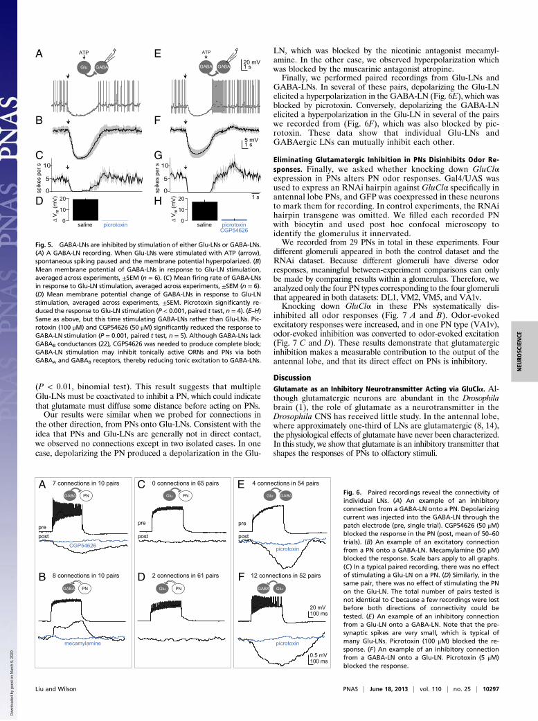
Glutamatergic LNs Inhibit GABAergic LNs. We next asked whetherGlu-LNs can inhibit GABA-LNs. This experiment was moti-vated by our observation that iontophoresed glutamate hyper-polarizes GABA-LNs (Fig. 3). As before, we drove P2X2expression specifically in Glu-LNs, and we stimulated Glu-LNswith ATP. Recordings from GABA-LNs showed that they werehyperpolarized and spontaneous firing was suppressed (Fig. 5A–C). These effects were abolished by picrotoxin (Fig. 5D).We then repeated this experiment, but this time stimulating
GABA-LNs rather than Glu-LNs. As in all these experiments,we coexpressed GFP with P2X2, and we could identify non–P2X2-expressing cells by their lack of GFP expression. We couldtherefore stimulate some GABA-LNs while recording from otherGABA-LNs that were not directly stimulated. These recordingsshowed robust inhibition (Fig. 5 E–G), which was blocked bypicrotoxin and CGP54626 (Fig. 5H).
Thus, GABA-LNs receive inhibition from both Glu-LNs andother GABA-LNs, providing further evidence that glutamateand GABA function in parallel as inhibitory neurotransmitters.
Paired Recordings Reveal Connections Made by Individual Glutamatergicand GABAergic Neurons.We next used paired whole-cell recordingsto investigate the connectivity of individual LNs. In every pairedrecording, we injected depolarizing current into one cell whilemonitoring the response of the nonstimulated cell. LNs do nothave axons, and PNs do not make axonal synapses in the antennallobe, and so connections between these neurons must representdendrodendritic interactions.In these recordings, the highest rate of connectivity was ob-
served between GABA-LNs and PNs. In most cases, depolariz-ing the GABA-LN hyperpolarized the PN (Fig. 6A), and theseresponses were abolished by CGP54626. In most cases, theseconnections were reciprocal: Depolarizing the PN depolarizedthe GABA-LN (Fig. 6B). These connections were blocked by thenicotinic antagonist mecamylamine, consistent with the fact thatPNs are cholinergic (24).Next, we performed paired recordings from Glu-LNs and PNs
(Fig. 6 C and D). We did not detect any connections from Glu-LNs onto PNs in 65 pairs, which is significantly different from theconnection rate in paired recordings with GABA-LNs and PNs(P < 0.001; two-sample binomial test). Our failure to detect theseconnections is difficult to explain by postulating a low rate ofconnectivity: Even if each PN received input from only 5 of the∼70 Glu-LNs, obtaining 0 hits in 65 attempts is improbable
2 mV100 ms
controlpicrotoxin
PN
100
50
0% b
lock
by
picr
otox
in
PNs GABA-LNs
A
100
0glut
amat
e re
spon
se (%
)
picrotoxinB
GABA-LN
50
wild type RNAiD
10
5
0gl
utam
ate
resp
onse
(mV
)
E
wild type RNAi2 min
C
Fig. 3. GluClα mediates a glutamate-gated chloride conductance in PNs andGABAergic LNs. (A) Whole-cell current-clamp recording from the soma of anantennal lobe PN (Left) and a GABA-LN (Right). A pulse of glutamate in theantennal lobe neuropil (arrow, 10–20 ms) hyperpolarizes both cells. Picrotoxin(100 μM) either abolishes or attenuates the response, depending on the re-cording. (B) Time course of the effect of picrotoxin on glutamate responses inPNs. Each line is a different PN recording. (C) Effect of picrotoxin on responsesto glutamate. Each symbol is a different recording, withmeans in blue. Overall,the effects of picrotoxin were similar in PNs (n = 12) and GABA-LNs (n = 7). (D)Responses to glutamate before and after applying 100 μM picrotoxin in a wild-type PN (Left) and a PN expressing GluClα RNAi (Right). Arrow indicates ionto-phoretic pulses. The residual deflection is a stimulus artifact. (E) Hyperpolarizingresponses to iontophoresis in both genotypes, before picrotoxin (black) andafter picrotoxin (blue). The response to glutamate is significantly smaller in RNAiflies versus wild type (P < 0.05, Student’s t test, n = 6wild type and 9 RNAi). Thepercent inhibition by picrotoxin is also significantly smaller (P < 0.0001,Student’s t test).
10 mV1 s
A
C
D
420
1 s
2 mV1 s
20 mV1 s
68
spik
es p
er s
B
F
H
I
G
PNGlu
ATP
PNGABA
ATP
PNGlu
ATP
PNGABA
ATP
420
68
spik
es p
er s
8
4
0Δ V
m (m
V)
saline picrotoxin saline picrotoxinCGP54626
E 8
4
0Δ V
m (m
V)
J
Fig. 4. PNs are inhibited by stimulation of either Glu-LNs or GABA-LNs. (A) Arecording from a P2X2-expressing Glu-LN, showing that ATP ejection (arrow)depolarized the cell and elicited a train of small spikes. (B) A recording froma PNshowing that, when Glu-LNs were stimulated with ATP (arrow), spontaneousspiking paused, and the membrane was slightly hyperpolarized. (C) Meanmembrane potential of PNs in response to Glu-LN stimulation, averaged acrossexperiments, ±SEM (n = 13). (D) Mean firing rate of PNs in response to Glu-LNstimulation, averaged across experiments, ±SEM (n = 9; some cells were ex-cluded because they did not spike during the analysis window). (E) Meanmembrane potential change of PNs in response to Glu-LN stimulation, averagedacross experiments, ±SEM. Picrotoxin significantly reduced the response to Glu-LN stimulation (P < 0.05, paired t test, n = 4). (F–J) Same as above, but this timestimulating GABA-LNs rather than Glu-LNs (n = 13 for H and J, and n = 9 for I).Picrotoxin (5 μM) and CGP54626 (50 μM) significantly reduced the membranepotential change in response toGABA-LN stimulation (P= 0.01, paired t test,n=8).
10296 | www.pnas.org/cgi/doi/10.1073/pnas.1220560110 Liu and Wilson
(P < 0.01, binomial test). This result suggests that multipleGlu-LNs must be coactivated to inhibit a PN, which could indicatethat glutamate must diffuse some distance before acting on PNs.Our results were similar when we probed for connections in
the other direction, from PNs onto Glu-LNs. Consistent with theidea that PNs and Glu-LNs are generally not in direct contact,we observed no connections except in two isolated cases. In onecase, depolarizing the PN produced a depolarization in the Glu-
LN, which was blocked by the nicotinic antagonist mecamyl-amine. In the other case, we observed hyperpolarization whichwas blocked by the muscarinic antagonist atropine.Finally, we performed paired recordings from Glu-LNs and
GABA-LNs. In several of these pairs, depolarizing the Glu-LNelicited a hyperpolarization in the GABA-LN (Fig. 6E), which wasblocked by picrotoxin. Conversely, depolarizing the GABA-LNelicited a hyperpolarization in the Glu-LN in several of the pairswe recorded from (Fig. 6F), which was also blocked by pic-rotoxin. These data show that individual Glu-LNs andGABAergic LNs can mutually inhibit each other.
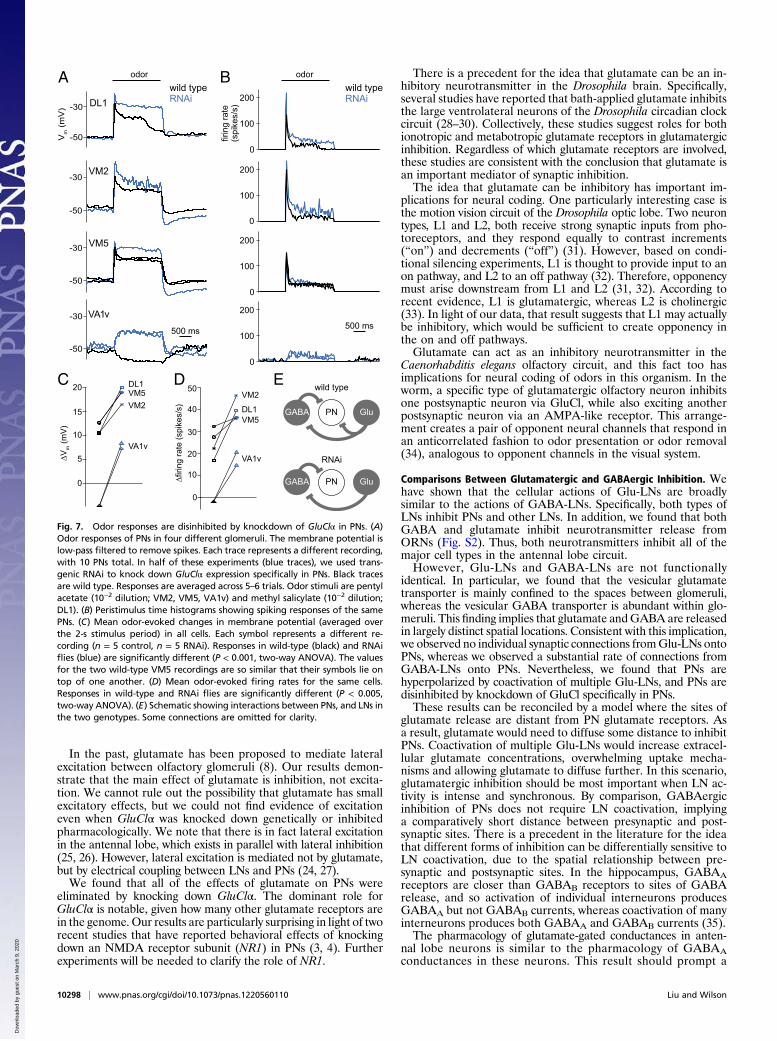
Eliminating Glutamatergic Inhibition in PNs Disinhibits Odor Re-sponses. Finally, we asked whether knocking down GluClαexpression in PNs alters PN odor responses. Gal4/UAS wasused to express an RNAi hairpin against GluClα specifically inantennal lobe PNs, and GFP was coexpressed in these neuronsto mark them for recording. In control experiments, the RNAihairpin transgene was omitted. We filled each recorded PNwith biocytin and used post hoc confocal microscopy toidentify the glomerulus it innervated.We recorded from 29 PNs in total in these experiments. Four
different glomeruli appeared in both the control dataset and theRNAi dataset. Because different glomeruli have diverse odorresponses, meaningful between-experiment comparisons can onlybe made by comparing results within a glomerulus. Therefore, weanalyzed only the four PN types corresponding to the four glomerulithat appeared in both datasets: DL1, VM2, VM5, and VA1v.Knocking down GluClα in these PNs systematically dis-
inhibited all odor responses (Fig. 7 A and B). Odor-evokedexcitatory responses were increased, and in one PN type (VA1v),odor-evoked inhibition was converted to odor-evoked excitation(Fig. 7 C and D). These results demonstrate that glutamatergicinhibition makes a measurable contribution to the output of theantennal lobe, and that its direct effect on PNs is inhibitory.
DiscussionGlutamate as an Inhibitory Neurotransmitter Acting via GluClα. Al-though glutamatergic neurons are abundant in the Drosophilabrain (1), the role of glutamate as a neurotransmitter in theDrosophila CNS has received little study. In the antennal lobe,where approximately one-third of LNs are glutamatergic (8, 14),the physiological effects of glutamate have never been characterized.In this study, we show that glutamate is an inhibitory transmitter thatshapes the responses of PNs to olfactory stimuli.
B
G
A
F
E
10
5
01 s
20 mV1 s
5 mV1 s
spik
es p
er s
GABAGlu
ATP
GABAGABA
ATP
20
10
0Δ V
m (m
V)
saline picrotoxin saline picrotoxinCGP54626
D 20
10
0Δ V
m (m
V)
H
10
5
0
C
spik
es p
er s
Fig. 5. GABA-LNs are inhibited by stimulation of either Glu-LNs or GABA-LNs.(A) A GABA-LN recording. When Glu-LNs were stimulated with ATP (arrow),spontaneous spiking paused and the membrane potential hyperpolarized. (B)Mean membrane potential of GABA-LNs in response to Glu-LN stimulation,averaged across experiments, ±SEM (n = 6). (C) Mean firing rate of GABA-LNsin response to Glu-LN stimulation, averaged across experiments, ±SEM (n = 6).(D) Mean membrane potential change of GABA-LNs in response to Glu-LNstimulation, averaged across experiments, ±SEM. Picrotoxin significantly re-duced the response to Glu-LN stimulation (P < 0.001, paired t test, n = 4). (E–H)Same as above, but this time stimulating GABA-LNs rather than Glu-LNs. Pic-rotoxin (100 μM) and CGP54626 (50 μM) significantly reduced the response toGABA-LN stimulation (P = 0.001, paired t test, n = 5). Although GABA-LNs lackGABAB conductances (22), CGP54626 was needed to produce complete block;GABA-LN stimulation may inhibit tonically active ORNs and PNs via bothGABAA and GABAB receptors, thereby reducing tonic excitation to GABA-LNs.
4 connections in 54 pairs
DGlu PN
2 connections in 61 pairs
A
CGP54626
7 connections in 10 pairs
pre
post
picrotoxin
F 12 connections in 52 pairs
GluGABA
CGlu PN
0 connections in 65 pairs
pre
post
E
picrotoxin
GABAGlu
pre
post
20 mV100 ms
B
mecamylamine
GABA PN
8 connections in 10 pairs
0.5 mV100 ms
GABA PN Fig. 6. Paired recordings reveal the connectivity ofindividual LNs. (A) An example of an inhibitoryconnection from a GABA-LN onto a PN. Depolarizingcurrent was injected into the GABA-LN through thepatch electrode (pre, single trial). CGP54626 (50 μM)blocked the response in the PN (post, mean of 50–60trials). (B) An example of an excitatory connectionfrom a PN onto a GABA-LN. Mecamylamine (50 μM)blocked the response. Scale bars apply to all graphs.(C) In a typical paired recording, there was no effectof stimulating a Glu-LN on a PN. (D) Similarly, in thesame pair, there was no effect of stimulating the PNon the Glu-LN. The total number of pairs tested isnot identical to C because a few recordings were lostbefore both directions of connectivity could betested. (E) An example of an inhibitory connectionfrom a Glu-LN onto a GABA-LN. Note that the pre-synaptic spikes are very small, which is typical ofmany Glu-LNs. Picrotoxin (100 μM) blocked the re-sponse. (F) An example of an inhibitory connectionfrom a GABA-LN onto a Glu-LN. Picrotoxin (5 μM)blocked the response.
Liu and Wilson PNAS | June 18, 2013 | vol. 110 | no. 25 | 10297
NEU
ROSC
IENCE
Dow
nloa
ded
by g
uest
on
Mar
ch 9
, 202
0
In the past, glutamate has been proposed to mediate lateralexcitation between olfactory glomeruli (8). Our results demon-strate that the main effect of glutamate is inhibition, not excita-tion. We cannot rule out the possibility that glutamate has smallexcitatory effects, but we could not find evidence of excitationeven when GluClα was knocked down genetically or inhibitedpharmacologically. We note that there is in fact lateral excitationin the antennal lobe, which exists in parallel with lateral inhibition(25, 26). However, lateral excitation is mediated not by glutamate,but by electrical coupling between LNs and PNs (24, 27).We found that all of the effects of glutamate on PNs were
eliminated by knocking down GluClα. The dominant role forGluClα is notable, given how many other glutamate receptors arein the genome. Our results are particularly surprising in light of tworecent studies that have reported behavioral effects of knockingdown an NMDA receptor subunit (NR1) in PNs (3, 4). Furtherexperiments will be needed to clarify the role of NR1.
There is a precedent for the idea that glutamate can be an in-hibitory neurotransmitter in the Drosophila brain. Specifically,several studies have reported that bath-applied glutamate inhibitsthe large ventrolateral neurons of the Drosophila circadian clockcircuit (28–30). Collectively, these studies suggest roles for bothionotropic and metabotropic glutamate receptors in glutamatergicinhibition. Regardless of which glutamate receptors are involved,these studies are consistent with the conclusion that glutamate isan important mediator of synaptic inhibition.The idea that glutamate can be inhibitory has important im-
plications for neural coding. One particularly interesting case isthe motion vision circuit of the Drosophila optic lobe. Two neurontypes, L1 and L2, both receive strong synaptic inputs from pho-toreceptors, and they respond equally to contrast increments(“on”) and decrements (“off”) (31). However, based on condi-tional silencing experiments, L1 is thought to provide input to anon pathway, and L2 to an off pathway (32). Therefore, opponencymust arise downstream from L1 and L2 (31, 32). According torecent evidence, L1 is glutamatergic, whereas L2 is cholinergic(33). In light of our data, that result suggests that L1 may actuallybe inhibitory, which would be sufficient to create opponency inthe on and off pathways.Glutamate can act as an inhibitory neurotransmitter in the
Caenorhabditis elegans olfactory circuit, and this fact too hasimplications for neural coding of odors in this organism. In theworm, a specific type of glutamatergic olfactory neuron inhibitsone postsynaptic neuron via GluCl, while also exciting anotherpostsynaptic neuron via an AMPA-like receptor. This arrange-ment creates a pair of opponent neural channels that respond inan anticorrelated fashion to odor presentation or odor removal(34), analogous to opponent channels in the visual system.
Comparisons Between Glutamatergic and GABAergic Inhibition. Wehave shown that the cellular actions of Glu-LNs are broadlysimilar to the actions of GABA-LNs. Specifically, both types ofLNs inhibit PNs and other LNs. In addition, we found that bothGABA and glutamate inhibit neurotransmitter release fromORNs (Fig. S2). Thus, both neurotransmitters inhibit all of themajor cell types in the antennal lobe circuit.However, Glu-LNs and GABA-LNs are not functionally
identical. In particular, we found that the vesicular glutamatetransporter is mainly confined to the spaces between glomeruli,whereas the vesicular GABA transporter is abundant within glo-meruli. This finding implies that glutamate andGABA are releasedin largely distinct spatial locations. Consistent with this implication,we observed no individual synaptic connections fromGlu-LNs ontoPNs, whereas we observed a substantial rate of connections fromGABA-LNs onto PNs. Nevertheless, we found that PNs arehyperpolarized by coactivation of multiple Glu-LNs, and PNs aredisinhibited by knockdown of GluCl specifically in PNs.These results can be reconciled by a model where the sites of
glutamate release are distant from PN glutamate receptors. Asa result, glutamate would need to diffuse some distance to inhibitPNs. Coactivation of multiple Glu-LNs would increase extracel-lular glutamate concentrations, overwhelming uptake mecha-nisms and allowing glutamate to diffuse further. In this scenario,glutamatergic inhibition should be most important when LN ac-tivity is intense and synchronous. By comparison, GABAergicinhibition of PNs does not require LN coactivation, implyinga comparatively short distance between presynaptic and post-synaptic sites. There is a precedent in the literature for the ideathat different forms of inhibition can be differentially sensitive toLN coactivation, due to the spatial relationship between pre-synaptic and postsynaptic sites. In the hippocampus, GABAAreceptors are closer than GABAB receptors to sites of GABArelease, and so activation of individual interneurons producesGABAA but not GABAB currents, whereas coactivation of manyinterneurons produces both GABAA and GABAB currents (35).The pharmacology of glutamate-gated conductances in anten-
nal lobe neurons is similar to the pharmacology of GABAAconductances in these neurons. This result should prompt a
200
100
0
200
100
200
100
0
500 ms
A B
DL1
VM2
VM5
VA1v500 ms
odor odorwild typeRNAi
C D
200
100
0
50
40
30
20
10∆firi
ng ra
te (s
pike
s/s)
20
15
10
5
0
VM5DL1
VM2
VA1v
DL1
VM2
VM5
VA1v
0
0fir
ing
rate
(spi
kes/
s)
E
RNAi
PNGABA Glu
PNGABA Glu
wild type
-50
-30
-50
-30
-50
-30
-50
-30
Vm (m
V)
wild typeRNAi
∆Vm (m
V)
Fig. 7. Odor responses are disinhibited by knockdown of GluClα in PNs. (A)Odor responses of PNs in four different glomeruli. The membrane potential islow-pass filtered to remove spikes. Each trace represents a different recording,with 10 PNs total. In half of these experiments (blue traces), we used trans-genic RNAi to knock down GluClα expression specifically in PNs. Black tracesare wild type. Responses are averaged across 5–6 trials. Odor stimuli are pentylacetate (10−2 dilution; VM2, VM5, VA1v) and methyl salicylate (10−2 dilution;DL1). (B) Peristimulus time histograms showing spiking responses of the samePNs. (C) Mean odor-evoked changes in membrane potential (averaged overthe 2-s stimulus period) in all cells. Each symbol represents a different re-cording (n = 5 control, n = 5 RNAi). Responses in wild-type (black) and RNAiflies (blue) are significantly different (P < 0.001, two-way ANOVA). The valuesfor the two wild-type VM5 recordings are so similar that their symbols lie ontop of one another. (D) Mean odor-evoked firing rates for the same cells.Responses in wild-type and RNAi flies are significantly different (P < 0.005,two-way ANOVA). (E) Schematic showing interactions between PNs, and LNs inthe two genotypes. Some connections are omitted for clarity.
10298 | www.pnas.org/cgi/doi/10.1073/pnas.1220560110 Liu and Wilson
reevaluation of studies that used picrotoxin to block inhibition inthe antennal lobe (22, 36–40). Given our results, it seems likelythat these studies were reducing both glutamatergic andGABAergic inhibition.
Interactions Between Glutamatergic and GABAergic Inhibition. It isperhaps surprising that knocking down GluClα in PNs had sucha substantial effect on PN odor responses, given that picrotoxinalone has comparatively modest effects (22, 36–39). The solutionto this puzzle may lie in our finding that glutamate regulates notonly PNs but also GABA-LNs. Importantly, GABA-LNs arespontaneously active and provide tonic inhibition to PNs (14,22). Hence, in the intact circuit, glutamatergic inhibition ofGABA-LNs should tend to disinhibit PNs (Fig. 7E). Picrotoxinprevents Glu-LNs from inhibiting GABA-LNs and should tendto potentiate GABAergic inhibition. The effects of GABA aremediated in part by GABAB receptors, which are not sensitive topicrotoxin. Thus, picrotoxin likely has bidirectional effects on thetotal level of inhibition in the circuit. By contrast, knockdown ofGluClα specifically in PNs should not directly affect GABA-LNsand should not produce these complex effects (Fig. 7E). Theseresults illustrate how a cell-specific genetic blockade of a neuro-transmitter system can have more dramatic effects than a globalpharmacological blockade of the same system.Our study reveals that an LN can have push-pull effects on
a single population of target cells. For example, Glu-LNs directlyinhibit PNs, but they should also disinhibit PNs, via the inhibitionof GABA-LNs. This architecture may allow for more robust gain
control and rapid transitions between network states and issimilar to the wiring of many cortical circuits, where corecruit-ment of excitation and inhibition is a common motif (41).Why might the existence of two parallel inhibitory transmitters
be useful? Our data argue that GABA and glutamate may acton different spatial and temporal scales. Because these two in-hibitory systems comprise different cells, receptors, and trans-porters, they can be modulated independently. Because theirproperties are encoded by different genes, they can also evolveindependently. This organization should confer increased flexi-bility in adapting synaptic inhibition to a changing environment.
MethodsImmunohistochemical procedures, in vivo whole-cell recordings, odor stim-ulation, and antennal nerve stimulation were performed essentially as de-scribed (22, 25, 42). Glutamate iontophoresis was performed by using a sharpglass microelectrode inserted into the antennal lobe neuropil. Stimulation ofLNs expressing the P2X2 receptor was achieved by pressure-ejecting ATPsolution onto their somata. Paired recordings were performed in an ex vivopreparation to improve optical and steric access. See SI Methods for exper-imental genotypes and all other experimental details.
ACKNOWLEDGMENTS. We thank A. DiAntonio for anti-VGlut antibody,D. E. Krantz for anti-VGAT antibody, G. Miesenböck for UAS-P2X2 flies, andmembers of the R.I.W. laboratory for feedback on the manuscript. This workwas supported by National Institutes of Health research project GrantR01DC008174. W.W.L. is supported by an Howard Hughes Medical Institute(HHMI) International Research Fellowship and a Presidential Scholarshipfrom Harvard Medical School. R.I.W. is an HHMI Early Career Scientist.
1. Daniels RW, Gelfand MV, Collins CA, DiAntonio A (2008) Visualizing glutamatergic cellbodies and synapses in Drosophila larval and adult CNS. J Comp Neurol 508(1):131–152.
2. Raghu SV, Borst A (2011) Candidate glutamatergic neurons in the visual system ofDrosophila. PLoS ONE 6(5):e19472.
3. Das S, et al. (2011) Plasticity of local GABAergic interneurons drives olfactory habit-uation. Proc Natl Acad Sci USA 108(36):E646–E654.
4. Sudhakaran IP, et al. (2012) Plasticity of recurrent inhibition in the Drosophila an-tennal lobe. J Neurosci 32(21):7225–7231.
5. Xia S, et al. (2005) NMDA receptors mediate olfactory learning and memory in Dro-sophila. Curr Biol 15(7):603–615.
6. Wu CL, et al. (2007) Specific requirement of NMDA receptors for long-term memoryconsolidation in Drosophila ellipsoid body. Nat Neurosci 10(12):1578–1586.
7. Miyashita T, et al. (2012) Mg(2+) block of Drosophila NMDA receptors is required forlong-term memory formation and CREB-dependent gene expression. Neuron 74(5):887–898.
8. Das A, et al. (2011) Identification and analysis of a glutamatergic local interneuronlineage in the adult Drosophila olfactory system. Neural Syst Circuits 1(1):4.
9. Littleton JT, Ganetzky B (2000) Ion channels and synaptic organization: Analysis of theDrosophila genome. Neuron 26(1):35–43.
10. Parmentier ML, Pin JP, Bockaert J, Grau Y (1996) Cloning and functional expressionof a Drosophila metabotropic glutamate receptor expressed in the embryonic CNS.J Neurosci 16(21):6687–6694.
11. Cully DF, Paress PS, Liu KK, Schaeffer JM, Arena JP (1996) Identification of a Dro-sophila melanogaster glutamate-gated chloride channel sensitive to the antiparasiticagent avermectin. J Biol Chem 271(33):20187–20191.
12. Masse NY, Turner GC, Jefferis GS (2009) Olfactory information processing in Dro-sophila. Curr Biol 19(16):R700–R713.
13. Wilson RI (2011) Understanding the functional consequences of synaptic specializa-tion: Insight from the Drosophila antennal lobe. Curr Opin Neurobiol 21(2):254–260.
14. Chou YH, et al. (2010) Diversity and wiring variability of olfactory local interneuronsin the Drosophila antennal lobe. Nat Neurosci 13(4):439–449.
15. Ramaekers A, Parmentier ML, Lasnier C, Bockaert J, Grau Y (2001) Distribution ofmetabotropic glutamate receptor DmGlu-A in Drosophila melanogaster central ner-vous system. J Comp Neurol 438(2):213–225.
16. Devaud JM, Clouet-Redt C, Bockaert J, Grau Y, Parmentier ML (2008) Widespreadbrain distribution of the Drosophila metabotropic glutamate receptor. Neuroreport19(3):367–371.
17. Silbering AF, et al. (2011) Complementary function and integrated wiring of the evolu-tionarily distinct Drosophila olfactory subsystems. J Neurosci 31(38):13357–13375.
18. Stocker RF, Lienhard MC, Borst A, Fischbach KF (1990) Neuronal architecture of theantennal lobe in Drosophila melanogaster. Cell Tissue Res 262(1):9–34.
20. Raymond V, Sattelle DB, Lapied B (2000) Co-existence in DUM neurones of two GluClchannels that differ in their picrotoxin sensitivity. Neuroreport 11(12):2695–2701.
21. Barbara GS, Zube C, Rybak J, Gauthier M, Grünewald B (2005) Acetylcholine, GABAand glutamate induce ionic currents in cultured antennal lobe neurons of the hon-eybee, Apis mellifera. J Comp Physiol A Neuroethol Sens Neural Behav Physiol 191(9):823–836.
22. Wilson RI, Laurent G (2005) Role of GABAergic inhibition in shaping odor-evokedspatiotemporal patterns in the Drosophila antennal lobe. J Neurosci 25(40):9069–9079.
23. Lima SQ, Miesenböck G (2005) Remote control of behavior through genetically tar-geted photostimulation of neurons. Cell 121(1):141–152.
24. Yaksi E, Wilson RI (2010) Electrical coupling between olfactory glomeruli. Neuron67(6):1034–1047.
25. Olsen SR, Bhandawat V, Wilson RI (2007) Excitatory interactions between olfactoryprocessing channels in the Drosophila antennal lobe. Neuron 54(1):89–103.
26. Shang Y, Claridge-Chang A, Sjulson L, Pypaert M, Miesenböck G (2007) Excitatorylocal circuits and their implications for olfactory processing in the fly antennal lobe.Cell 128(3):601–612.
27. Huang J, ZhangW, QiaoW, Hu A, Wang Z (2010) Functional connectivity and selectiveodor responses of excitatory local interneurons in Drosophila antennal lobe. Neuron67(6):1021–1033.
28. Hamasaka Y, et al. (2007) Glutamate and its metabotropic receptor in Drosophilaclock neuron circuits. J Comp Neurol 505(1):32–45.
29. McCarthy EV, et al. (2011) Synchronized bilateral synaptic inputs to Drosophila mel-anogaster neuropeptidergic rest/arousal neurons. J Neurosci 31(22):8181–8193.
30. Collins B, Kane EA, Reeves DC, Akabas MH, Blau J (2012) Balance of activity betweenLN(v)s and glutamatergic dorsal clock neurons promotes robust circadian rhythms inDrosophila. Neuron 74(4):706–718.
31. Clark DA, Bursztyn L, Horowitz MA, Schnitzer MJ, Clandinin TR (2011) Defining thecomputational structure of the motion detector in Drosophila. Neuron 70(6):1165–1177.
32. Joesch M, Schnell B, Raghu SV, Reiff DF, Borst A (2010) ON and OFF pathways inDrosophila motion vision. Nature 468(7321):300–304.
33. Takemura SY, et al. (2011) Cholinergic circuits integrate neighboring visual signals ina Drosophila motion detection pathway. Curr Biol 21(24):2077–2084.
34. Chalasani SH, et al. (2007) Dissecting a circuit for olfactory behaviour in Caeno-rhabditis elegans. Nature 450(7166):63–70.
35. Scanziani M (2000) GABA spillover activates postsynaptic GABA(B) receptors to con-trol rhythmic hippocampal activity. Neuron 25(3):673–681.
36. Wilson RI, Turner GC, Laurent G (2004) Transformation of olfactory representations inthe Drosophila antennal lobe. Science 303(5656):366–370.
37. Olsen SR, Wilson RI (2008) Lateral presynaptic inhibition mediates gain control in anolfactory circuit. Nature 452(7190):956–960.
38. Root CM, et al. (2008) A presynaptic gain control mechanism fine-tunes olfactorybehavior. Neuron 59(2):311–321.
39. Silbering AF, Galizia CG (2007) Processing of odor mixtures in the Drosophila antennallobe reveals both global inhibition and glomerulus-specific interactions. J Neurosci27(44):11966–11977.
40. Olsen SR, Bhandawat V, Wilson RI (2010) Divisive normalization in olfactory pop-ulation codes. Neuron 66(2):287–299.
41. Isaacson JS, Scanziani M (2011) How inhibition shapes cortical activity. Neuron 72(2):231–243.
42. Kazama H, Wilson RI (2008) Homeostatic matching and nonlinear amplification atidentified central synapses. Neuron 58(3):401–413.
Liu and Wilson PNAS | June 18, 2013 | vol. 110 | no. 25 | 10299
![REVIEW Open Access Traumatic brain injury: pathophysiology ...homeostasis [1], neurotransmitter release (e.g., glutamate excitotoxicity) [2], mitochondrial dysfunction [3], neur-onal](https://static.documents.pub/doc/80x56/6090d88d03865410793ff8f2/review-open-access-traumatic-brain-injury-pathophysiology-homeostasis-1.jpg)


