Glutamate Neurotransmission Excitatory Amino Acid Neurotransmitters Neurochemistry MS 532 September 11, 2014 Dr. Dan Savage BMSB 145A [email protected]Reference: Brady et al., Basic Neurochemistry 8 th ed., pp 342-366
Rapid decay of AMPA response: Lower affinity for GLU and a rapid desensitization of AMPA Rs
Slower decay of NMDA response: Higher affinity for GLU (slower dissociation kinetics)
Agonist & voltage-dependent activation of NMDA receptors
Also: NMDA requires binding of both glutamate and glycine to agonist recognition sites
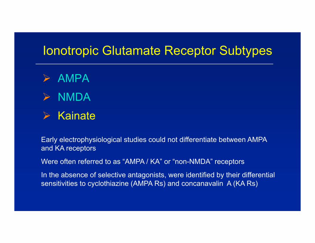
Ionotropic Glutamate Receptor Subtypes
AMPA
NMDA
Kainate
Early electrophysiological studies could not differentiate between AMPA and KA receptors
Were often referred to as “AMPA / KA” or “non-NMDA” receptors
In the absence of selective antagonists, were identified by their differential sensitivities to cyclothiazine (AMPA Rs) and concanavalin A (KA Rs)
[3H]-VKA Binding to Kainate Rs
iGluR
nAChR
Glutamate-R
25
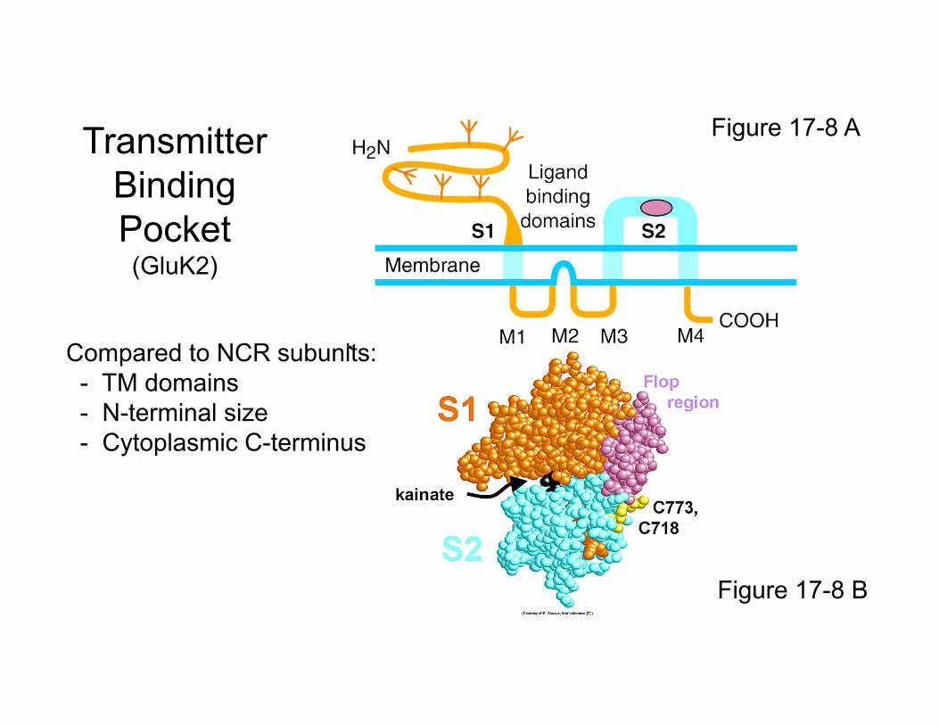
Figure 17-8 A
Figure 17-8 B
TransmitterBindingPocket(GluK2)
Compared to NCR subunits:- TM domains- N-terminal size- Cytoplasmic C-terminus
Hetero-oligomeric subunit combinations
the rule
Ionotropic glutamate receptor subunit types
nAChR
AMPAR
GluA1
GluA1
GluA2
GluA2
GluN1
NMDAR
Glutamate-gatedIon channels are
tetramersKainate-R
GluK2
GluK2
GluN1
GluN2
GluN2
GluK5
GluK528
NR1
NR1
NR2
NR2
GluN1
GluN1
GluN2
GluN2
The NMDA-R requires both glutamate and glycine (or D-serine) to be fully activated
Agonist: Glutamate
Glu
Glu
Co-agonist:Glycine or D-serine
“strychnine-insensitive”binding sites Gly
Gly
GluN3s also have Gly recognition sites29
Sources of Glycine or D-Serine
1. CSF contains micromolar glycine concentrations (but glial transporters could decrease levels near NMDARs)
2. Astrocytes wrapped tightly around glutamatergic synapses can release saturating concentrations of D-serine (Panatier et al. Cell.125, 775-784, 2006)
30
Figure 17-7
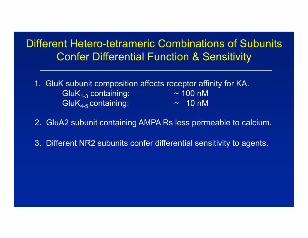
Different Hetero-tetrameric Combinations of Subunits Confer Differential Function & Sensitivity
2. GluA2 subunit containing AMPA Rs less permeable to calcium.
3. Different NR2 subunits confer differential sensitivity to agents.
3. Analgesia mGluR Group I antagonistsmGluR Group II agonists
4. Substance Abuse mGluR5 antagonists
5. Schizophrenia Group II mGluR agonists
6. Cognition Enhancers iGluR positive allosteric modulatorsmGluR Group I positive modulators
Principal Synaptic Inputs to the Dentate Granule Cell
Targets for Intervention:
1. Receptors on Granule Cell Dendrites- Positive allosteric modulators of AMPA receptors- Positive allosteric modulators of NMDA receptors
2. Receptors on Entorhinal Cortical Nerve Terminals- Group II/III mGluR autoreceptors- Group I mGluR receptors- Heterologous neurotransmitter receptors
DentateGranule
Cell(Glu)
EntorhinalCorticalNeuron(Glu)
Basket CellInterneuron
(GABA)
MedialSeptalNeuron(ACh)
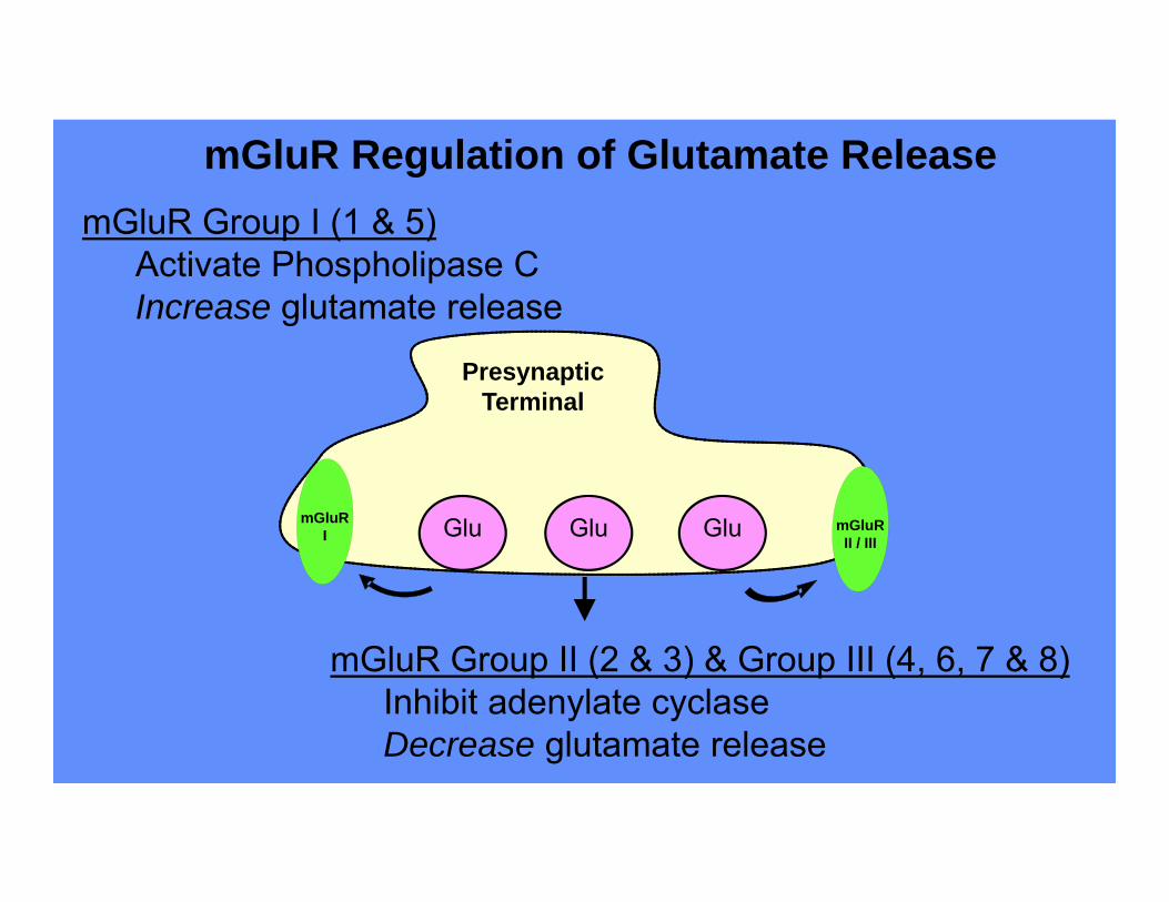
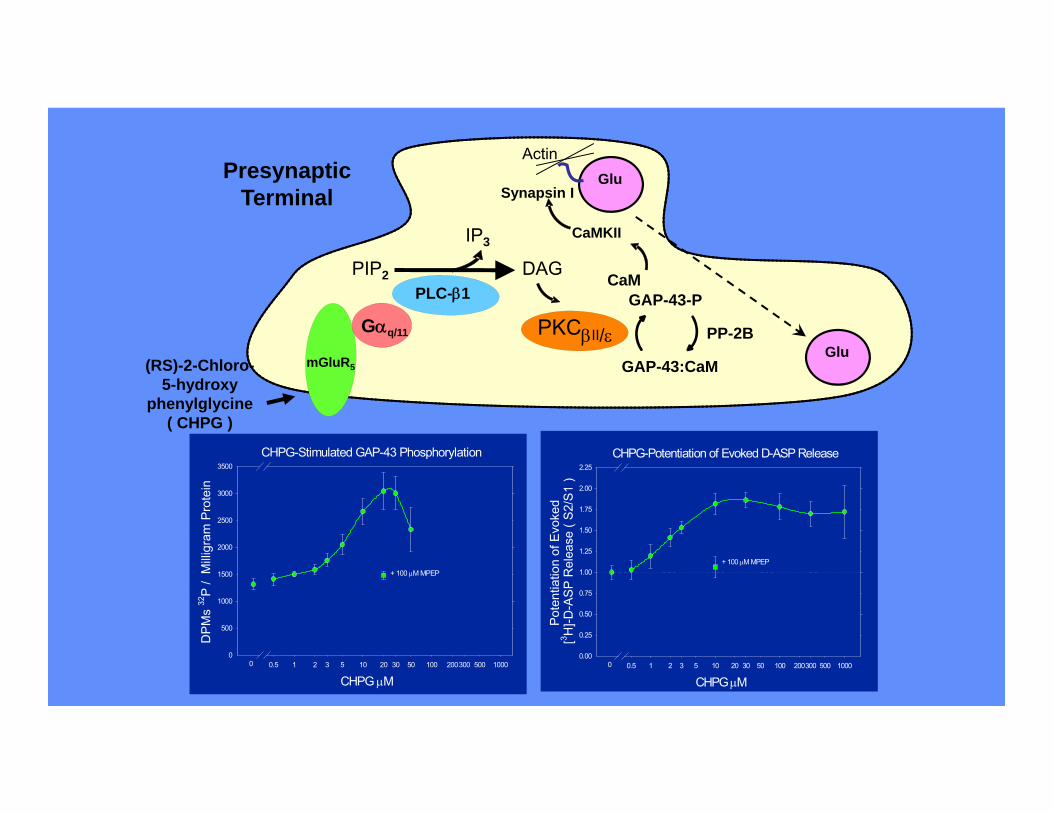
Receptors on Glutamate Nerve Terminals
Increase Glutamate Release
Group I mGluRs (1 & 5)
7 and 4/2 containing NCRsSerotonin 5HT3 receptors
Decrease Glutamate Release
Group II mGluRs ( 2 & 3)Group III mGluRs (4, 6, 7, 8)
Histamine H3 receptorsSerotonin 5HT4 and 5HT6 receptors