Food, drug, insect sting allergy, and anaphylaxis Glycation of a food allergen by the Maillard reaction enhances its T-cell immunogenicity: Role of macrophage scavenger receptor class A type I and II Anne Ilchmann, BSc, a Sven Burgdorf, PhD, b Stephan Scheurer, PhD, c Zoe Waibler, PhD, d Ryoji Nagai, PhD, e Anne Wellner, BSc, f Yasuhiko Yamamoto, MD, PhD, g Hiroshi Yamamoto, MD, PhD, g Thomas Henle, PhD, f Christian Kurts, MD, b Ulrich Kalinke, PhD, h Stefan Vieths, PhD, c and Masako Toda, PhD a Langen, Bonn, Dresden, and Hannover, Germany, and Tokyo and Kanazawa, Japan Background: The Maillard reaction occurs between reducing sugars and proteins during thermal processing of foods. It produces chemically glycated proteins termed advanced glycation end products (AGEs). The glycation structures of AGEs are suggested to function as pathogenesis-related immune epitopes in food allergy. Objective: This study aimed at defining the T-cell immunogenicity of food AGEs by using ovalbumin (OVA) as a model allergen. Methods: AGE-OVAwas prepared by means of thermal processing of OVA in the presence of glucose. Activation of OVA-specific CD4 1 T cells by AGE-OVAwas evaluated in cocultures with bone marrow–derived murine myeloid dendritic cells (mDCs) as antigen-presenting cells. The uptake mechanisms of mDCs for AGE-OVAwere investigated by using inhibitors of putative cell-surface receptors for AGEs, as well as mDCs deficient for these receptors. Results: Compared with the controls (native OVA and OVA thermally processed without glucose), AGE-OVA enhanced the activation of OVA-specific CD4 1 T cells on coculture with mDCs, indicating that the glycation of OVA enhanced the T-cell immunogenicity of the allergen. The mDC uptake of AGE-OVA was significantly higher than that of the controls. We identified scavenger receptor class A type I and II (SR-AI/II) as a mediator of the AGE-OVA uptake, whereas the receptor for AGEs and galectin-3 were not responsible. Importantly, the activation of OVA-specific CD4 1 T cells by AGE-OVA was attenuated on coculture with SR-AI/II–deficient mDCs. Conclusion: SR-AI/II targets AGE-OVA to the MHC class II loading pathway in mDCs, leading to an enhanced CD4 1 T-cell activation. The Maillard reaction might thus play an important role in the T-cell immunogenicity of food allergens. (J Allergy Clin Immunol 2010;125:175-83.) Key words: Food allergy, food allergen, Maillard reaction, T-cell immunogenicity, dendritic cells, macrophage scavenger receptor The Maillard reaction is a chemical reaction between reducing sugars and proteins and generates the so-called advanced glyca- tion end products (AGEs; ie, protein derivatives with glycation structures, such as Ne-carboxyethyl-lysine [CEL], Ne-carboxy- methyl-lysine [CML], pyrralin, and GA-pyridine). 1 Because the Maillard reaction occurs during storage and thermal processing of foods, a possible involvement of AGEs in the pathology of food allergy is of great concern. This assumption is corroborated by the fact that some patients with food allergy show anaphylactic reactions only against stored or heated foods. 2,3 Moreover, the presence of AGEs in food allergens could be linked to an increased binding ability of IgEs from patients allergic to the respective allergen. 4-7 These observations suggest that the Mail- lard reaction creates new pathogenesis-related immune epitopes in patients with food allergy. Several studies have shown diverse effects of AGEs on dendritic cells (DCs). For instance, AGEs derived from BSA augmented maturation of human DCs and increased their capacity to stimulate allogeneic T-cell activation. 8 In contrast, adrenocor- ticotropic hormone–derived AGEs were shown to inhibit the From a Junior Research Group 1 ‘‘Experimental Allergology,’’ d Junior Research Group 2 ‘‘Novel vaccination strategies and early immune responses,’’ and c the Division of Allergology, Paul-Ehrlich-Institut, Langen; b the Institute for Molecular Medicine and Experimental Immunology, Rheinische Friedrich-Wilhelms-Universita ¨t, Bonn; e the Department of Food and Nutrition, Laboratory of Biochemistry & Nutritional Sci- ence, Japan Women’s University, Tokyo; f the Institute of Food Chemistry, Technische Universita ¨t Dresden; g the Department of Biochemistry and Molecular Vascular Biol- ogy, Kanazawa University Graduate School of Medical Science; and h TWINCORE, Centre for Experimental and Clinical Infection Research, Hannover. Supported in part by Paul-Ehrlich-Institut and Deutsche Forschungsgemeinschaft (DFG Vi 165/6) Disclosure of potential conflict of interest: S. Burgdorf has received research support from the German Research Foundation. S. Vieths is an Associate of the Institute for Product Quality, Berlin; has received honoraria from Phadia, Uppsala, Sweden, and the Food Allergy Resource and Research Program, United States; is a consultant for MARS Chocolate UK Ltd; has received research support from the European Union (EuroPrevall), the German Research Foundation, the Research Fund of the German Food Industry, Monsanto Company, Pioneer Hi-Bred International, the Food Allergy Research & Resource Program, and the European Directorate for the Quality of Medicines and Health Care (EDQM); is an Executive Committee Member of the European Academy of Allergy and Clinical Immunology; is Chairman of the Allergen Standardization Subcommittee and Secretary of the Allergen Nomenclatures Sub- committee of the International Union of Immunological Societies (IUIS); is a Registered Expert with the European Agency for the Evaluation of Medicinal Products (EMEA) and the European Pharmacopoeia Commission; is Chairman of Technical Committee 275 of the European Committee for Standardization (CEN); and is a Member of the Food Allergy Working Group for the German Society for Allergy and Clinical Immunology. The rest of the authors have declared that they have no conflict of interest. Received for publication March 14, 2009; revised July 6, 2009; accepted for publication August 11, 2009. Available online October 28, 2009. Reprint requests: Masako Toda, PhD, Junior Research Group 1 ‘‘Experimental Allergol- ogy,’’ Paul-Ehrlich-Institut, Paul Ehrlich St 59, Langen 63225, Germany. E-mail: [email protected]. 0091-6749/$36.00 Ó 2010 American Academy of Allergy, Asthma & Immunology doi:10.1016/j.jaci.2009.08.013 175
Transcript
Food, drug, insect sting allergy, and anaphylaxis
Glycation of a food allergen by the Maillard reactionenhances its T-cell immunogenicity: Role of macrophagescavenger receptor class A type I and II
Anne Ilchmann, BSc,a Sven Burgdorf, PhD,b Stephan Scheurer, PhD,c Zoe Waibler, PhD,d Ryoji Nagai, PhD,e
Anne Wellner, BSc,f Yasuhiko Yamamoto, MD, PhD,g Hiroshi Yamamoto, MD, PhD,g Thomas Henle, PhD,f
Christian Kurts, MD,b Ulrich Kalinke, PhD,h Stefan Vieths, PhD,c and Masako Toda, PhDa Langen, Bonn, Dresden, and
Hannover, Germany, and Tokyo and Kanazawa, Japan
Background: The Maillard reaction occurs between reducingsugars and proteins during thermal processing of foods. Itproduces chemically glycated proteins termed advancedglycation end products (AGEs). The glycation structures ofAGEs are suggested to function as pathogenesis-related immuneepitopes in food allergy.Objective: This study aimed at defining the T-cellimmunogenicity of food AGEs by using ovalbumin (OVA) as amodel allergen.Methods: AGE-OVA was prepared by means of thermalprocessing of OVA in the presence of glucose. Activation of
From aJunior Research Group 1 ‘‘Experimental Allergology,’’ dJunior Research Group 2
‘‘Novel vaccination strategies and early immune responses,’’ and cthe Division of
Allergology, Paul-Ehrlich-Institut, Langen; bthe Institute for Molecular Medicine
and Experimental Immunology, Rheinische Friedrich-Wilhelms-Universitat, Bonn;ethe Department of Food and Nutrition, Laboratory of Biochemistry & Nutritional Sci-
ence, Japan Women’s University, Tokyo; fthe Institute of Food Chemistry, Technische
Universitat Dresden; gthe Department of Biochemistry and Molecular Vascular Biol-
ogy, Kanazawa University Graduate School of Medical Science; and hTWINCORE,
Centre for Experimental and Clinical Infection Research, Hannover.
Supported in part by Paul-Ehrlich-Institut and Deutsche Forschungsgemeinschaft (DFG
Vi 165/6)
Disclosure of potential conflict of interest: S. Burgdorf has received research support
from the German Research Foundation. S. Vieths is an Associate of the Institute for
Product Quality, Berlin; has received honoraria from Phadia, Uppsala, Sweden, and the
Food Allergy Resource and Research Program, United States; is a consultant for
MARS Chocolate UK Ltd; has received research support from the European Union
(EuroPrevall), the German Research Foundation, the Research Fund of the German
Food Industry, Monsanto Company, Pioneer Hi-Bred International, the Food Allergy
Research & Resource Program, and the European Directorate for the Quality of
Medicines and Health Care (EDQM); is an Executive Committee Member of the
European Academy of Allergy and Clinical Immunology; is Chairman of the Allergen
Standardization Subcommittee and Secretary of the Allergen Nomenclatures Sub-
committee of the International Union of Immunological Societies (IUIS); is a
Registered Expert with the European Agency for the Evaluation of Medicinal Products
(EMEA) and the European Pharmacopoeia Commission; is Chairman of Technical
Committee 275 of the European Committee for Standardization (CEN); and is a
Member of the Food Allergy Working Group for the German Society for Allergy and
Clinical Immunology. The rest of the authors have declared that they have no conflict
of interest.
Received for publication March 14, 2009; revised July 6, 2009; accepted for publication
August 11, 2009.
Available online October 28, 2009.
Reprint requests: Masako Toda, PhD, Junior Research Group 1 ‘‘Experimental Allergol-
ogy,’’ Paul-Ehrlich-Institut, Paul Ehrlich St 59, Langen 63225, Germany. E-mail:
� 2010 American Academy of Allergy, Asthma & Immunology
doi:10.1016/j.jaci.2009.08.013
OVA-specific CD41 T cells by AGE-OVA was evaluated incocultures with bone marrow–derived murine myeloid dendriticcells (mDCs) as antigen-presenting cells. The uptakemechanisms of mDCs for AGE-OVA were investigated by usinginhibitors of putative cell-surface receptors for AGEs, as well asmDCs deficient for these receptors.Results: Compared with the controls (native OVA and OVAthermally processed without glucose), AGE-OVA enhanced theactivation of OVA-specific CD41 T cells on coculture withmDCs, indicating that the glycation of OVA enhanced the T-cellimmunogenicity of the allergen. The mDC uptake of AGE-OVAwas significantly higher than that of the controls. We identifiedscavenger receptor class A type I and II (SR-AI/II) as amediator of the AGE-OVA uptake, whereas the receptor forAGEs and galectin-3 were not responsible. Importantly, theactivation of OVA-specific CD41 T cells by AGE-OVA wasattenuated on coculture with SR-AI/II–deficient mDCs.Conclusion: SR-AI/II targets AGE-OVA to the MHC class IIloading pathway in mDCs, leading to an enhanced CD41 T-cellactivation. The Maillard reaction might thus play an importantrole in the T-cell immunogenicity of food allergens. (J AllergyClin Immunol 2010;125:175-83.)
The Maillard reaction is a chemical reaction between reducingsugars and proteins and generates the so-called advanced glyca-tion end products (AGEs; ie, protein derivatives with glycationstructures, such as Ne-carboxyethyl-lysine [CEL], Ne-carboxy-methyl-lysine [CML], pyrralin, and GA-pyridine).1 Because theMaillard reaction occurs during storage and thermal processingof foods, a possible involvement of AGEs in the pathology offood allergy is of great concern. This assumption is corroboratedby the fact that some patients with food allergy show anaphylacticreactions only against stored or heated foods.2,3 Moreover, thepresence of AGEs in food allergens could be linked to anincreased binding ability of IgEs from patients allergic to therespective allergen.4-7 These observations suggest that the Mail-lard reaction creates new pathogenesis-related immune epitopesin patients with food allergy.
Several studies have shown diverse effects of AGEs ondendritic cells (DCs). For instance, AGEs derived from BSAaugmented maturation of human DCs and increased their capacityto stimulate allogeneic T-cell activation.8 In contrast, adrenocor-ticotropic hormone–derived AGEs were shown to inhibit the
maturation and T-cell stimulatory capacity of the human DCs.9
Together, these observations suggest that T-cell immunogenicityof antigens could be influenced by the Maillard reaction. How-ever, the effect of AGEs derived from food allergens on DCfunction, the subsequent activation of allergen-specific T cells,or both is poorly understood.
Antigen-specific T-cell activation is preceded by the uptake ofantigens by DCs. Receptors expressed on the cell surface mediatethe majority of antigen uptake by DCs.10,11 Importantly, antigen-presenting cells (APCs), such as DCs and macrophages, expressseveral receptors known to bind AGEs, such as the so-calledreceptor for AGEs (RAGE),12,13 galectin-3,14 macrophage scav-enger receptor class A type I and II (SR-AI/II),15,16 scavengerreceptor class B type I,17 and CD36.18 These receptors havebeen identified by investigating endothelial cells,12,13 macro-phages,15,16 or Chinese hamster ovary cells transfected with puta-tive receptors for AGEs.14,17,18 However, the receptors thatmediate the uptake of AGEs by DCs remain to be identified.
The aim of this study was to define the influence of the Maillardreaction on the T-cell immunogenicity of food allergens. We usedAGE-ovalbumin (OVA; ie, the Maillard reaction products ofglucose and the egg white allergen OVA) as a food allergen modelof AGEs. We found that AGE-OVA does not trigger the matura-tion of bone marrow–derived murine myeloid dendritic cells(mDCs) but enhances the activation of allergen-specific CD41 Tcells. Moreover, we demonstrated that the enhanced T-cellimmunogenicity of AGE-OVA depends on a SR-AI/II–mediateduptake of AGE-OVA by mDCs. Our findings support the signifi-cance of AGEs as pathogenesis-related factors in food allergy.
METHODS
MiceC57BL/6 J (B6) mice and SR-AI/II–deficient mice on a B6 background
were purchased from Jackson Laboratories (Bar Harbor, Me).19 RAGE-
deficient mice on a B6 background were kindly provided by Dr T. Shoji (Osaka
Medical College, Osaka, Japan).20 OT-II mice expressing a T-cell receptor
specific for the peptide OVA323–339 were kindly provided by Professor H.
Schild and Dr S. Sudowe (Johannes-Gutenberg-University, Mainz, Ger-
many).21 Mice were housed under pathogen-free conditions, and animal
experiments were performed in compliance with German legislation.
Preparation of AGE-OVA and AGE-BSAAGE-OVA and AGE-BSA (ie, the Maillard reaction products) were
prepared as described previously.22 Briefly, 1 mmol/L OVA or BSA (Sigma-
Aldrich, Steinheim, Germany) was incubated with 1 mol/L glucose in 100
mmol/L sodium phosphate buffer (pH 7.4) at 508C for 6 weeks. OVA
incubated under the same conditions but without glucose and native OVA
were used as controls. Protein concentrations of the final samples were mea-
sured by using a bicinchoninic acid assay kit (Pierce, Rockford, Ill). The pro-
tein concentration was further verified by analyzing valin concentrations using
ion-exchange chromatography with Ninhydrin postcolumn derivatization
after acid and enyzmatic hydrolysis because valin is not modified by the
Maillard reaction.23
Verification of glycation structures in AGE-OVAA protocol is described in the Methods section of this article’s Online
Repository at www.jacionline.org.
Preparation of recombinant OVAA protocol is described in the Methods section of this article’s Online
Repository.
Fluorescein isothiocyanate labeling of OVAsA protocol is described in the Methods section of this article’s Online
Repository.
Generation of bone marrow–derived mDCsA protocol is described in the Methods section of this article’s Online
Repository.
Assessment of mDC maturationA protocol is described in the Methods section of this article’s Online
Repository.
Assessment of T-cell activation and proliferationSplenic CD41 T cells were isolated from OT-II mice using an isolation kit
from Miltenyi Biotec (Bergisch Gladbach, Germany). CD41 T cells (8.03105
cells/mL) were cocultured with mDCs (1.63105 cells/mL) and stimulated
with either form of OVA for 24 hours to evaluate T-cell activation. In the ex-
periment with SR-AI/II–deficient mDCs the APCs (2.53106 cells/mL) were
first incubated with either form of OVA for 3 hours and then fixed with
0.008% glutaraldehyde before 21 hours of coculturing with CD41 T cells
(5.03106 cells/ml). After coculturing, the concentration of IL-2 in the super-
natants was measured by means of ELISA (eBioscience, San Diego, Calif).
CD41 T cells were first stained for 15 minutes with 10 mmol/L carboxyfluor-
escein succinimidyl ester (CFSE; Invitrogen, Karlsruhe, Germany) and then
cocultured with mDCs stimulated with either form of OVA to evaluate T-
cell proliferation. Cell proliferation was evaluated by measuring the intensity
of CFSE in the CD41 T cells with a flow cytometer, LSR II (BD Bioscience,
Heidelberg, Germany). Data were analyzed with FlowJo version 7 software
(Treestar, Inc, Ashland, Ore).
Assessment of the uptake of AGE-OVA by mDCsmDCs (1.03106 cells/mL) were incubated for 15 minutes with fluorescein
isothiocyanate (FITC) conjugates of AGE-OVA or of native OVA and OVA
thermally processed without glucose as controls. Lactose (150 mmol/L;
Sigma-Aldrich) was added to the mDCs 30 minutes before the addition of
AGE-OVA or the controls to inhibit a possible galectin-3–mediated uptake.24
Only samples with a comparable FITC/protein molar ratio were used to eval-
uate the uptake level of AGE-OVA and the controls. After incubation with
FITC conjugates of AGE-OVA or the controls, mDCs were stained with
both phycoerythrin-conjugated anti-mouse CD11b and allophycocyanin-con-
jugated anti-mouse CD11c mAbs. The FITC intensity of CD11b1CD11c1
cells was then analyzed by using flow cytometry. mDCs were first
fixed with 4% paraformaldehyde solution (Pierce) after incubation of
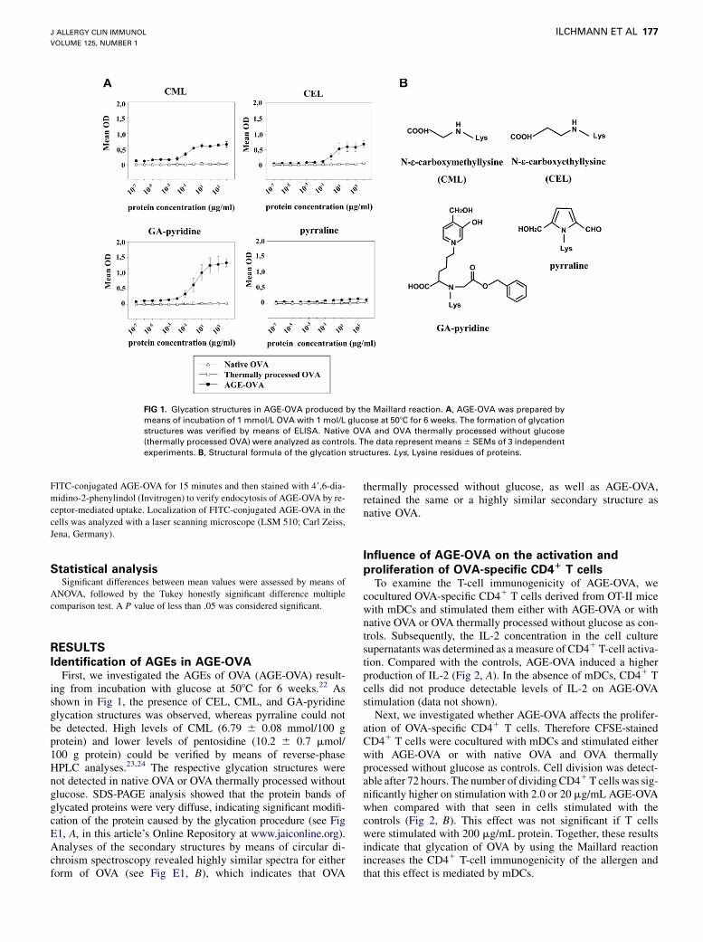
FIG 1. Glycation structures in AGE-OVA produced by the Maillard reaction. A, AGE-OVA was prepared by
means of incubation of 1 mmol/L OVA with 1 mol/L glucose at 508C for 6 weeks. The formation of glycation
structures was verified by means of ELISA. Native OVA and OVA thermally processed without glucose
(thermally processed OVA) were analyzed as controls. The data represent means 6 SEMs of 3 independent
experiments. B, Structural formula of the glycation structures. Lys, Lysine residues of proteins.
J ALLERGY CLIN IMMUNOL
VOLUME 125, NUMBER 1
ILCHMANN ET AL 177
FITC-conjugated AGE-OVA for 15 minutes and then stained with 49,6-dia-
midino-2-phenylindol (Invitrogen) to verify endocytosis of AGE-OVA by re-
ceptor-mediated uptake. Localization of FITC-conjugated AGE-OVA in the
cells was analyzed with a laser scanning microscope (LSM 510; Carl Zeiss,
Jena, Germany).
Statistical analysisSignificant differences between mean values were assessed by means of
ANOVA, followed by the Tukey honestly significant difference multiple
comparison test. A P value of less than .05 was considered significant.
RESULTS
Identification of AGEs in AGE-OVAFirst, we investigated the AGEs of OVA (AGE-OVA) result-
ing from incubation with glucose at 508C for 6 weeks.22 Asshown in Fig 1, the presence of CEL, CML, and GA-pyridineglycation structures was observed, whereas pyrraline could notbe detected. High levels of CML (6.79 6 0.08 mmol/100 gprotein) and lower levels of pentosidine (10.2 6 0.7 mmol/100 g protein) could be verified by means of reverse-phaseHPLC analyses.23,24 The respective glycation structures werenot detected in native OVA or OVA thermally processed withoutglucose. SDS-PAGE analysis showed that the protein bands ofglycated proteins were very diffuse, indicating significant modifi-cation of the protein caused by the glycation procedure (see FigE1, A, in this article’s Online Repository at www.jaiconline.org).Analyses of the secondary structures by means of circular di-chroism spectroscopy revealed highly similar spectra for eitherform of OVA (see Fig E1, B), which indicates that OVA
thermally processed without glucose, as well as AGE-OVA,retained the same or a highly similar secondary structure asnative OVA.
Influence of AGE-OVA on the activation and
proliferation of OVA-specific CD41 T cellsTo examine the T-cell immunogenicity of AGE-OVA, we
cocultured OVA-specific CD41 T cells derived from OT-II micewith mDCs and stimulated them either with AGE-OVA or withnative OVA or OVA thermally processed without glucose as con-trols. Subsequently, the IL-2 concentration in the cell culturesupernatants was determined as a measure of CD41 T-cell activa-tion. Compared with the controls, AGE-OVA induced a higherproduction of IL-2 (Fig 2, A). In the absence of mDCs, CD41 Tcells did not produce detectable levels of IL-2 on AGE-OVAstimulation (data not shown).
Next, we investigated whether AGE-OVA affects the prolifer-ation of OVA-specific CD41 T cells. Therefore CFSE-stainedCD41 T cells were cocultured with mDCs and stimulated eitherwith AGE-OVA or with native OVA and OVA thermallyprocessed without glucose as controls. Cell division was detect-able after 72 hours. The number of dividing CD41 T cells was sig-nificantly higher on stimulation with 2.0 or 20 mg/mL AGE-OVAwhen compared with that seen in cells stimulated with thecontrols (Fig 2, B). This effect was not significant if T cellswere stimulated with 200 mg/mL protein. Together, these resultsindicate that glycation of OVA by using the Maillard reactionincreases the CD41 T-cell immunogenicity of the allergen andthat this effect is mediated by mDCs.
FIG 2. AGE-OVA enhances the activation and proliferation of OVA-specific CD41 T cells. A, CD41 T cells
isolated from OT-II mice were cocultured with mDCs and stimulated with native OVA, OVA thermally pro-
cessed without glucose, or AGE-OVA for 24 hours. Concentration of IL-2 in the culture supernatant was
measured by means of ELISA. *P < .001. B, CFSE-stained CD41 T cells were cocultured with mDCs and stim-
ulated with either form of OVA. After 72 hours, CFSE intensity of CD41 T cells was measured by means of
flow cytometry. The data are representative of 3 independent experiments.
J ALLERGY CLIN IMMUNOL
JANUARY 2010
178 ILCHMANN ET AL
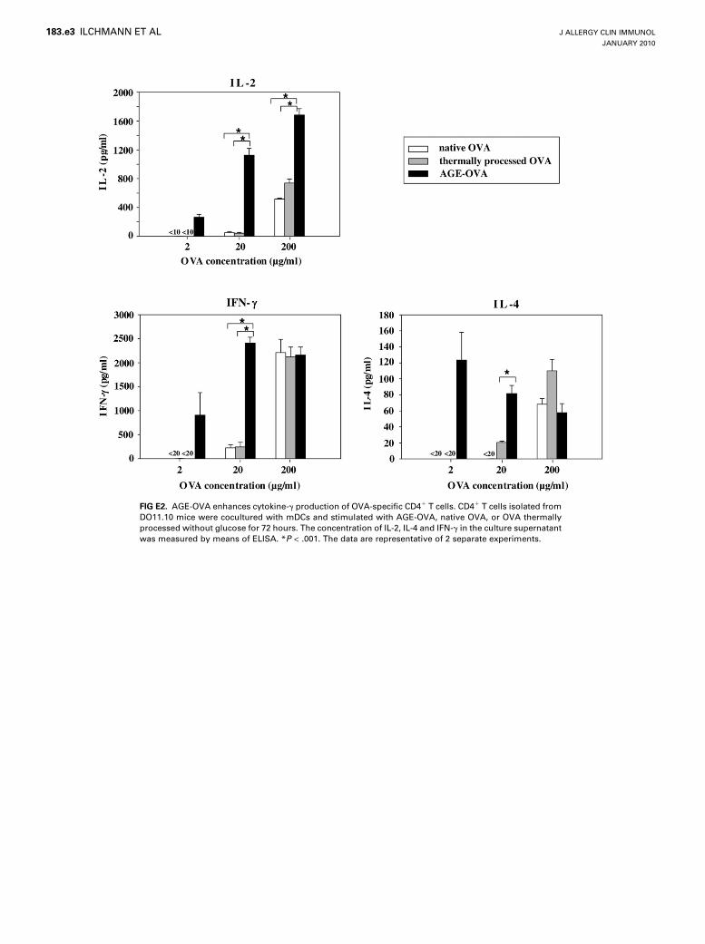
Next, we examined the influence of glycation structures oncytokine production by CD41 T cells. OVA-specific CD41 Tcells from DO11.10 mice were cocultured with mDCs andstimulated with either form of OVA. Production of TH2-typecytokines, such as IL-4, by allergen-specific CD41 T cells isa critical component in inducing food allergy. Because CD41
T cells from OT-II mice predominantly produce TH1-type cyto-kines, such as IFN-g, but hardly any TH2-type cytokines, forthe cytokine assay, we used CD41 T cells from DO11.10mice, which efficiently produce TH2-type cytokines. Comparedwith the controls, AGE-OVA induced higher production ofIL-2, IL-4, and IFN-g (see Fig E2 in this article’s OnlineRepository at www.jacionline.org). The results suggest thatglycation structures enhance production of both TH1- andTH2-type cytokines.
Influence of AGE-OVA on the maturation of mDCsTo investigate the mediator function of mDCs, we first
examined the influence of AGE-OVA on the maturation of theseAPCs. Expectedly, LPS-stimulated mDCs (LPS is a knowninducer of mDC maturation) displayed enhanced expression ofthe maturation markers CD40, CD80, CD86, and MHC class IImolecule. In contrast, the expression of these markers was notenhanced by either form of OVA (Fig 3, A). Moreover, theseOVAs also did not induce a detectable secretion of IL-12 p70or IL-10, which was observed when mDCs were stimulatedwith LPS (Fig 3, B). Together, these results suggest that theOVA allergens are not capable of stimulating the maturationof mDCs.
Uptake of AGE-OVA by mDCsTo further elucidate the mediator function of mDCs, we next
investigated whether glycation through the Maillard reactioninfluences the uptake of antigen by mDCs. Therefore these cellswere incubated with FITC conjugates of AGE-OVA or nativeOVA, OVA thermally processed without glucose, and recombi-nant OVA without any natural carbohydrate residues or glycationstructures as controls. The FITC intensity of mDCs was analyzedby means of flow cytometry as a measure of antigen uptake. Ifcompared with native OVA and thermally processed OVA, weobserved a fluorescence shift of approximately 5- to 10-fold inmDCs incubated with AGE-OVA, indicating higher uptake of theAGE (Fig 4, A). Importantly, the mDC uptake of recombinantOVAwas lower than that of native OVA (see Fig E3 in this article’sOnline Repository at www.jacionline.org). Subsequent confocalmicroscopic analyses showed that the majority of AGE-OVAwas not merely attached on the cell surface of mDCs but wasendocytosed into the cells (Fig 4B).
Involvement of SR-AI/II in the uptake of AGE-OVA
by mDCsNative OVA has natural mannose residues, and its uptake is
known to be mediated by the mannose receptor (MR).25,26 AGEsare known to bind to several cell-surface receptors, such asRAGE, SR-AI/II, and galectin-3.12-16 Hence we hypothesizedthat the uptake of AGE-OVA by mDCs is mediated by 1 ormore of these receptors for AGEs in addition to the MR for naturalmannose residues. To identify the responsible receptor or recep-tors for the AGE-OVA uptake by mDCs, we investigated
FIG 3. AGE-OVA does not induce the maturation of mDCs. mDCs were
stimulated with 20 mg/mL native OVA, OVA thermally processed without
glucose or AGE-OVA or with 10 mg/mL LPS. A, Expression of CD40, CD80,
CD86, and MHC class II molecules on mDCs was analyzed with flow cytom-
etry. Gray areas represent mDCs cultured without stimulation. B, Levels of
IL-10 and IL-12 p70 in the culture supernatants of mDCs were measured by
means of ELISA. The data are representative of 3 independent experiments.
J ALLERGY CLIN IMMUNOL
VOLUME 125, NUMBER 1
ILCHMANN ET AL 179
RAGE- and SR-AI/II–deficient mDCs, as well as the galectin-3inhibitor lactose and the MR inhibitor mannan.26,27
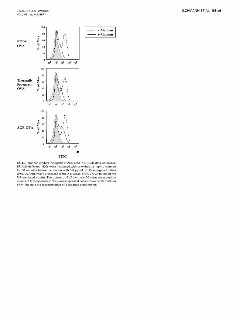
We observed almost identical uptake of FITC-conjugatedAGE-OVA in wild-type mDCs, RAGE-deficient mDCs, orlactose-treated wild-type mDCs (Fig 4, C and D), indicatingthat RAGE and galectin-3 would not be involved in the uptake.Likewise, the uptake of native OVA and OVA thermally processedwithout glucose was not affected in RAGE-deficient mDCs or inlactose-treated wild-type mDCs (see Fig E4 in this article’s On-line Repository at www.jacionline.org). In contrast, SR-AI/IIdeficiency resulted in a significant reduction of the AGE-OVAuptake by mDCs (Fig 5, A). The additional treatment of SR-AI/II–deficient mDCs with mannan led to a further reduction of theuptake of AGE-OVA (see Fig E5 in this article’s Online
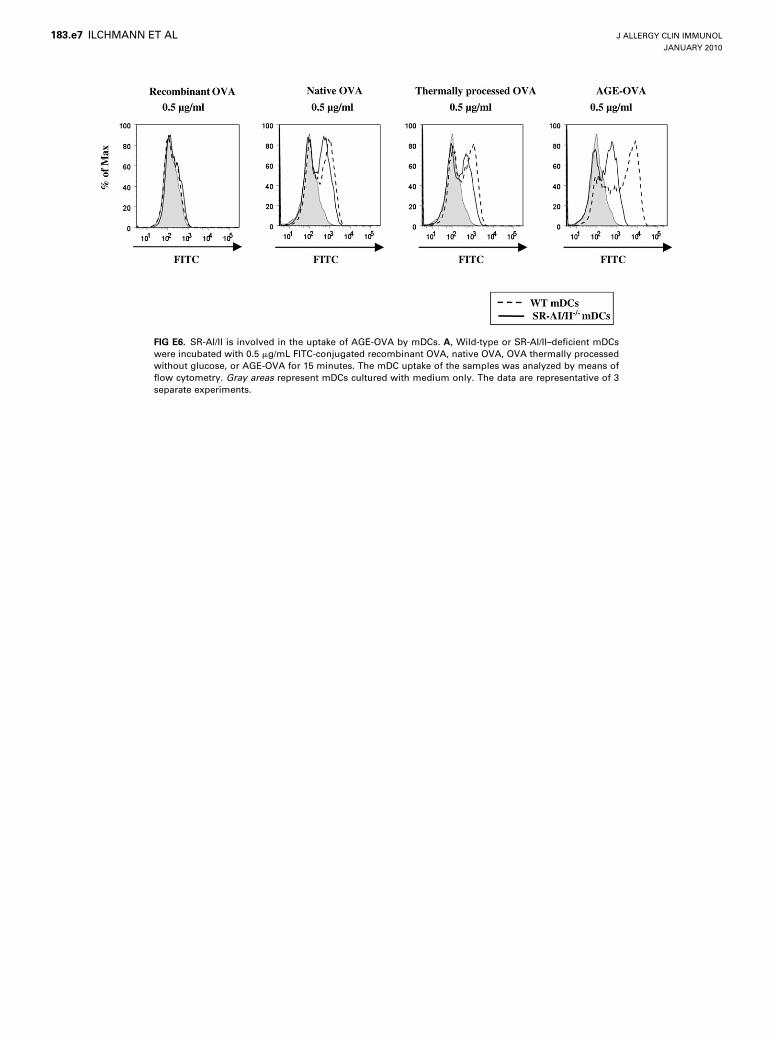
Repository at www.jacionline.org). Together, these resultssuggest that SR-AI/II and the MR, but not RAGE and galectin-3, are essential mediators of the mDC uptake of AGE-OVA. Un-expectedly, SR-AI/II deficiency also led to a slight reduction ofthe mDC uptake of native OVA and OVA thermally processedwithout glucose (Fig 5, A, and see Fig E6 in this article’s OnlineRepository at www.jacionline.org). The additional treatment ofSR-I/II–deficient mDCs with mannan resulted in a completesuppression of the uptake of the non-AGE forms of OVA (seeFig E5). The SR-AI/II deficiency, however, did not inhibit themDC uptake of recombinant OVA (see Figure E5, A).
Unlike OVA, BSA does not possess natural carbohydrateresidues.28 However, BSA should also be capable of binding toSR-AI/II as an AGE derivative. In accordance with this, weshow that the uptake of AGE-BSA was significantly attenuatedin SR-AI/II–deficient mDCs (see Fig E7 in this article’s OnlineRepository at www.jacionline.org). Consequently, we hypothe-sized that AGE-BSA should act as an inhibitor of the SR-AI/II–mediated uptake of OVA allergens. To prove this, we incubatedwild-type mDCs with native BSA or AGE-BSA together withthe different forms of OVA. In accordance with our hypothesis,AGE-BSA, but not native BSA, was capable of inhibiting theuptake of either form of OVA (Fig 6). Together, the results suggestthat SR-AI/II plays an important role in the enhanced mDCuptake of AGE-OVA.
SR-AI/II deficiency reduces the activation of OVA-
specific CD41T cells by mDCsTo further verify the obvious function of SR-AI/II in the
mDC-mediated enhanced activation of OVA-specific CD41 Tcells by AGE-OVA, we examined whether the expression ofSR-AI/II is a prerequisite for this effect. Therefore we cocul-tured OVA-specific CD41 T cells with SR-AI/II–deficient orwild-type mDCs that were pretreated with either form ofOVA. As shown in Fig 5, B, we observed significantly reducedproduction of IL-2 by OVA-specific CD41 T cells in response tonative OVA, OVA thermally processed without glucose, andAGE-OVA if the T cells were cocultured with SR-AI/II–defi-cient mDCs instead of wild-type mDCs. However, the reductionwas not observed for recombinant OVA. The capacity to stimu-late T cells appears not to be affected in SR-AI/II–deficientmDCs because SR-AI/II–deficient and wild-type mDCs expresscomparable levels of costimulatory CD40, CD80, CD86, andMHC class II molecules (see Fig E8 in this article’s OnlineRepository at www.jacionline.org). Wild-type and SR-AI/II–deficient mDCs induced comparable IL-2 production of OVA-specific CD41 T cells against recombinant OVA, also indicatingthe intact T-cell stimulatory capacity of the deficient mDCs. Inaccordance with this, the results suggest that mDC-expressedSR-AI/II is critical for the activation of OVA-specific CD41 Tcells by AGE-OVA.
DISCUSSIONThe majority of foods are modified by storage or processing.
The formation of AGEs produced by the Maillard reaction duringthermal processing of foods is suggested to exert important effectson the immunogenicity of food proteins. To our knowledge, thepresent study is the first to demonstrate that the formation ofAGEs enhances the CD41 T-cell immunogenicity of a food
FIG 4. The uptake of OVA by mDCs is increased by means of glycation. RAGE and galectin-3 are not
involved. A, Wild-type mDCs were incubated with 0.5 or 5.0 mg/mL FITC-conjugated native OVA, OVA ther-
mally processed without glucose, or AGE-OVA for 15 minutes. The mDC uptake of the samples was ana-
lyzed by means of flow cytometry. B, The mDC uptake of FITC-conjugated AGE-OVA was verified by
using confocal microscopy. C, Wild-type or RAGE-deficient mDCs were incubated with 5.0 mg/mL FITC-con-
jugated AGE-OVA for 15 minutes. The uptake of AGE-OVA was analyzed by using flow cytometry. D, Wild-
type mDCs were treated with or without 150 mmol/L lactose for 30 minutes before incubation with 5.0 mg/
mL FITC-conjugated AGE-OVA to prevent galectin-3–mediated uptake. The uptake of AGE-OVA by mDCs
was analyzed by means of flow cytometry. Gray areas represent mDCs cultured with medium only. The
data are representative of 3 independent experiments.
J ALLERGY CLIN IMMUNOL
JANUARY 2010
180 ILCHMANN ET AL
allergen. The results strongly suggest that the influence of theMaillard reaction needs to be carefully considered in the evalua-tion of the immunogenicity of food allergens.
Our results are in good accordance with previous studiesdemonstrating an increased CD41 T-cell immunogenicity of an-tigens chemically modified to function as SR-AI/II ligands.29-31
Moreover, it could be shown that native OVA endocytosedthrough scavenger receptors in macrophages predominantly acti-vates the allergen-specific CD41 T cells.26 However, the specificscavenger receptor or receptors participating in this OVA uptakewere not identified. Our results now substantiate that (1) SR-AI/IIis an important mediator of the mDC uptake of native OVA andAGE-OVA and (2) the subsequent activation of OVA-specificCD41 T cells occurs through SR-AI/II–mediated uptake (Fig 5).
SR-AI/II belongs to a large and diverse group of scavengerreceptors and functions as an endocytic receptor.32,33 The expres-sion of SR-AI/II is mostly restricted to myeloid cells.34 Thepresentation of soluble antigens by APCs is governed byendocytic receptors, which determine the intracellular routingof the endocytosed molecules.11,26,35 For example, antigens endo-cytosed by the MR in DCs were shown to be targeted to the MHCclass I loading pathway,26 whereas those endocytosed by theglycan-binding receptor DC-SIGN (CD209) were targeted tothe MHC class II loading pathway.35 We found that the glycation
of OVA does not enhance CD81 T-cell immunogenicity of theallergen. OVA-specific CD81 T cells cocultured with mDCssecreted comparable levels of IL-2 in response to AGE-OVAand native OVA (see Fig E9 in this article’s Online Repositoryat www.jacionline.org). Our results now suggest that SR-AI/IIis a receptor targeting its ligands to the MHC class II loadingpathway.
Importantly, the activation of OVA-specific CD41 T cells byrecombinant OVA was lower than that of native OVA and wasnot attenuated by SR-AI/II deficiency in the mDCs (Fig 5).Recombinant OVA has no natural carbohydrate or glycationstructures for SR-AI/II and other endocytic receptors. It couldpreviously be shown that pinocytosed antigens are transferredto the lysosome compartment, where antigens enter the MHCclass II loading pathway.26 The recombinant OVAwould be takenup by pinocytosis in mDCs, whereas native OVA and AGE-OVAwould be taken up not only by pinocytosis but also by SR-AI/II–mediated endocytosis. Because uptake of AGE-OVA by SR-AI/IIis higher than that of native OVA as a result of the glycation struc-tures, higher amounts of AGE-OVA would be targeted into theMHC class II loading pathway. This would increase the amountof OVA peptide/MHC class II complex on the surface of mDCsand subsequently induce enhanced OVA-specific CD41 T-cellactivation.
FIG 5. SR-AI/II is involved in the uptake of AGE-OVA by mDCs. A, Wild-type or SR-AI/II–deficient mDCs were
incubated with 5.0 mg/mL FITC-conjugated recombinant OVA, native OVA, OVA thermally processed with-
out glucose, or AGE-OVA for 15 minutes. The mDC uptake of the samples was analyzed by means of flow
cytometry. Gray areas represent mDCs cultured with medium only. B, Wild-type or SR-AI/II–deficient mDCs
were incubated for 3 hours with either form of OVA and then fixed with 0.008% glutaraldehyde before 21
hours of coculturing with CD41 T cells isolated from OT-II mice. The concentration of IL-2 in the culture su-
pernatant was measured by means of ELISA. *P < .01. **P < .001. The data are representative of 3
independent experiments.
J ALLERGY CLIN IMMUNOL
VOLUME 125, NUMBER 1
ILCHMANN ET AL 181
Despite the enhanced uptake of AGE-OVA by mDCs, theallergen did not stimulate the maturation of these cells in ourexperimental setting (Fig 3). Previously, other studies revealedboth stimulatory and inhibitory effects of AGEs on the maturationof human DCs.8,9 The different observations might be explainedby variations in the expression profiles of receptors for AGEs onthese DCs. The previously detected in vitro stimulation of humanDC maturation by AGE-BSA was ascribed to the expression ofRAGE.8 This is consistent with reports providing evidence thatRAGE acts as a receptor triggering the maturation of DCs.36,37
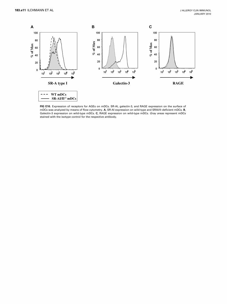
In our experiments RAGE expression was not detectable on thecell surface of the mDCs (see Fig E10 in this article’s OnlineRepository at www.jacionline.org). The absence of RAGE inmDCs might explain why AGE-OVA did not stimulate thematuration of the cells.
We found high levels of CEL, CML, and GA-pyridineglycation structures in AGE-OVA (Fig 1, A), which we recentlyalso detected in roasted peanuts (ie, an important food allergen;manuscript in preparation). CEL and CML are also representativeAGEs in thermally processed foods.1 Hence AGE-OVA appearsto be an appropriate model for studying the influence of food
allergen–derived AGEs on T-cell immunogenicity. Currently, itis still unknown which glycation structures bind to SR-AI/II orgalectin-3, whereas CML has been suggested as a major glycationstructure of RAGE ligands.38 The future identification of the exactglycation structures responsible for ligand binding to the differentAGE receptors will help to further decipher the influence of AGEson DC function and the T-cell immunogenicity of food allergens.
In addition, we found that AGE-OVA could enhance produc-tion of both TH1- and TH2-type cytokines by allergen-specificCD41 T cells (see Fig E2). The AGEs would enhance the activa-tion and subsequent cytokine production of allergen-specificCD41 T cells but not induce polarization of cytokine productionby the T cells. Recent findings suggest that regulatory T cells pro-ducing IL-10 consistently represent the dominant T-cell subsetspecific for food allergens in healthy subjects; in contrast, thereis a high frequency of allergen-specific IL-4–producing T cellsin allergic subjects.39,40 The glycated food allergens mightenhance activation of allergen-specific regulatory T cells formaintaining tolerance against the allergens in healthy subjectsbut might promote activation of allergen-specific TH2 cellsinducing allergic responses in allergic subjects.
FIG 6. AGE-BSA, but not native BSA, inhibits the uptake of AGE-OVA by mDCs. mDCs were incubated with
or without 50 mg/mL native BSA and AGE-BSA together with 5.0 mg/mL FITC-conjugated, native OVA, OVA
thermally processed without glucose, or AGE-OVA. After incubation for 15 minutes, the mDC uptake of
either form of OVA was analyzed by means of flow cytometry. Gray areas represent mDCs cultured with cell
culture medium only. The data are representative of 3 independent experiments.
J ALLERGY CLIN IMMUNOL
JANUARY 2010
182 ILCHMANN ET AL
In conclusion, we show that the Maillard reaction enhances theCD41 T-cell immunogenicity of the OVA food allergen. Itappears that AGEs of food allergens can be endocytosed bymDCs through SR-AI/II, enabling the subsequent presentationof the allergen to CD41 T cells. We also show that IL-4 produc-tion by allergen-specific CD41 T cells is enhanced by glycation ofa food allergen. Because IL-4 produced by CD41 T cells is a crit-ical component for IgE production by B cells and subsequentallergic responses, our results suggest that the Maillard reactioncould be capable of enhancing the allergenicity of food allergensin allergic subjects. Future in vivo studies on the T-cell immuno-genicity and allergenicity of AGEs will further elucidate theimportance of the Maillard reaction in food allergy.
We thank Dr Takuhito Shoji (Osaka Medical College, Osaka, Japan) for
providing RAGE-deficient mice; Doreen Werchau, Laura Sandner, and
Annette Jamin (Paul-Ehrlich-Institut) for technical assistance; and Stefan
Sch€ulke for preparation of recombinant protein. We also thank Professor Av
Mitchison (University College London, London, United Kingdom) and Dr
Stephan Steckelbroeck (Paul-Ehrlich-Institut) for helpful comments on this
study.
Key messages
d The T-cell immunogenicity of food allergens can be en-hanced by the Maillard reaction, indicating a criticalrole for thermal food processing in modulating or enhanc-ing the allergenicity of food proteins.
REFERENCES
1. Henle T. Protein-bound advanced glycation endproducts (AGEs) as bioactive
amino acid derivatives in foods. Amino Acids 2005;29:313-22.
2. Malanin K, Lundberg M, Johansson SG. Anaphylactic reaction caused by
neoallergens in heated pecan nut. Allergy 1995;50:988-91.
3. Berrens L. Neoallergens in heated pecan nut: products of Maillard-type
degradation? Allergy 1996;51:277-8.
4. Scheurer S, Lauer I, Foetisch K, San Miguel Moncin M, Retzek M, Hartz C, et al.
Strong allergenicity of Pru av 3, the lipid transfer protein from cherry, is related to
high stability against thermal processing and digestion. J Allergy Clin Immunol
2004;114:900-7.
5. Gruber P, Becker WM, Hofmann T. Influence of the Maillard reaction on the
allergenicity of rAra h 2, a recombinant major allergen from peanut (Arachis
hypogaea), its major epitopes, and peanut agglutinin. J Agric Food Chem 2005;
53:2289-96.
6. Chung SY, Champagne ET. Association of end-products with increased IgE bind-
ing of roasted peanuts. J Agric Food Chem 2001;49:3911-6.