198 of MT-associated proteins as antigens for the preparation of good quality immunological reagents for the identifi- cation of new spindle proteins. From monoclonals, from the serendipitous identification of a few well-characterized proteins in the spindle, and from auto- immune antibodies characteristic of patients with scleroderma, the list of spindle components is slowly growing. Table I describes the current state of knowledge, together with the criteria by which each protein has been identified as a spindle component. Biochemical genetics of the spindle A powerful approach to identifying the role played by a particular protein in mitosis would be a rigorous genetic dis- section of the spindle. Thus far, genetics has provided little help either in the identification of novel spindle com- ponents or in the elucidation of known ones. There is, however, active work on mitosis in Aspergillus ~7, Saccharomyces is and Drosophila ~9, taking advantage of the genetic elegance of these organisms. DNA clones corresponding to the kinetocbores of yeasts have been isolated and sequenced TM. It seems highly likely that molecular genetics and the study of mutant phenotype will in the near future join biochemistry and immun- ology as powerful approaches to the analysis of mitotic mechanisms. Acknowledgements This paper was written while visiting the MRC Laboratory of Molecular Biology, Cambridge, UK. I thank my host, J. V. Kilmartin, for his stimulating hospitality. Financial support from the Union Internationale Contre le Cancer is gratefully acknowledged. References I Mclntosh, J. R., McDonald, K. L.. Edwards, M. K. and Ross, B. M. (1979) J. CellBioL 83, 428-442; 443-461 2 Tippit, D. H., Pillus, L. and Pickett-Heaps, J. (1980) J. Cell Biol, 87, 531-545 3 Euteneuer, U., Jackson, W. T. and Mclntosh, J. R. 11982) J. ('ell Biol. 94, (~14-653 4 Margolis, R. C. and Wilson, L. (1978) Cell 13, TIBS - April 1984 1-8 5 Hill, T. L. and Kirschner, M. W. (1982) Int. Rev. CytoL 78, 1-125 6 Hays, T. S., Wise, D. and Salmon, E. D. 11982) J. Cell. Biol. 93, 374--382 7 Nicklas, R. B. 11983)J. ('ell Biol. 97, 542-548 8 Inoue, S. (1981) J. Cell Biol. 91, 131~147s 9 Cande, W. Z. (1982) Cell28, 15-22 10 Roobol, A., Havercroft, J. C. and Gull, K. 11982) J. Cell Sci. 55, 365-381 11 Mclntosh, J. R. (1983} Modern ~\ell Biol. 2, 115-142 12 De Brabender, M., Geuens, G., DeMey, J. and Jonian, M. 11981) (,'ell Motili~. I, 469-483 13 Hepler. P. K. and Wolniak, S. M. (1983) Modern ('_ell Biol. 2. 93-112 14 Schibler, M. J. and Pickett-Heaps, J. D. (1980) Eur. J. Cell BioL 22, 687~698 15 Hays, T. S. and Salmon, E. D. (1983) J. Cell BioL 97, 44a 16 Bulinski, J. and Borisy, G. G. (19811) J. BioL Chem. 225, 11570-11576 17 Oakley, B. R. and Morris, N. R. 11981) Cell 24, 83%845 18 Neff, N. F., Thomas, J. H., Grisafi, P. and Botsteim D. (1983) (~'e//3l, 655-67tl 19 Kemphues, K. J., Kaufman, T. C., Raft. R. A. and Raft. E. C. (1982) Cell 31,655~570 20 Bloom, K. S. and Carbon, J. 11982) Cell 29, 305-317 Glycoproteins Nathan Sharon The recent isolation and structural characterization of numerous glycosylated proteins from diverse sources, and the elucidation of the biosynthesis of their carbohydrate units, has provided a sound basis for the understanding of the roles" of these units especially as recognition determinants in normal and pathological processes. Although the occurrence in nature of proteins containing covalently bound carbohydrate was noted during the second part of the 19th century, the foundations of our current knowledge of glycoproteins were laid mainly during the 1950s and 1960s. By the early 1970s1'2 it was widely appreciated that glyco- proteins are found in all forms of life, with the possible exception of bacteria; and that most proteins are probably glycosylated. The two major bonds link- ing the carbohydrate to the protein were identified: the N-glycosyl linkage to asparagine and the O-glycosyl linkage to serine, threonine or hydroxylysine. It was demonstrated that although glyco- proteins contain only a few different monosaccharides, they vary markedly in their carbohydrate content, in the num- Nathan Sharon is at the Department of Biophysics, The Weizmann Institute of Science, Rehovot 76100, Israel. Currently he is Distinguished Visiting Scientist at the National Institute of Arthritis, Dia- betes, Digestive and Kidney Diseases, National Institutes of Health, Bethesda, MD 20205, USA. ber and degree of branching of their carbohydrate units, and in the distribu- tion of such units along the polypeptide chains. A fundamental difference be- tween the polypeptide backbone and the carbohydrate units was noted, in that whereas the former is always identi- cal in molecules of the same kind, the latter are quite variable. Therefore a carbohydrate unit attached to a parti- cular amino acid residue in a polypeptide chain of the same glycoprotein may show structural heterogeneity. Although it was thus concluded that glycoproteins exhibit microheterogeneity, at the time no structural data supporting this con- cept were available. As late as ten years ago little was known with certainty about the structure of the carbohydrate units of glycoproteins, except for units con- taining only two or three sugar residues. Glycoproteins were shown to be syn- thesized by the sequential action of glyco- syltransferases, located mainly in the Golgi apparatus, using sugar nucleotides as donor molecules. Preliminary results ~) 1984, Elsevier Science Publishers B.V. Amsterdam ff376 5067;84/$1P_.011 had been presented in the early 1970s on the possible involvement of lipid- linked intermediates in the biosynthesis of glycoproteins3. From studies of blood group antigens it was concluded that the structure of the carbohydrate units is determined by the availability of specific glycosyltransferases, so that these struc- tures are secondary gene products, the primary gene products being the above- mentioned enzymes. Several inherited diseases of metabolism, in particular some of the mucopolysaccharidoses, had been shown to be due to deficiencies of specific enzymes whose function is to hydrolyse the saccharide chains of the proteoglycans, a class of connective tissue glycoproteins. Concurrently, increasing attention has focused on the intriguing question of the biological functions of the carbohydrate units of glycoproteins. Demonstrated functions included imparting high vis- cosity to mucins, protection against pro- teolysis, serving as receptors for viruses, controlling the lifetime of glycoproteins in the circulatory system of higher animals, and regulating glycoprotein up- take by cells4. More important, it had been realized that sugar polymers can carry more information than polypep- tides or polynucleotides. It was thus assumed that the carbohydrates of glyco- proteins play a key role in recognition phenomena such as those that regulate cell differentiation and growth, and that derangement of this recognition might
Transcript
198
of MT-associated proteins as antigens for the preparation of good quality immunological reagents for the identifi- cation of new spindle proteins. From monoclonals, from the serendipitous identification of a few well-characterized proteins in the spindle, and from auto- immune antibodies characteristic of patients with scleroderma, the list of spindle components is slowly growing. Table I describes the current state of knowledge, together with the criteria by which each protein has been identified as a spindle component.
Biochemical genetics of the spindle A powerful approach to identifying
the role played by a particular protein in mitosis would be a rigorous genetic dis- section of the spindle. Thus far, genetics has provided little help either in the identification of novel spindle com- ponents or in the elucidation of known ones. There is, however, active work on mitosis in Aspergillus ~7, Saccharomyces is and Drosophila ~9, taking advantage of
the genetic elegance of these organisms. DNA clones corresponding to the kinetocbores of yeasts have been isolated and sequenced TM. It seems highly likely that molecular genetics and the study of mutant phenotype will in the near future join biochemistry and immun- ology as powerful approaches to the analysis of mitotic mechanisms.
Acknowledgements This paper was written while visiting
the MRC Laboratory of Molecular Biology, Cambridge, UK. I thank my host, J. V. Kilmartin, for his stimulating hospitality. Financial support from the Union Internationale Contre le Cancer is gratefully acknowledged.
References I Mclntosh, J. R., McDonald, K. L.. Edwards,
M. K. and Ross, B. M. (1979) J. CellBioL 83, 428-442; 443-461
2 Tippit, D. H., Pillus, L. and Pickett-Heaps, J. (1980) J. Cell Biol, 87, 531-545
3 Euteneuer, U., Jackson, W. T. and Mclntosh, J. R. 11982) J. ('ell Biol. 94, (~14-653
4 Margolis, R. C. and Wilson, L. (1978) Cell 13,
TIBS - April 1984
1-8 5 Hill, T. L. and Kirschner, M. W. (1982) Int.
Rev. CytoL 78, 1-125 6 Hays, T. S., Wise, D. and Salmon, E. D.
11982) J. Cell. Biol. 93, 374--382 7 Nicklas, R. B. 11983)J. ('ell Biol. 97, 542-548 8 Inoue, S. (1981) J. Cell Biol. 91, 131~147s 9 Cande, W. Z. (1982) Cell28, 15-22
10 Roobol, A., Havercroft, J. C. and Gull, K. 11982) J. Cell Sci. 55, 365-381
11 Mclntosh, J. R. (1983} Modern ~\ell Biol. 2, 115-142
12 De Brabender, M., Geuens, G., DeMey, J. and Jonian, M. 11981) (,'ell Motili~. I, 469-483
13 Hepler. P. K. and Wolniak, S. M. (1983) Modern ('_ell Biol. 2. 93-112
14 Schibler, M. J. and Pickett-Heaps, J. D. (1980) Eur. J. Cell BioL 22, 687~698
15 Hays, T. S. and Salmon, E. D. (1983) J. Cell BioL 97, 44a
16 Bulinski, J. and Borisy, G. G. (19811) J. BioL Chem. 225, 11570-11576
17 Oakley, B. R. and Morris, N. R. 11981) Cell 24, 83%845
18 Neff, N. F., Thomas, J. H., Grisafi, P. and Botsteim D. (1983) (~'e//3l, 655-67tl
19 Kemphues, K. J., Kaufman, T. C., Raft. R. A. and Raft. E. C. (1982) Cell 31,655~570
20 Bloom, K. S. and Carbon, J. 11982) Cell 29, 305-317
Glycoproteins Nathan Sharon
The recent isolation and structural characterization of numerous glycosylated proteins from diverse sources, and the elucidation of the biosynthesis of their carbohydrate units, has provided a sound basis for the understanding of the roles" of these units especially as recognition determinants in normal and pathological
processes.
Although the occurrence in nature of proteins containing covalently bound carbohydrate was noted during the second part of the 19th century, the foundations of our current knowledge of glycoproteins were laid mainly during the 1950s and 1960s. By the early 1970s 1'2 it was widely appreciated that glyco- proteins are found in all forms of life, with the possible exception of bacteria; and that most proteins are probably glycosylated. The two major bonds link- ing the carbohydrate to the protein were identified: the N-glycosyl linkage to asparagine and the O-glycosyl linkage to serine, threonine or hydroxylysine. It was demonstrated that although glyco- proteins contain only a few different monosaccharides, they vary markedly in their carbohydrate content, in the num-
Nathan Sharon is at the Department o f Biophysics, The Weizmann Institute o f Science, Rehovot 76100, Israel. Currently he is Distinguished Visiting Scientist at the National Institute of Arthritis, Dia- betes, Digestive and Kidney Diseases, National Institutes o f Health, Bethesda, MD 20205, USA.
ber and degree of branching of their carbohydrate units, and in the distribu- tion of such units along the polypeptide chains. A fundamental difference be- tween the polypeptide backbone and the carbohydrate units was noted, in that whereas the former is always identi- cal in molecules of the same kind, the latter are quite variable. Therefore a carbohydrate unit attached to a parti- cular amino acid residue in a polypeptide chain o f the same glycoprotein may show structural heterogeneity. Although it was thus concluded that glycoproteins exhibit microheterogeneity, at the time no structural data supporting this con- cept were available. As late as ten years ago little was known with certainty about the structure of the carbohydrate units of glycoproteins, except for units con- taining only two or three sugar residues.
Glycoproteins were shown to be syn- thesized by the sequential action of glyco- syltransferases, located mainly in the Golgi apparatus, using sugar nucleotides as donor molecules. Preliminary results
had been presented in the early 1970s on the possible involvement of lipid- linked intermediates in the biosynthesis of glycoproteins 3. From studies of blood group antigens it was concluded that the structure of the carbohydrate units is determined by the availability of specific glycosyltransferases, so that these struc- tures are secondary gene products, the primary gene products being the above- mentioned enzymes. Several inherited diseases of metabolism, in particular some of the mucopolysaccharidoses, had been shown to be due to deficiencies of specific enzymes whose function is to hydrolyse the saccharide chains of the proteoglycans, a class of connective tissue glycoproteins.
Concurrently, increasing attention has focused on the intriguing question of the biological functions of the carbohydrate units of glycoproteins. Demonstrated functions included imparting high vis- cosity to mucins, protection against pro- teolysis, serving as receptors for viruses, controlling the lifetime of glycoproteins in the circulatory system of higher animals, and regulating glycoprotein up- take by cells 4. More important, it had been realized that sugar polymers can carry more information than polypep- tides or polynucleotides. It was thus assumed that the carbohydrates of glyco- proteins play a key role in recognition phenomena such as those that regulate cell differentiation and growth, and that derangement of this recognition might
T I B S - A p r i l 1984
lead to pathological phenomena, includ- ing malignancy, although little direct evidence to support these ideas was available. Indeed, by 1975 it was stated that 'the specificity of many natural polymers is written in terms of sugar residues, not of amino acids or nucleotides "2.
Over 2000 publications on glycoproteins a year
Since 1976, the pace of research in this area has greatly accelerated. During this period, over 20 000 papers on glyco- proteins have been published, and the proportion of papers dealing with this subject in the total biomedical literature has more than doubled in comparison to the preceding 8 years. As a result of this enormous effort there has been a con- siderable increase in our knowledge of the structure, metabolism and functions of glycoproteins (for recent general reviews of the literature, see Refs 5-10).
A major contributing factor has been the development of improved techniques for the study of glycoproteins. Isolation and purification of glycoproteins has been facilitated by the use of lectins. They permit the separation of glycopro- teins that differ slightly in the structure of their carbohydrate units, or of variants of the same glycoprotein (e.g. oval- bumin) that differ in their glycosylation patterns. Numerous glycoproteins from such diverse sources as animals, plants, microorganisms and viruses have thus been isolated and characterized and several novel carbohydrate-peptide link- ages have been identified, for example L-arabinofuranosyl-hydroxyproline in plants I 1.
Endoglycosidases and chemical re- agents that relea~ oligosaccharides from glycoproteins or glyeopeptides, obtained by proteolytic digestion of glycoproteins, are now available. These oligosacchar- ides are readily purified to homogeneity by affinity chromatography on immobi- lized lectins and by high pressure liquid chromatography. In certain cases the purified oligosacchafides may be identi- fied by comparison with linear or branched synthetic compounds, some- thing not even dreamt of a decade ago, because of the enormous difficulties in synthetic oligosaccharide chemistry. Usually, however, classical techniques such as methylation analysis and hydro- lysis by glycosidases are employed for structural elucidation of the individual oligosaccharides. These are used in con- junction with gas-liquid chromatography, mass spectrometry and high resolution nuclear magnetic resonance (NMR),
199
~P--P-- Dol I l l
. . . . . . . 7 /
P
( III ) i ~ / p ( Vl )
( IV ) ( V )
GIc A Gal • Man m, k-Fuc
• GIcNAc [] GalNAc 91P NeuNAc
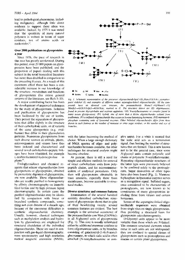
Fig. I. Schematic representation of' the precursor oligosaccharide-lipid GloManJGlcNAc)2-pyrophos- poryl dolichol (1) and examples of different mature asparagine-linked oligosaccharides. All the com- pounds have an identical core structure, the pentasaccharide Mane~(l-~6)[Mane~(l---~3)] Man~(l---*4)GlcNAcf3(1---~4)GlcNAc, marked in 1l. The structures shown are (11) oligomannose, found in procine thyroglobulin and soybean agglutinin; (lid N-ace~,llactosamine or complex found in many serum glycoproteins; (IV) hybrid, one of more than a dozen oligosaccharides isolated from ovalbumin; (V) a sulfated oligosaccharide that occurs in bovine luteinizing hormone; (V1) mannose-6- phosphate containing units of lysosomal enzymes. Other N-linked oligosaccharide~ differ from those shown in such features as the number of mannose or other sugar residues, or the number and size of branches.
with the latter becoming the method of choice. When a large enough dictionary of NMR spectra of oligo- and poly- saccharides becomes available, the other techniques for structural analysis might become obsolete.
At present, there is still a need for simple and efficient methods for removal of intact carbohydrate units from poly- peptide chains, and for microminiatur- ization of analytical procedures. Only then will glycoproteins obtainable in trace amounts, especially those from membranes, become accessible to struc- tural studies.
Diverse structures and common features Examination of the several hundred
recently described carbohydrate struc- tures of glycoproteins shows that in spite of their bewildering variety, several common features are evident. The best example is the universal occurrence of the pentasaccharide core Man3(GlcNAc)2 in all N-glycosyl units of glycoproteins (Fig. 1). This core is usually substituted either by additional mannose residues to form oligomannose units, or by branches consisting of galactosyl-~3-(1-4)-N-acetyl- glucosamine, to which sialic acid is often attached (N-acetyllactosamine or corn-
ptex units). For a while it seemed that the sialic acid acts as a termination signal, thus limiting the number of struc- tures that are formed. This is now known not to be the general case, since some N-glycosyl units contain polysialic acid chains or polymeric N-acetyllactosamine. Repeating oligosaccharide structures of the latter type were previously believed to be confined solely to the proteogly- cans. Sugar derivatives of other types have also been found (Fig. 1). Mannose 6-phosphate in lysosomal enzymes serves as a recognition signal. Sulfated sugars, once considered to be characteristic of proteoglycans, are now known to be constituents of many glycoproteins in- cluding those from viruses and certain hormones.
Scores of the asparagine-linked oligo- saccharide sequences were obtained from single sites on single glycoproteins, thus providing a structural basis for glycoprotein microheterogeneity.
O-Glycosyl units appear to be more variable than those which are N-linked. The common structural elements which occur in such units are not widespread; they are confined to special classes of glycoproteins such as proteoglycans and mucins or certain plant glycoproteins.
200
Rather unexpectedly, many of the carbohydrate sequences found in glyco- proteins, especially those occupying peripheral positions, are identical to those that occur in membrane glyco- lipids, such as the gangliosides. This suggests similar functions for these two classes of glycoconjugates12.
Three-dimensional structures The carbohydrate units of glycopro-
teins are often used by cells for inter- cellular and intracellular communication. The messages encoded in these struc- tures are transmitted through binding to complementary sites in antibodies or lectins.
Proper understanding of these carbo- hydrate-protein interactions requires detailed information about the conform- ation of carbohydrate molecules. Several approaches are being used to achieve this aim, including theoretical calcula- tions, high resolution NMR and X-ray crystallography. Here too, N-linked oligosaccharides have been the main subject of study. Marked differences were observed in the freedom of rotation and in the orientation of the different branches of these oligosaccharides, both in solution and in crystals. X-Ray crystal- lographic studies of the Fc fragments of human and rabbit IgG indicate that one of the oligosaccharide conformations is stabilized by interaction with the protein surface 13 (Fig. 2), Alterations in amino acid sequences may thus alter the anti- genic properties of a carbohydrate moiety or its ability to function as a receptor for lectins.
Biosynthesis The protein moieties of both mem-
brane-associated glycoproteins and secretory glycoproteins are initially synthesized in the rough endoplasmic reticulum. Our knowledge of the bio- synthesis of N-linked oligosaccharides is particularly advanced 14'L5 largely due to the availability of drugs such as tunica- mycin, swainsonine and castanosper- mine 16AT that inhibit specific steps of protein glycosylation and of cell mutants with glycosylation defects 1s'19. In con- trast to earlier views, all N-linked oligo- saccharides are formed from nucleotide sugars not by direct transfer to protein, but in two stages. In the first stage, a single precursor oligosaccharide Glc3Man9(GlcNAc)2, linked by pyro- phosphate to a special lipid, the long chain polyprenol dolichol (Fig. 1) is formed by the ordered stepwise transfer of monosaccharide residues from either nucleotide sugars or dolichol-linked sugars. A striking feature of the oligo- saccharide is that it contains the penta- saccharide core which is a constant part of all known N-linked carbohydrate units. Thus, both this biosynthetic pathway, known as the dolichol phosphate cycle, and the resulting structure have been conserved throughout evolution 2°. Although the exact role of the lipid- linked saccharides is not known, they appear to transport the hydrophilic sugars through the hydrophobic mem- brane to the location where attachment to polypeptide chains occurs.
The second stage of N-linked oligo- saccharide synthesis starts by the trans- fer of the lipid-linked oligosaccharide to
Fig. 2. The structure of the two-branched carbohydrate chains, in relation to the a-carbon backbone trace o f the Cn2 domains of rabbit lgG, to which they are covalently linked at residue Ash-297 (of each of the domains).
TIBS - Apr i l 1984
the amide of an asparaginyl residue that is part of a sequence Asn-X-Ser/Thr in a growing polypeptide chain as it is syn- thesized in and enters the lumen of the endoplasmic reticulum. N-Glycosylation is thus a cotranslational event. The same lipid intermediate is the precursor for both the oligomannose and the complex chains, so that a mechanism must exist for the removal of glucose and several mannose residues before the stepwise addition of N-acetylglucosamine, galac- tose, sialic acid and L-fucose residues can take place. Why the glucose residues are first added and then removed is not clear, although they may facilitate the transfer of the precursor oligosaccharide to protein, or control the later stages of the glycosylation process.
Most of the specific glycosidases that remove sequentially the three glucose residues (mainly in the endoplasmic reticulum) and of the bulk of the man- nose residues (mainly in the Golgi apparatus) from the polypeptide-bound oligosaccharide have been identified, but not yet characterized. In contrast, a number of glycosyltransferases that attach the outer sugars in the Golgi apparatus have been purified and investi- gated in detail.
Unlike the first stage of synthesis of asparagine-linked oligosaccharide units, which proceeds via a single pathway in most eukaryotes, processing is tremen- dously diverse. This remarkable variation among species, among oligosaccharides on different glycoproteins synthesized by individual cells, or even in the same cell, accounts for the enormous diversity of N-linked units. At present we do not know what determines the extent to which an oligosaccharide chain is pro- cessed. One factor may be the tertiary structure of the protein moiety which could limit the extent to which an oligo- saccharide is accessible to the modifying enzymes 14. Other factors which control processing include the high degree of the organization of the enzymes in the Golgi apparatus 21, and the three-dimen- sional shape of the oligosaccharides that serve as acceptors in branching and chain elongation reactions 22.
An important processing reaction is the attachment of phosphate to man- nose residues of oligomannose units, exclusively of newly synthesized lyso- somal acid hydrolases. It proceeds by a two-step reaction. Patients with the lyso- somal storage disease, the I-cell disease, lack the first enzyme in the phosphory- lating sequence 23. Thus, the disease is due to an oligosaccharide processing defect and although it is transmitted by
T I B S - A p r i l 1984
a single gene, many enzymes are affected. The failure to generate the mannose-6- phosphate recognition signal prevents the receptor-mediated targeting of the acid hydrolases to lysosomes and conse- quently the enzymes are secreted into the extracellular milieu, which is one of the biochemical abnormalities of the affected cells.
Although the synthesis of O-linked oligosaccharides does not, as a rule, require lipid intermediates, it is less well understood than that of N-linked units. O-Glycosylation is a post-translational event which proceeds through the sequential addition of monosaccharide residues by individual glycosytransfer- ases located in the Golgi apparatus iS. It is initiated prior to entry of glycoproteins into the early region of the Golgi appar- atus. The glycoproteins then migrate to the trans-Golgi where the late stages of the processing of both N- and O-linked units occur.
How the movement of proteins with- in the cell is controlled is not clear. Possibly membrane and secretory glyco- proteins are routed to their final destina- tion via the intracellular system of mem- branes and vesicles that are involved in the secretory pathways ~5~9.
Modification of protein conformation and activity
The demonstration that many struc- tures of the carbohydrate units of glyco- proteins have been conserved throughout evolution, and that protein glycosylation is a costly process for the organism in terms of energy and materials, strength- ened the belief that these units have important biological functions. The quest for such functions has been facili- tated by the availability of purified en- zymes that can remove or attach the sugars of glycoproteins, of specific glycosylation inhibitors and of cell mutants with known defects in glyco- sylation, particularly lectin-resistant cells 18
It is apparent that carbohydrates in glycoproteins do not have a single func- tion, and different glycoproteins have different requirement for carbohydrates (Table I). The ability of carbohydrates to modify, the physicochemical properties of glycoproteins has been known for a long time 24. Recent studies have shown that they are needed for folding and for the acquisition of the correct conforma- tion of certain proteins, as well as their ability to participate in subunit inter- actions. The carbohydrates of trans- membrane glycoproteins may help to orient and anchor them in the lipid
Table I. Functions of carbohydrates in glyco- proteins
Physicochemical Modify solubility, electrical charge, mass, size Stabilize protein conformation Protect against proteolysis
Biological recognition markers Signals for clearance of glycoproteins from the
circulatory system Markers for intracellular segregation of lyso-
somal enzymes Differentiation markers on cells Cell surface receptors for viruses, bacteria and
protozoa Mediators of non-immune phagocytosis
bilayer. Frequently carbohydrates pro- tect the protein against intracellular proteolysis during biosynthesis and transport, and are therefore also required for membrane insertion and secretion. In some cases, however, secretion occurs in the absence of glycosylation.
Whereas the biological activity of glycoprotein enzymes and lectins is independent of their carbohydrate moiety, this is not the case for glyco- protein hormones, as the hormonal activity of human chorionic gonadotropin and of the pituitary gonadotropins is lost on removal of the carbohydrate moi- eties 25. This is an exciting finding, being the first demonstration of a direct role of carbohydrate in the biological activi- ties of the protein.
The oligosaccharides in different viruses or protozoa may mask the pro- tein antigenic sites and help these infec- tious agents escape destruction by the immune system of the host.
Carbohydrates as recognition determinants
Carbohydrates may serve as recog- nition determinants on glycoproteins in solution as well as on cells, where they also exist in glycolipids. The classical example is the removal of asialoglyco- proteins from the circulatory system and their uptake by a galactose specific receptor (or lectin) on liver cells ~. During the last decade several other systems for lectin-mediated clearance and uptake have been discovered 26. One of these is pinocytosis of mannose/N-acetylglucos- amine terminated glycoproteins by macrophages. The macrophage surface lectin which recognizes such glyco- proteins is probably also responsible for the binding and phagocytosis of yeasts and certain bacteria. Carbohydrates also act as recognition markers for intra- cellular routing of internalized glyco- proteins to lysosomes, or of newly syn- thesized glycoproteins from the Golgi apparatus to appropriate organelles.
201
Mannose-6-phosphate is a traffic signal for the latter purpose.
The lifetime of proteins in the circu- latory system and their ultimate fate may be controlled by covalent attach- ment of carbohydrates to proteins or by modification of the sugars in glycopro- reins. Such techniques may be useful in enzyme replacement therapy for treat- ment of genetic diseases and also for delivering drugs into target organs.
Surface sugars are probably involved in intercellular adhesion, the first step in processes such as fertilization, cellular differentiation, organogenesis and meta- stases, and in host-parasite relationships. Firm evidence has been obtained only for their role in the latter process. The classical case dating to the early 1940s is the binding of influenza virus to sialic acid residues on cells. During the recent decade it has been shown that bacteria possess surface lectins which bind to carbohydrates on the surface of host cells, and that this step is essential for the initiation of infection 27. For example, oligomannose units of cell surface glyco- proteins are receptors for Escherichia coli and related organisms 2s. Similar structures also serve as attachment sites of the bacteria to macrophages and thus play an important role in phagocytosis in the absence of immune factors 2'~.
Concluding remarks This article has surveyed the historical
development and current status of glyco- protein research. Proteoglycans have been the only glycoproteins hardly touched upon. Some unsolved problems have also been mentioned. Other questions receiving considerable atten- tion include the following.
(1) ls microheterogeneity an expres- sion of the low fidelity of the mechanism of glycoprotein synthesis or does it have a biological significance?
(2) Are all glycoproteins synthesized in the same part of the cell?
(3) Why are N-linked carbohydrate units, and not O-linked ones, synthesized via lipid-linked intermediates?
(4) What determines the site of attach- ment to protein of N-linked and O- linked carbohydrate units, especially the polysaccharide chains of the proteo- glycans?
(5) How does the polypeptide back- bone control the pattern of N-glyco- sylation?
(6) Why is glycosylation sometimes, but not always, required for insertion of proteins into the plasma membrane and for secretion? How are glycoproteins inserted into the plasma membrane and
202
how is the distinction made between membrane-bound and secreted proteins?
(7) Why do most bacteria not syn- thesize glycoproteins?
Research in this area is now advancing at such a rapid rate that answers to the many mysteries that still surround glyco- protein structure, synthesis, and func- tion will undoubtedly be found soon.
A c k n o w l e d g e m e n t
I w i sh to t h a n k D r J e s se R o t h for his
hosp i t a l i t y a n d M r s V io l e t K a t z for the t yp ing of the m a n u s c r i p t .
R e f e r e n c e s
1 Sharon, N. (1974) Sci. Amer. 230 (5), 78-86 2 Sharon, N. (1975) Complex Carbohydrates:
Their Chemistry Biosynthesis and Functions, pp. 466, Addison-Wesley
3 Parodi, A. J. and Leloir, L. F. (1976) Trends Biochem. Sci. 1, 58-59
4 Ashwell, G. and Morell, A. G. (1977) Trends Biochem. Sci. 2, 76-78
5 Sharon, N. and Lis, H. (1982) Mol. Cell. Biochem. 42, 167-187; The Proteins 5, 1-144
6 Montreuil, J. (1982) in Comprehensive Bio- chemistry (2nd edn) 19, 1-188
7 Schauer, R. (ed.) (1982)Sialic acids'. Chemistry, Metabolism and Function, p. 344, Springer-Verlag
8 Berger, E. G., Buddecke, E., Kamerling, J. P., Kobata, A. and Vliegenthart. J. V. G. (1982) Experientia 38, 112%1162
9 Hughes, R. C. (1983) Glycoproteins (Outline Studies in Biology), pp. 95, Chapman and Hall
10 Ginsburg, V. and Robbins, P. W. (eds) (1984) Biology of Carbohydrates, Vol. 2, pp. 370, Wiley
11 Ashford, D. and Neuberger, A. (1980) Trends Biochem. Sci. 5, 245-247
12 J~imefelt, J., Finne, J., Krusius, V. and Rauvala, H. (1978) Trends Biochem. Sci. 3, 110-114
13 Sutton, B. J. and Phillips, D. C. (1983) Bioehem. Soc. Trans. 11, 130-132
14 Gibson, R., Kornfeld, S. and Schlesinger, S. (1980) Trends Biochem. Sci. 5,290-293
15 Hanover, J. A. and Lennarz. W. J. (1981) Arch. Biochem. Biophys. 211, 1-19
16 Elbein. A. D. (1981) Trends" Biochem. Sci. 6, 21%221
TIBS - Apri l 1984
17 Schwarz, R. T. and Datema, R. (1984) Trends Biochem. Sci. 9, 32-34
18 Briles, E. B. and Kornfeld, S. (1978) Trends Biochem. Sci. 3, 223-227
19 Schekman, R. (1982) Trends Biochem. Sci. 7, 243-246
20 Hughes, R. C. and Butters, T. D. (1981) Trends Biochem. Sci. 6, 228-230
21 Goldberg, D. E. and Kornfeld, S. (1983) J. Biol. Chem. 258, 315%3165
22 Schachter, H., Narasimhan, S., Gleeson, P. and Vella, G. (1983) Can. J. Biochem. 61, 104%1066
23 von Figura, K. and Hasilik, A. (1984) Trends Biochem. Sci. 9, 2%31
24 Allen, A. (1983) Trends Biochem. Sci. 8, 16%173
25 Kalyan, N. K. and Bahl, O. P. (1983) J. BioL Chem. 258, 67-74
26 Stahl, P. D. and Schlesinger, P. H. (1980) Trends Biochem. Sci. 5, 196-197
27 Ofek, I., Beachey, E. H. and Sharon, N. (1978) Trends Biochem. Sci. 3, 15%160
28 Firon, N., Ofek, I. and Sharon, N. (1983) Carbohydr. Res. 120, 235-249
29 Sharon, N. (1984) lmmunol. Today 5 (in press)
50 years ago For the last 15 mon ths , Professor A. Neubenrjer o f the Lister Institute, London, has b e e n our '50 Years Ago" editor. W e w o u l d like to t hank h i m warmly , a n d to w e l c o m e his successor: Dr S i g m u n d S c h w i m m e r o f the
U S D A Western Reg ional Research Laboratory, Berkeley, USA.
Enzyme kinetic constants: the double reciprocal plot
Dean Burk
O v e r t he pas t 50 y e a r s t he ar t ic le , ' T h e
D e t e r m i n a t i o n of E n z y m e D i s s o c i a t i o n
C o n s t a n t s ' j , w r i t t e n by L i n e w e a v e r a n d
B u r k w h e n in t h e i r l a te t w e n t i e s , ha s b e e n o n e o f t he m o s t f r e q u e n t l y - c i t e d
p a p e r s in b i o c h e m i s t r y . I t n o w a p p e a r s to b e r e q u i r e d r e a d i n g a n d s t u d y for
s t u d e n t s of b i o c h e m i s t r y all o v e r the w o r l d . B i o c h e m i s t r y c o u l d sca rce ly f u n c t i o n w i t h o u t d e t e r m i n a t i o n of the
c h a r a c t e r i z i n g c o n s t a n t s o f t he k ine t i c b e h a v i o r o f i ts g r a n d a r r a y of e n z y m e s .
R e f e r e e s ' c o m m e n t s
B e f o r e a t t e m p t i n g to re f lec t f u r t h e r u p o n s o m e of t he p r o b a b l e b a s e s for the f o r e g o i n g a s s e s s m e n t , i t is i n t e r e s t i n g to reca l l s o m e of t he p a r o c h i a l t h i n k i n g a b o u t e n z y m e s t h a t p r e v a i l e d fifty yea r s ago . W h e n o u r p a p e r w a s s u b m i t t e d for p u b l i c a t i o n in t he Journal o f the Ameri- can Chemical Society (JACS) i t u n d e r -
Dean Burk is at the Dean Burk Foundation lnc., 4719 Forty-Fourth Street, Washington DC 20016, USA.
w e n t the o r i g i n a l a n d u s u a l r o u n d of
t h r e e r e f e r e e s a n d t h e n a fu r the r unusua l r o u n d o f t h r e e m o r e r e fe rees . A l l w e r e
o p p o s e d to i ts p u b l i c a t i o n in J A C S for a
v a r i e t y of r e a s o n s (sc ient i f ic o r o the r - wise ) t h a t n o w a d a y s , if no t t h e n , m a k e
e n t e r t a i n i n g r e a d i n g :
'If the only contribution is a new method of plotting the results, it is doubtful whether the methods given are sufficiently new or valuable to warrant publication in such detail. For example . . . the fact that the others who are cited in the paper have been able to obtain the constants accurately by their own methods indicates that a new method is not urgently needed . . . . The authors should ask themselves whether they have anything new to contribute besides interesting mathematical exercises.'
'The plot of data for oxygenase activity (Azoto- bacter respiration) comes as a shock to a physiol- ogist who does not think of respiration in terms of oxygenase activity and who sees, in fact, only a hazy connection between the two . . . the readers of the Journal will get an erroneous impression if they are convinced by this article that respiration data can be treated satisfactorily by mathematical equations derived from the enzyme-substrate t h e o r y . . , the attempt to make use of the data of
photosynthesis in connection with the authors' enzyme-substrate theory is even less justifiable • . . the evidence [for this assumption] is very thin and is based more upon hopes than upon actualities . . . . '
'The authors have carried the matter of referring to reaction types by Roman numerals to the point of making the manuscript almost unreadable.'
'It would be well to treat the deviation of the velocity equations in more detail, since the present treatment will be difficult for non-mathematical readers . . . . In its present form, the Journal of Physical Chemistry might be the most suitable publication medium.'
'This paper is essentially a review article, con- taining no new experimental data, and seems somewhat better adapted to Chemical Reviews."
'It does not seem possible to shorten this paper without omitting ,some of the material included, since the presentation is already very concise. In fact, it would probably be desirable to publish this paper in some other journal which would have more space to permit a more complete discussion of the ideas presented.'
E d i t o r ' s c o m m e n t a n d dec i s ion
E d i t o r o f JACS , A r t h u r B. L a m b ( sha l l w e l o o k u p o n his l i ke a g a i n ? ) , was
m o r e s a n g u i n e , a n d qu i t e as f a r - see ing as w h e n , as a m e m b e r of an A l l i e d C o m m i s s i o n in G e r m a n y a f t e r W o r l d W a r I he m a d e off w i t h a s a m p l e of the
H a b e r a m m o n i a syn thes i s ca ta lys t . F r o m this , t he A m e r i c a n syn the t i c a m m o n i a i n d u s t r y w a s e v e n t u a l l y es tab- l i shed , a l o n g w i t h the U S D A F i x e d N i t r o g e n R e s e a r c h L a b o r a t o r y w h e r e t he w o r k u n d e r d i scuss ion h e r e was l a t e r c a r r i ed out . L a m b w r o t e on 17 O c t o b e r 1933, f r o m his JACS office a t H a r v a r d :