16
GVHD SKIN ATLAS* MARIA L. TURNER, M.D. EDWARD W. COWEN, M.D. Dermatology Branch, NIH * Not for publication

GVHD SKIN ATLAS*
MARIA L. TURNER, M.D.EDWARD W. COWEN, M.D.Dermatology Branch, NIH
* Not for publication
ERYTHEMATOUS RASHES
• Papular• Lichen planus-like• Papulosquamous• Poikiloderma• Keratosis pilaris-like• Erythroderma

PAPULAR RASH (A) – discrete to confluent erythematous 3-4 mm papules more often associated with acute GVH but also seen beyond the acute stage such as following donor lymphocyte infusions. (B) finer, morbilliform eruption, 7 months post-transplant that was histologically compatible with GVH. Biopsies for both show “lichenoid dermatitis.”
A
B
B

LICHEN PLANUS-LIKE – Markedly hyperpigmented, flat-topped papules and small plaques. Close-up reveals purplish papules coalescing into annular or ring-like small plaques with markedly hyperpigmentedcenters. These lesions look exactly like annular lichen planus clincicallyand histologically.

PAPULO-SQUAMOUS RASH –Papules and small scaly plaques on eyelids, vermillion border of lips and on cheeks. Location and reticulated pattern suggest lupus erythematosus. Another differential diagnosis is that of a phototoxic reaction to medication such as Voriconazole. Histology is “lichenoid.”

POIKILODERMA – Reticulated pattern, epidermal atrophy and 3 COLORS (red, brown, and white. Histology is “lichenoid.”
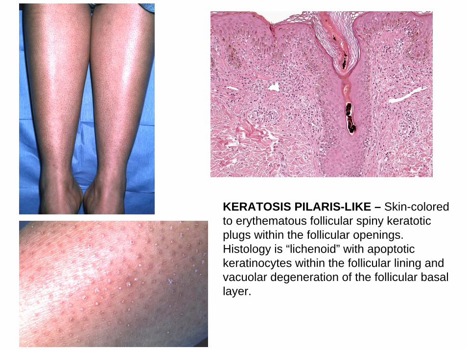
KERATOSIS PILARIS-LIKE – Skin-colored to erythematous follicular spiny keratoticplugs within the follicular openings. Histology is “lichenoid” with apoptotic keratinocytes within the follicular lining and vacuolar degeneration of the follicular basal layer.

ERYTHRODERMA – At least 80% of the body is red and scaly; may be mistaken for drug reaction or cutaneous T-cell lymphoma

SUPERFICIAL SCLEROSIS (MOVABLE)
This reflects increased fibrosis in the dermis.• Lichen sclerosus-like• Morphea-like

A
B
Lichen Sclerosus-like –discrete to coalescent gray to white papules with follicular plugs. (B) is from the mid-back in (A)

Morphea-like sclerosis – Typical site of superficial or morphea-like sclerosis in addition to dyspigmentation. The white areas (pigment loss) are sclerotic but can be moved over the clavicle.

DEEP SCLEROSIS (FIXED)
REFLECTS FIBROSIS OF DERMIS AND DEEPER LAYERS SUCH AS FASCIA AND FAT SEPTAE
• Diffuse over a wide area of skin• Tendon – “grooving”• Subcutaneous lobular fibrosis –
“rippling”

SCLEROTIC FIXED – Multiple manifestations: 1) inability to fully extend at the elbow joints; 2) “grooving” (black arrow) denotingtendon sclerosis; 3) “dimpling” or “rippling” (white arrow) from fibrosis of fat septae; 4) smooth, waxy skin is indurated on palpation. Papulo-squamous patch on upper mid back (green arrow).
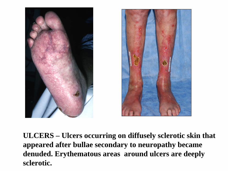
ULCERS – Ulcers occurring on diffusely sclerotic skin that appeared after bullae secondary to neuropathy became denuded. Erythematous areas around ulcers are deeply sclerotic.


RESPONSE ASSESSMENT CGVHD OF SKIN
Dimensions Outcome
Erythematous rash of any sort % BSA
Superficial sclerosis % BSA
Deep sclerosis % BSA
Ulcers – measure longest diameters __ cm
Pruritus/Itching 0 1 2 3 4 5 6 7 8 9 10

![Atlas of the Diseases of the Skin, Part I 1, 1878.] REVIEWS AND CORRESPONDENCE. 225 gj^biefos. Atlas of the Diseases of the Skin. Part I. By Balmanno Squire, M.B., Surgeon to the British](https://static.documents.pub/doc/80x56/5ae0ec937f8b9ac0428e1215/atlas-of-the-diseases-of-the-skin-part-i-1-1878-reviews-and-correspondence.jpg)


