24
1 © LLTECH 2011 LLTECH LightCT Scanner Skin Atlas of images July 2012
| Date post: | 31-Jul-2015 |
| Category: |
Health & Medicine |
| Upload: | lltech |
| View: | 563 times |
| Download: | 2 times |
1 © LLTECH 2011
LLTECH Light-‐CT Scanner Skin -‐ Atlas of images July 2012

2 © LLTECH 2011
• OpAcal in-‐depth biopsies of gross Assue within minutes
• 1 µm 2D and 3D histopathological resoluAon
• Easy exploraAon, acquisiAon and rendering in DICOM format
• Safe, non-‐invasive and non-‐destrucAve process
Light-‐CT™ key benefits
Fast and non-‐invasive 3D in-‐depth structural and cellular imaging
3 © LLTECH 2011
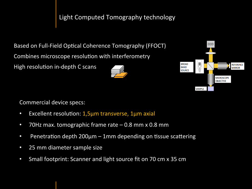
Based on Full-‐Field OpAcal Coherence Tomography (FFOCT)
Combines microscope resoluAon with interferometry
High resoluAon in-‐depth C scans
Commercial device specs:
• Excellent resoluAon: 1,5µm transverse, 1µm axial
• 70Hz max. tomographic frame rate – 0.8 mm x 0.8 mm
• PenetraAon depth 200µm – 1mm depending on Assue sca\ering
• 25 mm diameter sample size
• Small footprint: Scanner and light source fit on 70 cm x 35 cm
Light Computed Tomography technology

4 © LLTECH 2011
OpAcal acquisiAon unit, moving verAcally • User friendly
acquisiAon so^ware
• DICOM 2D and 3D Viewer
Movable tray with sample holder
X,Y moving stage
JoysAck for easy control of X,Y, Z movements
White Light Source
Integrated wide field camera to take sample picture before
imaging
Light-‐CT™ Scanner for ex-‐vivo cellular imaging
5 © LLTECH 2011
SKIN IMAGING

6 © LLTECH 2011
Normal skin morphology
Epidermis
Collagen
Pilosebaceous unit
Sweat gland
Adipocytes
b
a
c
e
d
500 µm
© LLTech 2012 Courtesy of Hopitaux Universitaires de Genève, Genève, Switzerland
7 © LLTECH 2011
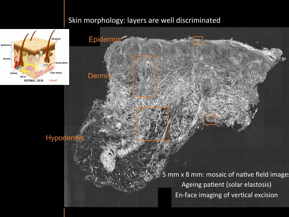
Hypodermis
Skin morphology: layers are well discriminated
5 mm x 8 mm: mosaic of naAve field images Ageing paAent (solar elastosis)
En-‐face imaging of verAcal excision
Epidermis
Dermis
1
3
2
4

8 © LLTECH 2011
4 - Sweat gland
100 µm
1 - Epidermis
100 µm
2 - Dermis
200 µm
Stratum corneum
Stratum spinosum
Stratum basale
KN KN KN
KN: keratinocyte nuclei
Collagen Adipocytes
3 – Pilosebaceous unit
200 µm
HF SG
SG
SG
SG
HF: hair follicle
SG: sebaceous gland
Structures are well discriminated with structural and cellular details

9 © LLTECH 2011
Stratum corneum Stratum spinosum
Stratum basale Reticular region
Epidermis
Dermis
En-‐face skin slicing shows structural and cellular details
Melanin papillary caps
KeraAnocyte nuclei
Blood vessels
Collagen
Corneocytes
100 µm 100 µm
100 µm 100 µm
© LLTech 2012 Courtesy of Hopitaux Universitaires de Genève, Genève, Switzerland

10 © LLTECH 2011
Skin verAcal reconstrucAon from en-‐face slicing for layers assesment and measurement
Stratum corneum
Stratum spinosum
Dermis
Papillary melanin caps BV: Blood vessels
BV
BV BV
KN KN KN
KN: KeraAnocyte nuclei
50 µm
© LLTech 2012 Courtesy of Hopitaux Universitaires de Genève, Genève, Switzerland

11 © LLTECH 2011
3D reconstrucAon from en-‐face slicing
Wrinkle Stratum corneum Stratum spinosum
Epidermis
800 µm x 800 µm
Dermis
Dermal papilla Collagen
800 µm x 800 µm
Epidermis and dermis separated by sodium bromide
Blood vessel
50 µm
Reconstructed verAcal slice
50 µm
Reconstructed verAcal slice
© LLTech 2012 Courtesy of Hopitaux Universitaires de Genève, Genève, Switzerland

12 © LLTECH 2011
VerAcal reconstrucAon of skin model
50 µm
Stratum corneum
Stratum spinosum
Dermis
KeraAnocyte nuclei
© LLTech 2012
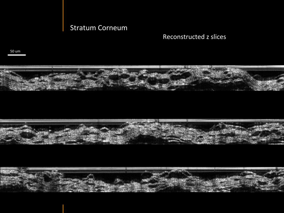
13 © LLTECH 2011
Stratum Corneum Reconstructed z slices
50 um
14 © LLTECH 2011
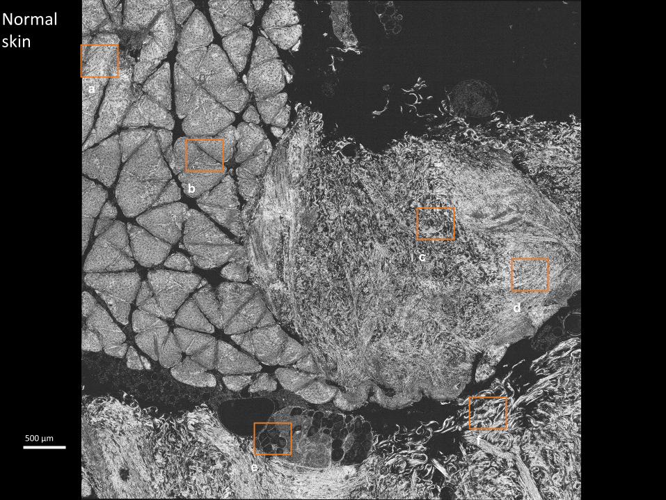
Normal skin
b
a
e
c
f
d
500 µm

15 © LLTECH 2011
Normal skin zoom
a b c
d e f
Stratum corneum Stratum spinosum Dermis
Collagen -‐ dermis Adipocytes Deep dermis
100 µm
16 © LLTECH 2011
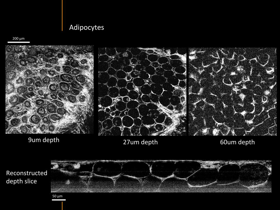
Adipocytes
9um depth 27um depth 60um depth
Reconstructed depth slice
200 µm
50 µm

17 © LLTECH 2011
Skin pathologies: basal cell carcinoma discriminaAon at structural level
Dense peritumoral stroma
© LLTech 2012 Courtesy of Hopitaux Universitaires de Genève, Genève, Switzerland

18 © LLTECH 2011
Epidermis
Dermis
Hypodermis
A
A A
C
BCC
PSU
PSU
BCC: Basal cell carcinoma A: Adipocytes C: Collagen PSU: Pilosebaceous unit
Skin pathologies: basal cell carcinoma discriminaAon at structural level
1 mm
© LLTech 2012 Courtesy of Hopitaux Universitaires de Genève, Genève, Switzerland
C

19 © LLTECH 2011
Zoom on basal cell carcinoma: discriminaAon at cellular level
200 µm
100 µm
Peritumoral stroma
High cell density
© LLTech 2012 Courtesy of Hopitaux Universitaires de Genève, Genève, Switzerland
20 © LLTECH 2011
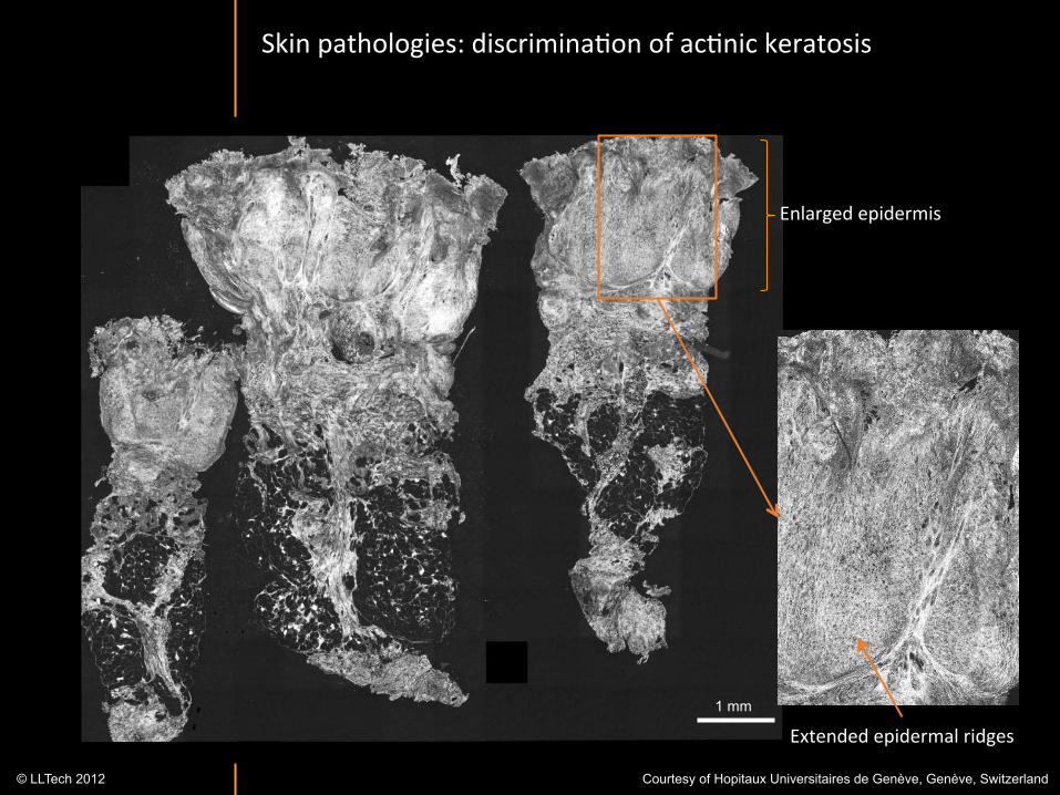
Skin pathologies: discriminaAon of acAnic keratosis
Enlarged epidermis
Extended epidermal ridges 1 mm
© LLTech 2012 Courtesy of Hopitaux Universitaires de Genève, Genève, Switzerland

21 © LLTECH 2011
Skin in-‐vivo: fingerprint imaging in 3D
100 µm
Sweat ducts
Fingerprints 3D reconstrucAon FOV 800 µm x 800 µm
Corneocytes
En-‐face image of fingerprints

22 © LLTECH 2011
Skin in-‐vivo: sweat duct imaging in 3D
VerAcal reconstrucAon from en-‐face images
Sweat duct 3D reconstrucAon
Stratum corneum
Stratum spinosum
SD: sweat ducts
SD SD
100 µm

23 © LLTECH 2011
Skin in-‐vivo: skin layers imaging en face and in verAcal reconstrucAon
Stratum corneum Stratum spinosum Dermis
Stratum corneum
Stratum spinosum
Dermis 100 µm EvaluaAon of stratum corneum thickness:
15 µm

24 © LLTECH 2011
Contact
LLTECH
LLTech 6, place de la Madeleine
75008 Paris, France
www.lltechimaging.com [email protected]
Phone: +33 9 72 16 33 40
LLTech, Inc. 103 Carnegie Center Drive
Suite 300 Princeton, NJ 08540, USA
www.lltechimaging.com
Phone :+1 609 955 3506


![Atlas of the Diseases of the Skin, Part I 1, 1878.] REVIEWS AND CORRESPONDENCE. 225 gj^biefos. Atlas of the Diseases of the Skin. Part I. By Balmanno Squire, M.B., Surgeon to the British](https://static.documents.pub/doc/80x56/5ae0ec937f8b9ac0428e1215/atlas-of-the-diseases-of-the-skin-part-i-1-1878-reviews-and-correspondence.jpg)


