23
DR. MURALI. U. M.S ; M.B.A. PROF. OF SURGERY D.Y.PATIL MEDICAL COLLEGE MAURITIUS. HAEMORRHAGE
| Date post: | 10-Aug-2015 |
| Category: |
Health & Medicine |
| Upload: | uthamalingam-murali |
| View: | 56 times |
| Download: | 0 times |
DR. MURALI. U. M.S ; M.B.A.PROF. OF SURGERYD.Y.PATIL MEDICAL COLLEGEMAURITIUS.
HAEMORRHAGE

DEFINITION
Haemorrhage means escape of blood
outside its containing vessel.

CLASSIFICATION
Depending on nature of the vessel involved
Depending on the timing of haemorrhage
Depending on the duration of Haemorrhage
Depending on the nature of bleeding
Depending upon type of Intervention

1A - SOURCE - ARTERIAL
Bright red
Emitted as spurting jet
Can lead to severe blood loss
Often hard to control

1B - SOURCE – VENOUS
Darker red
Steady and copious flow
Color becomes further darker with oxygen desaturation
Usually easy to control

1C- SOURCE – CAPILLARY
Bright red
Rapid and oozing
Blood loss becomes serious if continues for hours
Generally minor & easy to control
2A - TIMING - PRIMARY
Occurs at the time of surgery
Cause is injury to vessels
May be arterial, venous or capillary
More common in surgery on malignancies
2B - TIMING - REACTIONARY
Bleeding within 24 hours ( usually 4-6 hrs ) of surgery
Cause is slipping of ligature, dislodgement of clot or cessation of reflex vasospasm
Bleed starts when there is a rise in the arterial or venous pressure.

2C - TIMING – SECONDARY
Occurs after 7-14 days of surgery
Cause is sloughing of vessel due to infection, pressure necrosis or malignancy.
1st a warning stain followed by a sudden severe bleed
Common after hemorrhoids surgery, GI surgery & amputations.
3 – DURATION
Acute Haemorrhage: occurs suddenly. eg. Oesophageal variceal bleeding due to portal HT.
Chronic Haemorrhage.
4A – NATURE / TYPE
External Haemorrhage or Revealed :
External or visible bleed – soft tissue injuries
Bleeding from the limb vessels, wound,nose etc.
4B – NATURE / TYPE
Internal Haemorrhage or Concealed :
Internal or invisible bleed – Blunt or Penetrating trauma
May remain concealed as in ruptured spleen or liver
Concealed hemorrhage may become revealed as in haemetemesis or melaena in peptic ulcer bleed
5 – TYPE OF INTERVENTION
Surgical Haemorrhage: is the result of injury and amenable to surgical control, or from angioembolism.
Non-Surgical Haemorrhage: is general ooze from all raw surface due to coagulopathy, it can not be stopped by surgical mean, require correction coagulation abnormalities.

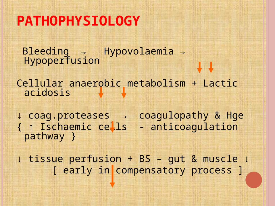
PATHOPHYSIOLOGY
Bleeding → Hypovolaemia → Hypoperfusion Cellular anaerobic metabolism + Lactic
acidosis ↓ coag.proteases → coagulopathy & Hge{ ↑ Ischaemic cells - anticoagulation
pathway } ↓ tissue perfusion + BS – gut & muscle ↓ [ early in compensatory process ]
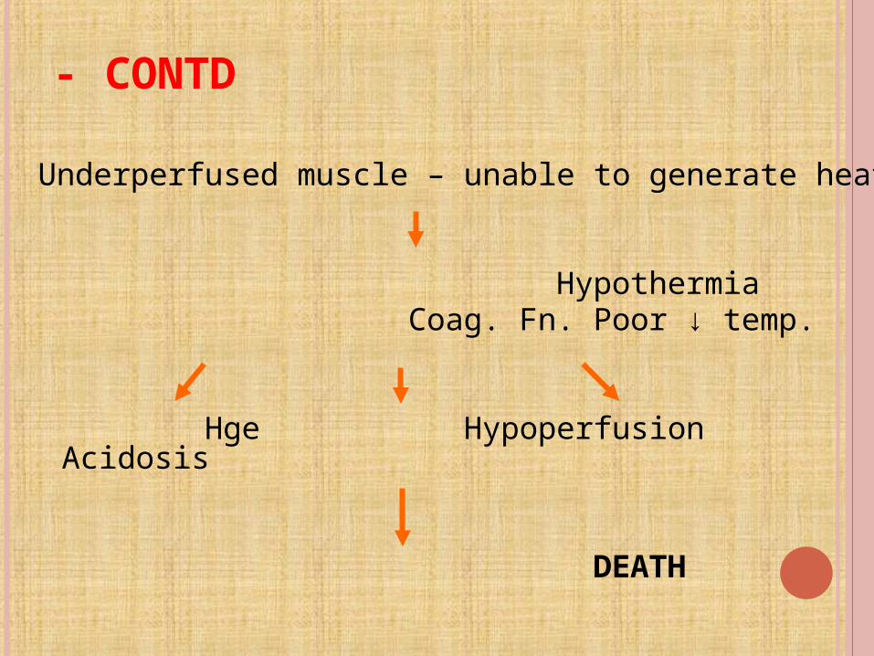
- CONTD
Underperfused muscle – unable to generate heat
Hypothermia Coag. Fn. Poor ↓ temp.
Hge Hypoperfusion Acidosis
DEATH

CLINICAL FEATURES
Pallor, thirsty, cyanosis Tachycardia, tachypnoea Cold clammy skin due to vasoconstriction Dry face, dry mouth and goose skin
appearance (due to contraction of arrector pilorum).
Rapid thready pulse, hypotension Oliguria Features related to specific causes
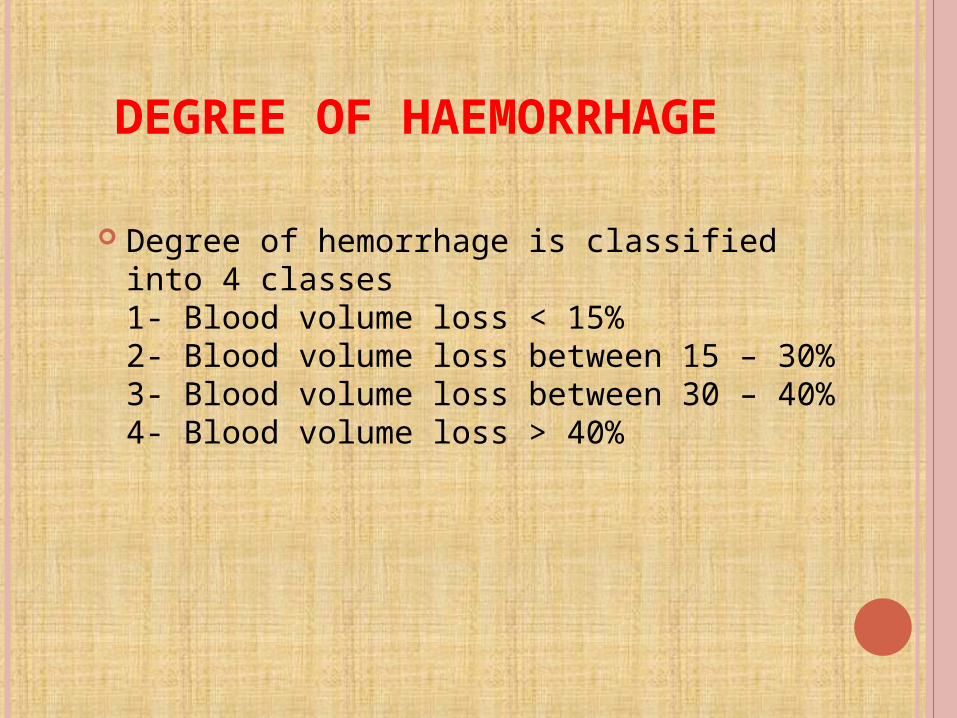
DEGREE OF HAEMORRHAGE
Degree of hemorrhage is classified into 4 classes 1- Blood volume loss < 15% 2- Blood volume loss between 15 – 30% 3- Blood volume loss between 30 – 40% 4- Blood volume loss > 40%
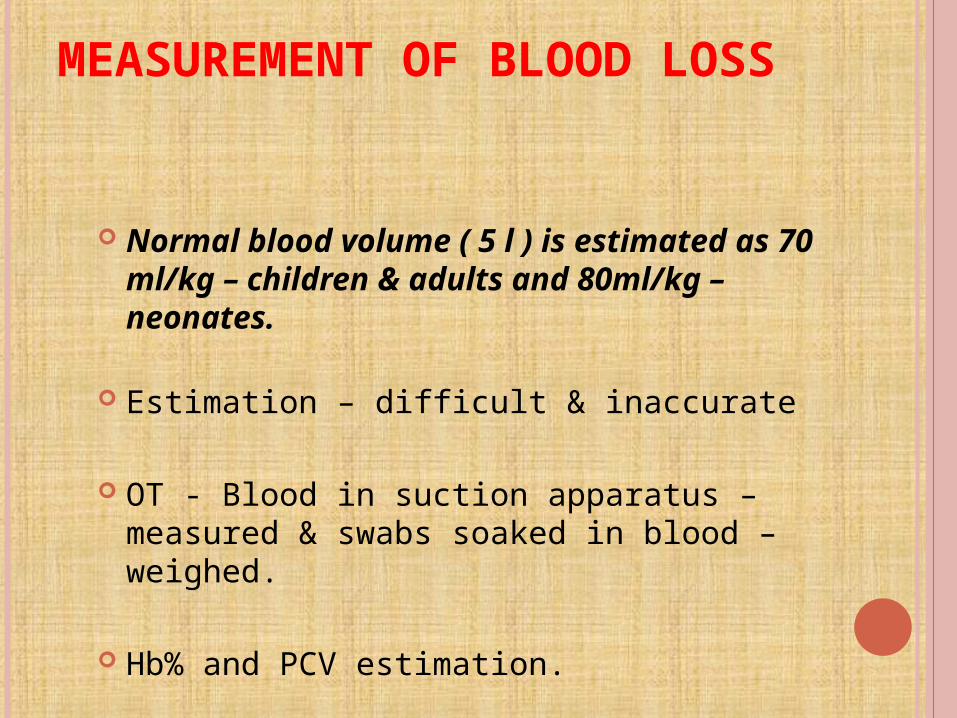
MEASUREMENT OF BLOOD LOSS
Normal blood volume ( 5 l ) is estimated as 70 ml/kg – children & adults and 80ml/kg – neonates.
Estimation – difficult & inaccurate
OT - Blood in suction apparatus – measured & swabs soaked in blood – weighed.
Hb% and PCV estimation.
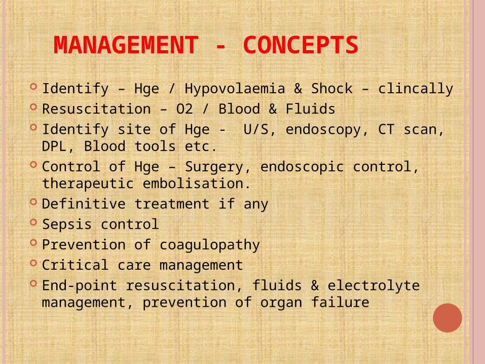
MANAGEMENT - CONCEPTS
Identify – Hge / Hypovolaemia & Shock – clincally Resuscitation – O2 / Blood & Fluids Identify site of Hge - U/S, endoscopy, CT scan, DPL,
Blood tools etc. Control of Hge – Surgery, endoscopic control,
therapeutic embolisation. Definitive treatment if any Sepsis control Prevention of coagulopathy Critical care management End-point resuscitation, fluids & electrolyte
management, prevention of organ failure
“When there is blood loss, replace with blood”
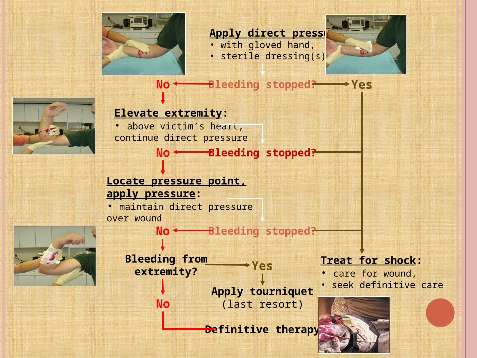
Apply direct pressure:• with gloved hand,• sterile dressing(s).
Bleeding stopped? YesNo
Elevate extremity:• above victim’s heart,continue direct pressure
Locate pressure point,apply pressure:• maintain direct pressureover wound
Treat for shock:• care for wound,• seek definitive care
Bleeding stopped?
Bleeding stopped?
No
Bleeding fromextremity?
No
Apply tourniquet(last resort)
Yes
No
Definitive therapy