Page 1
Lecture 1 June 15, 2018
©AllinaHealthSystems 1
Hand and Wrist Injuries in the Athlete
Allina Health Sports Medicine ConferenceJune 15, 2018
Diagnosis, Treatment, and Return to Play Guidelines
Patrick H. Smock, MDJune 15th, 2018
Financial Disclosures
No relevant financial disclosures.
No off-label use will be discussed.
Page 2
Lecture 1 June 15, 2018
©AllinaHealthSystems 2
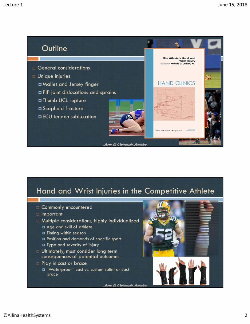
Outline
General considerations
Unique injuries
Mallet and Jersey finger
PIP joint dislocations and sprains
Thumb UCL rupture
Scaphoid fracture
ECU tendon subluxation
Hand and Wrist Injuries in the Competitive Athlete
Commonly encountered Important Multiple considerations, highly individualized
Age and skill of athlete Timing within season Position and demands of specific sport Type and severity of injury
Ultimately, must consider long term consequences of potential outcomes
Play in cast or brace “Waterproof” cast vs. custom splint or cast-
brace
Page 3
Lecture 1 June 15, 2018
©AllinaHealthSystems 3
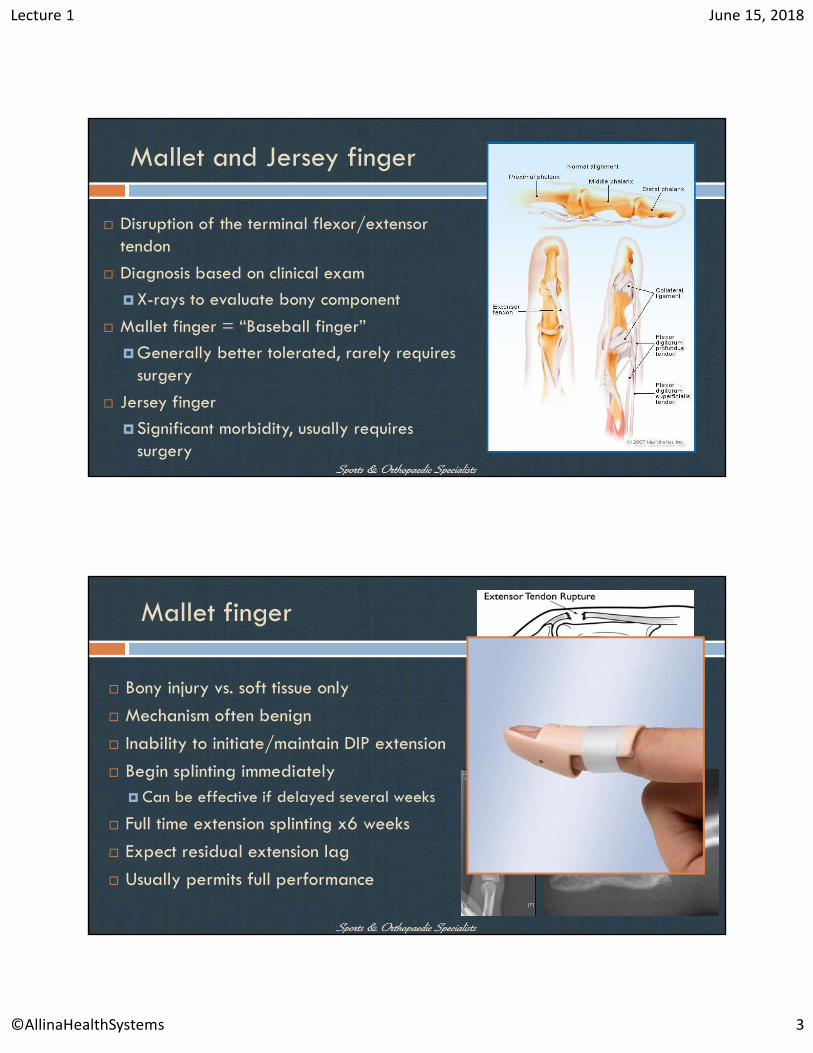
Mallet and Jersey finger
Disruption of the terminal flexor/extensor tendon
Diagnosis based on clinical exam
X-rays to evaluate bony component
Mallet finger = “Baseball finger”
Generally better tolerated, rarely requires surgery
Jersey finger
Significant morbidity, usually requires surgery
Mallet finger
Bony injury vs. soft tissue only
Mechanism often benign
Inability to initiate/maintain DIP extension
Begin splinting immediately Can be effective if delayed several weeks
Full time extension splinting x6 weeks
Expect residual extension lag
Usually permits full performance
Page 4
Lecture 1 June 15, 2018
©AllinaHealthSystems 4
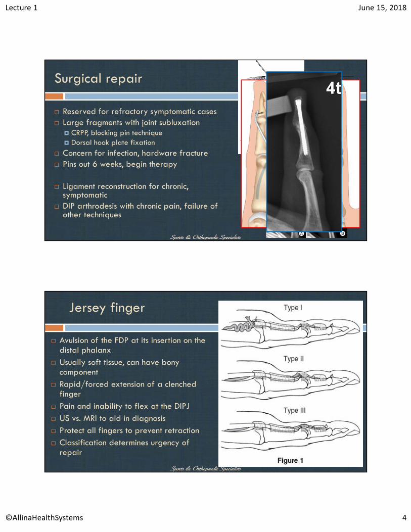
Surgical repair
Reserved for refractory symptomatic cases Large fragments with joint subluxation
CRPP, blocking pin technique Dorsal hook plate fixation
Concern for infection, hardware fracture Pins out 6 weeks, begin therapy
Ligament reconstruction for chronic, symptomatic
DIP arthrodesis with chronic pain, failure of other techniques
Jersey finger
Avulsion of the FDP at its insertion on the distal phalanx
Usually soft tissue, can have bony component
Rapid/forced extension of a clenched finger
Pain and inability to flex at the DIPJ US vs. MRI to aid in diagnosis Protect all fingers to prevent retraction Classification determines urgency of
repair
Page 5
Lecture 1 June 15, 2018
©AllinaHealthSystems 5
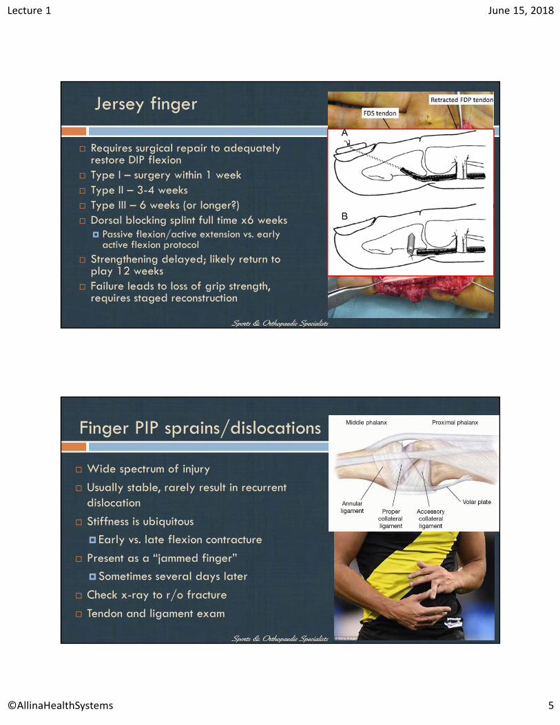
Jersey finger
Requires surgical repair to adequately restore DIP flexion
Type I – surgery within 1 week Type II – 3-4 weeks Type III – 6 weeks (or longer?) Dorsal blocking splint full time x6 weeks
Passive flexion/active extension vs. early active flexion protocol
Strengthening delayed; likely return to play 12 weeks
Failure leads to loss of grip strength, requires staged reconstruction
Finger PIP sprains/dislocations
Wide spectrum of injury
Usually stable, rarely result in recurrent dislocation
Stiffness is ubiquitous
Early vs. late flexion contracture
Present as a “jammed finger”
Sometimes several days later
Check x-ray to r/o fracture
Tendon and ligament exam
Page 6
Lecture 1 June 15, 2018
©AllinaHealthSystems 6
PIP sprain/dislocation
Stable injuries begin immediate static extension splinting and OT
Splinting 24/7 x 4weeks, remove for hygiene and ROM protocol
Overnight splinting x4 additional weeks to prevent late contracture
Return to play dictated by pain, swelling, strength
Protected play within first 4 weeks, with splint/wrap/buddy taping
Lots of counseling!
PIP sprain/dislocation
Unstable dislocations splinted in reduced position and extension block splint
AROM allowed within stable ROM
Extension block gradually reduced under instruction of hand therapy
Surgery reserved for open/ irreducible/ recurrent dislocations or chronic instability
Usually hyperextension deformity – volar plate repair
Collateral ligament repair/reconstruction
Page 7
Lecture 1 June 15, 2018
©AllinaHealthSystems 7
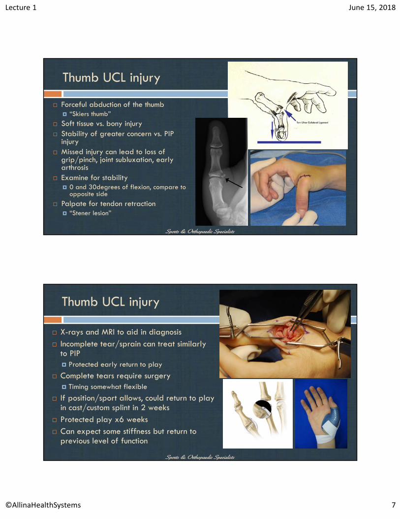
Thumb UCL injury
Forceful abduction of the thumb “Skiers thumb”
Soft tissue vs. bony injury Stability of greater concern vs. PIP
injury Missed injury can lead to loss of
grip/pinch, joint subluxation, early arthrosis
Examine for stability 0 and 30degrees of flexion, compare to
opposite side
Palpate for tendon retraction “Stener lesion”
Thumb UCL injury
X-rays and MRI to aid in diagnosis Incomplete tear/sprain can treat similarly
to PIP Protected early return to play
Complete tears require surgery Timing somewhat flexible
If position/sport allows, could return to play in cast/custom splint in 2 weeks
Protected play x6 weeks Can expect some stiffness but return to
previous level of function
Page 8
Lecture 1 June 15, 2018
©AllinaHealthSystems 8
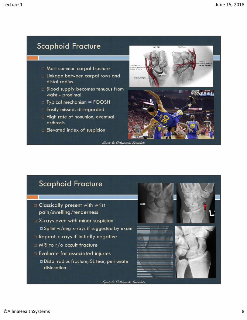
Scaphoid Fracture
Most common carpal fracture Linkage between carpal rows and
distal radius Blood supply becomes tenuous from
waist - proximal Typical mechanism = FOOSH Easily missed, disregarded High rate of nonunion, eventual
arthrosis Elevated index of suspicion
Scaphoid Fracture
Classically present with wrist pain/swelling/tenderness
X-rays even with minor suspicion Splint w/neg x-rays if suggested by exam
Repeat x-rays if initially negative
MRI to r/o occult fracture
Evaluate for associated injuries Distal radius fracture, SL tear, perilunate
dislocation
Page 9
Lecture 1 June 15, 2018
©AllinaHealthSystems 9
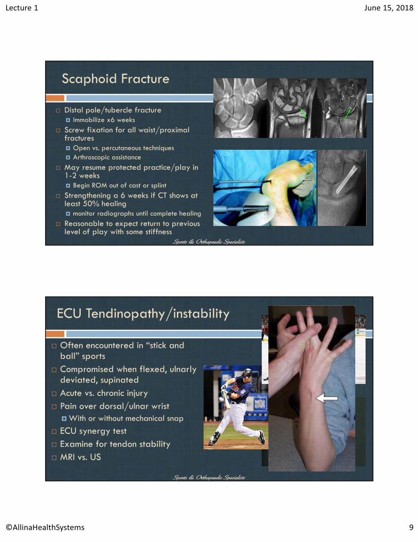
Scaphoid Fracture
Distal pole/tubercle fracture Immobilize x6 weeks
Screw fixation for all waist/proximal fractures Open vs. percutaneous techniques Arthroscopic assistance
May resume protected practice/play in 1-2 weeks Begin ROM out of cast or splint
Strengthening a 6 weeks if CT shows at least 50% healing monitor radiographs until complete healing
Reasonable to expect return to previous level of play with some stiffness
ECU Tendinopathy/instability
Often encountered in “stick and ball” sports
Compromised when flexed, ulnarly deviated, supinated
Acute vs. chronic injury Pain over dorsal/ulnar wrist
With or without mechanical snap
ECU synergy test Examine for tendon stability MRI vs. US
Page 10
Lecture 1 June 15, 2018
©AllinaHealthSystems 10
ECU Tendinopathy/instability
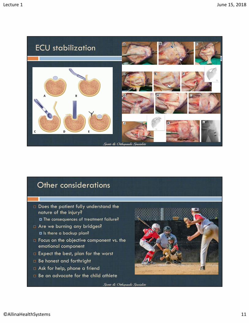
ECU tendinopathy/instability
Tendinosis without subluxation Conservative management mainstay
Brief immobilization, NSAID’s, modalities
Injection
Debridement/release/reconstruction of the ECU subsheath only in severe/recalcitrant cases
With ECU tendon instability Reduce tendon along with associated DRUJ dislocation if
present
Immobilization (above elbow? Wrist position?)
Depending on severity, immobilize 1-2 weeks followed by 1-2 more weeks of motion recovery
Therapy with taping/bracing
Return to strength/swinging approx. 6 weeks
Page 11
Lecture 1 June 15, 2018
©AllinaHealthSystems 11
ECU stabilization
Other considerations
Does the patient fully understand the nature of the injury? The consequences of treatment failure?
Are we burning any bridges? Is there a backup plan?
Focus on the objective component vs. the emotional component
Expect the best, plan for the worst Be honest and forthright Ask for help, phone a friend Be an advocate for the child athlete
Page 12
Lecture 1 June 15, 2018
©AllinaHealthSystems 12
J Hand Surg Am. 2014 Oct;39(10):1992-8. Return to football and long-term clinical outcomes after thumb ulnar collateral ligament suture anchor repair in collegiate athletes.Werner BC1, Hadeed MM1, Lyons ML1, Gluck JS1, Diduch DR1, Chhabra AB2.
Am J Sports Med. 2017 Jan;45(1):195-200. Injuries to the Collateral Ligaments of the Metacarpophalangeal Joint of the Thumb, Including Simultaneous Combined Thumb Ulnar and Radial Collateral Ligament Injuries, in National Football League Athletes.Werner BC1, Belkin NS2, Kennelly S3, Weiss L3, Barnes RP3, Rodeo SA2, Warren RF2, Hotchkiss RN2.
Arthroscopy. 2017 Dec;33(12):2154-2158Clinical and Radiologic Outcomes After Scaphoid Fracture: Injury and Treatment Patterns in National Football League Combine Athletes Between 2009 and 2014.Moatshe G1, Godin JA2, Chahla J3, Cinque ME3, Kennedy NI3, Sanchez G4, Beaulieu-Jones BR4, LaPrade RF5, Provencher MT6.
Hand Clin. 2012 Aug;28(3):269-78Scaphoid fracture in the elite athlete.Belsky MR1, Leibman MI, Ruchelsman DE.
References
Orthopedics. 2013 Jun;36(6):815-9.Opinions regarding the management of hand and wrist injuries in elite athletes.Dy CJ1, Khmelnitskaya E, Hearns KA, Carlson MG.
J Am Acad Orthop Surg. 2001 Nov-Dec;9(6):389-400.Acute hand and wrist injuries in athletes: evaluation and management.Morgan WJ1, Slowman LS.
J Am Acad Orthop Surg. 2016 Dec;24(12):853-862.Diagnosis, Treatment, and Return to Play for Four Common Sports Injuries of the Hand and Wrist.Goldfarb CA1, Puri SK, Carlson MG.
Hand Clin. 2012 Aug;28(3):395-401Phalangeal fractures: displaced/nondisplaced.Gaston RG1, Chadderdon C.
Clin Sports Med. 2016 Oct;35(4):597-608. Return to Play After Hand and Wrist Fractures.Halim A1, Weiss AP2.
References
Page 13
Lecture 1 June 15, 2018
©AllinaHealthSystems 13
Thank You!
Patrick H. Smock, MD
SAOS Hand Wrist & Elbow
St. Paul, MN
[email protected]
www.sportsandortho.com
facebook.com/sportsandortho
@sportsandortho