30
Hand & Wrist Injuries in the Young Athlete Bryce Bell, MD Assistant Professor Pediatric Hand Surgery Department of Orthopedic Surgery
Hand & Wrist Injuries
in the Young Athlete
Bryce Bell, MD
Assistant Professor
Pediatric Hand Surgery
Department of Orthopedic Surgery
Disclosure
• I have no relevant disclosures or conflicts
of interest related to this topic
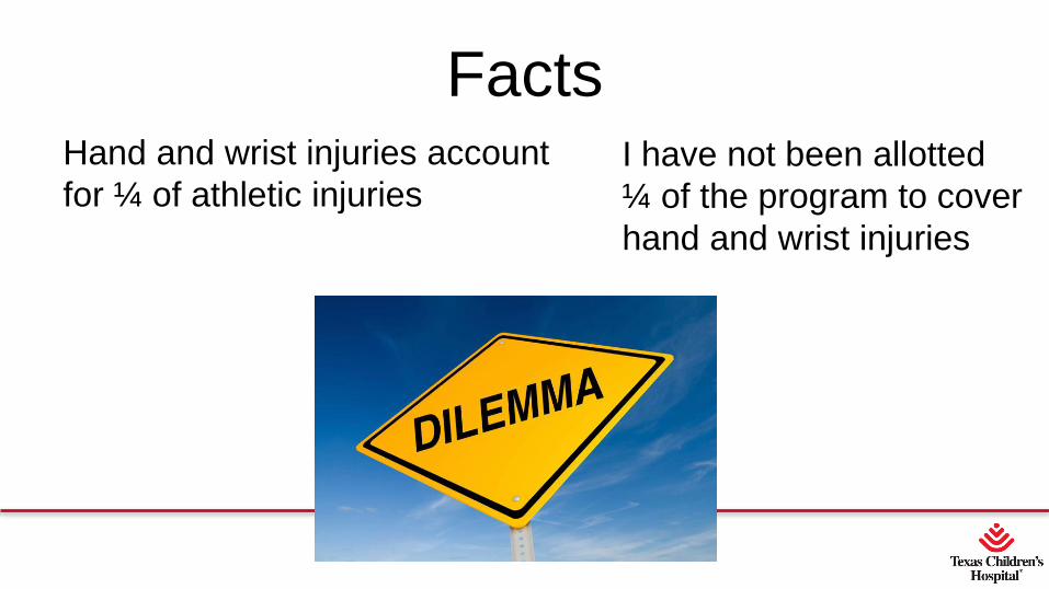
FactsHand and wrist injuries account
for ¼ of athletic injuriesI have not been allotted
¼ of the program to cover
hand and wrist injuries
Objectives
• Discuss epidemiology of hand fractures
• Understand basics of hand and wrist anatomy
– The key to all of Orthopaedics!
• Initial evaluation and treatment
• Principles to guide follow-up care
• When to seek out specialty care
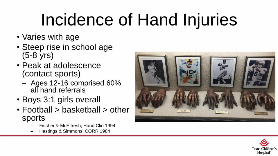
Incidence of Hand Injuries• Varies with age
• Steep rise in school age (5-8 yrs)
• Peak at adolescence (contact sports)– Ages 12-16 comprised 60%
all hand referrals
• Boys 3:1 girls overall
• Football > basketball > other sports
– Fischer & McElfresh, Hand Clin 1994
– Hastings & Simmons, CORR 1984
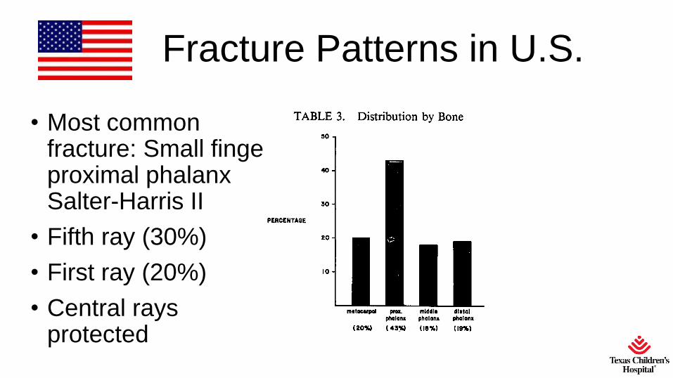
Fracture Patterns in U.S.
• Most common fracture: Small finger proximal phalanx Salter-Harris II
• Fifth ray (30%)
• First ray (20%)
• Central rays protected
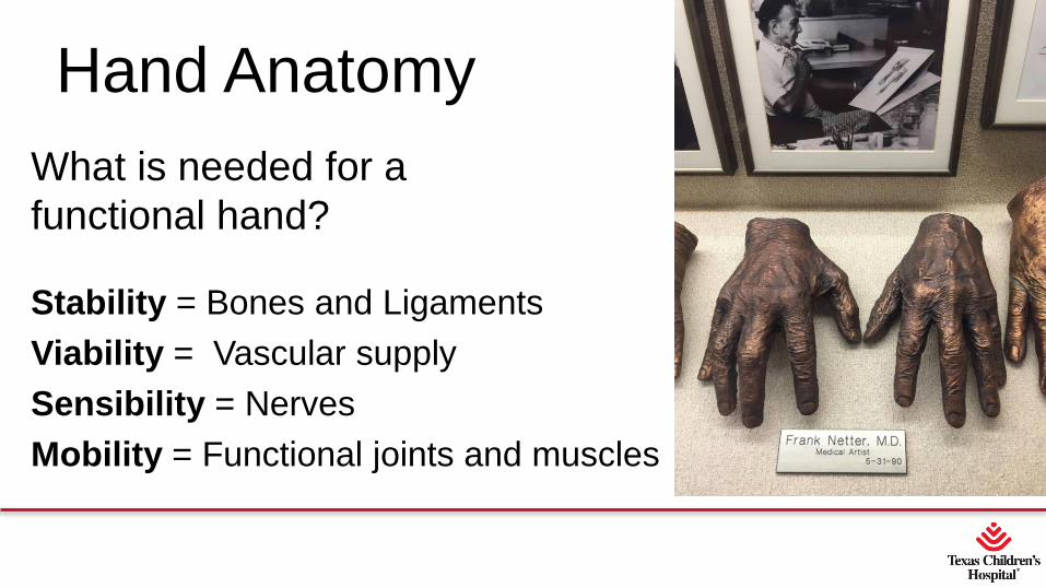
Hand Anatomy
What is needed for a
functional hand?
Stability = Bones and Ligaments
Viability = Vascular supply
Sensibility = Nerves
Mobility = Functional joints and muscles
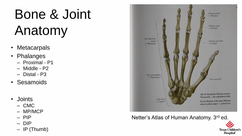
Bone & Joint
Anatomy• Metacarpals
• Phalanges– Proximal - P1– Middle - P2
– Distal - P3
• Sesamoids
• Joints– CMC– MP/MCP
– PIP
– DIP– IP (Thumb)
Netter’s Atlas of Human Anatomy. 3rd ed.
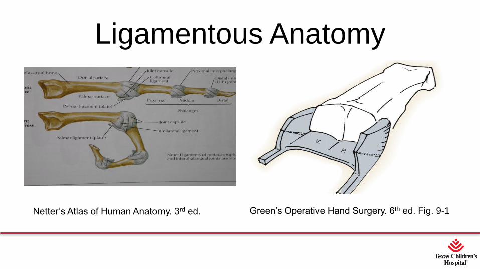
Ligamentous Anatomy
Netter’s Atlas of Human Anatomy. 3rd ed. Green’s Operative Hand Surgery. 6th ed. Fig. 9-1
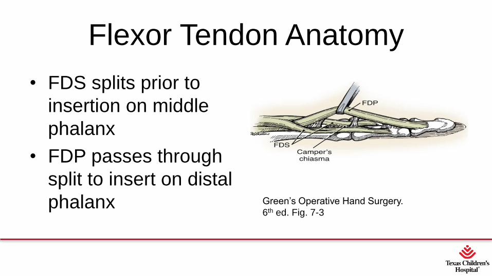
Flexor Tendon Anatomy
• FDS splits prior to
insertion on middle
phalanx
• FDP passes through
split to insert on distal
phalanx Green’s Operative Hand Surgery.
6th ed. Fig. 7-3
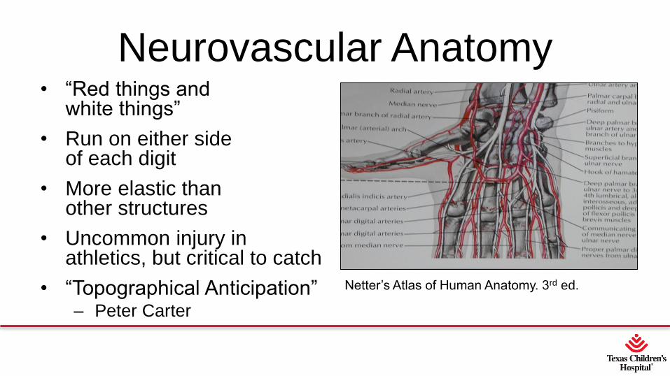
Neurovascular Anatomy• “Red things and
white things”
• Run on either side of each digit
• More elastic than other structures
• Uncommon injury in athletics, but critical to catch
• “Topographical Anticipation”– Peter Carter
Netter’s Atlas of Human Anatomy. 3rd ed.
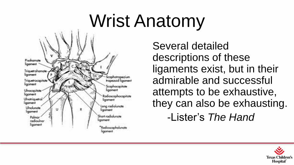
Wrist Anatomy
Several detailed descriptions of these ligaments exist, but in their admirable and successful attempts to be exhaustive, they can also be exhausting.
-Lister’s The Hand
Initial Evaluation and Treatment
The hand at rest is never truly so. For the fit
hand constantly moves in gesticulation,
nervousness, and personal mannerisms.
-Graham Lister, MD
Initial Evaluation and Treatment• History
– Understand mechanism Anticipate injury
• Inspection– No forceful reduction prior to X-ray
• Examination– Based on history and inspection
– Start with “uninjured” area. You won’t miss the obvious injury
• Immobilization
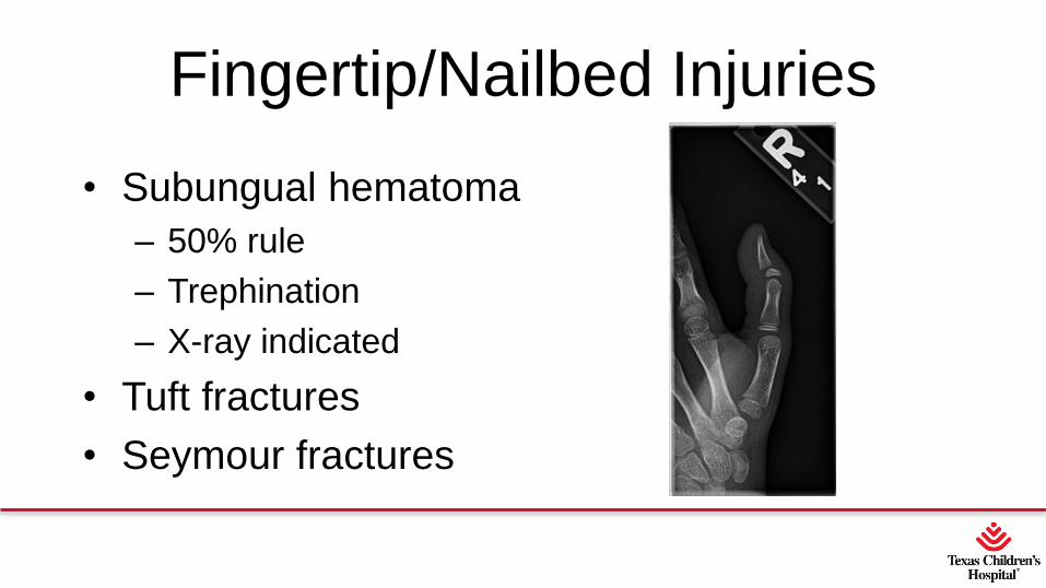
Fingertip/Nailbed Injuries
• Subungual hematoma
– 50% rule
– Trephination
– X-ray indicated
• Tuft fractures
• Seymour fractures
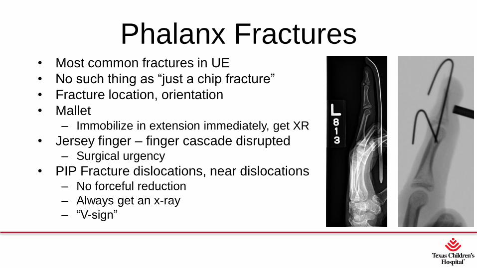
Phalanx Fractures• Most common fractures in UE
• No such thing as “just a chip fracture”
• Fracture location, orientation
• Mallet– Immobilize in extension immediately, get XR
• Jersey finger – finger cascade disrupted– Surgical urgency
• PIP Fracture dislocations, near dislocations– No forceful reduction
– Always get an x-ray
– “V-sign”
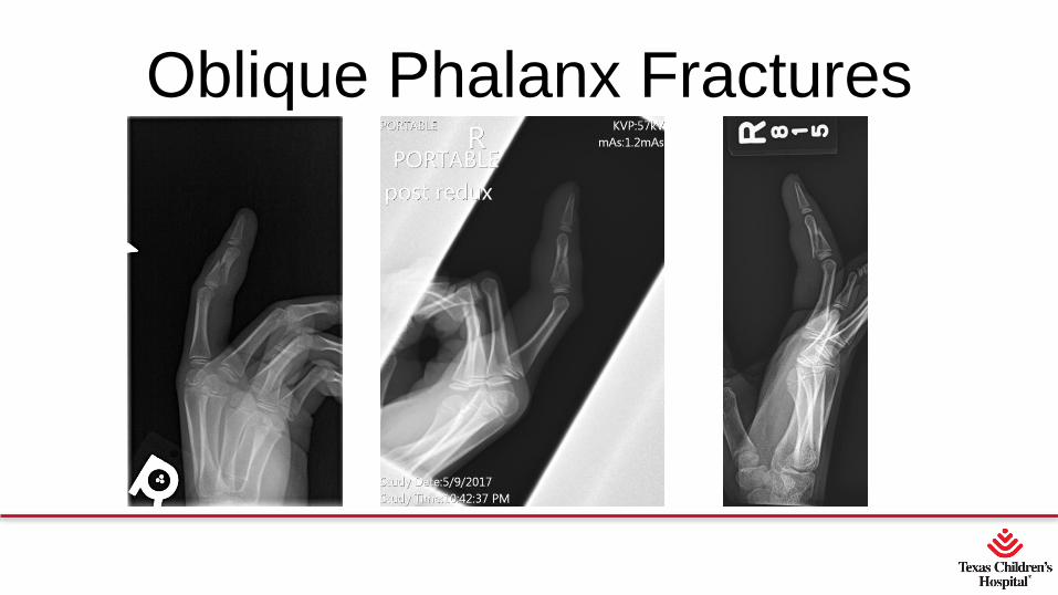
Oblique Phalanx Fractures
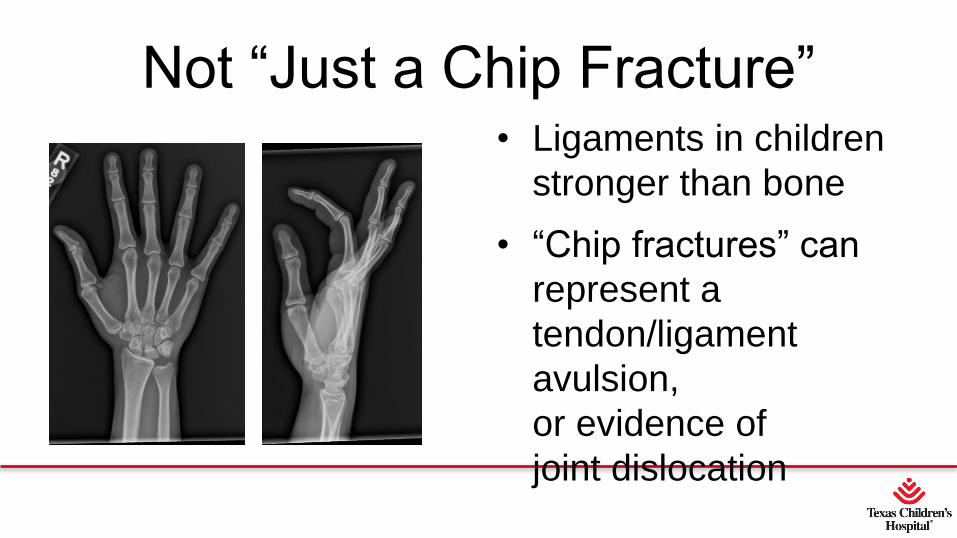
Not “Just a Chip Fracture”• Ligaments in children
stronger than bone
• “Chip fractures” can
represent a
tendon/ligament
avulsion,
or evidence of
joint dislocation
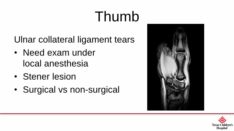
Thumb
Ulnar collateral ligament tears
• Need exam under
local anesthesia
• Stener lesion
• Surgical vs non-surgical
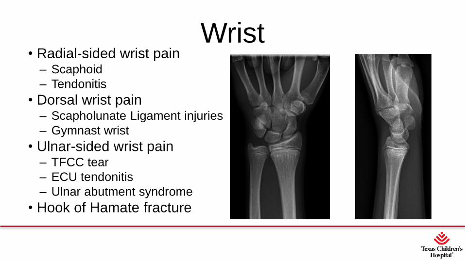
Wrist• Radial-sided wrist pain
– Scaphoid
– Tendonitis
• Dorsal wrist pain– Scapholunate Ligament injuries
– Gymnast wrist
• Ulnar-sided wrist pain– TFCC tear
– ECU tendonitis
– Ulnar abutment syndrome
• Hook of Hamate fracture
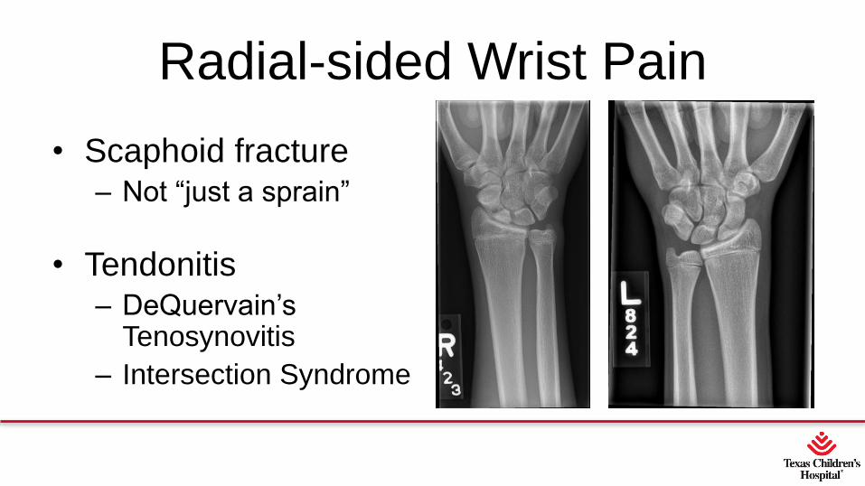
Radial-sided Wrist Pain
• Scaphoid fracture– Not “just a sprain”
• Tendonitis– DeQuervain’s
Tenosynovitis
– Intersection Syndrome

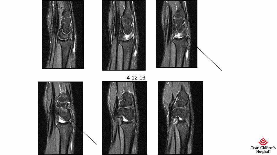
Dorsal Wrist Pain
Scapholunateligament tear
• Can be subtle or no signs on XR
• Persistent dorsal wrist pain, despite rest, negative XR MRI
http://www.pgatour.com/instruction/2014/06/11/golf-instruction-tips-hitting-from-waste-
bunkers.html
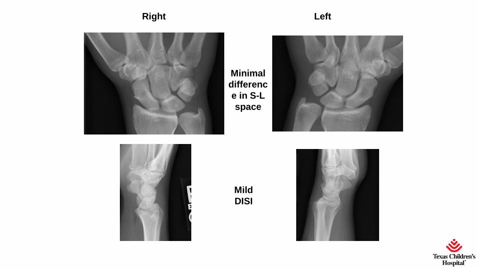
Right Left
Mild
DISI
Minimal
differenc
e in S-L
space
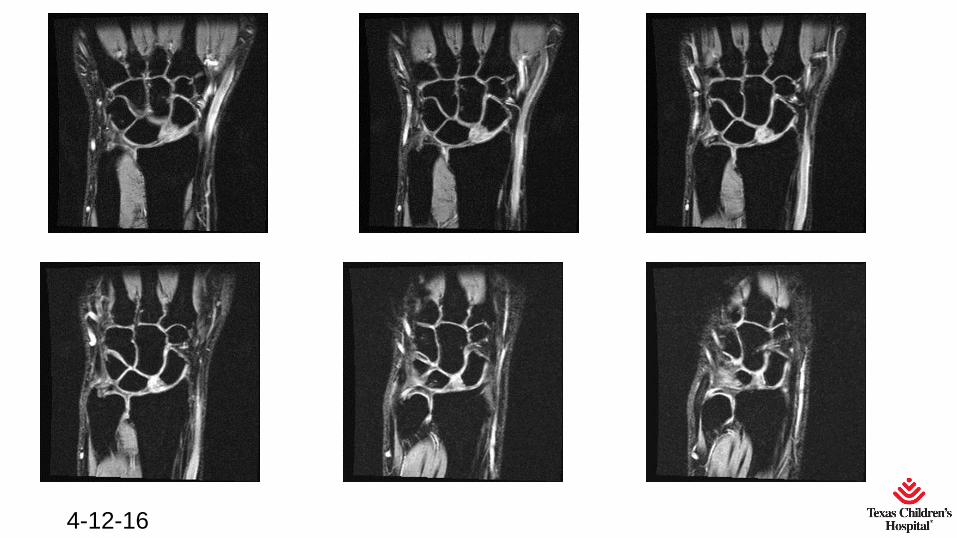
4-12-16
4-12-16
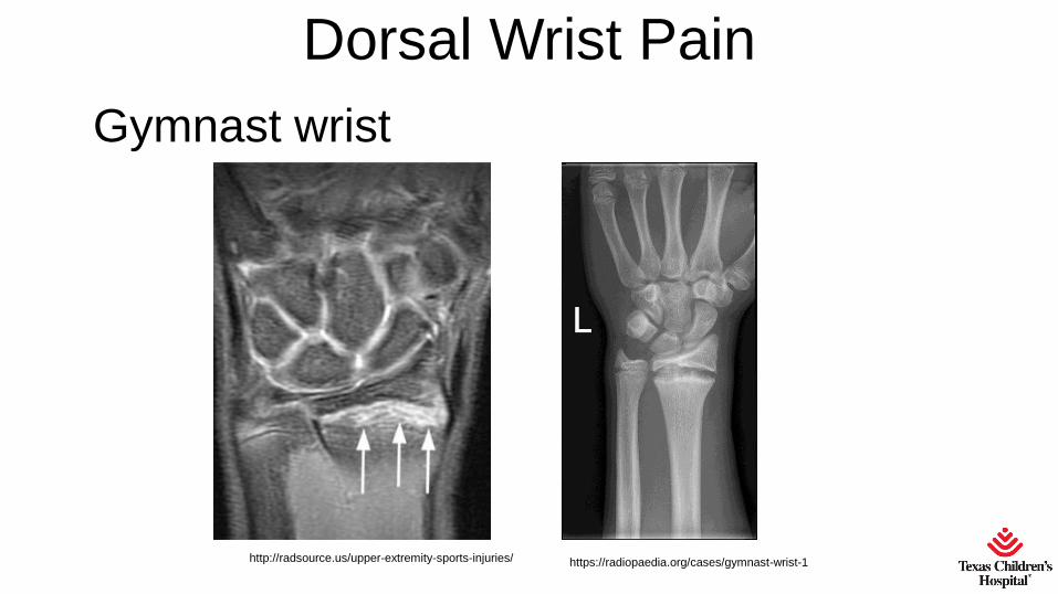
Dorsal Wrist Pain
Gymnast wrist
http://radsource.us/upper-extremity-sports-injuries/ https://radiopaedia.org/cases/gymnast-wrist-1
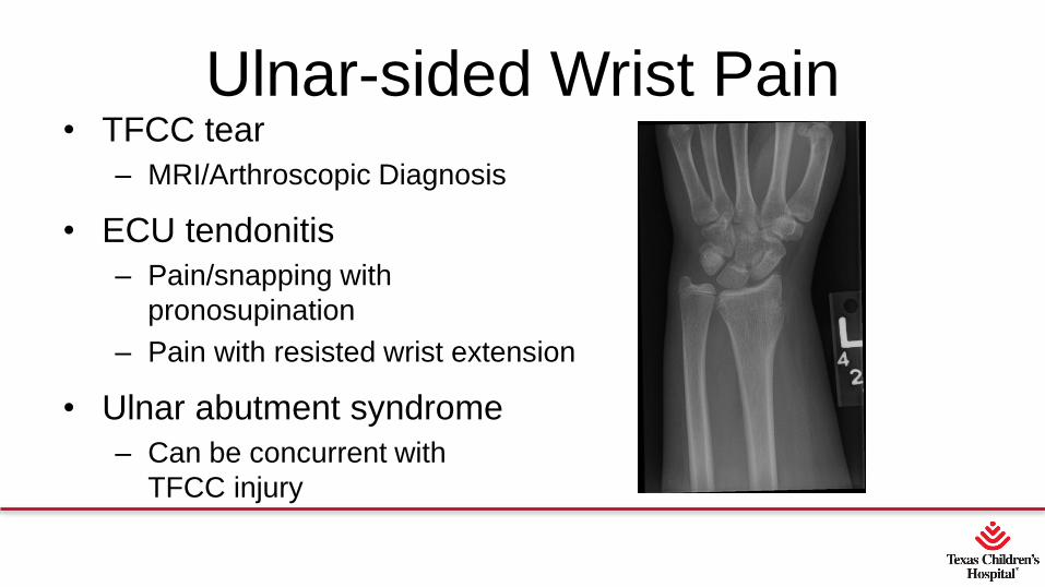
Ulnar-sided Wrist Pain• TFCC tear
– MRI/Arthroscopic Diagnosis
• ECU tendonitis
– Pain/snapping with
pronosupination
– Pain with resisted wrist extension
• Ulnar abutment syndrome
– Can be concurrent with
TFCC injury
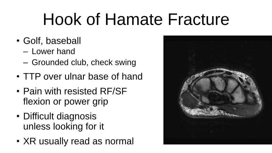
Hook of Hamate Fracture• Golf, baseball
– Lower hand
– Grounded club, check swing
• TTP over ulnar base of hand
• Pain with resisted RF/SF flexion or power grip
• Difficult diagnosis unless looking for it
• XR usually read as normal
Conclusions• Pain is not normal
• Immobilize and get the athlete evaluated– All dislocation events
– Any major swelling, ecchymosis
– All wrist pain not markedly improved or resolved within days
• It’s not “just a sprain”
• It’s not “just a chip fracture”
• Early evaluation and treatment is the fastest (and safest) way back to the field

Thank You


