70
HERNIA Jacek Szeliga MD, PhD

HERNIA
Jacek Szeliga MD, PhD

Hernia: The protrusion of tissue through a defect in fascial
and/or muscular layer(s) that normally contain it.
The sine qua non of a hernia is a bulge.
16th century illustration of femoral hernia
Source: Undetermined

The sac:- mouth- neck- body- fundus

Types of
abdominal wall
herniaLocation Congenital Acquired
Epigastric Upper midline *
Umbilical Umbilicus * ?
Inguinal/femoral Groin * *
Incisional Anywhere *
Lumbar Petit’s ∆ *
Interparietal Lateral hypogastric *
Obturator Obturator foramen *
Spigelian Arcuate x semilunar
lines
? ?
Traumatic Anywhere *
Diastasis Upper midline Not a hernia Not a hernia

Why Do Hernias Occur?
1. There is a congenital developmental defect
– Failure of fascial opening to close (e.g. umbilical)
– Failure of process to obliterate itself (e.g. processusvaginalis)
2. There is an acquired weakness
– Deterioration/thinning of fascia with age
– Loss of tissue (injury, infection, poor wound healing, etc.)
3. Repeated increase in abdominal pressure

Repeated INCREASE in abdominal pressure is usually due to
• Chronic cough
• Straining
• Bladder neck or urethral obstruction
• Pregnancy
• Vomiting
• Sever muscular effort
• Ascetic fluid
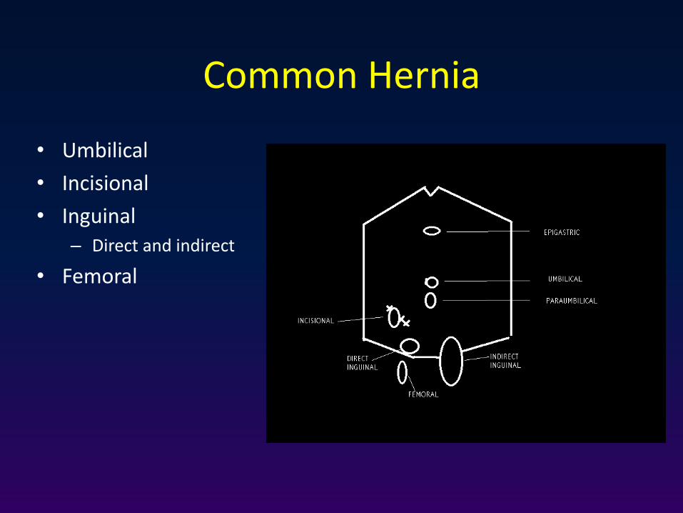
Common Hernia
• Umbilical
• Incisional
• Inguinal– Direct and indirect
• Femoral


Clinical types of hernia
- reducible hernia is one in which the contents of the sac return to the
abdomen spontaneously or with manual pressure when the patient is
recumbent.
- irreducible hernia is one whose contents or part of contents cannot be
returned to the abdomen, without serious symptoms.
- incarcerated hernia: is one whose contents cannot be returned to the
abdomen, with severe symptoms.
- strangulated hernia: denotes compromise to the blood supply of the
contents of the sac. Incarcerated hernia and strangulated hernia are the
two stages of a pathologic course
Hernias are trapped by the narrow neck

Clinical types of hernia
Richter’s hernia (intestinal wall hernia) a hernia that has
strangulated or incarcerated a part of the intestinal wall
without compromising the lumen.

Clinical types of hernia
Sliding hernia is one in which the wall of a viscus forms a portion
of the wall of the hernia sac. It is may be colon (on the left),
caccum (on the right) or bladder (on either side). Belongs to
irreducible hernia.

Clinical types of hernia
Littre hernia: a hernia that has incarcerated the intestinal
diverticulum (usually Meckel diverticulum).

Clinical types of hernia
Maydls hernia: ‘W’ loop of intestine

Groin hernia
• Indirect inguinal and scrotal
• Direct inguinal
• Femoral

Groin Hernias incidence
- Groin hernias are found in 5% of male population.
- Represents 86% of all hernia cases.
- It occurs 5 times more often in males than females.
- Inguinal 96% (indirect 75%, direct 25%).
- Bilateral in 20% of cases
- Right sided hernias are more frequent than left sided ones
- Femoral 4%.

Inguinal Anatomy
• Floor– Transversalis fascia
– Medially the conjoint tendon
• Roof– External oblique aponeurosis
– Laterally the conjoint tendon
– Skin and superficial fascia
• Above – Conjoint tendon
• Below– The inguinal ligament

Inguinal Anatomy
• Three nerves– Ilio-inguinal (on not in)
– Sympathetic fibers
– Genitofemoral
• Three layers of fascia– Internal spermatic (transversalis f.)
– Cremasteric (conjoint tendon)
– External spermatic (ext. oblique)

Inguinal Anatomy
• Three arteries– Testicular (from the aorta)
– Artery of the vas (external iliac)
– Cremasteric (inferior epigastric)
• Three other structures– The vas deferens
– The pampniform plexus of veins
– Lymphatics (to aortic nodes)

Inguinal

Inguinal/femoral

Direct Inguinal HerniaIndirect Inguinal Hernia
Bulge from the posterior wall of the inguinal canal
Pass through inguinal canal.
Cannot descent into the scrotum.Can descend into the scrotum.
Medial to inferior epigastric vessels.Lateral to inferior epigastric vessels.
Reduced: upward, then straight backward.
Reduced: upward, then laterally and backward.
Not controlled: after reduction by pressure over the internal (deep) inguinal ring.
Controlled: after reduction by pressure over the internal (deep) inguinal ring.
The defect may be felt in the abdominal wall above the pubic tubercle.
The defect is not palpable (it is behind the fibers of the external oblique muscle).
After reduction: the bulge reappears exactly where it was before.
After reduction: the bulge appears in the middle of inguinal region and then flows medially before turning down to the scrotum.
Common in old age.Common in children and young adults.

Femoral herniaInguinal hernia
1- more common in females1- more common in male
2- pass through the femoral canal2- pass through the inguinal canal
3- neck of the sac is below and lateralthe pubic tubercle
3- neck of the sac is above and medial the pubic tubercle
4- more common to be strangulated4- less common to be strangulated
5- must be treated surgically5- can be treated without surgery
6- the two diagnostic signs of hernia -6- the two diagnostic signs of hernia +
7- the sac mainly contains ; omentum7- the sac mainly contain ; bowel
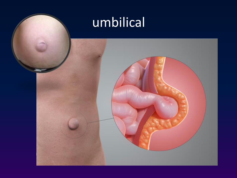
umbilical

Incisional
Hernia Complications
• Incarceration
• Strangulation
• Intestinal obstruction

Pertinent History
• Duration/onset
• Symptoms
– Local
– Obstructive
• Nausea, emesis, pain, distension, obstipation
• Prior Incarceration
• Related comorbidity
– Cough/Urinary flow/Constipation
– Operative risk

Pertinent exam
• Distension
– Bowel obstruction
• Scars
– Incisional hernias
– Recurrence
– Contraindications for certain approaches
• Rectal--blood/masses

HerniaExamination:
1. Inspection for site, size, shape and color.
2. Palpation for surface, temp, tenderness,
composition and reducibility.
3. Expansible cough impulse.
4. General exam: for common causes of increase
intra abdominal pressure


Surgical Exam
• Location
• Reducibility
• Tenderness
• Skin changes
• Palpable edges
• Genitalia
• Rectal
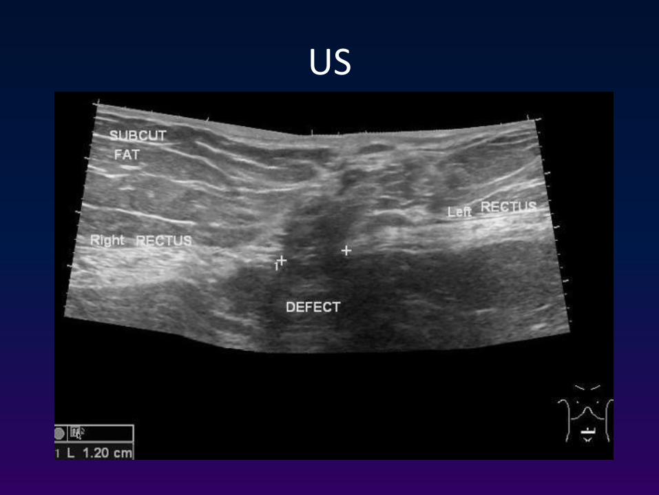
US
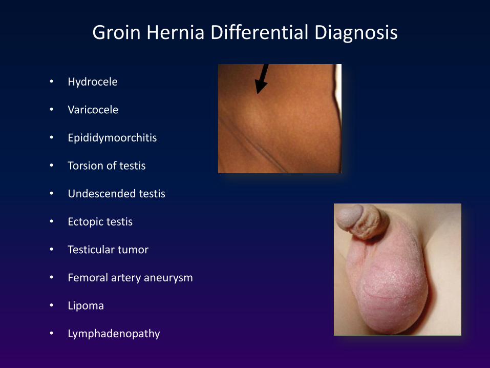
Groin Hernia Differential Diagnosis
• Hydrocele
• Varicocele
• Epididymoorchitis
• Torsion of testis
• Undescended testis
• Ectopic testis
• Testicular tumor
• Femoral artery aneurysm
• Lipoma
• Lymphadenopathy

Fluid collection in scrotum.
Contained in peritoneal sac that may
or may not communicate with
peritoneal cavity via processus
vaginalis.
‘Communicating’ hydrocele if
peritoneal communication is present.
Differentiated from true hernia by
finding of normal (i.e., no bulge in)
inguinal canal.
Hydrocele

Umbilical hernia
• Signs and symptoms
• Age; doesn’t appear until the umbilical cord has separated
and healed
• No specific symptoms
• Have wide neck and reduce easily, rarely give intestinal
obstruction
• Nature history; 90 % disappear spontaneously during the first
year

Inspection
Site; in the center of the umbilicus
Size and shape; size can vary from vary small to very large.
Shape is usually hemispherical.
Palpation
Composition; contain bowel, which makes it resonant to percussion.
They reduce spontaneously when the child lies down.
Reducibility; easy
Cough impulse; invariably present.
Examination

Epigastric hernia
• Incidence 1-5%
• Men > women
• Pre-peritoneal fat protrusion through decussating fibers at
linea alba
• Between xiphoid and umbilicus
• 20% multiple
• Repair primarily

Incisional Hernia
Can occur ANYWHERE an incision has been made, no matter how small.

Incisional Hernia
Can develop in the original incision site because of dehiscence or failure of wound healing, or
Can develop at the sites where sutures are passed through the tissue during closure (Swiss cheese-type hernia), or
Both

Incisional hernia risk factors
– Technical
– Wound infection
– Smoking
– Hypoxia/ ischemia
– Tension
– Obesity
– Malnutrition

Incisional hernia - diagnosis
Signs and symptoms
Previous operation or accidental trauma
Age; all ages, but more common in old age
Symptom; lump, pain, intestinal obstruction (distention, colic, vomiting,constipation, sever pain in the lump)
Examination
reducible lump
expansile cough impulse
if the lump dose not reduse and dose not have cough impulse, than it may be not a hernia
Differential diagnosis
Tumor
Chronic abscess
Hematoma
Foreign body granuloma

Incarcerated incisional hernia
Cannot be reduced.
Tender
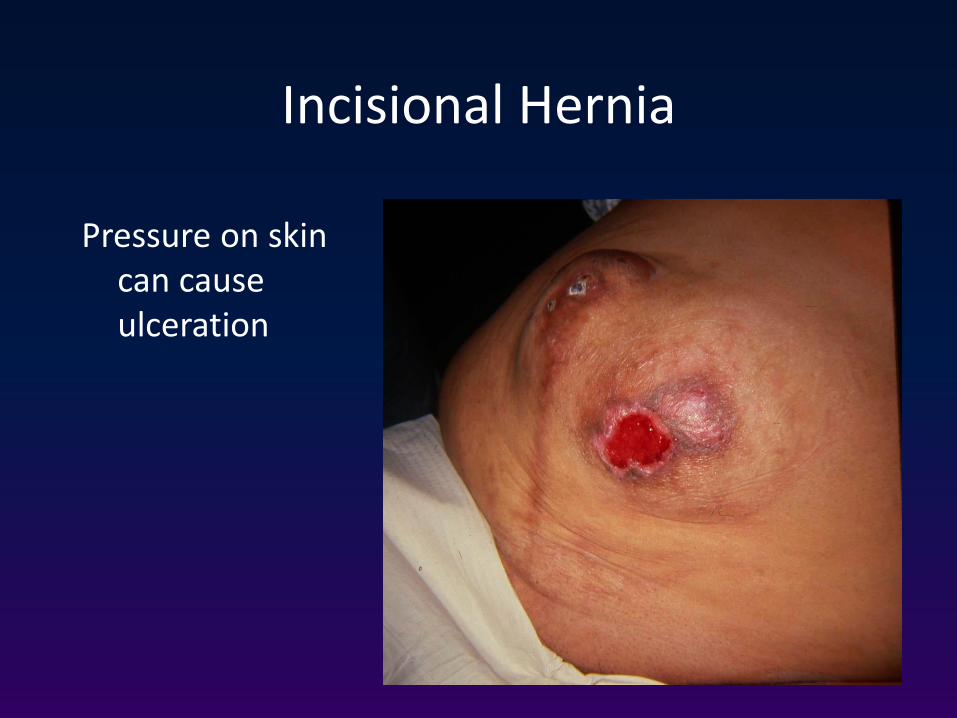
Incisional Hernia
Pressure on skin can cause ulceration

Rare: Spighellian hernia
• Hernia through subumbilical portion of semi-lunar line
• Difficult to diagnose– Clinical suspicion (location)
– CT scan
• Repair primarily or with mesh

Rare: Lumbar hernia
• Congenital, spontaneous or traumatic
• Grynfeltt’s triangle– 12th rib, internal oblique and sacrospinalis muscle
– Covered by latissimus dorsi
• Petit’s triangle– Latissimus dorsi, external oblique and iliac crest
– Covered by superficial fascia

Pelvic hernia
• Obturator hernia
– Most commonly in women
– Howship-Romberg sign (obturator nerve neuropathy due to
compression of it, by an obturator hernia. Patients present with pain and paresthesia along the inner aspect of the thigh, down to the knee).
• Sciatic hernia
• Perineal hernia

Hernia Management
Investigations:
– None required for routine uncomplicated case
– Plain X-ray for suspected bowel obstruction
– Ultrasound in case of diagnostic uncertainty
– Herniogram rarely used
– Routine pre-op investigations
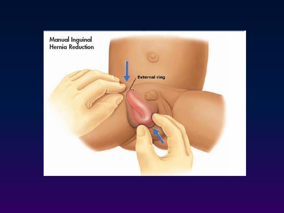
Reduction
• Uncomplicated: Manual Gentle pressure over hernia Gentle traction over the mass sedation and trendelenburg position.
• Complicated (strangulated): no attempt should be made to reduce the hernia because of potential reduction of gangrenous segment of bowel with the hernial sac.

Surgery
The primary goals of surgery are to:
– Repair the hernia
– Minimize the chance of recurrence
– Return the patient to normal activities quickly
– Improve quality of life
– Minimize postsurgical discomfort and the adverse effects
of surgery
Hernia Treatment
• Surgery
– To relieve symptoms
– To prevent complications
• Operations
– Open hernia repair
– Laparoscopic hernia repair
• Pre-peritoneal
• Intra- abdominal

Open Hernia Repair
• Day-case surgery
• Anaesthesia
– General
– Local
• Operations
– Tension free Mesh repair (Lichtenstien)
– Darn repairs (Shouldice, Bassini)

• Surgical repairs of inguinal hernia generally fall into 3 categories:
– Open repair without a mesh implant (i.e., sutured)
– Open repair with a mesh
– Laparoscopic repair with a mesh
• Several procedures have been employed within each of these
categories.
• The nearly universal adoption of mesh (except in pediatric cases)
means that the most relevant questions about hernia repair involve
various mesh procedures.
Types of Surgical Repair for Inguinal Hernias
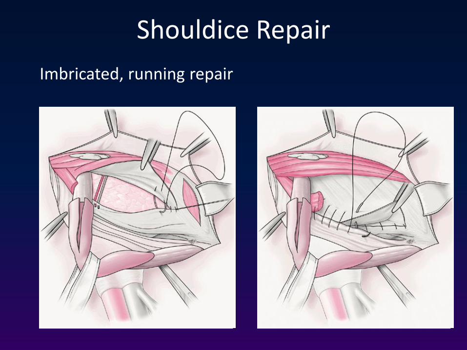
Shouldice Repair
Imbricated, running repair

Open Mesh-Based Repair of an Inguinal Hernia
Before After
Mesh
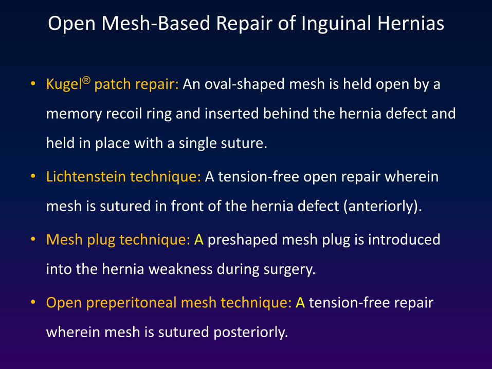
• Kugel® patch repair: An oval-shaped mesh is held open by a
memory recoil ring and inserted behind the hernia defect and
held in place with a single suture.
• Lichtenstein technique: A tension-free open repair wherein
mesh is sutured in front of the hernia defect (anteriorly).
• Mesh plug technique: A preshaped mesh plug is introduced
into the hernia weakness during surgery.
• Open preperitoneal mesh technique: A tension-free repair
wherein mesh is sutured posteriorly.
Open Mesh-Based Repair of Inguinal Hernias

Lichtenstein repair
• Tension free
• Less painful
• Foreign body

Mesh plug

• PROLENE™ Hernia System: A one-piece mesh device constructed of an
onlay patch connected to a circular underlay patch by a mesh cylinder.
• Read-Rives repair: A tension-free repair wherein mesh is placed just over
the peritoneum.
• Stoppa technique: A large polyester mesh is interposed in the
preperitoneal connective tissue between the peritoneum and the
transversalis fascia to prevent visceral sac extension through the
myopectineal orifice.
• Trabucco technique: A hernia repair procedure that involves placing a
single preshaped mesh without using sutures.
Open Mesh-Based Repair of Inguinal Hernias

PHS

• Intraperitoneal onlay mesh technique IPOM: A mesh is placed under the
hernia defect intra-abdominally to circumvent a groin dissection.
• Totally extraperitoneal patch TEP: The peritoneal cavity is not entered, and
a mesh is used to cover the hernia from outside the preperitoneal space.
• Transabdominal preperitoneal patch TAPP: A laparoscopic repair procedure
wherein the surgeon enters the peritoneal cavity, incises the peritoneum,
enters the preperitoneal space, and places the mesh over the hernia; the
peritoneum is then sutured and tacked closed.
Laparoscopic Mesh-Based Repair Procedures for Inguinal Hernias
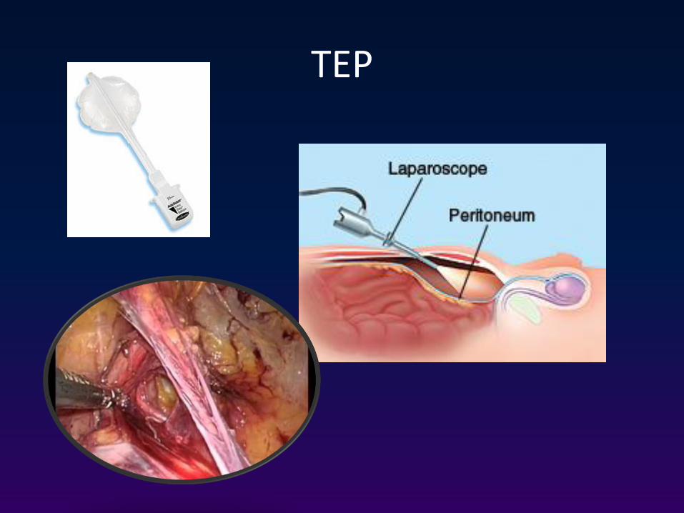
TEP

TAPP

TEP &TAPP
Surgical repair – ventral hernias
Primary suture repair:
• not recommended > 3cm
• high recurrence (25-63%)

Ventral (epigastric) hernia - Mesh placement techniques

IPOM
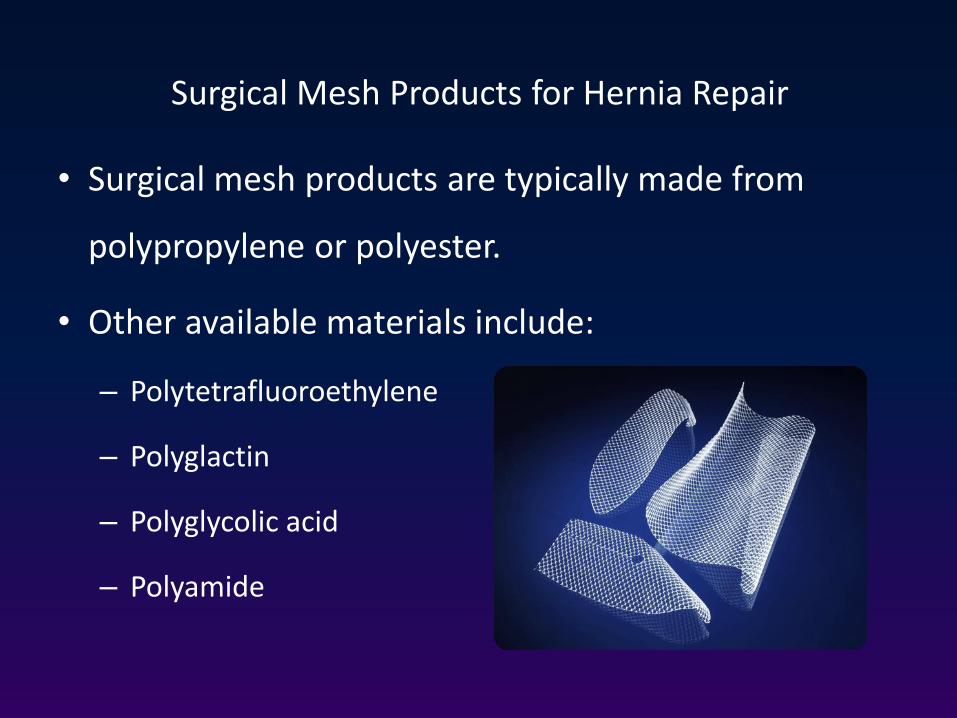
• Surgical mesh products are typically made from
polypropylene or polyester.
• Other available materials include:
– Polytetrafluoroethylene
– Polyglactin
– Polyglycolic acid
– Polyamide
Surgical Mesh Products for Hernia Repair

Seven important properties of mesh are:
1. Withstands physiologic stresses over time
2. Conforms to the abdominal wall
3. Mimics normal tissue healing
4. Resists the formation of bowel adhesions and erosions into visceral structures
5. Does not induce allergic reaction or foreign body reactions
6. Resists infection
7. Is noncarcinogenic
Properties of Mesh Products for Hernia Repair

Surgery Complications
• Trauma– Nerve
– Artery (testicular atrophy)
– Intestine
• Haemorrhage– Haematoma (infection)
• Infection– Wound infection
– Chest Infection

Thank you


