Loyola University Chicago Loyola University Chicago
Loyola eCommons Loyola eCommons
Dissertations Theses and Dissertations
1972
Histochemical and Gravimetric Analyses of the Pineal Gland of Histochemical and Gravimetric Analyses of the Pineal Gland of
the Albino Rat During the Estrous Cycle, Pregnancy and the Albino Rat During the Estrous Cycle, Pregnancy and
Pseudopregnancy Pseudopregnancy
Anthony V. Fasano Loyola University Chicago
Follow this and additional works at: https://ecommons.luc.edu/luc_diss
Part of the Medicine and Health Sciences Commons
Recommended Citation Recommended Citation Fasano, Anthony V., "Histochemical and Gravimetric Analyses of the Pineal Gland of the Albino Rat During the Estrous Cycle, Pregnancy and Pseudopregnancy" (1972). Dissertations. 1135. https://ecommons.luc.edu/luc_diss/1135
This Dissertation is brought to you for free and open access by the Theses and Dissertations at Loyola eCommons. It has been accepted for inclusion in Dissertations by an authorized administrator of Loyola eCommons. For more information, please contact [email protected].
HISTOCHE!•:ICAL AND GRAVIHE'I'RIC .AN.ALYSES OF THE PIImAL
GLAND OF THE ALBINO RAT DURING 'rHE ESTnOUS
CYCLE, PREGNANCY AND PSEUDOPREGNA~CY
by
Anthony V. Fasano
A Dissertation Submitted to the Faculty of the Graduate School
of Loyola University of Chicago in Partial Fulfillment of
the Requirements for the Degree of
Doctor of Philosophy
February
1972
ti~~r..Jtf
LOYOLA UNfV'~;::snY
LIFE
Anthony Vincent Fasano was born in Montclair, New Jersey on December 28, 19J6, the son of Nr. and Mrs. Benjamin V. Fasano.
He attended elementary and high school in Newark, New Jersey, and was graduated from Colorado State College, Greeley, Colorado in June, 1964. During the ensuing three years, Mr. Fasano was engaged as an analytical chemist for Fisher Scientific Company, Fair Lawn, ::-.iew Jersey and as a research chemist for the John L. Smith Memorial for Cancer Research in Maywood, New Jersey.
In January, 19 67, .r.:r. Fasano began his graduate study in the Department of Physiology of Fairleigh Dickinson University, Teaneck, New Jersey. He remained there until he entered the Department of Anatomy, Loyola University of Chicago, Stritch School of !•Iedicine, !-1aywood, Illinois, September, 1967. Here he continued his scientific investigations for the doctorate of philosophy degree in anatomy.
From 1968 to 1971, Mr. Fasano has had a National Defense Education Act fellowship. He is a member of the American Association for the Advancement of Science, American Society of Zoologists, and the New York Museum of Natural History.
During the period from 1954 to 1962, Mr. Fasano was an active member of the United States Naval Reserve from which he received an Honorable Discharge in August, 1962. In 1965, he married Linda Jacobsen and is the father of a boy, Steven Michael, now seventeen months old. and a girl, Dana Nicole, one month old.
111
A.CKNOWLEDGEMENTS
No research endeavor is ever the work of a lone
invest~gator; it is the culmination of the talents, guidance
and assistance of a multitude of people. · The author of this
dissertation wishes to thank a few of his fellow workers for
freely rendering their talents. First and foremost, an especial
thanks to may adviser, Professor Joseph T. Velardo for his
painstaking help and guidance; to Mrs. Velardo for her
understanding and always constant encouragement; and to Dr.
Barbara Kasprow for her help, instruction and guidance. A
warm thanks to Dr. Leslie Emmert, Francis Kovarik and Grover
Ericson for their assistance with both the animal work and the
preparatory written work for this dissertation; to my committee
for their ever friendly and patient guidances and to Mrs. Canuti,
Mrs. Schultz, Mrs. Smelte, and Mr. and Mrs. Kovarik for their
pleasant support. Last, but not least, a very grateful thanks
to my wife, Linda, who gave up so much and did so much so that
this investigator could reach his goal.
iv
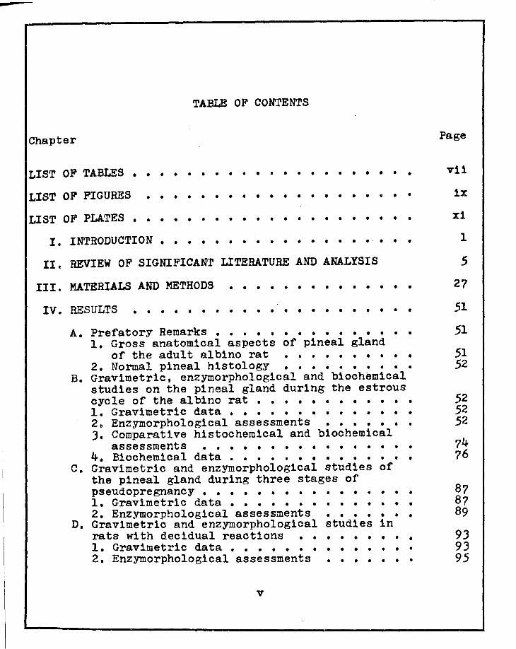
TABLE OF CONTENTS
Chapter Page
LIST OF TABLES • • • • • • • • • • • • • • • • • • • • • vii
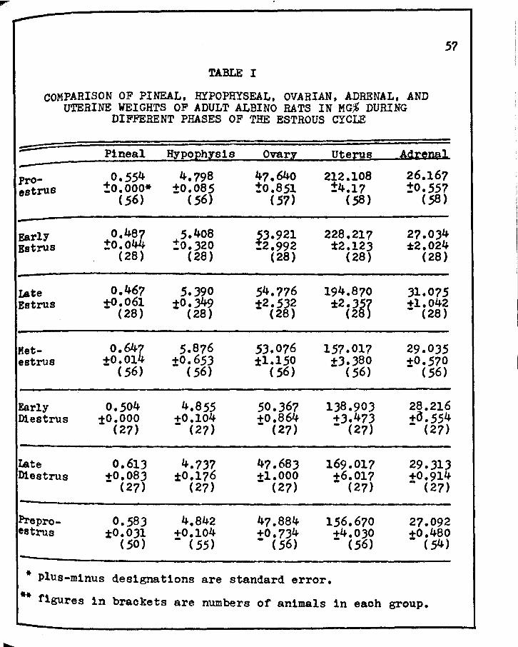
I. COMPARISO:i OF PINEAL, HYPOPHYSEAL, OVARIAN, ADRENAL, &'{D UTERINE WEIGHTS OF ADULT ALBINO RATS IN KG,% DURING DIFFERENT PHASES OF THE ESTROUS CYCLE • • • • • • • • • • • • • • • •
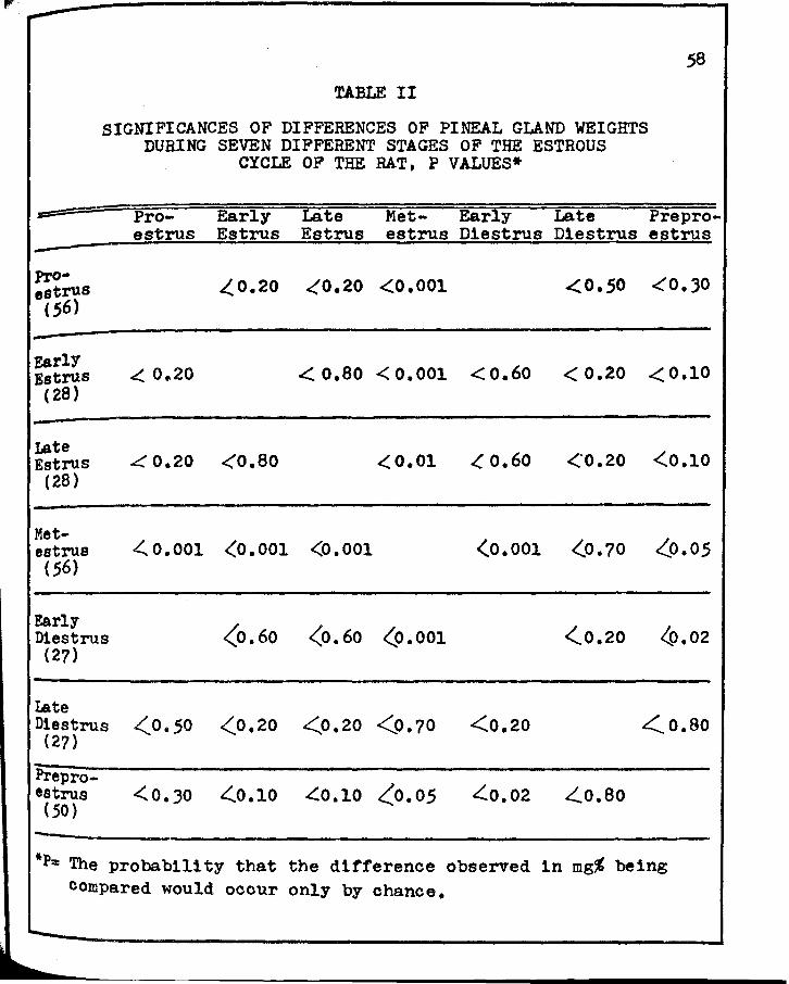
II. SIGNIFICANCES OF DIFFERENCES OF PINEAL GLAND WEIGHTS DURING SEVEN DIFFERENT STAGES OF ·rHE ESTROUS CYCLE OF THE RAT • • • • • • • • • •
III. SIGNIFICANCES OF DIFFERENCES OF HYPOPHYSEAL WEIGHTS DURING SEVEN DIFFEREHT STAGES OF THE ESTROUS CYCLE OF THE RAT • • • • • • • • • •
• •
• •
• •
IV. SIGNIFICANCES OF DIFFEREHCES OF OVARIA:-J WEIGHTS DURING SEVEN DIFFERENT STAGES OF rHE ESTROUS
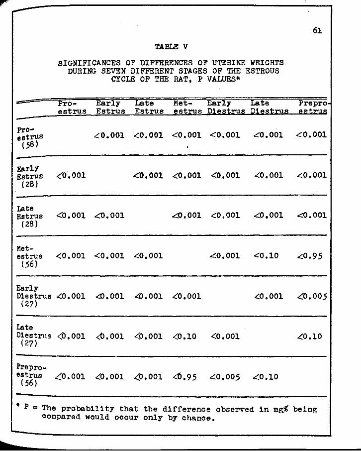
V. SIGrJIFICANCES OF DIFFERENCES OF UTERHrE WEIGHTS DURING SEVEi'l DIFFERENT s·rAGES OF 'rIIB ESrROUS CYCLE OF THE RAT • • • • • • • • • • • • • • • • 61
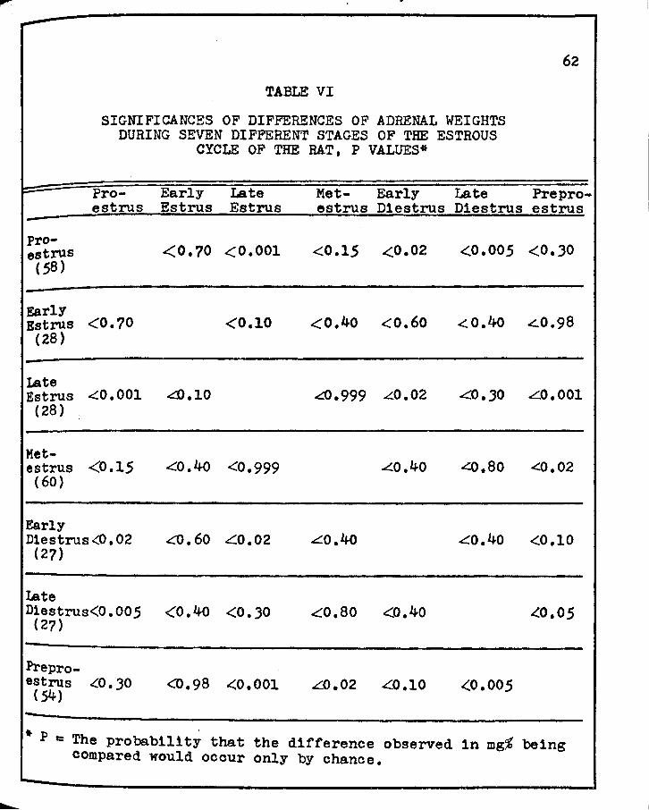
VI. SIGNIFICANCES OF DIFFERENCES OF ADRENAL WEIGHTS DURING SEVEN DIFFERENT STAGES OF THE ESTROUS CYCLE OF THE RAT • • • • • • • • • • • • • • • • 62
VII. SEMI-QUANTITATIVE HIS·roCIIEMICAL ESTI!'"iATES OF SUCcnuc DEHYDROGENASE ( SDH)' LACTIC DEHYDROGENASE (LDH)' ALKALrm PHOSPHATASE (ALK. P'TASE)' .AHD ACID PHOSPHA1'ASE (ACID P'TASE) OF THE PIHEAL GLAND OF THE RAT DURHJG THE Es·rROUS CYCLE • • • • ?8
v11
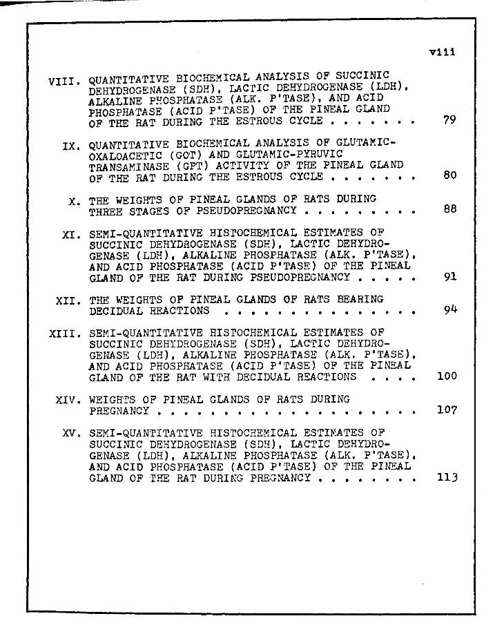
r VIII. QUANTITATIVE BIOCHEMICAL ANALYSIS OF SUCCINIC
DEHYDROGENASE ( SDH), LACrIC DEHYDROGENASE ( LDH), ALKALINE PHOSPHA'l'ASE (ALK. P'TASE), AND ACID PHOSPHATASE (ACID P'TASE) OF THE PINEAL GLAND OF THE RAT DURING THE ESTROUS CYCLE • • • • • • •
v111
79
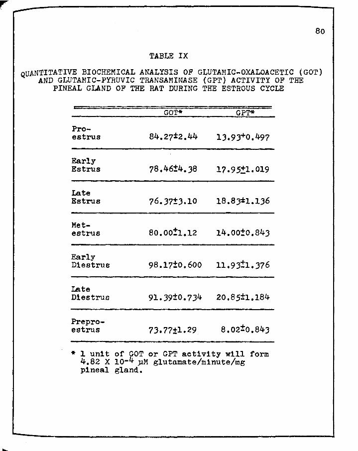
IX. QUANTITATIVE BIOCHEMICAL ANALYSIS OF GLUTAMICOXALOACETIC (GOT) AND GLUTAMIC-PYRUVIC TRANSAMINASE ( GPT) ACTIVITY OF THE PINEAL GLAND OF THE RAT DURING THE ESTROUS CYCLE • • • • • • • 80
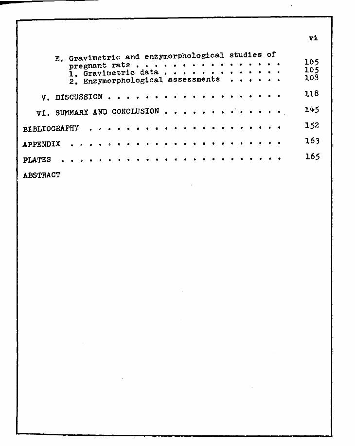
X. THE WEIGHTS OF PINE.AL GLANDS OF RATS DURING THREE STAGES OF PSEUDOPREGNANCY • • • • • • • • e
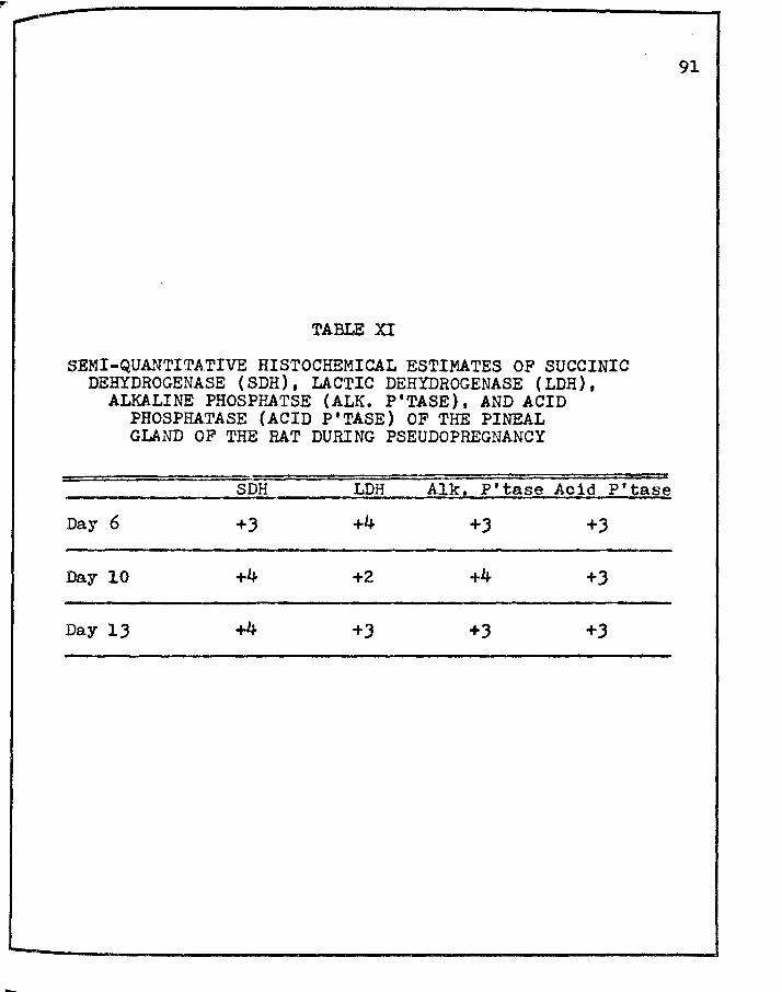
XI. SEMI-QUANTITATIVE HIS·rOCHEMICAL ESTIMATES OF SUCCINIC DEHYDROGENASE (SDH), LACTIC DEHYDROGENASE (LDH), .ALKALINE PHOSPHATASE (ALK. P'TASE), AND ACID PHOSPHATASE (ACID P'TASE) OF THE PINEAL
88
GLAND OF THE RAT DURING PSEUDOPREGNANCY • • • • • 91
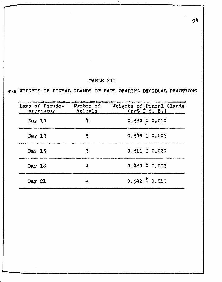
XII. THE WEIGHTS OF PINEAL GLANDS OF RATS BEARING DECIDUAL REACTIONS • • • • • • • • • • • • • • •
XIII. SEMI-QUANTITATIVE HISrOCHEMICAL ESTIMATES OF SUCCINIC DEHYDROGENASE (SDH), LACTIC DEHYDROGENASE (LDH), ALKALINE PHOSPHATASE (ALK. P'T.ASE), AND ACID PHOSPHATASE (ACID P'TASE) OF THE PINEAL GLAND OF THE RAT WITH DECIDUAL RE.ACTIONS • • • • 100
XIV. WEIGHT'S OF PINEAL GLANDS OF RATS DURING PREGNANCY • • • • • • • • • • • • • • • • • • • •
XV. SEMI-QUANTITATIVE HISTOCHEMICAL ESTIMATES OF SUCCINIC DEHYDROGENASE (SDH), LACTIC DEHYDROGENA.SE (LDH), ALKALINE PHOSPHATASE (ALK. P'TASE), AND ACID PHOSPHATASE (ACID P'TASE) OF THE PIN!<'---AL
107
GLAND OF rHE RAT DURING PREGNANCY • • • • • • • e 113
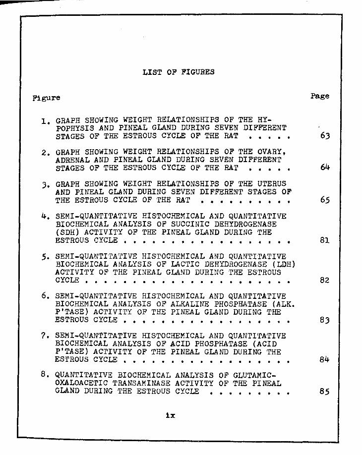
LIST OF FIGURES
Figure Page
1. GRAPH SHOWING WEIGHT RELATIONSHIPS OF THE HYPOPHYSIS AND PINEAL GLAND DURING SEVEN DIFFERENT STAGES OF THE ESTROUS CYCLE OF THE RAT • • • • • 63
2. GRAPH SHOWING WEIGHT RELATIONSHIPS OF THE OVARY, ADRENAL AND PINEAL GLAND DURING SEVEN DIFFERENT STAGES OF THE ESTROUS CYCLE OF THE RAT • • • • • 64
J. GRAPH SHOWING WEIGHT RELATIONSHIPS OF THE UTERUS AND PI.t-.TEAL GLAND DURING SEVEN DIFFERENT STAGES OF THE ESTROUS CYCLE OF ·rHE RAT • • • • • • • • • • 65
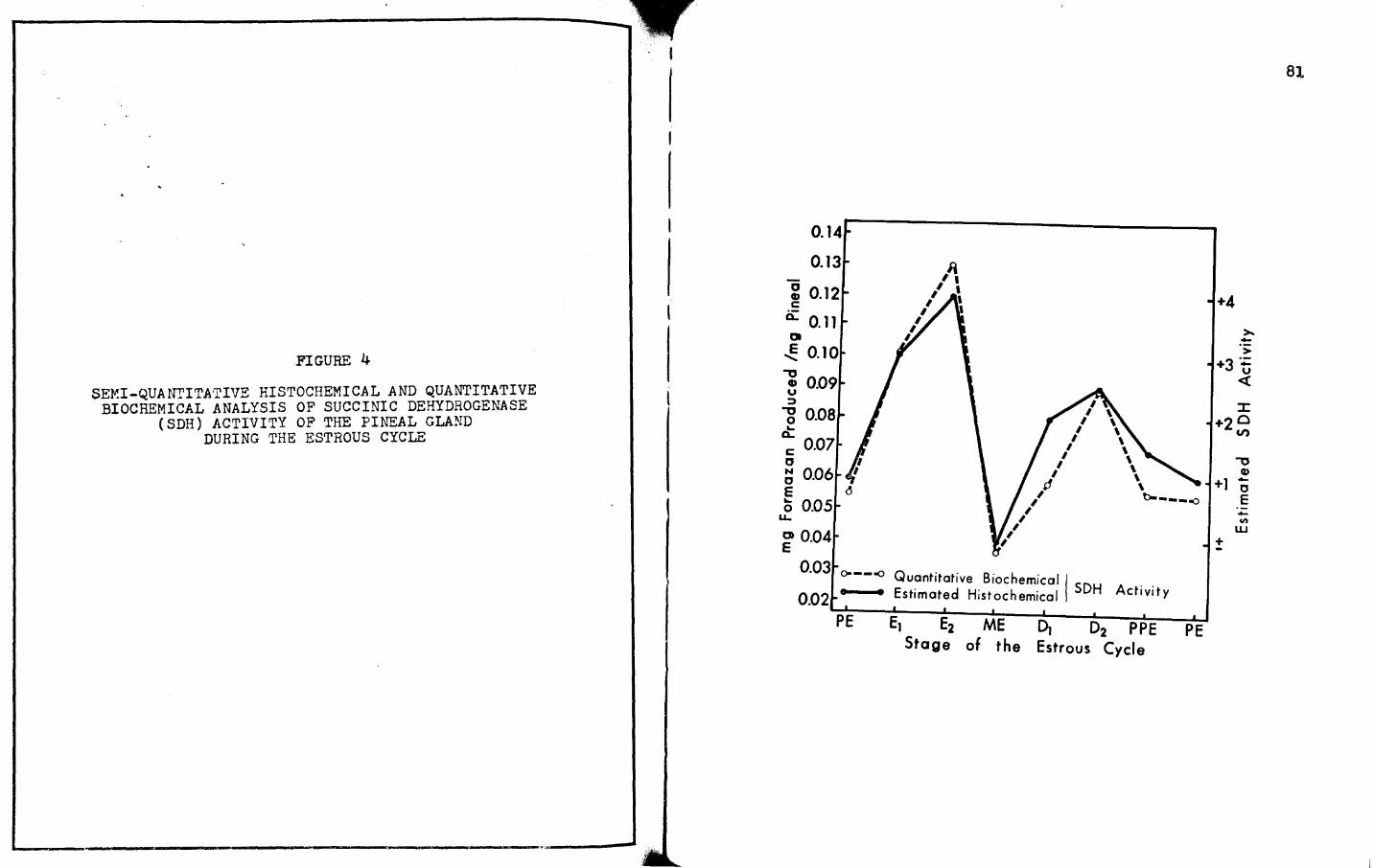
4. SEMI-QUANTITATIVE HISTOCHEMICAL AND QUANTITATIVE BIOCHEMICAL ANALYSIS OF SUCCINIC DEHYDROGENASE (SDH) ACTIVITY OF THE PINEAL GLAND DURING THE ESTROUS CYCLE • • • • • • • • • • • • • • • • • • 81
5. SEMI-QUANTI'I'ATIVE His·roCHEMICAL AND QUANTI·rATIVE BIOCHEMICAL ANALYSIS OF LACTIC DEHYDROGENASE (LDH) AC'rIVITY OF THE PINEAL GLAND DURING THE ESTROUS CYCLE • • • e • • • • • • • • • • • • • • • • • • 82
60 SEMI-QUANTITATIVE HISTOCHEMICAL AND QUANTITA'rIVE BIOCHEMICAL ANALYSIS OF ALKALINE PHOSPHATASE (ALK. P'TASE) ACTIVITY OF THE PINEAL GLAND DURING THE ESTROUS CYCLE • • • • • • • • • • • • • • • • • • BJ
?. SEMI-QUANTITATIVE HISTOCHEMICAL AND QUANTITATIVE BIOCHEMICAL ANALYSIS OF ACID PHOSPHATASE (ACID P'TASE) ACTIVITY OF THE PINEAL GLAND DURING THE ESTROUS CYCLE • • • , • • • • • • , • • • • • • • 84
B. QUANTITATIVE BIOCHEMICAL ANALYSIS OF GLUTAMICOXALOACETIC TRANSAMINASE ACTIVITY OF THE PINEAL GLAND DURING THE ESTROUS CYCLE • • • • • • • • • 85
ix
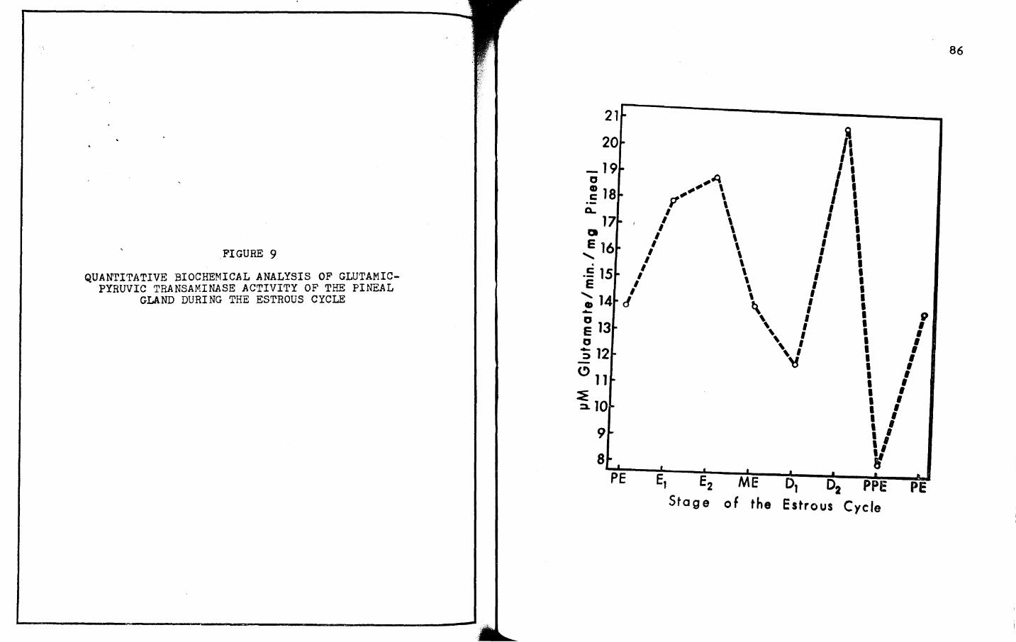
9. QUANTITATIVE BIOCHEMICAL ANALYSIS OF GLUTAMICPYRUVIC TRANSAMINASE ACTIVITY OF ·rHE PINEAL
x
GLAND DURING THE ESTROUS CYCLE. • • • • • • • • • 85
10. SEMI-QUANTITArIVE HISTOCHEMICAL EVALUATION OF SUCCINIC DEHYDROGENASE (SDH) AND LACI'IC DEHYDROGENASE (LDH) ACTIVITIES OF THE PINEAL GLAND DURING THREE STAGES OF PSEUDOFREGNANCY • • • • • • • • • 86
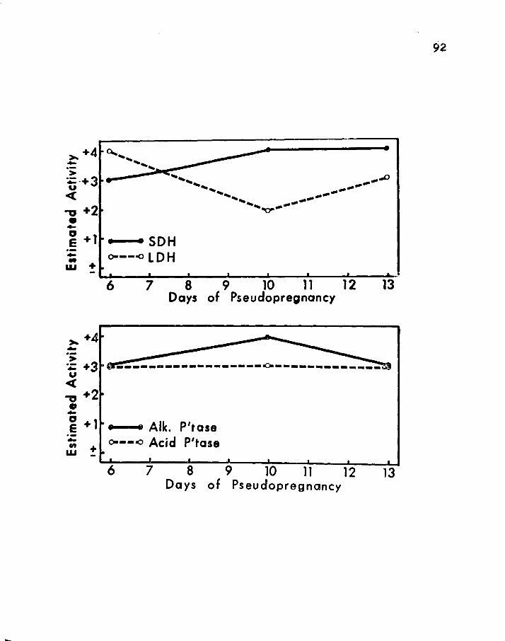
11. SEMI-QUANTI·rATIVE HISTOCHEMICAL EVALUATION OF ALKALINE PHOSPHATASE (ALK. P'TASE) AND ACID PHOSPHATASE (ACID P'TASE) ACTIVITIES OF rHE PINEAL GLAND DUR! NG THREE STAGES OF PSEUDO PREGNANCY • • 92
12. SEMI-QUANTI·I'ATIVE HISTOCHEMICAL EVALUATION OF SUCCINIC DEHYDROGENASE (SDH) ACTIVITY OF THE PINEAL GLAND IN RATS BEARING DECIDUAL REACrIONS • 101
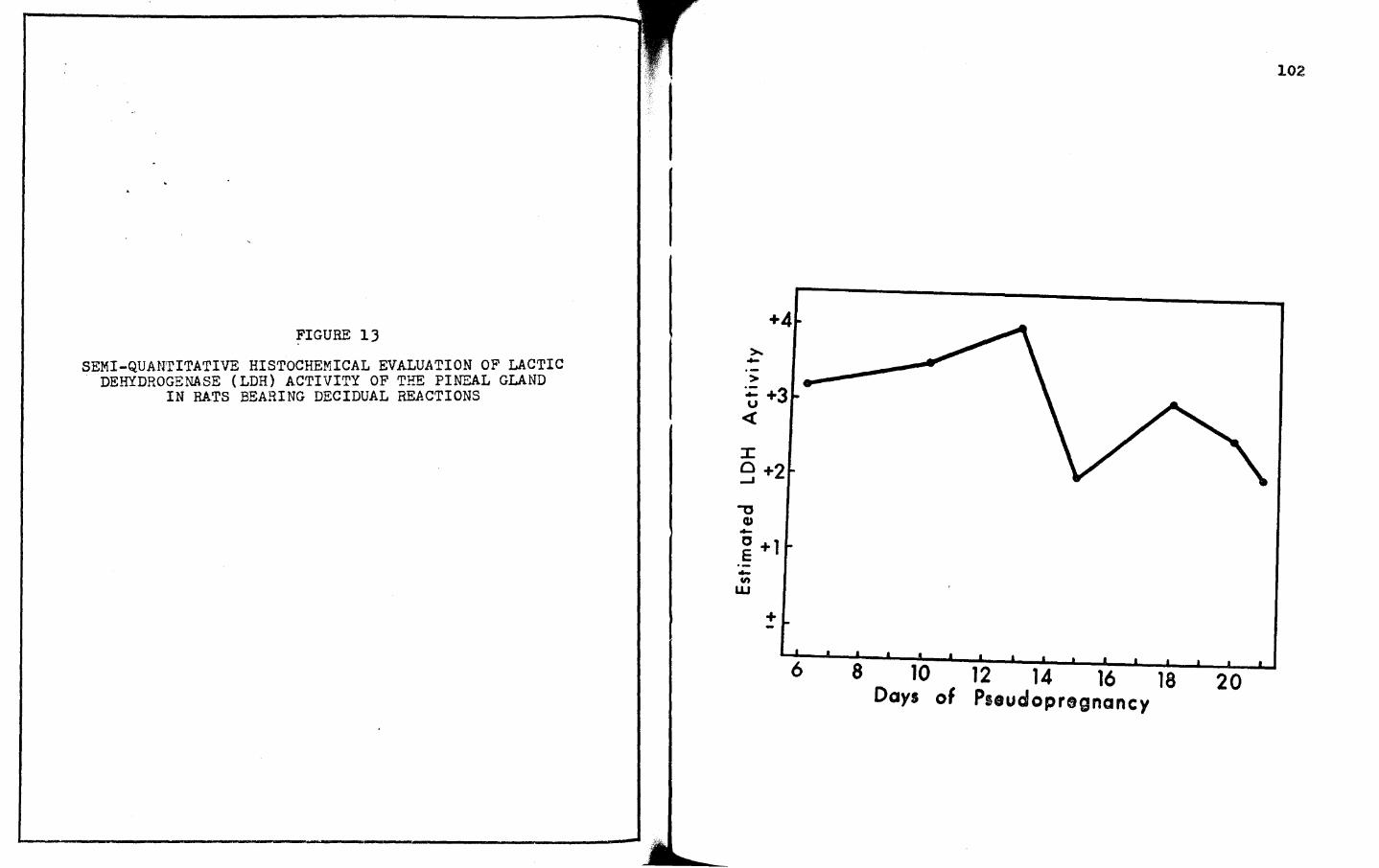
lJ. SEMI-QUANTITATIVE HISTOCHEMICAL EVALUATION OF LACTIC DEHYDROGENASE (LDH) ACTIVITY OF THE PINEAL GLAND IN RATS BEARING DECIDUAL REACTIONS • • • • 102
14. SEMI-QUANTITATIVE HISTOCHEMICAL EVALUATION OF ALKALI~TE PB.OSPF.ATASE (ALK. P'TASE) ACTIVITY OF THE PINEAL GLAND IN RATS BEARING DECIDUAL REAC·rIONS • • • • • • • • • • • • • • • • • • • • lOJ
15. SEMI-QUAITTITATIVE HISTOCHEMICAL EVALUATION OF ACID PHOSPHATASE (ACID P'TASE) ACTIVITY OF THE PINEAL GLAND IN RATS BEARING DECIDUAL REACTIONS • 104
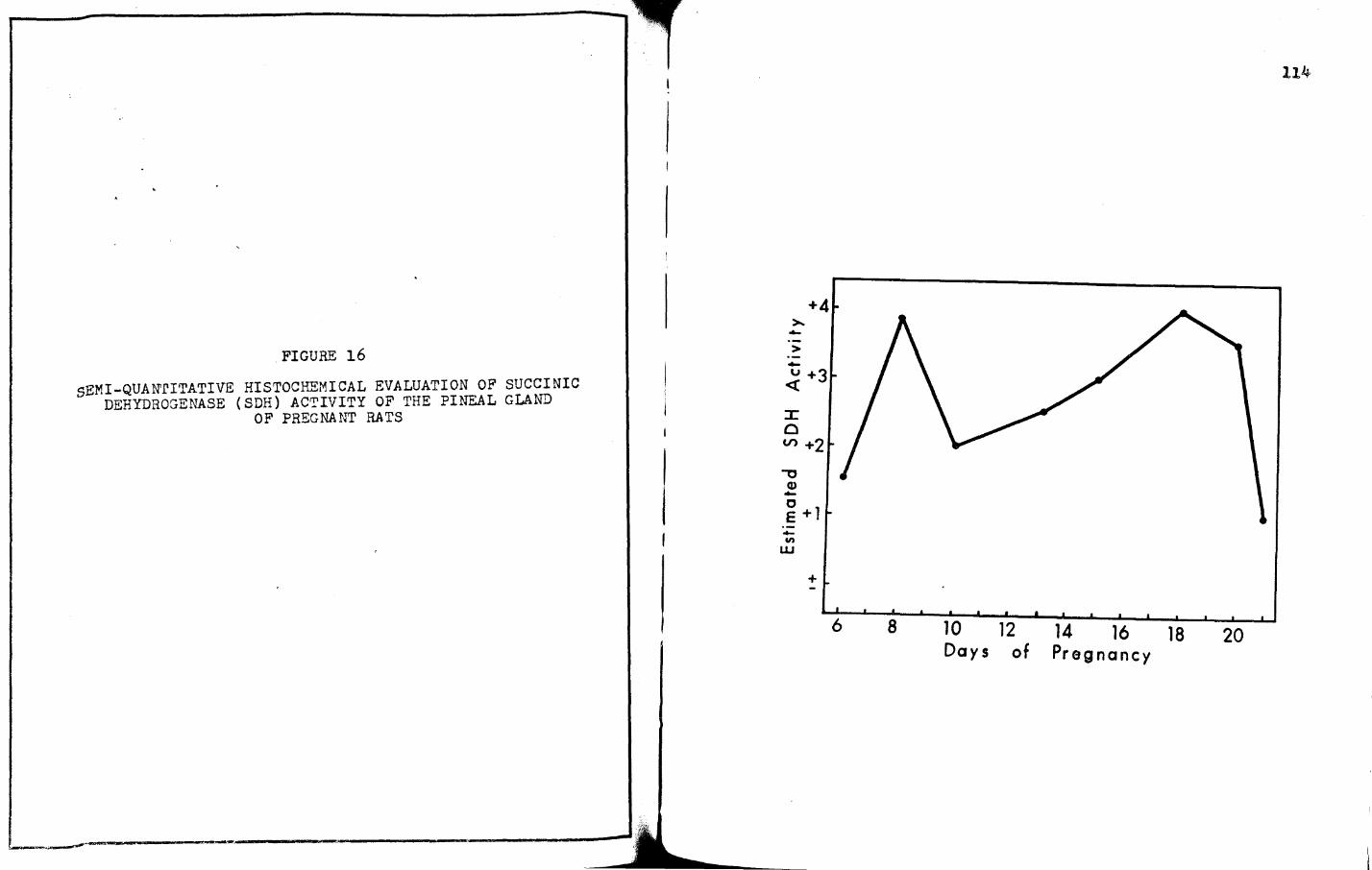
16. SEMI-QUANrITATIVE HISTOCHEMICAL EVALUATION OF SUCCINIC DEHYDROGENASE (SDH) ACTIVITY OF THE PINEAL GLAND OF PREGNANT RATS • • • • • • • • • •
17. SEMI-QUANTirATIVE HISrOCHEMICAL EVALUATION OF LACTIC DEHYDROGENASE (LDH) ACTIVITY OF THE PINEAL
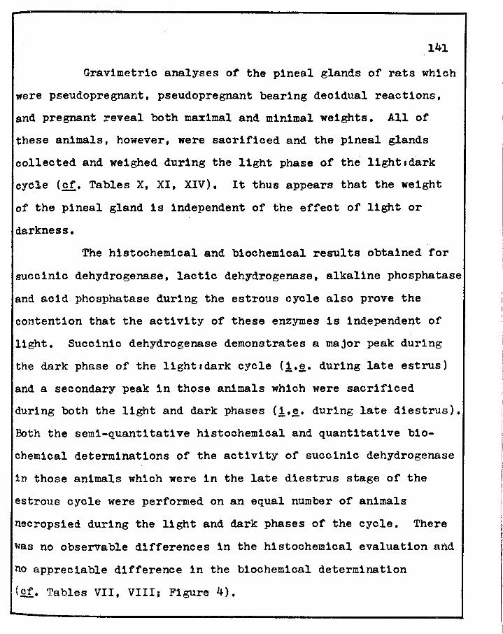
and acid phosphatase during the estrous cycle also prove the
contention that the activity of these enzymes is independent of
light. Succinic dehydrogenase demonstrates a major peak during
the dark phase of the lightadark cycle (i.~. during late estrus)
and a secondary peak in those animals which were sacrificed
during both the light and dark phases {i.~. during late diestrus),
Both the semi-quantitative histochemical and quantitative bio
chemical determinations of the activity of succinic dehydrogenase
1n those animals which were in the late diestrus stage of the
estrous cycle were performed on an equal number of animals
necropsied during the light and dark phases of the cycle. There
was no observable differences in the histochemical evaluation and
no appreciable difference in the biochemical determination
(£.!. Tables VII, VIII; Figure 4).
r -142
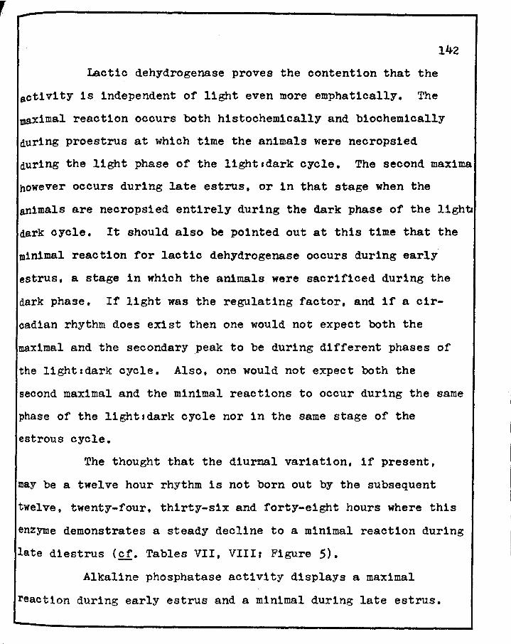
Lactic dehydrogenase proves the contention that the
activity is independent of light even more emphatically. The
maximal reaction occurs both histochem1cally and biochemically
during proestrus at which time the animals were necropsied
during the light phase of the lightcdark cycle. The second maxima
however occurs during late estrus, or in that stage when the
animals are necropsied entirely during the dark phase of the lighb
dark cycle. It should also be pointed out at this time that the
minimal reaction for lactic dehydrogenase occurs during early
estrus, a stage in which the animals were sacrificed during the
dark phase. If light was the regulating factor, and if a cir
cadian rhythm does exist then one would not expect both the
maximal and the secondary peak to be during different phases of
the 11ghtadark cycle. Also, one would not expect both the
second maximal and the minimal reactions to occur during the same
phase of the lightsdark cycle nor in the same stage of the
estrous cycle.
The thought that the diurnal variation, if present,
may be a twelve hour rhythm is not born out by the subsequent
twelve, twenty-four, thirty-six and forty-eight hours where this
enzyme demonstrates a steady decline to a minimal reaction during
late diestrus (cf. Tables VII, VIIIr Figure 5).
Alkaline phosphatase activity displays a maximal
reaction during early estrus and a minimal during late estrus.
143
Therefore, a maximal reaction 1s observed during the dark phase
of the 11ghtsdark cycle (early estrus) and a minimal reaction
during the dark phase (late estrus) of the lightsd.ark cycle
(£.!• Tables VII, VIIIr Figure 5).
Acid phosphatase, as alkaline phosphatase, also has a
maximal reaction during the dark phase (early estrus) and a
minimal reaction during the dark phase (late estrus). However,
there is a second minimal reaction which occurs during metestrus
and during this stage the animals were necrops1ed during the
light phase of the 11ght:dark cycle (cf. rables VII, VIII;
Figure 7).
A's a result of these findings the changes which occur
in the pineal gland weights and in the enzymes studied appear to
occur without the mediating effect of light, at least in a
12112 11ghtsdark environment.
During pseudopregnancy, prolonged pseudopregnancy in
rats bearing decidual reactions, and during pregnancy, the enzymes
studied h1stochemically also demonstrated varying degrees of
activity ranging from maximal to minimal. All of these animals
were necrops1ed during the iight phase of the lightsdark cycle
(.Qf. Tables XI, XII, XV; Figures 10-19). There, then, does not
appear to be any strong, compelling reason to assume that these
Particular enzymes studied demonstrate a diurnal rhythms rather,
1t appears that their activity is a function of the aforementioned
144
criteria.
Like most studies, the present dissertation unlocks the
door to more questions than were present in the initial stages
of these investigations. As with most works, and the present is
certainlY no exception, each investigation adds to the sum of
our knowledgee
CHAPTER VI
SUMMARY AND CONCLUSIONS
rhe main thrust of the dissertation involves a number
of studies on the histochemistry of the pineal gland of the rat
during seven stages of the estrous cycle: early estrus (A1 ),
late estrus (A 2 ), metestrus (B), early diestrus (C), late
diestrus (D), preproestrus (E) and proestrus (F). An important
offshoot of these studies on the pineal gland branched into
three aspects: pseudopregnancy, prolonged pseudopregnancy (rats
with decidual reactions), and pregnancy. Accompanying the first
part of this dissertation are a number of b1ochere1cal observations
on the pineal gland. Also, a large series of gravimetric data
is included on weight relationships of the pineal gland, the
pituitary body, adrenals, ovaries and uteri of the rat during the
estrous cycle.
Specifically, this dissertation is concerned with a
gravimetric analysis and a histocherrJ.1cal elucidation of the
activity of succinic dehydrog;enase (SDH), lactic dehydrogenase
(LDH), alkaline phosphatase (Alk. P'tase), and acid phosphatase
(Acid P'tase) activity in the pineal gland of the albino rat
during seven different stages of the estrous cycle, pseudopreg-
145
nancy, prolonged pseudopregnancy in rats bearing decidual
reactions, and in pregnancy.
146
l. Gravimetric analysis of the pineal gland revealed a
maximal weight, in mg%, during metestrus with a minimal weight
occurring during late estrus, the latter weighing one-third
those of metestrus.
2. When the pineal gland is lightest in weight, the
ovary and adrenal glands are heaviest and the hypophysis and
uterine weights are approximately at the mid-point in their
weight range. Conversely, when the pineal gland is heaviest,
the hypophysis ls heaviest, but the ovaries and adrenals are at
the mid-ranges, whereas the uterus ls at a low range.
J. The pineal gland is quite cyclical in weight changes
during the estrous cycle.
4. During the three stages of pseudopregnancy, the
weight of the pineal gland is typical of those of proestrus on
the initial and terminal days of pseudopregnancy (days six and
thirteen)J on day ten, the weight of the pineal appears similar
to that found during late estrus.
5. For the most part, the pineal gland weights during
prolonged pseudopregnancy in rats bearing decidual reaction show
weight relationships which are typical of those observed during
Preproestrus, proestrus and early estrus; the one exception
being day fifteen of prolonged pseudopregnancy which appears to
147
have a mean weight typical of those of early diestrus.
6. rhe weights of the pineal gland of pregnant rats
are highest on days six and eight (being somewhat typical of
rnetestrus), lighest on days eighteen, twenty, and twenty-one
(being soBewhat typical of early estrus), and appear cyclic in
nature during the period of gestation with the lowest weights
occurring during the last four to five days of pregnancy.
7. The four enzymes studied are localized in the
cytoplasm of the pinealocytes.
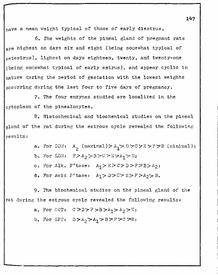
8. Histochemical and biochemical studies on the pineal
gland of the rat during the estrous cycle revealed the following
results:
a. For SDH: A2 (maximal)> Al·~ D '7C 7E 7 F 7B (minimal);
b. Por LDfi: F /7 A2 > B >C > E ?Al"'?" D;
c. For Alk. P'tase: A1 > E> C> D >F 7B>1'2;
d. For Acid p•·tase: A1/ D/C> E? F >A2> B.
9. The biochemical studies on the pineal gland of the
rat during the estrous cycle revealed the following results:
a. For GOI': C >D'7F >B>A1 >A 2 >E;
b. For GP'l': D 7A2 ?A1 "'7 B '?' F >C "?E;
148
10. Histochemical analyses of the pineal gland of the
rat during three stages of pseudopregnancy studied revealed the
following on days: 6 10 !l a. For SDH: +J +4 +4
b. For LDH: +4 +2 +J
c. For Alk. P'tase: +J +4 +3
d~ For Acid P'tase: +J +3 +J
11. Histochemical determinations of the pineal glands
of pseudopre~nant rats bearing decidual reactions displayed the
following intensities on days:
6
a. For SDH: +2
b. For LDH1 +J.2
c. For Alk, P'tase: +J
d. For Acid P'tase: +J.5
10
+J.5
+J.5
+2.5
+J
.u +J
+4
+J
+J.5
12 +1.5
+2
+4
+4
18
+4
+J
+2
+J
20
+2.5
+2.5
+2
+2
21
+2
+2
+1.5
+2.5
12. Histochemical analyses of the pineal gland during
pregnancy displayed the following intensities on days:
a. For SDH:
b. For LDH:
c, For Alk. P'tase:
d. For Acid P'tase:
6 8
+1.5 +J.8
+2. 5 :+ 2
10
+2
+J
!l
+2.5
+J.5
.Ll 18 20
+2
+4
+4 +J.5
+2 +J.5
21
+l
+J
±
+2
+1.5 +1.5 +J +4 +2 +2.5 +2.5
+2.5 +2.5 +2.5 +J.5 +4 +J +J
l~
lJ. High or maximal intensities of lactic dehydrogenase
precede those of succ1n1o dehydrogenase in the pineal glands of
rats during the estrous cycle, pseudopregnancy, prolonged
pseudopregnancy and during pregnancy.
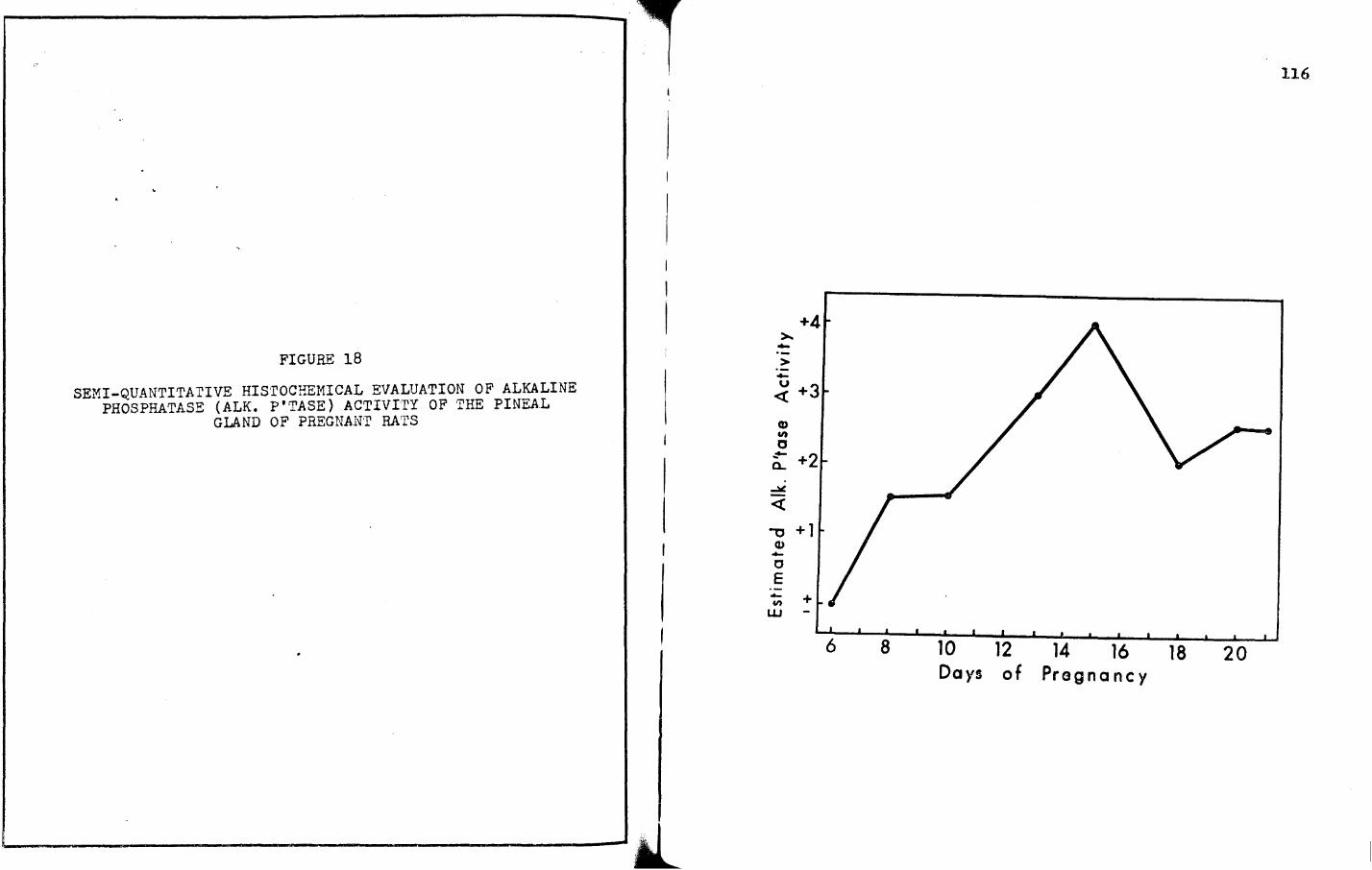
14. Maximal phosphatase activities in the pineal gland
either precede or are coincident with maximal intensities for
succinic dehydrogenase.
15. During the estrous cycle, the maximal succinic
dehydrogenase activity was coincident with the events of heat
and ovulation in the rat, while during pseudopregnancy there
appears to be a relationship between the activity of this enzyme
and that of the corpus luteum of pseudopregnancy.
16. Succ1n1c dehydrogenase maxima during prolonged
pseudopregnancy and pregnancy indicate a possible relationship
between this enzyme and the increased production and secretion
of relaxin.
17. Experimentally-derived data clearly indicate that
it is possible to demonstrate and compare the cyclical nature
of the pineal gland, both gravimetrically and histochemically,
the cyclicity of the estrous cycle 1n albino rats. Consequently,
numerous correlative relationships and interrelationships were
established, ranging from (a) high intensities of enzymes in the
pineal gland with high concentrations of similar enzymes 1n
reproductive tracts of rats during estrous and in estrogen1zed,
150
ovar1ectom1zed rats, to (b) specific concentrations of p1neal-
eontaining enzymes with specific aspects of adenohypophyseal
gonadal relationships.
18. Likewise, experimentally-derived data from this
dissertation make it possible to establish numerous correlations
between pineal enzymorphological and reproductive h1stomorpho
log1cal observations during pseudopregnancy, prolonged pseudo
pregnancy and pregnancy. Notable examples include specific
intensities of the dehydrogenases and phosphatases coinciding
with the development, maturation and subsequent necrosis of the
antimesometrial, mesometrial and metrial gland. Also, correlative
relationships were established between pineal enzymatic inten-
s1 t1es and the biological career of pregnancy.
19. Enzymorpholog1oal studies on the pineal gland
reveal that lactic dehydrogenase, a glycolytic enzyme in a system
which produces a small amount of ATP, always precedes succinic
dehydrogenase, an enzyme of the tricarboxyl1c acid cycle known
for its high production of ATP, in the metabolic scheme.
20. The results from these investigations clearly
reveal that the pineal gland enzymology and grav1metr1cs can be
related to the dynamics of reproductive mechanisms, both
endocr1nolog1cally and neuroendocr1nolog1cally.
21. These data form a workable standard reference
baseline for several important subsequent investigations which
151
can be performed on the pineal gland, the reproductive mechanism,
and the interrelationship of one with the other.
BIBLIOGRAPHY
Albert, A., L. o. Underdahl, L. F. Green and N. Lorenz 1954 Male hypogonadism. V. The testis in adult patients with multiple defects of pituitary function. Proc. Staff Mayo. Clln., ~1 317-323.
Axelrod, J., and H. Weissbach 1961 Purification and properties of hydroxyindole-0-methyltransferase. J. Biol. Chem., · 2361 211-213.
Axelrod, J., R.H. Wurtman ands. H. Snyder 1965 Control of hydroxyindole-0-methyltransferase activity in the rat pineal gland by environmental lighting. J. Biol. Chem., 240 (2)1 949-954. -
Bargmann, w. 1949 Uber die Neurosekretorische Verknupfung von Hypothalamus und Neurohypophyse. z. Zellforsch., ~· 610.
Barka, T. 1960 A simple azo-dye method for histochemical demonstration of acid phosphatase. Nature, 1871 248-249.
Berger, L., and D. Broida 1969 The colorimetric determination of lactic dehydrogenase. Sigma Tech. Bull., Number 500.
Bernard, Iren, and G. Csaba 1970 Localization of biogenic monoamines in tissue cultures of rat pineal. Acta Biol. Acade Sci. (hung.),~ (2)1 235-241.
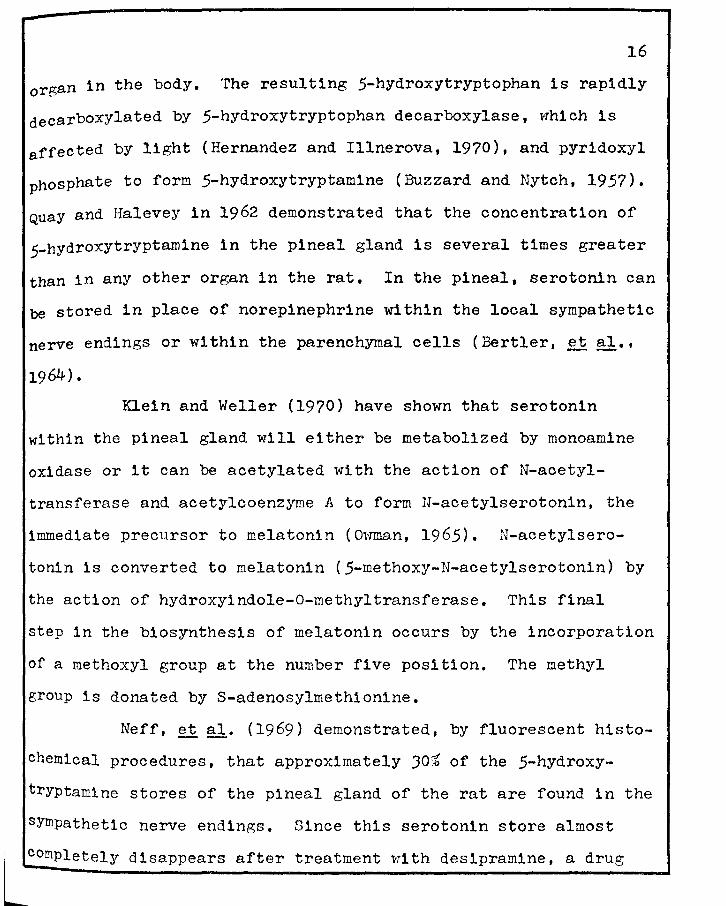
Bertler, A., B. Falck and c. Owman 1964 Studies on the 5-hydroxytryptamine stores in the pineal gland of the rat. Acta Physiol. Scand., .2,.2 (suppl. 234)1 1-18.
Bessey, o. A., o. H. Lowry and M. J. Brock 1946 A method for the rapid determination of alkaline phosphatase with five cubic millimeters of serum. J. Biol. Chem., 1641 )21-329.
Bever, A. T., J. T. Velardo and F. L. Hisaw 1953a Effect of estrogens on lactic dehydrogenase-DPNH oxidase system of ovariectomized rats. Fed. Proc., 121 14 (Abstract).
152
153
Bever, A. T., J. T. Velardo and F. L. Hisaw 195Jb The effect of progesterone on the lactic dehydrogenase-DPNH oxidase systerm of ovariectomized rat uteri. J. Clin. Endocrinology Metab., !1 (7)1 835-836 (Abstract).
Bloom, F. E., and L. L. Iversen 1970 Localizing JH-GABA in nerve terminals of rat cerebral cortex by electron microscopic autoradiography. Health, Education, and Welfare Reprint.
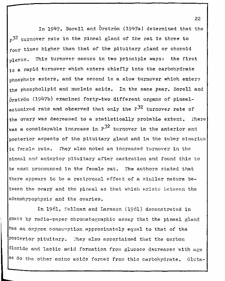
Borell, Ulf, and Ake Orstrom 1947a The turnover of phosphate in the pineal body compared with that of other parts of the· brain. Biochem. J., 411 J98-403.
Borell, Ulf, and Ake Orstrom 1947b On the function of the pineal body. Acta Physiol. Scand., !J1 62-71.
Burnstone, M. s. 1959 Histochemical demonstration of acid phosphatase activity in osteoclasts. J. Histochem. Cytochem., 1 ( 1 ) I J9-4 l •
Buzzard, J. A., and D. D. Nytch 1957 Some characteristics of rat kidney 5-hydroxytryptophan decarboxylase. J. Biol. Chem., 2271 225-2JO.
Chessman, D. w. 1970 Structural elucidation of a gonadotropininhibiting substance from the bovine pineal gland. Biochem & Biophys. Acta, 2071 247-253.
Chessman, D. w., and B. L. Fariss 1970 Isolation and characterization of a gonadotropin inhibiting substance from the bovine pineal gland. Proo. Soc. Exper. Biol. & Med., .!aJ1 1254-1256.
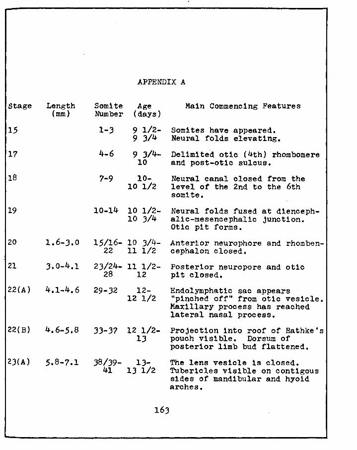
Christie, G. A. ·1964 Developmental stages in somite and postsomite rat embryos, based on external appearance and including some features of the macroscopic development of the oral cavity. J. Morphol., 1141 26J-286,
Chu, Elizabeth w. 1965 Effect of environmental illumination on estrous cycles of rodents. Acta Cytol., 21 221-227.
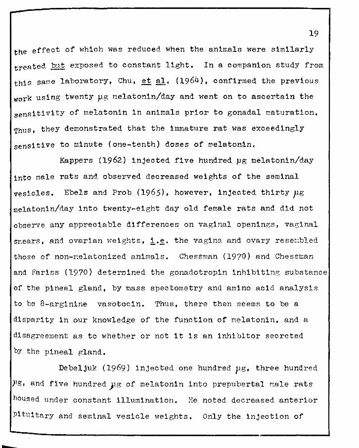
Chu, Elizabeth w., R. J. Wurtman and J. Axelrod 1964 An inhibitory effect of melatonin on the estrous phase of the estrous cycle of the rodent. Endocrinology, 22• 2J8-242.
II
r -~----------------------------------------..
154
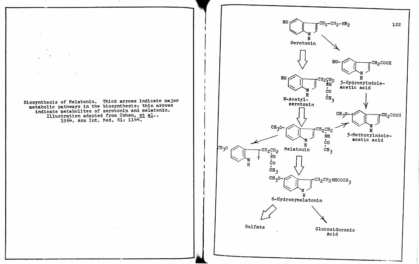
Cohen, R. A., R. J. Wurtman, J. Axelrod ands. H. Snyder 1964 Some clinical, biochemical and physiological actions of the pineal gland. Ann. Int. Med., .2!,1 1144-1161.
Crosby, Elizabeth c., Trypena Humphrey and E. M. Lauer 1962 Dlencephalon. Ins Correlative Anatomy of the Nervous System. The Macmillan Co., New York, Chapter VI1 pp 268-271.
Dallenbach-Hellweg, G., J. v. Battista and F. D. Dallenbach 1965 Immunohistological and histochemical localization of relaxin in the metrial gland of the pregnant rat. Amer. J. Anat., 1171 4JJ-450.
Danielii, J. F. 1953 Cytochemistry1 a critical approach. Chapman and Hall, London.
Das Gupta, T. K. 1968 The anatomy of the pineal organ in the syrian hamster. Acta Anat., 1!1 28-JS.
Dawson, A. B., and J. T. Velardo 1955 A histochemical study of lipids of the corpora lutea of the rat during pseudopregnancy. Amer. J. Anat., 2.11 JOJ-329.
Debeljuk, L. 1969 Effect of melatonin on the gona.dotrophic function cf the male rat under constant illumination. Endocrinology, ~ (4)1 937-939.
Dixon, J. s., and D. Bulmer 1971 The fine structure of cells in the rat metrial gland. J. Anat., lQ§. (1)1 123-lJJ.
Eadie, M. J., J. H. Tyrer, J. R. Kukums and w. D. Hooper 1970 Aspects of tetrazolium salt reduction relevant to quantitative histochemistry. Histochemie, z.!.1 170-180.
Ebels, I., and N. Prop 1965 A study of the effect of melatonin on the gonads, the estrous cycle and the pineal organ of the rat. Acta Endocrinology, !±21 567-577.
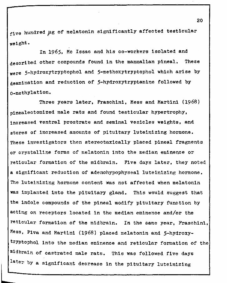
Fraschin1, F.,B. Mess and L. Martini 1968 Pineal gland, melatonin and the control of lutein1zing hormone secretion. Endocrinology, ~· 919-92J.
Fraschini, F., B. Mess, F. Piva, and L. Martini 1968 Brain receptors sensitive to indole compounds• functions in the control of lutein1zing hormone secretion. Science, 122. (3819)1 1104-1105.
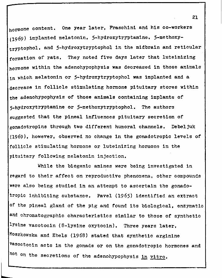
Fraschin1, F., F. Piva and L. Martini 1969 Indole compounds and FSH secretion. Prog. 5lst Meet. Endocrine Soc. p. 80 (Abstract).
155
Gomor1, F. 1951 Alkaline phosphatase or cell nuclei. J. Lab. & Clin. Med., .J.Z• 526-531.
Harris, H. F. 1900 On the rapid conversion of haematoxylin into haematin 1n staining reactions. J. Appl. Mier., J1 777-780.
Hellman, B., and4s. Larsson 1961 Utilization or uniformly
labelled cl -glucose in the pineal body of goats. Acta Endocr1nol. (Koh., J§.1 J5J-360.
Hernandez, J., and H. Illnerova 1970 decarboxylase activity in the rat 20 days of postnatal lifer effect cr1nology, ~· J4J-J48.
5-hydroxytryptophan pineal during the first of light. Neuroendo-
Hoffman, R. A., and R. J. Reiter 1965 Pineal glands influence on gonads of male hamsters. Science, 1481 1609-1611.
Hoffman, R. A., and R. J. Reiter 1966 Response or some endocrine organs of female hamsters to pinealectomy and light. Life Soi., j1 1147-1151.
Hoffmann, Joan c. 1967 Effect of light deprivation on rat estrous cycle. Neuroendocr1nology, 21 1-10.
Hoffmann, Joan c. 1968 Effect of photoperiod on estrous cycle length 1n the rat. Endocrinology, .§.ls 1355-1357.
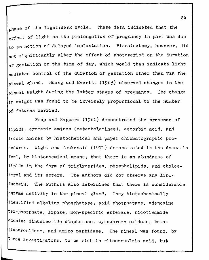
Huang, c. Y., and A. v. Everitt 1965 The effect or pregnancy on pineal weight in the rat. J. Endocrinology,Jg,1 261-262.
Jordan, H. E. 1921 A note on the cytology of the pineal body of the sheep. Anat. Rec., ~1 275-285.
Kamber!, I. A., R. s. ~1cal and J. c. Porter 1971 Effect of melatonin and serotonin on the release of FSH and prolact1n. Endocrinology, .§..§.. (6)1 1288-1293.
Kappers, J. A. 1960 The development, topographical relations and innervation of the epiphys1s cerebr1 in the albino rat. z. Zellforsch., j£1 163-215.
156
Kappers, J. A. 1962 Melatonin, a pineal compound. Preliminary investigations on its function in the rat. Gen. Comp. Endocrinology, 2r 610-611.
Kappers, J. A., and J. P. Shade 1965 Structure and function of the epiphysis cerebri. Progr. Brain Res., lQr 87-153.
Kitay, J. E., and M. D. Altschule 1954 The Pineal Gland. Harvard University Press, Cambridge.
Klein, D. c., and J, Rowe 1970 Pineal glard in organ culture. 1. Inhibition by harmine of serotonin- 4-c production. Molec. Pharmacol., 6 (2)1 164-171.
Klein, D. C,, and Joan L. Weller 1970 Indole metabolism in the pineal glands A circadian rhythm in N-acetyltransferase. Science, 169; 1093-1095.
Le Gros Clark, w. E. 1939-1940 The nervous and vascular relations of the pineal gland. J. Anat., 1!±1 471-492.
Lerner, A. B,, J. D. Case, T. Takabash1, T. H. Lee and w. Mori 1958 Isolation of melatonin, the pineal gland factor that lightens melanocytes. J. Amer. Chem. Soc., 801 2587.
Lerner, A. B., J, D. Case and R. v. Heinzeilman 1959 Structure of melatonin. J. Amer. Chem. Soc., §1.1 6084-6085.
Lovenberg, w., E. Jequier and A. Sjoerdsma 1967 Tryptophan hydroxylationr Measurement in pineal gland, brain stem, and carcinoid tumor. Science, !i2• 217-219.
Machado, c. R. s. 1967 Histochemical study of catecholamines and 5-hydroxytryptamine in the developing pineal body. Anat. Rec., 1.22• 282.
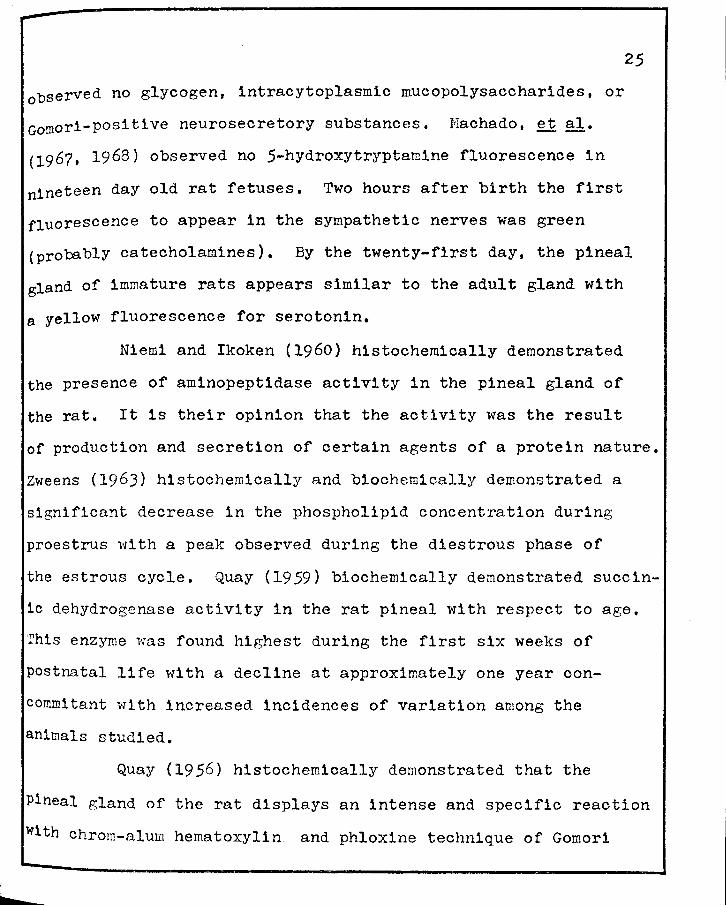
Machado, c. R. s., L. E. Wragg and A. B. M. Machado 1968 A histochemical study of sympathetic innervation of 5-hydroxytryptamine in the developing pineal body of the rat. Brain Res.,~· 310-318.
Manocha, s. L., and G. H. Bourne 1968 Histochemical mapping of lactate dehydrogenase and monoamine oxidase in the medulla oblongata and cerebellum of the squirrel monkey (Sa1mir1 sciureus). J. Neurochem., 1.2• lOJJ-1040.
Mc Cord, c. P., and F. P. Allen 1917 Evidence associating pineal gland function with alterations 1n pigmentation. J. Exper. Zool., ~· 207-224.
Mc Issac, w. M., G. Farrel, R. G. Taborsky and A. N. Taylor 1965 Indole compounds• Isolation from pineal tissue. Science, 1481 102-lOJ.
Mc Kay, D. G., A. T. Hert1g, w. A. Bardawil and J. T. Velardo 1956a Histochemical observations on the endometrium. I. Normal endometrium. Obstet. & Gynec. ~ (l)a 22-39.
157
Mc Kay, D. G., A. T. Hertig, w. A. Bardaw11 and J. T. Velardo 1956b Histochemical obsertrations on the endometrium. II. Abnormal endometrium. Obstet. & Gynec. ~ (2)a 140-156.
Milofsky, A. 1957 The fine structure of the pineal in the ratf with special reference to parenchyma. Anat. Rec., 1271 4JS-4J6.
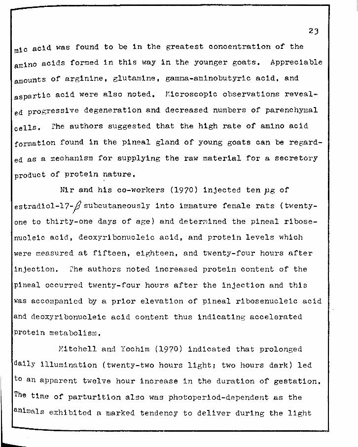
Mitchell, J. A., and J. M. Yochim 1970 Influence of environmental lighting on duration of pregnancy in the rat. Endocrinology, §21 472-480.
Moore, R. Y., A.. Heller, R. J. Wurtman and J. Axelrod 1967 Visual pathway mediating pineal response to environmental lighting. Science, ~· 220-223.
Moszkowska, A., and I. Ebels 1968 A study of the antigonadotrophic action of synthetic arginine vasotoc1n. Exper1ent1a, ~· 610-611.
Motta, M., F. Fraschini and L. Martini 1967 Endocrine effects of pineal gland and melatonin. Proc. Soc. Exp. Biol. Mede, 1261 4Jl-4J5.
Neff, N. H., R. E. Barrett and E. Costa 1969 Kinetic and fluorescent histochemical analysis of the serotonin compartments in the rat pineal gland. Europ. Je Pharmaeol, j1 J48-J56.
Niemi, M., and M. Ikoken 1960 Histochemical evidence of aminopept1dase activity in the rat pineal gland. Nature, 1851 928.
N1r, I. and N. Kaiser, N. Hirschmann and F. G. Sulman 1970 The effect of 17-,,6'-estradiol on pineal metabolism. Life Sciences, 2• 851-858.
158
o'Steen, w. K. 1970 Retinal and optic nerve serotonin and retinal degeneration as influenced by photoper1od. Exper. Neurol., ,g,za 194-205.
owman, c. 1960-1961 Secretory activity of the fetal pineal gland of the rat. Acta Morphologioa Neerlando-Soand1navica, J• 367-394.
Qwman, c. 1965 Localization of neuronal and parenchymal monoamines under normal and experimental conditions in the mammalian pineal gland. Prog. Brain Res., .!Q.1 423-45J.
Pavel, s. 1965 Evidence of lysine vasotocin in the pig pineal. Endocrinology, 1.1• 812-817.
Pellegrino de Iraldi, A., and L. M. Zieher 1966 Noradrenaline and dopamine content of normal decentralized and denervated pineal glands of rats. Life Sci., 2• 589-593.
Prop, N., and A. J. Kappers 1961 Demonstration of some compounds present in the pineal organs of the albino rat by histochemical methods and paper chromatography. Acta Anat., !±21 90-109.
Quay, w. B. 1956 The demonstration of a secretory material and cycle in the parenchymnl cells of the mammalian pineal organ. Exper. Cell Res., !Q1 541-544.
Quay, w. B. 1957 Cytochemistry of pineal lipids in rat and man. J. Histohem. Cytochem., ja 145-153.
Quay, w. B. 1958 Pineal blood content and its experimental modification. Amer. J. Phys1ol. 12.,2• 391-395.
Quay, w. B. 1959 Experimental modifications and changes with age of pineal succinic dehydrogenase activity. Amer. J. Physiol. 1961 951-955.
Quay, w. B. 1961 Reduction of mammalian weight and lipid during continuous light. Gen. Comp. Endoorinol., l• 211-217.
Quay, w. B. 196Ja Circadian rhythm in rat pineal serotonin and its modification by estrous cycle and photoperiod. Gen. Comp. Endocrinol., Js 473-479.
Quay, w. B. 1963b Cytologic and metabolic parameters of pineal inhibition by continuous light 1n the rat (Rattus norveg1cus} z. Zellforsch., .Q.Qs 479-490.
l~
Quay, w. B. 196.5a Histological structure and cytology of the pineal organ in birds and mammals. Progr. Brain Res., !.Q1 49-86.
Quay, w. B. 1965b Retinal and pineal hydroxyindole-0-methyltransferase activity in vertebrates. Life Sol., ~· 98J-991.
Quay, w. B. 1969 Evidence for a pineal contribution in the regulation of vertebrate reproductive systems. Gen. Comp. Endocrinology Suppl., £• 101-110.
Quay, w. B. 1970 Endocrine effects of the mammalian pineal. Amer. Zoologist, !Q.1 237-246.
Quay, w. B., and A. Halevy 1962 Experimental mod1f1cat1ons of the rat pineal content of serotonin and related amines. Phys1ol. Zool., 12• 1-7.
Ralph, c. L., and H. J. Lynch 1970 A quantitative melatonin b1oassay. Gen. Comp. Endocrinology, !ja 334-338.
Reiter, R. J. 1968 Morphological studies on the reproductive organs of blinded male hamsters and the effects of pinealectomy or superior cervical ganglionectomy. Anat. Rec., 1601 13-19.
Reiter, R. J., ands. Sorrentino Jr. 1970 Reproductive effects of the mammalian pineal. Amer. Zoologist, !.Q.• 247-258.
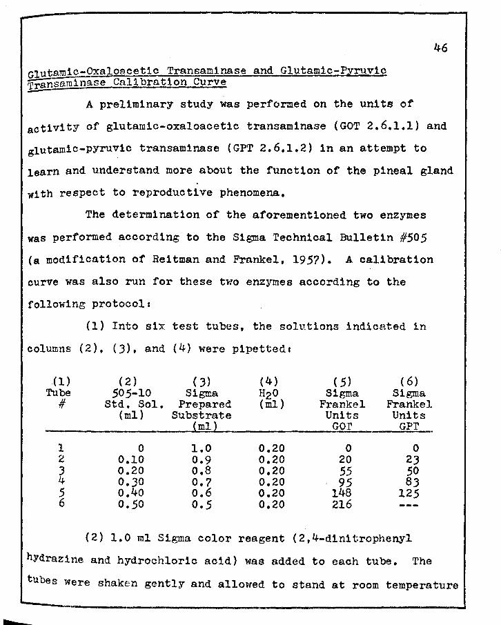
Reitman, s., ands. Frankel 1957 Colorimetric method for the determination of serum transaminase activity. Amer. J. Cl1n. Pathol., _g§,a 56-63.
Relkin, R. 1966 The pineal gland. New Engl. J. Med., 2741 944-950.
Rosa, c. G., and J. T. Velardo 1953 Histochemical localization of succinic dehydrogenase and of lactic dehydrogenase-DPNH ox1dase in the vagina of the rat during the estrous cycle. Anat. Rec., 117 (3)1 532-533 (Abstract).
Rosa, c. G., and J. T. Velardo 1954 Histochemical demonstration of succ1nic dehydrogenase activity in tissue sections by a modified technique. J. Histochem. Cytochem., 21 110-114.
l~
Rosa, c. G., and J. T. Velardo 1954a The influence of estrad1ol-17:,.& upon the lactic dehydrogenase-DPNH oxidase system (LDH) in the va~ina of the castrated rat. Anat. Rec., 1201 771 (Abstract).
Rosa, c. G., and J. T. Velardo 1954b Localization of succin1c dehydrogenase in the uterus of the rat during the estrous cycle. Anat. Rec., 1201 808-809 (Abstract).
Rosa, c. G., and J. T. Velardo 1955 The influence of estradiol upon succ1nic dehydrogenase in the vagina of the castrated rat. Anat. Rec., 1211 415 (Abstract).
Rosa, c. G., and J. T. Velardo 1958 Localization of oxidative enzyme systems in the uterus.and vagina of the rat. Nature, 181 (4605)1 J48-J49.
Rosa, c. G., and J. T. Velardo 1959 Histochemical observations of oxidative enzyme systems in the uterus and vagina of the rat. Ann. N.Y. Acad. Soi., 22• 491-503.
Roth, w. D., R. J. Wurtman and M. D. Altschule 1962 Morphologic changes in the pineal parenchymal cells of rats exposed to continuous light and darkness. Endocrinology, 1!• 88-92.
Shelton, Emma and Mary E. Rice 1957 Comparison of reduction of two tetrazolium salts with succinoxidase activity of tissue homogenates. J. Natl. Cancer Inst., 18: 117-125.
Sommers, s. c. 1958 The pituitary and hypothalamus. Ins The Endocrinology of Reproduction. J. T. Velardo, ed. Oxford University Press, New York, Oxford, Chapter Vs pp. 59-97.
Sze, P. Y., and R. A. Lovell 1970 Reduction of level of Lglutamic acid decarboxylase by (-am1nobutyr1c acid 1n mouse brain. J. Neurochem., lZ• 657-664.
Trentini, G. P., and c. B. Silva 1965 Some histological and histochemical changes in the pineal gland of the rat after removal of the superior cervical ganglia. B1och1m1ca e B1ologia Sperimentale, 41 J-8.
Van de Veerdonk, F. c. G. 1965 Separation method for melatonin in pineal extracts. Nature, 2081 1J24-1J25.
161
Velardo, J. T. 1951 Effects of growth inhibitors on response of rat's uterus to estrogen. Proc. Soc, Exper. Biol, & Med., 1§. (3)1 872-874,
Velardo, J. T. 1954 Inhibitory action of progesterone and am1nopterin on estradiol-1nduced alkaline phosphatase activity 1n the reproductive tract of the ovariectomized rat. Anat. Rec., 120 (3)1 770 (Abstract).
Velardo, J. T. 1958 Dynamics of endocrinology, Ina The Endocrinology of Reproduction. J. T. Velardo, ed. Oxford University Press, New York, Oxford, Chapterls pp-J-7.
Velardo, J, T. 1971 Personal communication.
Velardo, J, T,, A. B. Dawson, A. G, Olsen and F. L. Hisaw 1953 Sequence of histological changes in the uterus and vagina of the rat during prolongation of pseudopregnancy associated with the presence of deciduomata, Am. J. Anat,, 2.J1 273-305.
Velardo, J, T., and c. G. Rosa 1963 Enzymes in the female genital system. VII/3 of Handbuch der Histochemie, Walther Graumann and Kalhe1ng Neuwmann, eds. Stuttgart, Germany, Gustan Fischer Verlag, pp 1-190.
Wislock1,G, B., and E. w. Dempsey 1948 The chemical histology and cytology of the pineal and neurohypophysis. Endocrinology, !!:ga 56-72.
Wislocki, G, B., L. P. Weiss, M. H, Burgos and R. A. Ellis 1957 The cytology, histochemistry and electron microscopy of the granular cells of the metrial gland of the gravid rat, J. Anat., .21• 130-140.
Wolfe, D. E, 1965 The epiphyseal cells an electron microscopic study of its intercellular relationships and intracellular morphology in the pineal body of the albino rat. Prog. Brain Res., !Qi 332-386.
Wolfe, D. E., L. T. Potter, K. C, Richardson and J, Axelrod 1962 Localizing tr1t1ated norepinephrine in sympathetic axons by electron microscopic autoradiography. Science, 1381 440-442.
Wright, P. A. L,, and G. M. Mackenzie 1971 The histochemistry of the pineal gland of the domestic fowl, J, Anat,, !Q§,1 261-273.
162
Wurtman, R. J., and Axelrod 1965 The pineal gland. Sci. Amer., 21J {l)s 50-60.
Wurtman, R. J., J. Axelrod and E. w. Chu 196J Melatonin, a pineal substances effect on the rat ovary. Science, 1411 277-278.
Wurtman, R. J., J. Axelrod, E. w. Chu, A. Heller and R. Y. Moore 1967 Medial forebrain bundle les1onsa blockage of effects of light on rat gonads and pineal. Endocrinology, 811 ~9-~4. -
Wurtman, R. J., J. Axelrod and J. E. F1soher 1964 Melatonin synthesis in the pineal glanda effect of light mediated by the sympathetic nervous system. Science, 1421 1J28-1JJO.
Wurtman, R. J., J. Axelrod and L. Phillips 196J Melatonin synthesis in the pineal glands control by light. Science, 1421 1071-1073.
Wurtman, R. J., J. Axelrod, s. H. Snyder and E. w. Chu 1965 Changes of the enzymatic synthesis of melatonin in the pineal during the estrous cycle. Endocrinology, .z.21 798-800.
Wurtm.an, R. J., F. Larin, J. Axelrod, H. M. Shein and K. Rascasca 1968 rarmation of melatonin and 5-hydroxy1ndole acetic acid from C -tryptophan by rat pineal glands in organ culture. Nature, 2171 953-954.
Zweens, J. 1963 Influence of the estrous cycle and ovariectomy on the phosphol1pid content of the pineal gland. Nature, 121• 1114-1115. '
Zweig, M., and J. Axelrod 1969 Relationship between catecholamines and serotonin in sympathetic nerves of the rat pineal gland. J. Neurobiol., 11 87-97.
Stage Length (mm)
15
17
18
19
APPENDIX A
Somite Age Number (days)
1-J
4-6
7-9
9 1/2-9 3/4
9 J/4-10
10-10 1/2
10-14 10 1/2-10 3/4
Main Commencing Features
Somites have appeared. Neural folds elevating.
Delimited otlc (4th) rhombomere and post-otic sulous.
Neural canal closed from the level of the 2nd to the 6th somite.
Neural folds fused at diencephalic-mesencephalic junction. Otio pit forms.
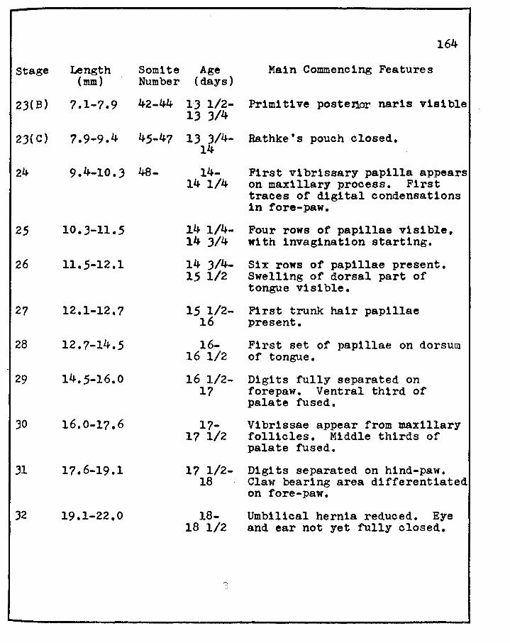
First vibrissary papilla appears on maxillary process. First traces of digital condensations in fore-paw.
Four rows of papillae visible, with invag1nation starting.
Six rows of papillae present. Swelling of dorsal part of tongue visible.
First trunk hair papillae present.
First set of papillae on dorsum of tongue.
Digits fully separated on forepaw. Ventral third of palate fused.
Vibrissae appear from maxillary follicles. Middle thirds of palate fused.
Digits separated on hind-paw. Claw bearing area differentiated on fore-paw.
Umbilical hernia reduced. Eye and ear not yet fully closed.
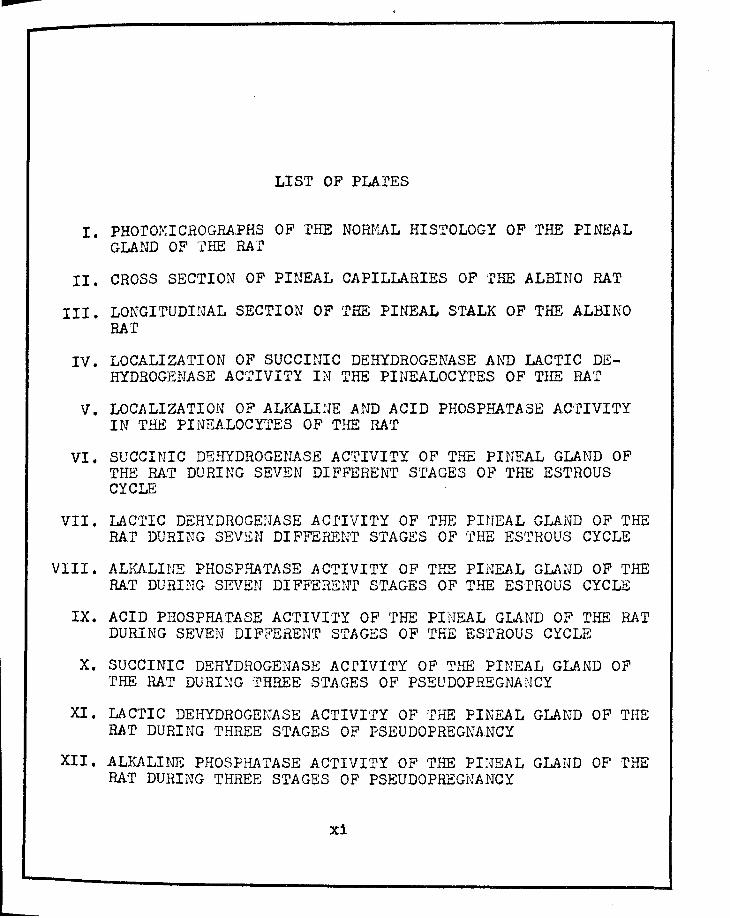
PLATE I
PHOTOMICROGRAPHS OF THE NORMAL HISTOLOGY OF THE PINEAL GLAND OF RAT. X 2.000;
Figure 20 Pineal Gland. Hematoxylin-Eosin. Two types of cells are observed• ' 1) light cell (1) with a round nucleus, prominent nucleolus and a pale staining cytoplasm; and 2) a dark cell (d) with irregularly shaped nucleus, no apparent nucleolus and a darker staining cytoplasm. Note also lipid vacuoles surrounding capillaries (c).
Figure 21 Pineal Gland. Bargmann's chrom-alum hematoxyl1n. Longitudinal section of a pineal capillary (c), containing red blood cells and lined with neurosecretory material of moderate staining intensity.
~\I ]!
d.~
"# .,
I
PLATE I
'lt •• I
Figure 20
• Figure 21
165
i . I
i . ! I
PLATE II
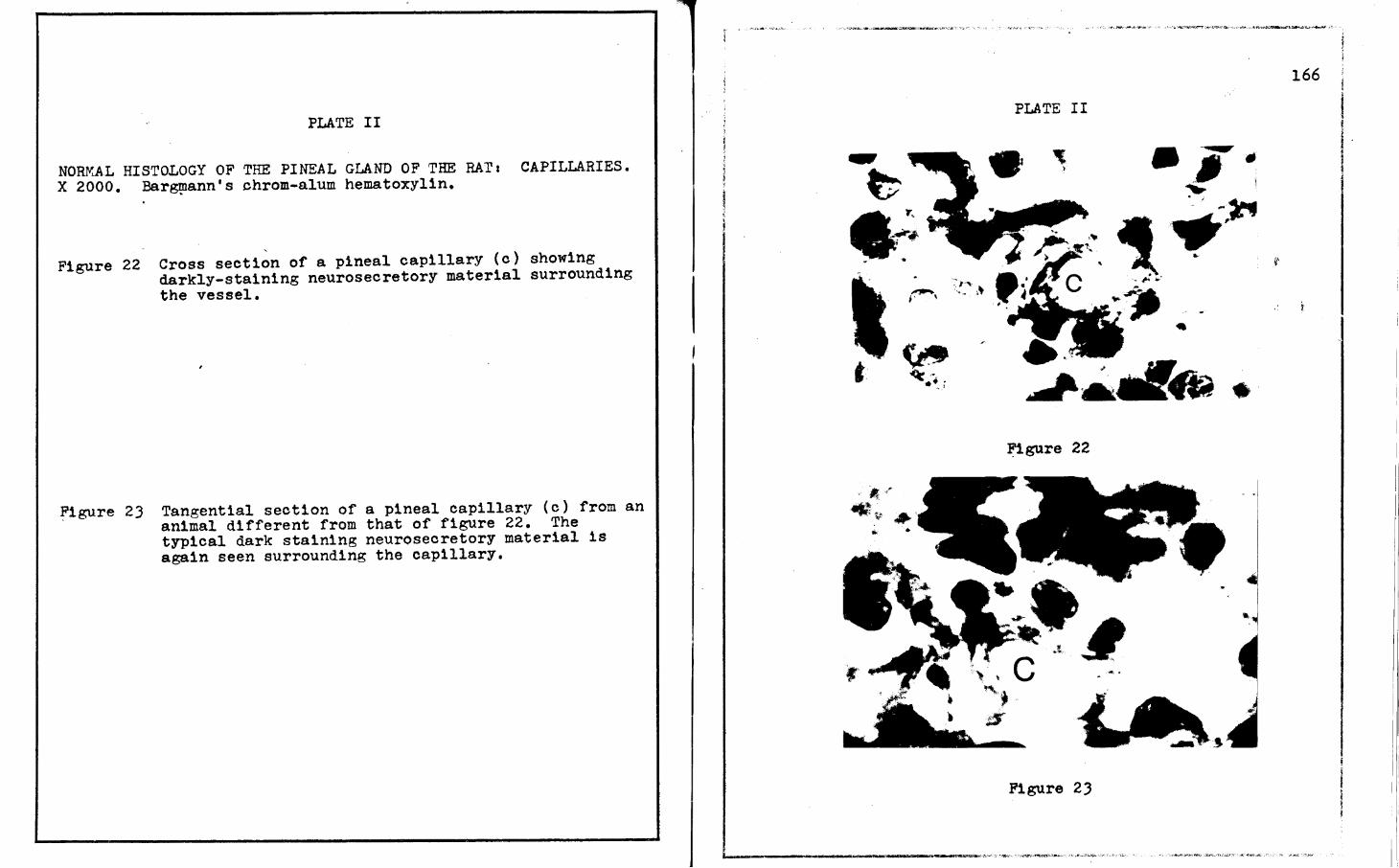
NORf'iAL HISTOLOGY OF THE PINEAL GLAND OF THE RAT 1 CAPILLARIES. X 2000. Bar~ann's chrom-alum hematoxylin.
Figure 22 Cross section of a pineal capillary (c) showing darkly-staining neurosecretory material surrounding the vessel.
~igure 23 Tangential section of a pineal capillary (c) from an animal different from that of figure 22. The typical dark staining neurosecretory material 1s again seen surrounding the capillary.
PLATE II
•
Figure 22
Figure 2J
• I
. ·I I
166 I
I ! I . l
PLATE III
LONGITUDINAL SECTION OF THE PINEAL STALK OF THE RAT. X 2000. Bargmann's chrom-alum hematoxylin • .
rigure 24 Dark staining neurosecretory material is seen here 1n an irregular pattern along this portion of the
Figure 25 '
pineal stalk.
In this section of the pineal stalk the darklystaining neurosecretory material is seen 1n discrete lines.
167
PLATE III
4149
Figure 24
Figure 25
PLATE IV
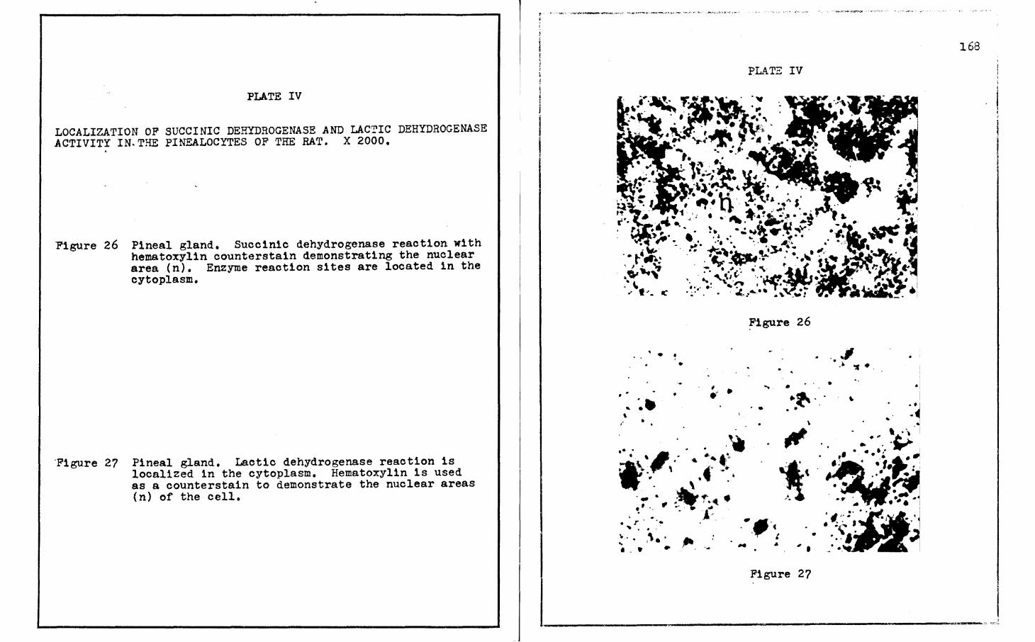
LOCALIZATION OF SUCCINIC DEHYDROGENASE AND LACTIC DEHYDROGENASE .ACTIVITY IN.THE PINEALOCYTES OF THE RAT. X 2000.
Figure 26 Pineal gland. Succinic dehydrogenase reaction with hematoxylin counterstain demonstrating the nuclear area (n). Enzyme reaction sites are located in the cytoplasm.
'Figure 27 Pineal gland. Lactic dehydrogenase reaction is localized 1n the cytoplasm. Hematoxylin is used as a countersta1n to demonstrate the nuclear areas (n) of the cell.
LOCALIZATION OF ALKALINE AND ACID PHOSPHATASE ACTIVITY IN THE PINEALOCYTES OF THE RAT. X 2000.
,Figure 28 Pineal gland. Alkaline phosphatase reaction is localized in the cytoplasm of the p1nealocytes. Hematoxylin is used as a counterstain to demonstrate the nuclear areas (n).
Figure 29 Pineal gland. Acid phosphatase activity within the pinealocytes is localized in the cytoplasm. Hematoxyl1n is used as a counterstain to demonstrate nuclear areas (n).
SUCCINIC DEHYDROGENASE ACTIVITY OF THE PINEAL GLAND OF THE RAT DURING SEVEN.DIFFERENT STAGES OF THE ESTROUS CYCLE. X 2000.
Figure JO Proestrus. This gland shows a distinctive array of diformazan granules with an inappreciable intensity of monoformazan material.
Figure Jl Early Estrus. A small scatter of diformazan granules with an almost maximal intensity of monoformazan material is evident.
-Figure 32 Late Estrus. Maximal diformazan deposition with
considerable intensities of monoformazan material readily observable 1n the background.
Figure 33 Metestrus. Small scatter of diformazan granules with only trace intensities of monoformazan material is discernible.
Figure J4 Early Diestrus. Increased intensity of both mono- and diformazan material is seen when comparing with metestrus.
Figure 35 Late D1estrus. Intense d1formazan granules in a scattered array are observed. Increased intensity of monoformazan material is visible 1n the perinuclear areas of the pinealocytes.
Figure ,36
Figure 37
Preproestrus. There is a marked reduction 1n the intensity of the diformazan granules. An appreciable intensity of only monoformazan material is evident.
Control Section. Control sections are uniformly negative for each of the seven stages of the estrous cycle, also during pseudopregnanoy, prolonged pseudopregnancy and pregnancy.
Figure Jl
Figure 33
. ' ., .
•
,.
•
' ~ ,,
. , ..
.._. • J
·v • ' . . ' ' • . . .r
PLATE VI
"!
"I
Figure 35
Figure J6
Figure . 37
170
PLATE VII
LACTIC DEHYDROGENASE ACTIVITY OF THE PINEAL GLAND OF THE RAT DURING SEVEN-DIFFERENT STAGES OF THE ESTROUS CYCLE. X 2000.
Figure .38
Figure 39
Proestrus. A maximal intensity of both mono- and diformazan'material is readily discernible.
Early Estrus. A marked decrease 1s observed in the intensity of both the mono- and diformazan reaction sites.
Figure 40 Late Estrus. A near maximal intensity is seen for both the mono- and diformazan material.
Figure 41 Metestrus. Slight reduction in intensity of the diformazan granules with a marked reduction in monoformazan material is demonstrated.
Figure 42 Early Diestrus. Further reduction in the overall reaction with scattered diformazan granules of moderated intensity and a slight elevation in the monoformazan distribution and intensity is shown.
Figure 4.3 Late Diestrus. Marked reduction in the intensities of both mono- and diformazan material is demonstrated.
Figure 44
Figure 45
Preproestrus. This reaction shows an overall increase in the number of diformazan granules with a markedly increased intensity of monoformazan material.
Control Sections. Control sections were uniformly negative for each of the seven stages of the estrous cycle, also during pseudopregnancy, prolonged pseudopregnanoy and pregnancy.
Figure 38
.Figure 39
Figure 40
Figure 41
.. •
~
•
• •
. • >
, l . ·' .
... " • • •
! • . . . • ._ . ,.
• .. • .. . ' ..
PL>\T:S VII
• • I
.. '· •
,..
•
•
Figure 42
Figure 43
Figure 44
. Figure 45
.
,. • ,, ..- ...
' ... ' • • . .. • ... .. ..
'
'iJ ~. ~#
,.
~ • c.,. •
• . . ' . .... •
171
• • • ~
._ ·~ ~ , I . .. " . • ,. JI/' ,.
• ·y
. .
.. ' .. -~· ~-· t ·6,, • •
' :a ...
.. • •
PLATE VIII
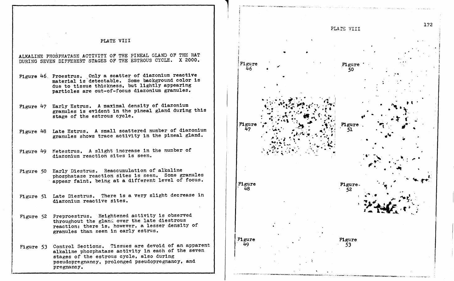
ALKALINE PHOSPHATASE ACTIVITY OF THE PINEAL GLAND OF THE RAT DURING SEVEN DIFFERENT STAGES OF THE ESTROUS CYCLE. X 2000.
Figure 46,
Figure 47
Figure 48
Figure 49
Figure 50
Figure 51
Figure 52
Proestrus. Only a scatter of diazonium reactive material is'detectable. Some background color is due to tissue thickness, but lightly appearing particles are out-of-focus diazonium granules.
Early Estrus. A maximal density of diazonium granules is evident in the pineal gland during this stage of the estrous cycle.
Late Estrus. A small scattered number of diazonium granules shows trace activity 1n the pineal gland.
Metestrus. A slight increase in the number of d1azon1um reaction sites is seen.
Early Diestrus. Reaccumulation of alkaline phosphatase reaction sites 1s seen. Some granules appear faint, being at a different level of focus.
Late Diestrus. There is a very slight decrease in diazonium reactive sites.
Preproestrus. Heightened activity is observed throughout the glanQ over the late d1estrous reaction; there is, however, a lesser density of granules than seen in early estrus.
Figure 53 Control Sections. Tissues are devoid of an apparent alkaline phosphatase activity in each of the seven stages of the estrous cycle, also during pseudopregnancy, prolonged pseudopregnancy, and pregnancy.
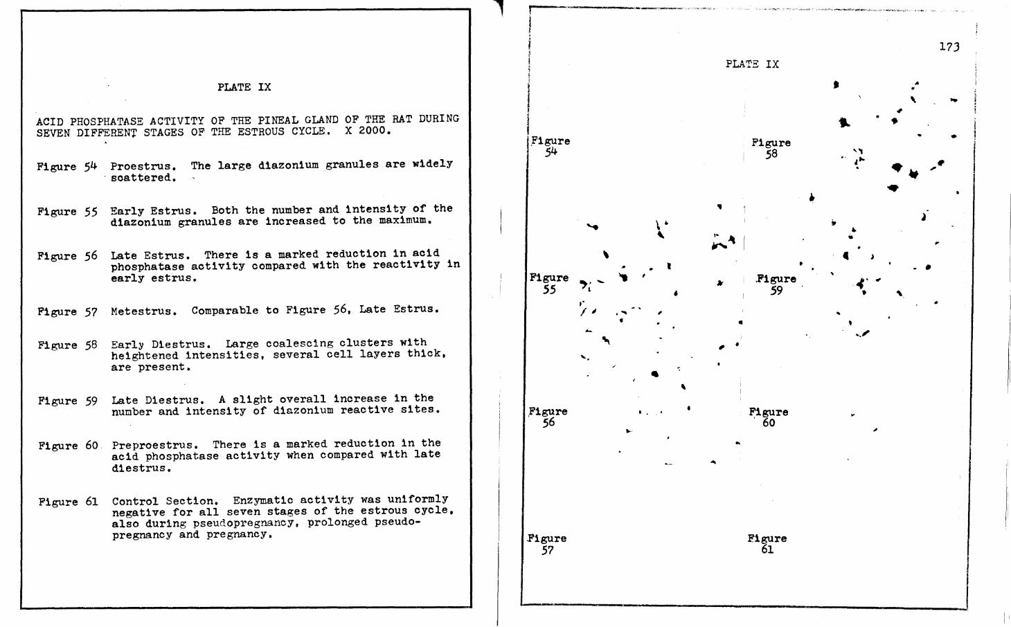
ACID PHOSPHATASE ACTIVITY OF THE PINEAL GLAND OF THE RAT DURING SEVEN DIFFERENT STAGES OF THE ESTROUS CYCLE. X 2000.
Figure 54 Proestrus. The large d1azon1um granules are widely , scattered.
Figure 55 Early Estrus. Both the number and intensity of the diazonium granules are increased to the maximum.
Figure 56 Late Estrus. There is a marked reduction in acid phosphatase activity compared with the reactivity in early estrus.
Figure 57 Metestrus. Comparable to Figure 56, Late Estrus.
Figure 58 Early Diestrus. Large coalescing clusters with heightened intensities, several cell layers thick, are present.
Figure 59 Late Diestrus. A slight overall increase in the number and intensity of diazonium reactive sites.
Figure 60 Preproestrus. There is a marked reduction in the acid phosphatase activity when compared with late diestrus.
Figure 61 Control Section. Enzymatic activity was uniformly negative for all seven stages of the estrous cycle, also during pseudopregnancy, prolonged pseudopregnancy and pregnancy.
... f -- ------~·--~----·
l
I I I.Figure ~ 54
Figure 55
Figure ' 56
.Figure 57
.....
\
.,. -' ,.
I;
..:..
"'\ ... .
\ . ' • , ,
' . "' , •
.I
' . .. . ' • ...
PLATE IX
!• "
"' •
.. I , •
..
Figure 58
. Figure 59
Figure ' 60
Eigure 61
..
•
• .. ,, . ' , .. .
" • • • c
.. 4' •
.. ' ... i'
...
17J
,.
' ,.
., • • i - •
j ·- • , .. ~
,,
l . • ., '\
•
PLATE X
SUCCINIC DESYDROGENASE ACTIVITY OF THE PINEAL GLAND OF THE RAT DURING THREE STAGES OF PSEUDOPREGNANCY. X 2000.
Figure 62 Day Six. Scatter of diformazan granules is observed <in the perinuclear areas. Intense monoformazan material is readily observable in some areas.
Figure 63 Day Ten. Marked increase in intensity of the monoformazan material with a slight increase in diformazan deposition is demonstrated, giving the maximal overall reaction.
Figure 64 Day Thirteen. Increased intensity of diformazan granules with a slight decrease in intensity of the monoformazan material is shown.
Figure 63
PLl~TE X
Ji'igure 64
174
I I
--------~----------------'
PLATE XI
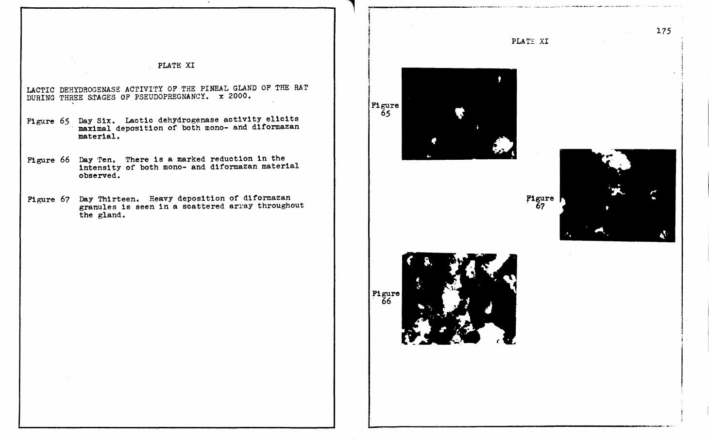
LACTIC DEHYDROGENASE ACTIVI·rY OF THE PINEAL GLAND OF THE RAT DURING THREE STAGES OF PSEUDOPREGNANCY. x 2000 • . Figure 65 Day Six. Lactic dehydrogenase activity elicits
maximal depo~1tion of both mono- and diformazan material.
Figure 66 Day ·ren. There is a marked reduction in the intensity of both mono- and diformazan material observed.
Figure 67 Day Thirteen. Heavy deposition of diformazan granules is seen in a scattered array throughout the gland.
l
Figure 65
Figure ' 66
PLAT~ XI
figure 67
175
PLATE XII
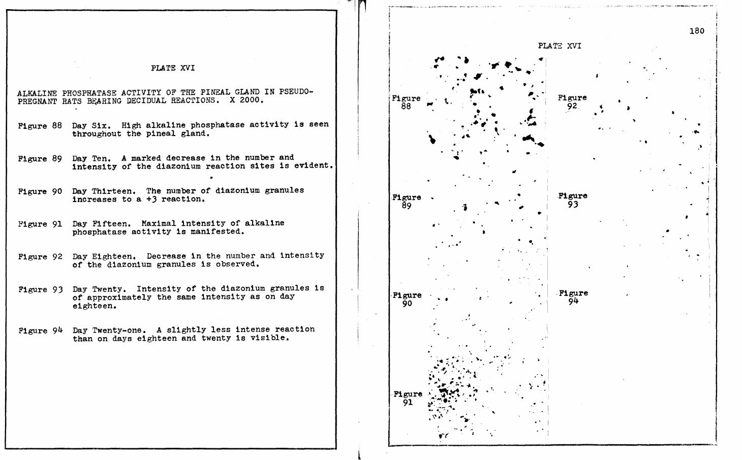
ALKALINE PHOSPHArASE ACTIVI·rY OF THE PINEAL GLAND OF THE RAT DURING TH?EE STAGES OF PSEUDOPREGNANCY. X 2000.
Figure 68 Day Six. Abundance of small diazonium granules several cell layers deep is observed.
Figure 69 Day Ten. This figure shows maximal intensity for an alkaline phosphatase reaction.
Figure 70 Day Thirteen. A slight decrease ls observed in the intensity of the diazon1um granule deposition.
figure 68
.Figure 69
•
' • • I... • ,. .... ~ . ' . '-. ...
• • <I •• ; -.. . -.. . .
• ~ . ~~: ..
• • • • .. • . -., .... , •
• ..
•
· .
.. •
....
•
•
PLATE XII
r1gure 70
176
..
~ . ', . . '. • .. :
•
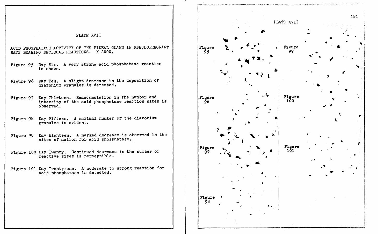
PLATE XIII
ACID PHOSPHATASE ACTIVI'rY OF THE PINEAL GLAND OF THE RAT DURING THREE. STAGES OF PSEUDOPREGNANCY. X 2000.
Figure ?l
Figure 72
Day Six. A high intensity of d1azon1um granules is seen several cell layers thick.
Day Ten. Approximately the same intensity as days six and thirteen. Diazonium granules are not easily distinguishable as they are at different depths of this field. ·
Figure ?J Day Thirteen. The intensity is comparable to those of days six and ten. Acid phosphatase reactive sites are several cell layers thick. All the stages of acid phosphatase activity observed during pseudopregnancy were semi-quantitatively assigned a value of +J.
I I f Figure
?l
Figure 72
• . ' • • .J
-- ••
"" •
..
'
.,
' . ,
' ' ..
. '
PLATE XIII
e I
•
,Figure 73
• ., '
177
. '
' <.
• ..
PLATE XIV
SUCCINIC DEHYDROGENASE ACTIVITY OF THE PINEAL GLAND IN PSEUDOPRE~NAN'l' RATS BEARING DECIDUAL REAC'I'IONS. X 2000.
Figure 74. Day Six. Moderate diformazan granule intensity with trace amounts of monoformazan material is seen throughout the gland.
Figure 75
Figure 76
Figure 77
Figure 78
Figure 79
Figure 80
Day ~en. A near maximally intense reaction showing mono- and diformazan material is readily observable in t~e perinuclear areas.
Day Thirteen. Decreasing intensities of succinic dehydrogenase activity sites are demonstrated.
Day Fifteen. Minimal succ1n1c dehydrogenase activity with a scatter of diformazan granules and trace monoformazan material.
Day Eighteen. Maximal intensities of both mono- and diformazan reaction sites are readily observable.
Day Twenty. A marked decrease in the intensities of both mono- and diformazan material is discernible.
Day Twenty-one. Continual decrease in the number of diformazan granules and negligible monoformazan material is visible.
LACTIC DEHYDROGENASE ACTIVITY OF THE PINEAL GLAND IN PSEUDOPREGNANr B.ATS 'BEARING DECIDUAL REACTIONS. X 2000.
Figure 81 Day Six. High diformazan granule intensity and moderate monoformazan intensity is observed.
Figure 82 Day Ten. Increased intensities of both mono- and diformazan material is evident.
Figure 8J Day Thirteen. Maximal lactic dehydrogenase intensities are observed in the pinealocytes.
Figure 84 Day Fifteen. A precipitous decrease in both monoand diformazan reaction sites is noticeable.
Figure 85 Day Eighteen. A marked increase is seen in the intensity of diformazan granules with a moderate intensity of monoformazan material.
Figure 86
Figure 87
Day Twenty. A decrease in the intensity of diformazan granules with trace monoformazan material is revealed.
Day Twenty-one. A continual decrease of diformazan granule intensities and moderate monoformazan material are detectable. When compared to day twenty, it can be observed that there is a slight increase in the concentration of the monoformazan material on day twenty-one, but a decrease in the number of diformazan granules. On day twenty, the diformazan granules are small and evenly distributed throughout all of the pineal gland, while on day twenty-one, the diformazan granules are large with scattered coalescing clusters.
SUCCINIC DEHYDROGENASE ACTIVITY OF THE PINEAL GLAND OF PREGNANT RATS. X 2000.
Figure 102 Day Six. A less than moderate intensity of both mono- and diformazan material is readily seen 1n the perinuclear areas.
Figure 103 Day Eight. A near maximal intensity of both monoand diformazan material is apparent.
Figure 104 Day Ten. ·rhere is a marked reduction in diformazan granules with a moderate intensity of monoformazan.
Figure 105 Day Thirteen. Reaccumulation of diformazan granules 1s observed with a slightly less than moderate intensity for monoformazan.
Figure 106 Day Fifteen. Moderate intensity of diformazan granules is seen in the perinuclear areas. Monoformazan material is intense in some areas.
Figure 107 Day Eighteen. A maximal reaction of small diformazan granules is demonstrated. Monoformazan background intensity is high.
Figure 108 Day Twenty. There is an intense monoformazan deposition throughout the pineal gland. Diformazan granules are slightly decreased in intensity.
Figure 109 Day Twenty-one. Marked reduction in monoformazan depostion is readily seen. Diformazan granules are scattered throughout the pineal gland.
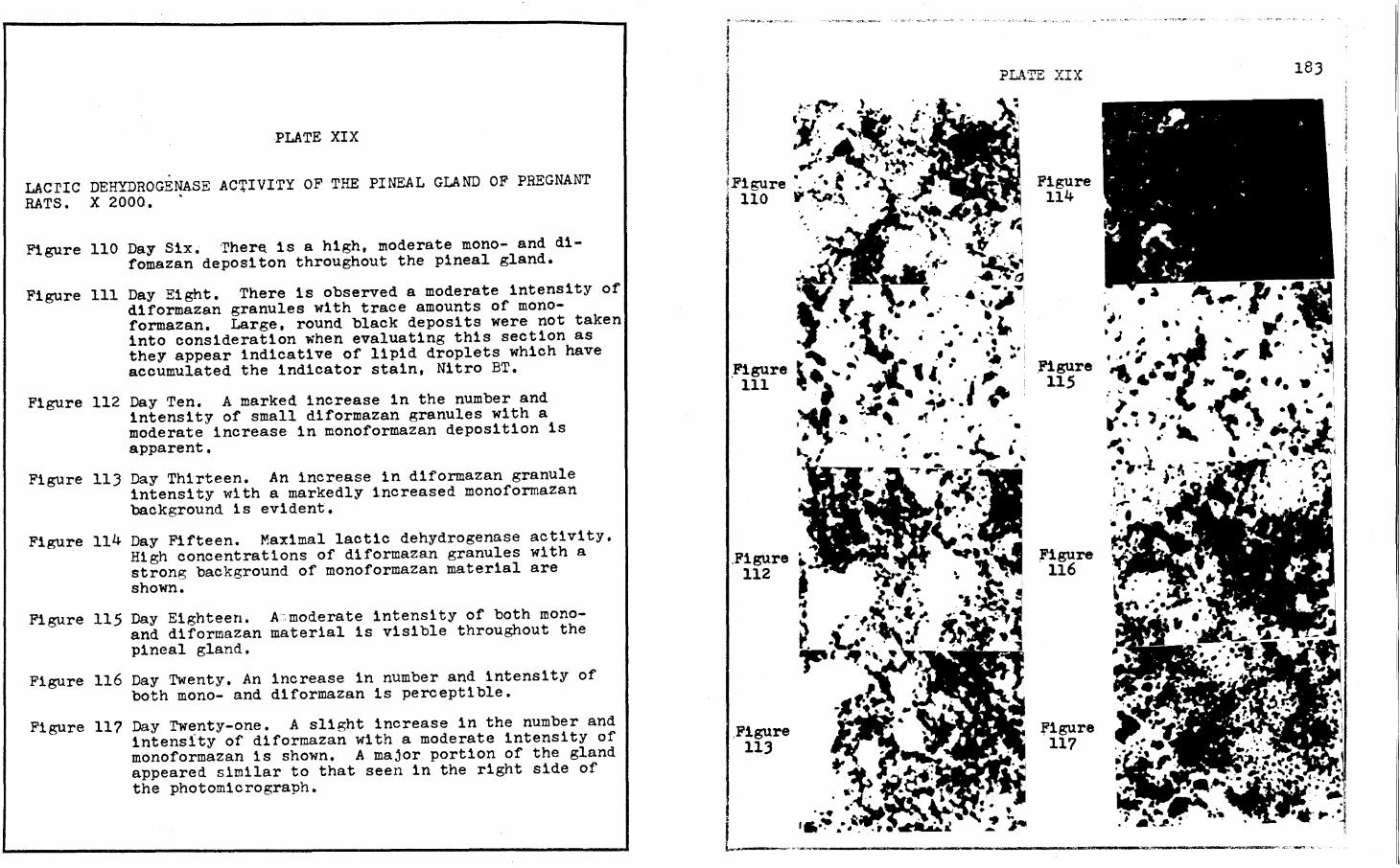
LACrIC DEHYDROGENASE AC'.UVITY OF THE PINEAL GLAND OF PREGNANT RATS. X 2000. .
Figure 110 Day Six• ·rher~ is a high, moderate mono- and difomazan depositon throughout the pineal gland.
Figure 111 Day Eight. There is observed a moderate intensity of diformazan granules with trace amounts of monoformazan. Large, round black deposits were not taken into consideration when evaluating this section as they appear indicative of lipid droplets which have accumulated the indicator stain, Nitro BT.
Figure 112 Day Ten. A marked increase in the number and intensity of small diformazan granules with a moderate increase in monoformazan deposition is apparent.
Figure 113 Day Thirteen. An increase in diformazan granule intensity with a markedly increased monoformazan background is evident.
Figure 114 Day Fifteen. Maximal lactic dehydrogenase activity. High concentrations of diformazan granules with a strong background of monoformazan material are shown.
Figure 115 Day Eighteen. A:. moderate intens1 ty of both m.onoand d1formazan material is visible throughout the pineal gland.
Figure 116 Day Twenty. An increase in number and intensity of both mono- and d1formazan 1s perceptible.
Figure 11? Day Twenty-one. A slight increase in the number and intensity of diformazan with a moderate intensity of monoformazan is shown. A major portion of the gland appeared similar to that seen in the right side of the photomicrograph.
I •.- ,_ I ..
J • fl..•r• ~•' J ' .. . ..
i 'j'. . .c' . ·. ' i. ~ I ·' Ai1. •.. · l ~-, ... ~l~". ~Figure ". · ~ • t "' ·•' I 110 P~:~. i '
ACID PHOSPHATASE ACTIVITY OF THE PINEAL GLAND OF PREGNANT RATS. X 2000,.
Figure 126 Day Six.. A scatter of d1azon1um granules is observed. Background coloration is the result of tissue thickness and is not indicative of sites of acid phosphatase action.
Figure 127 Day Eight. Large, intense but widely spaced diazonium gran~les are evident.
Figure 128 Day Ten. Acid phosphatase reaction sites are observed more than one cell layer in thickness.
Figure 129 Day Thirteen. Increase in the number and intensity of the d1azonium granules ls apparent.
Figure 130 Day Fifteen. Near maximal intensity of diazon1um deposition is visible,
Figure 131 Day Eighteen. Maximal, intense d1azon1um granule deposition is observed several cell layers in thickness.
Figure 132 Day Twenty. Strong acid phosphatase reaction ls seen. Diazonium granules are intense but scattered.
Figure 133 Day Twenty-one. Intense, but scattered, deposition of diazon1um granules is demonstrated.