

Loyola University Chicago Loyola University Chicago
Loyola eCommons Loyola eCommons
Dissertations Theses and Dissertations
1972
Histochemical and Gravimetric Analyses of the Pineal Gland of Histochemical and Gravimetric Analyses of the Pineal Gland of
the Albino Rat During the Estrous Cycle, Pregnancy and the Albino Rat During the Estrous Cycle, Pregnancy and
Pseudopregnancy Pseudopregnancy
Anthony V. Fasano Loyola University Chicago
Follow this and additional works at: https://ecommons.luc.edu/luc_diss
Part of the Medicine and Health Sciences Commons
Recommended Citation Recommended Citation Fasano, Anthony V., "Histochemical and Gravimetric Analyses of the Pineal Gland of the Albino Rat During the Estrous Cycle, Pregnancy and Pseudopregnancy" (1972). Dissertations. 1135. https://ecommons.luc.edu/luc_diss/1135
This Dissertation is brought to you for free and open access by the Theses and Dissertations at Loyola eCommons. It has been accepted for inclusion in Dissertations by an authorized administrator of Loyola eCommons. For more information, please contact [email protected].
This work is licensed under a Creative Commons Attribution-Noncommercial-No Derivative Works 3.0 License. Copyright © 1972 Anthony V. Fasano
HISTOCHE!•:ICAL AND GRAVIHE'I'RIC .AN.ALYSES OF THE PIImAL
GLAND OF THE ALBINO RAT DURING 'rHE ESTnOUS
CYCLE, PREGNANCY AND PSEUDOPREGNA~CY
by
Anthony V. Fasano
A Dissertation Submitted to the Faculty of the Graduate School
of Loyola University of Chicago in Partial Fulfillment of
the Requirements for the Degree of
Doctor of Philosophy
February
1972
ti~~r..Jtf
LOYOLA UNfV'~;::snY
LIFE
Anthony Vincent Fasano was born in Montclair, New Jersey on December 28, 19J6, the son of Nr. and Mrs. Benjamin V. Fasano.
He attended elementary and high school in Newark, New Jersey, and was graduated from Colorado State College, Greeley, Colorado in June, 1964. During the ensuing three years, Mr. Fasano was engaged as an analytical chemist for Fisher Scientific Company, Fair Lawn, ::-.iew Jersey and as a research chemist for the John L. Smith Memorial for Cancer Research in Maywood, New Jersey.
In January, 19 67, .r.:r. Fasano began his graduate study in the Department of Physiology of Fairleigh Dickinson University, Teaneck, New Jersey. He remained there until he entered the Department of Anatomy, Loyola University of Chicago, Stritch School of !•Iedicine, !-1aywood, Illinois, September, 1967. Here he continued his scientific investigations for the doctorate of philosophy degree in anatomy.
From 1968 to 1971, Mr. Fasano has had a National Defense Education Act fellowship. He is a member of the American Association for the Advancement of Science, American Society of Zoologists, and the New York Museum of Natural History.
During the period from 1954 to 1962, Mr. Fasano was an active member of the United States Naval Reserve from which he received an Honorable Discharge in August, 1962. In 1965, he married Linda Jacobsen and is the father of a boy, Steven Michael, now seventeen months old. and a girl, Dana Nicole, one month old.
111
A.CKNOWLEDGEMENTS
No research endeavor is ever the work of a lone
invest~gator; it is the culmination of the talents, guidance
and assistance of a multitude of people. · The author of this
dissertation wishes to thank a few of his fellow workers for
freely rendering their talents. First and foremost, an especial
thanks to may adviser, Professor Joseph T. Velardo for his
painstaking help and guidance; to Mrs. Velardo for her
understanding and always constant encouragement; and to Dr.
Barbara Kasprow for her help, instruction and guidance. A
warm thanks to Dr. Leslie Emmert, Francis Kovarik and Grover
Ericson for their assistance with both the animal work and the
preparatory written work for this dissertation; to my committee
for their ever friendly and patient guidances and to Mrs. Canuti,
Mrs. Schultz, Mrs. Smelte, and Mr. and Mrs. Kovarik for their
pleasant support. Last, but not least, a very grateful thanks
to my wife, Linda, who gave up so much and did so much so that
this investigator could reach his goal.
iv
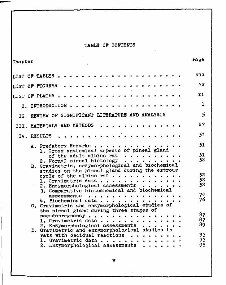
TABLE OF CONTENTS
Chapter Page
LIST OF TABLES • • • • • • • • • • • • • • • • • • • • • vii
LIST OF FIGURES • • • • • • • • • • • • • • • • • • • • 1x
LIST OF PLATES • • • • • • • • • • • • • • • • • • • • •
I. INTRODUCTION ••• • • • • • • • • • • • • • • • • •
II. REVIEW OF SIGNIFICANT LITERATURE AND ANALYSIS
III. MATERIALS AND METHODS • • • • • • • • • • • • • •
IV. RESULTS • • • • • • • • • • • • • • • • • • • • •
xi
1
5
27
51
A. Prefatory Remarks • • • • • • • • • • • • • • • 51 1. Gross anatomical aspects of pineal gland
of the adult albino rat • • • • • • • • • • 51 2. Normal pineal histology • • • • • • • • • • 52
Be Gravimetric, enzymorphological and biochemical studies on the pineal gland during the estrous cycle of the albino rat • • • • • • • • • • • • 52 1. Gravimetric data • • • • • • • • • • • • • • 52 2. Enzymorphological assessments • • • • • • • 52 J. Comparative histochemical and biochemical
assessments • • • • • • • • • • • • • • • • 74 4. Biochemical data • • • • • • • • • • • • • • 76
c. Gravimetric and enzymorphological studies of the pineal gland during three stages of pseudopregnancy • • • • • • • • • • • • • • • • 87 1. Gravimetric data • • • • • • • • • • • • • • 8? 2. Enzymorphological assessments • • • • • • • 89
D. Gravimetric and enzymorphological studies in rats with decidual reactions • • • • • • • • • 93 1. Gravimetric data • • • • • • • • • • • • • • 93 2. Enzymorpholog1cal assessments • • • • • • • 95
v
vi
E. Gravimetric and enzymorphological studies of pregnant rats • • • • • • • • • • • • • • • 105 1. Gravimetric data • • • • • • • • • • • • • 105 2. Enzymorphological assessments • • • 108
v. DISCUSSION • • • • • • • • • • • • • • • • • • 118
VI. SUMMARY AND CONCLUSION • • • • • • • • • • • • • 145
BIBLIOGRAPHY • • • • • • • • • • • • • • • • • • • • • 152
APPENDIX • e e • • • • • • • • • • • • • • • • • • • • 163
PLATES e • e • • • • 0 • • • • • • • • • • • • • • • • 165
ABSTRACT

LIST OF ·rABLES
Table
I. COMPARISO:i OF PINEAL, HYPOPHYSEAL, OVARIAN, ADRENAL, &'{D UTERINE WEIGHTS OF ADULT ALBINO RATS IN KG,% DURING DIFFERENT PHASES OF THE ESTROUS CYCLE • • • • • • • • • • • • • • • •
II. SIGNIFICANCES OF DIFFERENCES OF PINEAL GLAND WEIGHTS DURING SEVEN DIFFERENT STAGES OF ·rHE ESTROUS CYCLE OF THE RAT • • • • • • • • • •
III. SIGNIFICANCES OF DIFFERENCES OF HYPOPHYSEAL WEIGHTS DURING SEVEN DIFFEREHT STAGES OF THE ESTROUS CYCLE OF THE RAT • • • • • • • • • •
• •
• •
• •
IV. SIGNIFICANCES OF DIFFEREHCES OF OVARIA:-J WEIGHTS DURING SEVEN DIFFERENT STAGES OF rHE ESTROUS
Page
57
58
59
CYCLE OF THE RAr • • • • • • • • • • • • • • • • 60
V. SIGrJIFICANCES OF DIFFERENCES OF UTERHrE WEIGHTS DURING SEVEi'l DIFFERENT s·rAGES OF 'rIIB ESrROUS CYCLE OF THE RAT • • • • • • • • • • • • • • • • 61
VI. SIGNIFICANCES OF DIFFERENCES OF ADRENAL WEIGHTS DURING SEVEN DIFFERENT STAGES OF THE ESTROUS CYCLE OF THE RAT • • • • • • • • • • • • • • • • 62
VII. SEMI-QUANTITATIVE HIS·roCIIEMICAL ESTI!'"iATES OF SUCcnuc DEHYDROGENASE ( SDH)' LACTIC DEHYDROGENASE (LDH)' ALKALrm PHOSPHATASE (ALK. P'TASE)' .AHD ACID PHOSPHA1'ASE (ACID P'TASE) OF THE PIHEAL GLAND OF THE RAT DURHJG THE Es·rROUS CYCLE • • • • ?8
v11
r VIII. QUANTITATIVE BIOCHEMICAL ANALYSIS OF SUCCINIC
DEHYDROGENASE ( SDH), LACrIC DEHYDROGENASE ( LDH), ALKALINE PHOSPHA'l'ASE (ALK. P'TASE), AND ACID PHOSPHATASE (ACID P'TASE) OF THE PINEAL GLAND OF THE RAT DURING THE ESTROUS CYCLE • • • • • • •
v111
79
IX. QUANTITATIVE BIOCHEMICAL ANALYSIS OF GLUTAMICOXALOACETIC (GOT) AND GLUTAMIC-PYRUVIC TRANSAMINASE ( GPT) ACTIVITY OF THE PINEAL GLAND OF THE RAT DURING THE ESTROUS CYCLE • • • • • • • 80
X. THE WEIGHTS OF PINE.AL GLANDS OF RATS DURING THREE STAGES OF PSEUDOPREGNANCY • • • • • • • • e
XI. SEMI-QUANTITATIVE HIS·rOCHEMICAL ESTIMATES OF SUCCINIC DEHYDROGENASE (SDH), LACTIC DEHYDROGENASE (LDH), .ALKALINE PHOSPHATASE (ALK. P'TASE), AND ACID PHOSPHATASE (ACID P'TASE) OF THE PINEAL
88
GLAND OF THE RAT DURING PSEUDOPREGNANCY • • • • • 91
XII. THE WEIGHTS OF PINEAL GLANDS OF RATS BEARING DECIDUAL REACTIONS • • • • • • • • • • • • • • •
XIII. SEMI-QUANTITATIVE HISrOCHEMICAL ESTIMATES OF SUCCINIC DEHYDROGENASE (SDH), LACTIC DEHYDROGENASE (LDH), ALKALINE PHOSPHATASE (ALK. P'T.ASE), AND ACID PHOSPHATASE (ACID P'TASE) OF THE PINEAL GLAND OF THE RAT WITH DECIDUAL RE.ACTIONS • • • • 100
XIV. WEIGHT'S OF PINEAL GLANDS OF RATS DURING PREGNANCY • • • • • • • • • • • • • • • • • • • •
XV. SEMI-QUANTITATIVE HISTOCHEMICAL ESTIMATES OF SUCCINIC DEHYDROGENASE (SDH), LACTIC DEHYDROGENA.SE (LDH), ALKALINE PHOSPHATASE (ALK. P'TASE), AND ACID PHOSPHATASE (ACID P'TASE) OF THE PIN!<'---AL
107
GLAND OF rHE RAT DURING PREGNANCY • • • • • • • e 113
LIST OF FIGURES
Figure Page
1. GRAPH SHOWING WEIGHT RELATIONSHIPS OF THE HYPOPHYSIS AND PINEAL GLAND DURING SEVEN DIFFERENT STAGES OF THE ESTROUS CYCLE OF THE RAT • • • • • 63
2. GRAPH SHOWING WEIGHT RELATIONSHIPS OF THE OVARY, ADRENAL AND PINEAL GLAND DURING SEVEN DIFFERENT STAGES OF THE ESTROUS CYCLE OF THE RAT • • • • • 64
J. GRAPH SHOWING WEIGHT RELATIONSHIPS OF THE UTERUS AND PI.t-.TEAL GLAND DURING SEVEN DIFFERENT STAGES OF THE ESTROUS CYCLE OF ·rHE RAT • • • • • • • • • • 65
4. SEMI-QUANTITATIVE HISTOCHEMICAL AND QUANTITATIVE BIOCHEMICAL ANALYSIS OF SUCCINIC DEHYDROGENASE (SDH) ACTIVITY OF THE PINEAL GLAND DURING THE ESTROUS CYCLE • • • • • • • • • • • • • • • • • • 81
5. SEMI-QUANTI'I'ATIVE His·roCHEMICAL AND QUANTI·rATIVE BIOCHEMICAL ANALYSIS OF LACTIC DEHYDROGENASE (LDH) AC'rIVITY OF THE PINEAL GLAND DURING THE ESTROUS CYCLE • • • e • • • • • • • • • • • • • • • • • • 82
60 SEMI-QUANTITATIVE HISTOCHEMICAL AND QUANTITA'rIVE BIOCHEMICAL ANALYSIS OF ALKALINE PHOSPHATASE (ALK. P'TASE) ACTIVITY OF THE PINEAL GLAND DURING THE ESTROUS CYCLE • • • • • • • • • • • • • • • • • • BJ
?. SEMI-QUANTITATIVE HISTOCHEMICAL AND QUANTITATIVE BIOCHEMICAL ANALYSIS OF ACID PHOSPHATASE (ACID P'TASE) ACTIVITY OF THE PINEAL GLAND DURING THE ESTROUS CYCLE • • • , • • • • • • , • • • • • • • 84
B. QUANTITATIVE BIOCHEMICAL ANALYSIS OF GLUTAMICOXALOACETIC TRANSAMINASE ACTIVITY OF THE PINEAL GLAND DURING THE ESTROUS CYCLE • • • • • • • • • 85
ix

9. QUANTITATIVE BIOCHEMICAL ANALYSIS OF GLUTAMICPYRUVIC TRANSAMINASE ACTIVITY OF ·rHE PINEAL
x
GLAND DURING THE ESTROUS CYCLE. • • • • • • • • • 85
10. SEMI-QUANTITArIVE HISTOCHEMICAL EVALUATION OF SUCCINIC DEHYDROGENASE (SDH) AND LACI'IC DEHYDROGENASE (LDH) ACTIVITIES OF THE PINEAL GLAND DURING THREE STAGES OF PSEUDOFREGNANCY • • • • • • • • • 86
11. SEMI-QUANTI·rATIVE HISTOCHEMICAL EVALUATION OF ALKALINE PHOSPHATASE (ALK. P'TASE) AND ACID PHOSPHATASE (ACID P'TASE) ACTIVITIES OF rHE PINEAL GLAND DUR! NG THREE STAGES OF PSEUDO PREGNANCY • • 92
12. SEMI-QUANTI·I'ATIVE HISTOCHEMICAL EVALUATION OF SUCCINIC DEHYDROGENASE (SDH) ACTIVITY OF THE PINEAL GLAND IN RATS BEARING DECIDUAL REACrIONS • 101
lJ. SEMI-QUANTITATIVE HISTOCHEMICAL EVALUATION OF LACTIC DEHYDROGENASE (LDH) ACTIVITY OF THE PINEAL GLAND IN RATS BEARING DECIDUAL REACTIONS • • • • 102
14. SEMI-QUANTITATIVE HISTOCHEMICAL EVALUATION OF ALKALI~TE PB.OSPF.ATASE (ALK. P'TASE) ACTIVITY OF THE PINEAL GLAND IN RATS BEARING DECIDUAL REAC·rIONS • • • • • • • • • • • • • • • • • • • • lOJ
15. SEMI-QUAITTITATIVE HISTOCHEMICAL EVALUATION OF ACID PHOSPHATASE (ACID P'TASE) ACTIVITY OF THE PINEAL GLAND IN RATS BEARING DECIDUAL REACTIONS • 104
16. SEMI-QUANrITATIVE HISTOCHEMICAL EVALUATION OF SUCCINIC DEHYDROGENASE (SDH) ACTIVITY OF THE PINEAL GLAND OF PREGNANT RATS • • • • • • • • • •
17. SEMI-QUANTirATIVE HISrOCHEMICAL EVALUATION OF LACTIC DEHYDROGENASE (LDH) ACTIVITY OF THE PINEAL
114
GLAND OF PREGNANT RATS • • • • • • • • • • • • • 115
18. SEMI-QUANTITATIVE HISTOCHEMICAL EVALUATION OF ALKALINE PHOSPHATASE (ALK. P'TASE) ACTIVITY OF THE PINEAL GLAND OF PREGNANT RATS • • • • • • • • 116
19. SEMI-QUANTI'rATIVE HISTOCHEMICAL EVALUATION OF ACID PHOSPHATASE (ACID P'TASE) AC·rIVITY OF THE PINEAL GLAND OF PREGNANT RATS • • • • • • • • • • 117
--
LIST OF PLArEs
I. PHOT0rHCROGRAPHS OF THE NORMAL HISTOLOGY OF THE PINEAL GLAND OF I'HE RAr
II. CROSS SECTION OF PINEAL CAPILLARIES OF THE ALBINO RAT
III. LONGITUDINAL SECTION OF 'rHE PINEAL STALK OF THE ALBINO BAT
IV. LOCALIZATION OF SUCCINIC DEHYDROGENASE AND LACTIC DEHYDROGENASE ACTIVITY IN THE PINEALOCYTES OF THE RA'f
v. LOCALIZATION OF ALKALI~m AND ACID PHOSPHATASE AC·rIVITY IN THE PINEALOCYTES OF THE RAT
VI. SUCCINIC DEHYDROGENASE ACTIVITY OF THE PINEAL GLAND OF THE RAT DURING SEVEN DIFFEREti.l"T STAGES OF THE ESTROUS CYCLE
VII. LACTIC DEHYDROGENASE ACrIVITY OF THE PINEAL GLAND OF THE BAI' DURING SEVEN DIFFEREiH STAGES OF THE ESTROUS CYCLE
VIII. ALKALINE PHOSPHATASE ACTIVITY OF THE PDJEAL GLAND OF THE RAT DURING SEVEN DIFFERENT STAGES OF THE ES'TROUS CYCLE
IX. ACID PHOSPHATASE ACTIVITY OF THE PINEAL GLAND OF THE RAT DURING SEVEN DIFFERENT STAGES OF THE ESTROUS CYCLE
X. SUCCINIC DEHYDROGENASE ACrIVI'11Y OF THE PINEAL GLAND OF rHE RAT DURDW THREE STAGES OF PSEUDOPREGNANCY
XI. LACTIC DEHYDROGENASE ACTIVI'I'Y OF THE PINEAL GLAND OF THE RAT DURING THREE STAGES OF PSEUDOPREGNANCY
XII• ALKALINE PHOSPHATASE ACTIVITY OF THE PDJEAL GLAND OF ·rHE RAT DURING THREE STAGES OF PSEUDOPREGNANCY
xi
CHAPTER I
INTRODUC'rION
The ep1phys1s cerebr1, commonly known as the pineal
gland, has come to the forefront, in recent years, in the search,
through laboratory investigations, for more knowledge regarding
the physical makeup of biological systems. This organ, which
acts or reacts partially as an endocrine organ and as neural
tissue, has been associated with the timing of ovulation in mam
mals as well as with the onset of puberty.
Since much is known of the estrous cycle of the albino
rat, and since this cycle in some ways resembles that of the
human female, the rat appeared to be an excellent animal for the
study of the interrelationships of different enzyme activities of
the pineal gland and the cycling reproductive organs.
Perusal of the literature reveals a paucity of knowledge
concerning the enzyme biochemistry and histochemistry of the
Pineal gland. It has been shown hlstochemically that the pineal
gland of the rat contains aminopeptidase (Niemi and Ikoken, 1960),
which could denote secretions of compounds with a protein base;
phospholipids (Zweens, 196J), which are a possible indication of
1
2
lipid metabolism and stores; and lipids (Quay, 1965) which have
been identified as a possible source of compounds to supply
energy necessary for protein synthesis, and basic metabolic
activity of the parenchymal cells of the eplphysis cerebri.
Interestingly enough, the report by Zweens (1963) ls the only
study which attempted to correlate the phospholipids of the pineal
gland with the different stages of the estrous cycle of the rat.
He demonstrated that phospholipids were lowest just before ov
ulation and commenced rising to a peak during mid-cycle
(diestrus).
Since it has been demonstrated that there is a shift 1n
the phospholipid concentration in the pineal gland during the
estrous cycle, the thought emerged that other specific substances
might, to one degree or another, also vary. rhus it seemed of
paramount interest to ascertain the roles, relationships and
possible interrelationships of e. number of key compounds with a
view toward relating them between reproductive mechanisms and
levels of concentrations of these substances in the pineal gland.
The purpose of this investigntion, therefore, ls to
assess a series of important enzymes of the pineal gland in the
albino rat during the seven distinctive stages of the estrous
cycle, and to determine whether or not there are associative
changes in the pineal gland during the superiBposed events of
pseudopregnancy and pregnancy.
J
Alkaline phosphatase and acid phosphatase were studied
in an attempt to determine possible sources of high energy phos
phate bonds; lactic dehydrogenase was examined as it is an enzyme
which converts lactate to pyruvate, the latter in turn enters the
citric acid cycle; and succinic dehydrogenase was analyzed as it
is a tricarboxylic acid cycle enzyme which converts succ1nate to
fumara.te.
Realizing both alkaline and acid phosphatase enzymes
denote sources of high energy phosphate bonds, and lactic de
hydrogenase and succinic dehydrogenase demonstrate metabolic
activity, the two phosphatases being necessary for the dehydro
genases to function efficiently, this study would then denote a
distinct interrelationship. Since this investigation is being
performed within the critical stages of the estrous cycle, it
thus becomes possible to establish other correlations between
the enzymes studied in the pineal gland and reproductive varia
tions in the albino rat.
With these base-line studies established, new avenues
of investigations can then attempt to demonstrate possible neural
effects on reproductive organs and conversely reproductive effects
on neural structures, studies worthy of future investigative
attempts.
Thus in the main, this study will attempt to assess
specific (control) enzymatic activities which occur in the

4
pineal gland of the albino rat throughout the estrous cycle as
well as the activity which occurs when the animal is pregnant or
pseudopregnant.
CHAPTER II
REVIEW OF SIGNIFICANT LITERATURE AND ANALYSIS
The pineal gland was first described before 200 A. D.
by Galen who stated that the pineal was probably a gland similar
to lyinph glands. This idea continued until the seventeenth
century when Descartes designated the pineal as the seat of the
rational soul which received information via the eyes and
produced animal humors which controlled the response by muscles.
This idea eained prominence regardless of the fact that some
investigators like Bartholin, a Galenist physician of the era,
contended that the pineal was a gland sphincter which served to
filter lymph fro~ the veins (Kitay, 1954).
The idea that the pineal might be an endocrine organ
was not formulated until the nineteenth century when a physician,
Pellizzi, described two cases of nubertas parecox and declared
that they were due to pineal tumors. Similar case reports led to
the formulation of theories concerning the hormonal function of
this organ. These were: (1) that the glands stinulated bodily
and sexual development, (2) that it inhibits bodily and sexual
development, and (J) that it had no effect at all. A landmark
discovery of pineal function was made by Mc Cord and Alan who in
5

1917 ascertained that extracts of cattle pineal glands when
added to water containing tadpoles blanched the skin of the
tadpoles (Kitay, 1954).
6
This led Lerner, et al. (1958), of Yale University, to
atte~pt to isolate the blanching agent from the pineal of cattle
1n an attempt to control the dermatological condition known as
vit1ligo. These investigators isolated this blanching agent,
but unfortunately found it had no effect on human pigmentation.
rhey, however, named the blanching agent melatonin for its
ability to blanch the melanophores of the frog skin at concentra
tions of 10-lJ gm/ml. As a blanching agent, this compound, on
a weight basis, was found to be one hundred times as active as
adrenalin or noradrenalin, two hundred times as active as tri-
iodothyrinine, and five hundred times as active as serotonin
(5-hydroxytryptamine).
Two years later, Axelrod and Weissbach (1961) working
at the National Institutes of Health, purified and subsequently
characterized the properties of hydroxyindole-0-methyltransferase
(HIO!:T), the pineal specific enzyme which converts N-acetyl-sero
tonin to melatonin. In this work they ascertained that melatonin
formation occurred more rapidly with N-acetylserotonin than with
serotonin. They also demonstrated that S-adenosyl-methionine,
Which acts as a methyl donor, and hydroxyindole-0-methyltransfer
ase are necessary for the reaction to go to com~letion.

7
In this country, until the monograph of Kttay in 1954,
most investigators considered the pineal gland a vestigal organ,
but the isolation of pineal specific compounds, which could
react or act as hormones, stimulated new interest in this gland.
Investigations of the pineal gland now not only progressed in
biochemical laboratories but also in the anatomical and neuro
anatomical laboratories.
In 1960, Kappers traced the nerve pathways to the
pineal gland and found only sympathetic fibers from the superior
cervical ganglia innervated this gland. He stated that the few
fibers which enter the pineal stalk from the brain turned back
ward toward the midbrain. There then appears to be an evolution
ary change in the pineal from other neural structures in that it
1s innervated by motor endin~s rather than by the brain itself.
The postganglion1c sympathetic fibers which enter the pineal
gland from the superior cervical ganglia enter via the nervi
conarii and the blood vessels, A large number of sympathetic
nerves which enter the gland terminate among the parenchymal
cells.
Wolfe, et al. (1962), have showr: electron microscop
ically, using tritiated norepinephrine, that the pineal granular
vesicles of the rat represent storage sites for norep1nephr1ne
in the s~npathetic nerve endings. Pelegrino de Iraldi and Zieher
(1966) went on to show that the pineal gland of the rat also
8
contains dopamine as well as norep1nephr1ne. Sympathetic denerv
ation of the pineal gland was found to decrease the norepinephrine
concentration to non-detectable levels.
Quay (1957) demonstrated that in the epiphysis cerebri
there are two types of parench~al cells. The first is character
ized by abundant lipid droplets composed primarily of ethanol
soluble carbonyl lipids coated with phospholipids and a second
type distinguishable by its content of phospholipid cytoplasmic
matrix containing few lipid droplets or vacuoles. rhe author
stated that the lipids were seen to be frequently associated with
the pineal capillaries and this frequency of association possibly
suggests endocrine activity.
Trentini and Silva (1965) have shown two distinct
areas in the pineal gland of the rat after superior cervical
ganglionectomy. These authors described a central part, medulla,
consisting predominantly of dark cells and a peripheral part,
cortex, consisting predominantly of the clear variety of cells.
It was only after Quay realized that the pineal gland
had much fascination for an in-depth study did he turn his at
tention to the basic biology of the pineal including its embryo
logy. Quay (1965) demonstrated that the parenchymal cells de
velop from neural ectoderm and are usually arranged in solid
clusters, cords, or as incompletely separated lobules. Meso-
dermal derivatives enter the pineal via the vascular system

which passes through the pineal meningeal covering to penetrate
the interior of this gland.
9
Milofsky (1957) demonstrated, electron microscopically,
that the sympathetic nerves not terminating among the parenchymal
cells end either in or on the blood vessels of the epiphysis
cerebri. This same author has shown that the endoplasmic
reticular membrane is mostly smooth, but some are present which
are studded with ribose nucleoprotein, which confirms the light
microscopic findings of Wislocki and his co-workers (1948).
Jordan, in 1921, demonstrated by means of light
microscopy that in the cytoplasm of the parenchymal cells there
are organelles suggestive of active metabolism and protein
synthesis. Wolfe (1965) affirmed the results of Jordan by
demonstrating electron nlcroscoplcally an abundance of ribose
nucleoprotein studded endoplai::nic reticular membrane, thus
suggesting protein synthesis within the pineal gland.
Das Gupta (1962) working with the hamster pineal also
de~onstrated two types of cells within the pineal gland. One
type he designated as glial cells, and characterized the~ as
having irregularly shaped nuclei without prominent nucleoli and
a slightly dark staining cytoplasm. The second type, the light
cell or parenchY!Jlal cell, was characterized as having a light
staining cytoplasm and a round nucleus with a prominent nucleolus.
In an extension of his earlier studies, Quay (1963 a)
Und t er ook a m1mber of experiments in an attempt to characterize
--
10
some of the basic biologic properties of the pineal gland. In
50 doing, he was able to show that the pineal is a gland which
has circadian rhythms. He derived highly quantifiable evidence
for this concept by studying the 5-hydroxytryptamine (serotonin)
concentrations in male rats. Quay ascertained that from a noc
turnal minimum of approximately 10 ng/pineal,the concentrations
~radually increased to a mid-day max1mUII1 of 90 ng/ pineal.
Immediately following the onset of darkness, the pineal 5-hydroxy
tryptamine (5-HT) concentration decreased at the rate of 25 ng/
hour to the nocturnal level of 10 ng around midnight. He also
reported that there are modifications 1n the concentration of
5-hydroxytryptamine in the pineal that can be correlated with
certain phases of the estrous cycle.
Early morning levels of 5-hydroxytryptamine were
found higher on the day in which the animals showed a cornified
vaginal smear (estrus) than on the following day. ·rhe late
evening minimum of 5-hydroxytryptamine concentration was sig
nificantly higher on the day the animals were in the proestrus
phase of the cycle than on the day the animals demonstrated a
diestrus smear.
O'Steen (1970) demonstrated, in the retina of adult
female rats, a possible relationship between 5-hydroxytryptamine
and Photoperiods after intra.ocular injections of tritiated
5-hydroxytryptophan, the precursor of serotonin. •rhis author
demonstrated that serotonin is influenced by photoperiod
fluctuations and suggests that this amine may be related to
light-induced changes in neuroendocrine function.
Quay, also in 1963 {196Jb), devised a number of ex-
11
periments showing that in male rats kept in constant light, there
is a definite metabolic inhibition manifested by decreases in
glycogen content, succinic dehydrogenase activity, and respiratory
activity. Pineal ATP content, p32-phosphate uptake, and
5-hydroxyindoleacetic acid {HIAA) content did not appear to be
modified. He suggested pineal inhibition by continuous light
primarily involves the citric acid cycle, accumulation of metab
olites and lipids, and the synthesis of protein. This investi
gation suggested a possible influence of environmental lighting
on pineal metabolism.
In 1965, Axelrod, Wurtman and Snyder showed that
hydroxyindole-0-methyltransferase (HIOMT) activity varied with
environmental lighting. Female rats which were exposed to
continuous darkness showed an increase in hydroxyindole-0-
methyl transferase concentrations up to a plateau which is from
two to ten times greater than normal values. Conversely,
hydroxyindole-0-methyltransferase concentrations decrease to
about one-third of the normal value in rats kept in continuous
light. By subjecting rats to alternating twelve hour periods
of light and dark, these investigators demonstrated a twenty
four hour circadian rhythm of hydroxyindole-0-methyltransferase
activity of approximately three-fold. No such diurnal rhythm
has yet been demonstrated for any other enzyme in the pineal.
12
Hoffmann (1968) exposed female rats to twelve hours,
fourteen hours, and sixteen hours of light per day beginning at
sixty days of age. Rats exposed to twelve hours of light per
daY were predominantly four day cycling animals (70%) with 20%
being of irregular cyc11city and 10% being of five day cycling
phenomena. At sixteen hours of light per day, 21% of the rats
observed manifested four day cycles, 33% were irregular and 46%
had five day cycles. Thus, Hoffmann suggested the possibility
that longer daily photoperiods raise the threshold at which
steroid secretions trigger the ovulatory luteiniz1ng hormone (LH)
release.
Since it was now demonstrated that the pineal gland is
affected by environmental lighting and it is innervated by the
superior cervical ganglia, it now seemed mandatory to find out
the pathway or a possible pathway by which environmental lighting
affects pineal function. V.oore and his co-workers (1967) dem
onstrated that cutting of certain fibers within the optic tract
abolishes the pineal response to light without causing blindness.
Normal visual response occurs via the retina, optic tract,
lateral geniculate body, and optic radiations to the primary
visual cortex. Moore, et al. demonstrated that fibers regu
lating pineal function leave the optic tract before the lateral
geniculate body, pass through the hypothalamus via the medial

-13
forebra1n bundle down through the brain stem to the upper
thoracic spinal cord levels, out the pregangl1onic sympathetic
fibers to the superior cervical ganglia where they synapse, and
then pass to the pineal gland as postganglion1c fibers. Cutting
of the medial forebra1n bundle will abolish the pineal response
to light while leaving the animal with complete vision (Wurtman,
et al., 1967). Sectioning of the optic tract after the fibers to --the median forebrain bundle have left blinds the animal, but does
not alter the pineal response to light.
Wurtman, et al. (1964) have also shown that the pineal
response to light is lost by bilateral superior cervical ganglion-
ectomy or destruction of their preganglionic roots. These same
authors al8o demonstrated that the hydroxyindole-0-rnethyltrans
ferase response to environmental light is lost by bilateral
enucleation which indicates the locus of photic input is in the
retina and not in the pineal.
Since it has been shown that environmental lighting
does, in fact, affect the pineal gland, it seems important at
this time to state some of the findings that show the effect of
environmental lighting on the pineal. Quay (1961) demonstrated
that both male and female rats housed in continuous light or
with long daily photoperiods have decreased pineal weights and
stores of lipids. In 1962, Roth, et al. demonstrated, under
the same conditions, decreases in the size of the parenchymal
--

14
cells. In 1965 and 1966, Hoffman and Reiter showed that rats
housed under continuous light have the gonadal inhibitory
influence of the pineal blocked. ~irtrnan, Axelrod and Phillips
(l96J) and Axelrod, et al. (1965) showed that animals housed under
constant light conditions demonstrated decreased amounts of
melatonin synthesis and decreased hydroxyindole-0-methyltrans
ferase activity. Darkness generally has the reverse effect.
Pineal monoaMine oxidase 1s unaffected by environmental lighting.
Increased hydroxyindole-0-methyltransferase activity in animals
in constant darkness is probably representative of increased
synthesis of the enzyme protein.
Reiter (1968) has shown that blinding of male hamsters
leads to decreased testicule.r and accessory organ weights of
approximately 10% and 33% respectively within eight weeks. The
atrophic testes exhibited a complete loss of spermatogenesis and
and apparent reduction of androgenic secretion. Hoffman (1967)
blinded female rats at twenty-one days of ae;e and subsequently
observed, up to eight months, normal uterine weights, and
decreased ovarian and pituitary weights in these animals. Rats
blinded at ninety de.ys of age, after estrous cycling had been
established, bege.n showing prolonged vaginal cycles and many
of them showed significantly decreased uterine, adrenal,
ovarian, and pituitary weights. Chu (1965) observed increased
1 incidences of phases of heat (estrus) in rats and mice kept in
~onstant light. Darkness had the opposite effect, The author

15
also states that there was good correlation between the vaginal
smear and vaginal epithelial histology.
Motta, et al. {1967) have shown that plnealectomy in
male rats resulted in no change in pituitary weights, little
change in testicular weights, and a significant increase in the
weights of the prostrate and seminal vesicles, the effect of
which was reversed by two hundred ug/day injections of melatonin.
Prepubertal female rats injected with melatonin resulted in
retardation of vaginal canalization and decreased uterine and
pituitary weights.
Although it is known that light does affect the pineal,
essentially nothing is known about the relation between the
physical characteristic of light sources and their ability to
modify pineal function, f.·E· degree of light, wavelength, etc.
In order to understand the affect of melatonin on
other organs in the body, it is important to first understand
the biosynthesis of this compound. The biosynthesis of melatonin
is first initiated by the uptake of circulating trypotophan into
the parenchyrnal cells. 'l'his amino acid is then hydroxylated at
the number five position by tryptophan hydroxylase and dihydro
nicotinamide adenine dinucleotide phosphate (NADPHz) {Wurtman,
~ gl.,1968). Tryptophan hydroxylase has been shown by Lovenberg,
~ gl. (1967) that, in the rat, the activity of this enzyme is
higher per unit weight of pineal tissue than any other tissue or
16
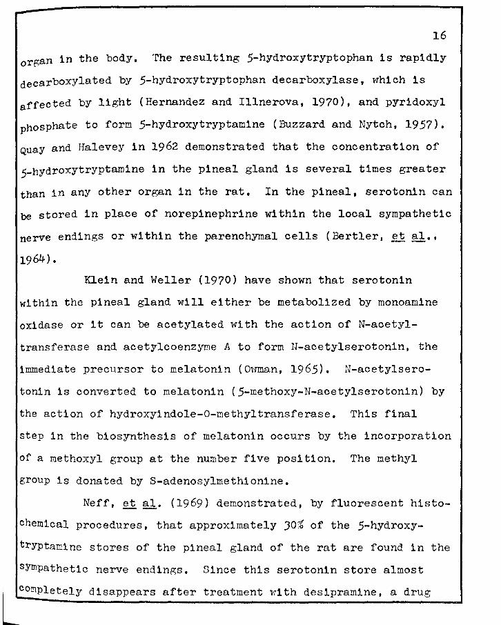
organ in the body. The resulting 5-hydroxytryptophan is rapidly
decarboxylated by 5-hydroxytryptophan decarboxylase, which is
affected by light (Hernandez and Illnerova, 1970), and pyr1doxyl
phosphate to form 5-hydroxytryptamine (Buzzard and Nytch, 1957).
Quay and Halevey in 1962 demonstrated that the concentration of
5-hydroxytryptamine in the pineal gland is several times greater
than in any other organ in the rat. In the pineal, serotonin can
be stored in place of norepinephrine within the local sympathetic
nerve endings or within the parenchyrnal cells (Bertler, et al.,
1964).
Klein and Weller (1970) have shown that serotonin
within the pineal gland will either be metabolized by monoamine
oxidase or it can be acetylated with the action of N-acetyl-
transferase and acetylcoenzyme A to form N-acetylserotonin, the
immediate precursor to melatonin (Omnan, 1965). N-acetylsero
tonin is converted to melatonin (5-methoxy-N-acetylserotonin) by
the action of hydroxyindole-0-methyltransferase. This final
step in the biosynthesis of melatonin occurs by the incorporation
of a roethoxyl group at the number five position. The methyl
group is donated by S-adenosylmethionine.
Neff, et al. (1969) demonstrated, by fluorescent histo
chemical procedures, that approximately JO~ of the 5-hydroxy
tryptamine stores of the pineal gland of the rat are found in the
sympathetic nerve endings.
~mpletely disappears after
Since this serotonin store almost
treatment with desipramine, a drug

I
17
which blocks amine transport in neurons, it was concluded by
the authors, that 5-hydroxytryptamine is synthesized in the
parenchymal cells and subsequently taken into the axons. Zweig
and Axelrod (1969) have shotm that blockage of norepinephrine
synthesis results in an elevation in pineal serotonin content.
Bernad and Csaba. (1970) have shown both serotonin and histamine
in the intracytoplasmic granules of the pinealocyte through
tissue culture preparations and fluorescence microscopy of the
pineal gland of the rat. These granules also contained muco
polysaccharides and protein, but no catecholamines could be
demonstrated.
Klein and Rowe (1970) observed that inhibition of the
oxidation of c14-serotonin ln organ culture of pineal glands of
rats with harmine resulted in enhanced N-acetylation. The
resulting high levels of N-acetylseroton1n caused increased
melatonin production. These investigators also reported decreased
production of hydroxyindoleacetic acid, hydroxytryptophol, and
methoxytryptophol. The hydroxyindole-0-methyltransferase
activity in the glands of harmine-treated rats was no different
than in non-treated controls. This would indicate that there
was an increase in melatonin production by a mechanism not
dependent upon increased production of hydroxyindole-0-methyl
~ransferase.
As previously mentioned, melatonin is effective at
L

18
verY low concentrations as a blanching agent and is found in
verY low concentrations in the pineal gland of all animals
studied as well as in the retinas of amphibians, fishes, reptiles
and some birds (Quay, 1965). At this time however, there is no
truly effective way of assaying for melatonin; therefore,
demonstration of hydroxyindole-0-methyltransferase activity or
changes of hydroxyindole-0-methyltransferase activity is
considered sufficient to establish the melatonin forming ability
of this tissue (Van de Veerdonlr, 1965). Current methods for
possible assaying of melatonin include incubation of homogenized
tissue with N-acetylserotonin and a tagged methyl donor (cl4_
methyl-S-adenosylmethionine) and radioimmunoassay for the tagged
melatonin; or by melanophore response of the larvae of ar.iphib1ans
(Ralph and Lynch, 1970). Hydroxyindole-0-methyltransferase
is the rate controlling enzyme in these reactions.
Melatonin has been suggested to be a pineal hormone
which has an inhibitory effect on the reproductive organs in
mammals. In 1963, Wurtman, Axelrod a.nd Chu studied melatonin to
determine the effect of this compound on the ovary of the rat.
They injected one to twenty pg melatonin/day into twenty-eight
day old female rats and found delayed vaginal opening, decreased
ovarian weights and decreased incidence of cornified vaginal
smears (estrus). They showed that circulating melatonin was
~ •electi vcly talren up and retained by the pineal and the ovary,
19
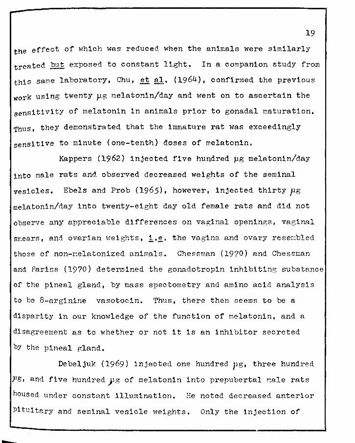
the effect of which was reduced when the animals were similarly
treated 1B! exposed to constant light. In a companion study from
this same laboratory, Chu, et al. (1964), confirmed the previous
work using twenty µg melatonin/day and went on to ascertain the
sensitivity of melatonin in animals prior to gonadal ~aturation.
Thus, they demonstrated that the immature rat was exceedingly
sensitive to minute (one-tenth) doses of melatonin.
Kappers (1962) injected five hundred µg melatonin/day
into male rats and observed decreased weights of the seminal
vesicles. Ebels and Prob (1965), however, injected thirty pg melatonin/day into twenty-eight day old female rats and did not
observe any appreciable differences on vaginal openings, vaginal
smears, and ovarian weights, 1.e. the vagina and ovary reserebled
those of non-melatonized animals. Chessman (1970) and Chessman
and Fariss (1970) deter~lned the gonadotropin inhibiting substance
of the pineal gland, by mass spectometry and amino acid analysis
to be 8-arginine vasotocin. Thus, there then seems to be a
disparity in our knowledge of the function of melatonin, and a
disagreement as to whether or not it is an inhibitor secreted
by the pineal gland.
Debeljuk (1969) injected one hundred pg, three hundred
pg, and five hundredyg of melatonin into prepubertal male rats
housed under constant illumination. He noted decreased anterior
Pituitary and seminal vesicle weights. Only the injection of
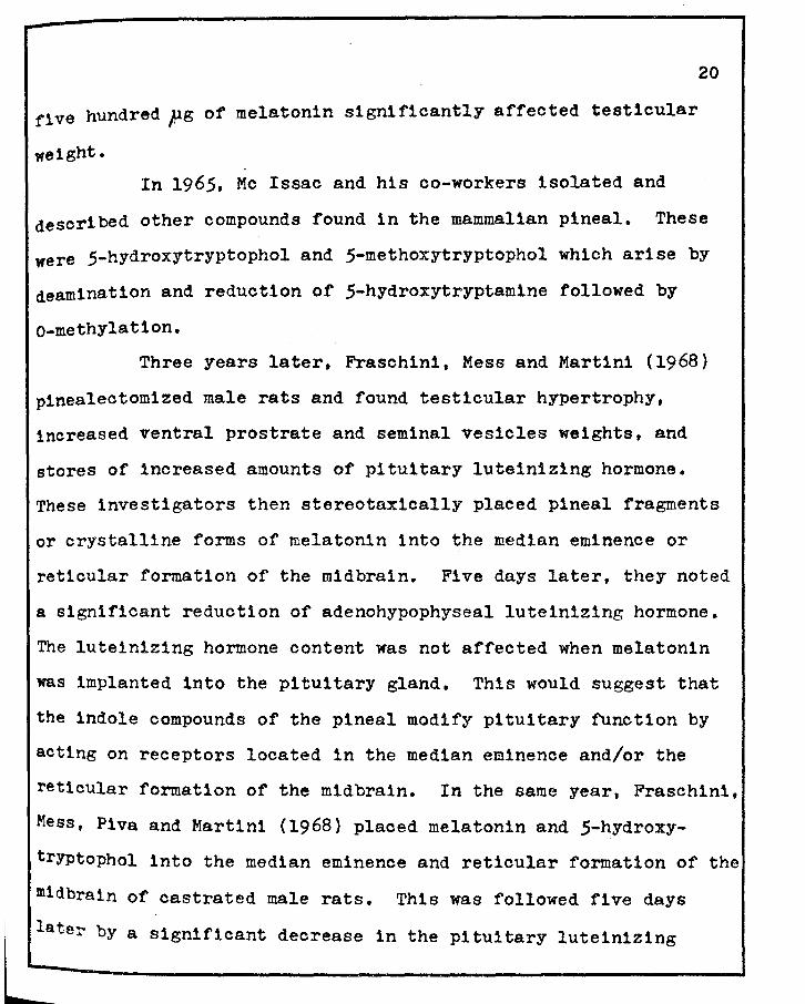
five hundred pg of melatonin significantly affected testicular
weight.
20
In 1965, Mc Issac and his co-workers isolated and
described other compounds found in the mammalian pineal. These
were 5-hydroxytryptophol and 5-methoxytryptophol which arise by
deamination and reduction of 5-hydroxytryptamine followed by
o-methylat1on.
Three years later, Fraschini, Mess and Martini (1968)
p1nealectomized male rats and found testicular hypertrophy,
increased ventral prostrate and seminal vesicles weights, and
stores of increased amounts of pituitary lute1n1z1ng hormone.
These investigators then stereotaxically placed pineal fragments
or crystalline forms of melatonin into the median eminence or
reticular formation of the midbrain. Five days later, they noted
a significant reduction of adenohypophyseal luteinizing hormone.
The lutein1zing hormone content was not affected when melatonin
was implanted into the pituitary gland. This would suggest that
the indole compounds of the pineal modify pituitary function by
acting on receptors located in the median eminence and/or the
reticular formation of the midbrain. In the same year, Fraschini,
Mess, P1va and Martini (1968) placed melatonin and 5-hydroxy
tryptophol into the median eminence and reticular formation of the
m1dbra1n of castrated male rats. This was followed five days
later by a significant decrease in the pituitary lute1n1z1ng
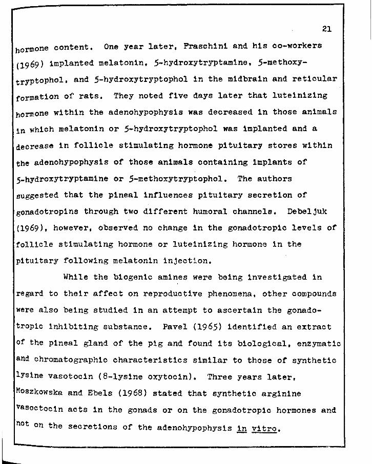
21
hormone content. One year later, Frasch1n1 and his co-workers
(1969) implanted melatonin, 5-hydroxytryptam1ne, 5-methoxy
tryptophol, and 5-hydroxytryptophol in the m1dbrain and reticular
formation of rats. They noted five days later that lute1nizing
hormone within the adenohypophysis was decreased in those animals
1n which melatonin or 5-hydroxytryptophol was implanted and a
decrease in follicle stimulating hormone pituitary stores within
the adenohypophysis of those animals containing implants of
5-hydroxytryptamine or 5-methoxytryptophol. The authors
suggested that the pineal influences pituitary secretion of
gonadotropins through two different humoral channels. Debeljuk
(1969), however, observed no change in the gonadotropic levels of
follicle stimulating hormone or lute1niz1ng hormone 1n the
pituitary following melatonin injection.
While the biogen1c amines were being investigated in
regard to their affect on reproductive pheno~ena, other compounds
were also being studied in an attempt to ascertain the gonado
tropic inhibiting substance. Pavel (1965) identified an extract
of the pineal gland of the pig and found its biological, enzymatic
and chromatographic characteristics similar to those of synthetic
lysine vasotoc1n (8-lysine oxytocin). Three years later,
Moszkowska and Ebels (1968) stated that synthetic arginine
vasoctocin acts in the gonads or on the gonadotropic hormones and
~ on the secretions of the adenohypophysis .!!! vitro.
22
In 1947, Borell and Orstrom (1947a) determined that the
pJ2 turnover rate in the pineal gland of the rat is three to
four times higher than that of the pituitary gland or choroid
plexus. This turnover occurs in two principle ways: the first
is a rapid turnover which enters chiefly into the carbohydrate
phosphate esters, and the second is a slow turnover which enters
the phospholipid and nucleic acids. In the same year, Borell and
orstrom (1947b) examined forty-two different organs of pineal
ectomized rats and observed that only the p32 turnover rate of
the ovary was decreased to a statistically probable extent. rhere
was a considerable increase in p32 turnover in the anterior and
posterior aspects of the pituitary gland and in the tuber cinerium
in fe~ale rats. rhey also noted an increased turnover in the
pineal and anterior pituitary after castration and found this to
be most pronounced in the female rat. The authors stated that
there appears to be a reciprocal effect of a similar nature be-
tween the ovary and the pineal as that which exists between the
adenohypophysis and the ovaries.
In 1961, Hellman and Larsson (1961) demonstrated in
goats by radio-paper chromatographic assay that the pineal gland
has an oxygen consumption approximately equal to that of the
Posterior pituitary. rhey also ascertained that the carbon
dioxide and lactic acid formation from glucose decreases with age
as do the other amino acids formed from this carbohydrate. Gluta-
23
mic acid was found to be in the greatest concentration of the
amino acids formed in this way in the younger goats. Appreciable
amounts of arginine, glutamine, gamna-aminobutyric acid, and
aspart1c acid were also noted. ~icroscopic observations reveal
ed progressive degeneration and decreased numbers of parenchymal
cells. rhe authors suggested that the high rate of amino acid
formation found in the pineal gland of young goats can be regard
ed as a mechanism for supplying the raw material for a secretory
product of protein nature,
Nir and his co-workers (1970) injected ten pg of
estradiol-17-,f'subcutaneously into immature female rats (twenty
one to thirty-one days of age) and deter~ined the pineal ribose-
nucleic acid, deoxyribonucleic acid, and protein levels which
were measured at fifteen, eighteen, and twenty-four hours after
injection. rhe authors noted increased protein content of the
pineal occurred twenty-four hours after the injection and this
was accompanied by a prior elevation of pineal ribosenucleic acid
and deoxyribonucleic acid content thus indicating accelerated
protein metabolism.
Mitchell and Yochim (1970) indicated that prolonged
daily illumination (twenty-two hours light: two hours dark) led
to an apparent twelve hour increase in the duration of gestation,
The time of partur1t1on also was photoperiod-dependent as the
animals exhibited a marked tendency to deliver during the light
24
phase of the light:dark cycle. 'rhese data indicated that the
effect of light on the prolongation of pregnancy in part was due
to an action of delayed implantation. Pinealectomy, however, did
not significantly alter the effect of photoperiod on the duration
of gestation or the time of day, which would then indicate light
mediates control of the duration of gestation other than via the
pineal gland. Huang and Everitt (1965) observed changes in the
pineal weight during the latter stages of pregnancy. The change
in weight was found to be inversely proportional to the number
of fetuses carried.
Prop and Kappers {1961) demonstrated the presence of
lipids, aromatic amines (catecholamines), ascorbic acid, and
1ndole amines by histochemical and paper chromatographic pro
cedures. Wight and Vackenzie {1971) demonstrated in the domestic
fowl, by histochemical means, that there is an abundance of
lipids in the form of triglycerides, phospholipids, and choles
terol and its esters. ?he authors did not observe any lipo
fuchsin. The authors also determined that there is considerable
enzyme activity in the pineal gland, ·rhey histochemically
identified alkaline phosphatase, acid phosphatase, adenosine
tr1-phosphate, lipase, non-specific esterase, n1cotinamide
denine d1nucleotide diaphorase, cytochrome oxidase, beta
glucuronidase, and an:1no peptidase. The pineal was found, by
~se investigators, to be rich in r1bosenucleic acid, but
25
observed no glycogen, intracytoplasmic mucopolysaccharides, or
Gornor1-pos1tive neurosecretory substances. Machado, et al.
(1967, 1968) observed no 5-hydroxytryptamine fluorescence in
nineteen day old rat fetuses. Two hours after birth the first
fluorescence to appear in the sympathetic nerves was green
(probably catecholamines). By the twenty-first day, the pineal
gland of immature rats appears similar to the adult gland with
a yellow fluorescence for serotonin.
Niemi and Ikoken (1960) histochemically demonstrated
the presence of aminopeptidase activity in the pineal gland of
the rat. It is their opinion that the activity was the result
of production and secretion of certain agents of a protein nature.
Zweens (1963) h1stochem1cally and biochemically demonstrated a
significant decrease in the phospholipid concentration during
proestrus with a peak observed during the diestrous phase of
the estrous cycle. Quay (1959) biochemically demonstrated succin
ic dehydrogenase activity in the rat pineal with respect to age.
This enzyme was found highest during the first six weeks of
postnatal life with a decline at approximately one year con-
co~mi tant with increased incidences of variation among the
animals studied.
Quay (1956) histochemically demonstrated that the
Pineal gland of the rat displays an intense and specific reaction
With chrom-alum hematoxylin. and phloxine technique of Gomori

(l941) for staining pancreatic islets. The author states that
the parenchymal cells of the pineal gland in the rat appeared
to take part in a secretory process which is in agreement with
owman (1960-1961) who studied parenchymal cell secretory sub
stances in the fetal rat.
26
The literature is replete with numerous studies of the
pineal gland, but is most unfortunately devoid of any pertinent
and critical information of the pineal gland during specific
reproductive mechanisms, 1·~· from the estrous cycle throughout
its entirety to pregnancy and to pseudopregna.ncy. The major
aim of this dissertation is therefore involved in an elucidation
of the pineal gland of the rat (a) during the estrous cycle,
(b) during pseudopregnancy and prolonGed pseudopregnancy and
(c) during pregnancy.

CHAPTER III
MATERIALS AND METHODS
The investigations carried out for this dissertation
utilized 506 adult, Charles River, Caesarean-Originated, Barrier
sustained Sprague-Dawley, derived female albino rats. Of this
number, J62 rats, sixty-nine to ninety-one days of age, were
utilized for the studies involving the pineal gland and the
estrous cycle. A total of fifty-two rats, ninety to lJO days of
age, were investigated for the possible modulating roles of
pregnancy on the pineal gland, and a total of ninety-two rats,
ages ninety to 130 days, were likewise pursued for the possible
influences of pseudopregnancy and prolonged pseudopregnancy (1·~·
rats containing decidual reactions and manifesting extended
luteal life beyond the normal range of pseudopregnancy, lJ.O
days) on the pineal gland. Of the ninety-two on the pseudo
pregnancy studies, thirty were utilized as pseudopregnancy
controls (1·~· without endometr1al traumatization) and sixty-two
were pursued with experimentally-induced decidual reactions.
Food (Purina Rat Chow) and water were supplied ad
lJbitum and the animals were housed in a 72°F room with a cool
White fluorescent light source ()000-5000 i) on a 7100 A.M. to
27
-28
71 00 P.M. light cycle. Rats were numbered and housed six in a
cage. Due to the importance of light on the pineal gland, the
cages were oriented parallel to the light source so as to assure
complete penetration of light into the cages,
In an effort to obtain as complete a reproductive
history as possible, the animals were staged according to estrous
cycle phases. Vaginal smears were taken twice a day at 8100 A.H.
and 5100 P.M. for three weeks, subsequently stained with Giemsa
solution, and were read and recorded. ro determine the onset of
estrus, aninals were smeared at 9100 P.V. (or at 12100 A.K. if
an estrus smear was not observed at 9100 P.n.) on the evening
following a proestrous smear, and the first indication of a
predominance of :v:aginal cornified cells with degenerative nuclei
was taken as the zero hour of estrus.
Rats were staged as carefully as possible so as to
assure the critical selection of a sufficiently, quantifiable
number for each of the six distinctively different estrous cycle
stages. For each of the critical phases, a special effort was
made to obtain animals prior to, during, and immediately after
the sought after phases. It was, therefore, possible to obtain
good diagnostic criteria for each of the stages under study. For
each rat, therefore, the time of the onset of estrus, the hours
after the onset of estrus and the hours within each stage were known prior to and at the time of necropsy.
/'

29
The six distinctive stages of the estrous cycle of the
albino rat were further subdivided into a seventh stage. This
was done by dividing the phase of estrus of the cycle into two
parts, the first encompassing hours zero through six and the
second, hours six through twelve of the stage of estrus. Diestrus
was ordinarily divided into early diestrus, zero through twenty
five hours within the stage, and late diestrus, twenty-five to
fifty hours within the stage. In this study, the histochemical
and biochemical tests were performed on the following stages of
the cycle with the hours after onset of estrus as indicated in
brackets: Early Estrus (E1 , zero to six hours); Late Estrus
(E2 , six to twelve hours); Jv:etestrus ( r~E, twelve to eighteen
hours); Early Diestrus ( D1, eighteen to fifty hours); I.ate Di
estrus (D2 , fifty-one to seventy-six hours); Preproestrus
(PPE, seventy-seven to ninety hours); and Proestrus (PE, ninety
to ninety-seven hours).
At the selected stage of the estrous cycle, the rats
were weighed on a rorbal rorsion Balance to the nearest gram,
necropsied by decapitation, and the following tissues were
removed: the pineal, hypothalamus, hypophysis, thyroids,
adrenals, ovaries, uterus, and vagina. These tissues were
rapidly renoved, cleaned, weighed on a FPE Precision balance to
. the nearest tenth of a milligram and either fixed in Bouins'
~tive or frozen on dry lee for subsequent hlstomorphologlcal

...
JO
studies. Tissues fixed in Bouins' for eighteen hours were washed
in tap water, dehydrated through graded alcohols, and embedded
in Tissuemat (M. P. = 53 ! 0.5°c).
Pineal glands of the rats were exposed by cutting first
through the sutura fontal1s and secondly along the os temporalis
on both sides. The calvarium was then lifted dorsally to expose
the cerebrum and cerebellum. The pineal, which lies in the tri
angular space formed at the junction of the cerebral hemispheres
and the cerebellu~, was rapidly removed, cleaned of connecting
dura mater and blood vessels, weighed on a Roller Smith torsion
balance to the nearest tenth of a milligram and either frozen or
placed in Bouins' fixative for histochemical and biochemical
assessments and histological characterization, respectively.
Gravimetric Data
Weights taken for each organ removed at necropsy, with
the exception of the vagina and hypothalamus, were analyzed for
the arithmetical mean, standard deviation, and standard error;
these data were then applied to the Student's "t" test for
significance using the Olivetti-Underwood Programma 101. This
was done in an attempt to grav1metrically ascertain if a
correlation exists between the pineal gland and changes of the
Other endocrine organs as influenced by the estrous cycle •
bz
.31
Histornorpholo12:y - Bouins' fixed tissues were dehydrated and embedded ln
paraffin, sectioned at five µ and stained with henatoxylin-eosin
(Harris, 1900) for observing general morphology and chrom-alum
hematoxylin (.Bargmann, 1949) for demonstrating neurosecretory
substances.
Paraffin sections were deparaffinized and placed in
Bouins' fixative containing 3.5i chrom-alum for twelve to four
teen hours at J7°C. Sections were then washed in running tap
water until colorless and oxidized for two minutes in one part
2.5:b potassium permanganate, one part 5,'t sulfuric acid, and six
to eight parts distilled water, Sections were washed in distilled
water, bleached in 1)6 oxalic acid solutions for one minute, re-
washed in distilled water, and stained for ten minutes. The
stain used in this method consisted of a mixture of 50 ml 1%
aqueous hematoxylin, 50 ml JZ aqueous chrom-alum[cr2(S04) 3 (NH4) 2
S04·24 H2~, 2 ml 5% aqueous potassium dichro~ate, and 1 ml
5% aqueous sulfuric acid. rhe stain was allowed to ripen for
forty-ei~ht hours at 0-4°c and filtered before use. The slides
were differentiated in acid alcohol and/or ammonium hydroxide to
develop nuclear color to a sharp contrast. Sections were then
washed in running tap water for three minutes, counterstained
With 0.5~ aqueous phloxine solution for three minutes and rinsed
in 5{ aqueous solution of phosphotungstic acid for two minutes.

....
bn
J2
Following counterstain1ng, the sections were washed in running
taP water for five minutes, dehydrated, and mounted with Permount,
Deep purple staining of chrom-alum hematoxylin pos
itive substances indicated sites of the neurosecretory material.
The nuclei stained with a slightly less purple intensity and the
cytoplasm stained a pale pinkish red.
Enzyme HistocheMistry
A minimum of three frozen pineal glands for each stage
of the estrous cycle, pseudopregnancy, prolonged pseudopregnancy
and pregnancy were sectioned at ten f on an International cryo
stat and subjected to the following histochemical tests: alka
line phosphatase (naphthol phosphate method, Gomori, 1951); acid
phosphatase (azo-coupl1ng method, Barka, 1960); succinic de
hydrogenase, a Krebs cycle enzyrne which converts succinate to
fumarate (Rosa and Velardo, 19 54); and lactic dehydrogennse.
which converts pyruvate to lactate (Vianocha and 3-ourne, 1968),
SuccinJ_c Dehydroge11.a§e Histochernistr;x-
The method of Rosa and Velardo (1954) with a minor
modification was used for the histochemical demonstration of
succinic dehydrogenase (SDH l.J.99.1.). Pineal glands removed
at necropsy were frozen on dry ice and stored at -70°c until
ready for use.
In preparation for sectioning, the tissues i.:ere mounted
JJ
bY freezing on the stage of an International cryostat. Sections
were cut at tenp and mounted on coverslips in spaced serial
sections and air-dried for one-half hour.
Sections were then incubated for two hours at :n°c in
a media composed of JO ml 0.1 M phosphate buffer containing
o,lfa sodium cyanide at pH 8,2, 4.1.ul 0,5 !': sodium succ1nate,
and 30 mg nitro blue tetrazolium. The buffer was prepared by
dissolving 1 gm sodium cyanide in 500 ml Q.l M disodiurn phosphate
solution, adjusting the solution to pH 8.2 by adding, with
constant stirring, 0,1 ;r monosodlum phosphate, then adding pH
8.2 0,1 li phosphate buffer to a final volume of one liter.
Following incubation, the slides were rinsed in distilled water
and fixed for three hours in 10}~ neutral formalin •
.A control slide was also run with the same chemicals
in the incubation media with the exceptiog of the substrate,
sodium succinate.
Criteria used for evaluating the histochemical reaction
were as follows 1 (a) heavy deposits of violet to black di for-
mazan granules were indicative of high succinic dehydrogenase
activity sites; (b) areas staining pink were considered to have
lower activity, their color probably indicative of an inter
mediate state of formazan production (Eadie, 1970).
The theory concerning the formation of the formazan
crystals indicates that a hydro~en atom is removed from the
..
34
number two and three carbon atom; a hydrogen atom reacts with
nicotlnamlde adenine dinucleotide (NAD) which is reduced to form
dlhydronicotinamide adenine dinucleotide (NADH). Since this
coenzyme operates by virtue of reversible oxidation and reduction
reactions, the dihydronicotinarnide adenine dinucleotide is ox
idized to again form nicotinamide adenine dinucleotide and a
hydrogen atom is incorporated into the nitro blue tetrazolium
molecule to form the insoluble formazan. Nicotinamide adenine
d1nucleot1de operates as a hydrogen and electron transfer agent.
one of the two hydrogens lost when succinate is oxidized, is
incorporated into the n1cotinamide adenine dinucleotide molecule,
the reduction occurring in the para position, while the second
hydrogen atom enters the media.
Since oxaloacetic acid is an inhibitor to succinic de-
hydrogenase by virtue of its similar chemical structure to
succinatc, sodium cyanide was used in the incubation me,~~.a in
order to trap by cyanhydrin formation any oxc.loacetic acid
possibly for~ed in the tissue during processing and/or incubation.
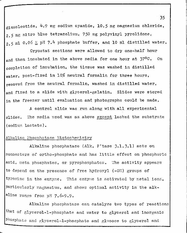
19.cttc DehYC!.:roe;enase Histochemistrx
The histochemical determination of lactic dehydrogenase
(LDH 1.1.1.27) was demonstrated according to the slightly mod
ified procedure of Vanocha and Bourne (1968). The media used
in this procedure was composed of 112.07 mg sodium lactate
(lactic acid, sodium salt). 66. J4 mg nicot1namide adenine
-35
dinucleotide, 4.9 mg sodium cyanide, 10.5 mg magnesium chloride,
2 • 5 mg n1 tro blue tetrazolium, 7 50 mg polyvinyl pyrolidone,
z.5 ml 0.06 li pH 7.4 phosphate buffer, and 10 ml distilled water.
Cryostat sections were allowed to dry one-half hour
and then incubated in the above media for one hour at J7°C. On
completion of incubation, the tissue was washed in distilled
water, post-fixed in 10% neutral formalin for three hours,
removed from the neutral formalin, washed in distilled water,
and fixed to a slide with glycerol-gelatin. Slides were stored
in the freezer until evaluation and photographs could be made.
A control slide was run along with all experimental
slides. The media used was as above except lacked the substrate
(sodiun lactate).
Alkaline Phosnhatase Histochemistry
Alkaline phosphatase (Alk. P'tase J.l.J.l) acts on
monoesters of ortho-phosphate and has little effect on phosphoric
acid, meta phosphates, or pyrophosphates. The activity appears
to depend on the presence of free hydroxyl (-OH) groups of
tyrosine in the enzyme. This enzyne is activated by metal ions,
Particularly magnesium, and shows optimal activity in the alk
aline range from pH 7.6-9.9.
Alkaline phosphatase can catalyze two types of reactions
that of glycerol-1-phosphate and water to glycerol and inorganic
Phosphate and glycerol-1-phosphate and glucose to glycerol and
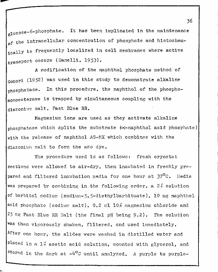
--J6
glucose-6-phosphate. It has been implicated in the maintenance
of the intra.cellular concentration of phosphate and histochem
icallY is frequently localized in cell membranes where active
transport occurs (Danelli, 1953).
A modification of the naphthol phosphate method of
Gomori (1952) was used in this study to demonstrate alkaline
phosphatase. In this procedure, the naphthol of the phospho
roonoesterase is trapped by simultaneous coupling with the
diazoni u~n salt, Fast Blue RR.
Magnesium ions are used as they activate alkaline
phosphatase which splits the substrate P<-naphthol acid phosphate)
with the release of naphthol AS-!·~X which combines with the
diazonium salt to forn the azo dye.
The procedure used is as follows: fresh cryostat
sections were allowed to air-dry, then incubated in freshly pre
pared and filtered incubation rr.edia for one hour at 37°c. Nedia
was prepared by combining in the following order, a 2.-b solution
of barbitol sodium (sodiuD.-5,5-diethylbarbituate), 10 mg ne.phthol
acid phosphate (sodium salt), 0. 2 ml 10,::& magnesium chloride and
25 mg Fast Blue RR Salt (the final pH being 9.2). The solution
was then vigorously shaken, filtered, and used immediately.
After one hour, the slides were washed in distilled water and
Placed in a li acetic acid solution, mounted with glycerol, and
stored in the dark at -4°c until analyzed. A purple to purple-

37
black precipitate denoted sites of alkaline phosphatase activity.
Controls minus the o<-naphthol acid phosphate were run with each
experimental section.
Acid Phos£hatase Histochemistry
Acid phosphatase (Acid P'tase J.l.J.2) catalyzes the
hydrolysis of most phosphomonoesters, of creatine phosphate and
of amino-phosphate. It is activated by manganese and has an
optimal pH range of 4.5-5.2. Dimethyl formamide was used to
dissolve the substrate. The reaction sequence is the same as
for alkaline phosphatase.
A modification of Burnstone's (1959) method for the
histochemical demonstration of acid phosphate was used. The
procedure involved use of fresh frozen cryostat sections which
were air-dried on cover slips. The incubation media consisted
of 5 mg naphthol acid phosphate (naphthol AS-MX phosphate),
0.25 ml dimethyl formamide, 25 ml 0.2 M pH 5.2 acetate buffer,
0.1 ml 10% manganese choloride, JO mg Fast Red Violet LB (d1azon-
1um salt), and 25 ml distilled water to a final volume of 50 ml.
Experimental and control (minus o<-naphthol acid
Phosphate) tissue sections were concurrently processed.
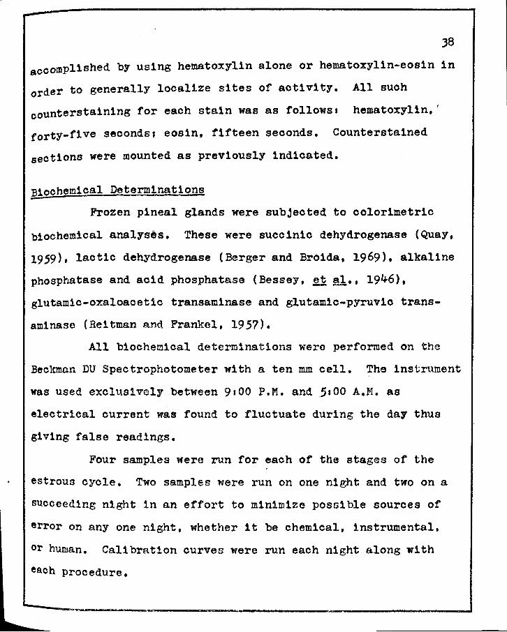
£...ountersta1n1ng
Counterstain1ng of the histochemical procedure was
J8
accomplished by using hematoxyl1n alone or hematoxyl1n-eos1n in
order to generally localize sites of activity. All such
countersta1n1ng for each stain was as follows a hematoxylin,'
forty-five seconds; eos1n, fifteen seconds. Countersta1ned
sections were mounted as previously 1nd1oated.
Biochemical Determinations
Frozen pineal glands were subjected to colorimetric
biochemical analyses. These were succinic dehydrogenase (Quay,
1959). lactic dehydrogenase (Berger and Broida, 1969). alkaline
phosphatase and acid phosphatase (Bessey, ~ !!1•• 1946),
glutam1c-oxaloacet1c transaminase and glutam1c-pyruv1o trans
aminase (Reitman and Frankel, 1957).
All biochemical determinations were performed on the
Beckmc.n DU Spectrophotometer with a ten mm cell. The instrument
was used exclusively between 9100 P.M. and 5a00 A.M. as
electrical current was found to fluctuate during the day thus
giving false readings.
Four samples were run for each of the stages of the
estrous cycle. Two samples were run on one night and two on a
succeeding night in an effort to minimize possible sources of
error on any one night, whether it be chemical, instrumental,
or human. Calibration curves were run each night along with
each procedure.

bn
39
succ1n1c Dehydrogenase Biochemistry -Pineal succ1nic dehydrogenase (SDH) activity was
determined according to a modification or the procedure of Quay
(1959). Pineal glands were individually homogenized in 0.50 ml
o.025 ~ sodium phosphate buffer containing 0.1% sodium cyanides
o.50 ml 5.4.% sodium succinate containing M/100 aluminum chloride
and M/1000 calcium ohlorider and 0.50 ml 1.0% tetrazolium
2-(p-iodophenyl)-J-(p-n1trophenyl)-5-phenyltetrazol1um chloride.
The above solution, with homogenized pineal, was incubated 1n a
J70C water bath for two hours with constant shaking.
After completion of this incubation, 10% neutral
formalin was added to terminate the formazan production and the
solution taken to dryness 1n a 68°c oven. The colored form~zan
was extracted with A.c.s. (American Chemical Society) grade
ethyl acetate and the optical density was determined at 490 mp.
on a Beckman DU Spectrophotometer. Ethyl acetate was used as a
reference blank.
The micromoles of tetrazol1um reduced per pineal was
calculated according to the method of Shelton and Rice (1957).
The formula used is D=KCL where C is the molar concentration of
substance in solution, L 1s the length of the light path 1n
centimeters (1n this case, one), Dis the optical density of the
sample, and K 1s a constant found to be 5.34 at O.D.490 by
Shelton and Rice. The resulting molar concentration of the
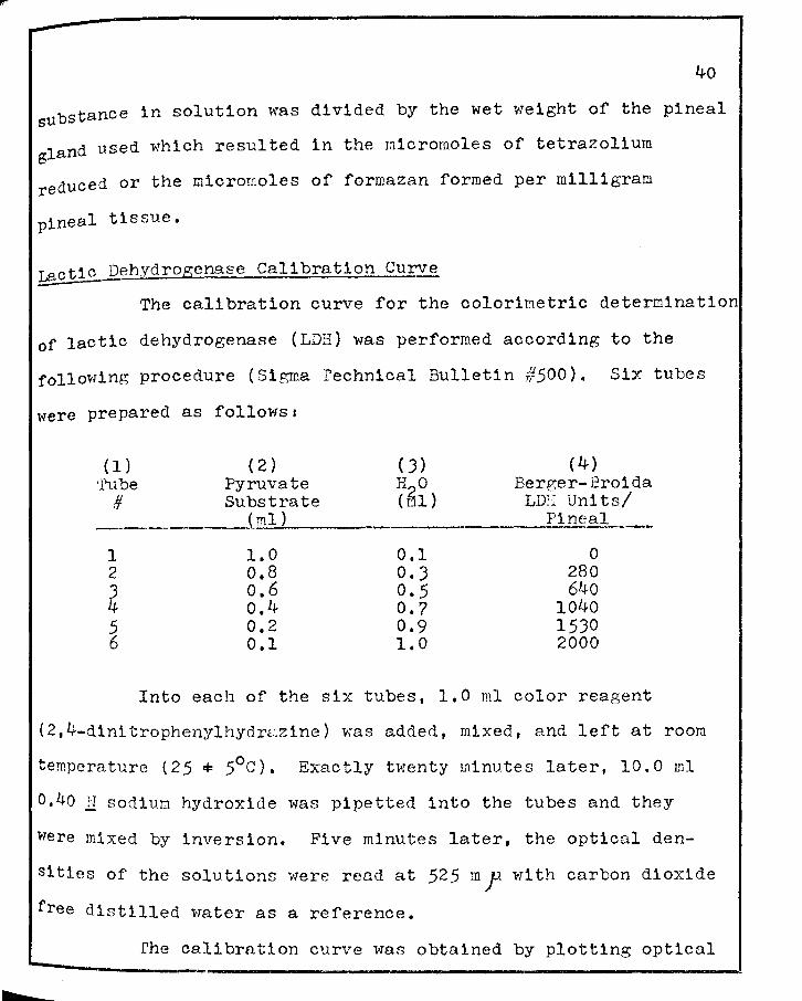
40
substance in solution was divided by the wet weight of the pineal
gland used which resulted in the micromoles of tetrazolium
reduced or the micromoles of formazan formed per milligram
pineal tissue.
r..actic Dehydrogenase Calibration Curve -The calibration curve for the colorimetric determination
of lactic dehydrogenase (LDH) was performed according to the
following procedure (Sigma rechnical Bulletin #500). Six tubes
were prepared as followsi
( 1 ) ( 2) ( J) ( 4) ·rube Pyruvate H O Berger-Broida
# Substrate (61) LDII Uni ts/ (ml) :Pi n~gl._ __
1 1.0 0.1 0 2 o.8 O.J 280 J o.6 0.5 640 4 o.4 0.7 1040 5 0.2 0.9 1530 6 0.1 1.0 2000
Into each of the six tubes, 1.0 ml color reagent
(2,4-d1nitrophenylhydr2zine) was added, mixed, and left at room
temperature (25,,.. 5°c). Exactly twenty minutes later, 10.0 ml
0,40 11 sodiul:l hydroxide was pipetted into the tubes and they
were mixed by inversion. Five minutes later, the optical den
sities of the solutions were read at 52.5 m .f wl th carbon dioxide
free distilled water as a reference.
rhe calibration curve was obtained by plotting optical

density readings against the corresponding units of lactic
dehydrogenase from column four above.
Lactic dehydrogenase in this procedure catalyzes the
conversion of pyruvic acid to lactic acid according to the
reaction1
pyruvic acid + NADPH2 lac tic acid + NAD
41
rhe speed of the reaction is proportional to the amount of lactic
dehydrogenase present. Pyruvic acid reacts with 2,4-dinitro
phenylhydrazine to form an intensely colored hydrazone. Lactic
acid, dihydronicotinamide adenine d1nucleot1de phosphate and
nicotinamide adenine dinucleotide do not add a significant amount
of optical density to the solution; therefore, the standardized
pyruvate substrate which yields the same hydrazone optical den
sity will be accurate and reproducible. 2he amount of pyruvate
remaining after the incubation is inversely proportional to the
amount of lactic dehydrogenase present in the reaction. One unit
of lactic dehydrogenase activity will reduce 4.8 X 10⁻⁴ pr;;
pyruvate/rc.inute/rng pineal.
Total Lactate Deh;ydrogenase B1ocher.1istr:v
Frozen pineal glands were homogenized with 0.50 ml
carbon dioxide free distilled water and diluted one part sample
Plus four parts distilled water (1:4). This was added to 1.0 ml
Sigma standardized pyruvate substrate and 1.0 mg dihydron1cotin
am1de adenine d1nucleot1de and placed in a water bath at J7°C.
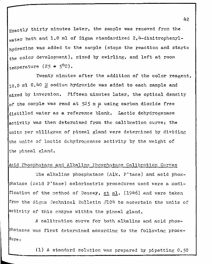
--42
Exactly thirty minutes later, the sample was removed from the
water bath and 1.0 ml of Sigma standardized 2,4-dinitrophenyl
hydrazine was added to the sample (stops the reaction and starts
the color development), mixed by swirling, and left at room
temperature (25 • 5°C).
Twenty minutes after the addition of the color reagent,
10.0 ml 0.40 N sodium hydroxide was added to each sample a.nd
mixed by inversion. Fifteen minutes later, the optical density
of the sample was read a.t 525 m p using carbon dioxide free
distilled water as a reference blank. Lactic dehydrogenase
activity was then determined from the calibration curve; the
units per milligram of pineal gland were deterr:iined by dividing
the units of lactic dehydrogenase activity by the weight of
the pineal gland,
Acid Phosphatase and Al1calinc __ Phosp_tiatase Calibration Curves
'I1he alkaline phosphatase ( Alk. P' tase) and acid phos
phatase (Acid P'tase) colorimetric procedures used were a wodi
f1cation of the method of Bessey, et al, (1946) and were taken
from the Signa rechnical Bulletin ifl04 to ascertain the uni ts of
activity of this enzyme within the pineal gland,
A calibration curve for both alkaline and acid phos
Phatases was first determined according to the following proce
dure:
(1) A standard solution was prepared by pipetting 0.50
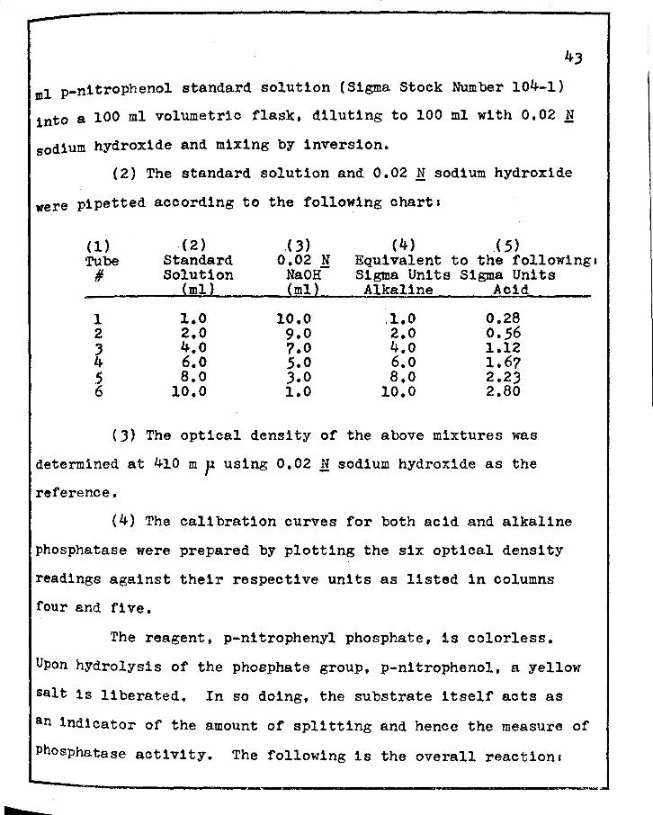
b
43
ml p-nltrophenol standard solution (Sigma Stock Number 104-1)
into a 100 ml volumetric flask, diluting to 100 ml with 0.02 N
sodium hydroxide and mixing by inversion.
(2) The standard solution and 0.02 N sodium hydroxide
were pipetted according to the following charts
(1) . ( 2) « 3) (4) ( 5) Tube Standard 0.02 N Equivalent to the followings # Solution Na OH Sigma Units Sigma Units
(ml) (ml) Alkaline Acid
1 l.O 10.0 .1.0 0.28 2 2.0 9.0 2.0 0.56 J 4.o 7.0 4.o 1.12 4 6.o 5.0 6.o 1.67 5 a.o J.O a.o 2.23 6 10.0 1.0 10.0 2.80
(J) The optical density of the above mixtures was
determined at 410 m p using 0.02 M sodium hydroxide as the
reference.
(4) The calibration curves for both acid and alkaline
phosphatase were prepared by plotting the six optical density
readings against their respective units as listed 1n columns
four and fl ve.
The reagent, p-nitrophenyl phosphate, 1s colorless.
Upon hydrolysis of the phosphate group, p-nitrophenol, a yellow
salt is liberated. In so doing, the substrate itself acts as
an indicator of the amount of splitting and hence the measure of
Phosphatase activity. The following is the overall reactions
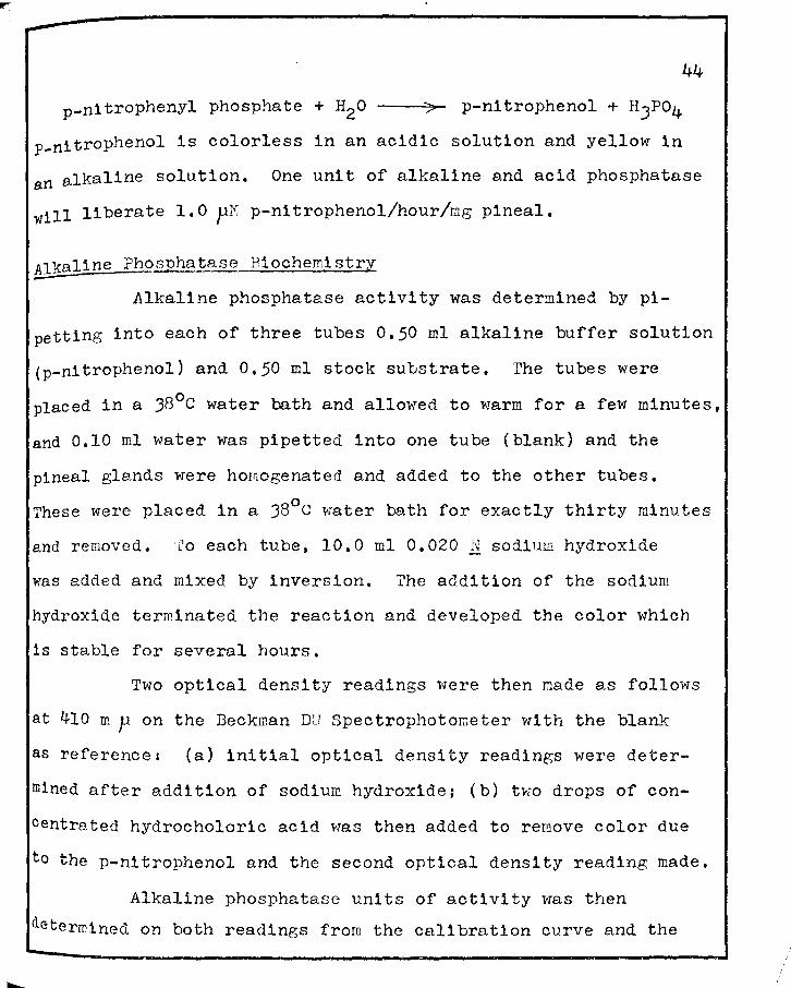
44
p-n1trophenyl phosphate + H2o
p-nitrophenol is colorless in an acidic solution and yellow in
an alkaline solution. One unit of alkaline and acid phosphatase
will liberate 1. 0 pI•i p-ni trophenol/hour/mg pineal.
,!_lkaline Ph_?~~ase Biochemistry
Alkaline phosphatase activity was determined by pi-
petting into each of three tubes 0,50 ml alkaline buffer solution
(p-ni trophenol} and 0. 50 ml stock substrate. ·rhe tubes were
placed in a JB 0 c water bath and allowed to warm for a few minutes,
and 0,10 ml water was pipetted into one tube (blank) and the
pineal glands were homogenated and added to the other tubes.
These were placed in a J8°c water bath for exactly thirty minutes
and removed. lo each tube, 10,0 ml 0,020 N sodium hydroxide
was added and mixed by inversion. ?he addition of the sodium
hydroxide terminated the reaction and developed the color which
is stable for several hours.
Two optical density readings were then made as follows
at 410 m p on the Beckman DU Spectrophotometer with the blank
as reference: (a) initial optical density readings were deter
mined after addition of sodium hydroxide: (b) two drops of con-
centre.tea hydrocholoric acid was then added to remove color due
to the p-n1trophenol and the second optical density reading made.
Alkaline phosphatase units of activity was then
determined on both readings from the calibration curve and the
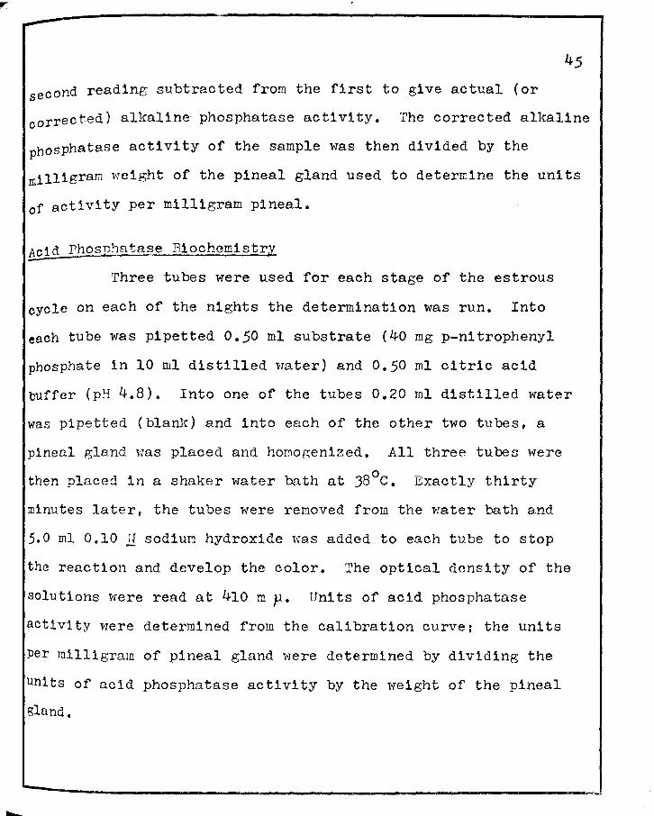
second reading subtracted from the first to give actual (or
corrected) alkaline phosphatase activity. The corrected alkaline
phosphatase activity of the sample was then divided by the
milligram weight of the pineal gland used to determine the units
of activity per milligram pineal.
!£id Phosphatase Biochemistry
Three tubes were used for each stage of the estrous
cycle on each of the nights the determination was run. Into
each tube was pipetted 0.50 ml substrate (40 mg p-nitrophenyl
phosphate in 10 ml distilled water) and 0.50 ml citric acid
buffer (pH 4.8). Into one of the tubes 0.20 ml distilled water
was pipetted (blanlc) and into each of the other two tubes, a
pineal gland was placed and homogenized. .All three tubes were
then placed in a shaker water bath at J8°c. Exactly thirty
minutes later, the tubes were removed from the water bath and
5.0 ml 0.10 ~1 sodium hydroxide was added to each tube to stop
the reaction and develop the color. The optical density of the
solutions were read at 410 mp. Units of acid phosphatase
activity were determined from the calibration curve; the units
Per milligram of pineal gland were determined by dividing the
Units of acid phosphatase activity by the weight of the pineal
gland.
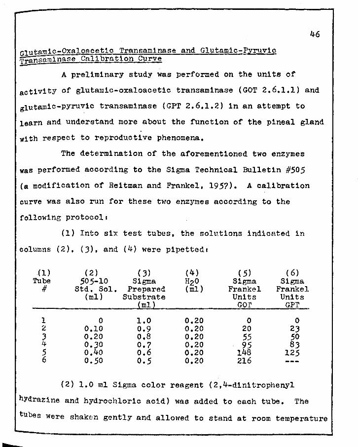
bz
Glutamic-Oxaloacetic Transaminase and Glutamic-Pyruvic Transaminase Calibration curve
46
A preliminary study was performed on the units of
activity of glutamic-oxaloacetic transaminase (GOT 2.6.1.1) and
glutam1c-pyruv1c transaminase (GPT 2.6.1.2) in an attempt to
learn and understand more about the function of the pineal gland
with respect to reproductive phenomena.
The determination of the aforementioned two enzymes
was performed according to the Sigma Technical Bulletin #505
(a modification of Reitman and Frankel, 1957). A. calibration
curve was also run for these two enzymes according to the
following protocols
(1) Into six test tubes, the solutions indicated 1n
columns (2), (J), and (4) were pipetteds
(1) (2) ( J) (4) ( 5) ( 6) Tube 505-10 Sigma H20 Sigma Sigma
# Std. Sol. Prepared (ml) Frankel Frankel (ml) Substrate Units Units
(ml) Gor GPr
1 0 1.0 0.20 0 0 2 0.10 0.9 0.20 20 2J 3 0.20 o.s 0.20 55 50 4 0.30 0.7 0.20 95 SJ 5 o.4o o.6 0.20 148 125 6 0.50 0.5 0920 216
(2) 1.0 ml Sigma color reagent (2,4-dinitrophenyl
hydrazine and hydrochloric acid) was added to each tube. The
tubes were shaken gently and allowed to stand at room temperature

(25 ± 5°c) for twenty minutes.
(J) 10.0 ml o.40 N sodium hydroxide was added and
mixed by inversion.
(4) After five minutes, the optical density was
determined at 505 m p using distilled water as a reference.
(5) A calibration curve was then constructed plotting
the optical density readings against the corresponding units of
glutamic-oxaloacetic transaminase found in column five.
(6) A second curve was also set up plotting the
optical density readings against the corresponding units of
glutamic-pyruv1c transaminase found 1n column six.
Glutamic-oxaloacetic transaminase is an enzyme which
catalyzes the conversion of aspart1c acid ando(-ketoglutaric
acid to oxaloacetic acid and glutamic ac1d according to the
equation:
aspartic acid +
o(~ketoglutaric acid
oxaloacetic acid +
glutamio acid
Glutam1c-pyruvic transaminase is an enzyme which
catalyzes the conversion of alanine and oo<-ketoglutaric acid to
pyruvic acid and glutamio acid according to the equations
alanine +0\.-ketoglutaric acid ,GPT, pyruvic acid + glutamic acid
In the procedure used, the amount of oxaloacetic acid or pyruvate
formed in one hour is determined colorimetrically by the for
mation of a hydrazone which 1s highly colored. Theoretically,

b
48
one unit of activity of either glutam1o-oxaloacet1c transaminase
or glutamic-pyruv1c transaminase will form 4.82 X 10⁻⁴ pM of
glutamate per minute at pH 7.5.
Glutamic-Oxaloacetic Transaminase Biochemistry -Pineal glands were homogenized in l.O ml of Sigma
@ standardized aspartate-o(-ketoglutarate substrate (pH 7.5) and
placed in a shaker water bath at 37°c. Exactly one hour later,
the samples were removed from the bath and l.O ml of Sigma
standardized 2,4-dinitrophenyl hydrazine was added to each tube
(which stops the enzymatic reaction and starts the color
reaction). The samples were then mixed by inversion and allowed
to stand at room temperature {25 ! 5oc}. Exactly twenty minutes
later, 10,0 ml o.40 N sodium hydroxide was added and each tube
mixed. Five minutes later, the optical densities of the solutionc·
were read at 505 mp, utilizing distilled water as a reference
blank, The units of glutam1c-oxaloacet1c transaminase activity
were determined from the calibration curver the units per
milligram of pineal gland were determined by dividing the units o
glutamic-oxaloacetic acid activity by the weight of the pineal
gland,
&lutamic-Pyruvic Transaminase Biochemistry
Pineal glands were homogenized in 1.0 ml Sigma stand
ardized aspartate-o(-ketoglutarate~substrate (pH 7.5) and placed

-
0 in a shaker i·rater bath at 37 c. Exactly thirty minutes later,
1 • o ml of Sigrra standardized 2, 4-dini trophenyl hydrazine (which
stops the enzymatic activity and starts the color reaction) was
added to the samples, shaken, and left at roor:i temperature.
rwentY minutes after the addition of the color reagent 10.0 ml
o.40 .ll sodium hydroxide was added and each tube was mixed by
inversion. The optical densities were read after five minutes
at 505 r:1 p with distilled water as a reference blank. The uni ts
of glutamic-pyruvic transaminase activity were determined from
the calibration curve; the units per milligram of pineal gland
were determined by dividing the units of glutamic-pyruvic trans-
aminase activity by the weight of the pineal gland.
Pseudopregnancy was induced by vibration of the cervix
during estrus. A group of pseudopregnant rats were endometrially
traumatized by scratching the antimesometrial aspect of the
endo~etriu~ on the fifth day after vibration of the cervix to
induce a decldual reaction of the uterus according to the pro
cedure of Velardo, et al., 1955. Both groups of pseudopregnant
rats were necropsied on days six, eight, ten, and thirteen. The
Pineal glands were removed as previously detailed, and either
fixed in Bouins' or frozen and subjected to the previously
mentioned histo-and cytochemical analyses.

Pregnant rats were necropsied on six, eight, ten,
thirteen, fifteen, eighteen, twenty, and twenty-one days of
pregnancy. Pineal glands were removed and studied gravimetri
callY and histochemically as previously stated.
Photomicrographs
Photomicrographs of representative histochemical
reactions were taken using an American Optical microscope,
equipped with an oil immersion objective with a 5X adapter,
50
Kodak 35 millimeter camera loaded with Kodak Panatomic X Film
(FX-402 35 millimeter), and utilizing a Waatten Number 58 filter.
Exposure time and shutter speed were employed as per the American
Optical Photomicrographic Equipment Eanual. Developing and
printing were perforoed by the investigator.
CHAPTER IV
RESULTS
A. Prefatory Remarks
1. Gross Anatomical As ects of Pinea Gland of th lbino Rat •
.!!l ~. the ep1phys1s ~ebr1 (pineal gland) 1n the
adult mammal is a readily observable spherical body in the
triangular space formed by the occipital poles of the cerebral
hemispheres and the tentor1um cerebelli which forms the roof of
the cerebellum, In man, this gland 1s seen to lie between the
two superior collicul1. The pineal gland is connected to the
third ventricle, from whence 1 t or1g1nntes, by the pineal stall~
which is a relatively long, thin and solid appendage except at
its base where there is a very short recessus pinealis. The
recess is bordered dorsally by the habenular commissure and
ventrally by the posterior commissure.
Surrounding the pineal gland is a thick covering of dura
mater and an encapsulating layer of pia mater. Meningeal
mesenchyme surrounds the pineal gland, thus forming a thin sheath
of connective tissue which invades the organ as connective tissue
septa containing blood vessels (Crosby, ~al., 1962),
51

b
52
Arterial blood to the pineal gland in adult mammals is
supplied from medial branches of the posterior choroidal artery
which enter first the tentorium cerebelli. At this point, these
blood vessels become intimately associated with the nervi conari1
in which are carried the postganglionic sympathetic nerves from
the superior cervical ganglia. Both the blood vessels and nerves
enter the pineal body together. The nerves ramify throughout
the entire structure and the arterial branches terminate as
small capillaries deep within the body of the pineal gland
(Le Gros Clark, 1939-1940).
Venous drainage from the pineal gland is via small
capillaries from the body of the pineal. These venous tributaries
empty into larger veins located in the pia mater. The larger
veins eventually empty into either the superior sag1ttal sinus or
the sinus confluens. Sexual differences in the blood content of
the pineal gland of the rat have not been observed (Quay, 1958).
The pineal gland or the rat is a small, conical shaped
structure measuring approximately one millimeter in length and
one-half m1111meter in width. The actual weight range is from
0.9-1.9 mg w1th a mean weight of approximately 1.4-1.5 mg. The
weights of the pineal varies with the different stages of the
estrous cycle £!. Table I, p. 57.
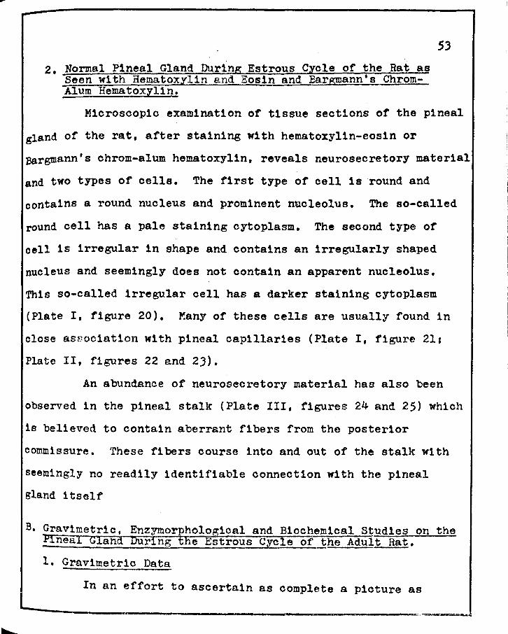
2. Normal Pineal Gland During Estrous Cycle of the Rat as Seen with Hematoxylin and Eos1n and BarEIIlann•s ChromAlum Hematoxylin.
53
Microscopic examination of tissue sections or the pineal
gland of the rat, after staining with hematoxylin-eosin or
Bargmann's chrom-alum hematoxylin, reveals neurosecretory material
and two types of cells. The first type of cell is round and
contains a round nucleus and prominent nucleolus. The so-called
round cell has a pale staining cytoplasm. The second type of
cell is irregular in shape and contains an irregularly shaped
nucleus and seemingly does not contain an apparent nucleolus.
This so-called irregular cell has a darker staining cytoplasm
(Plate I, figure 20). Many of these cells are usually found in
close as~ociat1on with pineal capillaries (Plate I, figure 211
Plate II, figures 22 and 2J).
An abundance of neurosecretory material has also been
observed in the pineal stalk (Plate III, figures 24 and 25) which
1s believed to contain aberrant fibers from the posterior
comm1ssure. These fibers course into and out of the stalk with
seemingly no readily identifiable connection with the pineal
gland itself
B. Gravimetric, Enzymorphological and Biochemical Studies on the Pineal Gland DUring the Estrous Cycle of the Adult Rat.
1. Gravimetric Data
In an effort to ascertain as complete a picture as
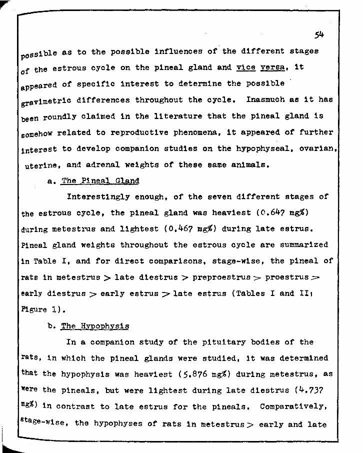
possible as to the possible influences of the different stages
of the estrous cycle on the pineal gland and ~ versa, it
appeared of specific interest to determine the possible
gravimetric differences throughout the cycle. Inasmuch as it has
been roundly claimed in the literature that the pineal gland is
somehow related to reproductive phenomena, it appeared of further
interest to develop companion studies on the hypophyseal, ovarian,
uterine, and adrenal weights of these same animals.
a. The Pineal Gland
Interestingly enough, of the seven different stages of
the estrous cycle, the pineal gland was heaviest (0.64? mg%)
during metestrus and lightest (0.467 mg%) during late estrus.
Pineal gland weights throughout the estrous cycle are summarized
1n Table I, and for direct comparisons, stage-wise, the pineal of
rats 1n metestrus > late diestrus > preproestrus > proestrus-:;:::
early d1estrus > early estrus ::;:> late estrus (Tables I and II t
Figure 1).
b. The HIROphysis
In a companion study of the pituitary bodies of the
rats, in which the pineal glands were studied, it was determined
that th~ hypophysis was heaviest (5.8?6 mg'.t) during metestrus, as
were the pineals, but were lightest during late diestrus (4.737
mg%) 1n contrast to late estrus for the pineals. Comparatively,
~ase-wtse, the hypophyses of rats 1n metestrus > early and late

55
estrus phases (almost identical, 5.408 and 5.390 mg% respectively)
> early diestrus and preproestrus (again almost identical,
4.855 and 4.842 mg% respectively).> proestrus>- late diestrus
(Tables I and IIIs Figure 1).
c .. The Ovaries
Companion gravimetric studies on the ovaries revealed
that those of rats in late estrus were heaviest (54.776 mg%) while
those in proestrus and late diestrus were lightest, the latter
two being almost comparable (!·~·47.640 and 47.683 mg% respect
ively). Comparatively, stage-wise, the ovaries of rats in late
est:rus > early estrus > metestrus > early diestrus > preproestrus
/- proestrus and late diestrus (Tables I and IV; Figure 2).
d. l'he Uteri
Studies on a much 1.nvestigated reproductive tract,
target-organ, the uterus, identified additional points of
1nteresta Of the seven different stages studied, the uteri of
early estrus were the heaviest (227.217 mg%), whereas those of
early diestrus were the lightest (138.903 m~). Comparatively,
stage-wise, the uteri of rats in early estrus > proestrus > late
estrus )> late d1estrus /=" metestrus > preproestrus '/>" early diestrus
(Tables I and V; Figure 3).
e. Xhe Adrenal Glands
Inasmuch as the adrenal glands have been importantly
elated to reproductive function, 1t thus became of essential

......
56
significance to include similar studies on the gravimetrics of
these glands. Interestingly enough, as with the ovaries, the
adrenal glands of rats in late estrus were heaviest (Jl.075 mg~)
and those of animals in proestrus were lightest (26.167 mg%).
comparatively, stage-wise, late estrus> late diestrus > metestrus
~ early diestrus ;:> early estrus and preproestrus > proestrus
(Tables I and VIi Figure 2).
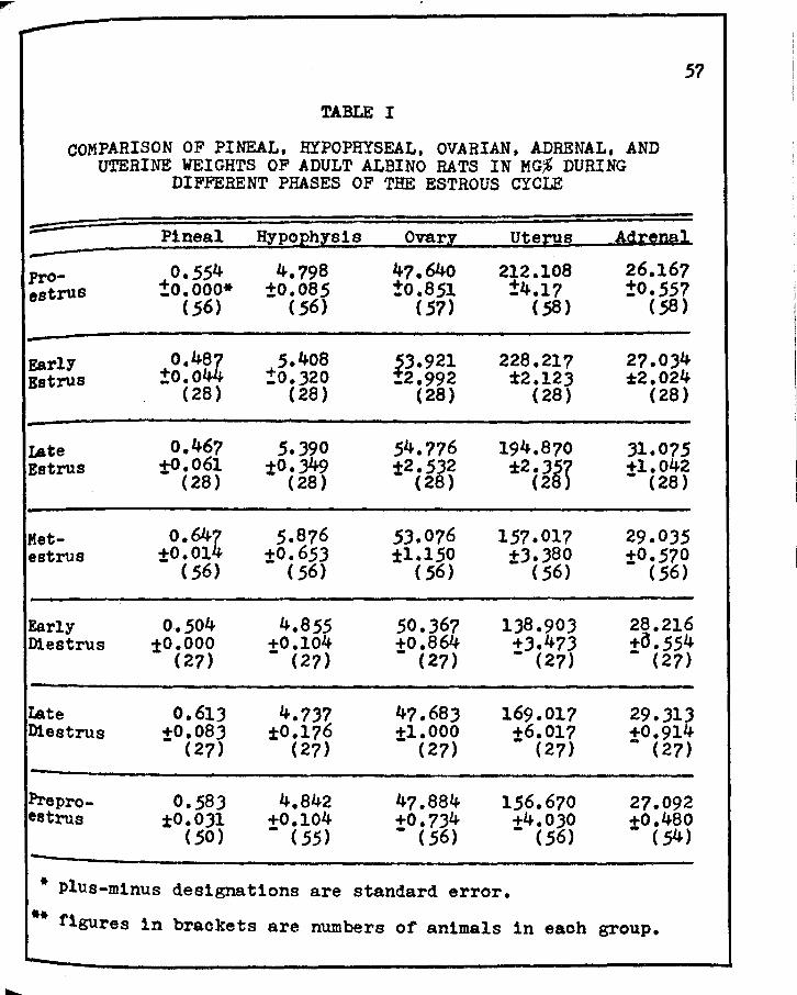
---57
TABLE I
COMPARISON OF PINEAL, HYPOPHYSEAL, OVARIAN, ADRENAL, AND UTERINE WEIGHTS OF ADULT ALBINO BATS IN MG,% DURING
DIFFERENT PHASES OF THE ESTROUS CYCLE
- Pineal HypoEhlsis ovarz Uterus Agt~rml -PrO- 0.554 4.798 47.640 212.108 26.167 estrus !0.000* ±0.085 ±0.851 !4.17 ±0.557
(56) ( 56) (57) ( 58) (58)
EarlY o.4~ 5.408 ~J.921 228.217 27.034 Estrus :to.o !O.J20 _2.992 ±2.123 ~2.024
(28) ( 28) (28) (28) (28)
Late o.467 s.390 54.776 194.870 )l.075 Estrus t0.061 :tO.J49 :t2.532 ±2.3.5r +l.042
(28) (28) (28) (28 - (28)
Met- o.644 5.876 53.076 157. 017 29.035 estrus t0.01 :t0.653 ±1.150 +3.380 :t0.570
( 56) ( 56) ( 56) - ( .56) (56)
Early 0.504 4.855 50.367 1J8.903 28.216 Di estrus t0.000 +0.104 +o.864 +3.47J +a.554
(27) - (27) - (27) - (27) - (27)
I.ate 0.613 4.737 47.683 169.017 29.JlJ D1estrus +0.083 ±0.176 :tl.000 +6.017 +0.914
- (27) (27) (27) - (27) - (27)
Pre pro- 0.583 4.842 4?.884 156.670 27.092 estrus ±0.031 +0.104 +0.734 +4.030 +0.480
(50) - ( 55) - ( 56) - (56) - ( 54) -
* Plus-minus designations are standard error.
** figures in brackets are numbers of animals in each group • ......_
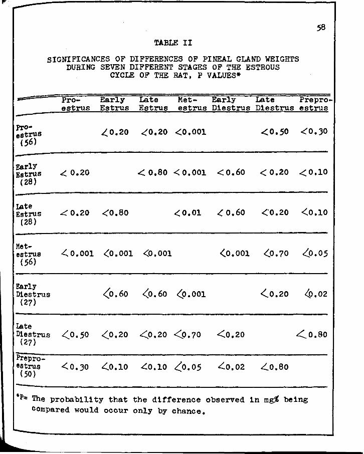
Jr'
.58
TABLE II
SIGNIFICANCES OF DIFFERENCES OF PINEAL GLAND WEIGHTS DURING SEVEN DIFFERENT STAGES OF THE ESTROUS
CYCLE OF THE RAT, P VALUES*
= Pro- Early Late Met- Early Late Pre pro-estrus Estrus Estrus estrus Di estrus Di estrus estrus
Pro- L.. 0.20 <_'0.20 <0.001 <o.so <(O.JO estrus ( 56)
EarlY ..( 0.20 <:. o.so < 0.001 <0.60 < 0.20 <0.10 Estrus
( 28)
Ia.te <o.ao <.. o. 60 <·0.20 <0.10 Estrus ~0.20 <( 0.01 (28)
Met-estrus .(0.001 <0.001 <0.001 (0.001 (0.70 z_o.05 (56)
Early <0.60 <0.60 (0.001 <.0.20 4>.02 Diestrus
(27)
I.ate Di estrus <0.50 <0.20 (27)
<.0.20 <0.70 <0.20 .:.(_ 0. so
Pre pro-.(0.10 <(o. 05 ~0.02 estrus <0.30 4(0.10 ~0.80
(50)
*P= The probability that the difference observed in mg% being compared would occur only by chance.

--59
TABLE III
SIGNIFICANCES OF DIFFERENCES OF HYPOPHYSEAL WEIGHTS DURING SEVEN DIFFERENT STAGES OF THE ESTROUS
CYCLE OF THE RAT, P VALUES*
::=:: Pro- Early Late Met- Early Late Pre pro-estrus Estrus Estrus estrus Di estrus Di estrus estrus
-PrO-
~0.20 <0.20 <0.20 <:0.70 ~0.80 <.o.ao estrus (,56)
-
EarlY <0.10 C::::0.80 <0.70 <:0.20 <.0.10 <..0.20 Estrus
(28)
Iste <0.20 <o.ao <0.60 <.0.20 <0.20 <.0.20 Estrus
(28)
Met-estrus <0.20 <0.70 <0.60 <'0.20 4(0.10 <.0.20
(56)
Early <o.4o Diestrus <:0.70 <(0.20 <0.20 <0.20 c('0.60
(27)
Late Di estrus
(27) <o.ao <0.10 (0,20 <0.10 <0.60 <0.70
Pre pro-estrus <o.so <0.20 <0.20 <0.20 <o.4o <0.70 (55)
-
* P = The prob:ib111ty that the difference observed 1n mg% being compared would ocour only by chance.
-b

-60
TABLE IV
SIGNIFICANCES OF DIFFERENCES OF OVARIAN WEIGHTS DURING SEVEN DIFFERENT STAGES OF THE ESTROUS
CYCLE OF THE RAT, P VALUES*
Pro- Early Late Met- Early Late Pre pro-estrus Estrus Estrus estrus D1estrus D1estruE1 estrus
Pro-<.0.0.5 <0.02 .(0.001 ~0.05 <0.975 <:0.90 estrus
(57) -
Early Estrus <(0.05 ~0.99 <o.ao ~0.,30 <0.10 ~0.10
(28)
Late Estrus ~0.02 <0.99 ~0.60 <:0.,30 ~0.02 <0.02
(28)
Met-estrus <::'.0. 001 <0.80 A(O. 60 <'0.10 .c:::o.001 <0.001
(56)
Early Di estrus c(0.05 <O.JO (27)
L.,O.JO ~0.10 4(0.05 ~0.05
Late D1estrus <0.975 <0.10 (27)
(0.02 <.0.001 ~0.05 ~0.90
Pre pro-estrus <0.90 (0.10 <:0.02 <0.001 <.0.05 <0.90 (56)
,_
* P = The probab111ty that the difference observed 1n mg,% 'being compared would occur only by chance •
....._
L
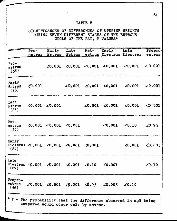
==-
-PrO-estrus c sa)
F.arlY Estrus
(28)
Late Estrus
(28)
Met-estrus
(56)
Early
TABLE V
SIGNIFICANCES OF DIFFERENCES OF UTERINE WEIGHTS DURING SEVEN DIFFERENT STAGES OF THE ESTROUS
CYCLE OF THE RAT, P VALUES*
Pro- Early Late Met- Early Late estrus Estrus Estrus estrus 121e~tnu1 Cl~~ttJJa
<0. 001 <'.'0.001 <0.001 <0.001 .co.001
<0.001 <'0.001 <0.001 <0.001 <0.001
<"0.001 <:0.001 ..c..o.001 <0.001 <0.001
<0.001 <0.001 <0.001 c:::o .001 <'0.10
D1estrus .(0. 001 <0.001 4).001 <0.001 ~0.001 (2?)
Late Di estrus <0.001 <t>.001
(2?) <o.001 <0.10 <0.001
Pre pro-estrus <_0.001 c(O. 001 <,t>.001 4>.95 <.0.005 .c::o.10
(56)
61
Pre pro eatz:.us
<0.001
£0.001
<0.001
.(0.9 5
<('0.005
~0.10
* P = The probability that the difference observed in mg% being compared would occur only by chanoe.
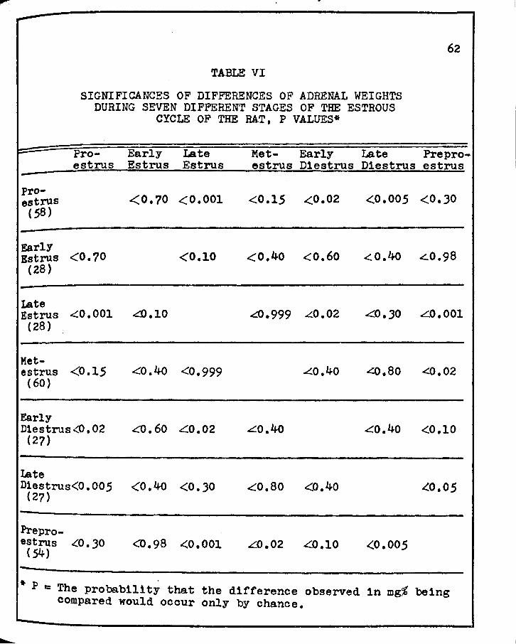
-62
TABLE VI
SIGNIFICANCES OF DIFFERENCES OF ADRENAL WEIGHTS DURING SEVEN DIFFERENT STAGES OF THE ESTROUS
CYCLE OF THE RAT, P VALUES*
~ Pro- Early Late Met- Early Late Pre pro-estrus Estrus Estrus estrus D1estrus Dlestrus estrus
-pro-
<0.70 .c(0.001 <0.15 ..(0.02 <0.005 <.O.JO estrus (58)
F.arlY <0.40 <0.60 <.0.4o <::.0.98 Estrus <0.70 <0.10
(28)
Late Estrus <'.0. 001 ~.10 4>.999 L.0.02 &.JO L0.001
(28)
Met-estrus <0.15 <0.40 <0.999 .L.0.40 ..(0.80 <:0.02 (60)
Early D1estrus <O. 02 <0.60 ~0.02 L0.40 ~0.40 <0.10
(2?)
Late D1estrus<O. 00 5 <0.40 <0 • .30 (27)
.co.so <0.40 ~0.05
Pre pro-estrus c::::o.30 <0.98 .(0.001 L0.02 ~0.10 <0.005 ( 54) -
* P = The probability that the difference observed 1n mg$ being compared would occur only by chance.
~

FIGURE 1
GRAPH SHOWING WEIGHT RELATIONSHIPS OF THE HYPOPHYSIS AND PINEAL GLAND DURING SEVEN DIFFERENr STAGES
OF THE ESTROUS CYCLE OF THE RAT
o.?r------------7
0.6 -~ C)
E - 0.5 -~ C)
·a; ~ 0.4
c Cl> c
a: 0.3
• •Pineal f <>---o Hypophyseal Weights
-~ 6 C)
E --5 ...c C)
Cl>
~ 4_
c G> en ~
...c 3 g-
Q. ~
:i:
0.2 ~P~--~E'----t--~~-=---~~_,_ __ .j2
t -E2 ME D1 02 PPE PE Stage of the Estrous Cycle

.•
FIGURE 2
GRAPH SHOWING WEIGHT RELATIONSHIPS OF THE OVARY, ADRENAL AND PINEAL GLAND DURING SEVEN DIFFERENT STAGES
OF THE ESTROUS CYCLE OF THE RAT
a1r----------------------0.6
-a-4t
:o.s --~ .~0.4
~ 0 0.3
CD c
0..
0.2
0. 1
............ ""<>---~----o
,~ ........ ,, ......... _, ___ _._ ___ .... __ _____ __... ......._... ........
• •Pineal I o----o Ovarian Weights .,. __ •Adrenal
....... ___ ..,.
70
-.. 60 Q
e -.,. 50~
Q
~ 40_
D c CD ._
-0 30~
PPEE--te--~""-Atc--;:t--~--.-'----.1j10 1 E2 ME D1 D2 PPE PE Stage of the Estrous Cycle

FIGURE .3
GRAPH SHOWING WEIGHT RELATIONSHIPS OF THE UTERUS AND PINEAL GLAND DURING SEVEN DIFFERENT STAGES
OF THE ESTROUS CYCLE OF THE RAT
Q7i:------------------------
0.6
~ Cl E 0.5
-..c C> ·~ 0.4 ~
g 0.3 c
Q..
0.2
0.1
,,'\ ,, \
<f' \ \ \ \ \ \ I
I I I
\ I \ I \ I \ I \ I \ I \ I \ I \ ~ I \ I ' I \ I ' I \ I ', I \ I , I ~ I ~
' I ' I ' I
' I ' I
• •Pineal I \,l ~---o Uterine Weights
230
210
-
-..c C> CD
170 ~ Q) c '-Q)
::> 150
PPEE -e:~--t:-r:c--.~~-~-~130 t E2 ME D1 D2 PPE Stage f PE o the Estrous Cycle
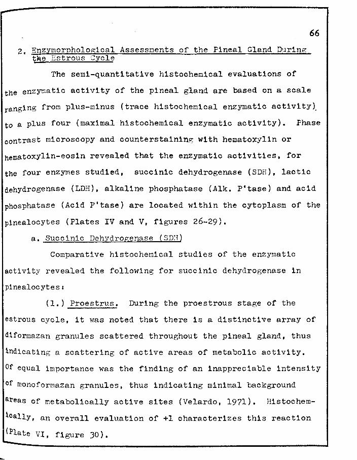
2. Enzymorpholo~ical Assessnents of the Pineal Gland Durin~ t~e Estrous Cycle
The semi-quantitative histochemical evaluations of
66
the enzymatic activity of the pineal gland are based on a scale
ranging from plus-minus (trace histochemical enzymatic activity).
to a plus four (maximal histochemical enzymatic activity). Phase
contrast microscopy and counterstaining with hematoxylin or
hematoxylin-eosin revealed that the enzymatic activities, for
the four enzymes studied, succinic dehydrogenase (SDH), lactic
dehydrogenase (LDH), alkaline phosphatase (Alk. P'tase) and acid
phosphatase (Acid P'tase) are located within the cytoplasm of the
pinealocytes (Plates IV and V, figures 26-29).
a. Succinic Dehydrogenase (SDH)
Comparative histochemical studies of the enzymatic
activity revealed the following for succinic dehydrogenase in
pinealocytess
(1.) Proestrus. During the proestrous stage of the
estrous cycle, it was noted that there is a distinctive array of
d1formazan granules scattered throughout the pineal gland, thus
indicating a scattering of active areas of metabolic activity.
Of equal importance was the finding of an inappreciable intensity
of monoforrnazan granules, thus indicating minimal background
areas of metabolically active sites (Velardo, 1971). llistochem
ically, an overall evaluation of +l characterizes this reaction
(Plate VI, figure 30).
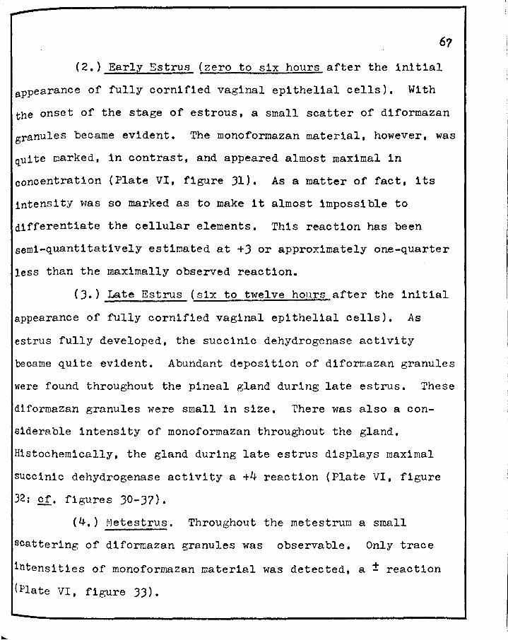
(2.) Early Estrus (zero to six hours after the initial
appearance of fully cornified vaginal epithelial cells). With
the onset of the stage of estrous, a small scatter of diformazan
granules became evident. The monoformazan material, however, was
quite marked, in contrast, and appeared almost maximal in
concentration (Plate VI, figure 31). As a matter of fact, its
intensity was so marked as to make it almost impossible to
differentiate the cellular elements. This reaction has been
semi-quantitatively estimated at +3 or approximately one-quarter
less than the maximally observed reaction.
(3.) Late Estrus (six to twelve hours after the initial
appearance of fully cornified vaginal epithelial cells). As
estrus fully developed, the succinic dehydrogenase activity
became quite evident. Abundant deposition of diforrr.azan granules
were found throughout the pineal gland during late estrus. These
diformazan granules were small in size. There was also a con
siderable intensity of monoformazan throughout the gland.
Histochemically, the gland during late estrus displays maximal
succinic dehydrogenase activity a +4 reaction (Plate VI, figure
32; £.£. figures 30-37).
(4,) Metestrus. Throughout the metestruro a small
scattering of diformazan granules was observable. Only trace
intensities of monoformazan material was detected, a ± reaction
(Plate VI, figure 33).

68
(5.) Early Diestrus (zero to twenty-five hours within
the stage of diestrus). During this phase of the estrous cycle,
an increased deposition of large diformazan granules was observed
in the pineal gland. A moderate intensity (+2.5) of material was
present (Plate VI, figure 34).
(6e) Late Diestrus (twenty-five to fifty hours within •
the stage of diestrus). As the diestrum fully developed, an
increased intensity of diformazan granules was found in a
scattered array throughout the pineal gland. Most of the
markedly intense succinic dehydrogenase activity was found in
perinuclear areas of the pinealocytes. Also noted was increased
intensity of the monoformazan background, a +2.5 composite
reaction (Plate VI, figure 35).
(7e) Preproestrus. During this phase of the estrous
cycle, a marked reduction in the intensity of diformazan granules
was quite evident. Also, there was an inappreciable intensity
of monoformazan material approximating a +l.5 overall reaction
(Plate VI, figure J6).
For the purpose of having this study rigidly controlled,
it appeared quite advisable to include parallel control sections,
~·~· those incubated without the substrate (sodium succ1nate) for
every pineal gland studied. Thus, for each determination of the
seven different stages of the estrous cycle, parallel controls
were incorporated. Such controls were uniformly devoid of
~------------~

ltnn
69
mono- and diformazan materials in each of the seven stages of
the estrous cycle. A typical control section is depicted in
Plate VI, figure J7. For immediate comparisons, both a summary
table and an illustrative graph are presented (Table VI1
Figure 4).
be Lactic Dehydrogenase CLPHl
The semi-quantitative histochemical analysis or lactic
dehydrogenase activity revealed the following for this enzyme in
pinealocytes 1
(1.) Proestrus. During this phase of the estrous
cycle, there is evidence of a maximally intense diformazan granule
reaction throughout the pineal gland. Likewise, there was a
maximally intense monoformazan reaction. The semi-quantitative
histochemical assessment of this stage of the estrous cycle was
the maximal designation, +4 (Plate VII, figure J8).
(2.) Early Estrus (zero to six hours). Coincident with
the onset of heat, 1·~· early estrus, a marked decrease in
intensity of lactic dehydrogenase became quite noticeable. Both
the mono- and diformazan material had overall intensities of
+l (Plate VII, figure 39).
(J.) Late Estrus (six to twelve hours). With the full
development of estrus, an almost maximally intense diformazan
reaction was observable (+J.5); likewise, a near maximal
monoformazan reaction was equally evident, thus giving an overall

70
~ 5 reaction (Plate VII, figure 40). +~·
(4.) Metestrus. During this phase, following late
estrus, only a slight reduction in d1formazan granules and
intensity was observed, a +J reaction. The monoformazan, although
present, was less pronounced. The intensity of the +J reaction
was for the most part due primarily to the d1formazan (Plate
VII, figure 41).
(5.) Early Diestrus (zero to txent~-f1ve hours). With
the onset of the d1estrum, a further reduction 1n overall
intensity was observed. Scattered diformazan granules of
moderate intensities were widespread. The monoformazan distri
bution and intensity, however, was slightly elevated over that
observed during the previous phase of the metestrum, thus giving
an overall +2.5 reaction (Plate VII, figure 42).
(6.) Late Diestrus (twenty-five to fifty hours). As the
diestrum progressed, the intensity of lactic dehydrogenase in
the pinealocytes, as assessed by the intensities of mono- and
diformazan, became markedly reduced from the previous estimate of
+2.5 down to +o.5 (Plate VII, figure 4J).
(?.) Preproestrus. As the diestrum waned, and the
initial appearance of estrus emerged, there was an overall
increase in the number of dlformazan granules. There was also a
marked increase in the intensity of monoformazan material,
giving the overall reaction of a +l {Plate VII, figure 44).
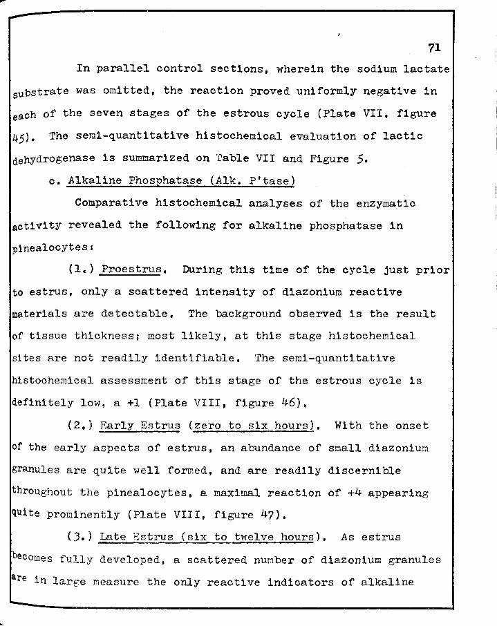
71
In parallel control sections, wherein the sodium lactate
substrate was omitted, the reaction proved uniformly negative in
e~ch of the seven stages of the estrous cycle (Plate VII, figure
45). The semi-quantitative histochemical evaluation of lactic
dehydrogenase is summarized on ·rable VII and Figure 5.
c. Alkaline Phosphatase (Alk. P'tase)
Comparative histochemical analyses of the enzymatic
activity revealed the following for alkaline phosphatase in
pinealocytes i
{le) Proestrus. During this time of the cycle just prior
to estrus, only a scattered intensity of diazonium reactive
materials are detectable. The background observed is the result
of tissue thickness; most likely, at this stage histochemical
sites are not readily identifiable. The semi-quantitative
histochemical assessment of this stage of the estrous cycle is
definitely low, a +l (Plate VIII, figure 46).
( 2.) Early Estrus (zero to six hours_2. With the onset
of the early aspects of estrus, an abundance of small diazonium
granules are quite well formed, and are readily discernible
throughout the pinealocytes, a maximal reaction of +4 appearing
quite prominently (Plate VIII, figure 47).
(J.) Late Estrus {six to twelve hours). As estrus
becomes fully developed, a scattered nunber of diazonium granules
re in large measure the only reactive indicators of alkaline
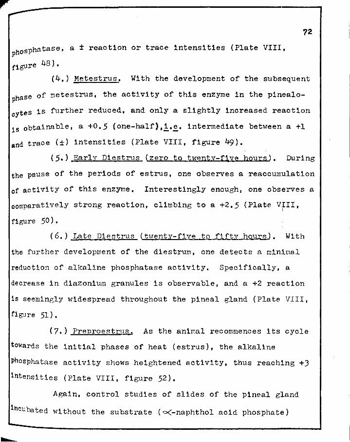
phosphatase, a ± reaction or trace intensities (Plate VIII,
r1gure 48).
72
(4.) Metestrus. With the development of the subsequent
phase of ~etestrus, the activity of this enzyme in the pinealo
cytes is further reduced, and only a slightly increased reaction
15 obtainable, a +0.5 (one-half),!·~· intermediate between a +l
and trace (±) intensities {Plate VIII, figure 49).
(5.) Early Diestrus (zero to twenty-fiye hours). During
the pause of the periods of estrus, one observes a reaccumulation
of activity of this enzyme. Interestingly enough, one obs1erves a
comparatively strong reaction, climbing to a +2.5 (Plate VIII, I
figure 50).
(6.) Late Diestrus (twenty-five to fifty hou~. With
the further development of the diestrum, one detects a mintmal
reduction of alkaline phosphatase activity. Specifically, a
decrease in diazonium granules is observable, and a +2 reaction
is seemingly widespread throughout the pineal gland (Plate VIII,
figure 51}.
(7.) Preproestrus, As the animal recommences its cycle
towards the initial phases of heat (estrus), the alkaline
Phosphatase activity shows heightened activity, thus reach1ng +J
intensities (Plate VIII, figure 52).
Again, control studies of slides of the pineal gland
incubated without the substrate ( o(-naphthol acid phosphate!)

73
clearly indicated that the pineal tissue sections so treated were
devoid of activity in each of the seven stages of the estrous
cycle. (Plate VIII, figure 5.3). Tabular and graphic represen
tations of these data are presented in Table VII and Figure 6.
d. Acid Phosuhatase (Acid P'tase)
Comparative histochenical analyses of the enzymatic
activity revealed the following for acid phosphatase in
pinealocytes:
(1.) Proestrus. During this phase of the estrous cycle,
just prior to estrus, large scattered diformazan granules,
indicating acid phosphatase activity, begin to become prominent,
The granules, ho·wever, are of low intensity, a +1.5 (Plate IX,
figure 54).
( 2. ) _Early :!:strus (.zero to six hours). As estrus
develops, both the number and intensity of the diazonium granules
become maximally developed and intensified, thus giving a +4
reaction for acid phosphatase (Plate IX, figure 55).
(J.) Late Estrus (six to twelve hours). With the full
develop~ent of the phase of heat, late estrus, the activity of
acid phosphatase becomes markedly reduced, and appears only of a
+l intensity (Plate IX, figure 56).
(4.) retestrus. Interestingly enough, the activity of
BC1d phosphatase during the metestrum is quite comparable to
that observed during the previous stage of late estrus (Plate IX,

74
figures 56 and 57).
(5.) Early Diestrus (zero to twenty-five hou$..§..}. With
the onset and development of the diestrum, the d1azon1um granules
show large coalescing clusters, several cell layers deep, and of
heightened intensity, thus giving a +J reaction (Plate IX,
figure 58).
(6.) Late Diestrus (twenty-five to fifty hours).
Clearly, the activity of acid phosphatase shows a marked rise
during the further development of the diestrum, a +J.5 (Plate IX,
figure 59).
(7.) Preproestrus. With the reappearance of the initial
estrous phases, the acid phosphatase activity shows a marked
reduction to a +2 (Plate IX, figure 60).
The control studies, omitting the o<-naphthol acid
phosphate substrate, was uniformly negative throughout the seven
stages of the estrous cycle. A representative control section
of the pineal gland is shown in Plate IX, figure 61. The
semi-quantitative histochemical evaluation for acid phosphatase
is summarized on Table VII and Figure 7.
J. Comparative Histochemical and Bioohemical Assessments of the Pineal Gland of the Rat During the Estrous Cycle.
a. Succinic Dehydrogenase (SDH~.
Utilizing similar techniques for the histochemical and
biochemical assessments for succ1nic dehydrogenase, 1t was
determined that similar maxima and minima were obtained. The
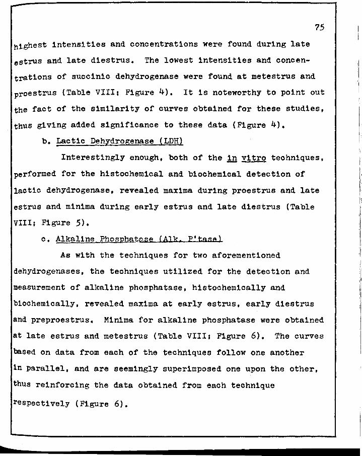
b
75
highest intensities and concentrations were found during late
estrus and late diestrus. The lowest intensities and concen
trations of succinic dehydrogenase were found at metestrus and
proestrus (Table VIII; Figure 4). It is noteworthy to point out
the fact of the similarity of curves obtained for these studies,
thus giving added significance to these data (Figure 4).
b. Lactic Dehydrogenase (LDH)
Interestingly enough, both of the !n vitro techniques,
performed for the histochemical and biochemical detection of
lactic dehydrogenase, revealed maxima during proestrus and late
estrus and minima during early estrus and late diestrus (Table
VIII; Figure 5).
c. Alkaline Phosphgtase (Alk. P'tase)
As with the techniques for two aforementioned
dehydrogenases, the techniques utilized for the detection and
measurement of alkaline phosphatase, histochemically and
biochemically, revealed maxima at early estrus, early diestrus
and preproestrus. Minima for alkaline phosphatase were obtained
at late estrus and metestrus (Table VIII1 Figure 6). The curves
based on data from each of the techniques follow one another
1n parallel, and are seemingly superimposed one upon the other,
thus reinforcing the data obtained from each technique
respectively (Figure 6).

76
d. Acid PhosEhatase (Acid P'tase)
Utilizing comparable histochemical and b1oohem1cal
techniques as were used for the estrous cycle studies on alkaline
phosphatase, except for the d1fferenoe in pH, these studies
revealed acid phosphatase maxima at early estrus and late diestru~
Minima for acid phosphatase were found at late estrus and
metestrus. It is of added interest to point out here the fact of
similarity of curves obtained for the histo- and biochemical
studies, thus showing remarkable agreements throughout the studies
of aoid phosphatase throughout the seven stages of the estrous
cycle of the rat (Table VIII1 Figure ?).
4. Preliminary Biochemical Data of the Pineal Gland During the Estrous Cycle of the Hat.
In a continuation of the biochemical studies, it
appeared of added interest to ascertain the biochemical
concentrations of two transaminases, glutamic-oxaloacetic
transaminase and glutamic-pyruvic transaminase, with the idea of
obtaining some preliminary data for future experimentation.
Furthermore, inasmuch as these two transam1nases are importantly
concerned with glutam1c acid metabolism and the central nervous
system, it seemed of interest to attempt to ascertain what role(s)
such might have in the pineal gland.
a. Glutam1c-Oxaloacet1c Transaminase (GOT)
Utilizing a oolorimetr1o test for glutam1c-oxaloacet1c

77
transaminase on the pineal gland of the rat during seven d1fferen
stages of the estrous cycle, it was ascertained that the peak,
maximal reaction was obtained at early diestrus, having ascended
during the stage of the metestrum. Thereafter, the peak descends
in two phasesr first, from the peak of early diestrus down 7% to late diestrus; secondly, almost 25% to the minimal reaction
obtained during preproestrus (Table IX1 Figure 8).
b. Glutam1c-Pyruvic Transaminase (GPT)
Utilizing a similar colorimetric test for glutamlc
pyruvic transaminase on the pineal gland of the rat during the
seven different stages of the estrous cycle, an initial early
cyclical rise of glutam1o-pyruvic transaminase activity was
obtained during late estrus, followed by a steady decline through
metestrus and early d1estrus, after which the enzymatic activity
ascended to its maximal peak during late diestrus, thereafter
falling precipitously at the initial phase of estrus, 1.e. at
preproestrus. Succinctly, the dominant and maximal peak activity
occurred at late diestrus whereas the lowest was clearly
associated with preproestrus (Table IX; Figure 9).

78
TABLE VII
SEMI-QUANTITATIVE HISTOCHEMICAL ESTIMATES OF SUCCINIC DEHYDROGENASE (SDB), LACTIC DEHYDROGENASE (LDH),
AL.T\ALINE PHOSPHATASE (ALK. P'TASE), AND ACID PHOSPHATASE (ACID P'TASE) OF THE PINEAL
GLAND OF THE RAT DURING THE ESTROUS CYCLE
SD11 LDB Alk. P'tase Acid P'tase
Early Estrus +3 +l +4 +4
Late Estrus +4 +3.5 ± +l
Met-estrus ± +J +0.5 +l
Early Dlostrus +2 +2.5 +2.5 +3
Late Di estrus +2.5 +o.s +2 +3.5
Pre pro-estrus +1.5 +2 +3.2 +2
Pro-estrus +l +4 +l +l.5

,.-..-
79
TABLE VIII
QUANTITATIVE BIOCHEMICAL ANALYSIS OF SUCCINIC DEHYDROGENASE (SDH), LACTIC DEHYDROGENASE (LDH), ALKALINE PHOSPHATASE
(ALK. P'TASE), AND ACID PHOSPHATASE (ACID P'TASE) OF THE PINEAL GLAND OF THE RAT DURING THE ESTROUS CYCLE
-- SDH* LDH** *** Alk. P'tase "~ Ao1d P'tase -EarlY
0.10±0.006 JJ.2±0.111 1.02±0.463 1.16±0.015 Estrus -Late
0.13±0.006 + 3.57±0.197 0.71±0.013 Estrus 117.2-0.312 -
Met-0.034±0.003 67.9±0.070 4.00±0.190 0.76±0.016 estrus
Early o.oao±o.003 62 • .s±o.100 6.45±0.775 1.04±0.016 Di estrus
!ate Di estrus 0.089±0.009 ;2.6±0.052 s.a 5:to. s49 1.13±0.011
Pre pro-estrus 0.055±0.004 60.5±0.172 6.59±1.123 o.aa±o.010
Pro-estrus 0.054±0.006 123.7±0.877 4.93±0.570 0.86±0.008
* l unit of SDH activity equals the number of )lM of f ormazan formed/mg pineal gland.
** 1 unit of LDH activity will reduce 4.8 X io-4 )lM pyruvate/ minute/mg pineal gland.
*** 1 unit of Alk. and Acid P'tase activity will liberate l)lM P-n1trophenol/hour/mg pineal gland (1 JlM = 0.1391 mg).
-
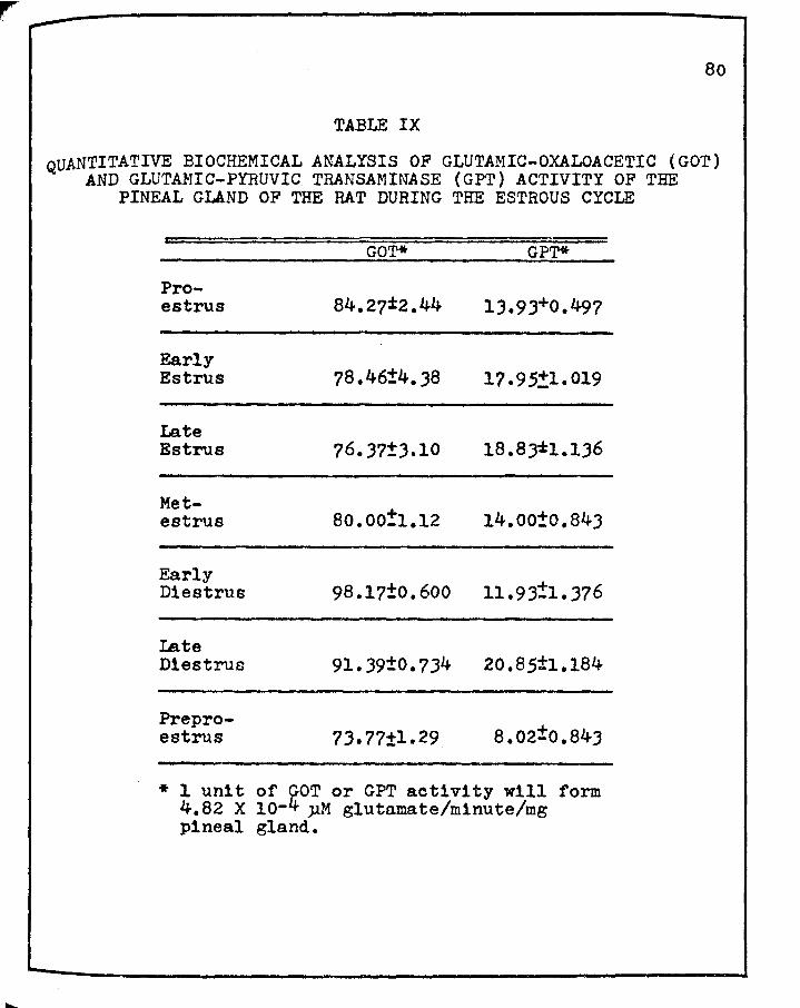
80
TABLE IX
QUANTITATIVE BIOCHEMICAL ANALYSIS OF GLUTAMIC-OXALOACETIC (GOT) AND GLUTAMIC-PYRUVIC TRANSAMINASE (GPT) ACTIVITY OF THE
PINEAL GLAND OF THE RAT DURING THE ESTROUS CYCLE
GOT* GPT*
Pro-estrus 84.27±2.44 13.93+0.497
Early 78.46±4.38 Estrus 17.95+1.019
Late Estrus 76.J?±J.10 18.8)*1.l.36
Met-80.00!1.12 estrus 14.oo±o.84J
Early 11. 9 ;:1. 37 6 Diestrus 98.17±0.600
I.ate Di estrus 91.39±0.734 20.85±1.184
Pre pro-a.02±0.843 estrus 73. 77±1. 29.
* 1 unit of goT or GPT activity will form 4.82 X 10- µM glutamate/minute/mg pineal gland.
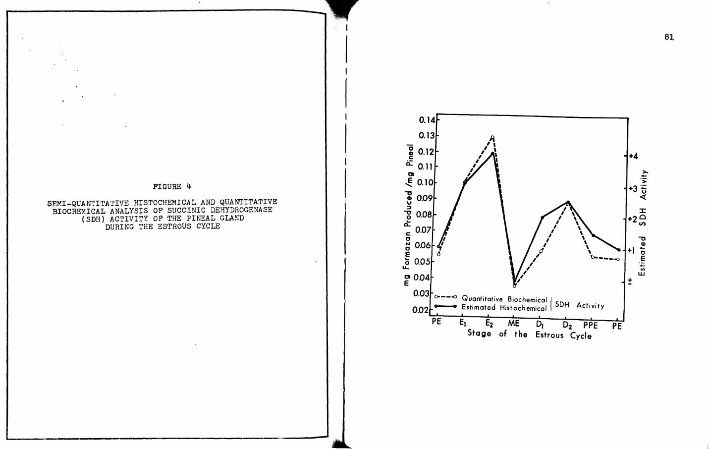
r--------;w'"
FIGURE 4
SEMI-QUANrITATIVE HISTOCHEMICAL AND QUANTITATIVE BIOCHEMICAL ANALYSIS OF SUCCINIC DEHYDROGENASE
(SDH) ACTIVITY OF THE PINEAL GLAND DURING THE ESTROUS CYCLE
o.14r-------------o.13
~ 0.12 c
0: 0.11 a
..!: 0.10 ,, ~ 0.09 :::>
-g 0.08 ct c 0.07
" ~ 0.06 E 0 0.05
u..
~ 0.04
/ I
I I
I I
I I ,,,,.i
,' , 0.03 o----o Q . . .
uant1tat1ve Biochemical I 0.02 • 1 Estimated Histochemical I SDH Activity
PE E1 E2 ME 01 02 PPE Stage of the Estrous Cycle
PE
+4
> +3 :;: u
<(
::c: +20
Cl')
,, Cl>
+1 ; E
+ -..... Cit
w
81

f'IGURE 5
SEMI-QUANTITATIVE HISTOCHEMICAL AND QUANTITATIVE BIOCHEMICAL ANALYSIS OF LACTIC DEHYDROGENASE
(LDH) ACTIVITY OF THE PINEAL GLAND DURING THE ESTROUS CYCLE
130 o----o Quantitative Biochemical I -• • Estimat--ed Histochemical LDH Activity
120 Q
- 110 " • -~ 100 Q..
Q 90 E
c 80 E
' 70
50
30
PE El E2 ME D
l I I I I I I I I I I I I I I
~ I
I I
I I
1 D2 PPE PE Stage of the E strous Cycle
+4
-u <(
·2 a _, .,,
CD -+1 e
+
-"' w
82

.----------------Y
FIGURE 6
SEMI-QUANTITATIVE HISTOCHEMICAL AND QUANTITATIVE BIOCHEMICAL ANALYSIS OF ALKALINE PHOSPHATASE
(ALK. P'TASE) ACTIVITY OF THE PINEAL GLAND DURING THE ESTROUS CYCLE
0 QJ
c: ·-a..
O'l E
....... ...:
...c
.......
8
7
6
I I
I I
I I
I I
I I
I
0 5 l c Cl>
...c Q.
~ 4 z
I
a.
~ 3
o----=> Quantitative Biochemical
1 2 • 'Estimated Histochemical Alk. P'tase Activity
PE E1 E2 ME D1 D2 PPE PE Stage of the Estrous Cycle
+4
-> +3:;:
u <{
CD
+2 ~
+
-..
.,, Q) -0 E -ut
w
BJ

FIGURE 7
.SEMI-QUANTITATIVE HISTOCHEMICAL AND QUANTITATIVE BIOCHEMICAL ANALYSIS OF ACID PHOSPHATASE (ACID
P'TASE) ACTIVITY OF THE PINEAL GLAND DURING THE ESTROUS CYCLE
1.2
c 1.1 cu
~ c
a... 1.0 C> E
....... ~0.9
0
~ 0.8 ,,L:,
a. 0 .!: 0] z
I Q.
~ 0.6
o----o Guan ti tative Biochemical I . 0.5 • •Estimated Histochemical Acid P'tase Activity
PE E1 E2 ME D1 D2 PPE Stage of the Estrous Cycle
PE
>.~ > -+4~ cu U)
c +3 :a... ,,
u +2 <{ ,,
cu -+1 c E
+ -U)
w
84

FIGURE 8
QUAN'rITATIVE BIOCHEMICAL ANALYSIS OF GLUTAMICOXALOACE'l'IC TRANSAMINASE ACTIVITY OF THE
PINEAL GLAND DURING THE ESTROUS CYCLE
L _________________ ___.
11or---------------
0 Q) c ·- 100 .. a..
O> E
........ 90 c
E
Q)
- 80--0 E 0
0 70-
:E :i
60 .. . PE
I . . . t
El E2 . .
ME 01 02 PPE PE Stage of the Estrous Cycle
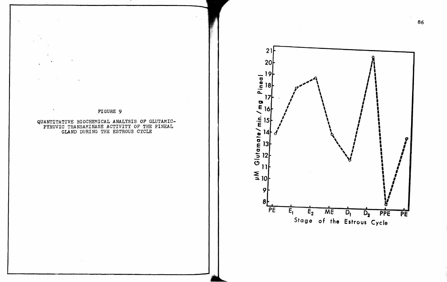
FIGURE 9
QUANTITATIVE BIOCHEMICAL ANALYSIS OF GLUTAMICPYRUVIC TRANSAMINASE ACTIVITY OF THE PINEAL
GLAND DURING THE ESTROUS CYCLE
21r"" -----------
ft I I
-19· ,.,, " ,,-'\ ~ 18. ,, \
a.. ff \ I I
I \ I ' 17· ' / \ I I Q I \ I I E 16.. I \ I I
......._ I \ I I . I \ I I
.E 15 .. 11
\ 11 I E \ I
/ \ I I
20•
~ 14• cf '" i \ ~ 13- \, I 1 f 0 ' I I I :; 12... ', I •• I
~ I I
9 ..
8· • . PE
. • . .
I I I I I I I I I I I I . ' I I ., ~ . ..
E2 ME D1 02 PPE PE Stage of the Estrous Cycle
86
8?
C Gravimetric and Enzy~ornholo~ical Studies of the Pineal Gland • Jurin.cc L'hree Stas:;:es of Pseudoprei::nancy in the Hat.
Inasmuch as the normal duration of pseudopregnancy lasts
for approxi~ately thirteen days, it seemed desirable to study
three phasesi (a) early, six days; (b) mid-point, at ten days;
and (c} the terminal day, at thlrte~n days. In this way, one
could obtain a rather inforIBa ti ve index of ac ti vi ty during the
major aspects of pseudopregnancy in the rat.
1. Gravimetric ~ata of Pineal Glands of Rats During 'P"seudopre~nancy.,
Contrasting the weights of the pineal glands of
pseudopregnant rats with those of the estrous cycle, one finds two
interesting phenomenas first, the pineal glands of early
pseunopregnancy (day six) and those of the tern::inal day of
pseudopregnancy (day thirteen) closely approximate the weights of
the pineal glands during proestrus (cf. Tables I and X);
secondly, the weights of the pineal glands of rats of advancP-d
pseudopregnancy (day ten) are quite variable, and closely resemble
the arithrratic mean of the pineal glands of rats in the late
phase of estrus (cf. Tables I and X).

TABLE X
THE WEIGHTS OF PINEAL GLANDS OF RATS DURING THREE STAGES OF PSEUDOPREGNANCY
=nays of PseuCiopregnancy
Day 6
Day 10
Day lJ
Num"5er =or Animals
8
7
lJ
weights of Pineal Glanas (mg~ !; S .E.)
0.540 ± 0.027
o.461 ! 0.253
+ 0.546 - 0.028
88
89
2. r.:nzyrr;orpholoi:;ical Assessment8 of the Pineal Gland During Pseudopregnancy,
a. Su.cc1n1c Dehydrogenase ( SmiJ
During the early phase of pseudopregnancy, the succinic
dehydrogenase activity in the pinealocyte appears almost
maxi~ally (+J), and achieves maxlnal and sustained activity at
the mid-point through the terminal day of pseudopregnancy (fable
X: ?igure 10; Plate X,figures 62-64).
For each period of pseudopregnancy, controls without
substrate were incorporated. rhe results of the controls, l·~·
without the sodium su.ccinate substrate, were uniformly negative.
b. Lactic Dehydro.o:enase (LDR)
A maximal reaction for lactic dchydrogenase activity
( +4) is found in the pinealocytes throughout the gland during
the early phase of pseudopreKnancy, 1·~· day six (Plate XI,
figure 65). As pseudopregnancy continues, the reaction for lactic
dehydrogenase becomes much less intense, the mono- and diformazan
~ranules manifesting only +2 reactions (Plate XI, fia;ure 66).
With the nearing of the cessation of pseudopregnancy, at thirteen
days, the reactive mono- and diformazan granules show heightened
activity and are typically +J in intensity (Table 10; Figure 10;
Plate XI, figure 67).
As previously stated, controls were studied along with
euch period of pseudopregnancy. The pineal sections incubated
' 1thout the sodium lactate substrate were uniformly negative.

90
c. Alkaline Phosphatase (Alk. P'tase)
The pineal ~land during these three periods of
pseudopregnancy contains pinealocytes that are heavily laden with
alkaline phosphatase (Plate XII, figures 68-70). The alkaline
phosphatase activity is oax1mum in intensity (+4) on day ten, and
is only about 25t reduced during the early and ter~inal stages of
pseudopregnancy, +J reactions (Table X; Figure 11).
Control sections incubated without the o(-naphthol acid
phosphate substrate manifested ne.p;ati ve reactions.
d. Acld Phosphatase (Acid f'tese)
Throughout the three periods of pseudopregnancy, days
six, ten, and thirteen, the deposition and intensity of 3Cid
phosphatnse was of hi~h moderate characterization, +3 intensities.
The control sections, without the o(-naphthol acid phosphate,
were uniformly negative (Table X; Figure 11; Plate XIII, figures
71-73).
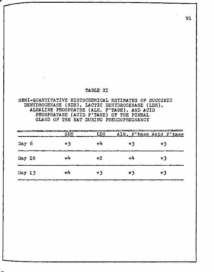
--
TABLE XI
SEMI-QUANTITATIVE HISTOCHEMICAL ESTIMATES OF SUCCINIC DEHYDROGENASE (SDH), LACTIC DEHYDROGENASE (LDH),
ALKALINE PHOSPHATSE (ALK. P'TASE), AND ACID PHOSPHATASE (ACID P'TASE) OF THE PINEAL GLAND OF THE RAT DURING PSEUDOPREGNANCY
SDH LDH Alk, P'tase Acid P'tase
Day 6 +3 +4 +J +J
Day 10 +4 +2 +4 +J
Day 13 +4 +J +3 +3
91
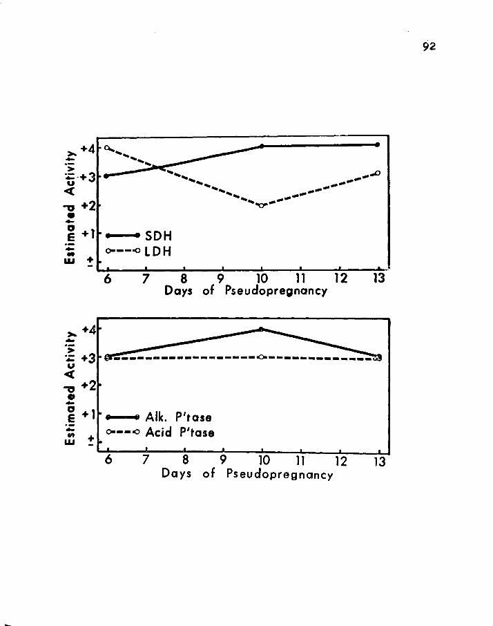
>- +4 - a... ... ...... 9 I
...... > ___ _.....-•)!IC<: ... : :e::-+3 F ...... _.., U ...... --...... --
<( ...... ----......... ----,, +2 • -
"'O"
" e+l .. ,_ .. •SDH -- o----o LOH w + -
6 7 8 9 10 11 12 Days of Pseudopregnancy
>- +4 • -·-
13
> ~ +3 r------------------.0.------------~ u <( .,, +2 • -~ + 1 • • Alk. P'tase -:;; o----o Acid P'tase w ~
6 7 8 9 10 11 12 Days of Pseudopregnancy
13
92

93
D cravirietric and Enz:ymorpholos:r1cql Studies of the Pineal Glands . -in Rats with Decidual 3eactions.
1. Grav1reetr1c Data of the Pineal Gland of Rats Bearin~ Decldual Reactions.
It is of interest to point up the fact of similarities
between pseudopregnant rats with and without decidual reactions
and with non-pseudopregnant, normally cycling albino rats. As
stated in the previous section (Cl.), rats on the terminal day
of pseudopregnancy have pineal gland weights that closely
approximate those of rats during the proestrous phase. Likewise,
the pineal glands of rats with decidual reactions on day thirteen
and during the terminal phase of pseudopregnancy (day twenty-one)
resemble in weight those of rats in proestrus (cf. Tables I, X,
and XII). As pseudopree:nnncy ndvr.i.nces to prolonged pseudopreg-
nancy (!.~· extending beyond the nor~al phase-length of pseudo
pregnancy, lJ.O days), it appears from these data that the
weights of the pineal glands resemble those of rats in early
diestrus and early and late estrus, 1·~· rats of fifteen and
eighteen days of prolonged pseudopregnancy status, respectively
Uables I and XII). Al though only three to five animals are
utilized per point, the trend of the gravimetrics is both
interesting and informative.
Specifically, it should be pointed out that the weights
of the pines.l glands of rats with decidual reactions for the most
Part resemble those of rats during preproestrus, proestrus, and
estrus (cf .Tables I and XII .
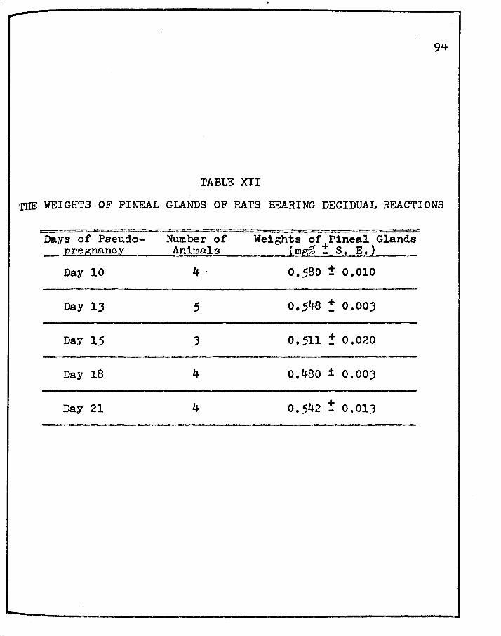
94
TABLE XII
THE WEIGHTS OF PINEAL GLANDS OF BATS BEARING DECIDUAL REACTIONS
Days of Pseudo- Number of Weights of Pineal Glands 12regnanc~ Animals 'mg~ :t s 1 E.)
Day 10 4· 0.580 ± 0.010
Day lJ 5 0.548 : 0.003
Day 15 J o. 511 ! 0.020
Day 18 4 0,480 ± 0.003
Day 21 4 0.542 ! 0.013

2. Enzy~orpholo~1cal Assessments of the Pineal Glands of Pseudopre~nant Rats Bearin~ Dec1dual ieactlons.
a. Succ1n1c Dehydrogenas~ (SDH)
The pineal glands of rats bearing decidual reactions
95
show for the most part moderate to maximal succinic dehydrogenase
intensities in pinealocytes, a notable exception being those of
day fifteen of prolonged pseudopregnancy wherein the reaction is
slightly less than moderate, a +1.5 (Figure 12; Plate XIV,
fieJres 74-80). Initially, during the development of the
antimesometrial reaction (A~~) of the decidual reaction, the
succinic dehydrogenase reaction within the pinealocytes appears
as a moderate reaction, a +2. On day ten during which time the
antimeso~etrial reaction has reached its peak with some areas
showing initial regression, and the mesometrinl reaction (I:a)
becomins quite enlarged, the pinealocytes reach almost naximal
intensity (+J.5) for succinic dehydrogenase (Plate XIV, figure
75).
Interestingly enoueh, during the next forty-eight to
seventy-two hours, while the rnesometrial reaction reaches its
peak in development and the metrial gland (EG) beco~cs organized,
the pinealocytes begin to show decreasing intensities of this
enzyne (Plate XIV, figure 76), With the full development of the
metrial gland on day fifteen of prolonged pseudopregnancy, the
succinic dehydrogenase activity within the pinealocytes reaches
its lowest intensity thrrn1ghout the superimposed event, a +1.5
(Plate XIV, fip;ure 77), Thereafter, on day eighteen, when both
the antimeso~etrial reaction and metrial reaction are almost
totally necrotic and during which tine the metrial gland ls the
most pro~inent, the pinealocytes show maximal intensities (+4)
for succinic dehydrogenase (Plate XIV, fip;ure 78). As the
metrial gland undergoes necrosis, dissolution and liquefaction,
the pinealocytes show a marked drop in succinic dehydrogenase,
from a peak reaction of a +4 to a +2.5 on day twenty (Plate XIV,
figure 79) and finally at the time of metrial gland involution,
1.e. day 21, the pinealocytes reveal a further decrease to a +2 - -(moderate)reaction (Table XIII; Figure 12; Plate XIV, figure 80).
It is of further interest to point out here that the
very strong (+J.5) and maximal succinic dehydrogenase reactions
(+4) obtained on days ten and eighteen in the pinealocytes are
truly typical of those seen during early and late estrus phases
of the estrous cycle.
Control sections of pineal glands incubated without
sodium succ1nate were entirely devoid of any detectable succinic
dehydrogenase activity.
b. Lac tic JJehydror.r;en8.se ( LDI1)
Interestingly enough, whereas the ~aximal reaction (+4)
for succinic dehydrogenase in the pinealocytes was on day
eighteen, the maximal in intensity (+4) for lactic dehydrogenase
in the pineal parenchyrnal cells appears on day thirteen
...

97
(cf. Figures 12 and lJs Plate XIV, figure 78 and Plate XV, -figure 83). As with succ1nio dehydrogenase, the lactic
dehydrogenase intensities in these cells reveal a picture of
increasing intensity from days six through ten (Q!. Figures 12
and 1J1 Plate XJI, figures 81 and 82). Thereafter, the lactic
dehydrogenase picture shows slight variation from that of
suoc1n1c dehydrogenase. In the lactic dehydrogenase series, on
day thirteen the peak is reached (Figure 131 Plate X:V, figure
8J), but in the succinic dehydrogenase series a slight decrease
1s observed (Figure 12). On day fifteen, however, a precipitous
decrease in intensity of both succ1nic dehydrogenase and lactic
dehydrogenase is observed (£!. Figures 12 and lJr Plates XIV and
XV, figures 77 and 84). Both enzymes show a rise in intensity
on day eighteen, being maximal (+4) for succ1n1c dehydrogenase,
and very strong (+J) for lactic dehydrogenase, but approximately
25~ less than the maximal for succ1n1c dehyd.rogenase (£!. Figures
12 and 13t Plates XIV and XJI, figures 78 and 85). Thereafter,
the intensities for succinic dehydrogenase and lactic dehydrogen
ase decrease in steps to a +2.5 on day twenty (£!. Figures 12
and 13; Plates XIV and XV, figures 80 and 87; Table XIII).
For each pineal gland, a control without the sodium
lactate substrate was incubated, and as previously noted, resulted
in a negative reaction.
II.

98
c. A1kal1ne Phosphatase (Alk. P'tasel
The p1nealocytes throughout the superimposed physio
logical event of pseudopregnancy, with the exception of the
terminal day 1·~· day twenty-one, reveal a moderate to maximal
intensity for alkaline phosphatase. The reaction is maximal on
daY fifteen, falls precipitously to a moderate intensity on day
eighteen (1·~· a +2), plateaus at same through day twenty, and
becomes slightly less intense on day twenty-one, scoring the
lowest reaction for alkaline phosphatase at this time, a +1.5
(£!. Table XIII: Figure 141 Plate XVI, figures 88-94).
Control pineal sections incubated without the
o(-naphthol acid phosphate were totally devoid of any alkaline
phosphatase activity.
d. Acid Phosphatase {Acid P'ta~e)
Histochemical studies for acid phosphatase in the pineal
glands of pseudopregnant rats bearing decidual reactions clearly
indicate that the p1nealocytes react most strongly for this
enzyme. From day six through thirteen, very strong to near
maximal reactions are obvious (+J.5, day six; +J.O, day tenr
+J.5, day thirteen). A maximal peak reaction of a +4 becomes
quite prominent on day fifteen, after which linear, precipitous
decreases become quite noticeable on days eighteen and twenty,
a +J and +2, respectively. On day twenty-one, the terminal day
of prolonged pseudopregna.ncy, a strong, moderate acid phosphatase

reaction in the pinealocytes emerges from the previous days
minimal reaction, thus increasing from +2 to +2.5 (Table XIII;
Figure 15; Plate XVII, figures 95-101). Control pineal gland
sections incubated without o(-naphthol acid phosphate proved
uniformly negative.
99

100
TABLE XIII
SEMI-QUANTITATIVE HISTOCHEMICAL ESTIMATES OF SUCCINIC DEHYDROGENASE (SDH), LACTIC DEHYDROGENASE (LDH),
ALKALINE PHOSPHATASE (ALK. P'TASE), AND ACID PHOSPHATASE (ACID P'TASE) OF THE PINEAL GLAND OF THE RAT WITH DECIDUAL REACTIONS
~ r;~~ Ir!C 1 i"l'tase Acid P~tase
Day 6 +2 +J.2 +3 +J.5
Day 10 +J.5 +3.5 +2.5 +3
Day 13 +J +4 +J +3.5
Day 15 +1.5 +2 +4 +4
Day 18 +4 +3 +2 +J
Day 20 +2.5 +2.5 +2 +2
Day 21 +2 +2 +1.5 +2.5

l"IGURE 12
SEMI-QUANTITATIVE HISTOCHEMICAL EVALUATION OF SUCCINIC . DEHYDROGENASE ( SDH) ACTIVI·rY OF THE PINEAL GLAND
IN RATS BEARING DECIDUAL REACTIONS
-> -u <( +3
:z: 0 V) +2 ,, Q) .. " .§ +1 .. .,, w
+ -
101
6 8 10 12 14 16 18 20 Days of Pseudopregnancy
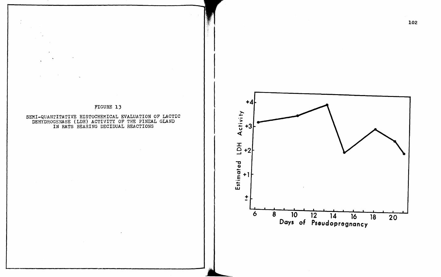
FIGURE 13
SEMI-QUANTITATIVE HISTOCHEMICAL EVALUATION OF LACTIC DEHYDROGENASE (LDH) ACTIVITY OF THE PINEAL GLAND
IN RATS BEARING DECIDUAL REACTIONS
+4 >. -> ~ +3 u <{
:c 0 +2 ...J
.,., Q) -~ +1 -en
w
+ -6
102
8 10 12 14 16 18 Days of Pseudopregnancy
20

FIGURE 14
SEMI-QUANrirATIVE HISTOCHEMICAL EVALUATION OF ALKALINE ~HOSPHATASE (ALK. P'TASE) ACTIVITY OF THE PIW"~L
GLAND IN RATS BEARING DECIDUAL REACTIONS >-. +4 -·-> -u
<{ +3 Cl> U)
0 -.. a.. . +2 ~
-0
~ +1 0 E -Cl)
w +
l
lOJ
6 8 10 12 14 16 18 Days of Pseudopregnancy
20

FIGURE 15
SEMI-QUANTITATIVE HISTOCHEMICAL EVALUATION OF ACID PHOSPHATASE (ACID P'TASE) ACTIVITY OF THE PIW"~L
GLAND IN RATS BEARING DECIDUAL REACTIONS
>-. +4 -> -u
<( +3 G> Cit 0 -..
Q.. +2
" u <(
-g +1 -0 E -&I) + w
104
6 8 10 12 14 16 18 20 Days of Pseudopregnancy

Gravimetric and Enzvmornholo~lca E. 0 Pre~nant Rats.
Prefatory nemarlrn
105
The major aim of this dissertation was focused on the
pineal gland of the rat during the estrous cycle, and to a much
lesser extent on the influence of pseudopregnancy and decidual
tissue on the pineal gland. Following the studies on pseudo-
pregnancy and decidual tissue, however, it became of interest to
attempt a small, pilot study on the influence of pregnancy on the
pineal gland, owing primarily to the fact that the observations
on pseudopregnancy and prolonged pseudopregnancy with decidual
tissue proved of remarkable significance. Immediately noteworthy
are the facts of increased intensities of succinlc dehydrogenase,
lactic dehydrogenase, alkaline phosphatase, and acid phosphatase
during prolonged pseudopregnancy. The question naturally
proE:pted the investigation of the pineal gland of rats during the
biological life span of pregnnncy. Specifically, it appeared of
great interest to ascertain whether or not the presence of viable
fetuses in addition to decidua could in some way further modify
the enz~~atic intensities of each of the enzymes studied.
1. Gravirnctric Do.ta of Pineal Glands of Prep:rn=mt Bats
The gravimetric studies on the pineal glands of pregnant
rats reveal that there is a marked rise in the weights during
the early aspects of pregnancy. Notably, a marked rise in
ineal weights is seen over that of estrus, during which time
.. -106
fertilization occurs. On days six and eight of pregnancy, the
pineal e;land weights are 0.630 and 0.732 mg~ 1 respectively
(Table XIV). Thereafter, weights of the pineal glands are truly
representative of those observed during metestrus, particularly
daY thirteen of pregnancy, and of proestrus (days ten and fifteen)
and early estrus (days eighteen, twenty, and twenty-one).
Comparatively, these data are best viewed by examining
Tables X, XII, and XIV).

107
TABLE XIV
WEIGHTS OF PINEAL GLANDS OF RATS DURING PREGNANCY*
uays of Number or Pregnanc;y** Animals
Number of' Fetuses
weigff'fS""or Pine~ (mg% ! S, E.)
DaY 6 5 9 0.630 : 0.018
Day 8 8 8 0.732 '! 0.043
Day 10 4 lJ 0.528 + 0.045
Day 13 5 8 0.676 ! 0.055
Day 15 6 9 0 • .549 '! 0.022
Day 18 7 10 o.490 ± 0.036
Day 20 5 10 o.492 ! 0.027
Day 21 6 11 o.484 : 0.020
* Obtained from Charles River Breeding Laboratories, North Wilmington, Massachusetts.
** Verified as to duration of pregnancy according to the criteria of Christie, 1964, cf. Appendix: A.

109
pinealocytes in the gland of the pregnant rat is of comparatively
iow succinlc dehydrogenase activity, a +1.5. Following this to
daY eight of pregnancy, during which time the development of
differentiated germ layers is nearing completion (totally on d~y
nine), the succinic dehydrogenase picture in the pinealocytes
becomes almost maximally intense, a +J,8). During the early
development of the central nervous system (CNS), starting from
approximately day ten, one observes an increase in intensity in
almost linear fashion through day eighteen, at which time much of
the central nervous system 1s complete. The succ1n1c dehydrogen
ase pattern goes from a +l to a maximal +4 reaction during this
time (Plate XVIII, figures 102-107). During the time when the
fetus is normally achieving its definitive form, eighteen to
eighteen and one-half days, and the time of parturition (days
twenty-one to twenty-two), the succ1nic dehydrogenase reaction
decreases, v.s. to an all-time low of a +l (Table XV; Figure 16:
Plate XVIII, figures 102-109).
b. Lactic Dehydrogenase (LDH}
The pinealocytes of the rat from day six through day
twenty-one .of pregnancy show moderate to maximal intensities for
lactic dehydrogenase. The semi-quantitative histochemical
estimates for lactic dehydrogenase reveal a strongly moderate
intensity on day six (+2.5), a decrease on day eight (+l}, and
a steady increase in intensities in three steps to the maximal
110
reaction day fifteen (+4), cf. Table XV: Figure l?a Plate XIX,
r1gures 110-114. Whereas the minimal reaction (+l) appears on
daY twenty-one for succ1nic dehydrogenase, the minimal lactic
dehydrogenase intensities (+2) appear on days eight and eighteen
(of. Table XV). Interestingly enough, the reaction for lactic ........
dehydrogenase shows another upswing after the second minima,
ascending to a +3.5 on day twenty and slightly less to a +J on
day twenty-one (Table XVr Figure l?r Plate XIX, figures 115-117).
The lactic dehydrogenase intensities in the pinealocytes seem to
rebound in deep intensities immediately after each of the minima
(£!. Plate XIX, figures 110-117). Control pineal gland sections
1noubated without the substrate, sodium lactate, proved entirely
negative.
The maximal reactions for succinic dehydrogenase and
lactic dehydrogenase appear in the pregnant animal after much of
the central nervous system has been formed in the embryo, a
rather interesting finding, especially so when one attempts a
correlation between the pineal gland and the events of reproduc
tion. Another interesting finding is that of the maxima for
succ1n1c dehydrogenase and lactic dehydrogenase when the corpora
lutea vera are the largest in the pregnant rat (Velardo, 1958).
Thus, a rather interesting series of events, making this aspect
a worthy point of departure for future studies.

111
c. Alkaline Phosphatase (Alk. P'tase)
The alkaline phosphatase activity of the pineal gland
f the pregnant rat shows a most remarkable picture, starting
1th trace (!) intensities on day six, becoming almost moderately
intense (+1.5) on days eight and ten, thereafter becoming more
1ntense on day thirteen (+J) and subsequently becoming maximally
intense (+4) on day fifteen. The alkaline phosphatase reaction
decreases almost 50% on day eighteen (+2) and rises only meagerly
thereafter, 1.e. a +2.5 on days twenty and twenty-one (Table XV;
Figure 18r Plate XX, figures 118-125). Control pineal gland
sections incubated without the substrate, c:<-naphthol acid
phosphate, were entirely negative for alkaline phosphatase.
The general observations for the dehydrogena.ses again
erta1n here, and one begins to see quite interesting relation
ships among succinic dehydrogenase, lactic dehydrogenase, and
lkaline phosphatase in the p1nealocytes during pregnancy.
d. Acid Phosphatase-(Acid P'tase)
The results for acid phosphatase quite closely
parallel those obtained for lactic dehydrogenase (cf. Table XV;
Figures 17 and 19). rhe acid phosphatase reaction in the
P1nealocytes on day six appears moderately intense (+2), becomes
slightly more intense on day eight (+2.5), remains similarly
intense on day ten, thereafter becoming substantially more
intense on days thirteen (+J), fifteen (+J.5) and maximally so on
-112
daY eighteen (+4). The pinealocytes are heavily laden with acid
phosphatase (diazonium granules) on day eighteen, and appear only
slightly less so on days twenty and t·wenty-one ( +J), cf. Table
XV; ?igure 19: Plate XXI, figures 126-lJJ. Control pineal gland
sections incubated without the substrate, o(-naphthol acid
phosphate, proved devoid of any acid phosphatase activity.
It ls of interest to point up here the remarkable
finding of the near uniformity of intensities of the four enzyDes
studied, at the tine of rraxlmum corpus luteum size during
pregnancy, and at the time of near completion of foetal internal
develop~ent and definitive foetal fore (Christie, 1964).
These studies point the way for an extensive analysis
of the pineal sland during the whole biological career of
pregnancy, with an attempt to ascertain and possibly correlate
the several neuroendocrinal rclRtionships (and interrelationships)
aruong the pineal gland, the hypothalamus, the ovaries, the uterus,
and the products of conception as well.

TABLE XV
SEMI-QUANT.ID\ L1IVE HISTOCHEMICAL ESTIMATES OF SUCCINIC DEHYDROGENASE ( SDH) , LAC'l,IC DEHYDROGENASE ( LDH)
ALKALINE PHOSPHATASE (ALK, P'TASE), AND ACID PHOSPHATASE (ACID P'TASE) OF THE PINEAL
GLAND OF THE RAT DURING PREGNANCY
SDH LDH Alk, P'tase A~id P'tase
Day 6 + - +2 +1.5 +2.5
Day 8 +J.8 +2 +1.5 +2.5
Day 10 +2 +J +1.5 +2.5
Day 13 +2.5 +J.5 +J +J
Day 15 +2 +4 +4 +J.5
Day 18 +4 +2 +2 +4
Day 20 +J.5 +J.5 +2.5 +J
Day 21 +l +3 +2.5 +J
llJ
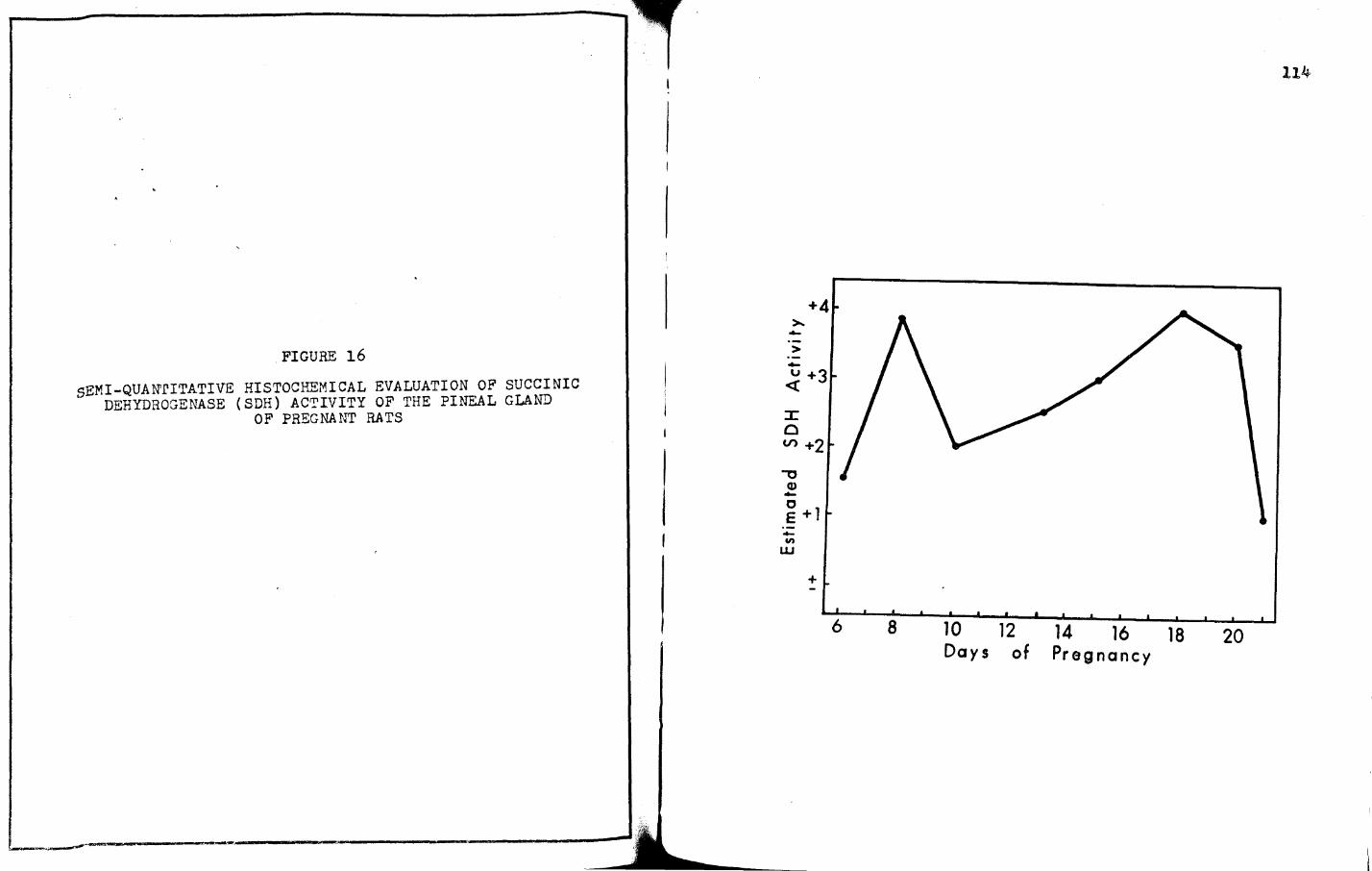
FIGURE 16
sEMI-QUAN'rITATIVE HISTOCHEMICAL EVALUATION OF SUCCINIC DEHYDROGENASE (SDH) ACTIVITY OF THE PINEAL GLAND
OF PREGNANT RATS
----~---=,---------------------~------------~~------------...
+4
-·-> -u +3 <(
:x: 0 V> +2 ,,
CD -0 E +1 -Cl)
w
+
114
6 8 10 12 14 16 18 20 Days of Pregnancy

FIGURE 17
SEMI-QUANTITATIVE HISTOCHEMICAL EVALUATION. OF LACTIC DEHYDROGENASE (LDH) ACTIVIrY OF THE PI~AL GLAND
OF PREGNANT RATS
I
I
>--> ·--u <(
::x:: 0 _. .,,
Cl> -" E -.,, w
115
+4
+3
+2
+1
+ -
6 8 10 12 14 16 18 20 Days of Pregnancy
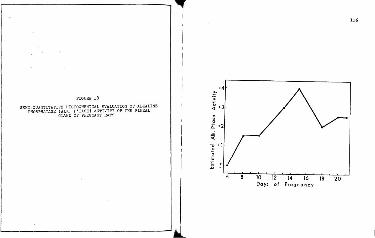
FIGURE 18
SEMI-QUANTITA1'IVE His·roCHE!1ICAL EVALUATION OF ALKALINE PHOSPHATASE (ALK. P'TASE) ACTIVITY OF 'I1HE PINEAL
GLAND OF PREGNAl'n RATS
~-----__.lit_
+4 >.. -> ·--:( +3
CD en 0 ~ +2 a..
~
<t: -0 +1
Cl> -c E - + U)
w -
116
6 8 10 12 14 16 18 20 Days of Pregnancy

-
11?
.?:- +4 > -:f'.'IGURE 19 u
<{
SEMI-QUA~TITATIVE HISTOCHEMICAL EVALUATION OF ACID . PHOSPHATASE (ACID P'·rASE) ACTIVITY OF THE
PINEAL GLAND OF PREGNANT RATS
+3 CD .,, 0 -.. a_
"'lJ +2 u
<{
~ +1 -'
0
I E -
I .,, + w -
6 8 10 12 14 16 18 20 Days of Pregnancy
.._.__ ___ __...J_

CHAPTER V
DISCUSSIOr~
As an organ of uncertain endocrine function, the pineal
gland has been at the forefront long before the study of
endocrinology became a reality. The suspicions that the pineal
gland might in some way be associated with gonadal function
emerged from two pieces of evidences first, true parenchymatous
pineal tumors are usually associated with depressed gonadal
funct1oni secondly, pineal tumors which eventually become
widespread throughout the gland, so as to cause enormous
destruction of it, are usually associated with precocious puberty
(Kitay, 1954). Thus the concept emerged that the pineal gland is
the source of a horn.one which could affect the gonads. That the
pineal gland is enshrouded in rather complex- or at least hard to
elucidate-physiological phenomena is well documented (Quay, 1969;
Sommers, 1958; Albert, et!±_., 1954; Kitay and Altschule, 1954;
Kappers and Shade, 1965; Cohen et al., 1964: Rilkin, 1966; and
Wurtman and Axelrod, 1965).
However complex the pineal may be, it is not surprising
to note that endocrinologists and general medical scientists have
been in agreement for the past two thousand years regarding the
118

119
obscurity of the function(s) of this gland. The stark facts
point up one basic concepts the physiological functions of the
pineal gland in mammals remain to be elucidated. This is not to
convey the thought that practically nothing is known; to the
contrary, some major pieces of knowledge exist. It is for exactly
the reason of the obscurity of knowledge pertaining to the pineal
that this dissertation came into being. The facts that were
available made it all the more challenging, for like all new
basic sciences in general ''Endocrinology is not truly beyond the
pioneering stages" (Velardo, 19 58).
In a careful appraisal of the information extant,
several challenging pieces of information are available:
First, embryologically, it is well known that the pineal
glB.nd develops from an evagination of the roof of the dicncephalon
and that the pineal parenchymal cell is most likely derived fror'l
the pril'li ti ve ependyr.ial lining of the third ventrical. Neurop.;lia
is its chief supporting structure. The surprising fact, however,
emerges from the concept that while it is developed from neural
tissue, the pineal gland has no direct innervation from the
brain in mammals.
Secondly, although nerve fibers from the epithalamic
region (habenular) penetrate the pineal, it has been shown that
these fibers are exceptional (for their straying characteristics)
and en:crge from the habenular commissure, loop through the pineal
-120
without synapsing, and return to the opposite side of the brain.
While habe:nular fibers do not in fact innervate the pineal, it
is known that postganglionic sympathetic nerve fibers, which have
cellular origins in the superior cervical ganglia, reach the
pineal gland in association with an abundant blood supply. The
8 ympathetic nerves terminate chiefly in the interstitial tissue
spaces of the pineal, and not unco~~only in direct contact with
a pinealocyte, thus making a substantial arrangement for
allowing neurohurnoral substances released from sympathetic
neuronal endings in these spe.ces to diffuse into the pinealocytes.
Giving the picture of a true endocrine gland is the fact that the
pinealocytes are in close association with a basement membrane
in contac~ with the interstitial space, and this basereent
rner:ibrane in turn is in contnct vd th a capillary basement rnembr:;ne
and a fenestrated capillary endotheliun, This cyto~orphological
architecture allows for control of the pinealocytes by the
autonomic nervous system O!olfe, 1965).
Thirdly, at least three active substances have been
isolated from the pineal gland: melatonin, norepinephrine and
serotonin. It has been roundly demonstrated by histochemical
and pharmacological techniques that the sympathetic nerve fiber
endin~s of the pineal gland contain norepinephrine and serotonin.
In the pineal gland, however, serotonin occurs in sympathetic
nerve endings. It has also been roundly established that

-121
pinealocytes synthesize rather copious quantities of serotonin
which diffuse into the interstitial space, the latter being
bound by the nerve endings.
It should be emphasized that serotonin and norepi-
nephrine are not unique to the pineal gland. Specifically, it
should be emphasized that melatonin is synthesized only in the
pinealocytes. As was stated earlier, Lerner and his associates,
in the Department of Derffiatology at Yale University School of
~edicine, attempting to isolate the active principle in the pineal
gland which caused blanching of the skin of frogs, came upon
such an amine and thus discovered melatonin. Of especial
significance is the fact that melatonin, a most active ~elano-
phore contra.cting principle, acts on cells to cause their granules
to cluster with a dramatic lightenin~ of the skin. This important
aspect, serving as a biological assay, gave rise to a number of
sip;nificant observations which ultimately led to the chemical
isolation and synthesis of .melatonin (Lerner, et al. , 19 .59;
Cohen, et al., 1961).
Fourthly, melatonin is formed from ~-acetylserotonin,
having been derived in turn from serotonin, cf. accompanying - -
diagram.
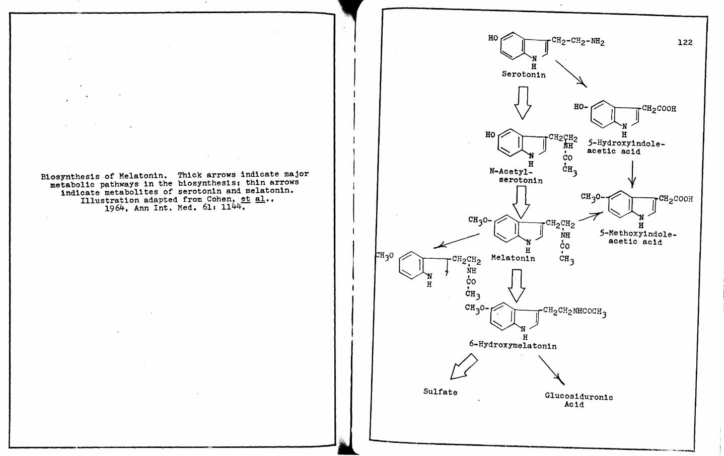
B1osynthes1s of Melatonin. Thick arrows indicate major metabol1c pathways 1n the biosynthesis; thin arrows
indicate metabolites of serotonin and melatonin. Illustration adapted from Cohen, et al.,
1964, Ann Int. Med. 611 1144:- -
Sulfate Glucos1duron1c Acid
122
123
The enzyme which methylates N-acetylserotonin, hydroxyindole-
o-methyl transferase (HIOMT), 1s found only in the pineal gland.
Inasmuch as hydroxyindole-0-methyltransferase oan be made to
vary under specific experimental conditions, it provides ~
fide clues indicating certain of the factors which regulate ............... pineal activity. It is well established that hydroxyindole-0-
methyltransferase ls found within the pinealocytes. The concen
tration of this enzyme depends upon the physiological activity of
the sympathetic nerves which are in association with the pineal
gland, for it is known that sectioning of these nerves markedly
diminishes the hydroxyindole-0-methyltransferase activity.
Likewise, transplantation of the gland to an ectopic location
remote from the nerve supply to the pineal gland_results in
markedly reduced hydroxylndole-0-methyltransferase activity.
Fifthly, the endocrine influence of the mammalian
pineal gland seemingly depends upon alterations in the photoperioc
and possible other environmental factors (Quay, 1970).
Sixthly, the reproductive systems of mammalian species
are morphologically and physiologically altered by the pineal
gland. It is now well known that removal of the pineal gland
accelerates gonadal growth in immature animals. and causes a
transient. but less well pronounced enlargement of the repro
ductive organs when the p1nealectomy is performed after adulthood,
Evidence is also at hand showing that shortened light cycles tend
124
to make the pineal remarkably antigonadotropic. It has also been
shown that chronic administration of pineal extracts or pineal
substances elicits effects which are opposite to those of
pinealectomy. Specifically, such extracts restrict gonadal
developr~ent as well as gonadal growth (Reiter and Sorrentino,
19?0).
To be sure, one could add a number of other associated
facts, and one could present commentaries on some controversial
and discredited concepts concerning the pineal gland, ~·B·
adrenoglomerulotroplc, cardiovascular, thymic, hypophyseal,
thyroidal effects, etc. Suffice it to say that the six categories
above are in the main some of the most basically discussed and in
varying degrees yet tenable concepts regardin1; the pineal. For
added details on the pineal, including nu~erous non-tenable
points, the reader's attention is invited to the review of the
literature herein presented, wherein a number of additional
points, facts and contentions are also presented. In the main,
the above six concepts present a balanced foundation upon which
to erect further exploratory studies including those of the
Present dissertation.
The aforementioned facts are quite revealing of the
Paucity of basic information p~rtaining to the status of knowledge
of the pineal gland. It, therefore, became of paramount interest
to initiate a series of experiments that would in fact beam new

125
1nfor~ative insi~hts into the pineal gland. In the process of
11 tter:iptintr, to set up sorre ne1·r experiments, IT'UCh helpful guidance
vias derived from (a) the status of published knowledge on the
subject, and (b) some specific questions that are important to
our further elucidation of pineal histophysiology.
Specifically, it appeared of interest to ascertain
something of the "form and function" relationships of the pineal
~land during specific reproductive mechanisms in the rat. First,
it appeared of interest to answer certain basic questions
regarding the role of the pineal gland during the estrous cycle.
Is the pineal gland in any way modified as a result of the estrous
cycle chan~es in the reproductive tract? Can one detect
histor:wrphological changes? Are there some 00.sic and crl ti cal
weight changes within the pineal gland? Can one demonstrate
specific changes in certain groups of enzymes, ~·3· dehydro
genases and phosphatases, during estrous cycliclty? Are there
any detectable biochemical changes? fhus, the aforementioned
questions set the pace. Almost concurrently, a second and a
third group of questions evolved.
Of no less importance than the planned estrous cycle
stuoies, the thought of investigating differences in pineal
Weights and historr.orphology during pseudopregnancy becarr:e quite
important. Of added significance, the whole idea of pursuing
studies of the pineal gland during pseudopregnancy in rats bearing

126
decidual reactions soon became of a realistic nature to stimulate
another set of questions.
With the original goals set forth to attempt to uncover
some basic, and quantifiable data on the pineal gland of the rat
during the estrous cycle and in pseudopregnant rats with and
without decidual reactions, it sequentially appeared of interest
to pursue a number of studies on the pineal gland during
pregnancy. The one basic question which was quite stimulating
pressed forwards Can basic changes be demonstrated in the pineal
gland throughout the biological career of pregnancy? The
challenging concept lu:dmlg behind such thinking was to initially
examine the pineal gland during a time when the life of the
corpus luteum is seemingly quiescent, as it is during the estrous
cycle when luteal function cannot be demonstrated, to a time
when the rat 1s reproductively dependent upon the corpus luteum
as it is during pseudopregnancy, dec1dual development and much
of pregnancy.
In a series of experiments, commene1ng with the estrous
cycle, two major thrusts were aimed at elucidating basic data
pertinent and associated with estrous cycle phenomena.
Specifically, 1t was determined that there are statistically
s1gn1f1cant changes 1n the weight of the pineal gland in each of
the seven stages of the estrous cycle. Gravimetric data at
hand reveals that the pineal glands of rats in metestrus are

127
heaviest while those in late estrus are lightest, weighing about
one-third less.
Examination of the gra.vimetric data suggests rather
convincingly that the pineal gland is quite cyclical in weight
relationships in regard to the estrous cycle, and shows most
noteworthy correlation with changes in the cycle. During that
tin:e (estrus) when the pineal gland is lightest in weight, the
ovaries are undergoing an enormous amount of endocrine activity,
just prior to ovulation. The ovaries are responding to pituitart
gonadotropins, and pineal gland secretion of melatonin can, in
fact, control the level of at least one pituitary gonadotropin,
luteinizing hormone(LH), as borne out in the researches of
?raschini, Pi va and .<artini ( 19 69). These workers showed quite
convincingly that melatonin can in fact reduce the adenohypo
physeal stores of luteinizing hormone.
Recently, Ka':lberi, Vical and Porter (1971) reported
that ~elatonin, as well as serotonin, when injected into the
third ventricle of the brain of male rats, appears to stimulate
the release of prolactin and inhibit the release of follicle
st1Mulating hormone (FSfl) as judged by the changes in the
concentrations of these hornones in the plasma of recipient
animals. Careful analysis of their radioimmunoassays suggests
that melatonin or serotonin "rw.y have suppressed" the discharge
of prolnctin inhibiting factor (PIP) and follicle releasing
hormone (FRH) and thereby indirectly affected the release of
prolact1n and follicle stimulating hormone.
128
In pseudopregnant rats without decidual reactions, in
initial and terminal phases of pseudopregnancy, the weights of
the pineal gland are typical of the weights of pineals of
proestrous rats, whereas those of the mid-aspect (day ten of
pseudopregnancy) are more typical of late estrus, !·~· during the
time one would normally expect the secretion of melatonin to be
at a level to physiologically control lutein1z1ng hormone.
The pineal glands of rats with decidual reactions are
equally of interest. Previously, it was shown and discussed that
the pineal gland of the rat on the terminal day of pseudopregnancy
(day thirteen) has a pineal gland weight typical of that of a
proestrous ratr likewise, the pineal gland of the pseudopragnant
rat bearing a deoidual reaction on day thirteen is of a similar
weight relationship. Moreover, for the most part, the pineal
gland weight relationships throughout prolonged pseudopregnancy
(!·~· of pseudopregnant rats bearing decidual reactions) are
typical of preproestrus, proestrus and early estrusr one notable
exception being on day fifteen of prolonged pseudopregnancy, at
Which time the weight relationship resembles that of early
diestrus. Again, the point can be made that during luteal
functional activity and during the time of decidual development,
the pineal gland is comparatively smallest.
129
The pineal gland weights during pregnancy present a
most noteworthy conditiont namely that there is a truly rr.arked
rise initially. During both initial ti~est days six and eightt
pineal ueights a.ppear somewhat typical of metestrus; the pineal
glands are the heaviest at these timest respectively. Thereaftert
at day ten, the weight drops to that seen during proestrus, but
again rises to the metestrus pattern on day thirteen, after
which the weight pattern drops to proestrous (day fifteen) and
estrous patterns (days eighteen to twenty-one). The noteworthy
concept here is that during pregnancy, the weights of the pineal
gland are truly typical of those obtained during the three
shortest phases of the estrous cycle.
An additional cor'1mentary on the gravimetric data
concerns thG hypophyseal, ovarian, uterine, and adrenal 1veights.
On one hand, one observes that when the pineal gland weight is
the heaviest, weights for the pituitary bod;>r are also highest;
~t. on the other hand, at the time of high~st pineal weights,
the weights of the ovaries and adrenals fluctuate about the
mid-point ranses, while the uterine weights are almost at their
lonest point. Conversely, when one observes that the weight of
the pineal gland is li~htestt one finds in association the
heaviGst ovarian and adrenal Hcightst and at this time the
Pituitary body and uterine weights fluctuate about the mid-point
ranr.res. 11 future arbe it will focus on the mircroanato1ny and

lJO
histochemistry of these removed organs so as to look for addi
tional relationships.
Subsequent investigations could develop major inroads
1nto the study of the pineal gland with detailed blostatistical
analyses. Also, in a future study, precise wet and dry pineal
weight measurements could be determined so as to get another
index of activity of this gland. This would facilitate a much
more accurate assessment of the changes which occur throughout
the estrous cycle, pseudopregnancy, prolonged pseudopregnancy
and pregnancy.
Although the gravimetric data forms a small but
interesting part of this dissertation, it does point up areas
that can in fact provide take-off points for correlated form and
function studies, especially at the times when the pineal ls
heaviest and lightest, and perhaps when the pineal can be shown
to be changing in predictable increments and decrements. The
present attempt, although in minor proportion to the major
histochemical theme, does provide worthy and quantifiable data,
and even more important, avenues for future research 1n this area.
The histochemical and biochemical studies provide a
major inroad into some of the hitherto, non-explained concepts
regarding the pineal gland during reproductive mechanisms.
Especially important and noteworthy are the estrous cycle studies.
It is of clear significance that both the histochemical and

r
l~
biochemical studies pertaining to succinic dehydrogenase (SDH)
reveal that of the seven stages investigated, the highest
intensities and concentrations within the pinealocytes were
found to occur at the time of estrus. This is the time during
which an enormous amount of metabolic energy is required for the
great crescendo of endocrine activity associated with the period
of heat and the events of ovulation (Velardo, 1951; Bever, et al.,
1954; Rosa and Vela~do, 1954a and b, 1955: 1958, 1959; Velardo
and·Rosa, 1963). It is also of notable significance that the
lowest intensities and concentrations of this dehydrogenase were
determined to occur immediately following ovulation, 1·~· 1n the
estrous cycle stages designated as metestrus (.£!. preceding
references of Rosa and Velardo, and Velardo and Rosa).
Realizing the significance of the finding pertaining to
succ1nic dehydrogenase, 1t naturally appeared of interest to
ascertain the pattern of lactic dehydrogenase (LDH) activity
during the estrous cycle. Histochemical and biochemical deter
minations for lactic dehydrogenase proved to be quite rewarding.
Whereas the maximal peak for succinic dehydrogenase occurred at
estrus and the minimal value at metestrus, the maximal and
minimal points for lactic dehydrogenase occurred at proestrus
and late diestrus, respectively. Here, one should emphasize that
the metabolic energy cycle leading to lactate production precedes
that of the tr1carboxyl1c acid-succ1nate energy yielding reaction.
132
Again, realizing the enormous amount of metabolic work and
energy required for the accomplishment of endocrine expression
associated with the periods of heat and ovulation, it is indeed
quite significant to point up the findings suggesting a biological
order of prioritiess first, during proestrus, lactic dehydrogenase
appears maximally; secondly, as proestrus advances to estrus,
succinic dehydrogenase appears in maximal designations, 1n both
h1stochemically demonstrable intensities and biochemically
determined concentrations. Such has also been found in the
reproductive tracts of estrous, intact and estrogenized,
ovariectomized rats (Bever, Velardo and Hisaw, 1953 a, br Rosa
and Velardo, 1953, 1954 a, br 1959r and Velardo and Rosa, 196J).
Observations on alkaline and acid phosphatase revealed
that the pineal gland was maximal for both of these enzymes
during early estrus. The high intensities and concentrations of
these two phosphatases markedly decrease after early estrus,
decreasing to minimal reactions during late estrus and metestrus.
The data on the phosphatases are of exciting interest, partic
ularly since it has been repeatedly shown that (a) alkaline
Phosphatase is associated with rapidly growing tissues in the
reproductive tracts of animals and man, especially during periods
of estrus or in ovariectomized animals given estrogens (Velardo,
1954r Mc Kay, et al., 1966 a, b; and Velardo and Rosa, 196J);
(b) acid phosphatase is also associated with rapidly growing

13J
tissues in health and disease, and it has been shown to occur
in near peak concentrations and 1ntensit1es 1n the reproductive
tracts during estrous phases and after estrogenizat1on in a num
ber of species including the human female (cf. Velardo and Rosa,
196J); and (c) the results for alkaline and acid phosphatase
obtained in the pineal gland are in essential agreement with
those ~ound in the reproductive tract.
O~ added interest are the results of the preliminary
colorimetric determination of glutamic-oxaloacetic transaminase
(GOT) and glutamic-pyruvio transaminase {GPT). Both of these
enzymes are known to catalyze the reaction resulting in the
formation of glutamic acid. This amino acid, in turn, can
either be metabolized to glutamine or to gamma-aminobutyric acid.
Current investigations on gamma-aminobutyric acid (GABA) have
recently become of great interest stemming from the hypothesis
that this acid probably plays an important functional role in
the modulation (or inhibition) of neurotransmitter substances in
the mammalian central nervous system (Bloom and Iverson, 1970;
Sze and Lovell, 1970).
The high concentration of glutamic-oxaloacetic trans
aminase and glutamic-pyruvic transaminase in the pineal gland
during the diestrum could possibly be indicative of increased
gamma-aminobutyric acid production. If this is indeed true, the
increased concentration of gamma-aminobutyric acid may be an
134
indicator of pineal inhibition other than via melatonin and the
biogenic amines as has been suggested and refuted. {Wurtman,
et al., 196Jr Chu, et al., 1964r Kappers, 1962r Ebels and Prop, --19651 Chessman, 1970r Chessman and Farriss, 1970: Debeljuk, 1969r
Moszkowska and Ebels, 1968: etc.).
During pseudopregnancy, as with the estrous phases of
the cycle, the semi-quantitative histochemical estimates for
succinic dehydrogenase, lactic dehydrogenase, alkaline phospha
tase, and acid phosphatase in the pineal gland are very strong to
maximal, the notable exception being lactic dehydrogenase on
day ten of pseudopregnancy at which time only a moderate intensity
(+2) is obvious. Previously, it was noted that during the estrous
cycle the maximum for lactic dehydrogenase in the pineal gland
appears before that of suecinic dehydrogenase, and the same
situation occurs during pseudopregnancy. Specifically, lactic
dehydrogenase is maximal on day six whereas succinic dehydrogenase
appears to be maximal on day ten. It ls at this time that the
corpus luteum of pseudopregnancy is nearly maximum in size, as
has been reported by Dawson and Velardo (1955). The phosphatases
are likewise of interest for they are of very strorgintenslties
(all +J reactions with a maximum +4 for alkaline phosphatase
occurring only on day ten). This correlates quite nicely with
the high concentrations of these phosphatases 1n the reproductive
tract when the corpus luteum is functional (£.!:. Velardo and Rosa,

135 i96J1 Dawson and Velardo, 1955).
Inasmuch as it appeared quite important to correlate
some data with the pineal studies in pseudopregnant rats bearing
decidual reactions, it will perhaps be sufficient to preface
those remarks here, and then provide a commentary on the possible
correlation of the pineal with the physiological expression of
prolonged pseudopregnancy. It was quickly seen that the succinic
dehydrogenase reaction (+2) and those of lactic dehydrogenase,
alkaline phosphatase and acid phosphatase (+J,to +J.S reactions)
are moderate to strong at the time or the 1n1t1al developing
stages of the antimesometrial reaction, the latter normally seen
on day six of pseudopregnancy in rats with dec1dual reactions
(c~. Velardo, Dawson, Olson and Hisaw, 1953 for decidual develop
ment). It was also pointed up that on day thirteen the intensi
ties for all four of these enzymes are near maximal intensities,
and it is at this time that the prolonged pseudopregnancy study
of Velardo, et al. (1953) indicated that the mesometrial reaction
reaches peak development. It is also at this time that the
metrial gland becomes organized and proceeds at a fast develop
mental pace (Velardo, Dawson, Olson and Hisaw, 1953). The
present study clearly identifies this as the time during which
the enzymatic intensities in the pineal gland show variable but
1et substantial intens1tiess (a) succinic dehydrogenase varies
from a +3 on day thirteen, thereafter declines to a +1.5 on day
I I
r l
1J6
fifteen, rises to a +4 on day eighteen, and declines to a +2.5
and a +2 on days twenty and twenty-one; (b) the lactic dehydrogen
ase pattern is almost exact, with the notable exception of a +J
rather than a +4 on day eighteen; (c) the alkaline phosphatase
intensities increase from a +J (on day thirteen) to a +4 on day
fifteen, thereafter plateau at a +2 on days eighteen and twenty
and slightly decrease on day twenty-one to a +1.5; and lastly
(d) acid phosphatase shows a near maximal (+J.5) reaction (on
day thirteen), increases to a maximal reaction day fifteen, there
after decreases in intensity from a +J to a +2 to a +2.5 on days
eighteen, twenty, and twenty-one, respectively.
The cogency for repeating the data from the thirteenth
day of pseudopregnancy resides in the fact of the coincidence of
time of the initial aspect of prolonged pseudopregnancy, for 1t
is at this time that if large decidual reactions are induced, the
animals do not return to estrus, and reappearance of estrus is
delayed until days twenty-one to twenty-two, the normal duration
of prolonged pseudopregnancy. Specifically, some rather
interesting histochemical data from the pineal gland studies pose
some remarkable thoughts regarding some of the non-explored areas
of reproductive endocrinology. Numerous thoughts emerge1 (a)
What accounts for the sudden, remarkable decrease in intensity
of succinic dehydrogenase at the time of the metrial gland
development, and the noteworthy rise in intensity to maximal

137
strength within seventy-two hours following the minimal reaction
observed throughout the course of prolonged pseudopregnancy?
(b) Can one ascribe such to the elusive, but present hormone
called relaxin, a hormone secreted by the reproductive tract
primarily during luteal activity? (cf. Velardo, et al., 1963;
Wislocki, et !!l• 1957: Dixon and Bulmer, 1971); (c) Can the
variations in enzymatic activity as observed in the pineal gland
during pseudopregnancy be duplicated with injections of relaxin,
estrogens and progestins alone or in dual combinations, or in
concert?; and (d) Can one show even more dramatic changes in the
pineal gland during pregnancy?
To be sure, the thought emerged to pursue these studies
in pregnant animals so as to determine the pinealsreproductive
interrelationships during the time when reproductive activity and
luteal life are seemingly dominant. The results obtained were
much more intriguing than any of the data obtained from pineal
glands of rats during pseudopregnancy, with and without decidual
reactions. Whereas the maximal reactions of each of these enzymes
during pseudopregnancy was between days six and ten, and whereas
the maximal reactions for these enzymes during prolonged pseudo
Pregnancy (pseudopregnant animals bearing decidual reactions)
appeared as early as day thirteen for lactic dehydrogenase,
followe~ by equally maximally intense reactions for alkaline and
acid phosphatase on day fifteen, and thence followed seventy-two

138
hours later, on day eighteen, by maximal reactions for succin1c
dehydrogenase, the histochemical reactions during pregnancy
showed most remarkable and intriguing patterns for max1mas on
daY fifteen both lactic dehydrogenase and alkaline phosphatase
are maximal; on day eighteen, both succinic dehydrogenase and
acid phosphatase are maximal. Even more intriguing is the fact
that none of the maxima are actually observed until after the
onset of the second half of pregnancy, specifically the m1d- to
near-terminal aspects of pregnancy. These data make it rather
obvious that the maximal reactions for lactic dehydrogenase and
alkaline phosphatase not only precede those of succinic
dehydrogenase and acid phosphatase but are importantly prominent
at the time of full metrial gland development. Note wells the
maxima for alkaline phosphatase, +4 reaction, occurs on day
fifteen in both pregnancy and prolonged pseudopregnancy states.
The question reappearss Is there a correlation between relaxin
of the metr1al gland and the pineal gland - specifically between
relax1n and melatonin? Is the correlation in any way governed
by the high intensities of lactic dehydrogenase and alkaline
Phosphatase within the pineal gland?
Another interesting aspect regarding maxima is the fact
that the maximal reactions for succinic dehydrogenase during
Prolonged pseudopregnancy and pregnancy occur on the same day,
day eighteen. The acid phosphatase reaction maximizes on day

1J9
eighteen, seventy-two hours later than that seen during prolonged
pseudopregna.ncy. The interesting aspects !!.2!! emerge• seemingly
exhaustion of the ant1mesometrial reaction, and the necrosis of
the mesometrial reaction can in some way be correlated with the
maximal reactions for lactic dehydrogenase and alkaline phospha
tase. Moreover, the full development of the metrlal gland, too,
ean 1n some way be correlated with the pineal gland peak reactions
of lactic dehydrogenase, alkaline phosphatase and during the
subsequent seventy-two hours with the rising to peak activities
of succinio dehydrogenase and acid phosphatase. Dallenba.ch
Hillweg, Battista and Dallenbach (1965): Wislocki, Weiss, Burgos
and Ellis {1957); and Dixon and Bulmer (1971) have independently
shown by histochemical, immunofluoresoent, and electron micro
scopical evidence that the metrial gland is heavily laden at this
time, and present further evidence suggesting that relax1n is a
hormone of the metrial gland. Thus the future experiments of
choice must consider and incorporate relaxin with melatonin
so as to get a more lucid understanding of reproductive mechanism
ln the rat.
No discussion of the pineal gland would be complete
without first relating these aforementioned findings to the effect
Of light and the inherent capacity of the gland toward a circadian
(diurnal) rhythm. It is true that many chemical substances
Within the pineal gland do demonstrate a rhythm, and this rhythm
140
has been shown to be regulated by environmental lighting.
Naturally the question arises• Do the results obtained in this
1nvestigation demonstrate actual changes in the weights of the
pineal and in the activity of the enzymes studied or are these
variations a function of a epiphyseal circadian rhythm? To this
1nvestigator, the changes are actual changes which occur irregard
less of the lightadark cycle. This has been demonstrated
individually for the gravimetric study and the histochemical
and/or biochemical determinations as reported~rein.
Observations of the gravimetric results during the
estrous cycle indicate a maximal weight for the pineal gland
(in mg%) during metestrus with a second maximal peak during late
diestrus. The animals were necrops1ed during the light phase of
the lightsdark cycle during metestrus and during both the light
and dark phases of the 11ghtsdark cycle during late diestrus.
Concerning the maximal peaks, there does not appear to be a
correlation with a circadian rhythm in the pineal because during
the early diestrus phase, the animals were also necropsied during
the light and dark phases of the light cycle, and there is a
statistically significant difference between metestrus and early
d1estrus. Also, there is a statistically significant difference
between proestrus and metestrus, although both stages were
obtained after necropsies of animals during the light phase
(£.!:. Tables I, IIr Figure 1).
141
Gravimetric analyses of the pineal glands of rats which
were pseudopregnant, pseudopregnant bearing deoidual reactions,
and pregnant reveal both maximal and minimal weights. All of
these animals, however, were sacrificed and the pineal glands
collected and weighed during the light phase of the 11ghtadark
cyele (£.!.. Tables X, XI, XIV), It thus appears that the weight
of the pineal gland is independent of the effect of light or
darkness.
The histochemical and biochemical results obtained for
succ1nic dehydrogenase, lactic dehydrogenase, alkaline phosphatase
and acid phosphatase during the estrous cycle also prove the
contention that the activity of these enzymes is independent of
light. Succinic dehydrogenase demonstrates a major peak during
the dark phase of the lightadark cycle (i.~. during late estrus)
and a secondary peak in those animals which were sacrificed
during both the light and dark phases {i.~. during late diestrus),
Both the semi-quantitative histochemical and quantitative bio
chemical determinations of the activity of succinic dehydrogenase
1n those animals which were in the late diestrus stage of the
estrous cycle were performed on an equal number of animals
necropsied during the light and dark phases of the cycle. There
was no observable differences in the histochemical evaluation and
no appreciable difference in the biochemical determination
(£.!. Tables VII, VIII; Figure 4).
r -142
Lactic dehydrogenase proves the contention that the
activity is independent of light even more emphatically. The
maximal reaction occurs both histochem1cally and biochemically
during proestrus at which time the animals were necropsied
during the light phase of the lightcdark cycle. The second maxima
however occurs during late estrus, or in that stage when the
animals are necropsied entirely during the dark phase of the lighb
dark cycle. It should also be pointed out at this time that the
minimal reaction for lactic dehydrogenase occurs during early
estrus, a stage in which the animals were sacrificed during the
dark phase. If light was the regulating factor, and if a cir
cadian rhythm does exist then one would not expect both the
maximal and the secondary peak to be during different phases of
the 11ghtadark cycle. Also, one would not expect both the
second maximal and the minimal reactions to occur during the same
phase of the lightsdark cycle nor in the same stage of the
estrous cycle.
The thought that the diurnal variation, if present,
may be a twelve hour rhythm is not born out by the subsequent
twelve, twenty-four, thirty-six and forty-eight hours where this
enzyme demonstrates a steady decline to a minimal reaction during
late diestrus (cf. Tables VII, VIIIr Figure 5).
Alkaline phosphatase activity displays a maximal
reaction during early estrus and a minimal during late estrus.

143
Therefore, a maximal reaction 1s observed during the dark phase
of the 11ghtsdark cycle (early estrus) and a minimal reaction
during the dark phase (late estrus) of the lightsd.ark cycle
(£.!• Tables VII, VIIIr Figure 5).
Acid phosphatase, as alkaline phosphatase, also has a
maximal reaction during the dark phase (early estrus) and a
minimal reaction during the dark phase (late estrus). However,
there is a second minimal reaction which occurs during metestrus
and during this stage the animals were necrops1ed during the
light phase of the 11ght:dark cycle (cf. rables VII, VIII;
Figure 7).
A's a result of these findings the changes which occur
in the pineal gland weights and in the enzymes studied appear to
occur without the mediating effect of light, at least in a
12112 11ghtsdark environment.
During pseudopregnancy, prolonged pseudopregnancy in
rats bearing decidual reactions, and during pregnancy, the enzymes
studied h1stochemically also demonstrated varying degrees of
activity ranging from maximal to minimal. All of these animals
were necrops1ed during the iight phase of the lightsdark cycle
(.Qf. Tables XI, XII, XV; Figures 10-19). There, then, does not
appear to be any strong, compelling reason to assume that these
Particular enzymes studied demonstrate a diurnal rhythms rather,
1t appears that their activity is a function of the aforementioned

144
criteria.
Like most studies, the present dissertation unlocks the
door to more questions than were present in the initial stages
of these investigations. As with most works, and the present is
certainlY no exception, each investigation adds to the sum of
our knowledgee
CHAPTER VI
SUMMARY AND CONCLUSIONS
rhe main thrust of the dissertation involves a number
of studies on the histochemistry of the pineal gland of the rat
during seven stages of the estrous cycle: early estrus (A1 ),
late estrus (A 2 ), metestrus (B), early diestrus (C), late
diestrus (D), preproestrus (E) and proestrus (F). An important
offshoot of these studies on the pineal gland branched into
three aspects: pseudopregnancy, prolonged pseudopregnancy (rats
with decidual reactions), and pregnancy. Accompanying the first
part of this dissertation are a number of b1ochere1cal observations
on the pineal gland. Also, a large series of gravimetric data
is included on weight relationships of the pineal gland, the
pituitary body, adrenals, ovaries and uteri of the rat during the
estrous cycle.
Specifically, this dissertation is concerned with a
gravimetric analysis and a histocherrJ.1cal elucidation of the
activity of succinic dehydrog;enase (SDH), lactic dehydrogenase
(LDH), alkaline phosphatase (Alk. P'tase), and acid phosphatase
(Acid P'tase) activity in the pineal gland of the albino rat
during seven different stages of the estrous cycle, pseudopreg-
145
nancy, prolonged pseudopregnancy in rats bearing decidual
reactions, and in pregnancy.
146
l. Gravimetric analysis of the pineal gland revealed a
maximal weight, in mg%, during metestrus with a minimal weight
occurring during late estrus, the latter weighing one-third
those of metestrus.
2. When the pineal gland is lightest in weight, the
ovary and adrenal glands are heaviest and the hypophysis and
uterine weights are approximately at the mid-point in their
weight range. Conversely, when the pineal gland is heaviest,
the hypophysis ls heaviest, but the ovaries and adrenals are at
the mid-ranges, whereas the uterus ls at a low range.
J. The pineal gland is quite cyclical in weight changes
during the estrous cycle.
4. During the three stages of pseudopregnancy, the
weight of the pineal gland is typical of those of proestrus on
the initial and terminal days of pseudopregnancy (days six and
thirteen)J on day ten, the weight of the pineal appears similar
to that found during late estrus.
5. For the most part, the pineal gland weights during
prolonged pseudopregnancy in rats bearing decidual reaction show
weight relationships which are typical of those observed during
Preproestrus, proestrus and early estrus; the one exception
being day fifteen of prolonged pseudopregnancy which appears to
147
have a mean weight typical of those of early diestrus.
6. rhe weights of the pineal gland of pregnant rats
are highest on days six and eight (being somewhat typical of
rnetestrus), lighest on days eighteen, twenty, and twenty-one
(being soBewhat typical of early estrus), and appear cyclic in
nature during the period of gestation with the lowest weights
occurring during the last four to five days of pregnancy.
7. The four enzymes studied are localized in the
cytoplasm of the pinealocytes.
8. Histochemical and biochemical studies on the pineal
gland of the rat during the estrous cycle revealed the following
results:
a. For SDH: A2 (maximal)> Al·~ D '7C 7E 7 F 7B (minimal);
b. Por LDfi: F /7 A2 > B >C > E ?Al"'?" D;
c. For Alk. P'tase: A1 > E> C> D >F 7B>1'2;
d. For Acid p•·tase: A1/ D/C> E? F >A2> B.
9. The biochemical studies on the pineal gland of the
rat during the estrous cycle revealed the following results:
a. For GOI': C >D'7F >B>A1 >A 2 >E;
b. For GP'l': D 7A2 ?A1 "'7 B '?' F >C "?E;

148
10. Histochemical analyses of the pineal gland of the
rat during three stages of pseudopregnancy studied revealed the
following on days: 6 10 !l a. For SDH: +J +4 +4
b. For LDH: +4 +2 +J
c. For Alk. P'tase: +J +4 +3
d~ For Acid P'tase: +J +3 +J
11. Histochemical determinations of the pineal glands
of pseudopre~nant rats bearing decidual reactions displayed the
following intensities on days:
6
a. For SDH: +2
b. For LDH1 +J.2
c. For Alk, P'tase: +J
d. For Acid P'tase: +J.5
10
+J.5
+J.5
+2.5
+J
.u +J
+4
+J
+J.5
12 +1.5
+2
+4
+4
18
+4
+J
+2
+J
20
+2.5
+2.5
+2
+2
21
+2
+2
+1.5
+2.5
12. Histochemical analyses of the pineal gland during
pregnancy displayed the following intensities on days:
a. For SDH:
b. For LDH:
c, For Alk. P'tase:
d. For Acid P'tase:
6 8
+1.5 +J.8
+2. 5 :+ 2
10
+2
+J
!l
+2.5
+J.5
.Ll 18 20
+2
+4
+4 +J.5
+2 +J.5
21
+l
+J
±
+2
+1.5 +1.5 +J +4 +2 +2.5 +2.5
+2.5 +2.5 +2.5 +J.5 +4 +J +J
l~
lJ. High or maximal intensities of lactic dehydrogenase
precede those of succ1n1o dehydrogenase in the pineal glands of
rats during the estrous cycle, pseudopregnancy, prolonged
pseudopregnancy and during pregnancy.
14. Maximal phosphatase activities in the pineal gland
either precede or are coincident with maximal intensities for
succinic dehydrogenase.
15. During the estrous cycle, the maximal succinic
dehydrogenase activity was coincident with the events of heat
and ovulation in the rat, while during pseudopregnancy there
appears to be a relationship between the activity of this enzyme
and that of the corpus luteum of pseudopregnancy.
16. Succ1n1c dehydrogenase maxima during prolonged
pseudopregnancy and pregnancy indicate a possible relationship
between this enzyme and the increased production and secretion
of relaxin.
17. Experimentally-derived data clearly indicate that
it is possible to demonstrate and compare the cyclical nature
of the pineal gland, both gravimetrically and histochemically,
the cyclicity of the estrous cycle 1n albino rats. Consequently,
numerous correlative relationships and interrelationships were
established, ranging from (a) high intensities of enzymes in the
pineal gland with high concentrations of similar enzymes 1n
reproductive tracts of rats during estrous and in estrogen1zed,

150
ovar1ectom1zed rats, to (b) specific concentrations of p1neal-
eontaining enzymes with specific aspects of adenohypophyseal
gonadal relationships.
18. Likewise, experimentally-derived data from this
dissertation make it possible to establish numerous correlations
between pineal enzymorphological and reproductive h1stomorpho
log1cal observations during pseudopregnancy, prolonged pseudo
pregnancy and pregnancy. Notable examples include specific
intensities of the dehydrogenases and phosphatases coinciding
with the development, maturation and subsequent necrosis of the
antimesometrial, mesometrial and metrial gland. Also, correlative
relationships were established between pineal enzymatic inten-
s1 t1es and the biological career of pregnancy.
19. Enzymorpholog1oal studies on the pineal gland
reveal that lactic dehydrogenase, a glycolytic enzyme in a system
which produces a small amount of ATP, always precedes succinic
dehydrogenase, an enzyme of the tricarboxyl1c acid cycle known
for its high production of ATP, in the metabolic scheme.
20. The results from these investigations clearly
reveal that the pineal gland enzymology and grav1metr1cs can be
related to the dynamics of reproductive mechanisms, both
endocr1nolog1cally and neuroendocr1nolog1cally.
21. These data form a workable standard reference
baseline for several important subsequent investigations which

151
can be performed on the pineal gland, the reproductive mechanism,
and the interrelationship of one with the other.
BIBLIOGRAPHY
Albert, A., L. o. Underdahl, L. F. Green and N. Lorenz 1954 Male hypogonadism. V. The testis in adult patients with multiple defects of pituitary function. Proc. Staff Mayo. Clln., ~1 317-323.
Axelrod, J., and H. Weissbach 1961 Purification and properties of hydroxyindole-0-methyltransferase. J. Biol. Chem., · 2361 211-213.
Axelrod, J., R.H. Wurtman ands. H. Snyder 1965 Control of hydroxyindole-0-methyltransferase activity in the rat pineal gland by environmental lighting. J. Biol. Chem., 240 (2)1 949-954. -
Bargmann, w. 1949 Uber die Neurosekretorische Verknupfung von Hypothalamus und Neurohypophyse. z. Zellforsch., ~· 610.
Barka, T. 1960 A simple azo-dye method for histochemical demonstration of acid phosphatase. Nature, 1871 248-249.
Berger, L., and D. Broida 1969 The colorimetric determination of lactic dehydrogenase. Sigma Tech. Bull., Number 500.
Bernard, Iren, and G. Csaba 1970 Localization of biogenic monoamines in tissue cultures of rat pineal. Acta Biol. Acade Sci. (hung.),~ (2)1 235-241.
Bertler, A., B. Falck and c. Owman 1964 Studies on the 5-hydroxytryptamine stores in the pineal gland of the rat. Acta Physiol. Scand., .2,.2 (suppl. 234)1 1-18.
Bessey, o. A., o. H. Lowry and M. J. Brock 1946 A method for the rapid determination of alkaline phosphatase with five cubic millimeters of serum. J. Biol. Chem., 1641 )21-329.
Bever, A. T., J. T. Velardo and F. L. Hisaw 1953a Effect of estrogens on lactic dehydrogenase-DPNH oxidase system of ovariectomized rats. Fed. Proc., 121 14 (Abstract).
152
153
Bever, A. T., J. T. Velardo and F. L. Hisaw 195Jb The effect of progesterone on the lactic dehydrogenase-DPNH oxidase systerm of ovariectomized rat uteri. J. Clin. Endocrinology Metab., !1 (7)1 835-836 (Abstract).
Bloom, F. E., and L. L. Iversen 1970 Localizing JH-GABA in nerve terminals of rat cerebral cortex by electron microscopic autoradiography. Health, Education, and Welfare Reprint.
Borell, Ulf, and Ake Orstrom 1947a The turnover of phosphate in the pineal body compared with that of other parts of the· brain. Biochem. J., 411 J98-403.
Borell, Ulf, and Ake Orstrom 1947b On the function of the pineal body. Acta Physiol. Scand., !J1 62-71.
Burnstone, M. s. 1959 Histochemical demonstration of acid phosphatase activity in osteoclasts. J. Histochem. Cytochem., 1 ( 1 ) I J9-4 l •
Buzzard, J. A., and D. D. Nytch 1957 Some characteristics of rat kidney 5-hydroxytryptophan decarboxylase. J. Biol. Chem., 2271 225-2JO.
Chessman, D. w. 1970 Structural elucidation of a gonadotropininhibiting substance from the bovine pineal gland. Biochem & Biophys. Acta, 2071 247-253.
Chessman, D. w., and B. L. Fariss 1970 Isolation and characterization of a gonadotropin inhibiting substance from the bovine pineal gland. Proo. Soc. Exper. Biol. & Med., .!aJ1 1254-1256.
Christie, G. A. ·1964 Developmental stages in somite and postsomite rat embryos, based on external appearance and including some features of the macroscopic development of the oral cavity. J. Morphol., 1141 26J-286,
Chu, Elizabeth w. 1965 Effect of environmental illumination on estrous cycles of rodents. Acta Cytol., 21 221-227.
Chu, Elizabeth w., R. J. Wurtman and J. Axelrod 1964 An inhibitory effect of melatonin on the estrous phase of the estrous cycle of the rodent. Endocrinology, 22• 2J8-242.
II

r -~----------------------------------------..
154
Cohen, R. A., R. J. Wurtman, J. Axelrod ands. H. Snyder 1964 Some clinical, biochemical and physiological actions of the pineal gland. Ann. Int. Med., .2!,1 1144-1161.
Crosby, Elizabeth c., Trypena Humphrey and E. M. Lauer 1962 Dlencephalon. Ins Correlative Anatomy of the Nervous System. The Macmillan Co., New York, Chapter VI1 pp 268-271.
Dallenbach-Hellweg, G., J. v. Battista and F. D. Dallenbach 1965 Immunohistological and histochemical localization of relaxin in the metrial gland of the pregnant rat. Amer. J. Anat., 1171 4JJ-450.
Danielii, J. F. 1953 Cytochemistry1 a critical approach. Chapman and Hall, London.
Das Gupta, T. K. 1968 The anatomy of the pineal organ in the syrian hamster. Acta Anat., 1!1 28-JS.
Dawson, A. B., and J. T. Velardo 1955 A histochemical study of lipids of the corpora lutea of the rat during pseudopregnancy. Amer. J. Anat., 2.11 JOJ-329.
Debeljuk, L. 1969 Effect of melatonin on the gona.dotrophic function cf the male rat under constant illumination. Endocrinology, ~ (4)1 937-939.
Dixon, J. s., and D. Bulmer 1971 The fine structure of cells in the rat metrial gland. J. Anat., lQ§. (1)1 123-lJJ.
Eadie, M. J., J. H. Tyrer, J. R. Kukums and w. D. Hooper 1970 Aspects of tetrazolium salt reduction relevant to quantitative histochemistry. Histochemie, z.!.1 170-180.
Ebels, I., and N. Prop 1965 A study of the effect of melatonin on the gonads, the estrous cycle and the pineal organ of the rat. Acta Endocrinology, !±21 567-577.
Fraschin1, F.,B. Mess and L. Martini 1968 Pineal gland, melatonin and the control of lutein1zing hormone secretion. Endocrinology, ~· 919-92J.
Fraschini, F., B. Mess, F. Piva, and L. Martini 1968 Brain receptors sensitive to indole compounds• functions in the control of lutein1zing hormone secretion. Science, 122. (3819)1 1104-1105.

Fraschin1, F., F. Piva and L. Martini 1969 Indole compounds and FSH secretion. Prog. 5lst Meet. Endocrine Soc. p. 80 (Abstract).
155
Gomor1, F. 1951 Alkaline phosphatase or cell nuclei. J. Lab. & Clin. Med., .J.Z• 526-531.
Harris, H. F. 1900 On the rapid conversion of haematoxylin into haematin 1n staining reactions. J. Appl. Mier., J1 777-780.
Hellman, B., and4s. Larsson 1961 Utilization or uniformly
labelled cl -glucose in the pineal body of goats. Acta Endocr1nol. (Koh., J§.1 J5J-360.
Hernandez, J., and H. Illnerova 1970 decarboxylase activity in the rat 20 days of postnatal lifer effect cr1nology, ~· J4J-J48.
5-hydroxytryptophan pineal during the first of light. Neuroendo-
Hoffman, R. A., and R. J. Reiter 1965 Pineal glands influence on gonads of male hamsters. Science, 1481 1609-1611.
Hoffman, R. A., and R. J. Reiter 1966 Response or some endocrine organs of female hamsters to pinealectomy and light. Life Soi., j1 1147-1151.
Hoffmann, Joan c. 1967 Effect of light deprivation on rat estrous cycle. Neuroendocr1nology, 21 1-10.
Hoffmann, Joan c. 1968 Effect of photoperiod on estrous cycle length 1n the rat. Endocrinology, .§.ls 1355-1357.
Huang, c. Y., and A. v. Everitt 1965 The effect or pregnancy on pineal weight in the rat. J. Endocrinology,Jg,1 261-262.
Jordan, H. E. 1921 A note on the cytology of the pineal body of the sheep. Anat. Rec., ~1 275-285.
Kamber!, I. A., R. s. ~1cal and J. c. Porter 1971 Effect of melatonin and serotonin on the release of FSH and prolact1n. Endocrinology, .§..§.. (6)1 1288-1293.
Kappers, J. A. 1960 The development, topographical relations and innervation of the epiphys1s cerebr1 in the albino rat. z. Zellforsch., j£1 163-215.

156
Kappers, J. A. 1962 Melatonin, a pineal compound. Preliminary investigations on its function in the rat. Gen. Comp. Endocrinology, 2r 610-611.
Kappers, J. A., and J. P. Shade 1965 Structure and function of the epiphysis cerebri. Progr. Brain Res., lQr 87-153.
Kitay, J. E., and M. D. Altschule 1954 The Pineal Gland. Harvard University Press, Cambridge.
Klein, D. c., and J, Rowe 1970 Pineal glard in organ culture. 1. Inhibition by harmine of serotonin- 4-c production. Molec. Pharmacol., 6 (2)1 164-171.
Klein, D. C,, and Joan L. Weller 1970 Indole metabolism in the pineal glands A circadian rhythm in N-acetyltransferase. Science, 169; 1093-1095.
Le Gros Clark, w. E. 1939-1940 The nervous and vascular relations of the pineal gland. J. Anat., 1!±1 471-492.
Lerner, A. B,, J. D. Case, T. Takabash1, T. H. Lee and w. Mori 1958 Isolation of melatonin, the pineal gland factor that lightens melanocytes. J. Amer. Chem. Soc., 801 2587.
Lerner, A. B., J, D. Case and R. v. Heinzeilman 1959 Structure of melatonin. J. Amer. Chem. Soc., §1.1 6084-6085.
Lovenberg, w., E. Jequier and A. Sjoerdsma 1967 Tryptophan hydroxylationr Measurement in pineal gland, brain stem, and carcinoid tumor. Science, !i2• 217-219.
Machado, c. R. s. 1967 Histochemical study of catecholamines and 5-hydroxytryptamine in the developing pineal body. Anat. Rec., 1.22• 282.
Machado, c. R. s., L. E. Wragg and A. B. M. Machado 1968 A histochemical study of sympathetic innervation of 5-hydroxytryptamine in the developing pineal body of the rat. Brain Res.,~· 310-318.
Manocha, s. L., and G. H. Bourne 1968 Histochemical mapping of lactate dehydrogenase and monoamine oxidase in the medulla oblongata and cerebellum of the squirrel monkey (Sa1mir1 sciureus). J. Neurochem., 1.2• lOJJ-1040.
Mc Cord, c. P., and F. P. Allen 1917 Evidence associating pineal gland function with alterations 1n pigmentation. J. Exper. Zool., ~· 207-224.
Mc Issac, w. M., G. Farrel, R. G. Taborsky and A. N. Taylor 1965 Indole compounds• Isolation from pineal tissue. Science, 1481 102-lOJ.
Mc Kay, D. G., A. T. Hert1g, w. A. Bardawil and J. T. Velardo 1956a Histochemical observations on the endometrium. I. Normal endometrium. Obstet. & Gynec. ~ (l)a 22-39.
157
Mc Kay, D. G., A. T. Hertig, w. A. Bardaw11 and J. T. Velardo 1956b Histochemical obsertrations on the endometrium. II. Abnormal endometrium. Obstet. & Gynec. ~ (2)a 140-156.
Milofsky, A. 1957 The fine structure of the pineal in the ratf with special reference to parenchyma. Anat. Rec., 1271 4JS-4J6.
Mitchell, J. A., and J. M. Yochim 1970 Influence of environmental lighting on duration of pregnancy in the rat. Endocrinology, §21 472-480.
Moore, R. Y., A.. Heller, R. J. Wurtman and J. Axelrod 1967 Visual pathway mediating pineal response to environmental lighting. Science, ~· 220-223.
Moszkowska, A., and I. Ebels 1968 A study of the antigonadotrophic action of synthetic arginine vasotoc1n. Exper1ent1a, ~· 610-611.
Motta, M., F. Fraschini and L. Martini 1967 Endocrine effects of pineal gland and melatonin. Proc. Soc. Exp. Biol. Mede, 1261 4Jl-4J5.
Neff, N. H., R. E. Barrett and E. Costa 1969 Kinetic and fluorescent histochemical analysis of the serotonin compartments in the rat pineal gland. Europ. Je Pharmaeol, j1 J48-J56.
Niemi, M., and M. Ikoken 1960 Histochemical evidence of aminopept1dase activity in the rat pineal gland. Nature, 1851 928.
N1r, I. and N. Kaiser, N. Hirschmann and F. G. Sulman 1970 The effect of 17-,,6'-estradiol on pineal metabolism. Life Sciences, 2• 851-858.
158
o'Steen, w. K. 1970 Retinal and optic nerve serotonin and retinal degeneration as influenced by photoper1od. Exper. Neurol., ,g,za 194-205.
owman, c. 1960-1961 Secretory activity of the fetal pineal gland of the rat. Acta Morphologioa Neerlando-Soand1navica, J• 367-394.
Qwman, c. 1965 Localization of neuronal and parenchymal monoamines under normal and experimental conditions in the mammalian pineal gland. Prog. Brain Res., .!Q.1 423-45J.
Pavel, s. 1965 Evidence of lysine vasotocin in the pig pineal. Endocrinology, 1.1• 812-817.
Pellegrino de Iraldi, A., and L. M. Zieher 1966 Noradrenaline and dopamine content of normal decentralized and denervated pineal glands of rats. Life Sci., 2• 589-593.
Prop, N., and A. J. Kappers 1961 Demonstration of some compounds present in the pineal organs of the albino rat by histochemical methods and paper chromatography. Acta Anat., !±21 90-109.
Quay, w. B. 1956 The demonstration of a secretory material and cycle in the parenchymnl cells of the mammalian pineal organ. Exper. Cell Res., !Q1 541-544.
Quay, w. B. 1957 Cytochemistry of pineal lipids in rat and man. J. Histohem. Cytochem., ja 145-153.
Quay, w. B. 1958 Pineal blood content and its experimental modification. Amer. J. Phys1ol. 12.,2• 391-395.
Quay, w. B. 1959 Experimental modifications and changes with age of pineal succinic dehydrogenase activity. Amer. J. Physiol. 1961 951-955.
Quay, w. B. 1961 Reduction of mammalian weight and lipid during continuous light. Gen. Comp. Endoorinol., l• 211-217.
Quay, w. B. 196Ja Circadian rhythm in rat pineal serotonin and its modification by estrous cycle and photoperiod. Gen. Comp. Endocrinol., Js 473-479.
Quay, w. B. 1963b Cytologic and metabolic parameters of pineal inhibition by continuous light 1n the rat (Rattus norveg1cus} z. Zellforsch., .Q.Qs 479-490.

l~
Quay, w. B. 196.5a Histological structure and cytology of the pineal organ in birds and mammals. Progr. Brain Res., !.Q1 49-86.
Quay, w. B. 1965b Retinal and pineal hydroxyindole-0-methyltransferase activity in vertebrates. Life Sol., ~· 98J-991.
Quay, w. B. 1969 Evidence for a pineal contribution in the regulation of vertebrate reproductive systems. Gen. Comp. Endocrinology Suppl., £• 101-110.
Quay, w. B. 1970 Endocrine effects of the mammalian pineal. Amer. Zoologist, !Q.1 237-246.
Quay, w. B., and A. Halevy 1962 Experimental mod1f1cat1ons of the rat pineal content of serotonin and related amines. Phys1ol. Zool., 12• 1-7.
Ralph, c. L., and H. J. Lynch 1970 A quantitative melatonin b1oassay. Gen. Comp. Endocrinology, !ja 334-338.
Reiter, R. J. 1968 Morphological studies on the reproductive organs of blinded male hamsters and the effects of pinealectomy or superior cervical ganglionectomy. Anat. Rec., 1601 13-19.
Reiter, R. J., ands. Sorrentino Jr. 1970 Reproductive effects of the mammalian pineal. Amer. Zoologist, !.Q.• 247-258.
Reitman, s., ands. Frankel 1957 Colorimetric method for the determination of serum transaminase activity. Amer. J. Cl1n. Pathol., _g§,a 56-63.
Relkin, R. 1966 The pineal gland. New Engl. J. Med., 2741 944-950.
Rosa, c. G., and J. T. Velardo 1953 Histochemical localization of succinic dehydrogenase and of lactic dehydrogenase-DPNH ox1dase in the vagina of the rat during the estrous cycle. Anat. Rec., 117 (3)1 532-533 (Abstract).
Rosa, c. G., and J. T. Velardo 1954 Histochemical demonstration of succ1nic dehydrogenase activity in tissue sections by a modified technique. J. Histochem. Cytochem., 21 110-114.
l~
Rosa, c. G., and J. T. Velardo 1954a The influence of estrad1ol-17:,.& upon the lactic dehydrogenase-DPNH oxidase system (LDH) in the va~ina of the castrated rat. Anat. Rec., 1201 771 (Abstract).
Rosa, c. G., and J. T. Velardo 1954b Localization of succin1c dehydrogenase in the uterus of the rat during the estrous cycle. Anat. Rec., 1201 808-809 (Abstract).
Rosa, c. G., and J. T. Velardo 1955 The influence of estradiol upon succ1nic dehydrogenase in the vagina of the castrated rat. Anat. Rec., 1211 415 (Abstract).
Rosa, c. G., and J. T. Velardo 1958 Localization of oxidative enzyme systems in the uterus.and vagina of the rat. Nature, 181 (4605)1 J48-J49.
Rosa, c. G., and J. T. Velardo 1959 Histochemical observations of oxidative enzyme systems in the uterus and vagina of the rat. Ann. N.Y. Acad. Soi., 22• 491-503.
Roth, w. D., R. J. Wurtman and M. D. Altschule 1962 Morphologic changes in the pineal parenchymal cells of rats exposed to continuous light and darkness. Endocrinology, 1!• 88-92.
Shelton, Emma and Mary E. Rice 1957 Comparison of reduction of two tetrazolium salts with succinoxidase activity of tissue homogenates. J. Natl. Cancer Inst., 18: 117-125.
Sommers, s. c. 1958 The pituitary and hypothalamus. Ins The Endocrinology of Reproduction. J. T. Velardo, ed. Oxford University Press, New York, Oxford, Chapter Vs pp. 59-97.
Sze, P. Y., and R. A. Lovell 1970 Reduction of level of Lglutamic acid decarboxylase by (-am1nobutyr1c acid 1n mouse brain. J. Neurochem., lZ• 657-664.
Trentini, G. P., and c. B. Silva 1965 Some histological and histochemical changes in the pineal gland of the rat after removal of the superior cervical ganglia. B1och1m1ca e B1ologia Sperimentale, 41 J-8.
Van de Veerdonk, F. c. G. 1965 Separation method for melatonin in pineal extracts. Nature, 2081 1J24-1J25.
161
Velardo, J. T. 1951 Effects of growth inhibitors on response of rat's uterus to estrogen. Proc. Soc, Exper. Biol, & Med., 1§. (3)1 872-874,
Velardo, J. T. 1954 Inhibitory action of progesterone and am1nopterin on estradiol-1nduced alkaline phosphatase activity 1n the reproductive tract of the ovariectomized rat. Anat. Rec., 120 (3)1 770 (Abstract).
Velardo, J. T. 1958 Dynamics of endocrinology, Ina The Endocrinology of Reproduction. J. T. Velardo, ed. Oxford University Press, New York, Oxford, Chapterls pp-J-7.
Velardo, J, T. 1971 Personal communication.
Velardo, J, T,, A. B. Dawson, A. G, Olsen and F. L. Hisaw 1953 Sequence of histological changes in the uterus and vagina of the rat during prolongation of pseudopregnancy associated with the presence of deciduomata, Am. J. Anat,, 2.J1 273-305.
Velardo, J, T., and c. G. Rosa 1963 Enzymes in the female genital system. VII/3 of Handbuch der Histochemie, Walther Graumann and Kalhe1ng Neuwmann, eds. Stuttgart, Germany, Gustan Fischer Verlag, pp 1-190.
Wislock1,G, B., and E. w. Dempsey 1948 The chemical histology and cytology of the pineal and neurohypophysis. Endocrinology, !!:ga 56-72.
Wislocki, G, B., L. P. Weiss, M. H, Burgos and R. A. Ellis 1957 The cytology, histochemistry and electron microscopy of the granular cells of the metrial gland of the gravid rat, J. Anat., .21• 130-140.
Wolfe, D. E, 1965 The epiphyseal cells an electron microscopic study of its intercellular relationships and intracellular morphology in the pineal body of the albino rat. Prog. Brain Res., !Qi 332-386.
Wolfe, D. E., L. T. Potter, K. C, Richardson and J, Axelrod 1962 Localizing tr1t1ated norepinephrine in sympathetic axons by electron microscopic autoradiography. Science, 1381 440-442.
Wright, P. A. L,, and G. M. Mackenzie 1971 The histochemistry of the pineal gland of the domestic fowl, J, Anat,, !Q§,1 261-273.

162
Wurtman, R. J., and Axelrod 1965 The pineal gland. Sci. Amer., 21J {l)s 50-60.
Wurtman, R. J., J. Axelrod and E. w. Chu 196J Melatonin, a pineal substances effect on the rat ovary. Science, 1411 277-278.
Wurtman, R. J., J. Axelrod, E. w. Chu, A. Heller and R. Y. Moore 1967 Medial forebrain bundle les1onsa blockage of effects of light on rat gonads and pineal. Endocrinology, 811 ~9-~4. -
Wurtman, R. J., J. Axelrod and J. E. F1soher 1964 Melatonin synthesis in the pineal glanda effect of light mediated by the sympathetic nervous system. Science, 1421 1J28-1JJO.
Wurtman, R. J., J. Axelrod and L. Phillips 196J Melatonin synthesis in the pineal glands control by light. Science, 1421 1071-1073.
Wurtman, R. J., J. Axelrod, s. H. Snyder and E. w. Chu 1965 Changes of the enzymatic synthesis of melatonin in the pineal during the estrous cycle. Endocrinology, .z.21 798-800.
Wurtm.an, R. J., F. Larin, J. Axelrod, H. M. Shein and K. Rascasca 1968 rarmation of melatonin and 5-hydroxy1ndole acetic acid from C -tryptophan by rat pineal glands in organ culture. Nature, 2171 953-954.
Zweens, J. 1963 Influence of the estrous cycle and ovariectomy on the phosphol1pid content of the pineal gland. Nature, 121• 1114-1115. '
Zweig, M., and J. Axelrod 1969 Relationship between catecholamines and serotonin in sympathetic nerves of the rat pineal gland. J. Neurobiol., 11 87-97.
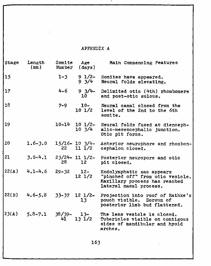
Stage Length (mm)
15
17
18
19
APPENDIX A
Somite Age Number (days)
1-J
4-6
7-9
9 1/2-9 3/4
9 J/4-10
10-10 1/2
10-14 10 1/2-10 3/4
Main Commencing Features
Somites have appeared. Neural folds elevating.
Delimited otlc (4th) rhombomere and post-otic sulous.
Neural canal closed from the level of the 2nd to the 6th somite.
Neural folds fused at diencephalic-mesencephalic junction. Otio pit forms.
20 1.6-3.0 15/16- 10 3/4- Anterior neurophore and rhomben-22 11 1/2 cephalon closed.
21 J.0-4.1 2J/24- 11 1/2- Posterior neuropore and otic
22(A)
22(B)
2J(A)
28 12 pit closed.
4.1-4.6 29-32 12-
4.6-5.8
12 1/2
33-37 12 1/2-lJ
J8/J9- 1.3-41 1.3 1/2
163
Endolymphatic sac appears "pinched off" from otio vesicle. Maxillary process has reached lateral nasal process.
Projection into roof of Rathke's pouch visible. Dorsum of posterior limb bud flattened.
The lens vesicle is closed. Tuberlcles visible on cont1gous sides of mandibular and hyoid arches.
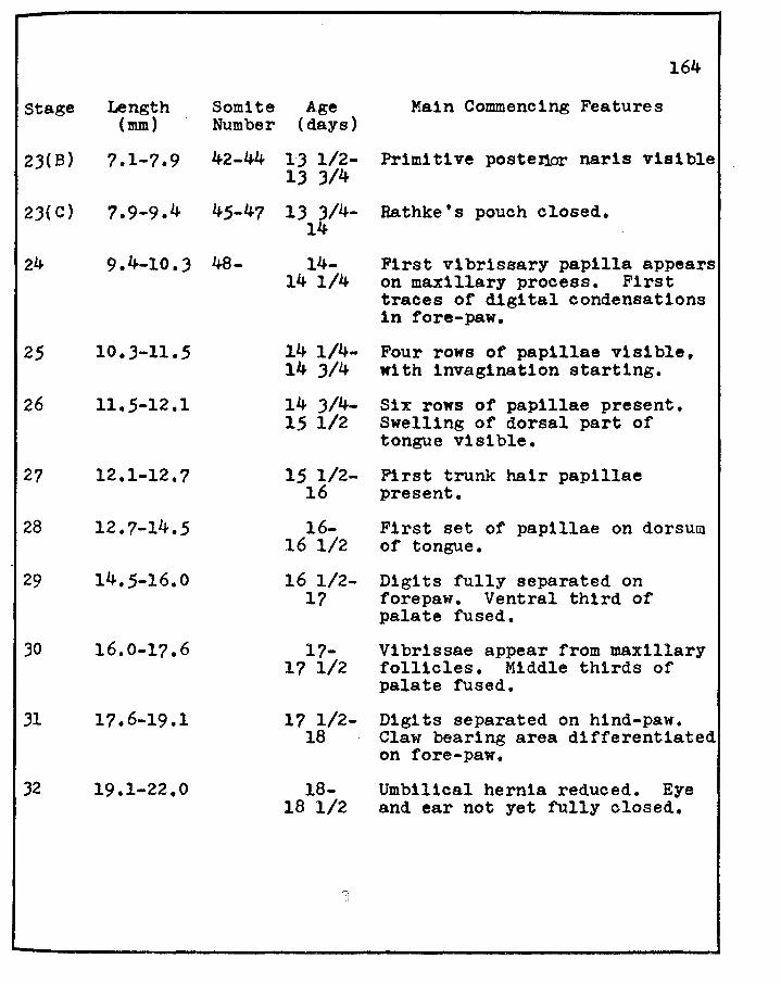
164
Stage Length (mm)
Somite Age Number (days)
Main Commencing Features
2J(B)
2J(C)
24
25
26
27
28
29
30
Jl
32
7.1.7.9 42-44 13 1/2- Primitive postenor nar1s visible 1) J/4
7.9-9.4 45-47 1) J/4- Bathke's pouch closed. 14
9.4-10.J 48-
10.J-11.5
11.5-12.1
12.1-12.7
12.7-14.5
14.5-16.0
16.0-17.6
17.6-19.1
19.1-22.0
14-14 1/4
14 1/4-14 )/4
14 J/4-15 1/2
15 1/2-16
16-16 1/2
16 1/2-17
17-17 1/2
17 1/2-18
18-18 1/2
First vibrissary papilla appears on maxillary process. First traces of digital condensations in fore-paw.
Four rows of papillae visible, with invag1nation starting.
Six rows of papillae present. Swelling of dorsal part of tongue visible.
First trunk hair papillae present.
First set of papillae on dorsum of tongue.
Digits fully separated on forepaw. Ventral third of palate fused.
Vibrissae appear from maxillary follicles. Middle thirds of palate fused.
Digits separated on hind-paw. Claw bearing area differentiated on fore-paw.
Umbilical hernia reduced. Eye and ear not yet fully closed.

PLATE I
PHOTOMICROGRAPHS OF THE NORMAL HISTOLOGY OF THE PINEAL GLAND OF RAT. X 2.000;
Figure 20 Pineal Gland. Hematoxylin-Eosin. Two types of cells are observed• ' 1) light cell (1) with a round nucleus, prominent nucleolus and a pale staining cytoplasm; and 2) a dark cell (d) with irregularly shaped nucleus, no apparent nucleolus and a darker staining cytoplasm. Note also lipid vacuoles surrounding capillaries (c).
Figure 21 Pineal Gland. Bargmann's chrom-alum hematoxyl1n. Longitudinal section of a pineal capillary (c), containing red blood cells and lined with neurosecretory material of moderate staining intensity.
~\I ]!
d.~
"# .,
I
PLATE I
'lt •• I
Figure 20
• Figure 21
165
i . I
i . ! I
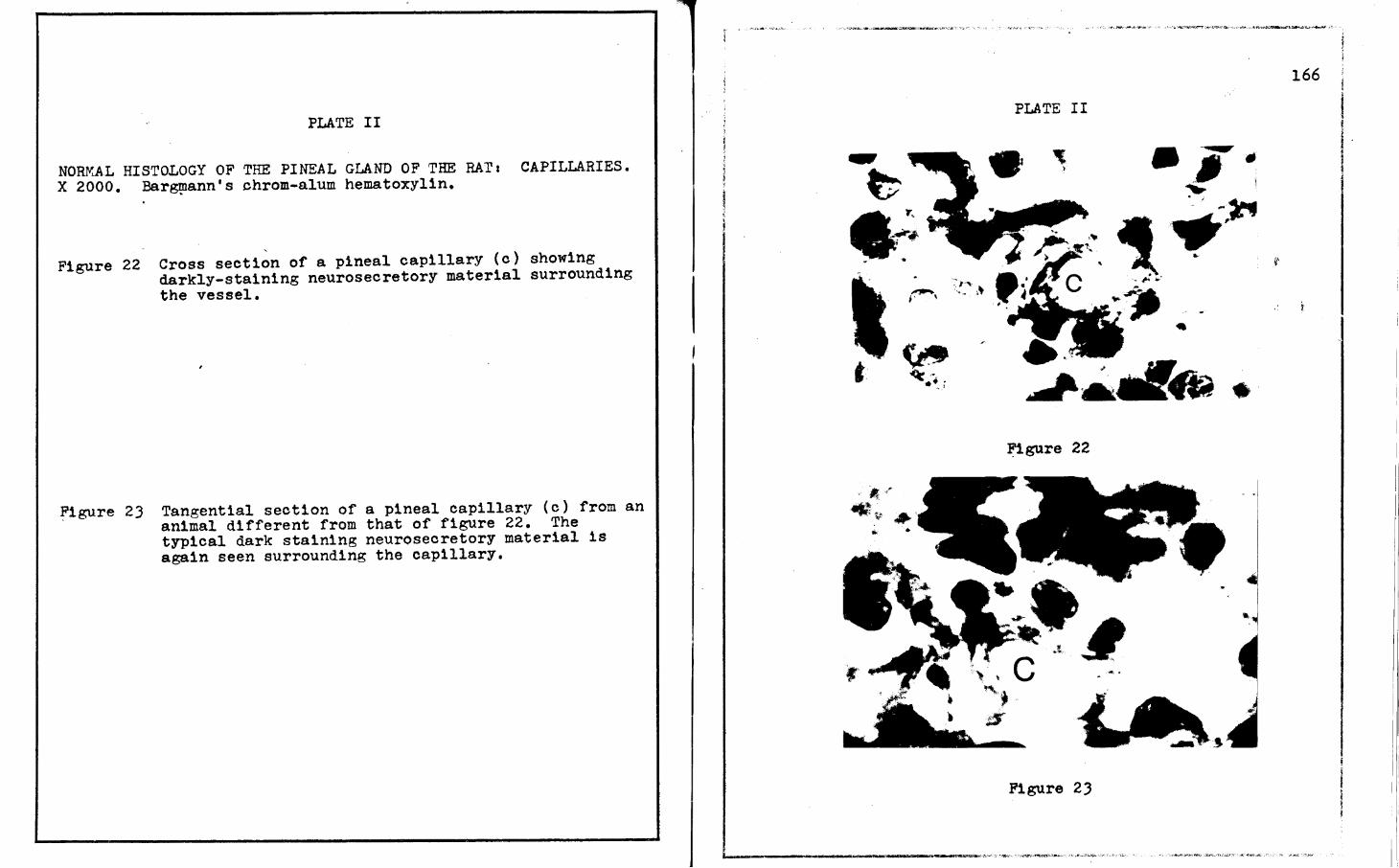
PLATE II
NORf'iAL HISTOLOGY OF THE PINEAL GLAND OF THE RAT 1 CAPILLARIES. X 2000. Bar~ann's chrom-alum hematoxylin.
Figure 22 Cross section of a pineal capillary (c) showing darkly-staining neurosecretory material surrounding the vessel.
~igure 23 Tangential section of a pineal capillary (c) from an animal different from that of figure 22. The typical dark staining neurosecretory material 1s again seen surrounding the capillary.
PLATE II
•
Figure 22
Figure 2J
• I
. ·I I
166 I
I ! I . l

PLATE III
LONGITUDINAL SECTION OF THE PINEAL STALK OF THE RAT. X 2000. Bargmann's chrom-alum hematoxylin • .
rigure 24 Dark staining neurosecretory material is seen here 1n an irregular pattern along this portion of the
Figure 25 '
pineal stalk.
In this section of the pineal stalk the darklystaining neurosecretory material is seen 1n discrete lines.
167
PLATE III
4149
Figure 24
Figure 25
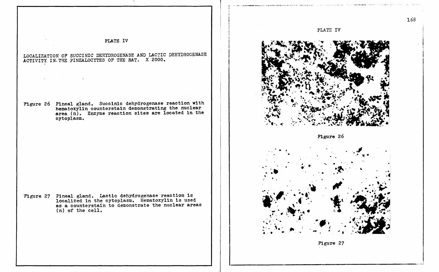
PLATE IV
LOCALIZATION OF SUCCINIC DEHYDROGENASE AND LACTIC DEHYDROGENASE .ACTIVITY IN.THE PINEALOCYTES OF THE RAT. X 2000.
Figure 26 Pineal gland. Succinic dehydrogenase reaction with hematoxylin counterstain demonstrating the nuclear area (n). Enzyme reaction sites are located in the cytoplasm.
'Figure 27 Pineal gland. Lactic dehydrogenase reaction is localized 1n the cytoplasm. Hematoxylin is used as a countersta1n to demonstrate the nuclear areas (n) of the cell.
168
PLATE IV
~1gure 26
• • •
.. .., . )• .... • •
·~ .. · • •
• . .
.. \ ,,
~~ .. . ... . ·~
•. ,, . . ... ... *. ,· ...... ~ " . .
. . \ . ·- • ,, "'~t • .._. .'· ..,. .. • . • j ...
• • Ja • r . . .. ._ . . . '
:·. ,,. . ,., ... . . . .. ·-· . c . ' •
, . j !
Figure 27
'-------------------------------......,.-.-.---._;,_ ,;-:-_·"~

PLATE V
LOCALIZATION OF ALKALINE AND ACID PHOSPHATASE ACTIVITY IN THE PINEALOCYTES OF THE RAT. X 2000.
,Figure 28 Pineal gland. Alkaline phosphatase reaction is localized in the cytoplasm of the p1nealocytes. Hematoxylin is used as a counterstain to demonstrate the nuclear areas (n).
Figure 29 Pineal gland. Acid phosphatase activity within the pinealocytes is localized in the cytoplasm. Hematoxyl1n is used as a counterstain to demonstrate nuclear areas (n).
• • • .. •• ' • . • •
. . .• ' • •
•
• • •
, .
PLATE V
• • . . •. . •• • .... t •
. . . -" .• • , . . .
• 0 I • . . . ' , ....... . . . . . . ,,, . ' . .. . . ~ ... • .. •
• •
• •
• • . -
,,
• • •... _, .. ,._ • f I
. . • I . . . ..
• .• . . . . .. . ... .. •· n ..
. ._, - . . . . . .. . '· .... .:·.. .. ' ~ , .· .. •• • 't !. • • •
• .. . . , . . ..
•• • • . ,,_ . . . , .... ,., .. . . . ' . . ' ' . .. . • •• • •
• • • •• • . . . ·'\ .. • 4't
•
'-
-
•
• • • : ...... · . . . . . ... • • • • •
•
Figure 28 .
•
, n "
..
Figure 29
I
•
. . .~. 4-•
• •
• . .
•
• . · .. .. •• . .. •
•

PLATE VI
SUCCINIC DEHYDROGENASE ACTIVITY OF THE PINEAL GLAND OF THE RAT DURING SEVEN.DIFFERENT STAGES OF THE ESTROUS CYCLE. X 2000.
Figure JO Proestrus. This gland shows a distinctive array of diformazan granules with an inappreciable intensity of monoformazan material.
Figure Jl Early Estrus. A small scatter of diformazan granules with an almost maximal intensity of monoformazan material is evident.
-Figure 32 Late Estrus. Maximal diformazan deposition with
considerable intensities of monoformazan material readily observable 1n the background.
Figure 33 Metestrus. Small scatter of diformazan granules with only trace intensities of monoformazan material is discernible.
Figure J4 Early Diestrus. Increased intensity of both mono- and diformazan material is seen when comparing with metestrus.
Figure 35 Late D1estrus. Intense d1formazan granules in a scattered array are observed. Increased intensity of monoformazan material is visible 1n the perinuclear areas of the pinealocytes.
Figure ,36
Figure 37
Preproestrus. There is a marked reduction 1n the intensity of the diformazan granules. An appreciable intensity of only monoformazan material is evident.
Control Section. Control sections are uniformly negative for each of the seven stages of the estrous cycle, also during pseudopregnanoy, prolonged pseudopregnancy and pregnancy.
Figure Jl
Figure 33
. ' ., .
•
,.
•
' ~ ,,
. , ..
.._. • J
·v • ' . . ' ' • . . .r
PLATE VI
"!
"I
Figure 35
Figure J6
Figure . 37
170

PLATE VII
LACTIC DEHYDROGENASE ACTIVITY OF THE PINEAL GLAND OF THE RAT DURING SEVEN-DIFFERENT STAGES OF THE ESTROUS CYCLE. X 2000.
Figure .38
Figure 39
Proestrus. A maximal intensity of both mono- and diformazan'material is readily discernible.
Early Estrus. A marked decrease 1s observed in the intensity of both the mono- and diformazan reaction sites.
Figure 40 Late Estrus. A near maximal intensity is seen for both the mono- and diformazan material.
Figure 41 Metestrus. Slight reduction in intensity of the diformazan granules with a marked reduction in monoformazan material is demonstrated.
Figure 42 Early Diestrus. Further reduction in the overall reaction with scattered diformazan granules of moderated intensity and a slight elevation in the monoformazan distribution and intensity is shown.
Figure 4.3 Late Diestrus. Marked reduction in the intensities of both mono- and diformazan material is demonstrated.
Figure 44
Figure 45
Preproestrus. This reaction shows an overall increase in the number of diformazan granules with a markedly increased intensity of monoformazan material.
Control Sections. Control sections were uniformly negative for each of the seven stages of the estrous cycle, also during pseudopregnancy, prolonged pseudopregnanoy and pregnancy.
Figure 38
.Figure 39
Figure 40
Figure 41
.. •
~
•
• •
. • >
, l . ·' .
... " • • •
! • . . . • ._ . ,.
• .. • .. . ' ..
PL>\T:S VII
• • I
.. '· •
,..
•
•
Figure 42
Figure 43
Figure 44
. Figure 45
.
,. • ,, ..- ...
' ... ' • • . .. • ... .. ..
'
'iJ ~. ~#
,.
~ • c.,. •
• . . ' . .... •
171
• • • ~
._ ·~ ~ , I . .. " . • ,. JI/' ,.
• ·y
. .
.. ' .. -~· ~-· t ·6,, • •
' :a ...
.. • •
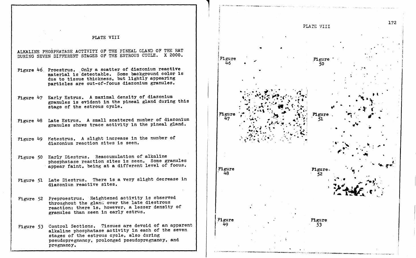
PLATE VIII
ALKALINE PHOSPHATASE ACTIVITY OF THE PINEAL GLAND OF THE RAT DURING SEVEN DIFFERENT STAGES OF THE ESTROUS CYCLE. X 2000.
Figure 46,
Figure 47
Figure 48
Figure 49
Figure 50
Figure 51
Figure 52
Proestrus. Only a scatter of diazonium reactive material is'detectable. Some background color is due to tissue thickness, but lightly appearing particles are out-of-focus diazonium granules.
Early Estrus. A maximal density of diazonium granules is evident in the pineal gland during this stage of the estrous cycle.
Late Estrus. A small scattered number of diazonium granules shows trace activity 1n the pineal gland.
Metestrus. A slight increase in the number of d1azon1um reaction sites is seen.
Early Diestrus. Reaccumulation of alkaline phosphatase reaction sites 1s seen. Some granules appear faint, being at a different level of focus.
Late Diestrus. There is a very slight decrease in diazonium reactive sites.
Preproestrus. Heightened activity is observed throughout the glanQ over the late d1estrous reaction; there is, however, a lesser density of granules than seen in early estrus.
Figure 53 Control Sections. Tissues are devoid of an apparent alkaline phosphatase activity in each of the seven stages of the estrous cycle, also during pseudopregnancy, prolonged pseudopregnancy, and pregnancy.
1 I
l ; r'igure ' 46 f
Figure I 48
Figure ' 49
PLA:rE VIII
•
• ..
- - I . . ~ . ., ... -. . - .. -'"•' .. / ' - .- . ~;.. , . ...:6t • ' . •· •, .• ·~·-r:. ,...~ .... ~ .). . , . ...,.
• :_:,? .. f ,•" ,:_. •. ~ • • ~ f I
.- . •; ""''. • , - ... .. t . •. •• I
•:.· # . "\ • • •' ' '• - . ,a • I ... • .... • .. .. <(" , .. . . t. ·~ # • '>t -:"' ~-~ .... 1 . ..... • . - .. ~. . .. ' t .. , J
. . . . , . .. . . .... . ,. ' . ,t ..... w '* .. • I .I- ; .•• ; • • . • . . .. •. ~.· . ' • • ' • I • • ~ .·• .. ,,,.. . .,,
'ti • ,,, ....... • ·J ·" ~ ~ . ~,.. . . . ·:, '. .... ..... . . .. . I • ,·. ~. .... • •· .. ~ ... • • .. • : • . - ' . ~
.
,Figure · 50
Figure , 51
Figure, 52
Figure 53
..
•
•
•
•
'f
. .
·~ J t
•• ..
~ ,. . ' . • '\ II .1-..
-.: t .. .;. •• ~ ~
• ..
.. -.. . ,. . • ••
' ... ~ ..
172
,,_,.,,,._*""~-:.,-,,...:>'>.~ot..c • .-.~~51~~c··-0-r1'"'-'·~ .~~~-.'·ro.:, .:,i.·.::1:-.,....w-:u.~~·--...,..._~~o.•.!.,·i.-•lo.-'' ,:n ,~ '· ··-~--,~....-°""""·' ,.,
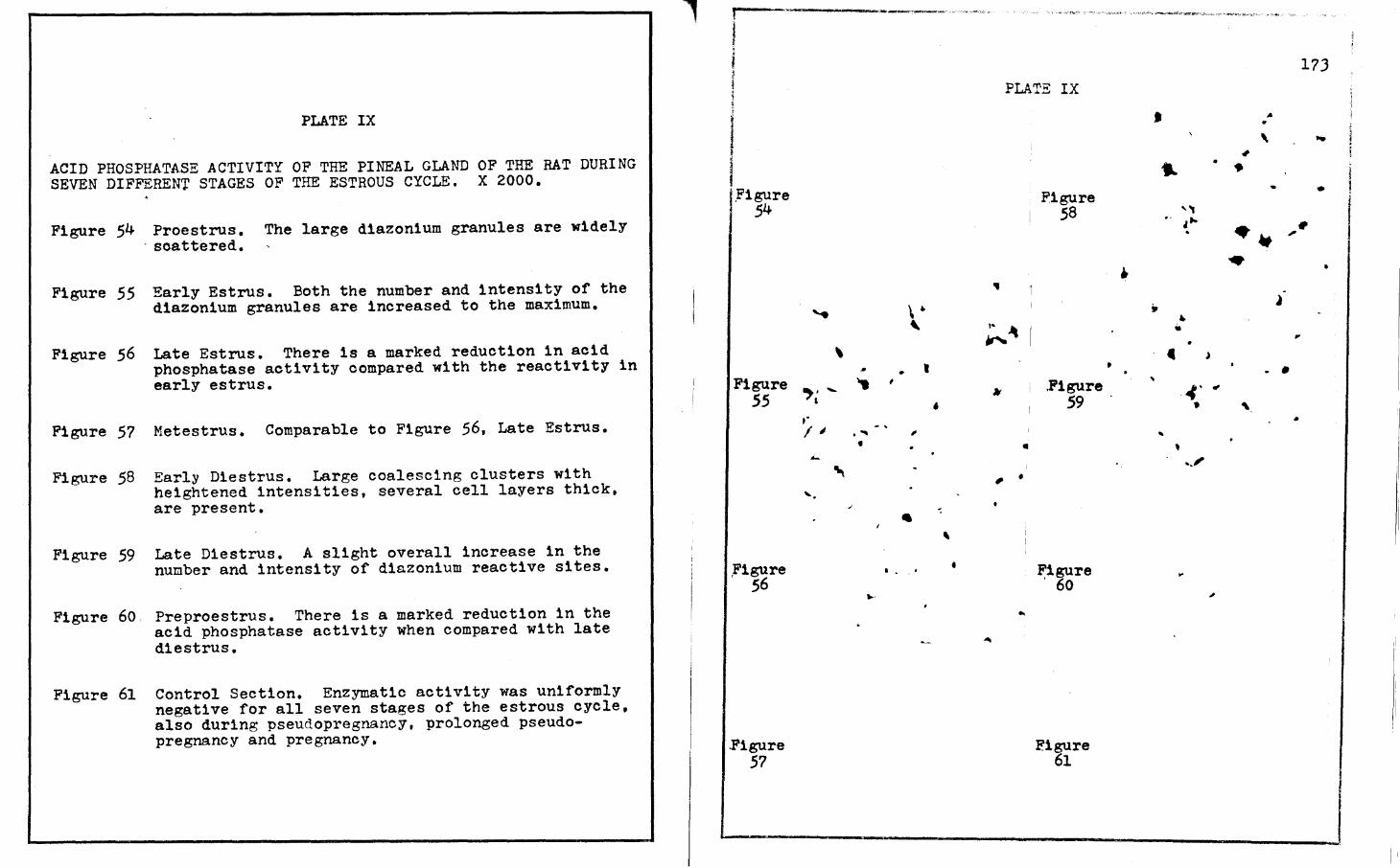
PLATE IX
ACID PHOSPHATASE ACTIVITY OF THE PINEAL GLAND OF THE RAT DURING SEVEN DIFFERENT STAGES OF THE ESTROUS CYCLE. X 2000.
Figure 54 Proestrus. The large d1azon1um granules are widely , scattered.
Figure 55 Early Estrus. Both the number and intensity of the diazonium granules are increased to the maximum.
Figure 56 Late Estrus. There is a marked reduction in acid phosphatase activity compared with the reactivity in early estrus.
Figure 57 Metestrus. Comparable to Figure 56, Late Estrus.
Figure 58 Early Diestrus. Large coalescing clusters with heightened intensities, several cell layers thick, are present.
Figure 59 Late Diestrus. A slight overall increase in the number and intensity of diazonium reactive sites.
Figure 60 Preproestrus. There is a marked reduction in the acid phosphatase activity when compared with late diestrus.
Figure 61 Control Section. Enzymatic activity was uniformly negative for all seven stages of the estrous cycle, also during pseudopregnancy, prolonged pseudopregnancy and pregnancy.
... f -- ------~·--~----·
l
I I I.Figure ~ 54
Figure 55
Figure ' 56
.Figure 57
.....
\
.,. -' ,.
I;
..:..
"'\ ... .
\ . ' • , ,
' . "' , •
.I
' . .. . ' • ...
PLATE IX
!• "
"' •
.. I , •
..
Figure 58
. Figure 59
Figure ' 60
Eigure 61
..
•
• .. ,, . ' , .. .
" • • • c
.. 4' •
.. ' ... i'
...
17J
,.
' ,.
., • • i - •
j ·- • , .. ~
,,
l . • ., '\
•

PLATE X
SUCCINIC DESYDROGENASE ACTIVITY OF THE PINEAL GLAND OF THE RAT DURING THREE STAGES OF PSEUDOPREGNANCY. X 2000.
Figure 62 Day Six. Scatter of diformazan granules is observed <in the perinuclear areas. Intense monoformazan material is readily observable in some areas.
Figure 63 Day Ten. Marked increase in intensity of the monoformazan material with a slight increase in diformazan deposition is demonstrated, giving the maximal overall reaction.
Figure 64 Day Thirteen. Increased intensity of diformazan granules with a slight decrease in intensity of the monoformazan material is shown.
Figure 63
PLl~TE X
Ji'igure 64
174
I I
--------~----------------'
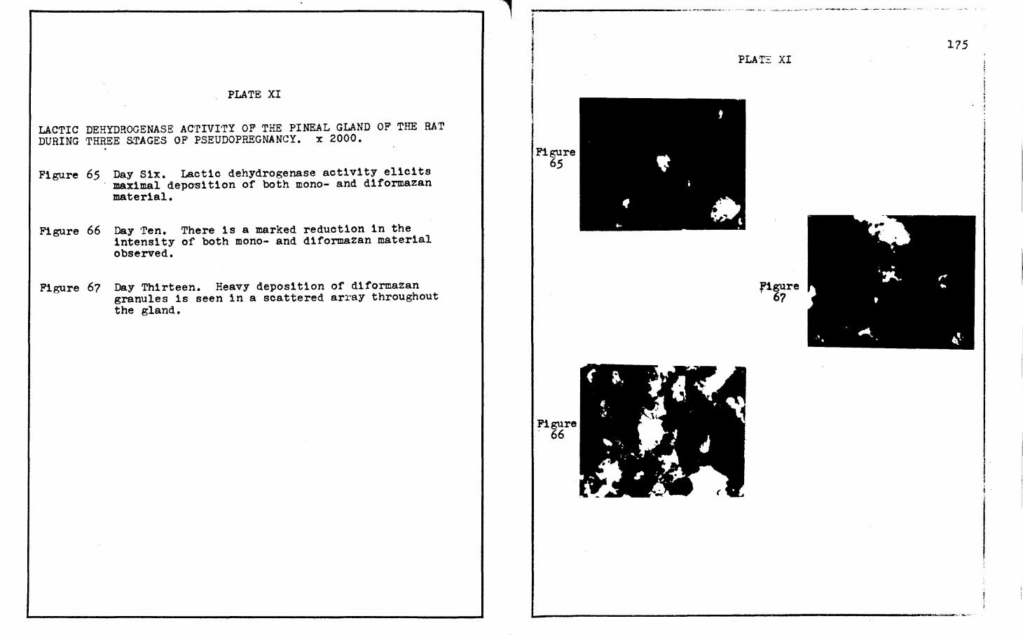
PLATE XI
LACTIC DEHYDROGENASE ACTIVI·rY OF THE PINEAL GLAND OF THE RAT DURING THREE STAGES OF PSEUDOPREGNANCY. x 2000 • . Figure 65 Day Six. Lactic dehydrogenase activity elicits
maximal depo~1tion of both mono- and diformazan material.
Figure 66 Day ·ren. There is a marked reduction in the intensity of both mono- and diformazan material observed.
Figure 67 Day Thirteen. Heavy deposition of diformazan granules is seen in a scattered array throughout the gland.
l
Figure 65
Figure ' 66
PLAT~ XI
figure 67
175

PLATE XII
ALKALINE PHOSPHArASE ACTIVI·rY OF THE PINEAL GLAND OF THE RAT DURING TH?EE STAGES OF PSEUDOPREGNANCY. X 2000.
Figure 68 Day Six. Abundance of small diazonium granules several cell layers deep is observed.
Figure 69 Day Ten. This figure shows maximal intensity for an alkaline phosphatase reaction.
Figure 70 Day Thirteen. A slight decrease ls observed in the intensity of the diazon1um granule deposition.
figure 68
.Figure 69
•
' • • I... • ,. .... ~ . ' . '-. ...
• • <I •• ; -.. . -.. . .
• ~ . ~~: ..
• • • • .. • . -., .... , •
• ..
•
· .
.. •
....
•
•
PLATE XII
r1gure 70
176
..
~ . ', . . '. • .. :
•

PLATE XIII
ACID PHOSPHATASE ACTIVI'rY OF THE PINEAL GLAND OF THE RAT DURING THREE. STAGES OF PSEUDOPREGNANCY. X 2000.
Figure ?l
Figure 72
Day Six. A high intensity of d1azon1um granules is seen several cell layers thick.
Day Ten. Approximately the same intensity as days six and thirteen. Diazonium granules are not easily distinguishable as they are at different depths of this field. ·
Figure ?J Day Thirteen. The intensity is comparable to those of days six and ten. Acid phosphatase reactive sites are several cell layers thick. All the stages of acid phosphatase activity observed during pseudopregnancy were semi-quantitatively assigned a value of +J.
I I f Figure
?l
Figure 72
• . ' • • .J
-- ••
"" •
..
'
.,
' . ,
' ' ..
. '
PLATE XIII
e I
•
,Figure 73
• ., '
177
. '
' <.
• ..

PLATE XIV
SUCCINIC DEHYDROGENASE ACTIVITY OF THE PINEAL GLAND IN PSEUDOPRE~NAN'l' RATS BEARING DECIDUAL REAC'I'IONS. X 2000.
Figure 74. Day Six. Moderate diformazan granule intensity with trace amounts of monoformazan material is seen throughout the gland.
Figure 75
Figure 76
Figure 77
Figure 78
Figure 79
Figure 80
Day ~en. A near maximally intense reaction showing mono- and diformazan material is readily observable in t~e perinuclear areas.
Day Thirteen. Decreasing intensities of succinic dehydrogenase activity sites are demonstrated.
Day Fifteen. Minimal succ1n1c dehydrogenase activity with a scatter of diformazan granules and trace monoformazan material.
Day Eighteen. Maximal intensities of both mono- and diformazan reaction sites are readily observable.
Day Twenty. A marked decrease in the intensities of both mono- and diformazan material is discernible.
Day Twenty-one. Continual decrease in the number of diformazan granules and negligible monoformazan material is visible.
1 I
PLA'l'E XIV 178
., '• .. . , ..• • • •
• • , . . .. I • •• ', ....
t . "'... ··a, . Figure • · · 74 ; •. 7. .....
•• • • • ~ ,_ ·.: . - . . . . . . ... _. ..
• •. 'I .- ••
• • • • • • : ... ••• ••• ••
. . '~"" ·.
. .. ., & ••.
• • . . , ., • •••
• .. . •
., '· • I
!
. ..... : •· .. .. : . . : ' . , ..... . . . ' .. ,,.. .. ~ ..
. . . . ..
Figure.~·· ·1.-. · • ' 77 ··' !" • . . •• . . - . : ...... . . .. •, '~· ... ( . .
•
... ·~ ·.,: . . . ~ ........
..
•
. .
Figure . 78
i• 4" I . . •• ...... ; . . ~ ...
• • • ,•. "9l • .1 .. • • • • '!, . '• •
• • • • ... . I
: ..... , ..... l ' . . \ .. ~igure; ; .... · • •• , . .,_.!~· ."~.:.. j
79 . • . ,f. . ........ .
: .-.P I
.•. I
•A' ... , . p • • .. • ,,. . . . . . . . . . . •. . . ' . ~· . . :.•·
... . . . ~ . ..
~ .•• I_ .. • t'6 • • • ·'' -\' . . • ~ ,, .... . ... . .. . .-{::,. • • ~ • . .r :.~
. ·. . . . . ; Figure .e. • · ~ V ~ , 80 • ' •-r .-• ' " '. I ., .. ,. . . .... ... ·' •.1.... • ,.. .• 41 . ,. .-. . ~ .. . . . . ' ..
" • -~i '. -. ·.; .. . . . ...... . • •! , ~

PLAI'E XV
LACTIC DEHYDROGENASE ACTIVITY OF THE PINEAL GLAND IN PSEUDOPREGNANr B.ATS 'BEARING DECIDUAL REACTIONS. X 2000.
Figure 81 Day Six. High diformazan granule intensity and moderate monoformazan intensity is observed.
Figure 82 Day Ten. Increased intensities of both mono- and diformazan material is evident.
Figure 8J Day Thirteen. Maximal lactic dehydrogenase intensities are observed in the pinealocytes.
Figure 84 Day Fifteen. A precipitous decrease in both monoand diformazan reaction sites is noticeable.
Figure 85 Day Eighteen. A marked increase is seen in the intensity of diformazan granules with a moderate intensity of monoformazan material.
Figure 86
Figure 87
Day Twenty. A decrease in the intensity of diformazan granules with trace monoformazan material is revealed.
Day Twenty-one. A continual decrease of diformazan granule intensities and moderate monoformazan material are detectable. When compared to day twenty, it can be observed that there is a slight increase in the concentration of the monoformazan material on day twenty-one, but a decrease in the number of diformazan granules. On day twenty, the diformazan granules are small and evenly distributed throughout all of the pineal gland, while on day twenty-one, the diformazan granules are large with scattered coalescing clusters.
lFigure I a1
i
.Figure SJ
PUTE ).'V
Figure ' 86
' • •'I""!- • ...... ., . · ......
; . . . ' . . •
179
. .. . .. . .. .. . . . ~T •• . • • I ··.· . , ." ..
. ... . . . ' ,·" , ... ·"'.·.·_]
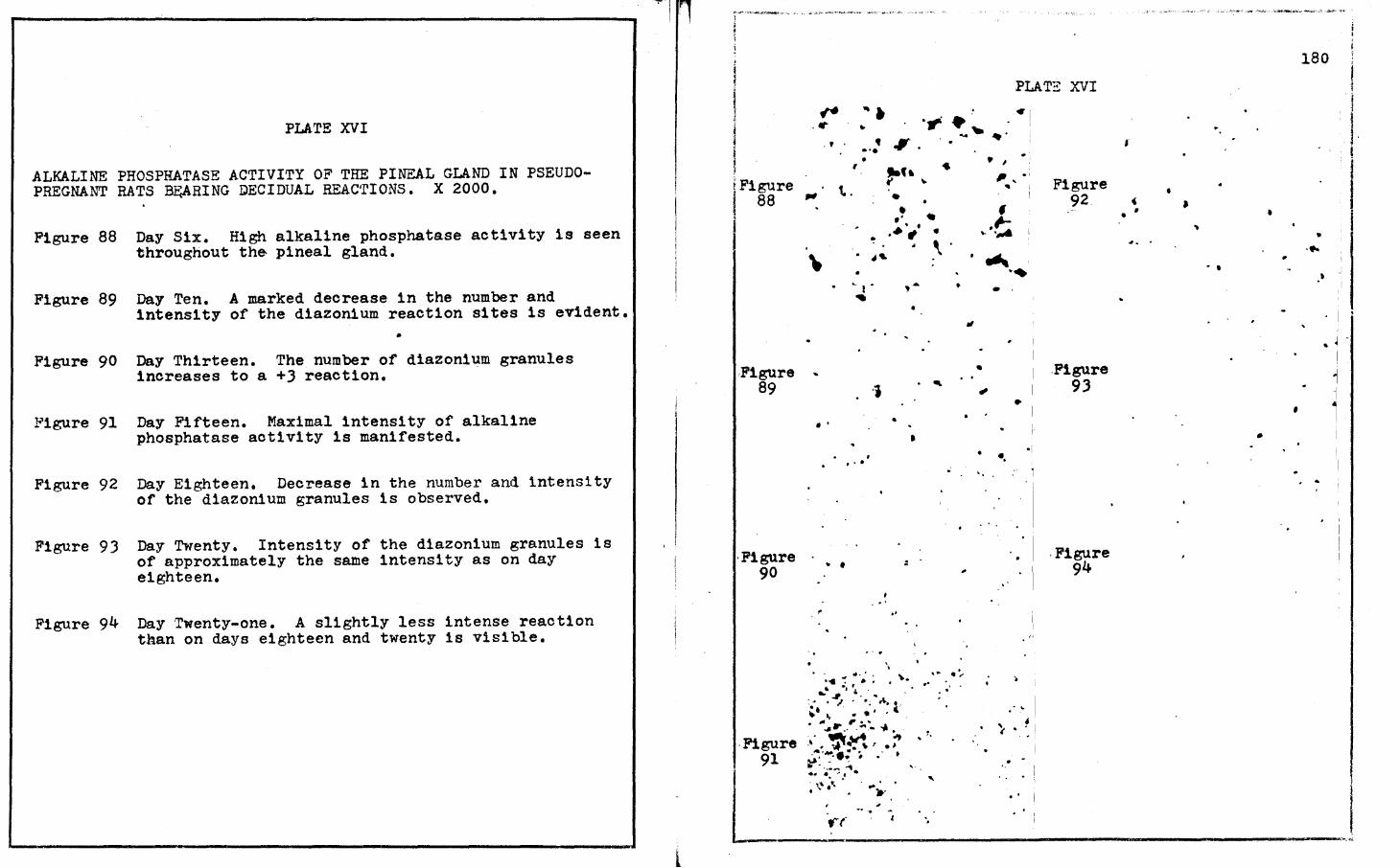
PLATE XVI
ALKALINE PHOSPHATASE ACTIVITY OF THE PINEAL GLAND IN PSEUDOPREGNANT RATS ~RING DECIDUAL REAC'rIONS. X 2000.
Figure 88 Day Six. High alkaline phosphatase activity is seen throughout th& pineal gland.
Figure 89 Day Ten. A marked decrease in the number and intensity of the diazonium reaction sites is evident.
Figure 90 Day Thirteen. The number of diazonium granules increases to a +J reaction.
Figure 91 Day Fifteen. Maximal intensity of alkaline phosphatase activity is manifested.
Figure 92 Day Eighteen. Decrease in the number and intensity of the diazonium granules is observed.
Figure 93 Day Twenty. Intensity of the diazonium granules is of approximately the same intensity as on day eighteen.
Figure 94 Day Twenty-one. A slightly less intense reaction than on days eighteen and twenty is visible.
PLATE XVI ., . ., . . .
-~ -· . ' . . .. . ..... ..• .., . .
• , jFigure i 88 ~ \..
I
,Figure 89
·Figure 90
• ,. -• • ... , .
• •
,. .
. .
~ . ,, t ..
... ,
·i
, ..
\
'
..
:
..
. ~ .. ":' . ··" : ' \.. ._ .J ,
.... _, 4. ~ '. ... , .... .f\.., ~· " . . .. . . ( .
•
.
•
\'
~
,. . ' .
. ""'~ r ~.'".:,. ~- .,. • ·Figure ·'·. ~Jrt, .~
91 ~·~··; # •· •
. '•
. . .. ' ... .. ..... . . <t ' .
• I ,, . ' ' Figure
f 92
~·~ • ...._
·~ • •
"
• . Figure . *' 93
•
• •. •I
'·
. ..
, · .· I . Figure
94
.- ~
' . ' .. · ..
• ' I
180
• ' J ,
• • .. •
*• . .. •
• • ·'
I
.. J -1 .
1 ·1
' 1
..
L \~·; .. ~ .......... ,,: . ' ...
-------·~~----~------~-------------------------------~----
l I I· i
.
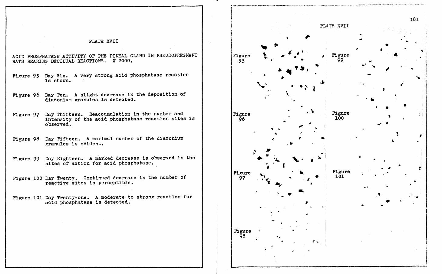
PLATE XVII
ACID PHOSPHATkSE ACTIVITY OF THE PINE.AL GLAND IN PSEUDOPREGNANT RATS BEARING DECIDUAL ·REACTIONS. x 2000.
Figure 95 Day Six. A very strong acid phosphatase reaction 1s shown.
Figure 96 Day Ten. A slight decrease in the deposition of diazonium granules is detected.
Figure 97 Day Thirteen. Reaccumulation 1n the number and intensity of the acid phosphatase reaction sites is observed.
Figure 98 Day Fifteen. A maximal number of the diazonium granules is eviden~.
Figure 99 Day Eighteen. A marked decrease is observed in the sites of action for acid phosphatase.
Figure 100 Day Twenty. Continued decrease reactive sites is perceptible.
in the number of
Figure 101 Day Twenty-one. A moderate to strong reaction for acid phosphatase is detected.
Figure 95
Figure 96
Figure 9?
figure 98
~'
"' f'
\. •
• • • • " c. ....
' , "'
' <
... " • ., '
. f ' ~ .. .. ~
........ ..
•
~
... ,
PLA'l'E. :XVI I
.. ,. ,, ' . Figure • ,
• 99 .. ~a.
~ ,.
... • . " i ..
\ •
• - . \. Figure 100
• ,. '
' ,. .. .~
~'
"\. - • ~
" Figure
' • .< • 101
• ... .. ' '
1 ...
181
I .. .. ... • ... • •
• ~ ; ! ~
' ~ l ,. I
'Ill I I ""
I ·i
- ; ·~
I' 1
• ~ ..
• • .,,
' ~ •
' , ~
• ' ~
• , !i
' ...
• ,. 4 ..
., . -' • .... •
a-.------~----------~--~~------~~---~------------------~---~~

PLATE XVIII
SUCCINIC DEHYDROGENASE ACTIVITY OF THE PINEAL GLAND OF PREGNANT RATS. X 2000.
Figure 102 Day Six. A less than moderate intensity of both mono- and diformazan material is readily seen 1n the perinuclear areas.
Figure 103 Day Eight. A near maximal intensity of both monoand diformazan material is apparent.
Figure 104 Day Ten. ·rhere is a marked reduction in diformazan granules with a moderate intensity of monoformazan.
Figure 105 Day Thirteen. Reaccumulation of diformazan granules 1s observed with a slightly less than moderate intensity for monoformazan.
Figure 106 Day Fifteen. Moderate intensity of diformazan granules is seen in the perinuclear areas. Monoformazan material is intense in some areas.
Figure 107 Day Eighteen. A maximal reaction of small diformazan granules is demonstrated. Monoformazan background intensity is high.
Figure 108 Day Twenty. There is an intense monoformazan deposition throughout the pineal gland. Diformazan granules are slightly decreased in intensity.
Figure 109 Day Twenty-one. Marked reduction in monoformazan depostion is readily seen. Diformazan granules are scattered throughout the pineal gland.
PLATE XVIII
i ;Figure ; 102
t
.Figure 103
Figur . 104
. '
,
,, •
.. ~: • I
~~, .. . ·'·.~;~•·. . .. .t , I . .
.1-• ~ .... -.v.. . . ' .. .. \ . ~-'
. Figure ... • -·: • ,• 105 •.• ·.·- -- .. ... • - ' . ' ·~
. . ' -, : .I - ' 4,.,. -: \. ... I I ... . • • I
.,T , • ... • .. ,, ' I·~
F1,:::ure 106
Figure 107
Figure . 108
182
... ... .. , .
. .
I
:. • #-~- -· . ~ -' "Ji. ,. • ; I ·.... , , ,. • . ,.... . . . ·.: - .. . ~ • ... I < .., ' !•a<A ,; ' . • . l Lv-~-~,r.~-~O:C<,~2"''>.!ll.".:...'"l"~•O:.ll,,.,,.i......,.-£'1,~~},.·.,c>c"''"'P•'.l!'."'i:',;."""'""'--$:-"'~~~,,,_.,..,."""-"''·~4·=""'c"~",.,_~,._W .. f"''· t'";" ,.,,.~~ll<'_.,-~·,<,,:~.,.__._, __ WC:(:;'...~>"°TY~'~,,.~~..i..:,u..;. ' .\l
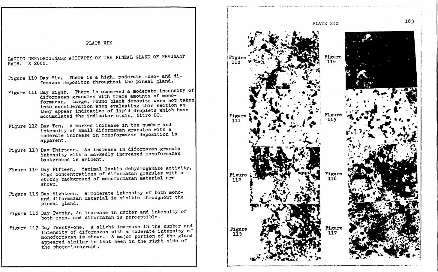
PLATE XIX
LACrIC DEHYDROGENASE AC'.UVITY OF THE PINEAL GLAND OF PREGNANT RATS. X 2000. .
Figure 110 Day Six• ·rher~ is a high, moderate mono- and difomazan depositon throughout the pineal gland.
Figure 111 Day Eight. There is observed a moderate intensity of diformazan granules with trace amounts of monoformazan. Large, round black deposits were not taken into consideration when evaluating this section as they appear indicative of lipid droplets which have accumulated the indicator stain, Nitro BT.
Figure 112 Day Ten. A marked increase in the number and intensity of small diformazan granules with a moderate increase in monoformazan deposition is apparent.
Figure 113 Day Thirteen. An increase in diformazan granule intensity with a markedly increased monoformazan background is evident.
Figure 114 Day Fifteen. Maximal lactic dehydrogenase activity. High concentrations of diformazan granules with a strong background of monoformazan material are shown.
Figure 115 Day Eighteen. A:. moderate intens1 ty of both m.onoand d1formazan material is visible throughout the pineal gland.
Figure 116 Day Twenty. An increase in number and intensity of both mono- and d1formazan 1s perceptible.
Figure 11? Day Twenty-one. A slight increase in the number and intensity of diformazan with a moderate intensity of monoformazan is shown. A major portion of the gland appeared similar to that seen in the right side of the photomicrograph.
I •.- ,_ I ..
J • fl..•r• ~•' J ' .. . ..
i 'j'. . .c' . ·. ' i. ~ I ·' Ai1. •.. · l ~-, ... ~l~". ~Figure ". · ~ • t "' ·•' I 110 P~:~. i '
I
.Figure 111
.Figure 112
,Figure 113
PLATE XIX
Figure 114
,Figure 115
;Figure 116
figure 117
183
L-·---· .. ,,,,.-·-·-----~----------... --------ll'W'S-·--~--,,...,·~'.'·~ t.. .•
PLATE XX
ALKALINE PHOSPHATASE ACTIVI·ry OF THE PINEAL GLAND OF PREGNANr RATS. X 2000.
Figure 118 Day Six. 'rrace alkaline phosphatase reaction is d:tscernible.
Figure 119 Day Eight. An increase, but still weak, intensity of diazonium granules is seen scattered througout the pineal cells.
Figure 120 Day Ten. This reaction is approximately equal in intensity to day eight.
Figure 121 Day Thirteen. High intensity of alkaline phosphatase reactive sites is evident.
Figure 122 Day Fifteen. Maximal intensity of diazonium granules is observed several cell layers deep.
Figure 123 Day Eighteen. A precipitous decrease in the intensity of the diazon1um granules is manifested.
Figure 124 Day Twenty. A slight increase in the alkaline phosphatase reaction prevails throughout the pineal gland.
Figure 125 Day Twenty-one. This reaction is approximately equal in intensity to day twenty, a strong, moderate reaction.
r·······--· .. ··~·-···" ., ··-·-~-·---·- ·. ~ ........... ~. ~-~~ . -- ..
\ i
l
l !
I Figure 118
Figure 119
'1gure 120
Figure 121
-. •
•
•
•
•
•
PLATZ XX
Figure. 12J
J,i'igure ·124
Figure 125
•
'
•
' • • • ...
• 41
•
..
•
•
•
I
" t
•
. .. •
184
. .. ' • • ,
J
. i • , I
I ! ' I

PLATE XXI
ACID PHOSPHATASE ACTIVITY OF THE PINEAL GLAND OF PREGNANT RATS. X 2000,.
Figure 126 Day Six.. A scatter of d1azon1um granules is observed. Background coloration is the result of tissue thickness and is not indicative of sites of acid phosphatase action.
Figure 127 Day Eight. Large, intense but widely spaced diazonium gran~les are evident.
Figure 128 Day Ten. Acid phosphatase reaction sites are observed more than one cell layer in thickness.
Figure 129 Day Thirteen. Increase in the number and intensity of the d1azonium granules ls apparent.
Figure 130 Day Fifteen. Near maximal intensity of diazon1um deposition is visible,
Figure 131 Day Eighteen. Maximal, intense d1azon1um granule deposition is observed several cell layers in thickness.
Figure 132 Day Twenty. Strong acid phosphatase reaction ls seen. Diazonium granules are intense but scattered.
Figure 133 Day Twenty-one. Intense, but scattered, deposition of diazon1um granules is demonstrated.
I ' t I i I Figure 126
Figure · 127
Figure 128
r< -,
• •
f'
•
• • •
. ~ .. • •
" ~ I
I .. ' . ..
Figure -129
.. ..
--
•
'
, .. ,.
,
•
• f , -
•
~ /. ~
•
• •
. -...
• .. -I
~
. . .. ,,
PLATZ XXI
•
•
• .,
•
Figure 130
Figure 1)1
Figure . 132
Figure 133
' . '\ , I
' •
...
185
.. •
' i
~ I f\ 0
• I I
, .. ..
'!· ~ l . -... ' . ... .
~ • • • ... 't ~ ...... . ' . . .. . ~
---~·· •' ' ~··,
•
• l:J... ' ( ... ' ... - ' . - .._. ..... ,.
• ·) • 1 t r ., . : :~ ·'. ...~'.: . I. • '- #.,
, ... - '• .. ' . . . ... .
. . ·r1 •. .. . .... .... , '~.\.
I•
• • , . • ~ •
I' .. ..
• •
• • • • • • • -"· • f •
• - •
l i !
Ii .· • ~-
.... • ,
• • . .. ·' .. • ,.
• •• • 1.---------------------------~~,,,....,.~·-cr..,,,-·; . .....,..;,~'""~-*'"-';

APPROVAL SHEET
The dissertation submitted by Anthony v. Fasano has
been read and approved by six members of the ·faculty of the
Graduate School· of Loyola University.
The final copies have been examined by the director
of the dissertation and the signature which appears below ver
ifies that fact that any necessary changes have been incorpor
ated, and that the dissertation is now given final approval with
reference to content, form and mechanical accuracy.
The dissertation is therefore accepted in partial
fulfillment of the requirement for the degree of Doctor of
Philosophy.
ii


