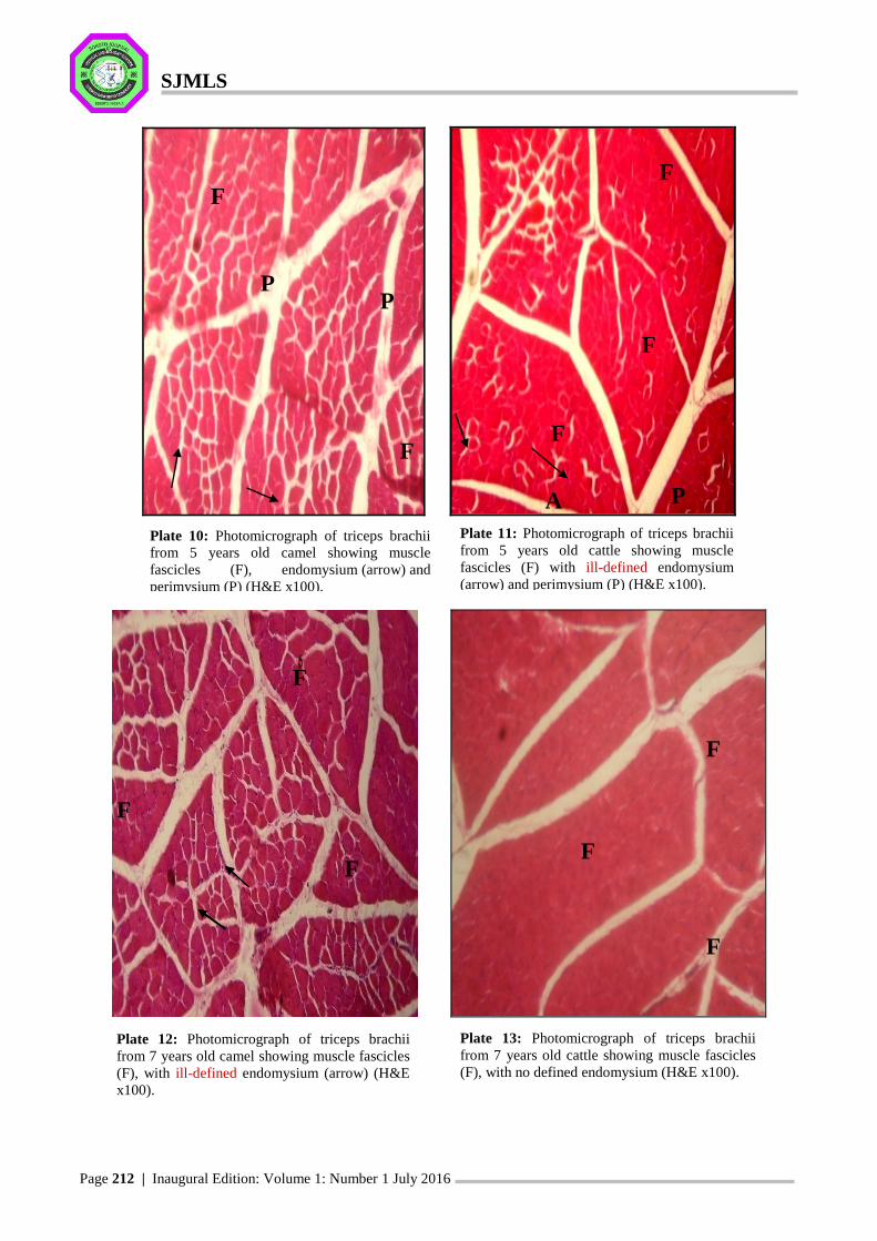
SJMLS Page 208 | Inaugural Edition: Volume 1: Number 1 July 2016 Sokoto Journal of Medical Laboratory Science 2016; 1(1): 208 – 214 Orginal Article SJMLS-1-2016-30 Histological differentiation of Triceps brachii muscle in cattle and one- humped camel: A comparative study S.A. Hena* 1 , M.L. Sonfada 1 , S.A. Shehu 1 , M. Jibir 2 and A. Bello 1 Department of Veterinary Anatomy, Faculty of Veterinary Medicine, Usmanu Danfodiyo University, Sokoto, Nigeria 1 , Department of Animal Science, Faculty of Agriculture, Usmanu Danfodiyo University, Sokoto, Nigeria 2 . Corresponding author: [email protected]; +234-806-052-4623 Abstract In this study forelimbs obtained from 25 male camels (Camelus dromedarius) and 25 male cattle (Zebu type) slaughtered at Sokoto Municipal Modern abattoir were studied. Each animal was within the age brackets of 6 months to 7 years. The triceps brachii muscle was dissected out and used for histological studies. Generally, the triceps brachii muscles from both the camel and cattle showed typical features of skeletal muscle, but comparatively, it was demonstrated that the camel’s muscles have well outlined muscle fascicle with prominent endomysium surrounding the muscle fibres and prominent muscle fibres than what was obtained in the cattle. The muscle fibres (grains) observed from the triceps brachii of camel was finer and clearer than those observed for the cattle. This could probably be one of the attributes making the camel muscle (meat) to look so appealing grossly and hence a good potential to be utilized in meat industry. Further work is hereby being recommended to be performed in the same area using electron microscopy so as to be able to establish the ultrastructural details of this muscle in these animal species. Key Words: Histological, Differentiation, Triceps Brachii, Cattle; One-humped Camel. Introduction The camel (Camelus dromedarius) is an important multipurpose livestock species uniquely adapted to harsh arid and semi-arid areas that can be used for meat, milk, wool, and hide production (Al-Juboori and Baker, 2012). Camels are greatly utilized as a source of meat, and the demand for camel meat appears to increase due to reasons related to human health. They produce meat with relatively less fat than other animals (Dawood and Alkanhal, 1995; Kurtu, 2004). Meat from young camels has been reported to be comparable in taste and texture to beef (Elgasim and Alkanhal, 1992; Kadim et al., 2008). Cattle are another important animal which is used as the major source of milk, meat, hides as well as draught power. Cattle can also be considered as multi-purpose livestock (Agada et al., 2010). In addition, they have played a major role in human culture by participating in recreation and religious ceremonies. Beef is an important animal husbandry food product, contributing roughly 30% of meat consumed in industrialized countries (Pelletier et al., 2010). Majority of meat from these animals comes from the skeletal muscles. Skeletal muscle tissue is named due to its attachment to bones. It consists of bundles of multinucleated fibres. Each fibre contains longitudinally disposed myofibrils in a matrix of sarcoplasm which is limited by a thin membrane, the sarcolemma. The nuclei are peripherally placed. The fibres appear to be cross-striated owing to alteration of thick and thin myofilaments of the myofibrils. Fibres usually do not extend the entire length of the muscle. They terminate by attaching to the investing connective tissue, although some of them may be arranged more or less end to end (Sisson and Grossman, 1975). Around each fibre external to the sarcolemma, is a film of connective tissue, the endomysium which is composed of fine reticular fibers (Dyce et al., 2010). Each bundle of fibres called fasciculus is surrounded by a greater quantity of connective tissue-the perimysium. The external sheath about the entire muscle is the epimysium (Williams, 1991; Dyce et al., 2010). The connective tissue dispersed in or about the muscles varies from dense to loose in consistency. Literature searches made in relation to the histological features of the triceps brachii muscle in camel and cattle was not
Transcript
SJMLS
Page 208 | Inaugural Edition: Volume 1: Number 1 July 2016
Sokoto Journal of Medical Laboratory Science 2016; 1(1): 208 – 214
Orginal Article
SJMLS-1-2016-30
Histological differentiation of Triceps brachii muscle in cattle and one-
humped camel: A comparative study
S.A. Hena*1, M.L. Sonfada1, S.A. Shehu1, M. Jibir2 and A. Bello1
Department of Veterinary Anatomy, Faculty of Veterinary Medicine, Usmanu Danfodiyo University, Sokoto,
Nigeria 1, Department of Animal Science, Faculty of Agriculture, Usmanu Danfodiyo University, Sokoto,
In this study forelimbs obtained from 25 male camels (Camelus dromedarius) and 25 male cattle (Zebu type) slaughtered at Sokoto Municipal Modern abattoir were studied. Each animal was within the age brackets of 6 months to 7 years. The triceps brachii muscle was dissected out and used for histological studies. Generally, the triceps brachii muscles from both the camel and cattle showed typical features of skeletal muscle, but comparatively, it was demonstrated that the camel’s muscles have well outlined muscle fascicle with prominent endomysium surrounding the muscle fibres and prominent muscle fibres than what was obtained in the cattle. The muscle fibres (grains) observed from the triceps brachii of camel was finer and clearer than those observed for the cattle. This could probably be one of the attributes making the camel muscle (meat) to look so appealing grossly and hence a good potential to be utilized in meat industry. Further work is hereby being recommended to be performed in the same area using electron microscopy so as to be able to establish the ultrastructural details of this muscle in these animal species.