26
HISTOLOGY CARDIOVASCULAR Dr.Elham Majeed Mahmood

HISTOLOGY CARDIOVASCULAR
Dr.Elham Majeed Mahmood

Objectives
By the time you have finished this topic and any additional reading you should
know:
1. The interrelationships and functions of the different parts of the cardiovascular
system,
2. The common structural plan seen in most components of the cardiovascular
system, and how their common structure is modified and adapted to fulfil
different functions in these different parts of the cardiovascular system.
3. The structure of the heart, arteries and veins, how to recognise the different
types of arteries, veins, and capillaries, and the essential differences are
between arteries and veins.
4. The structure of three different types of capillaries, and how their structure is
related to their function.

relationships and functions of the circulatory -Intersystem:
Oxygen and nutrients in the blood need to be pumped around the body by
the heart.
De-oxygenated blood needs to be pumped to the lungs to be re-oxygenated.
Waste products need to be taken to sites such as the kidney and liver (for
example) to be disposed of.
The circulatory system is also a transport system for cells (such as immune
cells), and proteins.

The right side of the heart receives blood from the body and pumps this blood
(via the pulmonary artery) to the lungs, maintaining the pulmonary circulation.
The left side of the heart receives blood from the lungs and pumps it to the rest of
the body, maintaining the systemic circulation.
The atria receive blood, and the ventricles eject it.
Blood is ejected under high pressure into arteries, which branch and distribute
blood into tissues via thin walled capillaries. Blood flow through capillaries is
regulated by arterioles that can be opened and closed.
Blood from capillaries is collected by venules which lead into small, then larger
veins. The largest veins return blood to the heart.
The structure of the heart, and blood vessels has a common structural plan.


structurecommon Basic The basic plan of all the blood vessels (except the capillaries) that make up the
cardiovascular system consists of these three layers.
Tunica intima
Tunica media
Tunica adventitia
The walls of the cardiovascular system
have a single layer of muscle while
those of the gastrointestinal tract for
example have two, or in some regions
three, layers of muscle.

Tunica adventitia This is the outermost coat. It consists of a simple squamous epithelium, basement
membrane, connective tissue, blood vessels, and sometimes smooth muscle cells.
This layer needs its own blood supply because it is quite thick. The blood vessels that
supply the tunica adventitia are called vasa vasorum (vessels of the vessels).
Tunica media This consists of concentric layers of smooth muscle fibres and elastin. Some small
blood vessels lack muscle fibres and elastin.
.

Tunica intima This is the innermost coat. In blood vessels the simple squamous lining cells
(epithelium) is called the endothelium. The tunica intima consists of the endothelium
and underlying basement membrane. A small amount of subendothelial connective
tissue and and internal elastic layer (lamina), is sometimes present in some blood
vessels.

HeartThe The heart is a muscular pump that propels blood at high pressure round the body
through the blood vessels. The heart contracts rhymically, and autonomously.
Contractions begin at the apex of the heart and spreads through to the postero-basal
region.
As with the rest of the circulatory system, the heart has three layers, as shown in the
diagram below and the photo on the right:
epicardium (tunica adventitia)
myocardium (tunica media)
endocardium (tunica intima)

Tunica Adventitia (Epicardium) This layer contains fibroelastic connective tissue,
blood vessels, lymphatics and adipose tissue.
The simple squamous epithelium of the
tunica adventitia layer is called the mesothelium
Tunica Media (Myocardium)
The tunica media layer is called the myocardium.
The myocardium is the largest of the three layers,
and contains cardiac muscle fibres, and loose
endomysial connective tissue that contains
lots of capillaries
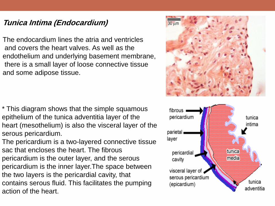
Tunica Intima (Endocardium)
The endocardium lines the atria and ventricles
and covers the heart valves. As well as the
endothelium and underlying basement membrane,
there is a small layer of loose connective tissue
and some adipose tissue.
* This diagram shows that the simple squamous
epithelium of the tunica adventitia layer of the
heart (mesothelium) is also the visceral layer of the
serous pericardium.
The pericardium is a two-layered connective tissue
sac that encloses the heart. The fibrous
pericardium is the outer layer, and the serous
pericardium is the inner layer.The space between
the two layers is the pericardial cavity, that
contains serous fluid. This facilitates the pumping
action of the heart.

Heart contraction:
First, impulses are generated by the sinoatrial node (SA), which is found in the wall
of the superior vena cava. It is a small mass of specialised cardiac muscle fibres
and associated connective tissue, and is supplied by nerve fibres from the
autonomic nervous system. Excitation of the SA node sets of a wave of
depolarisation around the atria via gap junctions between the muscle fibres.
Next the atrioventricular node (AV) starts impulse generation around the
ventricles. The AV node lies in the interatrial septum. Impulses are sent from the AV
node into the AV bundle, or bundle of his, which branches to form Purkinje fibres.
The AV node is also supplied by nerve fibres from the autonomic nervous system
that speed up and slow down the heart rate.
Purkinje fibres lie in the deepest layer of the endocardium and supply the papillary
muscles. Hence the apex of the heart contracts first, followed by the papillary
muscles, and then the wave of depolarisation spreads up the walls of the ventricles
from the base upwards, as shown in the diagram.

Heart contraction

Arteries There are three main types of arteries:
Elastic arteries
Muscular arteries
Arterioles
Elastic arteries: These arteries that receive blood directly from the heart - the aorta and the pulmonary
artery.:
They need to be elastic because:
They are relatively thin compared to their diameter.
When the heart contracts, and ejects blood into these arteries, the walls need to stretch
to accommodate the blood surge, storing energy. The arterial hydrostatic pressure that
results from ventricular contraction is the 'systolic blood pressure' (systole is greek for
contract).
Between heart contractions, the elastic walls recoil, to maintain blood pressure,
continuing to move blood even when ventricles are relaxed. The arterial hydrostatic
pressure between contractions is the 'diastolic blood pressure' (diastole is greek for
dilatation).The walls of these arteries have lots of elastin.

Tunica adventitia - has small 'vasa vasorum' as the large arteries need their own
blood supply.
Tunica media is broad and elastic with concentric fenestrated sheets of elastin,
and collagen and only relatively few smooth muscle fibres.
Tunica intima is made up of an epithelium, which is a single layer of flattened
endothelial cells, together with a supporting layer of elastin rich collagen. This layer
also has fibroblasts and 'myointimal cells' that accumulate lipid with ageing, and the
intima layer thickens, one of the first signs of atherosclerosis.

Muscular artery These arteries distribute blood to various parts of the body. These include arteries
such as the femoral and coronary arteries. The walls of these arteries have lots of
smooth muscle, which means that they are able to contract or relax (dilate) to
change the amount of blood delivered, as needed.
Comparing these arteries to the elastic arteries, the sheet of elastin is now much
reduced, and found at the border between the tunica intima and tunica media in a
layer called the internal elastic layer (IEL) which can be seen very clearly. Less well
defined is the external elastic layer (EEL), between the tunica media and tunica
adventitia. There is a well defined circular layer of smooth muscle in the tunica
media.
The tunica intima has an endothelium of flattened endothelial cells. The tunica
media is primarily a layer of smooth muscle, with some elastin an collagen. muscle
layer, and is sandwiched between the IEL and EEL. The Tunica Adventitia is very
broad, and mostly contains collagen and elastin.

Muscular artery

Arterioles: Larger arterioles have a lumen less than 100 to
300 µm in diameter. Arterioles are small arteries
that deliver blood to capillaries. Arterioles control
blood flow through capillary beds by contracting
or dilating the the size of the lumen, and
therefore the tunica media layer contains
concentric rings of smooth muscle to do this. This
compartment is important in determining your
blood pressure as the narrow diameter of these
blood vessels resists blood flow, and the back
pressure helps to stretch the walls of the arteries
during heart contractions.
The tunica intima is very thin, and mostly
consists of a single layer of squamous
epithelium.The tunica media consists almost
entirely of a single layer up to six layers of
smooth muscle cells, and there is no EEL. The
Tunica adventitia is about the same size as the
tunica media layer, merges in with surrounding
tissue.
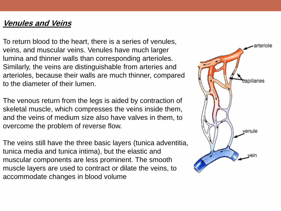
Veinsand Venules To return blood to the heart, there is a series of venules,
veins, and muscular veins. Venules have much larger
lumina and thinner walls than corresponding arterioles.
Similarly, the veins are distinguishable from arteries and
arterioles, because their walls are much thinner, compared
to the diameter of their lumen.
The venous return from the legs is aided by contraction of
skeletal muscle, which compresses the veins inside them,
and the veins of medium size also have valves in them, to
overcome the problem of reverse flow.
The veins still have the three basic layers (tunica adventitia,
tunica media and tunica intima), but the elastic and
muscular components are less prominent. The smooth
muscle layers are used to contract or dilate the veins, to
accommodate changes in blood volume

Venules These have a clear tunica intima layer, without any
elastic fibres, and a tunica media with one or two
layers of muscle fibres. The tunica adventitia
fuses with surrounding tissue.
Veins In a section that has both arteries and veins, the
artery and veins are very easy to tell apart. The
thickness of the walls of the veins is much less,
compared to the lumen, and the lumen is often
collapsed as shown here.

In this higher power image of part of the vein shown
above, can you identify the three layers of the vein:
Tunica Intima: A thin endothelial lining, (in some
veins, you may be able to see the valves).
Tunica Media: This layer contains 2-3 layers of
muscle cells.
Tunica Adventitia: This is the broadest layer. It
contains longitudinal collagen fibres, and vasa
vasorum.
Muscular veins Made of three layers, tunica intima (thin flattened
endothelial cells), the thick muscular wall (tunica
media) and the adventitia layer, which has vasa
vasorum.These blood vessels are much more
numerous than in arteries of a similar size.
Unlike muscular arteries, there is no internal or
external elastic layer surrounding the muscle layer

Capillaries Capillaries are small, normally around 3-4µm, but some
capillaries can be 30-40 µm in diameter. The largest capillaries
are found in the liver. (capillar comes from the greek for
hairlike).
Capillaries connect arterioles to venules. They allow the
exchange of nutrients and wastes between the blood and the
tissue cells, together with the interstitital fluid. This exchange
occurs by passive diffusion and by pinocytosis which means
'cell drinking'. Pinocytosis is used for proteins, and some lipids.
Also, importantly, white blood cells can move through
intercellular junctions, into the surrounding tissue to repair
damage, and fight infections. This route is also used by
metastasising cancerous cells. Capillaries have a single layer
of flattened endothelial cells, as shown here in the diagram.
There are no muscular or adventitial layers. The thinness of the
capillaries helps efficient exchange between the lumen of the
capillary and the surrounding tissue.

Continuous capillaries often have pericytes associated
with them. (perivascular cells - peri is greek for 'around')
lie just underneath the endothelium of blood capillaries,
and are a source of new fibroblasts.
There are three types of capillary:
continuous
fenestrated
discontinuous
Sinusoids found in the liver can be continuous,
fenestrated or discontinuous
Fenestrated capillaries
These are found in some tissues where there is extensive
molecular exchange with the blood such as the small
intestine, endocrine glands and the kidney. The
'fenestrations' are pores that will allow larger molecules
though.

These capillaries are more permeable than continuous capillaries.
The transmission and scanning electron microscopes below show pores
(fenestrae) in the capillary wall of the kidney glomeruli that are not resolved by the
light microscope.
At high magnification, the fenestrations of the endothelial cell can be seen as
'gaps' next the the basement membrane

Discontinuous Capillaries These are only found in the liver. They are formed between the endothelial cells of
the sinusoids and hepatocyte cells (Cell 1 and 2 in the picture). The hepatocytes
have lots of projections called microvilli that project into the space of Disse. This
produces large clefts or spaces between the two layers of cells, that allows proteins,
or even blood cells to pass through.
Sinusoids are a special type of capillary that have a wide diameter. These are found
in the liver, spleen, lymph nodes, bone marrow and some endocrine glands. They
can be continuous, fenestrated, or discontinuous.

THANK YOU…


