ORIGINAL RESEARCH ARTICLE published: 06 June 2013 doi: 10.3389/fimmu.2013.00137 Homing of antigen-presenting cells in head kidney and spleen – salmon head kidney hosts diverse APC types Dimitar B. Iliev 1 *, Hanna Thim 1 , Leidy Lagos 1 , Randi Olsen 2 and Jorunn B. Jørgensen 1 1 Norwegian College of Fisheries Science, University of Tromsø, Tromsø, Norway 2 Department of Medical Biology, University ofTromsø,Tromsø, Norway Edited by: Ken J. Ishii, National Institute of Bimedical Innovation, Japan Reviewed by: Ken J. Ishii, National Institute of Bimedical Innovation, Japan Mayda Gursel, Middle EastTechnical University, Turkey *Correspondence: Dimitar B. Iliev , Norwegian College of Fishery Science, University ofTromsø, Breivika, N-9037 Tromsø, Norway e-mail: [email protected]Lymph nodes and spleen are major organs where mammalian antigen-presenting cells (APCs) initiate and orchestrate Ag-specific immune responses. Unlike mammals, teleosts lack lymph nodes and an interesting question is whether alternative organs may serve as sites for antigen presentation in teleosts. In the current study, fluorescent ovalbumin (Ova) and CpG oligonucleotides (ODNs) injected intra-abdominally were detected in significant numbers of salmon head kidney (HK) MHCII+ cells over a period of 2 weeks while in spleen the percentage of these was transient and declined from day 1 post injection. In vitro stud- ies further shed light on the properties of the diverse MHCII+ cell types found in HK. The ultrastructure of a subpopulation of MHCII+ cells with a high capacity to endocytose and process Ova indicated that these were able to perform constitutive macropinocytosis. Upon stimulation with CpG ODNs these cells upregulated CD86 and gave very high levels of TNF mRNA indicating that these are professional APCs, related to macrophages and dendritic cells (DCs). A subpopulation of HK granulocytes expressed high levels of sur- face MHCII and upon CpG stimulation upregulated most of the tested APC marker genes. Although these granulocytes expressedTNF weakly, they had relatively high basal levels of IL-1β mRNA and the CpG stimulation upregulated IL-1β, along with its signaling and decoy receptors, to the highest levels as compared to other HK cell types. Interestingly, the high expression of IL-1β mRNA in the granulocytes correlated with a high autophagy flux as demonstrated by LC3-II conversion. Autophagy has recently been found to be implicated in IL-1β processing and secretion and the presented data suggests that granulocytes of salmon, and perhaps other teleost species, may serve as a valuable model to study the involvement of autophagy in regulation of the vertebrate immune response. Keywords: MHCII, APC, granulocytes, Atlantic salmon, CpG oligonucleotides, IL-1β,TNF, autophagy INTRODUCTION Antigen-presenting cells (APCs) are specialized to take up, process and present protein antigens (Ag) associated with MHCII mole- cules to Ag-specific T-cells. Professional APCs include dendritic cells (DCs), macrophages, and B-cells of which the DCs are dis- tinguished by their high co-stimulatory capacity (Hamilos, 1989; Thery and Amigorena, 2001). APCs are distributed throughout the organism, including the peripheral tissues where they cap- ture inbound Ags derived from potentially pathogenic agents. In order to achieve full co-stimulatory capacity, APCs need to be acti- vated through innate immune receptors such as toll-like receptors (TLRs) which recognize conserved molecular structures produced by microorganisms and viruses (Thery and Amigorena, 2001). In mammals, exposure of DCs to these pathogen-associated molecu- lar patterns (PAMPs) may trigger a process of maturation during which the antigen-loaded DCs migrate toward the secondary immune organs, upregulate MHCII-associated Ag peptides and co-stimulatory molecules on their surface and downregulate their capacity to endocytose Ag (Kaisho and Akira, 2003). In mammals, the major organs where maturing DCs migrate and which provide proper environment for Ag presentation are the lymph nodes and the spleen. The adaptive immune system has evolved at the level of early jawed vertebrates and most of its elements described in mammals, such as Ag-specific B- and, T-cell receptors and antibodies are also found in teleosts (Pancer and Cooper, 2006). In addition, a T-cell-dependent Ag response has been described in teleosts (Miller et al., 1986) and PAMPs, such as CpG oligonucleotides (ODNs), have shown a potential to induce maturation of salmon APCs in vitro (Iliev et al., 2010). Cells resembling mammalian DCs have been described in fish (Lovy et al., 2009; Aghaallaei et al., 2010; Lugo-Villarino et al., 2010; Bassity and Clark, 2012) and authors have suggested that the melanomacrophage centers which are found in the spleen and the head kidney (HK) (ante- rior kidney, pronephros) of different teleosts (Tsujii and Seno, 1990) may serve as sites for Ag presentation (Agius and Roberts, 2003). Still, there are considerable differences between the mam- malian and the teleost immune systems – for example, absence of lymph nodes and classical Ig class switch in teleosts. Therefore, more detailed knowledge about the phenotype and the function of piscine APCs will help gain insight into the evolution of the verte- brate adaptive immune system and will provide valuable informa- tion for development and optimization of immunotherapies for aquaculture use. www.frontiersin.org June 2013 |Volume 4 | Article 137 | 1
Transcript
ORIGINAL RESEARCH ARTICLEpublished: 06 June 2013
doi: 10.3389/fimmu.2013.00137
Homing of antigen-presenting cells in head kidney andspleen – salmon head kidney hosts diverse APC typesDimitar B. Iliev 1*, HannaThim1, Leidy Lagos1, Randi Olsen2 and Jorunn B. Jørgensen1
1 Norwegian College of Fisheries Science, University of Tromsø, Tromsø, Norway2 Department of Medical Biology, University of Tromsø, Tromsø, Norway
Edited by:Ken J. Ishii, National Institute ofBimedical Innovation, Japan
Reviewed by:Ken J. Ishii, National Institute ofBimedical Innovation, JapanMayda Gursel, Middle East TechnicalUniversity, Turkey
*Correspondence:Dimitar B. Iliev , Norwegian College ofFishery Science, University ofTromsø,Breivika, N-9037 Tromsø, Norwaye-mail: [email protected]
Lymph nodes and spleen are major organs where mammalian antigen-presenting cells(APCs) initiate and orchestrate Ag-specific immune responses. Unlike mammals, teleostslack lymph nodes and an interesting question is whether alternative organs may serve assites for antigen presentation in teleosts. In the current study, fluorescent ovalbumin (Ova)and CpG oligonucleotides (ODNs) injected intra-abdominally were detected in significantnumbers of salmon head kidney (HK) MHCII+ cells over a period of 2 weeks while in spleenthe percentage of these was transient and declined from day 1 post injection. In vitro stud-ies further shed light on the properties of the diverse MHCII+ cell types found in HK.The ultrastructure of a subpopulation of MHCII+ cells with a high capacity to endocytoseand process Ova indicated that these were able to perform constitutive macropinocytosis.Upon stimulation with CpG ODNs these cells upregulated CD86 and gave very high levelsof TNF mRNA indicating that these are professional APCs, related to macrophages anddendritic cells (DCs). A subpopulation of HK granulocytes expressed high levels of sur-face MHCII and upon CpG stimulation upregulated most of the tested APC marker genes.Although these granulocytes expressedTNF weakly, they had relatively high basal levels ofIL-1β mRNA and the CpG stimulation upregulated IL-1β, along with its signaling and decoyreceptors, to the highest levels as compared to other HK cell types. Interestingly, the highexpression of IL-1β mRNA in the granulocytes correlated with a high autophagy flux asdemonstrated by LC3-II conversion. Autophagy has recently been found to be implicatedin IL-1β processing and secretion and the presented data suggests that granulocytes ofsalmon, and perhaps other teleost species, may serve as a valuable model to study theinvolvement of autophagy in regulation of the vertebrate immune response.
INTRODUCTIONAntigen-presenting cells (APCs) are specialized to take up, processand present protein antigens (Ag) associated with MHCII mole-cules to Ag-specific T-cells. Professional APCs include dendriticcells (DCs), macrophages, and B-cells of which the DCs are dis-tinguished by their high co-stimulatory capacity (Hamilos, 1989;Thery and Amigorena, 2001). APCs are distributed throughoutthe organism, including the peripheral tissues where they cap-ture inbound Ags derived from potentially pathogenic agents. Inorder to achieve full co-stimulatory capacity, APCs need to be acti-vated through innate immune receptors such as toll-like receptors(TLRs) which recognize conserved molecular structures producedby microorganisms and viruses (Thery and Amigorena, 2001). Inmammals, exposure of DCs to these pathogen-associated molecu-lar patterns (PAMPs) may trigger a process of maturation duringwhich the antigen-loaded DCs migrate toward the secondaryimmune organs, upregulate MHCII-associated Ag peptides andco-stimulatory molecules on their surface and downregulate theircapacity to endocytose Ag (Kaisho and Akira, 2003). In mammals,the major organs where maturing DCs migrate and which provideproper environment for Ag presentation are the lymph nodes andthe spleen.
The adaptive immune system has evolved at the level of earlyjawed vertebrates and most of its elements described in mammals,such as Ag-specific B- and, T-cell receptors and antibodies arealso found in teleosts (Pancer and Cooper, 2006). In addition,a T-cell-dependent Ag response has been described in teleosts(Miller et al., 1986) and PAMPs, such as CpG oligonucleotides(ODNs), have shown a potential to induce maturation of salmonAPCs in vitro (Iliev et al., 2010). Cells resembling mammalianDCs have been described in fish (Lovy et al., 2009; Aghaallaeiet al., 2010; Lugo-Villarino et al., 2010; Bassity and Clark, 2012)and authors have suggested that the melanomacrophage centerswhich are found in the spleen and the head kidney (HK) (ante-rior kidney, pronephros) of different teleosts (Tsujii and Seno,1990) may serve as sites for Ag presentation (Agius and Roberts,2003). Still, there are considerable differences between the mam-malian and the teleost immune systems – for example, absenceof lymph nodes and classical Ig class switch in teleosts. Therefore,more detailed knowledge about the phenotype and the function ofpiscine APCs will help gain insight into the evolution of the verte-brate adaptive immune system and will provide valuable informa-tion for development and optimization of immunotherapies foraquaculture use.
In an earlier study, we found that Atlantic salmon HK hostsdistinct MHCII+ cell types including MHCII+ leukocytes whichendocytosed large amounts of dextran and a population of granu-lar cells with lower capacity to take up dextran but with high levelsof surface MHCII as shown by staining with an antibody specificfor the MHCII beta chain (Iliev et al., 2010). Additionally, the HKharbored MHCII/Ig double-positive cells, resembling B-cells.
In light of these findings, the current study has been intendedto further characterize salmon APCs in regard to their ability tomigrate to HK and spleen following in vivo uptake of Ag, their mor-phology, and their potential to express immune genes includingAPC markers, cytokines, and cytokine receptors. Fluorescent oval-bumin (Ova-FITC) injected in the abdominal cavity was observedboth in HK and spleen leukocytes 1 day post injection, while atlatter time points (5 and 14 days) it was found exclusively inMHCII+/IgM−HK cells. When HK cells were stimulated in vitro,distinct gene expression profiles were detected in different HKMHCII+ cell types. The cells which endocytosed high amountsof Ova expressed the highest levels of CD86 and TNF mRNAsuggesting that these are maturing APCs. On the other hand, apopulation of cells with morphology of polymorphonuclear gran-ulocytes which expressed high levels of surface MHCII and did nottake up significant amounts of Ova, upregulated very high levelsof IL-1β mRNA along with APC marker genes including CD83,CD40, and B7-H1 but they expressed relatively low levels of CD86.Autophagy has recently been found to be involved in productionand secretion of IL-1β (Dupont et al., 2011) and autophagy fluxanalysis using sorted cell populations showed that the intensity ofthe process was highest in the HK granulocytes suggesting thatthese cells might be specialized in production and secretion of thiscytokine.
MATERIALS AND METHODSFISHAtlantic salmon (Salmo salar) strain AquaGen standard (AquaGen, Kyrksæterøra, Norway) were obtained from the TromsøAquaculture Research Station (Tromsø, Norway). The fish werekept at about 10 °C in tanks supplied with running filtered waterand were fed on commercial, dry food (Skretting, Stavanger, Nor-way). All experiments were approved by the national committeefor animal experimentation (Forsøksdyrutvalget, Norway) andperformed according to its guidelines.
REAGENTSPhosphorothioate-modified CpG-B ODNs (5′-TCGTCGTTTTGTCGTTTTGTCGTT-3′) were purchased from Thermo Scientific.The antiserum against the β-chain of salmon MHCII was previ-ously described (Iliev et al., 2010). The antibody against salmonidIgM was obtained from CedarLane Labs, ON, Canada. A sec-ondary goat anti-rabbit antibody conjugated with AlexaFluor546,Ova-Alexa647, DQ Ova, and LysoSensor™ Green DND-189 werepurchased from Life technologies. Goat anti-rabbit-horseradishperoxidase antibody was obtained from Santa Cruz Biotechnologyand the eEF2 polyclonal antibody was purchased from Cell Signal-ing Technology. Polyclonal LC3 antibody, dextran-Cascade Blue,Ova-FITC, and May Grünwald–Giemsa reagents were obtainedfrom Sigma.
ISOLATION OF LEUKOCYTES FROM ATLANTIC SALMONThree groups of 15 individuals of ∼100 g were injected intra-abdominally with 100 µl of PBS, 500 µg of Ova-FITC, or with,500 µg of Ova-FITC+ 50 µg of CpG-Cy5. HK and spleen leuko-cytes were isolated 1, 5, and 14 days post injection as described(Iliev et al.,2010). Briefly, the HK and the spleen tissues were passedthrough 100-µm pore size cell strainers (Falcon) in L-15 mediumcontaining penicillin (10 U/ml), streptomycin (10 µg/ml), 2%fetal bovine serum (FBS), and heparin (20 U/ml). The result-ing suspension was placed on a 25/54% discontinuous Percollgradient and centrifuged at 400× g for 40 min at 4 °C. Thecells at the interface were collected and washed twice in L-15medium before further use. For the in vitro experiments, thedensity of the leukocyte suspensions was adjusted to 7× 10⁶
cells/ml and the cells were further incubated in 24-well plates inL-15, 5% FBS.
FLOW CYTOMETRYCells were pelleted at 4 °C and the staining was performed on ice.The cells were washed with ice-cold PBS and incubated simulta-neously with MHCIIβ rabbit antiserum (1000-fold dilution) andsalmon anti-IgM FITC-conjugated monoclonal antibody (200-fold dilution) for 1 h in PBS, 5% FBS on ice. The secondaryAlexa546 goat anti-rabbit antibody was diluted to 1 µg/ml in PBS,5% FBS and the cells were incubated for 30 min on ice prior towashing with PBS. For the in vitro endocytosis assays, HK leuko-cytes were incubated with 10 µg/ml of Ova-FITC or Ova-Alexa647,150 µg/ml of dextran-Cascade blue and 2 µM CpG-Cy5 ODNs for1 h. Prior to sorting, the whole HK leukocyte population was stim-ulated for 24 h with 2 µM CpG ODNs followed by 1 h incubationwith 10 µg/ml of or Ova-Alexa647 prior to staining as describedabove. The cells were analyzed and sorted using FACSAria (BectonDickinson).
To analyze the Ag-processing capacity of APCs found in salmonHK, cells were incubated with 5 µg/ml of Ova-DQ for 4 h. Afterremoval of non-adherent cells, the adherent ones were detachedand harvested using a 10 min treatment with PBS, 1 mM EDTA.The adherent and the non-adherent cells were stained for sur-face MHCII and analyzed separately with flow analysis. Ova-DQproteolysis was detected as green fluorescence.
REAL-TIME PCR (RT-PCR) ANALYSISRNA from sorted cells was isolated using RNeasy Mini Kit (Qia-gen). On-column DNase digestion was performed using RNase-Free DNase set (Qiagen, Hilden, Germany). For each sample 70 ngtotal RNA was reverse transcribed using the TaqMan ReverseTranscription Reagents kit (Applied Biosystems). The real-timePCR (RT-PCR) reactions were prepared with Power SYBR ®GreenPCR Master Mix or TaqMan ®Fast Universal PCR Master Mix(Applied Biosystems). The type of the reaction and the primerand the probe sequences are listed in Table 1. The reactions wererun in duplicate and included 5 µl of fivefold diluted cDNA.The reaction protocol and the data analysis have been previ-ously described (Iliev et al., 2010). EF1aB expression was usedas endogenous control and the data is presented as fold differ-ence values as compared to the non-stimulated MHC−/Ig−/Ova−sample.
Frontiers in Immunology | Antigen Presenting Cell Biology June 2013 | Volume 4 | Article 137 | 2
CONFOCAL MICROSCOPYHead kidney cells from fish injected with Ova-FITC and CpG-Cy5 attached to 15 mm coverslips were washed, with PBS andfixed for 15 min with 4% formaldehyde. Following permeabiliza-tion using 0.3% Triton X-100 for 15 min at room temperatureand blocking in PBS, 5% FBS for 30 min, the cells were incu-bated consecutively with 1:2000 dilution of MHCIIβ antiserumand a secondary Alexa546-conjugated antibody. The coverslipswere mounted in glycerol, 1% DABCO. Images were collected witha Zeiss Axiovert 200 microscope with a 40×, Apochroma objec-tive, equipped with an LSM510-META confocal module using theLSM5 software version 3.2 (Carl Zeiss Inc.).
For in vitro Ag uptake and sub-cellular localization, adherentHK mononuclear phagocytes were isolated as previously described(Iliev et al., 2010). To visualize endolysosomes, cells cultured inLab-Tek™ Chambered Coverglass slides (Nunc) were incubatedwith 1 µM LysoSensor™ Green DND-189 (Life technologies) for30 min, washed and incubated in L-15, 5% FBS with 10 µg/ml ofOva-Alexa647. Images of live cells were taken as described above.
WESTERN BLOTSamples from sorted cells (1× 10⁵ per sample) were pelleted(1500 g, 5 min), lyzed in LDS sample buffer (Invitrogen) and runon NuPAGE Novex Bis-Tris 12% gel (Invitrogen). The proteinswere transferred to PVDF membranes, blocked with 5% dry milkand incubated consecutively with a 1:1000 dilution of LC3 anti-body and a 1:10000 dilution of a goat anti-rabbit-horseradishperoxidase antibody. Stripping of the membranes was performedin 0.2 M NaOH for 10 min followed by washing, blocking, andreprobing with 1:1000 dilution of an eEF2 polyclonal antibody.
MORPHOLOGY OF SORTED HK CELLS – TRANSMISSION ELECTRONMICROSCOPY ANALYSISSorted HK subpopulations were pelleted in a microcentrifuge(1500 g, 5 min) and fixed in 8% formaldehyde. Cells were washedin 0.1% cacodylate buffer (pH 7.2), post fixed in mixture of 2%osmium tetroxide/1.5% potassium ferrocyanide in 0.1% cacody-late for 30 min. Staining was performed with 1% tannic acid and1% uranyl acetate, followed by dehydration in a graded seriesof ethanol (70%, 90%, 96%, 2× 100%). The cells were treatedwith acetonitrile as an intermediate step before infiltration withan Epon substitute (AGAR 100 resin, Agar Scientific, Stansted,
England) and polymerized at 60 °C overnight. Ultrathin sections(70 nm) were made using a Leica Ultracut S Ultramicrotome(Vienna, Austria) with a Diatome diamond knife (Biel, Switzer-land). The sections were mounted on carbon coated formvar filmson copper grids and contrasted with 5% uranyl acetate for 8 minand Reynolds lead citrate for 5 min. Micrographs were taken ona Jeol 1010 JSM (Tokyo, Japan) with a Morada 11 Mpixels digitalcamera (Olympus).
IMMUNOLABELING FOR ELECTRON MICROSCOPYSorted HK subpopulations were pelleted in a microcentrifuge(1500 g, 5 min) and fixed in 4% formaldehyde in 200 mM HEPESbuffer, pH 7.5, for 1 h. The samples were prepared for immuno-labeling according to standard methods. Ultrathin cryosectionscut on a Leica EMUC6 Ultramicrotome with an Ultracut Sin a Leica FCS cryochamber using a diamond knife (DiatomeLtd., Bienne, Switzerland) and mounted on Formvar-coated EM-grids. Immunocytochemical labeling was performed as described(Tokuyasu, 1986; Webster and Webster, 2007). The Anti-LC3 anti-body was used at 1:1000 dilution and was detected by proteinA-gold complexes. The dried sections were examined in a JEOLJEM 1010 transmission electron microscope (JEOL, Tokyo, Japan)operating at 80 kV. Control experiments were routinely includedin parallel by omission of the primary antibodies.
DATA ANALYSISData was analyzed with one-way and two-way ANOVA, as indi-cated, followed by Tukey post-tests. Statistical analyses were per-formed using the GraphPad Prism6 software. The value of p < 0.05was considered to be significant. Only groups with n > 3 wereincluded in the analysis.
RESULTSSOLUBLE ANTIGEN AND CpGs INJECTED IN THE ABDOMINAL CAVITYOF ATLANTIC SALMON ACCUMULATE PREDOMINATELY IN HK MHCII+CELLSTo analyze the Ova distribution in vivo, salmon (∼100 g) wereinjected intra-abdominally with 500 µg of Ova-FITC or with500 µg of Ova-FITC mixed with 50 µg of CpG-Cy5. Control fishwere injected with the same volume (100 µl) of PBS. HK andspleen leukocytes were isolated after 1, 5, and 14 days and follow-ing staining for surface MHCII and IgM were analyzed using flowcytometry. The results shown in Figure 1A demonstrate that Ovaand CpGs are detected in both spleen and HK cells 1 day postinjection. The percentage of Ova+ and CpG+ cells in HK andspleen were approximately equal at this time point (∼2% in bothorgans). Since, considerably more leukocytes were isolated fromHK, the absolute number of Ova+ cells was greater in this organas compared to spleen. Although there was relatively more CpG+than Ova+ cells in HK, there was a relatively good correlationin the uptake of the two substrates. It seems that the higher per-centage of CpG+ cells might be due to a higher intensity of theCpG-Cy5 fluorescence as compared to that emitted by Ova-FITC.Confocal microscopy analysis revealed that Ova-FITC and CpG-Cy5 colocalized in intracellular vesicles in MHCII+ cells fromHK harvested 24 h post injection (Figure 1B). These cells had amacrophage-like morphology as revealed by the presence of longand branching pseudopodia.
The dot plots presented in Figure 1C show cells isolated 1 daypost injection. Most of the Ova+ and CpG+ HK cells expressedsurface MHCII but not IgM. In contrast, in spleen an approxi-mately equal percentage of the Ova+ and the CpG+ cells wereMHCII− and MHCII+, and most of these were IgM−. The his-tograms in Figure 1D show the average percentage of the Ova+cells in the Ova-only and the Ova/CpG-injected groups. Theresults demonstrate that while on day one the percentages of Ova+cells in HK and spleen are comparable, on day 5 and day 14 thevalues in spleen decline whereas those in HK remain high for upto 14 days. The data also show that in HK at all tested time pointsmost of the Ova+ cells are MHCII+. The addition of 50 µg CpGsto the Ova, a dose that previously has been shown to upregulateexpression of immune genes in both HK and spleen (Strandskoget al., 2008), did not seem to affect the accumulation of Ova+cells in neither HK nor spleen. The experimental setup includedfive fish per group/time point; however, due to the large numberof samples, for some of the groups only two samples could beanalyzed which makes statistical analysis unreliable.
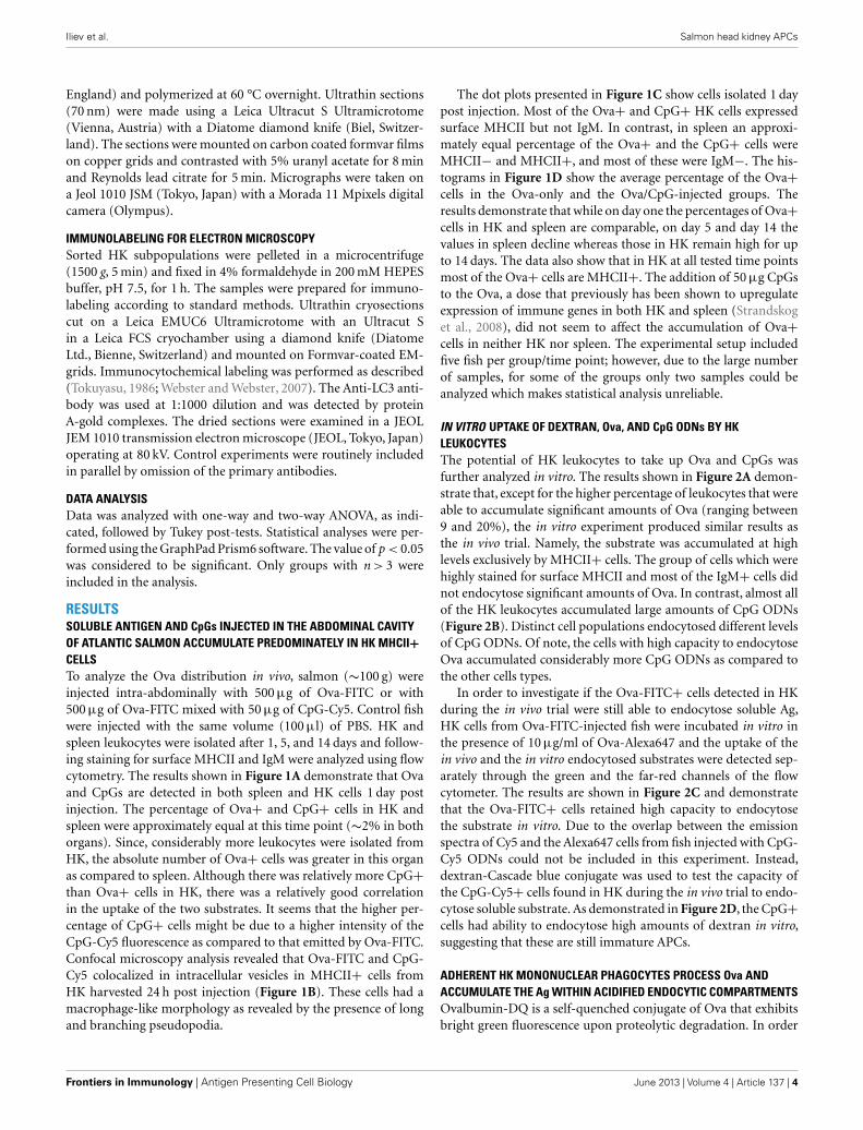
IN VITRO UPTAKE OF DEXTRAN, Ova, AND CpG ODNs BY HKLEUKOCYTESThe potential of HK leukocytes to take up Ova and CpGs wasfurther analyzed in vitro. The results shown in Figure 2A demon-strate that, except for the higher percentage of leukocytes that wereable to accumulate significant amounts of Ova (ranging between9 and 20%), the in vitro experiment produced similar results asthe in vivo trial. Namely, the substrate was accumulated at highlevels exclusively by MHCII+ cells. The group of cells which werehighly stained for surface MHCII and most of the IgM+ cells didnot endocytose significant amounts of Ova. In contrast, almost allof the HK leukocytes accumulated large amounts of CpG ODNs(Figure 2B). Distinct cell populations endocytosed different levelsof CpG ODNs. Of note, the cells with high capacity to endocytoseOva accumulated considerably more CpG ODNs as compared tothe other cells types.
In order to investigate if the Ova-FITC+ cells detected in HKduring the in vivo trial were still able to endocytose soluble Ag,HK cells from Ova-FITC-injected fish were incubated in vitro inthe presence of 10 µg/ml of Ova-Alexa647 and the uptake of thein vivo and the in vitro endocytosed substrates were detected sep-arately through the green and the far-red channels of the flowcytometer. The results are shown in Figure 2C and demonstratethat the Ova-FITC+ cells retained high capacity to endocytosethe substrate in vitro. Due to the overlap between the emissionspectra of Cy5 and the Alexa647 cells from fish injected with CpG-Cy5 ODNs could not be included in this experiment. Instead,dextran-Cascade blue conjugate was used to test the capacity ofthe CpG-Cy5+ cells found in HK during the in vivo trial to endo-cytose soluble substrate. As demonstrated in Figure 2D, the CpG+cells had ability to endocytose high amounts of dextran in vitro,suggesting that these are still immature APCs.
ADHERENT HK MONONUCLEAR PHAGOCYTES PROCESS Ova ANDACCUMULATE THE Ag WITHIN ACIDIFIED ENDOCYTIC COMPARTMENTSOvalbumin-DQ is a self-quenched conjugate of Ova that exhibitsbright green fluorescence upon proteolytic degradation. In order
Frontiers in Immunology | Antigen Presenting Cell Biology June 2013 | Volume 4 | Article 137 | 4
FIGURE 1 | Fluorescent Ova-FITC and CpG-Cy5 ODNs injected in theabdominal cavity of Atlantic salmon accumulate predominately in HKMHCII+ cells. The fish were injected with Ova-FITC alone or in combinationwith CpG-Cy5 and their accumulation in head kidney (HK) and spleenleukocytes was analyzed with flow cytometry after 1, 5, and 14 days. Thecontrols were injected with the same volume of PBS. (A) 24 h post injectionapproximately equal percentage of Ova+ and CpG+ cells was observed in HKand spleen and there was good correlation between the uptake of Ova andCpG ODNs. (B) HK cells from Ova/CpG-injected fish were stained for MHCIIand were analyzed with confocal microscopy. The arrow indicates thecolocalization between Ova and CpGs in endosomal compartments of
MHCII+ cells. (C) Ova and CpG ODNs accumulate mostly in MHCII+/IgM−cells in HK. MHCII- and IgM-stained cells were analyzed with flow cytometry.The dot plots show the correlation between the surface expression of MHCIIand IgM and the uptake of Ova and CpG ODNs in HK and spleen leukocytesisolated 24 h after injection. (D) The percentage of Ova+ cells in spleendeclines sharply between 1 and 5 days post injection whereas in HK itremains high for up to 14 days. The histograms show the mean percentage ofOva+ cells in MHCII± cells in both the Ova and the Ova/CpG groups asdetermined by flow cytometry (n varies between 2 and 5), Statistical analysiswas performed on samples taken on day 1 and day 5 since the number ofreplicates in all of these samples was >3, *P < 0.05.
to investigate the ability of salmon HK APCs to process Ag, HKcells were incubated with Ova-DQ for 4 h followed by flow analy-sis. Non-adherent and adherent cells were harvested and analyzedseparately after staining for surface MHCII. The results presentedin Figure 3A show that unlike non-adherent MHCII++ granu-locytes, adherent HK MHCII+ cells possess ability to effectivelytake up and process Ova.
The intracellular distribution of Ova was examined in adherentHK mononuclear phagocytes in which late endosomes and lyso-somes were labeled with LysoSensor. As shown in Figure 3B, theendocytosed Ova was detectable in acidic vesicles within 1 h afteraddition of the Ag to the cell culture medium. The accumulationof Ova in LysoSensor+ vesicles was more pronounced after 24 hof incubation.
DISTINCT HK CELL POPULATIONS ARE FOUND BASED ON SURFACEMHCII AND IgM EXPRESSION, OVA UPTAKE, AND MORPHOLOGYTo further characterize the different HK leukocyte subpopulationscells were sorted using a FACSAria instrument according to theirability to endocytose Ova and the surface MHCII and IgM expres-sion. The sorted samples were then analyzed using microscopy andRT-PCR.
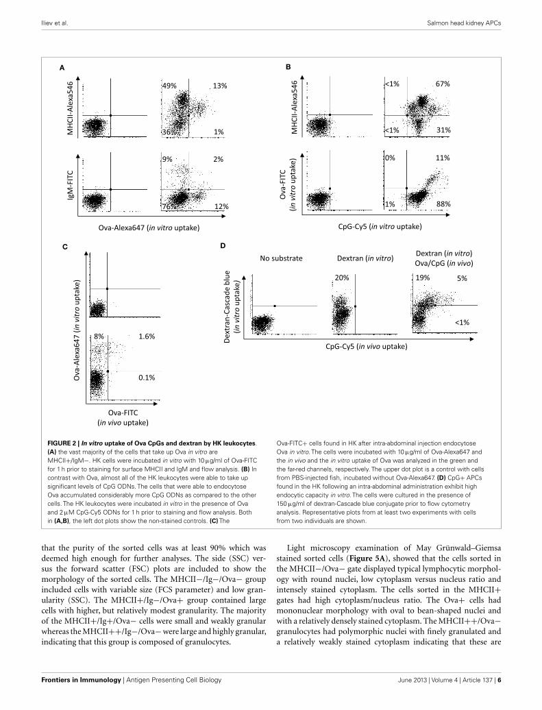
The pre-sort dot plots in Figure 4 show the setup of thegates. Unlike the Ova−/Ig+ cells which had typical lymphocytemorphology the Ova+/IgM+ cells were larger and granular andwere excluded from the analysis as they may also have includedmacrophages which had bound IgM through their Fc receptors. Asshown in the post-sort dot plots, nearly all of the IgM+ cells werealso positive for surface MHCII. The post-sort analysis indicated
No substrate Dextran (in vitro) Dextran (in vitro)
Ova/CpG (in vivo)
Ova-Alexa647 (in vitro uptake)
Ova
-FIT
C
(in
vit
ro u
pta
ke)
MH
CII
- Ale
xa5
46
D
CpG-Cy5 (in vitro uptake)
IgM
-FIT
C
MH
CII
-Ale
xa5
46
<1% 67%
31% <1%
0% 11%
88% 1%
C
FIGURE 2 | In vitro uptake of Ova CpGs and dextran by HK leukocytes.(A) the vast majority of the cells that take up Ova in vitro areMHCII+/IgM−. HK cells were incubated in vitro with 10 µg/ml of Ova-FITCfor 1 h prior to staining for surface MHCII and IgM and flow analysis. (B) Incontrast with Ova, almost all of the HK leukocytes were able to take upsignificant levels of CpG ODNs. The cells that were able to endocytoseOva accumulated considerably more CpG ODNs as compared to the othercells. The HK leukocytes were incubated in vitro in the presence of Ovaand 2 µM CpG-Cy5 ODNs for 1 h prior to staining and flow analysis. Bothin (A,B), the left dot plots show the non-stained controls. (C) The
Ova-FITC+ cells found in HK after intra-abdominal injection endocytoseOva in vitro. The cells were incubated with 10 µg/ml of Ova-Alexa647 andthe in vivo and the in vitro uptake of Ova was analyzed in the green andthe far-red channels, respectively. The upper dot plot is a control with cellsfrom PBS-injected fish, incubated without Ova-Alexa647. (D) CpG+ APCsfound in the HK following an intra-abdominal administration exhibit highendocytic capacity in vitro. The cells were cultured in the presence of150 µg/ml of dextran-Cascade blue conjugate prior to flow cytometryanalysis. Representative plots from at least two experiments with cellsfrom two individuals are shown.
that the purity of the sorted cells was at least 90% which wasdeemed high enough for further analyses. The side (SSC) ver-sus the forward scatter (FSC) plots are included to show themorphology of the sorted cells. The MHCII−/Ig−/Ova− groupincluded cells with variable size (FCS parameter) and low gran-ularity (SSC). The MHCII+/Ig−/Ova+ group contained largecells with higher, but relatively modest granularity. The majorityof the MHCII+/Ig+/Ova− cells were small and weakly granularwhereas the MHCII++/Ig−/Ova−were large and highly granular,indicating that this group is composed of granulocytes.
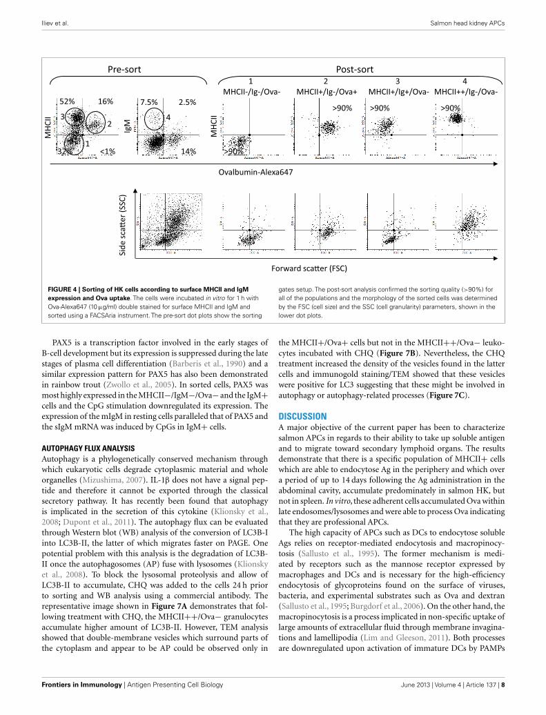
Light microscopy examination of May Grünwald–Giemsastained sorted cells (Figure 5A), showed that the cells sorted inthe MHCII−/Ova− gate displayed typical lymphocytic morphol-ogy with round nuclei, low cytoplasm versus nucleus ratio andintensely stained cytoplasm. The cells sorted in the MHCII+gates had high cytoplasm/nucleus ratio. The Ova+ cells hadmononuclear morphology with oval to bean-shaped nuclei andwith a relatively densely stained cytoplasm. The MHCII++/Ova−granulocytes had polymorphic nuclei with finely granulated anda relatively weakly stained cytoplasm indicating that these are
Frontiers in Immunology | Antigen Presenting Cell Biology June 2013 | Volume 4 | Article 137 | 6
FIGURE 3 | Adherent HK mononuclear phagocytes hydrolyze Ova andaccumulate the Ag within acidified endocytic compartments. (A) Headkidney leukocytes were incubated for 4 h with 5 µg/ml of Ova-DQ.Non-adherent and adherent cells were harvested and analyzed separatelywith flow cytometry. Gray contour – non-adherent MHCII++ granulocytes,black contour – adherent MHCII+ cells. The filled contour shows anon-stained control. Ova-DQ fluorescence is detected in the green channel.Similar results were obtained with cells from two individuals. (B) Adherenthead kidney mononuclear phagocytes attached to chambered coverglassslides were stained with LysoSensor Green (pKa 5.2) and incubated withOva-Alexa647 for the indicated periods prior to live imaging using a confocalmicroscope. The arrows in the magnified overlap regions indicateaccumulation of Ova within late endosome/lysosome compartments.
polymorphonuclear granulocytes. The ultrastructure of the sortedcells was examined with transmission electron microscopy (TEM)(Figure 5B). Except for the cell nuclei, a few small mitochondriaand abundance of free ribosomes, no other clear structures couldbe observed in the MHCII−/Ova− cells. The MHCII+/Ova+ cellshad long pseudopodia which upon fusion form macropinosomes(MPS) indicating that these cells perform macropinocytosis con-stitutively. The cells also had abundant mitochondria, extensiverough endoplasmic reticulum and, some of them, melanin gran-ules. In addition, in some cells, early/sorting endosomes (E/SE)could be observed (magnified inset). Typically, these appear asvesicles with attached tubules into which the receptors such asthose involved in the endocytosis of mannosylated substrates (i.e.,the mannose receptor) are recycled toward the cells surface (Wile-man et al., 1985; Jovic et al., 2010). Endocytosed proteins remainin the body of the E/SE and are transported toward degradationby lysosomal enzymes. The MHCII++/Ova− granulocytes con-tained many small and moderately sized vesicles (up to 400 nm)with varying density and round to elongated shape. Internal
membranes with lamellar or vesicular shape were visible in someof these lysosome-like vesicles. Interestingly, one of the observedvesicles appeared to be in a process of microautophagy as suggestedby the presence of an arm-like extension surrounding parts of theneighboring cytoplasm. No large pseudopodia, MPS, E/SE, andmelanin granules were observed in these cells. Also, compared tothe Ova+ cells these polymorphonuclear granulocytes containedfewer mitochondria and less RER.
DIFFERENT MHCII+ HK LEUKOCYTE SUBPOPULATIONS SHOWDISTINCT GENE EXPRESSION PROFILESRNA samples from sorted cells were used to analyze gene expres-sion in resting and in activated HK leukocytes subpopulations.CpG stimulation in higher vertebrate species directly activatesDCs, macrophages, and B-cells, leading to upregulation of co-stimulatory molecules and proinflammatory cytokines and anincreased capacity for antigen processing and presentation (Hart-mann et al., 1999). Similarly, in Atlantic salmon, in vitro stimu-lation of adherent mononuclear phagocytes with CpGs for 24 hinduced expression of proinflammatory cytokines and markergenes for mature APCs (Iliev et al., 2010). In the current study, thewhole HK leukocyte population was stimulated with CpG-B for24 h prior to sorting and gene expression analysis using RT-PCR.Among the leukocyte marker genes, the basal level of CD83, CD86,and B7-H1 was highest in the Ova+ cells (Figure 6A). However,the CpG treatment upregulated CD83 and CD40 most highly inthe MHCII++/IgM−/Ova− cells. The expression of CD208 andIFN-γ was highest in IgM+ cells.
Interestingly, there was a contrast in the expression patternsof the proinflammatory cytokines TNF2 and IL-1β1. The formerwas most highly expressed in the Ova+ cells whereas both thebasal and the CpG-upregulated IL-1β1 mRNA level were high-est the MHCII++/Ig−/Ova− cells. The expression of the twocytokines was further confirmed in sorted cells from two addi-tional fish, The TNF2 expression in non-stimulated MHCII− cellsfrom fish 3 and fish 4 was not detectable making it impossible tocalculate the “fold difference” expression of the cytokine in thesesamples. Nevertheless, as indicated by the ratio between the thresh-old cycle (Ct) values of the housekeeping gene and the cytokine, theMHCII+/Ova+ cells consistently expressed the highest levels ofTNF2 mRNA, whereas the MHCII++/Ova− cells gave the high-est levels of IL-1β1 mRNA when compared with other cell typesfrom the same individual (Figure 6B). The MHCII++ granulo-cytes also expressed highest levels of IL1R1 and IL1R2 mRNA. Onthe other hand, the TNF receptor family members decoy receptor3 (DCR3) and osteoprotegerin (OPG) were most highly upreg-ulated in the Ova+ cells. In contrast with IL-1β1 and TNF2,the anti-inflammatory cytokine IL-10 was approximately equallyupregulated by CpGs in Ova+ and MHCII++/IgM−/Ova− cells.IL-18 belongs to the IL-1 cytokine family and its expression wasrelatively high in Ova+ and in MHCII++/IgM−/Ova−; how-ever, unlike IL1B1, it was downregulated by the CpGs. Like IL-18, IL-15 was not upregulated by CpGs and its expression wasweakest in IgM+ cells. The CCR6 mRNA was detected only inthe MHCII−/IgM−/Ova− and in the MHCII+/Ova+ cells andin the latter, the CpG treatment downregulated its mRNA toundetectable level.
FIGURE 4 | Sorting of HK cells according to surface MHCII and IgMexpression and Ova uptake. The cells were incubated in vitro for 1 h withOva-Alexa647 (10 µg/ml) double stained for surface MHCII and IgM andsorted using a FACSAria instrument. The pre-sort dot plots show the sorting
gates setup. The post-sort analysis confirmed the sorting quality (>90%) forall of the populations and the morphology of the sorted cells was determinedby the FSC (cell size) and the SSC (cell granularity) parameters, shown in thelower dot plots.
PAX5 is a transcription factor involved in the early stages ofB-cell development but its expression is suppressed during the latestages of plasma cell differentiation (Barberis et al., 1990) and asimilar expression pattern for PAX5 has also been demonstratedin rainbow trout (Zwollo et al., 2005). In sorted cells, PAX5 wasmost highly expressed in the MHCII−/IgM−/Ova− and the IgM+cells and the CpG stimulation downregulated its expression. Theexpression of the mIgM in resting cells paralleled that of PAX5 andthe sIgM mRNA was induced by CpGs in IgM+ cells.
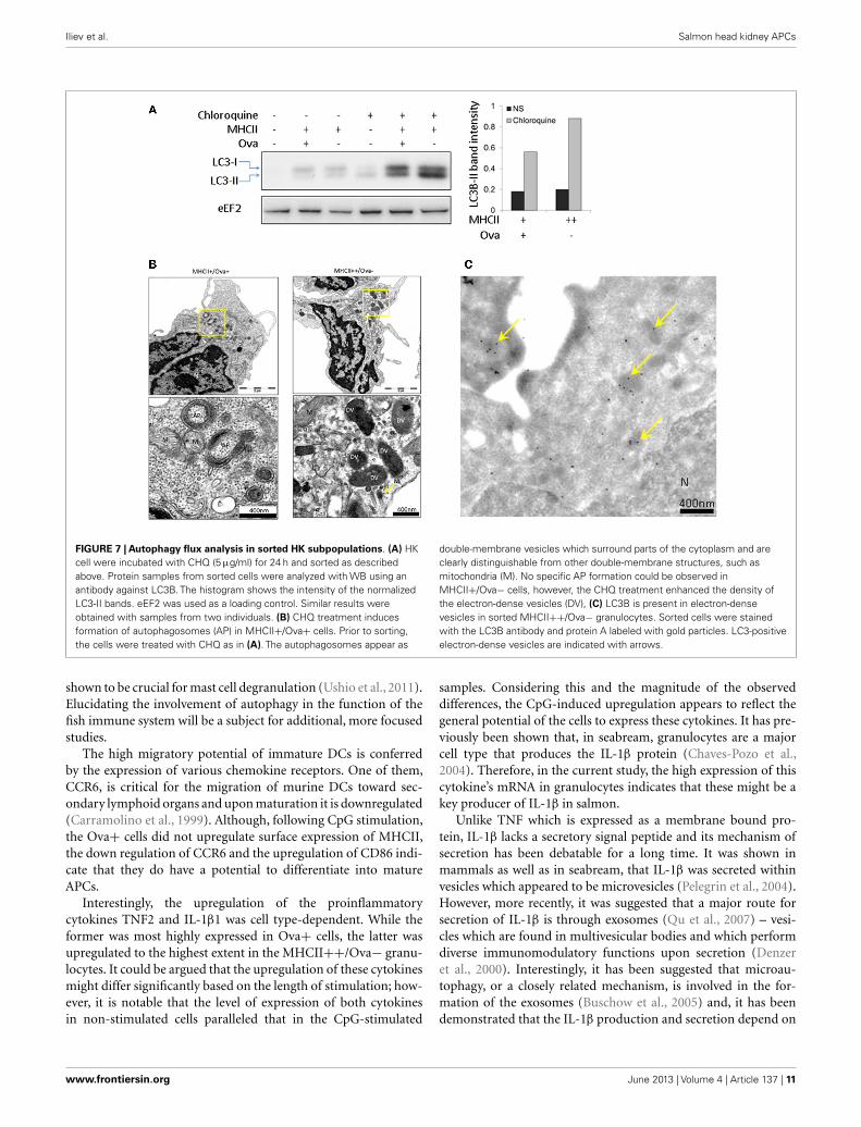
AUTOPHAGY FLUX ANALYSISAutophagy is a phylogenetically conserved mechanism throughwhich eukaryotic cells degrade cytoplasmic material and wholeorganelles (Mizushima, 2007). IL-1β does not have a signal pep-tide and therefore it cannot be exported through the classicalsecretory pathway. It has recently been found that autophagyis implicated in the secretion of this cytokine (Klionsky et al.,2008; Dupont et al., 2011). The autophagy flux can be evaluatedthrough Western blot (WB) analysis of the conversion of LC3B-Iinto LC3B-II, the latter of which migrates faster on PAGE. Onepotential problem with this analysis is the degradation of LC3B-II once the autophagosomes (AP) fuse with lysosomes (Klionskyet al., 2008). To block the lysosomal proteolysis and allow ofLC3B-II to accumulate, CHQ was added to the cells 24 h priorto sorting and WB analysis using a commercial antibody. Therepresentative image shown in Figure 7A demonstrates that fol-lowing treatment with CHQ, the MHCII++/Ova− granulocytesaccumulate higher amount of LC3B-II. However, TEM analysisshowed that double-membrane vesicles which surround parts ofthe cytoplasm and appear to be AP could be observed only in
the MHCII+/Ova+ cells but not in the MHCII++/Ova− leuko-cytes incubated with CHQ (Figure 7B). Nevertheless, the CHQtreatment increased the density of the vesicles found in the lattercells and immunogold staining/TEM showed that these vesicleswere positive for LC3 suggesting that these might be involved inautophagy or autophagy-related processes (Figure 7C).
DISCUSSIONA major objective of the current paper has been to characterizesalmon APCs in regards to their ability to take up soluble antigenand to migrate toward secondary lymphoid organs. The resultsdemonstrate that there is a specific population of MHCII+ cellswhich are able to endocytose Ag in the periphery and which overa period of up to 14 days following the Ag administration in theabdominal cavity, accumulate predominately in salmon HK, butnot in spleen. In vitro, these adherent cells accumulated Ova withinlate endosomes/lysosomes and were able to process Ova indicatingthat they are professional APCs.
The high capacity of APCs such as DCs to endocytose solubleAgs relies on receptor-mediated endocytosis and macropinocy-tosis (Sallusto et al., 1995). The former mechanism is medi-ated by receptors such as the mannose receptor expressed bymacrophages and DCs and is necessary for the high-efficiencyendocytosis of glycoproteins found on the surface of viruses,bacteria, and experimental substrates such as Ova and dextran(Sallusto et al., 1995; Burgdorf et al., 2006). On the other hand, themacropinocytosis is a process implicated in non-specific uptake oflarge amounts of extracellular fluid through membrane invagina-tions and lamellipodia (Lim and Gleeson, 2011). Both processesare downregulated upon activation of immature DCs by PAMPs
Frontiers in Immunology | Antigen Presenting Cell Biology June 2013 | Volume 4 | Article 137 | 8
FIGURE 5 | Morphology of sorted HK cells. (A) May Grünwald–Giemsastaining. (B) TEM images of sorted cells. Magnified regions are indicated witha dashed box and arrows. P, pseudopod; MPS, macropinosome; MG, melanin
granule; M, mitochondrion. RER, rough endoplasmic reticulum; N, nucleus;E/SE, early/sorting endosome. Different types of granules/vesicles inMHCII+/Ova-cells are indicated with asterisks.
or cytokines (Sallusto et al., 1995). In mammals, synthetic CpGODNs are known to be potent vaccine adjuvants due to theirpotential to induce maturation of DCs (Askew et al., 2000; Klin-man, 2003). This process is typically manifested by upregulationof surface MHCII and co-stimulatory molecules and downregula-tion of the Ag-uptake capacity trough inhibition of the receptor-mediated endocytosis and the macropinocytosis (Sallusto et al.,1995). It has previously been shown that in vitro stimulationof salmon mononuclear phagocytes with CpG-B upregulates alarge array of immune genes, including markers for mature APCs,and it lowers the capacity of these cells to take up dextran andOva (Iliev et al., 2010; Lagos et al., 2012). In the current study,injection with CpG ODNs did not seem to affect the migra-tion of cells toward HK and spleen since the number of Ova+cells in both organs did not differ significantly between the Ovaand the Ova CpG-injected groups. Furthermore, the CpG treat-ment did not detectably upregulate the surface expression ofMHCII and the HK CpG+ cells isolated following in vivo treat-ment were still able to endocytose significant amounts of dex-tran suggesting that these are still immature APCs. The CpGdose administered in vivo in the current study has been previ-ously shown to induce expression of immune genes in salmon
HK and spleen including IFN-γ and IL1β (Strandskog et al.,2008). However, the current data suggests that it may not bepotent enough to trigger maturation of salmon APCs under theseconditions.
While not very likely, it cannot be excluded that the sub-strates injected intra-abdominally may have diffused throughoutthe organism at high enough concentrations to be accumulated atdetectable levels in HK and spleen cells in situ. The in vitro exper-iments indicated that this was not the case since a significantlyhigher percentage of HK leukocytes were able to accumulate highamounts of Ova and CpG ODNs in vitro as compared to the in vivotrial. This indicates that cells had taken up the substrate in theperiphery prior to migrating toward the HK.
A previous study in which the expression of surface MHCIIwas analyzed in salmon HK and spleen leukocytes identifieda population of granular cells which expressed relatively highlevels of surface MHCII but had a low capacity to endocytosedextran and Ova (Iliev et al., 2011; Lagos et al., 2012). In the cur-rent study, the morphology of sorted cells, examined using MayGrünwald–Giemsa staining and TEM indicate that these cells aremost likely polymorphonuclear granulocytes. The relatively highMHCII expression on their surface and the upregulation of APC
FIGURE 6 | Gene expression in sorted HK cells. The cells were either leftnon-stimulated (NS) or treated with 2 µM CpG-B for 24 h prior toincubation with Ova-Alexa647 sorting as shown in Figure 3. Total RNA wasisolated immediately after sorting and the gene expression was analyzedwith Real-time PCR. (A) Gene expression is presented as fold differencevalues as compared to the non-stimulated MHCII−/Ig−/Ova− leukocytes.Data obtained with sorted cells from two individuals (F1 and F2) areshown in parallel. (B) The expression of TNF2 and IL-1β1 was analyzed intwo additional individuals and is presented as a Ct ratio against the
housekeeping gene. Note that in non-stimulated MHCII− cells from Fish 3and 4 the TNF2 expression was undetectable whereas it was detected inall of the tested MHCII+/Ova+ cells. The TNF2 expression was analyzedwith one-way ANOVA comparing the expression within the CpG-treatedgroup (n=4; a, p < 0.05 against all of the other cell types). The IL-β1expression was analyzed with two-way ANOVA comparing the sortedsamples across the non-stimulated and CpG-treated groups (n=4; b,p < 0.05. compared to CpG-stimulated MHCII− and IgM+ cells; c,p < 0.05 compared to non-stimulated MHCII++ cells).
marker genes including CD83, CD40, and B7-H1 is somewhat sur-prising since in mammals granulocytes are typically, short-livedeffector cells involved in clearance of bacterial and parasitic infec-tions (Kobayashi et al., 2005). Nevertheless, studies have demon-strated that under specific conditions, murine neutrophils mayupregulate MHCII and they can effectively present exogenous Agto T-cells (Abi Abdallah et al., 2011). In addition, high expressionof MHCII has been detected in seabream acidophilic granulocytes(functionally analogous to mammalian neutrophils) (Cuesta et al.,2006) and in zebrafish eosinophils (Wittamer et al., 2011) indicat-ing that fish granulocytes may, potentially, be involved in antigenpresentation.
In addition to exogenous substrate endocytosis, an alterna-tive mechanism for delivery of antigens to intracellular MHCIIcompartments is autophagy. This is a process through which cyto-plasmic constituents are delivered to lysosomes for degradation
and it has been implicated in induction of immune responseagainst intracellular parasites and in maintaining tolerance toautoantigens (Vyas et al., 2008; Klein et al., 2010). The relativelyhigh level of conversion of LC3-II, in MHCII++HK granulocytesindicates that these cells actively perform autophagy which may,potentially, be a source of peptides to be presented on MHCIImolecules.
The CHQ treatment induced the appearance of typical AP inthe MHCII+/Ova+ cell whereas in the MHCII++/Ova− granu-locytes such double-membrane structures containing cytoplasmicmaterial could not be observed. Nevertheless, the presence ofLC3 and heterogeneous material in electron-dense vesicles foundin the granulocytes indicates that these might be involved inautophagy, possibly microautophagy, or related processes suchas LC3-associated phagocytosis. In this regard, LC3-II has beenfound in secretory granules of mast cells and autophagy has been
Frontiers in Immunology | Antigen Presenting Cell Biology June 2013 | Volume 4 | Article 137 | 10
FIGURE 7 | Autophagy flux analysis in sorted HK subpopulations. (A) HKcell were incubated with CHQ (5 µg/ml) for 24 h and sorted as describedabove. Protein samples from sorted cells were analyzed with WB using anantibody against LC3B. The histogram shows the intensity of the normalizedLC3-II bands. eEF2 was used as a loading control. Similar results wereobtained with samples from two individuals. (B) CHQ treatment inducesformation of autophagosomes (AP) in MHCII+/Ova+ cells. Prior to sorting,the cells were treated with CHQ as in (A). The autophagosomes appear as
double-membrane vesicles which surround parts of the cytoplasm and areclearly distinguishable from other double-membrane structures, such asmitochondria (M). No specific AP formation could be observed inMHCII+/Ova− cells, however, the CHQ treatment enhanced the density ofthe electron-dense vesicles (DV), (C) LC3B is present in electron-densevesicles in sorted MHCII++/Ova− granulocytes. Sorted cells were stainedwith the LC3B antibody and protein A labeled with gold particles. LC3-positiveelectron-dense vesicles are indicated with arrows.
shown to be crucial for mast cell degranulation (Ushio et al., 2011).Elucidating the involvement of autophagy in the function of thefish immune system will be a subject for additional, more focusedstudies.
The high migratory potential of immature DCs is conferredby the expression of various chemokine receptors. One of them,CCR6, is critical for the migration of murine DCs toward sec-ondary lymphoid organs and upon maturation it is downregulated(Carramolino et al., 1999). Although, following CpG stimulation,the Ova+ cells did not upregulate surface expression of MHCII,the down regulation of CCR6 and the upregulation of CD86 indi-cate that they do have a potential to differentiate into matureAPCs.
Interestingly, the upregulation of the proinflammatorycytokines TNF2 and IL-1β1 was cell type-dependent. While theformer was most highly expressed in Ova+ cells, the latter wasupregulated to the highest extent in the MHCII++/Ova− granu-locytes. It could be argued that the upregulation of these cytokinesmight differ significantly based on the length of stimulation; how-ever, it is notable that the level of expression of both cytokinesin non-stimulated cells paralleled that in the CpG-stimulated
samples. Considering this and the magnitude of the observeddifferences, the CpG-induced upregulation appears to reflect thegeneral potential of the cells to express these cytokines. It has pre-viously been shown that, in seabream, granulocytes are a majorcell type that produces the IL-1β protein (Chaves-Pozo et al.,2004). Therefore, in the current study, the high expression of thiscytokine’s mRNA in granulocytes indicates that these might be akey producer of IL-1β in salmon.
Unlike TNF which is expressed as a membrane bound pro-tein, IL-1β lacks a secretory signal peptide and its mechanism ofsecretion has been debatable for a long time. It was shown inmammals as well as in seabream, that IL-1β was secreted withinvesicles which appeared to be microvesicles (Pelegrin et al., 2004).However, more recently, it was suggested that a major route forsecretion of IL-1β is through exosomes (Qu et al., 2007) – vesi-cles which are found in multivesicular bodies and which performdiverse immunomodulatory functions upon secretion (Denzeret al., 2000). Interestingly, it has been suggested that microau-tophagy, or a closely related mechanism, is involved in the for-mation of the exosomes (Buschow et al., 2005) and, it has beendemonstrated that the IL-1β production and secretion depend on
autophagy (Dupont et al., 2011; Harris et al., 2011). In view of this,the correlation between the high levels of LC3-II conversion andthe IL-1β1 mRNA in the MHCII++ granulocytes suggests thatthese cells may be specialized for production of IL-1β.
The results from the current study show that the expression ofTNF2 and IL-1β correlates very well with the expression of recep-tors for cytokines of these families. The granulocytes expressedexceptionally high levels of IL1R1 and IL1R2, the latter of which isa non-signaling decoy receptor for IL-1β, whereas the Ova+ cellsupregulated highly DCR3 and OPG which are TNF family decoyreceptors. The high upregulation of decoy receptors in cells thatexpress high levels of cytokines for these receptors may serve todampen excessive and potentially harmful effects of high concen-trations of the cytokines in the microenvironment around the cellsthat secrete them.
Overall, the B-cells did not show particularly high expressionof APC marker genes with the exception of CD208. In human,this molecule is specifically upregulated in mature DCs; howeverthis trait does not seem to be phylogenetically conserved since inmouse it is present in other cells types (Salaun et al., 2003). Theresults shown in the current paper indicate that CD208 may serveas an excellent marker for salmon HK B-cells.
Compared to the other two MHCII+ cell types, the IgM+cells expressed relatively low levels of proinflammatory cytokines.However, both the basal and the CpG-induced IFN-γ expressionwere very high in these cells. Although, in mammals, IFN-γ ispredominately produced by NK and T-cells, certain subtypes ofmammalian B lymphocytes are also known to express high levelsof this cytokine (Harris et al., 2000).
It has been previously shown that trout HK harbors both earlydeveloping B-cells and antibody-secreting cells and serves as amajor organ providing homing for mature plasma cells (Bromage
et al., 2004; Zwollo et al., 2005). The CpG-induced downregulationof mIgM and PAX5 (a transcription factor expressed in theearly/intermediate stages but not in the terminal stages of plasmacell differentiation) (Henderson and Calame, 1998) and the simul-taneous upregulation of sIgM in IgM+ cells indicate that thestimulation induces differentiation of mature plasma cells. It isnot very likely that CpG stimulation alone, without activationthrough the B-cell receptor could activate this process. However,provided these cells have already been activated through the B-cellreceptor, the CpGs may contribute to the process of their termi-nal differentiation. In this regard, it has been previously shownthat although Ag stimulation alone can activate B-cell prolifer-ation, a second stimulus, provided by innate immune receptors,such as TLRs is necessary for induction of Ab secretion (Vos et al.,2000).
In summary, the presented data indicates that salmon HK mayserve as a major secondary lymphoid organ to which APCs loadedwith Ag in the peripheral organs migrate. The description of dis-tinct gene expression profiles in different HK cell types will helpdefine markers for further characterization of different APC typesin salmon and other teleost species. The correlation between thehigh expression of IL-1β1 and the high autophagy flux in HKgranulocytes suggests that these cells might be specialized towardproduction and secretion of IL-1β1. The presented data also lay aground for further studies aimed at elucidating the significance ofautophagy for the immune response in teleosts.
ACKNOWLEDGMENTSWe thank prof. T. Johansen and Dr. T. Lamark (University ofTromsø) for critical reading of the manuscript and helpful dis-cussion. This study was supported by Aquaculture program ofThe Research Council of Norway (183196/S40 InNoVacc).
REFERENCESAbi Abdallah, D. S., Egan, C. E.,
Butcher, B. A., and Denkers, E.Y. (2011). Mouse neutrophils areprofessional antigen-presenting cellsprogrammed to instruct Th1 andTh17 T-cell differentiation. Int.Immunol. 23, 317–326. doi:10.1093/intimm/dxr007
Aghaallaei, N., Bajoghli, B., Schwarz,H., Schorpp, M., and Boehm,T. (2010). Characterization ofmononuclear phagocytic cellsin medaka fish transgenic for acxcr3a:gfp reporter. Proc. Natl. Acad.Sci. U.S.A. 107, 18079–18084.doi:10.1073/pnas.1000467107
Agius, C., and Roberts, R. J.(2003). Melano-macrophagecentres and their role in fishpathology. J. Fish Dis. 26,499–509. doi:10.1046/j.1365-2761.2003.00485.x
Askew, D., Chu, R. S., Krieg, A. M.,and Harding, C. V. (2000). CpGDNA induces maturation of den-dritic cells with distinct effectson nascent and recycling MHC-II
Barberis, A., Widenhorn, K., Vitelli, L.,and Busslinger, M. (1990). A novelB-cell lineage-specific transcriptionfactor present at early but not latestages of differentiation. Genes Dev.4, 849–859. doi:10.1101/gad.4.5.849
Bassity, E., and Clark, T. G. (2012).Functional identification of den-dritic cells in the teleost model,rainbow trout (Oncorhynchusmykiss). PLoS ONE 7:e33196.doi:10.1371/journal.pone.0033196
Bromage, E. S., Kaattari, I. M., Zwollo,P., and Kaattari, S. L. (2004). Plas-mablast and plasma cell productionand distribution in trout immunetissues. J. Immunol. 173, 7317–7323.
Burgdorf, S., Lukacs-Kornek, V., andKurts, C. (2006). The mannosereceptor mediates uptake of solublebut not of cell-associated antigen forcross-presentation. J. Immunol. 176,6770–6776.
Buschow, S. I., Liefhebber, J. M.,Wubbolts, R., and Stoorvogel,W. (2005). Exosomes containubiquitinated proteins. Blood
Carramolino, L., Kremer, L., Goya, I.,Varona, R., Buesa, J. M., Gutierrez,J., et al. (1999). Down-regulation ofthe beta-chemokine receptor CCR6in dendritic cells mediated by TNF-alpha and IL-4. J. Leukoc. Biol. 66,837–844.
Chaves-Pozo, E., Pelegrin, P., Garcia-Castillo, J., Garcia-Ayala, A., Mulero,V., and Meseguer, J. (2004). Aci-dophilic granulocytes of the marinefish gilthead seabream (Sparusaurata L.) produce interleukin-1beta following infection with Vib-rio anguillarum. Cell Tissue Res.316, 189–195. doi:10.1007/s00441-004-0875-9
Cuesta, A., Angeles Esteban, M., andMeseguer, J. (2006). Cloning, dis-tribution and up-regulation ofthe teleost fish MHC class IIalpha suggests a role for granu-locytes as antigen-presenting cells.Mol. Immunol. 43, 1275–1285.doi:10.1016/j.molimm.2005.07.004
Denzer, K., Kleijmeer, M. J., Heijnen,H. F., Stoorvogel, W., and Geuze, H.
J. (2000). Exosome: from internalvesicle of the multivesicular body tointercellular signaling device. J. Cell.Sci. 113(Pt 19), 3365–3374.
Dupont, N., Jiang, S., Pilli, M., Orna-towski, W., Bhattacharya, D., andDeretic, V. (2011). Autophagy-basedunconventional secretory pathwayfor extracellular delivery of IL-1beta. EMBO J. 30, 4701–4711.doi:10.1038/emboj.2011.398
Hamilos, D. L. (1989). Antigen present-ing cells. Immunol. Res. 8, 98–117.doi:10.1007/BF02919073
Harris, D. P., Haynes, L., Sayles, P.C., Duso, D. K., Eaton, S. M.,Lepak, N. M., et al. (2000). Recipro-cal regulation of polarized cytokineproduction by effector B and Tcells. Nat. Immunol. 1, 475–482.doi:10.1038/82717
Harris, J., Hartman, M., Roche, C.,Zeng, S. G., O’Shea, A., Sharp, F. A.,et al. (2011). Autophagy controlsIL-1beta secretion by targetingpro-IL-1beta for degradation.J. Biol. Chem. 286, 9587–9597.doi:10.1074/jbc.M110.202911
Frontiers in Immunology | Antigen Presenting Cell Biology June 2013 | Volume 4 | Article 137 | 12
Hartmann, G., Weiner, G. J., andKrieg, A. M. (1999). CpG DNA:a potent signal for growth,activation, and maturation ofhuman dendritic cells. Proc. Natl.Acad. Sci. U.S.A. 96, 9305–9310.doi:10.1073/pnas.96.16.9305
Henderson, A., and Calame, K. (1998).Transcriptional regulation duringB cell development. Annu. Rev.Immunol. 16, 163–200. doi:10.1146/annurev.immunol.16.1.163
Iliev, D. B., Jorgensen, S. M.,Rode, M., Krasnov, A., Harne-shaug, I., and Jørgensen, J. B.(2010). CpG-induced secretionof MHCIIbeta and exosomesfrom salmon (Salmo salar) APCs.Dev. Comp. Immunol. 34, 29–41.doi:10.1016/j.dci.2009.07.009
Iliev, D. B., Sobhkhez, M., Fremmer-lid, K., and Jorgensen, J. B. (2011).MyD88 interacts with interferonregulatory factor (IRF) 3 and IRF7 inAtlantic salmon (Salmo salar): trans-genic SsMyD88 modulates the IRF-induced type I interferon responseand accumulates in aggresomes.J. Biol. Chem. 286, 42715–42724.doi:10.1074/jbc.M111.293969
Jovic, M., Sharma, M., Rahajeng, J., andCaplan, S. (2010). The early endo-some: a busy sorting station forproteins at the crossroads. Histol.Histopathol. 25, 99–112.
Kaisho, T., and Akira, S. (2003).Regulation of dendritic cell func-tion through Toll-like receptors.Curr. Mol. Med. 3, 373–385.doi:10.2174/1566524033479366
Klein, L., Munz, C., and Lune-mann, J. D. (2010). Autophagy-mediated antigen processing inCD4(+) T cell tolerance and immu-nity. FEBS Lett. 584, 1405–1410.doi:10.1016/j.febslet.2010.01.008
Klinman, D. M. (2003). CpGDNA as a vaccine adjuvant.Expert Rev. Vaccines 2, 305–315.doi:10.1586/14760584.2.2.305
Klionsky, D. J.,Abeliovich, H.,Agostinis,P., Agrawal, D. K., Aliev, G., Askew,D. S., et al. (2008). Guidelines forthe use and interpretation of assaysfor monitoring autophagy in highereukaryotes. Autophagy 4, 151–175.
Lagos, L. X., Iliev, D. B., Helland, R.,Rosemblatt, M., and Jorgensen, J.B. (2012). CD40L – a costimula-tory molecule involved in the mat-uration of antigen presenting cellsin Atlantic salmon (Salmo salar).Dev. Comp. Immunol. 38, 416–430.doi:10.1016/j.dci.2012.07.011
Lim, J. P., and Gleeson, P. A. (2011).Macropinocytosis: an endo-cytic pathway for internalisinglarge gulps. Immunol. Cell Biol.89, 836–843. doi:10.1038/icb.2011.20
Lovy, J., Savidant, G. P., Speare, D.J., and Wright, G. M. (2009).Langerin/CD207 positive dendritic-like cells in the haemopoietictissues of salmonids. Fish Shell-fish Immunol. 27, 365–368.doi:10.1016/j.fsi.2009.01.006
Lugo-Villarino, G., Balla, K. M.,Stachura, D. L., Banuelos, K.,Werneck, M. B., and Traver, D.(2010). Identification of den-dritic antigen-presenting cells inthe zebrafish. Proc. Natl. Acad.Sci. U.S.A. 107, 15850–15855.doi:10.1073/pnas.1000494107
Miller, N. W., Deuter, A., and Clem,L. W. (1986). Phylogeny of lym-phocyte heterogeneity: the cellularrequirements for the mixed leuco-cyte reaction with channel catfish.Immunology 59, 123–128.
Mizushima, N. (2007). Autophagy:process and function.Genes Dev. 21, 2861–2873.doi:10.1101/gad.1599207
Pancer, Z., and Cooper, M. D. (2006).The evolution of adaptive immunity.Annu. Rev. Immunol. 24, 497–518.doi:10.1146/annurev.immunol.24.021605.090542
Pelegrin, P., CHAVES-Pozo, E., Mulero,V., and Meseguer, J. (2004). Pro-duction and mechanism of secre-tion of interleukin-1beta from themarine fish gilthead seabream.Dev. Comp. Immunol. 28, 229–237.doi:10.1016/j.dci.2003.08.002
Qu, Y., Franchi, L., Nunez, G., andDubyak, G. R. (2007). Nonclassi-cal IL-1 beta secretion stimulatedby P2×7 receptors is dependenton inflammasome activation andcorrelated with exosome release in
murine macrophages. J. Immunol.179, 1913–1925.
Salaun, B., de Saint-Vis, B., Clair-Moninot, V., Pin, J. J., Barthelemy-Dubois, C., Kissenpfennig, A., etal. (2003). Cloning and character-ization of the mouse homologueof the human dendritic cell mat-uration marker CD208/DC-LAMP.Eur. J. Immunol. 33, 2619–2629.doi:10.1002/eji.200324175
Sallusto, F., Cella, M., Danieli, C.,and Lanzavecchia, A. (1995). Den-dritic cells use macropinocytosisand the mannose receptor to con-centrate macromolecules in themajor histocompatibility complexclass II compartment: downregula-tion by cytokines and bacterial prod-ucts. J. Exp. Med. 182, 389–400.doi:10.1084/jem.182.2.389
Strandskog, G., Skjæveland, I.,Ellingsen, T., and Jørgensen,J. B. (2008). Double-strandedRNA- and CpG DNA-inducedimmune responses in Atlanticsalmon: comparison and syn-ergies. Vaccine 26, 4704–4715.doi:10.1016/j.vaccine.2008.06.054
Thery, C., and Amigorena, S. (2001).The cell biology of antigen presen-tation in dendritic cells. Curr.Opin. Immunol. 13, 45–51.doi:10.1016/S0952-7915(00)00180-1
Tokuyasu, K. T. (1986). Applica-tion of cryoultramicrotomy toimmunocytochemistry. J. Microsc.143, 139–149. doi:10.1111/j.1365-2818.1986.tb02772.x
Tsujii, T., and Seno, S. (1990).Melano-macrophage centers inthe aglomerular kidney of the seahorse (teleosts): morphologic stud-ies on its formation and possiblefunction. Anat. Rec. 226, 460–470.doi:10.1002/ar.1092260408
Ushio, H., Ueno, T., Kojima, Y.,Komatsu, M., Tanaka, S., Yamamoto,A., et al. (2011). Crucial rolefor autophagy in degranula-tion of mast cells. J. AllergyClin. Immunol. 127, 1267–1276.doi:10.1016/j.jaci.2010.12.1078
Vos, Q., Lees, A., Wu, Z. Q., Snap-per, C. M., and Mond, J. J.(2000). B-cell activation by T-cell-independent type 2 antigens asan integral part of the humoral
immune response to pathogenicmicroorganisms. Immunol. Rev.176, 154–170. doi:10.1034/j.1600-065X.2000.00607.x
Vyas, J. M., Van der Veen, A. G., andPloegh, H. L. (2008). The knownunknowns of antigen processing andpresentation. Nat. Rev. Immunol. 8,607–618. doi:10.1038/nri2368
Webster, P., and Webster, A. (2007).Cryosectioning fixed and cry-oprotected biological materialfor immunocytochemistry. Meth-ods Mol. Biol. 369, 257–289.doi:10.1007/978-1-59745-294-6_13
Wileman, T., Harding, C., and Stahl, P.(1985). Receptor-mediated endocy-tosis. Biochem. J. 232, 1–14.
Wittamer, V., Bertrand, J. Y., Gutschow,P. W., and Traver, D. (2011). Char-acterization of the mononuclearphagocyte system in zebrafish. Blood117, 7126–7135. doi:10.1182/blood-2010-11-321448
Zwollo, P., Cole, S., Bromage, E., andKaattari, S. (2005). B cell hetero-geneity in the teleost kidney: evi-dence for a maturation gradientfrom anterior to posterior kidney. J.Immunol. 174, 6608–6616.
Conflict of Interest Statement: Theauthors declare that the research wasconducted in the absence of any com-mercial or financial relationships thatcould be construed as a potential con-flict of interest.