Nowadays, a number of imaging modalities is available for the diagnosis and locali-zation of tumors; such techniques rely mainly on the detection of morphologicalchanges in the involved tissues. However, anatomical alterations are accompaniedand often preceded by metabolic modifications, which cannot be visualized byany of the available imaging modalities. The possibility of acquiring metabolicimages reporting about basic cellular pathways represents an outstanding break-through in medical imaging. Magnetic resonance spectroscopy (MRS) appears tobe the technique of choice for this purpose, as it allows, at least in principle, to iden-tify the presence and evolution of different metabolites in vivo [1-4]. Unfortunately,metabolites are present in low concentrations; this fact, coupled with the intrinsiclow sensitivity of MR, would lead to very long acquisition times, which arenot compatible with the time scale of metabolic transformations, preventing theextensive application of the technique.
Such a low sensitivity is due to the very small population difference between thenuclear spin levels at thermal equilibrium. Increasing this difference (and henceincreasing polarization) with respect to the equilibrium value leads to higher inten-sities in the nuclear magnetic resonance (NMR) spectra. Non-equilibrium states ofthe spin populations are referred to as hyperpolarized states [5].
The availability of hyperpolarized molecules allows to getaround the sensitivity issue of MRS, giving the opportunityof detecting metabolites in vivo and representing themin maps by means of the chemical shift imaging (CSI)modality [5-10]. Fast acquisitions are made possible by thehigh signal intensity and, as a consequence, metabolic trans-formations can be followed in real time on administrationof a given substrate.
1.1 General properties of hyperpolarized
contrast agentsUnlike standard magnetic resonance imaging (MRI) contrastagents, which act on water protons relaxation, hyperpolarizedmolecules are themselves the source of the NMR signal,yielding to signal intensity and SNR (signal-to-noise ratio)that linearly depend on their concentration and polarizationlevel. As a consequence, two major points must be takeninto account when dealing with this kind of agents:i) nuclear relaxation (which affects the polarization lifetime)and ii) irreversible loss of polarization after the applicationof radiofrequency pulses.
1.1.1 Relaxation timesThe unbalanced nuclear spin populations obtained after thehyperpolarization process are subjected to re-equilibration,restoring the thermal equilibrium situation in a time scalewhich depends on the relaxation time of the nucleus ofinterest. As a consequence, an essential property of a hyperpo-larized contrast agent relies on a long relaxation time of theresonance being detected, which will guarantee the mainte-nance of the hyperpolarized state for a time sufficient forimage acquisition.Heteronuclei are often characterized by longer relaxation
times with respect to protons because of their lower g values;for this reason, they are usually preferred. The use of low g
heteronuclei presents the further advantage of reducing theendogenous background, allowing the obtainment of imageswith higher SNR.
A tight control of the factors affecting nuclear relaxation hasto be pursued in order to attain very long T1 values. As intra-molecular dipolar interaction is the main relaxation mechanismin small organic molecules, perdeuteration of the molecule andselection of dipolarly isolated heteronuclei are useful indica-tions. Furthermore, small molecules are preferred since theirhigher mobility in solution is effective in maintaining low relax-ation rates. Finally, as chemical shift anisotropy (CSA) is oftenan important relaxation mechanism, very high magnetic fieldsare not recommended as the CSA contribution scales up withthe magnetic field strength.
1.1.2 Pulse sequencesImaging with hyperpolarized molecules puts unique require-ments on the acquisition sequences that can be used, mainlybecause of the (usually fast) magnetization decay from thehyperpolarized state to thermal equilibrium. This featurelimits the time available for each study (i.e., in the case ofpyruvate, the useful time window lasts just a few minutes afterthe injection). Furthermore, since radiofrequency pulses causedisruption of the magnetization, small flip angles have to beused when a series of images at consecutive times are needed(i.e., when following the time course of a given metabolictransformation). Otherwise, single shot sequences can beused for the acquisition of single high resolution images.Tailored multiband pulses can also be used: for example,pyruvate can be excited with small flip angles to preserve itsmagnetization, while the metabolic products can be excitedwith larger flip angles to increase their SNR [11]. Whateverthe kind of acquisition, fast imaging techniques are neededto cope with the magnetization decay. A number of fast pulsesequences has been implemented (mainly echo planar imaging(EPI), echo planar spectroscopic imaging (EPSI) and spiralCSI) to obtain high resolution images in times as short as1 s: this allows to detect the formation of metabolic productsin real time after administration of the hyperpolarized com-pound. Further points to keep in mind when dealing withhyperpolarized contrast agents are the need for wider gradientamplitudes (due to the lower gyromagnetic ratio of 13C withrespect to 1H) and spectral widths (due to the larger 13Cchemical shift dispersion). Details about pulse sequences formetabolic imaging have been recently reviewed [10]. Furtheroptimizations of sequences are still being carried out byseveral groups in order to achieve even higher resolution(spectral-spatial imaging [12]) in the short times dictatedby hyperpolarization.
One of the main issues with hyperpolarized MRI is relatedto the difficulty of telling apart the contributions to the signalarising from intra- and extracellular compartments. This maylead to uncertainties in the measurements of metabolic fluxesand/or exchange rates. Sequential injections of hyperpolarized1-13C-pyruvate followed by gadolinium-chelate have been
Article highlights.
. Hyperpolarization techniques have allowed us toovercome magnetic resonance imaging (MRI)/magneticresonance spectroscopy (MRS) sensitivity issues.
. Administration of hyperpolarized substrates allows us tofollow their metabolic fate in vivo in real time.
. Pyruvate has been shown to be the candidate of choicebecause it is a key molecule in mammalian metabolismand its conversion to lactate by lactate dehydrogenase(LDH) activity is highly increased in tumors.
. 1-13C-lactate detection in vivo on injection ofhyperpolarized 1-13C-pyruvate allows to visualize,localize and grade different types of tumors (lymphoma,prostate, breast, liver and brain cancer).
. Changes in lactate levels usually precede morphologicalchanges after tumor treatments, and their visualizationby 13C magnetic resonance spectroscopy imaging (MRSI)allows to monitor early response to therapy.
This box summarizes key points contained in the article.
A. Viale et al.
336 Expert Opin. Med. Diagn. (2012) 6(4)
Exp
ert O
pin.
Med
. Dia
gn. D
ownl
oade
d fr
om in
form
ahea
lthca
re.c
om b
y U
nive
rsity
of
Gla
sgow
on
04/3
0/13
For
pers
onal
use
onl
y.
proven to provide T1 shortening in extracellular compart-ments, allowing the build-up of a kinetic model that distin-guishes the intracellular space and can be used to directlymeasure the intracellular 13C kinetics [13]. Recently, anothermethod based on a spin tagging pulse sequence has beensuggested: intravascular tagged substrates are subjected toflow and therefore they are not detected. The same appliesto metabolites coming by perfusion, which are not tagged,while those arising from metabolic transformations of injectedsubstrate are [14].
1.2 Hyperpolarization techniquesThree main hyperpolarization methodologies are currentlybeing used to produce hyperpolarized molecules: i) opticalpumping and spin exchange of noble gases (3He and 129Xe),whose application appears to be limited to studies of the lungsand to a limited number of perfusion investigations [15-17] (aninteresting targeting approach which uses hyperpolarized129Xe entrapped in a cryptand has been developed) [18];ii) para hydrogen-induced polarization (PHIP), which, evenif cheap and easy to accomplish, suffers for the limitednumber of substrates which may be polarized, as unsaturatedprecursors of the molecules of interest are needed, and strongchemical requirements must be satisfied in order to obtainheteronuclear polarization useful for MRI applications [19-21];iii) dynamic nuclear polarization (DNP), which is the mostversatile technique, allowing to polarize (at least in principle)every nucleus in every molecule. For this reason, DNP has
become the technique of choice for the preparation of hyper-polarized contrast agents; it has been used to obtain a numberof metabolites of interest which are currently under investiga-tion for metabolic imaging (pyruvate, lactate, fumarate, fruc-tose, alanine, glutamine, glutamate, acetate, bicarbonate,ketoisocaproate) [6,10,22-30].
The DNP procedure can be described as follows: the mate-rial to be polarized is first dissolved in a glass-forming solvent(or used in a pure form if it forms a glass by itself whenlowering the temperature), doped with a stable radical species,eventually added with a paramagnetic Gd complex, andplaced into the magnetic field. The solution is then broughtto very low temperatures (1 -- 2 K) and irradiated with micro-wave radiation, at or near to the electron EPR resonancefrequency. Under these conditions, the electron polarization(which is much higher than that of the nuclei) is transferredto neighboring nuclei, and then to all the nuclei in the sampleby spin diffusion [31,32]. After the polarization transfer hastaken place, the microwave irradiation is switched off, thesample is raised above the liquid helium level and rapidlywarmed up (usually by dissolution in hot water) stillinside the magnetic field. It is then quickly used for theMR acquisition.
Among the molecules which have been polarized by DNP,1-13C-pyruvate has received much attention thanks to thehigh attainable polarization degree (20 -- 30%), the favorable13C T1 (the T1 for the carboxylic carbon atom is 55 s at1.5 T [28]), its implication in many major metabolic pathwaysand the fast in vivo metabolism. All these factors contribute toallow the detection of its in vivo metabolic transformations byCSI, with very interesting results in a number of applications,ranging from studies of heart diseases [33-40], to studies ofliver [41-47], to cancer diagnosis/stadiation [6,48-53] and moni-toring of response to treatment [54-63]. Applications to cancerimaging are described in more detail in Section 2.
2. Hyperpolarized 1-13C-pyruvate incancer imaging
Pyruvate is a key molecule in major metabolic and catabolicpathways in the mammalian cells. It is converted by the actionof the pyruvate dehydrogenase complex (PDH) into acetyl-coenzyme A (which then enters the Krebs cycle), with forma-tion of CO2. It is also converted to oxaloacetate by pyruvatecarboxylase. Under hypoxic conditions, pyruvate is metabolizedanaerobically, yielding lactate through the activity of lactatedehydrogenase (LDH), using NADH as coenzyme. Finally, itis converted to alanine through alanine transaminase (ALT)(Figure 1).
Lactate, alanine and CO2 can be detected in normal cellsafter injection of hyperpolarized 1-13C-pyruvate [6,27,28]. Expe-rimental in vitro and in vivo data together with magnetizationtransfer experiments demonstrated that the pyruvate--lactateflux is dominated by label exchange between pyruvate and thepre-existing lactate pool, rather than net flux. This must be
Alanine transaminase (ALT)
Pyruvate carboxylase
Pyruvate dehydrogenase (PDH)
Pyruvate
Glycolysis
Glucose
Lactate dehydrogenase (LDH)
Alanine
AcetylCoA
Lactate
Oxaloacetate
CO2
CO2
Figure 1. Schematic representation of the metabolism of
pyruvate.
Hyperpolarized 13C-pyruvate magnetic resonance imaging in cancer diagnostics
Expert Opin. Med. Diagn. (2012) 6(4) 337
Exp
ert O
pin.
Med
. Dia
gn. D
ownl
oade
d fr
om in
form
ahea
lthca
re.c
om b
y U
nive
rsity
of
Gla
sgow
on
04/3
0/13
For
pers
onal
use
onl
y.
kept under consideration when evaluating the rate constantsinvolved in the process; it also implies that significantly highlactate signals will be observed after injection of hyperpolarizedpyruvate mainly in those tissues which already contain highquantities of lactate, such as in tumors [9,10].The production of lactate is increased under hypoxic
conditions due to anaerobic glycolysis, and in tumors dueto the enhanced aerobic glycolysis known as the Warburgeffect [64,65]. The maps of the metabolic products arisingfrom pyruvate are therefore highly informative about thepresence and status of the tumor cells.The first report of in vivo tumor imaging obtained by
this technique appeared in 2006 and deals with the detectionand localization of a malignant sarcoma in rat [6]. The13C-pyruvate, alanine and lactate distribution maps afterpyruvate administration showed that the tumor could clearlybe revealed by the highest NMR 13C signal from lactateproduced within 30 s from the injection, while the alaninelevel was higher in the skeletal muscle and very low in thetumor (Figure 2).Many efforts have been carried out since then and the
methodology has been successfully applied to other types oftumors, such as lymphomas, prostate, breast, liver andbrain tumors.
2.1 1-13C-pyruvate imaging of adenocarcinoma
of the prostateHyperpolarized 13C-pyruvate CSI has been first applied to thestudy of transgenic adenocarcinoma of mouse prostate(TRAMP), which is a model of prostate cancer that wellmimics the human tumor with regards to both histopatho-logy and disease progression. 13C metabolic maps showedincreased lactate/pyruvate ratios in the tumor with respect tothe neighboring healthy tissues 10 s after the injection ofhyperpolarized pyruvate [48]. Differentiation of the histologicgrades on the basis of lactate levels was possible. Highest gradetumors gave rise to the highest lactate signals, with the excep-tion of necrotic regions were lower lactate levels were observeddue to decreased perfusion and pyruvate uptake. A linear coef-ficient of 0.95 was found in the relationship between lactatelevels and histologic grade. Higher lactate levels were detectedin metastatic lymph nodes as well. The accuracy of this
grading methodology was evident in the non-overlappingdata for high-grade tumors, low-grade tumors and normalprostates [49].
While single time-point imaging allowed localizing andassessing the grade of the murine prostate tumors, dynamicimaging has been shown to be effective in providing informa-tion about the cellularity and necrosis, perfusion and vascula-rity and more in general about the heterogeneity of the tumormasses. In fact, acquiring images at successive times, andhence following the dynamics of both pyruvate and lactate,allowed to determine the mean time (MT) of arrival ofpyruvate and lactate into the tumor (which was indicative ofperfusion and vascularity), the duration of lactate conversionand its persistency in the tumor [11,50].
In dogs, whose prostate size and anatomy is similar to thoseof humans, the optimization of the coil geometry and dataacquisition parameters permitted to show that pyruvate israpidly taken up by the tumor cells, but only partially con-verted to lactate. Thus, the lactate/pyruvate ratio is lower thanthat observed in rats and mice, but still high enough to allowthe spatial location of the tumor, giving confidence that theprotocol may actually be extended to clinical applications [51].
The evaluation of lactate levels after hyperpolarized pyru-vate injection for the diagnosis and grade assessment of pros-tate cancer is currently under intense scrutiny, as it representsa valuable complement to the (often inaccurate and invasive)techniques currently in use. The clinical phase of trial hasbeen undertaken in order to develop a method for monitoringthe therapeutic treatment of prostate cancer and possibly forchoosing the best therapy for each single subject [66].
2.2 1-13C-pyruvate imaging of brain tumorsThe positron emission tomography (PET) determination offluorodeoxyglucose (FDG) is not useful for the diagnosis ofbrain tumors due to the high FDG uptake levels of normalgrey matter. Although it is well established that high lactatelevels are found in human gliomas, MRS has not been success-fully applied to this kind of tumors because lactate in tumortissues cannot be easily distinguished, by means of 1H detectionfrom the one that accumulates in necrotic or cystic regions.Therefore, it would be of paramount importance to have accessto an alternative technique, which allows to univocally visualizelactate formation in brain tumors.
Figure 2. Pyruvate, lactate and alanine maps of a rat bearing a P22 tumor.Adapted by permission from the American Association for Cancer Research from [6].
A. Viale et al.
338 Expert Opin. Med. Diagn. (2012) 6(4)
Exp
ert O
pin.
Med
. Dia
gn. D
ownl
oade
d fr
om in
form
ahea
lthca
re.c
om b
y U
nive
rsity
of
Gla
sgow
on
04/3
0/13
For
pers
onal
use
onl
y.
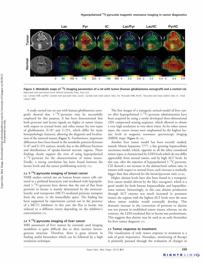
A study carried out on rats with human glioblastoma xeno-grafts showed that 1-13C-pyruvate may be successfullyemployed for this purpose. It has been demonstrated thatboth pyruvate and lactate signals are higher in tumor tissueswith respect to normal brain and other tissues for two typesof glioblastoma (U-87 and U-251, which differ for mainhistopathologic features), allowing the diagnosis and localiza-tion of the tumoral masses (Figure 3). Furthermore, importantdifferences have been found in the metabolic patterns betweenU-87 and U-251 tumors, mainly due to the different fractionsand distributions of uptake-limited necrotic regions. Thesefindings clearly support the view of using hyperpolarized1-13C-pyruvate for the characterization of tumor tissues.Finally, a strong correlation has been found between thelactate levels and the tumor proliferating activity [52].
2.3 1-13C-pyruvate imaging of breast cancerNMR studies carried out on human breast cancer cells cul-tured in a perfused bioreactor and incubated with hyperpola-rized 1-13C-pyruvate have shown that the rate of flux frompyruvate to lactate is mainly determined by the monocar-boxylic acid transporter (MCT)-mediated pyruvate transportfrom the extra- to the intracellular space. The finding hasbeen supported by experiments carried out in the presenceof a MCT1 inhibitor: in this case, the flux to lactate wasreduced to a different extent depending on the inhibitor’sconcentration [53].
2.4 1-13C-pyruvate imaging of liver cancerMRI assessment of liver tumors by currently used imagingmodalities is quite difficult due to their intrinsic hetero-geneous structure. Therefore, there is great interest infinding useful biomarkers which can be followed by a highresolution technique.
The first images of a transgenic animal model of liver can-cer after hyperpolarized 1-13C-pyruvate administration havebeen acquired by using a newly developed three-dimensional(3D) compressed sensing sequence, which allowed to obtaina very high resolution in very short times. As for other tumortypes, the cancer tissues were emphasized by the highest lac-tate levels in magnetic resonance spectroscopy imaging(MRSI) maps (Figure 4) [44].
Another liver tumor model has been recently studied,namely Morris hepatoma 7777, a fast growing hepatocellularcarcinoma model, which, opposite to all the other consideredtumor types, is characterized by LDH levels which do not differappreciably from normal tissues, and by high ALT levels. Inthis case, after the injection of hyperpolarized 1-13C-pyruvate,CSI showed a net increase in the alanine/total carbon ratio intumors with respect to normal livers; such increase is markedlybigger than that observed for the lactate/pyruvate ratio [45,46].
Higher alanine levels have also been found in a transgenicliver cancer model (driven by the Myc oncogene), which is agood model for both human hepatocellular and hepatoblas-toma tumors. Interestingly, in this case alanine productionthrough ALT enzyme was much increased in pretumortissues; the regions with the highest ALT levels were the areaswhere tumor nodules would eventually develop. Thisdramatic increase in the conversion of pyruvate to alaninewas not present in established tumor tissues, whereas, on thecontrary, the LDH-mediated flux to lactate was predominant.This suggests that alanine may be used as an early biomarkerfor liver tumor diagnosis [47].
2.5 Tumor response to treatmentThe visualization of early tumor response to treatment is atask of great importance. Nowadays, monitoring of therapyis primarily pursued through the evaluation of changes in
Figure 3. Metabolic maps of 13C imaging parameters of a rat with tumor (human glioblastoma xenograft) and a control rat.Reprinted with permission from Oxford University Press, from [52].
Lac: Lactate SNR; Lac/Pyr: Lactate over pyruvate ratio; Lac/tC: Lactate over total carbon ratio; Pyr: Pyruvate SNR; Pyr/tC: Pyruvate over total carbon ratio; tC: Total
carbon SNR.
Hyperpolarized 13C-pyruvate magnetic resonance imaging in cancer diagnostics
Expert Opin. Med. Diagn. (2012) 6(4) 339
Exp
ert O
pin.
Med
. Dia
gn. D
ownl
oade
d fr
om in
form
ahea
lthca
re.c
om b
y U
nive
rsity
of
Gla
sgow
on
04/3
0/13
For
pers
onal
use
onl
y.
tumor size. This approach has evident limitations: on the onehand, a size change may require days or weeks to becomeobservable, and on the other hand there might be a positiveresponse to treatment without any detectable morphologicalchange. It is straightforward to note that the knowledge onhow well a tumor is reacting to a given therapy, even after ashort time of treatment, is of great importance for assessinghow to proceed with the undertaken cure. Currently,FDG-PET is considered the golden standard in the evaluationof anti-tumor treatments as it monitors early glucose uptakechanges occurring in treated tumor cells. However, it is noteffective in every kind of tumors, and PET suffers from lowspatial resolution. Moreover, it implies the use of ionizingradiation that is not irrelevant in the case of frequent scanrepetition. The superb spatial resolution, coupled with thehigher safety of the technique, represents the great advantageof MRI. Furthermore, information about metabolism intumor cells (not achievable by FDG-PET) is undoubtedly abreakthrough in medical diagnosis.In general, the pyruvate--lactate flux through LDH
decreases in tumor cells after treatment with anticancer drugsand this result may be taken as a biomarker for monitoringthe success of the undertaken therapy.This has been demonstrated both in vitro and in mice
implanted with lymphoma EL-4 tumors, treated with etopo-side. A reduction of the conversion of pyruvate to lactate inapoptotic tumor cells was observed after 16 h of chemother-apy in the treatment of lymphoma-bearing mice. In culturedcells, the initial reduction was ascribed to the loss of coenzymeNAD(H) rather than to decreased LDH activity, as theenzyme activity measured in cell extracts was the same for
treated and untreated cells. The further decrease in fluxthrough LDH observed after 20 h of treatment, accompaniedby cellular necrosis, was on the other hand due to actualdiminished LDH activity [54].
These findings have been recently confirmed by comparingthe response to etoposide treatment measured by hyperpolar-ized 1-13C-pyruvate CSI and by FDG-PET both inlymphoma cells in vitro and in murine lymphoma tumorsin vivo. In cultured cells, FDG uptake was first in showingresponse to treatment thanks to translocation of glucosetransporters to cytosol 6 h after the treatment, while the rateconstant for flux between pyruvate and lactate showed asignificant decrease only after 14 h of treatment. In vivo, areduction of FDG uptake was observed after 16 h, whilethe decrease of the pyruvate--lactate flux became evident after24 h. There is a difference in the timing of the response whenmeasured by FDG-PET or by hyperpolarized 1-13C-pyruvateMRI. Importantly, the methodology may be applied alsoto those tumors (prostate, brain) where FDG uptake isinsensitive to treatment response [55].
In the case of lymphoma treatment, detection of apoptosisand early decrease in tumor size are often useful biomarkers;there are other cases for which more evidence of early responseto treatment is needed because the therapy does not immedi-ately affect the morphology of the tumor and/or does notcause apoptosis.
For example, early response to treatment with the vasculardisrupting agent combretastatin-A4-phosphate has beendetected by measuring lactate levels after injection of hyperpo-larized 1-13C-pyruvate in mice bearing lymphoma xenografts.In this case, the response to therapy was evidenced already fewhours after treatment and the persistency of the damage to thetumor tissues after treatment suspension was assessed bymeans of the lactate maps, even when dynamic contrastenhanced (DCE)-MRI measurements showed that perfusionhad recovered [56].
A further example of monitoring early response to treat-ment, in different kinds of tumors and in the absence of apo-ptosis, is the strong reduction in both lactate levels and rate ofpyruvate--lactate conversion that was observed in mice withxenografts of either breast cancer or glioblastoma afterinhibition of phosphatidylinositol 3-kinase (PI3K), whosepathway’s deregulation is known to be responsible for angio-genesis and proliferation in cancer cells, and whose inhibitioncauses tumor stasis rather than shrinkage, making it verydifficult to determine an early response to treatment by con-ventional imaging methods [57]. This has been also recentlyconfirmed in vivo in rats bearing invasive orthotopic (intracra-nial) glioblastoma xenografts, which is a more accurate pre-clinical model [58]. The same has been found after treatmentof PC-3MM2 prostate cancer bone metastasis murine modelswith imatinib, an inhibitor of tyrosine kinases which blockscell signaling and thus tumor growth, where a 30% reductionin lactate level was detected before any change in tumor sizecould be evidenced. In this case, the reduction was due to a
LactatePyruvate
TumorTumor
Tumor
KidneysKidneys
Kidneys
Figure 4. 3D-MRSI data set from a mouse with a moderate-
stage transgenic liver cancer at the level of the kidneys.Adapted with permission from John Wiley and Sons, from [44].
A. Viale et al.
340 Expert Opin. Med. Diagn. (2012) 6(4)
Exp
ert O
pin.
Med
. Dia
gn. D
ownl
oade
d fr
om in
form
ahea
lthca
re.c
om b
y U
nive
rsity
of
Gla
sgow
on
04/3
0/13
For
pers
onal
use
onl
y.
decrease in LDH expression and activity in non-apoptotictumor cells rather than to depletion of NADH coenzyme asreported for chemotherapy-treated cells [59]. Similar resultshave been found in the transgenic liver cancer model drivenby the Myc oncogene, where flux to lactate was depletedafter treatment with doxycyline, an inhibitor of the Mycexpression [47].
The hyperpolarized MRSI of 1-13C-lactate deriving from1-13C-pyruvate has also been used to test the anticancer prop-erties of dichloroacetate, which is known to activate PDH,shifting pyruvate metabolism from flux to lactate to flux tocarbonate. Mice implanted with highly glycolytic non-smallcell lung cancer (NSCLC) xenografts, treated with dichloroa-cetate, have shown decreased lactate levels as expected,demonstrating both that inhibition of fermentative glycolysismay constitute an effective anticancer therapy and that thismay be followed non-invasively in vivo by hyperpolarizedspectroscopic imaging [60].
Other types of anticancer treatments have been also investi-gated by hyperpolarized MRSI: in the TRAMP mice,androgen-sensitive tumors have been characterized bydecreased lactate/pyruvate ratios after androgen deprivationtherapy [61]; rat gliomas have shown lower lactate levels afterradiotherapy [62]; early response to temozolomide treatmenthas been detected by decreased lactate levels in rats bearinghuman glioblastoma multiforme xenografts as early as 1 dayafter therapy initiation, in spite of the continuous increaseof tumor volume during the first 5 -- 6 days after treatment(Figure 5) [63]. The application of hyperpolarized MRSI tothe monitoring of early response to treatment for brain
tumors is of particular interest, as FDG uptake is very highin normal brain tissue causing a bad contrast in PET images.
An interesting approach to the monitoring of treatmentresponse has been proposed by Witney et al., who showedthat the use of a combination of 1-13C-pyruvate and1,4-13C2-fumarate allows to visualize both the beginning ofthe apoptotic process, causing a decrease in lactate formation,and the onset of cellular necrosis, causing increase inmalate production, early after treatment of human breastadenocarcinoma cells with doxorubicin [67].
3. Conclusion
Use of hyperpolarized molecules allows to detect in vivometabolites, which have not been detectable up to now withconventional NMR techniques. Moreover, maps of a specificmetabolite can be visualized by MRSI, with great sensitivity,high resolution, fast acquisition and absence of ionizingradiation. The detection of metabolites’ levels may providenovel tumor biomarkers allowing early diagnosis, gradingand monitoring of response to treatment.
Among the different molecules which have been hyper-polarized and tested for MRI application, most attentionhas been devoted to 1-13C-pyruvate, a key molecule in cellularmetabolism. As pyruvate conversion to lactate in the glycolyticpathway is highly enhanced in tumor cells, the 1-13C-lactatelevels after injection of hyperpolarized 1-13C-pyruvate aremuch higher in tumor tissues and depend on the type and pro-gression of the tumor. Detection of 1-13C-lactate levels has thusbeen used for the identification and localization of different
T1 post-Gd
D0 D1
Treated ControlD2 D0 D1 D2
13C spectra
Lac/Pyr map
0.8
0.4
0
1
0.5
0
Lac LacPyr Pyr
Figure 5. Monitoring of response to temozolomide treatment in brain tumors: T1 post-Gd images, 13C spectra zoomed-in
around brain and Lac/Pyr overlay maps at D0 (pretreatment), D1 (1 day after the initiation of treatment) and D2 of a treated
rat and of a control animal receiving only the vehicle.Adapted with permission from John Wiley and Sons, from [63].
Hyperpolarized 13C-pyruvate magnetic resonance imaging in cancer diagnostics
Expert Opin. Med. Diagn. (2012) 6(4) 341
Exp
ert O
pin.
Med
. Dia
gn. D
ownl
oade
d fr
om in
form
ahea
lthca
re.c
om b
y U
nive
rsity
of
Gla
sgow
on
04/3
0/13
For
pers
onal
use
onl
y.
kinds of tumors (lymphomas, liver, breast, brain and prostatetumors) in animal models. Lactate levels are correlated withtumor progression and aggressiveness, and may vary early aftertreatment, even when no morphological changes are detectable.Translation of these results from animal models to humans hasalready started in the case of adenocarcinoma of the prostate:clinical Phase I trials are ongoing at the University ofCalifornia, San Francisco [66].
4. Expert opinion
The use of hyperpolarized molecules has opened new horizonsin the field of medical diagnosis as it allows, for the first time,to attain a real-time in vivo visualization of metabolictransformations. Metabolic imaging will yield an outstandingcontribution to molecular imaging and it represents a realbreakthrough that can drastically change our approach tomedical diagnosis. Metabolic changes precede anatomicalmodifications and therefore their detection makes early diag-noses and efficient monitoring of the undertaken therapeutictreatments possible.Up to now 1-13C-pyruvate has been shown to be the candi-
date of choice for tackling the tremendous challenge broughtabout by the availability of hyperpolarized molecules in thefield of tumor diagnosis. Could other substrates do better?Expectation is high to see if other hyperpolarized substratescan provide more specific and sensitive biomarkers. Anexample is given by the work carried out on 13C-fumarate.On healthy cells this substrate (as many others) does not yieldany metabolic information. Conversely, it becomes anefficient biomarker in the case of treated tumor cells as itstransformation, operated by fumarase, is an important indica-tion that the treated cells are responsive to the undertakentherapeutic treatment [29].
Currently, most of the applications dealing with1-13C-pyruvate rely on the levels of hyperpolarized lactatethat can be detected. However, the level of lactate is theresult of a sum of individual events and the stage of thedisease might make them unrelated. These events are:i) transport of pyruvate into the tumor cell; ii) flux to lactatethrough LDH; iii) label exchange between pyruvate andlactate (that basically depends on the intracellular concentra-tion of lactate); iv) transport of lactate into the extracellularcompartment. Could each of these events have more diagnos-tic information content than the evaluation of the overalllactate level? Work is in progress to find the way toseparate intra- from extracellular hyperpolarized signalseither by using properly designed acquisition protocols or bythe use of paramagnetic agents that ‘kill’ the extracellularcomponent. Results along this direction will be certainlyuseful in order to improve the diagnostic potential ofhyperpolarized pyruvate.
In summary, the authors are convinced that the use ofhyperpolarized molecules will have a tremendous impact inthe armory of diagnostic tools. It is just the tip of theiceberg. In less than a decade, DNP has been so muchimproved to allow the set-up of the beautiful experimentson 1-13C-pyruvate described in this survey, whose translationfrom animal models to humans is already under way.Together with other substrates, heteronuclei with longer T1
will probably compete with C-13 resonances. It is worth tomention that it has been reported that the N-15 resonancein choline displays a T1 of ca. 5 min [68].
Declaration of interest
The authors state no conflict of interest and have received nopayment in preparation of this manuscript.
A. Viale et al.
342 Expert Opin. Med. Diagn. (2012) 6(4)
Exp
ert O
pin.
Med
. Dia
gn. D
ownl
oade
d fr
om in
form
ahea
lthca
re.c
om b
y U
nive
rsity
of
Gla
sgow
on
04/3
0/13
For
pers
onal
use
onl
y.
BibliographyPapers of special note have been highlighted as
either of interest (�) or of considerable interest(��) to readers.
1. Ross BD. The biochemistry of living
tissues: examination by MRS.
NMR Biomed 1992;5:215-19
2. Negendank W. Studies of human tumors
by MRS: a review. NMR Biomed
1992;5:303-24
3. Howe FA, Barton SJ, Cudlip SA, et al.
Metabolic profiles of human brain
tumors using quantitative in vivo 1H
magnetic resonance spectroscopy.
Magn Reson Med 2003;49:223-32
4. Simonetti AW, Melssen WJ,
van der Graaf M, et al. A chemometric
approach for brain tumor classification
using magnetic resonance imaging and
spectroscopy. Anal Chem
2003;75:5352-61
5. Overhauser AW. Polarization of nuclei in
metals. Phys Rev 1953;92:411-15
6. Golman K, in ‘t Zandt R, Lerche M,
et al. Metabolic imaging by
hyperpolarized 13C Magnetic Resonance
Imaging for in vivo tumor diagnosis.
Cancer Res 2006;66:10855-60. The first report about use of
hyperpolarized pyruvate for tumor
diagnosis via lacate levels visualization.
7. Gallagher FA, Kettunen MI,
Brindle KM. Biomedical applications of
hyperpolarized 13C magnetic resonance
imaging. Prog Nucl Magn
Reson Spectrosc 2009;55:285-95
8. Kurhanewicz J, Vigneron DB, Brindle K,
et al. Analysis of cancer metabolism by
Imaging hyperpolarized nuclei: prospects
for translation to clinical research.
Neoplasia 2011;13:81-97
9. Kettunen MI, Hu D-E, Witney TH,
et al. Magnetization transfer
measurements of exchange between
hyperpolarized [1-13C]pyruvate and
[1-13C]lactate in a murine lymphoma.
Magn Reson Med 2010;63:872-80
10. Brindle KM, Bohndiek SE,
Gallagher FA, Kettunen MI. Tumor
imaging using hyperpolarized 13C
magnetic resonance spectroscopy.
Magn Reson Med 2011;66:505-19
11. Larson PEZ, Bok R, Kerr AB, et al.
Investigation of tumor hyperpolarized
[1-13C]-Pyruvate dynamics using
time-resolved multiband RF excitation
Echo-Planar MRSI. Magn Reson Med
2010;63:582-91
12. Lau AZ, Chen AP, Hurd RE,
Cunningham CH. Spectral-spatial
excitation for rapid imaging of DNP
compounds. NMR Biomed
2011;24:988-96
13. Smith MR, Peterson ET, Gordon JW,
et al. In Vivo imaging and spectroscopy
of dynamic metabolism using
simultaneous (13)C and (1)H MRI.
IEEE Trans Biomed Eng 2011;59:45-9
14. Chen AP, Hurd RE, Cunningham CH.
Spin tagging for hyperpolarized 13C
metabolic studies. J Magn Reson
2012;214:319-23
15. Altes TA, Salerno M. Hyperpolarized gas
MR Imaging of the lung.
J Thorac Imaging 2004;19:250-8
16. Oros AM, Shah NJ. Hyperpolarized
xenon in NMR and MRI.
Phys Med Biol 2004;49:R105-53
17. Matsuoka S, Patz S, Albert MS, et al.
Hyperpolarized gas MR Imaging of the
lung: current status as a research tool.
J Thorac Imaging 2009;24:181-8
18. Schroder L, Lowery TJ, Hilty C, et al.
Molecular imaging using a targeted
magnetic resonance hyperpolarized
biosensor. Science 2006;314:446-9
19. Natterer J, Bargon J. Parahydrogen
induced polarization. Prog Nucl Magn
Reson Spectrosc 1997;31:293-315
20. Duckett SB, Sleigh CJ. Applications of
the parahydrogen phenomenon:
a chemical perspective. Prog Nucl Magn
Reson Spectrosc 1999;34:71-92
21. Viale A, Aime S. Current concepts on
hyperpolarized molecules in MRI.
Curr Opin Chem Biol 2010;14:90-6
22. Jensen PR, Karlsson M, Meier S, et al.
Hyperpolarized amino acids for in vivo
assays of transaminase activity.
Chem Eur J 2009;15:10010-12
23. Jensen PR, Peitersen T, Karlsson M,
et al. Tissue-specific short chain fatty
acid metabolism and slow metabolic
recovery after ischemia from
hyperpolarized NMR in vivo.
J Biol Chem 2009;284:36077-82
24. Karlsson M, Jensen PR, in‘t Zandt R,
et al. Imaging of branched chain amino
acid metabolism in tumors with
hyperpolarized 13C ketoisocaproate.
Int J Cancer 2010;127:729-36
25. Wilson DM, Keshari KR, Larson PEZ,
et al. Multi-compound polarization by
DNP allows simultaneous assessment of
multiple enzymatic activities in vivo.
J Magn Reson 2010;205:141-7
26. Hu S, Zhu M, Yoshihara HAI, et al. In
vivo measurement of normal rat
intracellular pyruvate and lactate levels
after injection of hyperpolarized [1-13C]
alanine. Magn Reson Imaging
2011;29:1035-40
27. Kohler SJ, Yen Y, Wolber J, et al. In
vivo 13Carbon metabolic imaging at 3T
with hyperpolarized 13C-1-Pyruvate.
Magn Reson Med 2007;58:65-9
28. Golman K, in ‘t Zandt R, Thaning M.
Real-time metabolic imaging. Proc Natl
Am Soc 2006;103:11270-5
29. Gallagher FA, Kettunen MI, Hua D-E,
et al. Production of hyperpolarized
[1,4-13C2]malate from [1,4-13C2]
fumarate is a marker of cell necrosis and
treatment response in tumors. Proc Natl
Am Soc 2009;106:19801-6
30. Gallagher FA, Kettunen MI, Day SE,
et al. Magnetic resonance imaging of pH
in vivo using hyperpolarized 13C-labelled
bicarbonate. Nature 2008;453:940-4
31. Abragam A, Goldman M. Principles of
dynamic nuclear polarisation.
Rep Prog Phys 1978;41:395-467
32. Comment A, van den Brandt B,
Uffmann K, et al. Design and
performance of a DNP prepolarizer
coupled to a rodent MRI scanner.
Conc Magn Reson B 2007;31B:255-69
33. Golman K, Petersson JS, Magnusson P,
et al. Cardiac metabolism measured
noninvasively by hyperpolarized 13C
MRI. Magn Reson Med
2008;59:1005-13
34. Schroeder MA, Cochlin LE, Heather LC,
et al. In vivo assessment of pyruvate
dehydrogenase flux in the heart using
hyperpolarized carbon-13 magnetic
resonance. Proc Natl Am Soc
2008;105:12051-6
35. Merritt ME, Harrison C, Storey C, et al.
Inhibition of carbohydrate oxidation
during the first minute of reperfusion
after brief ischemia: NMR detection of
hyperpolarized 13CO2 and H13CO3-.
Magn Reson Med 2008;60:1029-36
36. Bhattacharya P, Ross BD, Bunger R.
Cardiovascular applications of
hyperpolarized contrast media and
Hyperpolarized 13C-pyruvate magnetic resonance imaging in cancer diagnostics
Expert Opin. Med. Diagn. (2012) 6(4) 343
Exp
ert O
pin.
Med
. Dia
gn. D
ownl
oade
d fr
om in
form
ahea
lthca
re.c
om b
y U
nive
rsity
of
Gla
sgow
on
04/3
0/13
For
pers
onal
use
onl
y.
metabolic tracers. Exp Biol Med
2009;234:1395-416
37. Schroeder MA, Atherton HJ, Ball DR,
et al. Real-time assessment of Krebs cycle
metabolism using hyperpolarized 13C
magnetic resonance spectroscopy.
FASEB J 2009;23:2529-38
38. Schroeder MA, Atherton HJ,
Cochlin LE, et al. The effect of
hyperpolarized tracer concentration on
myocardial uptake and metabolism.
Magn Reson Med 2009;61:1007-14
39. Lau AZ, Chen AP, Ghugre NR, et al.
Rapid multislice imaging of
hyperpolarized 13C Pyruvate and
bicarbonate in the heart.
Magn Reson Med 2010;64:1323-31
40. Malloya CR, Merritt ME, Sherry AD.
Could 13C MRI assist clinical decision-
making for patients with heart disease?
NMR Biomed 2011;24:973-9
41. Spielman DM, Mayer D, Yen Y-F, et al.
In vivo measurement of ethanol
metabolism in the rat liver using
magnetic resonance spectroscopy of
hyperpolarized [1-13C]pyruvate.
Magn Reson Med 2009;62:307-13
42. Hu S, Chen AP, Zierhut ML, et al. In
vivo Carbon-13 dynamic MRS and
MRSI of normal and fasted rat liver with
hyperpolarized 13C-pyruvate.
Mol Imaging Biol 2009;11:399-407
43. Merritt ME, Harrison C, Sherry AD,
et al. Flux through hepatic pyruvate
carboxylase and phosphoenolpyruvate
carboxykinase detected by hyperpolarized
13C magnetic resonance. Proc Natl
Am Soc 2011;47:19084-9
44. Hu S, Lustig M, Balakrishnan A, et al.
3D compressed sensing for highly
accelerated Hyperpolarized 13C MRSI
with in vivo applications to transgenic
mouse models of cancer.
Magn Reson Med 2010;63:312-21
45. Yen Y-F, Le Roux P, Mayer D, et al.
T2 relaxation times of 13C metabolites
in a rat hepatocellular carcinoma model
measured in vivo using 13C-MRS of
hyperpolarized [1-13C]pyruvate.
NMR Biomed 2010;23:414-23
46. Darpolora MM, Yen Y-F, Chu M-S,
et al. In vivo MRSI of hyperpolarized
[1-13C]pyruvate metabolism in rat
hepatocellular carcinoma. NMR Biomed
2011;24:506-13
47. Hu S, Balakrishnan A, Bok RA, et al.
13C-Pyruvate imaging reveals alterations
in glycolysis that precede c-Myc-induced
tumor formation and regression.
Cell Metabolism 2011;14:131-42. Injection of hyperpolarized pyruvate
allows to detect high alanine levels in
liver pretumor lesions, showing that
alanine may be used as a biomarker
for early diagnosis. Established tumors
showed high lactate levels which
strongly decreased after treatment.
48. Chen AP, Albers MJ, Cunningham CH,
et al. Hyperpolarized C-13 spectroscopic
imaging of the TRAMP mouse
at 3T -- initial experience.
Magn Reson Med 2007;58:1099-106
49. Albers MJ, Bok R, Chen AP, et al.
Hyperpolarized 13C lactate, pyruvate,
and alanine: non-invasive biomarkers for
prostate cancer detection and grading.
Cancer Res 2008;68:8607-15. The use of hyperpolarized pyruvate
allows diagnosis and grading of
prostate tumor via visualization of
lactate levels in murine models.
50. Lupo JM, Chen AP, Zierhuta ML, et al.
Analysis of hyperpolarized dynamic 13C
lactate imaging in a transgenic mouse
model of prostate cancer.
Magn Reson Imaging 2010;28:153-62
51. Nelson SJ, Vigneron D, Kurhanewicz J,
et al. DNP-Hyperpolarized 13C
magnetic resonance metabolic imaging
for cancer applications.
Appl Magn Reson 2008;34:533-44
52. Park I, Larson PEZ, Zierhut ML, et al.
Hyperpolarized 13C magnetic resonance
metabolic imaging: application to brain
tumors. Neuro-Oncol 2010;12:133-44
53. Harris T, Eliyahu G, Frydman L,
Degani H. Kinetics of hyperpolarized
13C1-pyruvate transport and metabolism
in living human breast cancer cells.
Proc Natl Am Soc 2009;106:18131-6
54. Day SE, Kettunen MI, Gallagher FA,
et al. Detecting tumor response to
treatment using hyperpolarized 13C
magnetic resonance imaging and
spectroscopy. Nat Med 2007;13:1382-7
55. Witney TH, Kettunen MI, Day SE,
et al. A comparison between radiolabeled
Fluorodeoxyglucose uptake and
hyperpolarized 13C-labelled pyruvate
utilization as methods for detecting
tumor response to treatment. Neoplasia
2009;11:574-82. The feasibility of monitoring response
to treatment by MRSI after injection
of hyperpolarized pyruvate is
confirmed by comparison with
FDG-PET data.
56. Bohndiek SE, Kettunen MI, Hu D, et al.
Detection of tumor response to a
vascular disrupting agent by
hyperpolarized 13C magnetic resonance
spectroscopy. Mol Cancer Ther
2010;9:3278-88
57. Ward CS, Venkatesh HS,
Chaumeil MM, et al. Noninvasive
detection of target modulation following
phosphatidylinositol 3-kinase inhibition
using hyperpolarized 13C magnetic
resonance spectroscopy. Cancer Res
2010;70:1296-305
58. Chaumeil MM, Ozawa T, Park I, et al.
Hyperpolarized 13C MR spectroscopic
imaging can be used to monitor
Everolimus treatment in vivo in an
orthotopic rodent model of glioblastoma.
Neuroimage 2012;59:193-201. The applicability of hyperpolarized