Jan-feb 2015 | vol. 1 | issue 1 www.hysteroscopy.info INSIDE THIS ISSUE WELLCOME 1 HYSTEROSCOPY PICTURES 2 Hysteroscopic pattern 2 Interview of the month 3 Tips and tricks 5 Highlights articles 6 Did You Know...? 7 What's your diagnosis? 8 Hystero Books 8 Congress 9 Welcome to our bimonthly newsletter This is the first issue of the only international newsletter devoted exclusively to hysteroscopy. Sometimes the best projects come as a result of an illusion. This is what happened with this newsletter. This publication is coming to life as a result of the illusion of some professionals from different countries with a common interest: the dissemination and expansion of clinical knowledge. Training in medicine has undergone a real revolution in the latest years. This is partly due to technological development and also to the ability that doctors have today to share experiences and knowledge in an immediate way. The ability to share this knowledge immediately is something that requires us to be connected to other professionals. In recent years the creation of interest groups, forums, and discussion groups, among others, have become the cornerstone of collective knowledge. The purpose of this publication is to offer the general gynecologists a window into the world of hysteroscopy, as well as to serve as a communication portal for experts in the field. It is important to note that this newsletter is an open forum where anyone interested in the project is welcome to participate and submit reviews and comments. Lastly, I just want to congratulate everyone who have believed in this project from the beginning and encourage them to be ambassadors of this publication and to continue their commitment to improve our knowledge on hysteroscopy. Warm regards, Dr. L. Alonso 1
Welcome to our bimonthly newsletterThis is the first issue of the only international newsletter devoted exclusively to hysteroscopy.
Sometimes the best projects come as a result of an illusion. This is what happened with this newsletter. This publication is coming to life as a result of the illusion of some professionals from different countries with a common interest: the dissemination and expansion of clinical knowledge.
Training in medicine has undergone a real revolution in the latest years. This is partly due to technological development and also to the ability that doctors have today to share experiences and knowledge in an immediate way. The ability to share this knowledge immediately is something that requires us to be connected to other professionals. In recent years the creation of interest groups, forums, and discussion groups, among others, have become the cornerstone of collective knowledge.
The purpose of this publication is to offer the general gynecologists a window into the world of hysteroscopy, as well as to serve as a communication portal for experts in the field. It is important to note that this newsletter is an open forum where anyone interested in the project is welcome to participate and submit reviews and comments.
Lastly, I just want to congratulate everyone who have believed in this project from the beginning and encourage them to be ambassadors of this publication and to continue their commitment to improve our knowledge on hysteroscopy.
Warm regards,
Dr. L. Alonso
1
TEAM COODINATORSPAIN
L. Alonso
EDITORIAL COMMITTEE
USAJ. Carugno
SPAINE. Cayuela
ITALYG. Gubbini
MEXICOJ. Alanis-Fuentes
PORTUGALJ. Metello
ARGENTINAA.M. Gonzalez
ITALYA .S. Laganà
VENEZUELAJ. Jimenez
SCIENTIFIC COMMITTEE
A. Tinelli (Ita)A. Duran (Spa)
M. Rodrigo (Spa)E. de la Blanca (Spa)
J. Rios (Spa)M. Bigozzi (Arg)
S. Haimovich (Spa)L. Nieto (Spa)
Copyright: All rights reserved.
The responsibility of the signed contributions is primarily of the authors and does not necessarily reflect the views of the editorial or scientific
committees.
If you are interested in sharing your cases or have a hysteroscopy image that you consider unique and want to share, send it to [email protected]
HYSTEROSCOPY
PICTURES
2
Among the different hysteroscopic patterns that have been described to assess endometrial cancer, it seems especially interesting the one developed by Dr F. Coloma. This classification describes three hysteroscopic patterns: ·Pseudohyperplastic pattern: Individualized aspect of multiple buds with sharp edges and atypical vascularization. ·Nodular pattern: Fixed nodule with solid aspect and a wide base with atypical poor vascularization. ·Malignant polyp: A polyp with signs of malignancy that affects the entire polyp or fragments of it.
These three patterns have an advanced sub-pattern, in which tissue breakdown, necrotic areas and fibrin deposits are appreciated.
Detailed aspect of the pseudohyperplastic pattern of endometrial cancer with
A book that contains all current knowledge about leiomyomas. What are the most important aspects of this new book? Uterine leiomyomas is the most common reproductive disease afflicting women. The health costs of uterine myomas are very high. So, this book was born after many discussions and scientific meetings in which we have identified many questioned highlights. After meeting with the top experts in such chosen topics, we decided to produce this atlas book. In writing a medical textbook, one of the most important aspects is the illustration. In old textbooks, from the beginning of the last century, there were anatomical tables and very didactic surgical figures. The students and doctors reading old textbooks have learned to interpret and memorize all images and draws. Our effort was to have a colored ilustration almost for each page. In fact, we were able to obtain on 281 pages, 232 illustrations. .
What are the conclusions reflected in the book regarding myomas and it's relation to fertility? Uterine myomas are generally involved in reproductive surgery. There are many studies and reviews that have analyzed this topic. Surely, the submucous myomas have the greatest negative influence on reproduction, so must always be removed. Regarding the impact of intramural fibroids on fertility, there are many reports that recommend removal of intramural myoma that deform the uterine cavity or interrupt normal myometrial peristalsis. However, it should always be evaluated with the couple the option to refer the woman to a surgical myomectomy. Furthermore, it is also important to determine the surgical approach. Endoscopic surgery by laparoscopy has showed a clear beneficial impact. On the benefits of robotically assisted surgery there are some concerns, especially when considering the cost-benefits. Many studies are currently ongoing and will hopefully clarify the benefits in the coming years.
Andrea TinelliNational Ministery of
Universityand research Vito FazziHospital, Lecce. Italy
INTERVIEW WITH...A new book has been recently published by Dr. A. Tinelli. With the title: Uterine Myoma, Myomectomy and Minimally Invasive Treatments. An outstanding group of worldwide experts have come together to provide a detailed discussion of basic research and clinical aspects of myomas.
3
“The young fellow gynecologists needs to follow the international literature
very carefully”
“Covering recent advances in our understanding of myoma behaviour
and an overview of the current options
for their minimally invasive treatments with endoscopy and new
devices, Uterine Myoma, Myomectomy
and Minimally Invasive Treatments brings together all the existing
knowledge on uterine myomas, the most common benign tumors in
women, including novel treatments being developed on the clinical
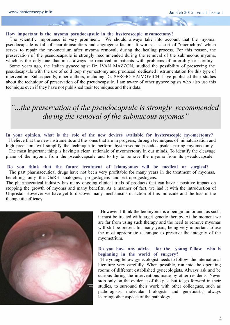
How important is the myoma pseudocapsule in the hysteroscopic myomectomy? The scientific importance is very prominent. We should always take into account that the myoma pseudocapsule is full of neurotransmitters and angiogenic factors. It works as a sort of "microchips" which serves to repair the myometrium after myoma removal, during the healing process. For this reason, the preservation of the pseudocapsule is strongly recommended during the removal of the submucous myoma, which is the only one that must always be removed in patients with problems of infertility or sterility. Some years ago, the Italian gynecologist Dr. IVAN MAZZON, studied the possibility of preserving the pseudocapsule with the use of cold loop myomectomy and produced dedicated instrumentation for this type of intervention. Subsequently, other authors, including Dr. SERGIO HAIMOVICH, have published their studies about the technique of preservation of the pseudocapsule. I am aware of other gynecologists who also use this technique even if they have not published their techniques and their data.
In your opinion, what is the role of the new devices available for hysteroscopic myomectomy? I believe that the new instruments and the ones that are in progress, through techniques of miniaturization and high precision, will simplify the technique to perform hysteroscopic pseudocapsule sparing myomectomy. The most important thing is having a clear rationale of myomectomy in our minds. To identify the cleavage plane of the myoma from the pseudocapsule and to try to remove the myoma from its pseudocapsule.
Do you think that the future treatment of leiomyomas will be medical or surgical? The past pharmaceutical drugs have not been very profitable for many years in the treatment of myomas, benefiting only the GnRH analogues, progestogens and estroprogestogens. The pharmaceutical industry has many ongoing clinical trials of products that can have a positive impact on stopping the growth of myoma and many benefits. As a manner of fact, we had it with the introduction of Ulipristal. However we have yet to discover many mechanisms of action of this molecule and the bias in the therapeutic efficacy.
“...the preservation of the pseudocapsule is strongly recommended during the removal of the submucous myomas”
However, I think the leiomyoma is a benign tumor and, as such, it must be treated with target genetic therapy. At the moment we are far from using such therapy and the need to remove myomas will still be present for many years, being very important to use the most appropriate technique to preserve the integrity of the myometrium.
Do you have any advice for the young fellow who is beginning in the world of surgery? The young fellow gynecologist needs to follow the international literature very carefully. When possible, run into the operating rooms of different established gynecologists. Always ask and be curious during the interventions made by other residents. Never stop only on the evidence of the past but to go forward in their studies, to surround their work with other colleagues, such as pathologists, molecular biologists and geneticists, always learning other aspects of the pathology.
Some things just can’t be learned from books. Some things can only be learned through experience. In this section the best hysteroscopists will share their tricks with you.
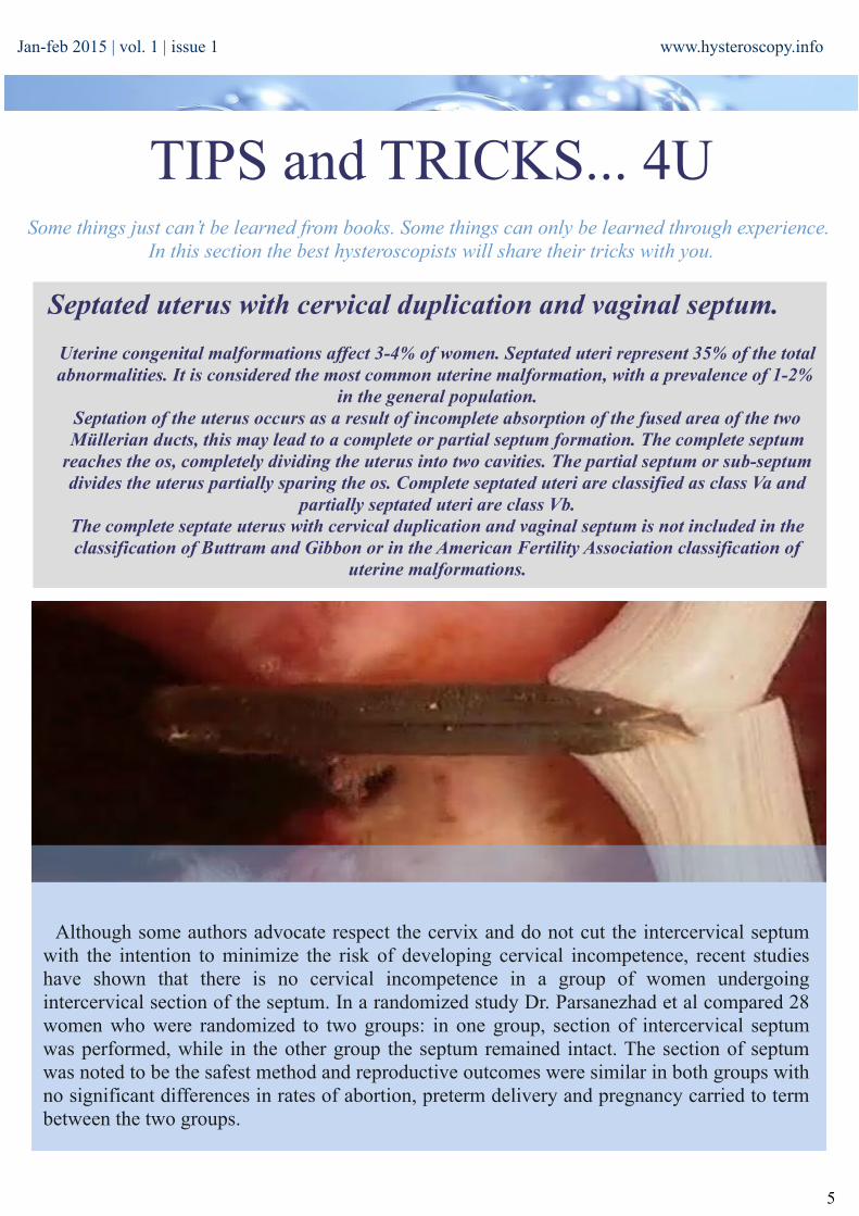
Septated uterus with cervical duplication and vaginal septum.
Uterine congenital malformations affect 3-4% of women. Septated uteri represent 35% of the total abnormalities. It is considered the most common uterine malformation, with a prevalence of 1-2%
in the general population.Septation of the uterus occurs as a result of incomplete absorption of the fused area of the two Müllerian ducts, this may lead to a complete or partial septum formation. The complete septum
reaches the os, completely dividing the uterus into two cavities. The partial septum or sub-septum divides the uterus partially sparing the os. Complete septated uteri are classified as class Va and
partially septated uteri are class Vb.The complete septate uterus with cervical duplication and vaginal septum is not included in the classification of Buttram and Gibbon or in the American Fertility Association classification of
uterine malformations.
5
Although some authors advocate respect the cervix and do not cut the intercervical septum with the intention to minimize the risk of developing cervical incompetence, recent studies have shown that there is no cervical incompetence in a group of women undergoing intercervical section of the septum. In a randomized study Dr. Parsanezhad et al compared 28 women who were randomized to two groups: in one group, section of intercervical septum was performed, while in the other group the septum remained intact. The section of septum was noted to be the safest method and reproductive outcomes were similar in both groups with no significant differences in rates of abortion, preterm delivery and pregnancy carried to term between the two groups.
HIGHLIGHT ARTICLESPublished in the last two months on different medias
Objective: To evaluate whether a correlation exists between the pain perceived during diagnostic anesthesia-free hysteroscopy and the characteristics of the cervical canal.Study design: Prospective observational pilot study of 255 women undergoing diagnostic hysteroscopy. Data analysis included characteristics of the patient and the cervical canal, and the pain experienced during the procedure, assessed by visual analog score (VAS). A multiple logistic regression was then carried out in order to exclude confounding factors.Conclusion: Cervical synechiae appear as a major factor influencing pain during hysteroscopy. While parity acts as a protective factor, the angle of the cervical canal does not seem to play an important role for pain during diagnostic hysteroscopy
Study design: Case reportAbstract: Although endometrial cancer, the most common gynecologic malignancy, is most often diagnosed in postmenopausal women, it affects young women who wish to preserve fertility. The purpose of this article is to describe 2 cases of stage IA endometrial cancer managed conservatively by a combination of hysteroscopic surgery and medical therapy for fertility-sparing purposes, one of which achieved successful pregnancy using assisted reproductive technology. A review of the existing literature on the use of hysteroscopic resection in conservative management of endometrial cancer to preserve fertility. The addition of hysteroscopic resection to conservative management of early-stage endometrial carcinoma may be a way to improve response and recurrence rates in women wishing to preserve fertility and can offer other additional benefits, such as a shorter time period to remission and a faster return to fertility. Key factors to success with this approach include an interdisciplinary approach, thorough patient counseling, and the availability of a team experienced in hysteroscopic resectionSaline infusion sonography (SIS) has become a valuable
diagnostic modality in gynaecology over the last three decades. SIS is now commonly employed for detailed evaluation of the uterine cavity as part of pre-treatment assessment in infertile women. The objective of this paper is review the scientific literature on SIS in infertility. Medline, Ovid and Cochrane databases were searched for relevant articles. The indications, technical aspects and the potential advantages of SIS are discussed. The efficacy and sensitivity of SIS are compared to hysteroscopy in the evaluation of uterine polyps, fibroids, intrauterine adhesions and uterine anomalies. Increasing evidence suggests the use of SIS prior to an in-vitro fertilization (IVF) cycle as it has increased sensitivity in the detection of intrauterine pathology. SIS is cost-effective and results in better patient satisfaction scores than hysteroscopy.
The possibility of progression to endometrial cancer is 1-3% for endometrial hyperplasia without atypia, about 8 % for simple
endometrial hyperplasia with atypia and 29% for complex hyperplasia with atypia.
A placental polyp is a polypoid mass that occurs as a result of the retention of decidual, fetal or placental tissue into the uterine cavity after an abortion or childbirth. It is estimated to occur in 1% after
term delivery and probably the incidence is higher in cases of abortion and preterm delivery.
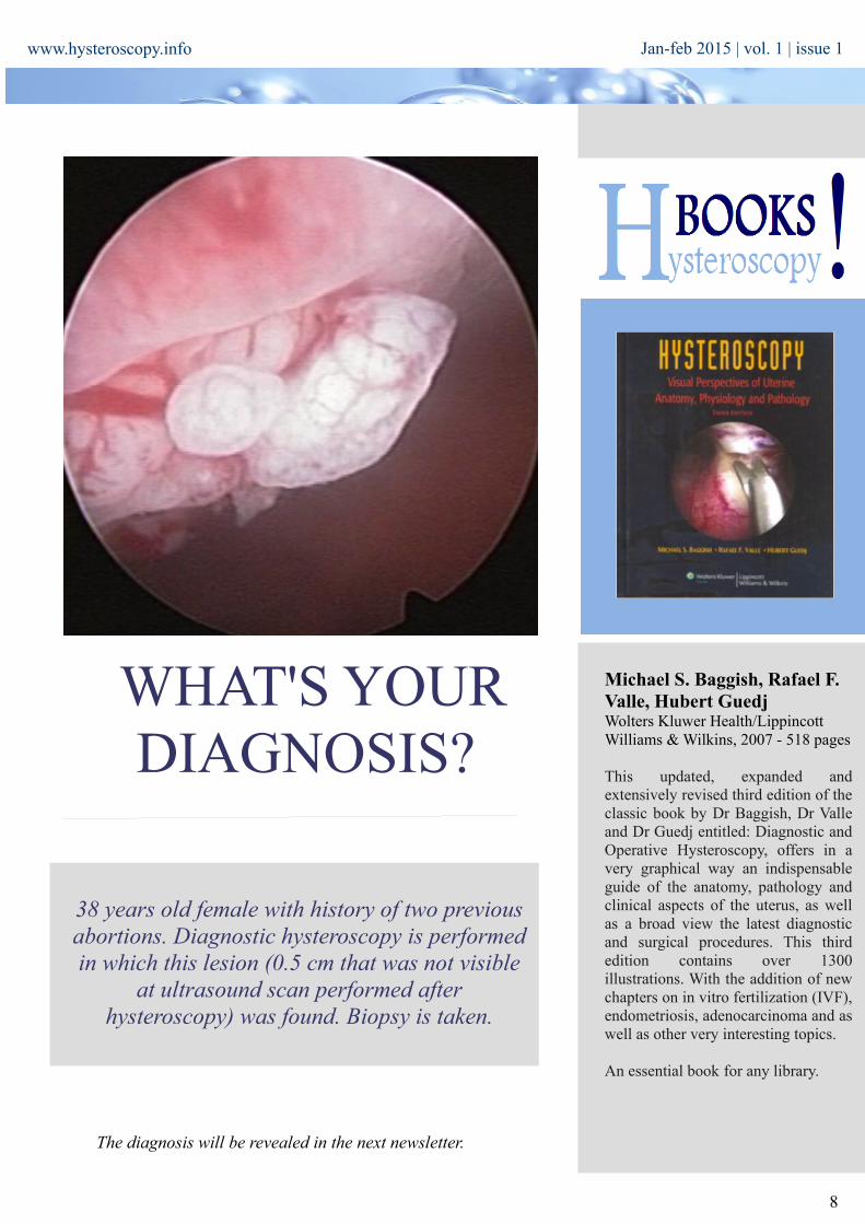
38 years old female with history of two previous abortions. Diagnostic hysteroscopy is performed in which this lesion (0.5 cm that was not visible
at ultrasound scan performed after hysteroscopy) was found. Biopsy is taken.
Michael S. Baggish, Rafael F. Valle, Hubert Guedj Wolters Kluwer Health/Lippincott Williams & Wilkins, 2007 - 518 pages
This updated, expanded and extensively revised third edition of the classic book by Dr Baggish, Dr Valle and Dr Guedj entitled: Diagnostic and Operative Hysteroscopy, offers in a very graphical way an indispensable guide of the anatomy, pathology and clinical aspects of the uterus, as well as a broad view the latest diagnostic and surgical procedures. This third edition contains over 1300 illustrations. With the addition of new chapters on in vitro fertilization (IVF), endometriosis, adenocarcinoma and as well as other very interesting topics.
An essential book for any library.
8
The diagnosis will be revealed in the next newsletter.