Identification of Small Molecules That Interfere with Radial Neuronal Migration and Early Cortical Plate Development Libing Zhou, Yves Jossin and Andre´ M. Goffinet Developmental Neurobiology Unit, Louvain University Medical School, Avenue East Mounier, 73, Box DENE7382, B1200 Brussels, Belgium Using a fetal brain slice culture system that recapitulates early cortical plate (CP) development, we screened the ‘‘Diversity Set’’ chemical library from the National Cancer Institute in order to identify molecules that interfere with radial migration and CP formation and identified 11 candidate molecules. Although most compounds had broadly similar effects, histological and immuno- histochemical studies with preplate and neuronal differentiation markers disclosed some differences in the anomalies induced, suggesting that the identified molecules may act on different targets. Selected compounds were tested for activity on signaling pathways known to be important during radial migration and CP development, namely reelin, phosphatidylinositol 3-kinase/Akt- protein kinase B(PKB)/glycogen synthase kinase-3ß (GSK3b), atypical protein kinases C (aPKC), and Cdk5. No perturbation of reelin signaling or GSK3b activity was detected. One molecule decreased the phosphorylation of Akt and focal adhesion kinase and may act via direct or indirect inhibition of Cdk5, whereas another inhibited phosphorylation of aPKCz /l and may interfere with cell polarity and leading edge formation or progression. These molecules potentially provide new tools to study a neuronal migration and CP development. Keywords: atypical PKC, Cdk5, chemical library, Dab1, reelin, slice culture Introduction The development of the cerebral cortex follows a complex sequence. Neuronal precursors proliferate in ventricular and subventricular zones located around the lateral ventricles. Postmitotic neurons leave the germinal zones and reach their destination by following complex routes (Caviness and Rakic 1978; Caviness 1982; Lambert de Rouvroit and Goffinet 2001; Nadarajah and Parnavelas 2002; Rakic 2003). The majority of cortical neurons migrate along radial fibers and settle, first, in the preplate (PP), where they contribute to a population of reelin-negative pioneer cells, and then in the cortical plate (CP), where they differentiate into glutamatergic neurons. Other cells, such as Cajal Retzius cells, are generated in the cortical hem and around the hilus of the hemispheric vesicles and migrate tangentially in a subpial location in the PP and cor- tical marginal zone (MZ) (Grove and Fukuchi-Shimogori 2003; Meyer and others 2004; Bielle and others 2005). c-aminobutyric acidergic interneurons, generated in the ganglionic eminences, move in 2 stages, first tangentially at the border of the hemispheric subventricular zone and then radially toward the CP (Anderson and others 1997; Nadarajah and Parnavelas 2002). Like other motile cells, neurons move by using at least 2 interdependent cellular mechanisms (Lambert de Rouvroit and Goffinet 2001). Migration begins with the projection of a leading edge, a highly sophisticated process that is based on recognition of microenvironmental cues and integration of these cues to regulate the actin treadmill (Guan and Rao 2003; Govek and others 2005). Migration itself occurs when the nucleus engages into the leading edge. This nuclear movement, often referred to as ‘‘nucleokinesis’’ (Morris and others 1998; Bellion and others 2003; Tsai and Gleeson 2005), is accompanied by a movement of the microtubule organizing center (MTOC) (Xie and Tsai 2004). Whereas nucleokinesis is thought to rely on microtubule dynamics, recent work shows that it is inhibited by blebbistatin, implicating myosin II in the process (Bellion and others 2005). At the end of radial migration, neurons settle in the PP and CP, the organization of which requires normal reelin signaling (Curran and D’Arcangelo 1998; D’Arcangelo 2001; Tissir and Goffinet 2003). Increasing numbers of small molecular weight inhibitors are becoming available to probe a wide variety of signaling path- ways. Although they lack the exquisite specificity of antibodies, or of plasmids encoding interfering RNA, dominant negative or positive proteins, small inhibitors have advantages: they often diffuse freely in cells where they act rapidly; they are relatively easy to use in vitro and in vivo; and they provide a starting point for pharmacological developments. Using a slice culture system that recapitulates several features of early cortical development, small inhibitors were used previously to demonstrate the role of atypical protein kinases C (aPKC) and Src family kinases in neuronal migration and reelin signaling ( Jossin and others 2003a). Here, we used that in vitro system to screen a chemical library and identified 11 original leading compounds that interfere with radial neuronal migration and/or early CP de- velopment. Such molecules should prove useful to define the signaling partners implicated in these developmental events. Materials and Methods Animals Animal procedures were carried out in accordance with institutional guidelines and ratified by competent animal ethics committees. Mice were outbred CD1, maintained on a normal irradiated diet with unrestricted access to water. Previous studies showed that embryonic development proceeds somewhat more rapidly in CD1 than in inbred mouse strains and that an immature CP is already present in the lateral telencephalic wall at E13.5. In order to obtain embryonic brains at the PP stage, prior to any significant migration to the CP, a CD1 colony was maintained on an inverted light cycle (light ‘‘off’’ from 7 AM to 5 PM), and females were inspected for the presence of vaginal plugs between 5 and 7 PM. Embryos at 13 days of pregnancy (E13) were used for slice preparation. The Chemical Library We used the Diversity Set developed by the Developmental Therapeu- tics branch of the National Cancer Institute (NCI). This set is composed Cerebral Cortex January 2007;17:211--220 doi:10.1093/cercor/bhj139 Advance Access publication February 15, 2006 Ó The Author 2006. Published by Oxford University Press. All rights reserved. For permissions, please e-mail: [email protected]
Transcript
Identification of Small Molecules ThatInterfere with Radial Neuronal Migrationand Early Cortical Plate Development
Libing Zhou, Yves Jossin and Andre M. Goffinet
Developmental Neurobiology Unit, Louvain University Medical
School, Avenue East Mounier, 73, Box DENE7382, B1200
Brussels, Belgium
Using a fetal brain slice culture system that recapitulates earlycortical plate (CP) development, we screened the ‘‘Diversity Set’’chemical library from the National Cancer Institute in order toidentify molecules that interfere with radial migration and CPformation and identified 11 candidate molecules. Although mostcompounds had broadly similar effects, histological and immuno-histochemical studies with preplate and neuronal differentiationmarkers disclosed some differences in the anomalies induced,suggesting that the identified molecules may act on differenttargets. Selected compounds were tested for activity on signalingpathways known to be important during radial migration and CPdevelopment, namely reelin, phosphatidylinositol 3-kinase/Akt-protein kinaseB(PKB)/glycogen synthase kinase-3ß (GSK3b), atypicalprotein kinases C (aPKC), and Cdk5. No perturbation of reelinsignaling or GSK3b activity was detected. One molecule decreasedthe phosphorylation of Akt and focal adhesion kinase and may act viadirect or indirect inhibition of Cdk5, whereas another inhibitedphosphorylation of aPKCz /l and may interfere with cell polarityand leading edge formation or progression. These moleculespotentially provide new tools to study a neuronal migration and CPdevelopment.
Keywords: atypical PKC, Cdk5, chemical library, Dab1, reelin,slice culture
Introduction
The development of the cerebral cortex follows a complex
sequence. Neuronal precursors proliferate in ventricular and
subventricular zones located around the lateral ventricles.
Postmitotic neurons leave the germinal zones and reach their
destination by following complex routes (Caviness and Rakic
1978; Caviness 1982; Lambert de Rouvroit and Goffinet 2001;
Nadarajah and Parnavelas 2002; Rakic 2003). The majority of
cortical neurons migrate along radial fibers and settle, first, in
the preplate (PP), where they contribute to a population of
reelin-negative pioneer cells, and then in the cortical plate (CP),
where they differentiate into glutamatergic neurons. Other
cells, such as Cajal Retzius cells, are generated in the cortical
hem and around the hilus of the hemispheric vesicles and
migrate tangentially in a subpial location in the PP and cor-
tical marginal zone (MZ) (Grove and Fukuchi-Shimogori 2003;
Meyer and others 2004; Bielle and others 2005). c-aminobutyric
acidergic interneurons, generated in the ganglionic eminences,
move in 2 stages, first tangentially at the border of the
hemispheric subventricular zone and then radially toward the
CP (Anderson and others 1997; Nadarajah and Parnavelas 2002).
Like other motile cells, neurons move by using at least 2
interdependent cellular mechanisms (Lambert de Rouvroit
and Goffinet 2001). Migration begins with the projection of
a leading edge, a highly sophisticated process that is based on
recognition of microenvironmental cues and integration of
these cues to regulate the actin treadmill (Guan and Rao
2003; Govek and others 2005). Migration itself occurs when
the nucleus engages into the leading edge. This nuclear
movement, often referred to as ‘‘nucleokinesis’’ (Morris and
others 1998; Bellion and others 2003; Tsai and Gleeson 2005), is
accompanied by a movement of the microtubule organizing
center (MTOC) (Xie and Tsai 2004). Whereas nucleokinesis is
thought to rely on microtubule dynamics, recent work shows
that it is inhibited by blebbistatin, implicating myosin II in the
process (Bellion and others 2005). At the end of radial
migration, neurons settle in the PP and CP, the organization of
which requires normal reelin signaling (Curran and D’Arcangelo
1998; D’Arcangelo 2001; Tissir and Goffinet 2003).
Increasing numbers of small molecular weight inhibitors are
becoming available to probe a wide variety of signaling path-
ways. Although they lack the exquisite specificity of antibodies,
or of plasmids encoding interfering RNA, dominant negative or
positive proteins, small inhibitors have advantages: they often
diffuse freely in cells where they act rapidly; they are relatively
easy to use in vitro and in vivo; and they provide a starting point
for pharmacological developments. Using a slice culture system
that recapitulates several features of early cortical development,
small inhibitors were used previously to demonstrate the role of
atypical protein kinases C (aPKC) and Src family kinases in
neuronal migration and reelin signaling ( Jossin and others
2003a). Here, we used that in vitro system to screen a chemical
library and identified 11 original leading compounds that
interfere with radial neuronal migration and/or early CP de-
velopment. Such molecules should prove useful to define the
signaling partners implicated in these developmental events.
Materials and Methods
AnimalsAnimal procedures were carried out in accordance with institutional
guidelines and ratified by competent animal ethics committees. Mice
were outbred CD1, maintained on a normal irradiated diet with
unrestricted access to water. Previous studies showed that embryonic
development proceeds somewhat more rapidly in CD1 than in inbred
mouse strains and that an immature CP is already present in the lateral
telencephalic wall at E13.5. In order to obtain embryonic brains at the PP
stage, prior to any significant migration to the CP, a CD1 colony was
maintained on an inverted light cycle (light ‘‘off ’’ from 7 AM to 5 PM),
and females were inspected for the presence of vaginal plugs between 5
and 7 PM. Embryos at 13 days of pregnancy (E13) were used for slice
preparation.
The Chemical LibraryWe used the Diversity Set developed by the Developmental Therapeu-
tics branch of the National Cancer Institute (NCI). This set is composed
Cerebral Cortex January 2007;17:211--220
doi:10.1093/cercor/bhj139
Advance Access publication February 15, 2006
� The Author 2006. Published by Oxford University Press. All rights reserved.
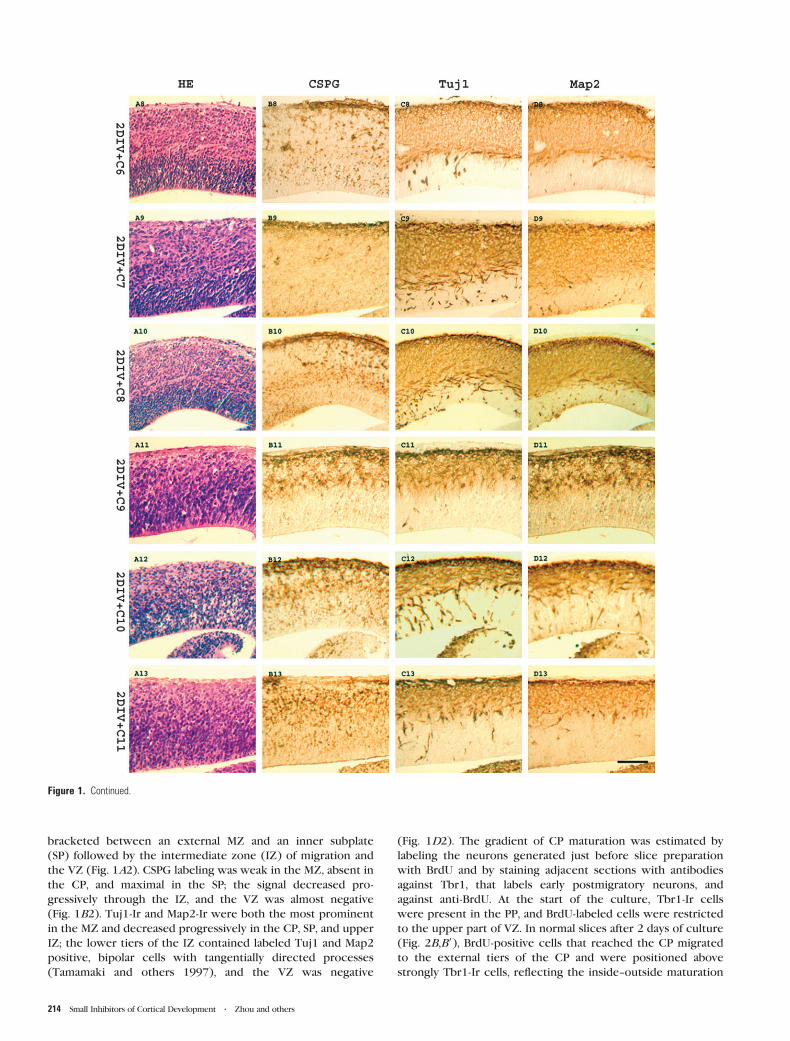
Figure 1. Effect of the selected molecules, C1--C11, on neuronal migration in slices. (A1--A13) HE staining to appreciate overall morphology; (B1--B13) Immunostaining with anti-CSPG antibody to assess PP splitting; (C1--C13) Immunostaining for Tuj1 to disclose neuronal differentiation; and (D1--D14) Anti-Map2 staining to show early dendritic maturationand tangential migration from ganglionic eminence. (A1--D1) Show control embryonic brain slices at E13 (0 DIV); (A2--D2) Show control slices cultured for 2 DIV with DMSO only.After 2 DIV (A2), a well developed CP appeared, and PP was split into MZ and CP (B2); postmitotic neurons were located in MZ, CP, SP, and upper IZ (C2); some tangentiallymigrating bipolar neurons appeared in upper IZ (D2). Compounds C1--C11 perturbed CP formation in vitro (A3--A13), resulted in defective PP splitting (B3--B13); and induced variableanomalies in Tuj1 and Map2 labeling, with occasional ectopic neurons in inner IZ and VZ (C3--C13, D3--D13). Bar = 100 lm.
Cerebral Cortex January 2007, V 17 N 1 213
bracketed between an external MZ and an inner subplate
(SP) followed by the intermediate zone (IZ) of migration and
the VZ (Fig. 1A2). CSPG labeling was weak in the MZ, absent in
the CP, and maximal in the SP; the signal decreased pro-
gressively through the IZ, and the VZ was almost negative
(Fig. 1B2). Tuj1-Ir and Map2-Ir were both the most prominent
in the MZ and decreased progressively in the CP, SP, and upper
IZ; the lower tiers of the IZ contained labeled Tuj1 and Map2
positive, bipolar cells with tangentially directed processes
(Tamamaki and others 1997), and the VZ was negative
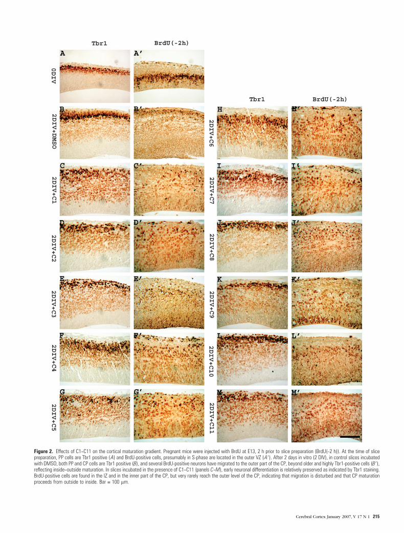
(Fig. 1D2). The gradient of CP maturation was estimated by
labeling the neurons generated just before slice preparation
with BrdU and by staining adjacent sections with antibodies
against Tbr1, that labels early postmigratory neurons, and
against anti-BrdU. At the start of the culture, Tbr1-Ir cells
were present in the PP, and BrdU-labeled cells were restricted
to the upper part of VZ. In normal slices after 2 days of culture
(Fig. 2B,B9), BrdU-positive cells that reached the CP migrated
to the external tiers of the CP and were positioned above
strongly Tbr1-Ir cells, reflecting the inside--outside maturation
Figure 1. Continued.
214 Small Inhibitors of Cortical Development d Zhou and others
Figure 2. Effects of C1--C11 on the cortical maturation gradient. Pregnant mice were injected with BrdU at E13, 2 h prior to slice preparation (BrdU(–2 h)). At the time of slicepreparation, PP cells are Tbr1 positive (A) and BrdU-positive cells, presumably in S-phase are located in the outer VZ (A9). After 2 days in vitro (2 DIV), in control slices incubatedwith DMSO, both PP and CP cells are Tbr1 positive (B), and several BrdU-positive neurons have migrated to the outer part of the CP, beyond older and highly Tbr1-positive cells (B9),reflecting inside--outside maturation. In slices incubated in the presence of C1--C11 (panels C--M), early neuronal differentiation is relatively preserved as indicated by Tbr1 staining.BrdU-positive cells are found in the IZ and in the inner part of the CP, but very rarely reach the outer level of the CP, indicating that migration is disturbed and that CP maturationproceeds from outside to inside. Bar = 100 lm.
Cerebral Cortex January 2007, V 17 N 1 215
gradient (Nowakowski and others 1975; Caviness and Rakic
1978; Caviness 1982).
Identification of Molecules That Affect CorticalDevelopment In Vitro
Following the screening procedure described in Materials and
Methods, we identified 77 compounds that were toxic to brain
slices when tested individually at a concentration of 2--10 lM.
Forty-seven of themwere known to be toxic from the literature.
These toxic molecules were not considered further. We also
identified 11 molecules that perturbed migration and/or the
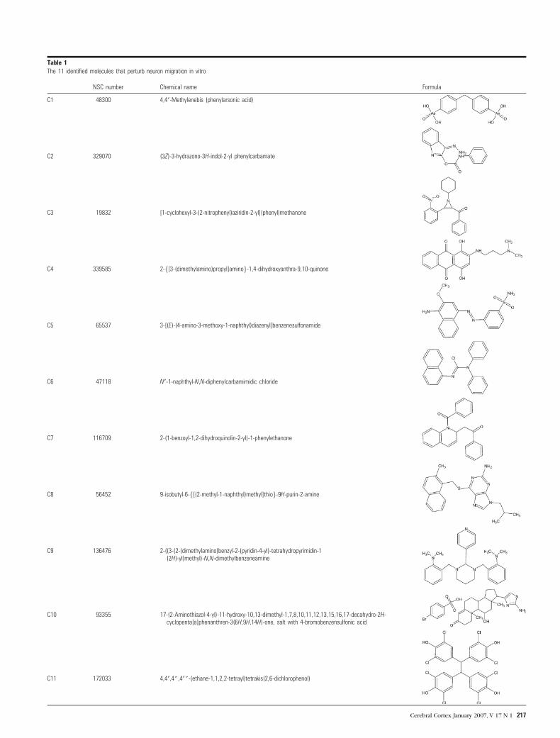
formation of the CP and were selected for further study. A
summary with references in the Developmental Therapeutics
Program library (NSC number), most reproducible active con-
centrations in the slice culture assay, chemical names, and for-
mulas, is provided in Table 1. In order to facilitate description,
the 11 candidate molecules will be referred to as compounds
C1--C11. As shown in Figure 1A3--A13, in the presence of
compounds C1--C11, the development of slices proceeded
relatively normally, in that the thickness of the telencephalic
wall and the overall cellular morphology were comparable with
those of normal slices. BrdU-labeling experiments confirmed
comparable tracer incorporation of labeled cells in normal slices
and in the presence of selected compounds (Fig. 2). In contrast,
architectonic development was drastically affected. The most
evident anomaly was the poor definition of the CP, that was
populated with obliquely oriented neurons, and traversed by
aberrant fiber bundles, a phenotype reminiscent of that ob-
served in reeler embryos (Caviness 1976; Lambert de Rouvroit
and Goffinet 1998) and in slices treated with PP2, a Src family
blocker, or with PKC inhibitors (Jossin and others 2003b). All
compounds resulted in some degree of defective PP splitting, as
estimated by CSPG staining, although some minor differences
between compounds were observed (Fig. 1B3--B13). Molecules
C5, C6, C10, and C11 resulted in an abnormal expression of
CSPG-Ir in the VZ, where no CSPG-Ir cells were detected in
normal slices. In slices incubated with C7, very little CSPG
staining was found outside the MZ. Some but not all molecules
perturbed the pattern of neuronal maturation estimated with
Tuj1 and anti-Map2 staining. In the presence of C1--C11, Tuj1-Ir
was prominent in the MZ, CP, SP, and upper IZ, like in control
slices (Fig. 1C3--C13). However, in the presence of compounds
C2--C8 and C10, intensively Tuj1-Ir neurons were also detected
in the deep IZ and the VZ. The results of Map2 immunostaining
(Fig. 1D1--D13) were quite similar to those obtained with Tuj1.
The presence of Tuj1 and Map2-Ir cells in the lower IZ and VZ
presumably reflected ectopic differentiation of postmitotic
neurons that failed to migrate past the IZ or to leave the VZ,
a feature that is not found in reelin-deficient brains or slices. In
most experiments, the orientation of slices did not consistently
allow visualization of tangentially migrating, Map2-positive cells
from ganglionic eminences, and the effect of C1--C11 on that
migratory stream could not be studied. The effects of C1--C11
on the gradient of CP maturation were studied using BrdU-
labeling experiments carried out as explained in Materials and
Methods. In slices treated with C1--C11, early postmigratory
cells stained normally with Tbr1 antibodies, confirming that
early neuronal differentiation was relatively unaffected. How-
ever, BrdU-positive cells migrated consistently in the interme-
diate zone or to the inner tiers of the CP, but did not reach
its outer part, indicating inverted, outside--inside maturation
(Fig. 2).
Compounds C1--C11 Do Not Perturb CorticalDevelopment In Vivo
To assess whether molecules C1--C11, active in vitro, also
affected development in vivo, they were administered intraper-
itoneally to pregnancy-dated females at E11, 12, and 13, and
fetuses were examined at E15. No overt malformation was
identified by macroscopic inspection, and no pathological
anomaly was found in their brains, which appeared histologi-
cally indistinguishable from those of normal E15 controls.
Effects of C1--C11 on Signaling Pathways Known to BeImplicated in Cortical Development
As a first attempt to identify putative targets of C1--C11, we
studied their effect on 4 pathways that are known to play
critical roles during early cortical development, namely, the
C10 93355 17-(2-Aminothiazol-4-yl)-11-hydroxy-10,13-dimethyl-1,7,8,10,11,12,13,15,16,17-decahydro-2H-cyclopenta[a]phenanthren-3(6H,9H,14H)-one, salt with 4-bromobenzensulfonic acid
continuously developed, and some prove very useful to probe
signaling in different settings. Here, we used an in vitro mouse
embryonic brain slice culture (Jossin and others 2003a) to
screen a chemical library, in order to identify molecules that
interfere with early cortical development, aiming to obtain
leading compounds from which series of analogs could be
developed. The chemical bank selected is the Diversity Set from
the Developmental Therapeutics Program of the National
Cancer Institute (NIH, USA). This set of 1992 compounds was
selected from the ~140 000 compounds of the NCI repository,
using chemical prediction programs. The screen, mostly con-
ducted at a target concentration of 10 lM, identified several
molecules with potent toxicity for embryonic brain tissue, that
were not considered for further analysis. It is worth noting that
this series included molecules such as camptothecin, bouvardin,
cucurbitacin, ellipticin, topotecan, the quinocarmycin analog
DX-52-1, and the mitomycin derivative T53, known to block cell
proliferation in cancer models. The screening resulted in the
selection of 11 molecules, named C1--C11, that perturb deeply
radial neuronal migration and/or CP formation and have not
been described previously. Very little chemical data are avail-
able on them (Table 1) and not informative in terms of putative
mechanisms of action. The fact that the chemical structures are
very different may suggest that they act on different bio-
chemical targets.
As a first attempt to define better the action of compounds C1--
C11, we performed histological studies using HE stain and well-
validated antibodies that reflect neuronal differentiation and
maturation and allow an estimation of the radial gradient of CP
maturation. Apart from some differences noted below, com-
pounds C1--C11 had largely similar effects. At the active dose,
none of them appears to affect dramatically cell death or pro-
liferation because a comparable development of telencephalic
tissue and similar BrdU incorporation occurred in slices cultured
with or without them. All 11 compounds inhibited PP splitting
to various extends and often dramatically. Defective PP splitting
is sometimes considered pathognomonic of defective reelin
signaling, and is not found, for example, in mice with defective
Cdk5 signaling. Because C1-C11 had no consistent effect on
Dab1 phosphorylation, other pathways besides reelin or down-
stream of Dab1 may be implicated in PP splitting. Another
common effect of C1--C11 was to perturb the architectonic
organization of the developing CP and to result in a maturation
that proceeds from outside to inside, whereas normal cortical
maturation proceeds from inside to outside. Despite these
common morphological features on PP splitting and CP forma-
tion, there were some differences in the malformations induced
by C1--C11, particularly in the numbers of ectopic, prematurely
differentiated neurons in the intermediate and ventricular zones.
These defectswere not observed in vivo following intraperitoneal
injections in pregnant mice. Several reasons may explain this
absence of effect, such as rapid degradation or difficulty to
cross the placenta. Preparation of more diffusible and/or stable
analogs is needed for further in vivo studies.
Morphological differences in vitro may reflect different
mechanisms of action of C1--C11, as indicated also by the
preliminary biochemical analysis. By probing key-signaling
molecules with known roles in cortical neuronal migration,
we identified 1 target affected by compound C10, namely,
phosphorylation of aPKC in the activation loop and 2 targets
consistently affected by compound C4, namely, phosphoryla-
tion of FAK at Ser372 and of Akt at Ser473. The inhibition of
phosphorylation of aPKC by C10 might reflect inhibition of
PDK1 or other kinases capable of phosphorylating this site
(Balendran and others 2000). aPKC inhibition perturbs migra-
tion in slices in culture, in which it generates a reeler-like
phenotype (Jossin and others 2003b). However, compound C10
Figure 3. Effect of C1--C11 on selected signaling pathways. At E13, embryonic brainslices were cultured with the 11 selected compounds for 1 DIV, and lysates wereanalyzed for phosphorylation of putative target proteins implicated in reelin signaling orneuronal migration. Data are plotted as the ratio of phosphorylated over total proteinand normalized to the control situation. Incubation with C4 decreased phosphorylationof Akt and FAK and culture in the presence of C10 inhibited phosphorylation of aPKCs.
218 Small Inhibitors of Cortical Development d Zhou and others
did not inhibit Dab1 phosphorylation and resulted in ectopic
neuronal maturation in the VZ, indicating that it perturbs
migration in a reelin-independent manner, or downstream of
Dab1. Observations in other systems have shown that aPKC is
required for translocation of the MTOC in the direction of and
possibly prior to the extension of a leading edge (Etienne-
Manneville and Hall 2003; Henrique and Schweisguth 2003;
Solecki and others 2004; Suzuki and others 2004), and this
mechanism is likely to be important in radial migration.
However, the phosphorylation of aPKC has not been studied
in this context. Phosphorylation of FAK at Ser372 is a recognized
target of the kinase Cdk5, which, together with its coactivators
p35 and p37, plays a key role in neuronal migration (Ohshima
and others 1996; Chae and others 1997; Ko and others 2001).
Phosphorylation of FAK by Cdk5 is important for microtubule
organization, nuclear movement, and neuronal migration (Xie
and others 2003; Nikolic 2004; Xie and Tsai 2004). To our
knowledge, no specific inhibitors of Cdk5 signaling are cur-
rently available because inhibitors such as Roscovitin also block
other Cdk-related enzymes. Like that of FAK(Ser372), the
inhibition of Akt(Ser473) phosphorylation by C4 was consistent
and significant. This could reflect a decrease in Akt activity.
However C4 does not decrease GSK3b phosphorylation at Ser9,
indicating that the inhibition of Akt is partial. High concen-
trations of C4 were toxic and did not allow us to test this
further. The observation that C4 prevents PP splitting, a pheno-
typic trait that is not present in Cdk5 mutant mice, indicates
that it also affects other unrecognized signaling components.
In sum, in the present work, we identified 11 original
compounds that interfere with cortical development in vitro.
Future work should focus on the development of analogs with
increased activity and on the identification of the biochemical
target of these molecules.
Notes
We thank the Developmental Therapeutics Program, Division of Cancer
Treatment and Diagnosis, National Cancer Institute, USA, particularly
R. Schultz and J. Johnson, for gift of the chemical library and selected
compounds and for advice, and Robert Hevner for gift of the anti-Tbr1
antibody. We also thank Esther Paıtre for technical assistance and
members of the Developmental Neurobiology laboratory for discussion.
YJ is Postdoctoral Researcher at the Fonds National de la Recherche
Scientifique. This work was supported by grants Fonds de la Recherche
Fondamentale Collective 2.4504.01, Action de Recherches Concertees
02/07-276, and by the Fondation Medicale Reine Elisabeth, all from