Copyright 1998 by the Genetics Society of America Identification of a Calcineurin-Independent Pathway Required for Sodium Ion Stress Response in Saccharomyces cerevisiae Raymond W. Ganster, Rhonda R. McCartney and Martin C. Schmidt Department of Molecular Genetics and Biochemistry, University of Pittsburgh School of Medicine, Pittsburgh, Pennsylvania 15261 Manuscript received February 12, 1998 Accepted for publication May 26, 1998 ABSTRACT The calcium-dependent protein phosphatase calcineurin plays an essential role in ion homeostasis in yeast. In this study, we identify a parallel ion stress response pathway that is independent of the calcineurin signaling pathway. Cells with null alleles in both STD1 and its homologue, MTH1, manifest numerous phenotypes observed in calcineurin mutants, including sodium, lithium, manganese, and hydroxyl ion sensitivity, as well as alpha factor toxicity. Furthermore, increased gene dosage of STD1 suppresses the ion stress phenotypes in calcineurin mutants and confers halotolerance in wild-type cells. However, Std1p functions in a calcineurin-independent ion stress response pathway, since a std1 mth1 mutant is FK506 sensitive under conditions of ion stress. Mutations in other genes known to regulate gene expression in response to changes in glucose concentration, including SNF3, RGT2, and SNF5, also affect cell growth under ion stress conditions. Gene expression studies indicate that the regulation of HAL1 and PMR2 expression is affected by STD1 gene dosage. Taken together, our data demonstrate that response to ion stress requires the participation of both calcineurin-dependent and -independent pathways. Y EAST cells are able to adapt to many environmental al. 1995), suggesting that the Na 1 /K 1 ratio is more stresses, including changes in the osmolarity of important to this enzyme’s activity than is the absolute their surroundings. When incubated in hypertonic me- concentration of sodium. This finding may explain why dia, yeast cells adapt by inducing the synthesis of high yeast cells undergoing sodium ion stress act both to concentrations of intracellular glycerol (Brewster et reduce intracellular sodium ion concentration and to al. 1993). Indeed, the enzyme responsible for glycerol retain intracellular potassium ions. Although the molec- synthesis, the product of the GPD1 gene, is essential for ular mechanism is unclear, the HAL3 gene appears to growth in hypertonic media (Albertyn et al. 1994). play a critical role in determining the intracellular Na 1 / Changes in osmolarity are detected by two independent K 1 ratio by regulating cation transport (Ferrando et osmosensors that in turn regulate the activity of the al. 1995). HOG1 mitogen activated protein (MAP) kinase pathway Yeast cells undergoing sodium ion stress increase both (Posas et al. 1996). Activation of the HOG1 MAP kinase the expression and the activity of the Pmr2p, a P-type cascade signals the induction of transcription from a ion pump thought to be responsible for Na 1 and Li 1 number of genes through stress response elements ion efflux (Weiland et al. 1995). Both of these responses (STREs) (Schuller et al. 1994). to Na 1 ion stress require the activity of the calcium Independent of their effects on osmolarity, certain signaling pathway. Ca 21 /calmodulin binds to and acti- ions are toxic to yeast cells due to their ability to inhibit vates Pmr2p, thereby directly stimulating Na 1 ion efflux specific metabolic pathways. Sodium and lithium ions (Weiland et al. 1995). In addition, Ca 21 /calmodulin are particularly toxic to yeast cells due to their inhibition binds to and activates the protein phosphatase cal- of the 39,59-bisphosphate nucleotidase enzyme that is cineurin. Although the phosphoprotein substrates of required for sulfate assimilation (Murguia et al. 1995, calcineurin have not yet been identified, calcineurin 1996). This enzyme is encoded by the MET22/HAL2 activity is required for the transcriptional induction of gene and was isolated initially as a methionine auxo- the PMR2 gene (Garciadeblas et al. 1993; Cunning- troph and subsequently by virtue of its ability to confer ham and Fink 1996). Recent studies have identified sodium tolerance when present on a high-copy plasmid a zinc finger transcription factor that is required for (Gaxiola et al. 1992; Glaser et al. 1993). The inhibition calcineurin-mediated stimulation of transcription of of the 39,59-bisphosphate nucleotidase activity by sodium the PMR2, PMC1, and FKS2 genes (Matheos et al. 1997; can be reversed by addition of potassium (Murguia et Stathopoulos and Cyert 1997). Thus, the calcineu- rin-dependent signaling pathway leading to increased expression of the PMR2 gene comprises one essential Corresponding author: Martin C. Schmidt, Department of Molecular response to Na 1 /Li 1 ion stress. Genetics and Biochemistry, University of Pittsburgh School of Medi- cine, Pittsburgh, PA 15261. E-mail: [email protected]Calcineurin is a highly conserved (Cyert et al. 1991) Genetics 150: 31–42 (September 1998)
Transcript
Copyright 1998 by the Genetics Society of America
Identification of a Calcineurin-Independent Pathway Required for Sodium IonStress Response in Saccharomyces cerevisiae
Raymond W. Ganster, Rhonda R. McCartney and Martin C. SchmidtDepartment of Molecular Genetics and Biochemistry, University of Pittsburgh School of Medicine, Pittsburgh, Pennsylvania 15261
Manuscript received February 12, 1998Accepted for publication May 26, 1998
ABSTRACTThe calcium-dependent protein phosphatase calcineurin plays an essential role in ion homeostasis in
yeast. In this study, we identify a parallel ion stress response pathway that is independent of the calcineurinsignaling pathway. Cells with null alleles in both STD1 and its homologue, MTH1, manifest numerousphenotypes observed in calcineurin mutants, including sodium, lithium, manganese, and hydroxyl ionsensitivity, as well as alpha factor toxicity. Furthermore, increased gene dosage of STD1 suppresses theion stress phenotypes in calcineurin mutants and confers halotolerance in wild-type cells. However, Std1pfunctions in a calcineurin-independent ion stress response pathway, since a std1 mth1 mutant is FK506sensitive under conditions of ion stress. Mutations in other genes known to regulate gene expression inresponse to changes in glucose concentration, including SNF3, RGT2, and SNF5, also affect cell growthunder ion stress conditions. Gene expression studies indicate that the regulation of HAL1 and PMR2expression is affected by STD1 gene dosage. Taken together, our data demonstrate that response to ionstress requires the participation of both calcineurin-dependent and -independent pathways.
Y EAST cells are able to adapt to many environmental al. 1995), suggesting that the Na1/K1 ratio is morestresses, including changes in the osmolarity of important to this enzyme’s activity than is the absolute
their surroundings. When incubated in hypertonic me- concentration of sodium. This finding may explain whydia, yeast cells adapt by inducing the synthesis of high yeast cells undergoing sodium ion stress act both toconcentrations of intracellular glycerol (Brewster et reduce intracellular sodium ion concentration and toal. 1993). Indeed, the enzyme responsible for glycerol retain intracellular potassium ions. Although the molec-synthesis, the product of the GPD1 gene, is essential for ular mechanism is unclear, the HAL3 gene appears togrowth in hypertonic media (Albertyn et al. 1994). play a critical role in determining the intracellular Na1/Changes in osmolarity are detected by two independent K1 ratio by regulating cation transport (Ferrando etosmosensors that in turn regulate the activity of the al. 1995).HOG1 mitogen activated protein (MAP) kinase pathway Yeast cells undergoing sodium ion stress increase both(Posas et al. 1996). Activation of the HOG1 MAP kinase the expression and the activity of the Pmr2p, a P-typecascade signals the induction of transcription from a ion pump thought to be responsible for Na1 and Li1
number of genes through stress response elements ion efflux (Weiland et al. 1995). Both of these responses(STREs) (Schuller et al. 1994). to Na1 ion stress require the activity of the calcium
Independent of their effects on osmolarity, certain signaling pathway. Ca21/calmodulin binds to and acti-ions are toxic to yeast cells due to their ability to inhibit vates Pmr2p, thereby directly stimulating Na1 ion effluxspecific metabolic pathways. Sodium and lithium ions (Weiland et al. 1995). In addition, Ca21/calmodulinare particularly toxic to yeast cells due to their inhibition binds to and activates the protein phosphatase cal-of the 39,59-bisphosphate nucleotidase enzyme that is cineurin. Although the phosphoprotein substrates ofrequired for sulfate assimilation (Murguia et al. 1995, calcineurin have not yet been identified, calcineurin1996). This enzyme is encoded by the MET22/HAL2 activity is required for the transcriptional induction ofgene and was isolated initially as a methionine auxo- the PMR2 gene (Garciadeblas et al. 1993; Cunning-
troph and subsequently by virtue of its ability to conferham and Fink 1996). Recent studies have identified
sodium tolerance when present on a high-copy plasmid a zinc finger transcription factor that is required for(Gaxiola et al. 1992; Glaser et al. 1993). The inhibition calcineurin-mediated stimulation of transcription ofof the 39,59-bisphosphate nucleotidase activity by sodium the PMR2, PMC1, and FKS2 genes (Matheos et al. 1997;can be reversed by addition of potassium (Murguia et
Stathopoulos and Cyert 1997). Thus, the calcineu-rin-dependent signaling pathway leading to increasedexpression of the PMR2 gene comprises one essential
Corresponding author: Martin C. Schmidt, Department of Molecularresponse to Na1/Li1 ion stress.Genetics and Biochemistry, University of Pittsburgh School of Medi-
cine, Pittsburgh, PA 15261. E-mail: [email protected] Calcineurin is a highly conserved (Cyert et al. 1991)
Genetics 150: 31–42 (September 1998)
32 R. W. Ganster, R. R. McCartney and M. C. Schmidt
heterodimer composed of one catalytic subunit and one grew poorly on media containing high concentrationsof NaCl but not KCl. Because this phenotype is similarregulatory subunit. The regulatory subunit, a calcium-
binding protein containing EF hands related to calmod- to that observed in calcineurin mutants, we tested thestd1D mth1D mutant for other calcineurin phenotypes.ulin, is required for calcineurin activity (Cyert and
Thorner 1992), although the molecular role played by Our results indicate that the Std1p is required for Na1
ion stress response in Saccharomyces cerevisiae but that itthis subunit has not yet been determined. The catalyticsubunit contains two distinct domains (Guerini 1997); functions in a signaling pathway that is distinct from
the calcineurin pathway.the N-terminal half comprises the phosphatase activesite conserved in numerous protein phosphatases, whilethe C-terminal domain contains an auto-inhibitory se-
MATERIALS AND METHODSquence that blocks phosphatase activity. In addition,the regulatory domain contains a calmodulin-binding Yeast strains, media, and genetic techniques: S. cerevisiae
strains utilized in this study are described in Table 1. Exceptmotif whose presence distinguished calcineurin fromwhere indicated, growth of yeast utilized standard mediaother protein phosphatases. Binding of calcium-calmod-(Rose et al. 1990) at 308. Glucose, sucrose, or raffinose were
ulin activates calcineurin by relieving the effects of the present at 2% (g/100 ml). Antimycin A was included at 1 mg/auto-inhibitory sequence. Indeed, truncation of the ml where indicated. Standard procedures were utilized for
genetic crosses, sporulation, and tetrad analysis (Rose et al.C-terminal domain results in a constitutively active cal-1990). Transformations of yeast strains utilized the lithiumcineurin enzyme (Hubbard and Klee 1989).acetate procedure (Gietz et al. 1995). FK506 was provided byIn mammals, calcineurin plays an important role inFujisawa USA Inc.
signal transduction in a number of cell types. In acti- Plasmid constructions: Increased gene dosage of STD1 wasvated T-cells, the primary target of calcineurin appears obtained by transforming cells with p6A5U, which contains
the 3.9-kb PvuII to SalI fragment from p6A (Ganster et al.to be members of the NFAT (nuclear factor of activated1993) bearing the entire STD1 gene cloned into the 2m plas-T-cells) family of transcription factors (Rao et al. 1997).mid YEp352 (Hill et al. 1986). The SNF5 centromeric plasmidIn resting T-cells, the NFAT proteins are phosphory-was constructed by placing the 4380-bp genomic EcoRI frag-
lated and thereby trapped in the cytoplasm. Dephos- ment (21159 to 13671, relative to the ATG codon) that con-phorylation by calcineurin unmasks the nuclear localiza- tains the entire SNF5 open reading frame and flanking se-
quences into the CEN plasmid pUN55 (Elledge and Davistion signal, thereby promoting translocation to the1988). The STD1 centromeric plasmid, pRG70U, was con-nucleus where NFAT cooperates with AP-1 (fos-jun) tostructed by inserting the 3429-bp SacI to EagI fragment fromactivate transcription of cytokine genes. In renal tubulep6A encompassing the entire STD1 gene and flanking se-
cells, calcineurin promotes Na1 ion retention by de- quences into the SacI and EagI sites of pUN55 (Elledge andphosphorylating DARP32 (Aperia et al. 1992), an inhibi- Davis 1988). The 2m CNB1 plasmid, pAMS283 (kindly pro-
vided by Angela Stathopoulos and Martha Cyert), con-tor of protein phosphatase 1. Protein phosphatase 1, intains a 1.4-kb genomic DNA fragment encompassing the entireturn, activates the Na1,K1-ATPase, which provides theCNB1 gene inserted into YEp352 (Hill et al. 1986).concentration gradient necessary for Na1 ion readsorp-
Spot dilution growth assays: Liquid cultures were grown fortion. Thus, in mammals and in yeast, calcineurin plays at least two cell generations to an OD600 between 0.2 and 0.4.a crucial role in Na1 ion homeostasis. Cell densities were normalized to an OD600 of 0.2, and 5–10
ml of 10-fold serial dilutions of the liquid cultures were spottedOur studies have focused on the Std1 protein, whoseonto solid media and incubated at 308 for 2–5 days.gene was cloned as a high-copy-number suppressor of
Strain constructions: All S. cerevisiae strains used in this studya mutation in the TATA binding protein (TBP) (Gans-
are in the S288c background. Strains with null alleles in STD1ter et al. 1993). STD1 (MSN3) was also isolated as a and MTH1 were constructed with plasmids pJH104 andhigh-copy-number suppressor of the growth defect ex- pJH124, respectively (Hubbard et al. 1994). The mth1D2 allele
was constructed using the 3796-bp EcoRV genomic DNA frag-hibited by snf4D2 mutants on raffinose antimycin mediament encompassing the MTH1 locus from which the 1791-bpand was shown to be a gene-dosage-dependent modula-EcoRI fragment (2986 to 1805, relative to the initiating ATGtor of glucose repression (Hubbard et al. 1994). Yeastcodon) had been deleted. A strain bearing the mth1D1::URA3
cells contain a single homologue of STD1 designated allele was transformed with the modified EcoRV fragment,MTH1. Null alleles in this pair of genes suggested that and 5-FOA media was used to select Ura2 transformants. The
resulting deletion of the 59 half of MTH1 was confirmed bythey acted redundantly because neither single knockoutSouthern blot. The mth1D2 allele does not confer any MTH1produced a detectable phenotype, whereas the doublefunction because cells bearing the mth1-D1::URA3 allele or theknockout strain was defective in the derepression of themth1D2 allele were equally defective for growth on raffinose
SUC2 gene. Std1p has also been shown to interact in antimycin media when present in combination with a std1the two-hybrid system and biochemically with both the null allele. A null allele of the SNF4 gene was engineered by
replacing the entire SNF4 coding sequence with the loxP-Snf1 kinase (Hubbard et al. 1994) and with TBP (Till-
kanMX-loxP cassette from plasmid pUG6 (Guldener et al.man et al. 1995). One hypothesis for these data posits1996). The loxP-kanMX-loxP cassette was amplified in a PCRthat Std1p couples the glucose starvation signal fromreaction using oligonucleotides with 22 and 19 bases comple-
the Snf1 kinase complex to transcriptional machinery. mentary to the kanMX cassette at the 39 end (lower case) andIn a search for additional phenotypes in cells lacking 40 bases complementary to the SNF4 locus at the 59 end (upper
case). The primer sequences were as follows: 59-CTGTGTStd1p function, we discovered that std1D mth1D cells
33Sodium Stress Response in S. cerevisiae
TABLE 1
S. cerevisiae strains used
Strain Genotype Source or reference
FY14 MATa ura3-52 trp1D63 Winston et al. (1996)FY86 MATa ura3-52 leu2-D1 his3-D200 Winston et al. (1996)MCY2099 MATa ura3-52 his3-D200 ade2-101 snf5-D2 Laurent et al. (1990)MSY182 MATa ura3-52 leu2-D1 his3-D200 trp1-D63 This studyMSY192 MATa ura3-52 leu2-D1 his3-D200 trp1-D63 std1::HIS3 mth1-D2 This studyMSY314 MATa ura3-52 leu2-D1 his3-D200 snf4::KAN This studyMSY317 MATa ura3-52 leu2D1 trp1D63 snf1::KAN This studyMSY401 MATa ura3-52 leu2-D1 his3-D200 trp1-D63 This studyMSY402 MATa ura3-52 leu2-D1 his3-D200 trp1-D63 snf3::hisG rgt2::HIS3 This studyMSY403 MATa ura3-52 leu2-D1 his3-D200 trp1-D63 rgt2::HIS3 This studyMSY404 MATa ura3-52 leu2 D1 his3-D200 trp1 D63 snf3::hisG This studyYPH499 MATa ura3-52 leu2-D1 his3-D200 trp1-D63 lys2-801 ade2-101 Sikorski and Hieter (1989)MCY300-1 MATa ura3-52 leu2-D1 his3-D200 trp1-D63 lys2-801 ade2-101 Cyert et al. (1991)
cna1-D1::hisG cna2-D1::HIS3DD12 MATa ura3-52 leu2-D1 his3-D200 trp1-D63 lys2-801 ade2-101 Cybert and Thorner (1992)
cnb1-D1::hisG
TAGCATTAGGAGGAAGCGAAAAGGAAAATACATAgcataggc ern blot analysis. The pmr2::lacZ plasmid pFR70 (Marquez
and Serrano 1996) was kindly provided by A. Rodriguez-cactagtggatctg and 59-TTTATTTATAGTATGTACACAAAAATCTCATCGGCTCGTTcagctgaagcttcgtacgc. A diploid strain Navarro.
Alpha factor toxicity: Yeast cultures that had been grown(FY14 3 FY86; Winston et al. 1996) was transformed with thePCR product, and G418-resistant clones were analyzed by PCR overnight in low-pH YEPD (Withee et al. 1997) were diluted
in the same media to an OD600 of 0.2. The cultures wereusing one primer complementary to the kanMX gene andone primer upstream of the SNF4 open reading frame. One grown an additional 2 hr, at which point (t 5 0) alpha factor
[synthesized Millipore (Bedford, MA) model 9050 plus Syn-strain that was positive by PCR was sporulated and segre-gants analyzed for growth on G418 and raffinose media con- thesizer] was added to a final concentration of 50 mm. Aliquots
were removed, diluted in water, and plated on solid YEPDtaining 1 mg/ml antimycin A. Of the 15 complete tetradsanalyzed, all 15 showed 2:2 segregation of G418 resistance media in order to determine viable cells per ml. Experiments
with 2m plasmids were conducted in low-pH SC media lackingand all G418-resistant segregants displayed a snf 2 phenotypeon raffinose-antimycin media. To confirm that the snf 2 phe- uracil.
Northern blot analyses: Liquid cultures (10 ml) were har-notype was due to disruption of the SNF4 locus, one snf4::KANhaploid strain was mated with a wild type, a snf5 strain, and vested in log phase, and total RNA was prepared by the hot
phenol method (Kohrer and Domdey 1991). A total of 10with a snf4D2 strain. Only the snf4D2 strain failed to comple-ment the snf4::KAN allele. A null allele of the SNF1 gene was mg of RNA isolated from each culture was subjected to electro-
phoresis in formaldehyde-1% agarose gels. The quantity andconstructed by PCR amplification of the kanMX cassette ofpUG6 with long flanking homology regions using the proce- integrity of the RNA samples were analyzed by ethidium bro-
mide staining in 0.1 m ammonium acetate. The RNA wasdure described by Wach (1996). The primers used to amplifythe 59 flank of the SNF1 locus were T-324: 59-GGCACATCAA electrotransferred to nylon membrane and hybridized to 32P-
labeled DNA sequences. The DNA probes included gel-puri-CAGGTAGCGTTATAGGGG and SK-B-1: 59-gcgtacgaagcttcagctggcggccgcGTTGACTTTATTAAGGGAGTGTAGC. The pri- fied DNA fragments of the entire HAL1 open reading frame
generated by PCR, the 2084-bp genomic HindIII fragmentmers used to amplify the 39 flank were SK-T1903: 59-tctgccggtctccctatagtgagtcgTGGTGGAACGTAAAAGAATGATATGG containing the SUC2 open reading frame, and the 240-bp
genomic BglII-KpnI fragment of TUB2. All fragments wereand B2260: 59-GGTTGTATTTTTGTCGCAGACTCCG. Nucle-otides written in the upper case are complementary to the radiolabeled by the random priming method.SNF1 locus and those written in lower case are complementaryto the kanMX cassette. The 59 and 39 flanking sequences wereamplified, gel purified, and used in a second PCR reaction RESULTSusing pUG6 as a template and primers T-324 and B2260. Theresulting 2.3-kb PCR product was used to transform a diploid STD1 gene function is required for ion stress re-(FY14 3 FY86; Winston et al. 1996) to G418 resistance. Disrup- sponse: Cells lacking STD1 gene function (std1D mth1D)tion of the SNF1 locus was confirmed by Southern blot. Tetrad
appear indistinguishable from wild-type cells on stan-analysis showed 2:2 segregation of the G418 resistance anddard rich (YEPD) and minimal (synthetic complete)cosegregation of a snf 2 phenotype in all tetrads analyzed. A
null allele of the SNF3 gene was constructed using plasmid media at all temperatures tested (data not shown). How-pBM3103, kindly provided by Sabire Ozcan and Mark John- ever, std1D mth1D strains display a defect in invertaseston, Washington University (Ozcan et al. 1996). A null allele derepression (Hubbard et al. 1994) and consequentlyof the RGT2 gene was created by PCR amplification using
show impaired fermentative growth on media with su-the identical primers and strategy described by Ozcan et al.crose or raffinose as the carbon source (Figure 1A). To(1996). Correct integration for both the snf3::hisG-URA3-hisG
allele and the rgt2::GFP-HIS3 allele were confirmed by South- identify new functions of the Std1 protein, a set of strains
34 R. W. Ganster, R. R. McCartney and M. C. Schmidt
Figure 1.—Effect of STD1 gene dosage on cell growth under ion stress conditions. Serial dilutions of yeast cultures werespotted onto standard synthetic complete media lacking uracil with 2% glucose, 2% sucrose plus 1 mg/ml antimycin A, or 2%glucose supplemented with either 1 m NaCl, 1 m KCl, or 0.3 m LiCl, as indicated. (A) The strains used in this experiment areMSY192 (std1D mth1D) transformed with YEp352 (2m vector). MSY182 (STD1 MTH1) transformed with either YEp352 or p6A5U(2m STD1), as indicated. (B) MSY192 (std1D mth1D) was transformed with either pUN55 (CEN vector), pRG70U (CEN STD1),YEP352, or p6A5U, as indicated.
that differ in STD1 gene dosage was analyzed for growth affected by STD1 gene dosage. Cells lacking any STD1gene function grow very poorly in media containingon a wide variety of different media. We have found
that std1D mth1D strains display impaired growth on these concentrations of Na1 and Li1. Low copy plasmidscontaining STD1 provide a limited growth advantage inmedia containing 1 m NaCl (Figure 1A). This effect is
not due to a defect in the HOG1-osmotic stress pathway the presence of 1 m NaCl, and the high copy plasmidencoding STD1 is required to promote a growth advan-because no growth defect is observed when this strain
is grown in the presence of 1 m KCl (Figure 1A) or tage in the presence of 0.3 m LiCl. The fact that thesestrains are isogenic except at the STD1 locus confirms1 m sorbitol (not shown). Furthermore, increased STD1
gene dosage on high copy number plasmids provides that the STD1 gene plays an essential role in ion stressresponse.wild-type cells with a growth advantage on media con-
taining 1 m NaCl. This Std1-mediated growth advantage Loss of Std1p function confers additional phenotypesfound in calcineurin mutants: The PP2B protein phos-was specific to sodium and was not observed on media
containing 1 m KCl (Figure 1A) or 1 m sorbitol (not phatase calcineurin is an important regulator of ionhomeostasis in yeast (Nakamura et al. 1993). In particu-shown).
To rule out any effect of strain variation on these lar, calcineurin mutants exhibit growth defects in thepresence of specific ions, including sodium, lithium,results, a single std1D mth1D strain transformed with low
and high copy plasmids containing the STD1 gene or manganese, and hydroxyl ions, but not when challengedby potassium or magnesium. Since std1 mth1 mutantsno insert was analyzed for growth in various media (Fig-
ure 1B). Cells lacking STD1 gene function show no also exhibited sodium and lithium sensitivity but notpotassium sensitivity, it was possible that other cal-growth defect on synthetic complete media alone or
supplemented with 1 m KCl. However, when cell growth cineurin phenotypes would be shared by std1 mth1 mu-tants. Three yeast strains that differ in STD1 gene dosageis dependent on the fermentation of sucrose, cells lack-
ing STD1 gene function display a distinct growth defect were compared for growth in the presence of 10 mm
manganese chloride and in media adjusted to pH 8.3that is equally well suppressed by low and high copySTD1 plasmids (Figure 1B). Growth in media supple- (Figure 2). Cells lacking STD1 gene function clearly
showed growth defects relative to the wild-type cellsmented with either 1 m NaCl or 0.3 m LiCl is greatly
35Sodium Stress Response in S. cerevisiae
Increased gene dosage of STD1 suppresses cal-cineurin mutations: Since std1 mth1 mutants shared sev-eral phenotypes with calcineurin mutants, it seemedpossible that the Std1p (and Mth1p) might act in thesame or in a parallel ion response pathway to cal-cineurin. If true, then increased gene dosage of STD1might be able to compensate for the loss of calcineurinfunction. To test this hypothesis, we transformed a setof isogenic strains that differ only at the CNA1, CNA2,and CNB1 loci with 2m plasmids containing either theSTD1 gene or no insert. These strains were then testedfor growth under a variety of ion stresses. Cells lackingFigure 2.—Loss of STD1 gene function causes additional
phenotypes observed in calcineurin mutants. Serial dilutions either both copies of the calcineurin catalytic subunitsof yeast cultures were spotted onto synthetic complete media or the single regulatory subunit all display a markedlacking uracil or the same media supplemented with 10 mm growth defect when grown on media containing 0.9 mMnCl2 or adjusted to pH 8.3, as indicated.
NaCl, 0.3 m LiCl, or 10 mm MnCl2 or in media adjustedto pH 8.3 (Figure 3). The growth defects observed in
under both of these conditions. Mutations in either the the calcineurin mutants, however, were in all cases sup-catalytic subunits (CNA1 and CNA2) or the regulatory pressed by increased gene dosage of STD1. The suppres-subunit (CNB1) of calcineurin also cause growth defects sion by 2m STD1 is observed in both calcineurin mutantunder these same ion stresses (Nakamura et al. 1993; strains as well as in wild-type cells whose calcineurinPozos et al. 1996) (Figure 3). Another phenotype of activity was inhibited by the immunosuppressant drugcalcineurin mutants is loss of viability in the presence FK506 (data not shown). Increased gene dosage ofof the alpha factor mating pheromone (Withee et al. MTH1, the STD1 gene homologue, was also able to1997). Pairs of isogenic strains lacking either calcineurin suppress calcineurin ion stress phenotypes (data not(Sikorski and Hieter 1989; Cyert et al. 1991; Cyert shown). However, a second calcineurin phenotype, lossand Thorner 1992) or STD1 gene function were incu- of viability in alpha factor, was not suppressed by in-bated with 50 mm alpha factor, and then cell viability creased gene dosage of STD1 (Figure 4B). In this experi-was determined over a 6-hr time course. Cells lacking ment, a cnb12 strain was transformed with high copycalcineurin (cnb1) lose viability in alpha factor with plasmids containing either the STD1 gene, the CNB1,10% of the cells being viable after 6 hr (Figure 4A). gene, or no insert. The CNB1 plasmid restored viabilityLikewise, the std1D mth1D strain also showed a similar to the cnb1 mutant, indicating that plasmid-based sup-loss of viability over the same time course and in contrast pression in this media was possible. Increased gene dos-to the isogenic wild-type strains. Thus cells lacking either age of MTH1 was also unable to suppress this cnb1 defectcalcineurin function or std1 mth1 function share an iden- (data not shown). The ability of the Std1p and thetical set of phenotypes with respect to ion stress and Cna1p and Cnb1p to interact directly was tested in the
Figure 3.—Increasedgene dosage of STD1 sup-presses ion stress-inducedgrowth defects in calci-neurin mutants. Serial dilu-tions of yeast cultures werespotted onto synthetic com-plete media lacking uracilwith 2% glucose or 2% glu-cose supplemented with 0.9m NaCl, 10 mm MnCl2, pH8.3, or 0.3 m LiCl, as indi-cated. The set of isogenicstrains used in this experi-ment, YPH499 (CNA1 CNA2CNB1), MCY300 (cna1Dcna2D), and DD12 (cnb1D),were transformed with ei-ther YEp352 (2m vector) orp6A5U (2m STD1), as indi-cated.
36 R. W. Ganster, R. R. McCartney and M. C. Schmidt
Figure 5.—Calcineurin and Std1p function in distinct ionstress response pathways. Serial dilutions of yeast cultures werespotted onto YEPD media supplemented with 0.3 m LiCl or0.1 m LiCl in the presence or absence of 1 mg/ml FK506, asindicated. Strains used were YPH499 (wild type), DD12 (cnb1),MSY192 (std1 mth1), and MCY2099 (snf5D2).
2m STD1 suppresses the ion stress phenotypes of cal-cineurin mutants are consistent with a model in whichStd1p acts in the same pathway but downstream of cal-cineurin. Alternatively, Std1p may function in a parallelpathway independent of calcineurin or downstream ofcalcineurin in a branched pathway. To distinguish thesepossibilities, yeast strains with null alleles in either cal-cineurin or in STD1 and MTH1 were exposed to LiClion stress in the presence and absence of the immuno-suppressant drug FK506 that specifically blocks the func-tion of calcineurin (Foor et al. 1992; Nakamura et al.1993). FK506 has no effect on the cell growth on YEPDplates (not shown), consistent with the observation thatcalcineurin is not required for growth on YEPD. Underconditions of ion stress, however, calcineurin function isrequired for optimal growth. Strains lacking calcineurin
Figure 4.—Alpha factor toxicity shared by both calcineurin function (cnb1) or STD1 gene function (std1 mth1) growand std1D mth1D strains. (A) Cells were grown overnight in poorly in the presence of 0.3 m LiCl (Figures 1B and 3).low-pH YEPD and diluted to an OD600 of 0.2 in the same
To distinguish between the different pathway models,media. Cells were grown an additional 2 hr, at which pointthese strains were exposed to mild ion stress conditions(time 0) alpha factor was added to a final concentration of
50 mm. Aliquots were removed at 2, 4, and 6 hr after addition (0.1 m LiCl) in the presence and absence of FK506of alpha factor and the number of viable cells per ml deter- (Figure 5). In the absence of FK506, the wild-type strainmined by plating dilutions onto YEPD. The strains used were has a distinct growth advantage over the calcineurinYPH499 (wild type, h), MCY300 (cnb1, j), MSY182 (wild type,
mutant on media containing 0.1 m LiCl. However, ins), and MSY192 (std1D mth1D, d). (B) Increased gene dosagethe presence of FK506, the wild type and calcineurinof STD1 fails to suppress alpha factor toxicity in calcineurin
mutants. Cells were grown overnight in low-pH SC-ura and mutant display very similar growth kinetics, consistentthen diluted in fresh media for an additional 2 hr of growth, with the earlier data showing that FK506 specificallyat which point (time 0) alpha-factor was added to a final inhibits calcineurin. In the absence of FK506, the std1concentration of 50 mm. Aliquots were removed and the num-
mth1 cells exhibit a growth defect relative to wild-typeber of viable cells per ml determined by plating dilutions oncells; however, it is not as severe as the calcineurin mu-SC-ura. The strains used were YPH499 (wild type) transformed
withYEp352 (.), DD12 (cnb1) transformed with either YEp352 tants. When calcineurin function is blocked by FK506,(e), p6A5U (2m STD1, n), or pAMS283 (2m CNB1, n). the std1 mth1 mutants exhibit a more severe defect
than the calcineurin mutant. These data demonstratethat the ion stress responses by calcineurin and Std1p
Std1p and Cna1p or Cnb1p failed to activate reporter are additive. If Std1p functioned downstream in thegene expression (data not shown), suggesting that these same pathway as calcineurin, then the std1D mth1D strainproteins may not stably interact in vivo. should be unaffected by FK506. However, the opposite
The Std1p and calcineurin function in distinct ion result is observed. Furthermore, if Std1p acted down-stress response pathways: The findings that std1 mth1 stream of calcineurin in a branched pathway, then onemutants exhibit the same ion stress and alpha factor would expect that mutation or biochemical inhibition
of calcineurin would produce an ion stress defect thattoxicity phenotypes as calcineurin mutants and that the
37Sodium Stress Response in S. cerevisiae
Figure 6.—Growth prop-erties of strains lacking theSnf3 and Rgt2 glucose sen-sors. Serial dilutions of yeastcultures were spotted ontosynthetic complete medialacking uracil with 2% glu-cose, 2% raffinose con-taining 1 mg/ml antimycinA, or 2% glucose supple-mented with either 1 m KCl,1 m NaCl, or 0.3 m LiCl, asindicated. The strains usedin this experiment wereMSY401 (wild type), MSY-404 (snf3), MSY403 (rgt2),and MSY402 (snf3 rgt2).
would not be affected by additional downstream muta- containing 0.3 m LiCl. Thus, we conclude that the glu-cose sensors SNF3 and RGT2 affect cell growth ratestions in either branch. However, loss of calcineurin func-
tion causes a further reduction of growth in a std1 mth1 during ion stress. Loss-of-function alleles in these genescause a growth defect on K1 but a growth advantagemutant. Therefore, Std1p cannot act exclusively in the
same ion stress response pathway as calcineurin, nor on Na1 media.The Snf1 kinase complex is not required for the re-can it act downstream of calcineurin in a branched
pathway. We conclude that Std1p must function in a sponse to sodium ion stress: The SNF1 gene encodesa serine-threonine protein kinase that is required forparallel ion stress response pathway.
Loss of the glucose sensors Snf3p and Rgt2p confers glucose derepression of many genes (Johnston andCarlson 1992). The Snf4 protein is a subunit of theion stress phenotypes: Earlier studies have shown that
Std1p functions in the glucose derepression pathway. kinase complex that is required for activation of theprotein kinase activity. We constructed snf1::KAN andTherefore, we tested whether other components known
to function in glucose signaling were also involved in snf4::KAN null alleles individually in a diploid strainand tested the growth properties of meiotic segregants.the ion stress response pathway defined by Std1p. We
tested the effect of null mutations in the genes encoding Haploid strains carrying the snf1::KAN or the snf4::KANallele displayed distinct growth phenotypes. The snf1the yeast glucose sensors, Snf3p and Rgt2p (Ozcan et
al. 1996), on growth under various ion stresses. Null null strain grows poorly on all media tested and is se-verely defective on low glucose media (0.1%) and onalleles of SNF3 and RGT2 were constructed in a diploid
strain, which was then subjected to tetrad analysis. Multi- raffinose antimycin media (Figure 7A), the phenotypethat defines the snf genes (Carlson et al. 1981). Inple tetrads were analyzed, and four representative hap-
loid strains from a single tetratype tetrad are shown in contrast, the snf4 null strain grows comparably to wild-type cells on rich media but is defective on raffinosethis experiment. The single snf3 and rgt2 strains display
normal growth on glucose media, whereas the double antimycin media. The snf1 and snf4 null strains weretested for growth under conditions of Na1, K1, and Li1snf3 rgt2 mutant displays a slow growth phenotype that
is specific for glucose media (Figure 6) and is not ob- ion stress in the presence of 2% glucose. None of theseions produced a noticeable growth defect in theseserved on media containing glycerol as the carbon
source (to be described in detail elsewhere). Both the strains, indicating that the Snf1 protein kinase complexis not required for response to ion stress under highsnf3 and snf3 rgt2 strains are defective for growth on
raffinose antimycin media, while the rgt2 strain grows glucose conditions.A functional Swi/Snf complex is required for halotol-comparably to the wild-type strain. The growth defect
of snf3 strain on raffinose antimycin is the phenotype erance: The chromatin remodeling Swi/Snf complex isrequired for the transcriptional induction of a diverseby which the SNF3 locus was originally identified (Neige-
born and Carlson 1984). When challenged with 1 m set of yeast genes. To determine whether the Swi/Snfcomplex was required for ion stress response, a strainKCl in the media, the single mutants grow with rates
similar to the wild-type strain, while the snf3 rgt2 strain carrying the snf5D2 allele (Laurent et al. 1990) wastested for growth relative to an unrelated wild-typehas a distinct growth defect. One surprise from this
experiment is the finding that loss-of-function muta- strain. The snf5D2 strain displayed a severe growth de-fect when grown in the presence of 1 m NaCl and 0.3 mtions in either SNF3 or RGT2 confer a growth advantage
on Na1 and Li1 containing media. Interestingly, the LiCl but not in the presence of 1 m KCl, indicatingthat the growth defect is Na1- and Li1-specific and notdouble snf3 rgt2 mutant grows more slowly than wild
type on 1 m NaCl but has a growth advantage in media osmotic (Figure 7A). Since strain differences could af-
38 R. W. Ganster, R. R. McCartney and M. C. Schmidt
Figure 7.—Ion stress re-sponse in strains lackingSnf1, Snf4, or Snf5 proteins.Serial dilutions of yeast cul-tures were spotted onto syn-thetic complete media with2% glucose, 2% raffinosecontaining 1 mg/ml antimy-cin A, or 2% glucose supple-mented with 1 m NaCl, 1 m
KCl, or 0.3 m LiCl, as indi-cated. (A) The strains usedin this experiment wereMSY182 (wild type), MSY-317 (snf1::KAN), MSY314(snf4::KAN ), and MCY2099(snf5D2). (B) Serialdilu-tions of MCY2099 (snf5D2)transformed with eitherpUN55 (CEN-vector) orpSNF5 (CEN SNF5) werespotted onto synthetic com-plete media lacking uracilwith and without 1 m NaCl.
fect this result, we also analyzed the growth properties ulation and a potential connection between STD1 andhalotolerance. In this experiment, the expression of theof the snf5D2 strain that was transformed with a single
copy plasmid containing either the wild-type SNF5 gene HAL1 gene in response to changes in extracellular Na1
ion concentration, glucose concentration, and STD1or no insert. These two strains were then isogenic at allloci except SNF5. This pair of strains shows no growth gene dosage was analyzed by Northern blot hybridiza-
tion. Total cellular RNA was prepared from wild-typedifference on glucose media (Figure 7B). The snf5 straindid have a severe growth defect on raffinose media cells, cells lacking STD1 gene function (std1D mth1D),
and wild-type cells carrying a high copy number STD1(Figure 7A) that is complemented by the SNF5 plasmid(not shown), indicating that the episomal copy of SNF5 plasmid. Cells grown in synthetic complete (lacking ura-
cil) 2% glucose media were shifted to media containingwas functional. Similarly, the growth defect observed inthe snf5D2 strain in the presence of 1 m NaCl is sup- 2% glucose plus 1 m NaCl or to 0.05% glucose for 3 hr
prior to RNA preparation. Equivalent RNA samples werepressed by the SNF5 plasmid, indicating that the defectin the snf5D2 strain observed in ion stress response is loaded in each lane as judged by ethidium bromide
staining of the ribosomal RNAs (Figure 8). HAL1 mRNAdue to the snf5D2 mutation. Therefore, we concludethat a functional Swi/Snf complex is required for ion is detected in all samples, and its accumulation is moder-
ately induced by exposure to 1 m NaCl for 3 hr. Interest-stress response. We also tested whether Snf5p acted inthe same ion stress response pathway as calcineurin ingly, the accumulation of the HAL1 mRNA was greatly
affected by both glucose limitation and STD1 gene dos-using the same strategy as was used for the Std1p (Figure5). The snf5D2 strain grew poorly in the presence of age. In this respect, HAL1 mRNA showed similar regula-
tion to that of the SUC2 gene, a well-characterized glu-0.1 m LiCl, and this growth was inhibited by the presenceof FK506. The finding that the effect of the snf5 muta- cose-repressed, STD1-induced gene. Not all genes are
responsive to increased STD1 gene dosage or glucosetion and loss of calcineurin activity are additive indi-cates that Snf5p and calcineurin function in distinct ion limitation, as evidenced by the constitutive expression
of the TUB2 mRNA. HAL1 and SUC2 regulation werestress response pathways.The halotolerance gene HAL1 is regulated by STD1 not identical because the low-glucose induction of the
HAL1 mRNA was not diminished by loss of STD1 func-gene dosage, glucose, and Na1 ion stress: Halotolerancein S. cerevisiae can be conferred by increased gene dosage tion, whereas the std1D mth1D strain shows a few-fold
reduction in SUC2 expression. However, full inductionof the HAL genes (Gaxiola et al. 1992; Ferrando et al.1995; Murguia et al. 1996). The HAL1 gene encodes a of HAL1 mRNA by NaCl did require Std1 protein func-
tion because the std1D mth1D strain accumulated lessprotein that affects the intracellular K1/Na1 ratio inresponse to Na1 ion stress (Rios et al. 1997). HAL1 HAL1 mRNA than wild type in media containing 1 m
NaCl. Also of note is that increased gene dosage of STD1mRNA accumulation increases as media glucose con-centration falls and is also repressed by the Tup1p (De- further induces HAL1 mRNA, but not SUC2 mRNA,
accumulation during glucose withdrawal, suggestingRisi et al. 1997). Since these properties are also sharedby SUC2, a gene induced by 2m STD1, the HAL1 gene that the glucose-mediated and Std1-mediated regula-
tion of these two genes is similar but distinct.appeared to be a potential target of STD1-mediated reg-
39Sodium Stress Response in S. cerevisiae
Figure 8.—HAL1 mRNA is regulated by sodium, glucose,and STD1 gene dosage. Total RNA was resolved on a 1%agarose-formaldehyde gel and analyzed by hybridization to32P-labeled DNA. Each row represents a single exposure ofthe same blot that was hybridized sequentially with sequencescomplementary to the SUC2, HAL1, and TUB2 mRNAs. As acontrol for equal loading of RNA, the ethidium-bromide-stained rRNAs for this gel are shown. RNA was prepared fromcultures grown in synthetic complete media lacking uraciland containing 2% glucose and shifted to the same mediacontaining 0.05% glucose or 2% glucose supplemented with1 m NaCl for 3 hr prior to RNA extraction. The strains usedwere MSY192 (std1D mth1D) transformed with YEP252 (D),MSY182 (STD1 MTH1) transformed with either YEP352 (wt)or p6A5U (2m), as indicated.
The PMR2 gene is regulated by STD1 gene dosage:Figure 9.—PMR2 expression is regulated by Std1p and cal-
The PMR2 gene encodes a plasma membrane P-type cineurin. (A) Cells transformed with plasmid pFR70 (Mar-
ion pump thought to be responsible for Na1 and Li1quez and Serrano 1996) were grown in synthetic completemedia lacking uracil and then diluted into YEPD media forion efflux (Weiland et al. 1995). PMR2 gene expression4 hr with (solid bars) or without (open bars) 1 m NaCl in theis induced by high concentrations of Na, calcium (Sta-
presence (1) or absence (2) of 1 mg/ml FK506. The strainsthopoulos and Cyert 1997), and low glucose (Alepuz
used in this experiment were MSY182 (wild type) and MSY192et al. 1997). We suspected that the PMR2 gene might (std1 mth1). (B) Wild-type cells (MSY401) transformed withbe a target of STD1 regulation since only a portion of its pFR70 and either YEp351 (2m vector) or p6A5 (2m STD1)
were grown in synthetic complete media lacking uracil andNa response is calcineurin dependent (Stathopoulos
leucine. Cultures were diluted in YEPD for 4 hr prior to har-and Cyert 1997). We tested the effect of changes invest. Protein extracts from two independent colonies of eachSTD1 gene dosage on PMR2 expression using a PMR2strain were assayed at two different protein concentrations.
promoter-lacZ reporter plasmid (Marquez and Ser- The mean value is plotted, and the error bars represent onerano 1996). Expression of b-galactosidase can be effi- standard deviation.ciently induced by incubation in 1 m NaCl for 4 hr(Figure 9A). Consistent with earlier studies using this
that increased gene dosage of STD1 induces the expres-reporter (Stathopoulos and Cyert 1997), only a frac-sion of both HAL1 and PMR2 provides a likely explana-tion of the Na induction of the PMR2 reporter is sensi-tion for the halotolerance conferred by 2m STD1 plas-tive to FK506, suggesting that other Na stress responsemids.pathways contribute to PMR2 induction. One of those
additional pathways is Std1p-dependent because muta-tion of STD1 and MTH1 reduces the Na induction of
DISCUSSIONPMR2. Addition of FK506 to the std1 mth1 mutant fur-ther reduces but does not eliminate Na induction of In this study we report the finding that std1D mth1Dthe PMR2 reporter gene, indicating that a calcineurin-, cells manifest numerous phenotypes that are sharedStd1-independent pathway exists. The presence of in- with calcineurin mutants. Both std1D mth1D mutantscreased gene dosage of STD1 induces PMR2 expression and calcineurin mutants display greatly impaired
growth in the presence of Na1 and Li1 ions but not inin the absence of Na stress (Figure 9B). Our finding
40 R. W. Ganster, R. R. McCartney and M. C. Schmidt
the presence of K1 ions. Both calcineurin mutants andstd1D mth1D mutants show a reduced tolerance to Mn1
ions and alkaline pH, and both mutants lose viabilityupon prolonged exposure to the mating pheromone,alpha factor. An additional genetic link between STD1and calcineurin is the finding that increased gene dos-age of STD1 can suppress the ion-mediated growthdefects observed in calcineurin mutants. The cellularresponse to Na1 ion stress endeavors to reduce intra-cellular Na1 concentrations by both limiting influx andincreasing efflux. Both of these responses utilize Ca21
signals and activated calcineurin. The Trk1p forms anion channel through which Na1 ions enter cells, and ithas been suggested that calcineurin-mediated dephos-phorylation of Trk1p may increase its specificity for K1
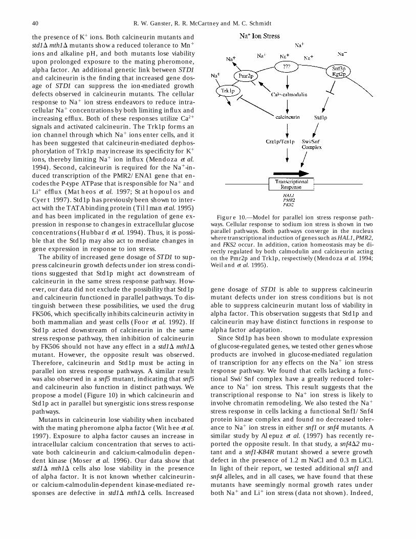
ions, thereby limiting Na1 ion influx (Mendoza et al.1994). Second, calcineurin is required for the Na1-in-duced transcription of the PMR2/ENA1 gene that en-codes the P-type ATPase that is responsible for Na1 andLi1 efflux (Matheos et al. 1997; Stathopoulos andCyert 1997). Std1p has previously been shown to inter-act with the TATA binding protein (Tillman et al. 1995)and has been implicated in the regulation of gene ex- Figure 10.—Model for parallel ion stress response path-
ways. Cellular response to sodium ion stress is shown in twopression in response to changes in extracellular glucoseparallel pathways. Both pathways converge in the nucleusconcentrations (Hubbard et al. 1994). Thus, it is possi-where transcriptional induction of genes such as HAL1, PMR2,ble that the Std1p may also act to mediate changes inand FKS2 occur. In addition, cation homeostasis may be di-
gene expression in response to ion stress. rectly regulated by both calmodulin and calcineurin actingThe ability of increased gene dosage of STD1 to sup- on the Pmr2p and Trk1p, respectively (Mendoza et al. 1994;
Weiland et al. 1995).press calcineurin growth defects under ion stress condi-tions suggested that Std1p might act downstream ofcalcineurin in the same stress response pathway. How-
gene dosage of STD1 is able to suppress calcineurinever, our data did not exclude the possibility that Std1pmutant defects under ion stress conditions but is notand calcineurin functioned in parallel pathways. To dis-able to suppress calcineurin mutant loss of viability intinguish between these possibilities, we used the drugalpha factor. This observation suggests that Std1p andFK506, which specifically inhibits calcineurin activity incalcineurin may have distinct functions in response toboth mammalian and yeast cells (Foor et al. 1992). Ifalpha factor adaptation.Std1p acted downstream of calcineurin in the same
Since Std1p has been shown to modulate expressionstress response pathway, then inhibition of calcineurinof glucose-regulated genes, we tested other genes whoseby FK506 should not have any effect in a std1D mth1Dproducts are involved in glucose-mediated regulationmutant. However, the opposite result was observed.of transcription for any effects on the Na1 ion stressTherefore, calcineurin and Std1p must be acting inresponse pathway. We found that cells lacking a func-parallel ion stress response pathways. A similar resulttional Swi/Snf complex have a greatly reduced toler-was also observed in a snf5 mutant, indicating that snf5ance to Na1 ion stress. This result suggests that theand calcineurin also function in distinct pathways. Wetranscriptional response to Na1 ion stress is likely topropose a model (Figure 10) in which calcineurin andinvolve chromatin remodeling. We also tested the Na1Std1p act in parallel but synergistic ions stress responsestress response in cells lacking a functional Snf1/Snf4pathways.protein kinase complex and found no decreased toler-Mutants in calcineurin lose viability when incubatedance to Na1 ion stress in either snf1 or snf4 mutants. Awith the mating pheromone alpha factor (Withee et al.similar study by Alepuz et al. (1997) has recently re-1997). Exposure to alpha factor causes an increase inported the opposite result. In that study, a snf4D2 mu-intracellular calcium concentration that serves to acti-tant and a snf1-K84R mutant showed a severe growthvate both calcineurin and calcium-calmodulin depen-defect in the presence of 1.2 m NaCl and 0.3 m LiCl.dent kinase (Moser et al. 1996). Our data show thatIn light of their report, we tested additional snf1 andstd1D mth1D cells also lose viability in the presencesnf4 alleles, and in all cases, we have found that theseof alpha factor. It is not known whether calcineurin-mutants have seemingly normal growth rates underor calcium-calmodulin-dependent kinase-mediated re-
sponses are defective in std1D mth1D cells. Increased both Na1 and Li1 ion stress (data not shown). Indeed,
41Sodium Stress Response in S. cerevisiae
we have tested the snf4D2 allele used by Alepuz et al. under conditions of ion stress. Further analysis of Snf3pand Rgt2p will be needed to determine if they play a(1997) and find no defect in Na1 ions stress response
(data not shown). It is difficult to reconcile these contra- direct role as sensors of ion stress.Increased gene dosage of STD1 confers a growth ad-dictory findings other than to attribute them to strain
differences. For instance, one important difference be- vantage under Na1 ion stress conditions. Since in-creased levels of Std1p have been shown to induce ex-tween our strains may be in the copy number or expres-
sion of the PMR2/ENA1 gene since this gene is found pression of the SUC2 gene (Hubbard et al. 1994;Tillman et al. 1995), we looked for halotolerance genesin a repeated gene cluster (Weiland et al. 1995). Also,
it is worth noting that neither strain used by Alepuz et that might be subject to Std1p regulation. Analysis ofglobal patterns of gene expression found that the HAL1al. (1997) contained null alleles of the snf1 or snf4 genes,
and perhaps some form of dominant negative inhibition gene was the only known halotolerance gene whoseexpression was increased more than twofold by the dele-could account for the Na1 ion stress defect observed in
their study. tion of TUP1 (DeRisi et al. 1997), a known repressor ofSUC2 expression. We examined the expression of HAL1Yeast encode 20 genes whose products are members
of the 12-transmembrane hexose transporter superfam- mRNA by Northern blot and found that its accumula-tion was regulated strongly by glucose concentrationily (Kruckeberg 1996). Two of these, the products of
the SNF3 and RGT2 genes, are structurally distinct due and by STD1 gene dosage. Increased gene dosage ofHAL1 has been shown to induce expression of PMR2/to the presence of a C-terminal hydrophilic domain.
Snf3p and Rgt2p are thought to function not as hexose ENA1, the Na1 efflux pump, and also to suppress the ionstress growth defects observed in calcineurin mutantstransporters but as glucose sensors (Ozcan et al. 1996).(Rios et al. 1997). We also tested the effect of STD1We show here that mutations in either of these genesgene dosage on PMR2 expression and found that Std1pactually enhance the Na1 stress response. We interpretis required for the full induction of PMR2 in responsethese results to mean that the Snf3p and Rgt2p areto Na stress. The Na response of the PMR2 gene is thenegative regulators of the Na1 stress response and theirresult of the additive effects of the calcineurin pathway,loss of function serves to increase expression of Na1
the Std1 pathway, and at least one additional pathway.stress response genes. Additional studies not shown hereThe high osmotic glycerol (HOG) MAP kinase pathwayindicate that the glucose-repressed gene SUC2 is dere-is also known to regulate expression of the PMR2 genepressed in the absence of SNF3 and RGT2 (X. Zhang
and most likely accounts for the calcineurin- and Std1-and M. C. Schmidt, unpublished results). We hypothe-independent Na response of the PMR2 gene (Marquezsize that loss of a glucose sensor likewise causes a dere-and Serrano 1996). We also show here that increasedpression of the Na1 stress response genes that are undergene dosage of STD1 induces both HAL1 and PMR2glucose control and thereby enhances the Na1 stressexpression, suggesting a possible mechanism for theresponse of the cells. In addition, we found that thehalotolerance and the suppression of calcineurin mu-snf3 rgt2 mutant is sensitive to high concentrations oftants that is conferred by 2m STD1. Our studies indicateK1. This result may stem from the fact that one of thethat yeast cells have developed overlapping but distinctcell’s Na1 stress responses, to increase intracellular K1
stress response pathways for coping with changes in(Gaxiola et al. 1992), may be deregulated in the snf3both cation and carbon source concentrations.rgt2 mutant. Cells that constitutively act to increase intra-
cellular K1 may find this maladaptive when presented We are grateful to Martha Cyert, Marian Carlson, and Fred
Winston for the gift of plasmids and strains, to Raman Venkatarama-with high concentrations of extracellular K1.nan for the gift of FK506, and to Kazi Islam for the synthesis ofCertain members of this 12-transmembrane hexosealpha factor. This work was supported by grant GM-46443 from thetransporter superfamily are ion/sugar symporters (Bald- National Institutes of Health.
win 1990). Although there is no evidence that the yeasthexose transporters couple ion and sugar transport,single amino acid mutations in Hxt1p and Hxt3p can LITERATURE CITEDconvert these proteins to cation transporters (Liang et
Albertyn, J., S. Hohmann, J. M. Thevelein and B. A. Prior, 1994al. 1998). We show here that mutations in the glucose GPD1, which encodes glycerol-3-phosphate dehydrogenase, is es-sensors confer a growth advantage under conditions of sential for growth under osmotic stress in Saccharomyces cerevisiae,
and its expression is regulated by the high-osmolarity glycerolNa1 ions stress and a growth disadvantage under con-response pathway. Mol. Cell. Biol. 14: 4135–4144.ditions of K1 ion stress. These effects are completely
Alepuz, P. M., K. W. Cunningham and F. Estruch, 1997 Glucoseindependent of glucose concentration, which was kept repression affects ion homeostasis in yeast through the regulation
of the stress-activated ENA1 gene. Mol. Microbiol. 26: 91–98.at 2% in these studies. One possible explanation forAperia, A., F. Ibarra, L. B. Svensson, C. Klee and P. Greengard,these data would be a model in which Snf3p and Rgt2p 1992 Calcineurin mediates a-adrenergic stimulation of Na1,
play dual roles as both glucose and cation sensors. The K1-ATPase activity in renal tubule cells. Proc. Natl. Acad. Sci.USA 89: 7394–7397.mechanism by which Na1 ion stress induces a calcium
Baldwin, S. A., 1990 Molecular mechanisms of sugar transportsignal in yeast cells is not known. Nor is it known by what across mammalian and microbial cell membranes. Biotechnol.Appl. Biochem. 12: 512–516.mechanism Std1p-mediated gene regulation is induced
42 R. W. Ganster, R. R. McCartney and M. C. Schmidt
Brewster, J. L., T. deValoir, N. D. Dwyer, E. Winter and M. C. ways regulate the sodium-extrusion gene PMR2/ENA1 during saltstress in yeast. FEBS Lett. 382: 89–92.Gustin, 1993 An osmosensing signal transduction pathway in
Matheos, D. P., T. J. Kingsbury, U. S. Ahsan and K. W. Cunning-yeast. Science 259: 1760–1763.ham, 1997 Tcn1p/Crz1p, a calcineurin-dependent transcrip-Carlson, M. C., B. C. Osmond and D. Botstein, 1981 Mutants oftion factor that differentially regulates gene expression in Sacchar-yeast defective in sucrose utilization. Genetics 98: 25–40.omyces cerevisiae. Genes Dev. 11: 3445–3458.Cunningham, K. W., and G. R. Fink, 1996 Calcineurin inhibits
Mendoza, I., F. Rubio, A. Rodriguez-Navarro and J. M. Pardo,VCX1-dependent H1/Ca11 exchange and induces Ca111994 The protein phosphatase calcineurin is essential for NaClATPases in yeast. Mol. Cell. Biol. 16: 2226–2237.tolerance of Saccharomyces cerevisiae. J. Biol. Chem. 269: 8792–Cyert, M. S., and J. Thorner, 1992 Regulatory subunit (CNB1 gene8796.product) of yeast Ca1/calmodulin-dependent phosphoprotein
Moser, M. J., J. R. Geiser and T. N. Davis, 1996 Ca21-calmodulinphosphatases is required for adaptation to pheromone. Mol. Cell.promotes survival of pheromone-induced growth arrest by activa-Biol. 12: 3460–3469.tion of calcineurin and Ca21-calmodulin-dependent protein ki-
Cyert, M. S., R. Kunisawa, D. Kaim and J. Thorner, 1991 Yeastnase. Mol. Cell. Biol. 16: 4824–4831.has homologs (CNA1 and CNA2 gene products) of mammalian
Murguia, J. R., J. M. Belles and R. Serrano, 1995 A salt-sensitivecalcineurin, a calmodulin-regulated phosphoprotein phospha-39(29),59-bisphosphate nucleotidase involved in sulfate activation.tase. Proc. Natl. Acad. Sci. USA 88: 7376–7380.Science 267: 232–234.
DeRisi, J. L., V. R. Iyer and P. O. Brown, 1997 Exploring theMurguia, J. R., J. M. Belles and R. Serrano, 1996 The yeast HAL2metabolic and genetic control of gene expression on a genomic
nucleotidase is an in vivo target of salt toxicity. J. Biol. Chem.scale. Science 278: 680–686.271: 29029–29033.
Elledge, S. J., and R. W. Davis, 1988 A family of versatile centro-Nakamura, T., Y. Liu, D. Hirata, H. Namba, S. Harada et al., 1993meric vectors designed for use in the sectoring-shuffle mutagene-
Protein phosphatase type2B (calcineurin)-mediated, FK506-sen-sis assay in Saccharomyces cerevisiae. Gene 70: 303–312.sitive regulation of intracellular ions in yeast is an important
Ferrando, A., S. J. Kron, R. Gabino, G. R. Fink and R. Serrano,
determinant for adaptation to high salt stress conditions. EMBO1995 Regulation of cation transport in Saccharomyces cerevisiaeJ. 12: 4063–4071.by the salt tolerance gene HAL3. Mol. Cell. Biol. 15: 5470–5481.
Neigeborn, L., and M. Carlson, 1984 Genes affecting the regula-Foor, F., N. Morin, A. M. Dahl, N. Ramadan, G. Chrebet et al., tion of SUC2 gene expression by glucose repression in Saccharo-
1992 Calcineurin mediates inhibition by FK506 and cyclosporin myces cerevisiae. Genetics 108: 845–859.of recovery from alpha-factor arrest in yeast. Nature 360: 682–684.
Ozcan, S., J. Dover, A. G. Rosenwald, S. Woelfl and M. Johnston,
Ganster, R., W. Shen and M. C. Schmidt, 1993 Isolation of STD1, 1996 Two glucose transporters in S. cerevisiae are glucose sensorsa high-copy-number suppressor of a dominant negative mutation that generate a signal for induction of gene expression. Proc.in the yeastTATA-binding protein. Mol. Cell. Biol. 13: 3650–3659. Natl. Acad. Sci. USA 93: 12428–12432.
Garciadeblas, B., F. Rubio, F. J. Quintero, M. A. Banuelos, R.Posas, F., S. M. Wurgler-Murphy, T. Maeda, E. A. Witten, T. C.
Haro et al., 1993 Differential expression of two genes encodingThai et al., 1996 Yeast HOG1 MAP kinase cascade is regulated
isoforms of the ATPase involved in sodium efflux in Saccharomyces by a multistep phosphorelay mechanism in the SNL1-YPD1-SSK1cerevisiae. Mol. Gen. Genet. 236: 363–368. “two-component” osmosensor. Cell 86: 865–875.
Gaxiola, R., I. F. de Larrinoa, J. M. Villalba and R. Serrano,Pozos, T. C., I. Sekler and M. S. Cyert, 1996 The product of
1992 A novel and conserved salt-induced protein is an impor- HUM1, a novel yeast gene, is required for vacuolar Ca21/H1
tant determinant of salt tolerance in yeast. EMBO J. 11: 3157– exchange and is related to mammalian Na1/Ca21 exchangers.3164. Mol. Cell. Biol. 16: 3730–3741.
Gietz, R. D., R. H. Schiestl, A. R. Willems and R. A. Woods, 1995 Rao, A., C. Luo and P. G. Hogan, 1997 Transcription factors ofStudies on the transformation of intact yeast cells by the LiAc/ the NFAT family: regulation and function. Ann. Rev. Immunol.SS-DNA/PEG procedure. Yeast 11: 355–360. 15: 707–747.
Glaser, H. U., D. Thomas, R. Gaxiola, F. Montrichard, Y. Surdin- Rios, G., A. Ferrando and R. Serrano, 1997 Mechanisms of saltKerjan et al., 1993 Salt tolerance and methionine biosynthesis tolerance conferred by overexpression of the HAL1 gene in Sac-in Saccharomyces cerevisiae involve a putative phosphatase gene. charomyces cerevisiae. Yeast 13: 515–528.EMBO J. 12: 3105–3110. Rose, M., F. Winston and P. Hieter, 1990 Methods in Yeast Genetics.
Guerini, D., 1997 Calcineurin: not just a simple protein phospha- Cold Spring Harbor Laboratory Press, Cold Spring Harbor, NY.Schuller, C., J. L. Brewster, M. R. Alexander, M. C. Gustin andtase. Biochem. Biophys. Res. Commun. 235: 271–275.
H. Ruis, 1994 The HOG pathway controls osmotic regulationGuldener, U., S. Heck, T. Fiedler, J. Beinhauer and J. H. Hege-
of transcription via the stress response element (STRE) of themann, 1996 A new efficient gene disruption cassette for re-Saccharomyces cerevisiae CTT1 gene. EMBO J. 13: 4382–4389.peated use in budding yeast. Nucleic Acids Res. 24: 2519–2524.
Sikorski, R. S., and P. Hieter, 1989 A system of shuttle vectors andHill, J. E., A. M. Meyers, T. J. Koerner and A. Tzagoloff, 1986yeast host strains designed for efficient manipulation of DNA inYeast/E. coli shuttle vectors with multiple unique restriction sites.Saccharomyces cerevisiae. Genetics 122: 19–27.Yeast 2: 163–167.
Stathopoulos, A. M., and M. S. Cyert, 1997 Calcineurin actsHubbard, M. J., and C. B. Klee, 1989 Functional domain structurethrough the CRZ1/TCN1-encoded transcription factor to regu-of calcineurin A: mapping by limited proteolysis. Biochemistrylate gene expression in yeast. Genes Dev. 11: 3432–3444.28: 1868–1874.
Tillman, T. S., R. W. Ganster, R. Jiang, M. Carlson and M. C.Hubbard, E. J. A., R. Jiang and M. Carlson, 1994 Dosage-depen-
Schmidt, 1995 STD1 (MSN3) interacts directly with the TATA-dent modulation of glucose repression by MSN3 (STD1) in Sac-binding protein and modulates transcription of the SUC2 genecharomyces cerevisiae. Mol. Cell. Biol. 14: 1972–1978.of Saccharomyces cerevisiae. Nucleic Acids Res. 23: 3174–3180.
Johnston, M., and M. Carlson, 1992 Regulation of Carbon and Phos-Wach, A., 1996 PCR-synthesis of marker cassettes with long flankingphate Utilization. Cold Spring Harbor Laboratory Press, Cold
homology regions for gene disruptions in S. cerevisiae. Yeast 12:Spring Harbor, NY.259–265.
Kohrer, K., and H. Domdey, 1991 Preparation of high molecularWeiland, J., A. M. Nitsche, J. Strayle, H. Steiner and H. K. Ru-weight RNA. Methods Enzymol. 194: 398–404.
dolph, 1995 The PMR2 gene cluster encodes functionally dis-Kruckeberg, A. L., 1996 The hexose transporter family of Saccharo- tinct isoforms of a putative Na1 pump in the yeast plasma mem-
myces cerevisiae. Arch. Microbiol. 166: 283–292. brane. EMBO J. 14: 3870–3882.Laurent, B. C., M. A. Treitel and M. Carlson, 1990 The SNF5
Winston, F., C. Dollard and S. L. Ricupero-Hovasse, 1996 Con-protein of Saccharomyces cerevisiae is a glutamine- and proline- struction of a set of convenient Saccharomyces cerevisiae strains thatrich transcriptional activator that affects expression of a broad are isogenic to S288C. Yeast 11: 53–55.spectrum of genes. Mol. Cell. Biol. 10: 5616–5625.
Withee, J. L., J. Mulholland, R. Jeng and M. S. Cyert, 1997 AnLiang, H., C. H. Ko and R. F. Gaber, 1998 Trinucleotide insertions, essential role of the yeast pheromone-induced Ca1 signal is to
deletions and point mutations in glucose transporters confer K1 activate calcineurin. Mol. Biol. Cell. 8: 263–277.uptake in Saccharomyces cerevisiae. Mol. Cell. Biol. 18: 926–935.
Marquez, J. A., and R. Serrano. 1996 Multiple transduction path- Communicating editor: M. Carlson