Image Guided Radiation Therapy and Stereotactic Body Radiation Therapy for Lung Cancer Kenneth Rosenzweig, MD Department of Radiation Oncology Mount Sinai School of Medicine September 20, 2010
Transcript
Image Guided Radiation Therapy and Stereotactic Body
Radiation Therapy for Lung Cancer
Kenneth Rosenzweig, MDDepartment of Radiation OncologyMount Sinai School of MedicineSeptember 20, 2010
•• 6 patients (11%) disseminated within 1 year of Rx6 patients (11%) disseminated within 1 year of Rx
Copyright restrictions may apply.
Timmerman, R. et al. JAMA 2010;303:1070-1076.
RTOG 0236: Patient Course After Initiation of Stereotactic Body Radiation Therapy
Copyright restrictions may apply.
Timmerman, R. et al. JAMA 2010;303:1070-1076.
RTOG 0236: Adverse Events Related to Stereotactic Body Radiation Therapy
Copyright restrictions may apply.
Protocol-Specified Adverse Events Related to Stereotactic Body Radiation Therapy
Rationale of High Dose per Fraction RT
• By radiobiologic principles, the higher dose per fraction, the greater the damage to the tumor (and normal structures)– Biologic equivalent dose (BED)
• BED = nd (1 + d/(α/β))• So assuming α/β
= 10, then 20 Gy x 3 is
equivalent to 180 Gy given in conventional fractionation
Results and BED
•Onishi, et al., J Thor Onc, 2007
Typical Verification Film
Techniques for IGRT Imaging
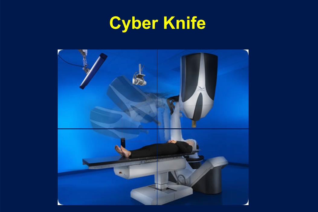
• Two dimensional imaging– Fluoroscopy-type imaging, Cyber Knife– Usually need fiducial marker (gold seed)
• Mega Voltage Cone Beam Imaging– Uses the treatment machine as a CT scan
• Kilo Voltage Cone Beam Imaging– Adds an extra machine to the treatment machine
that functions as a CT scanner
Varian kV Imaging system (OBI)• kV source, kV detector, and MV detector all mounted on
robotic arms
Cyber Knife
Technique for Lung SBRT• Simulation day
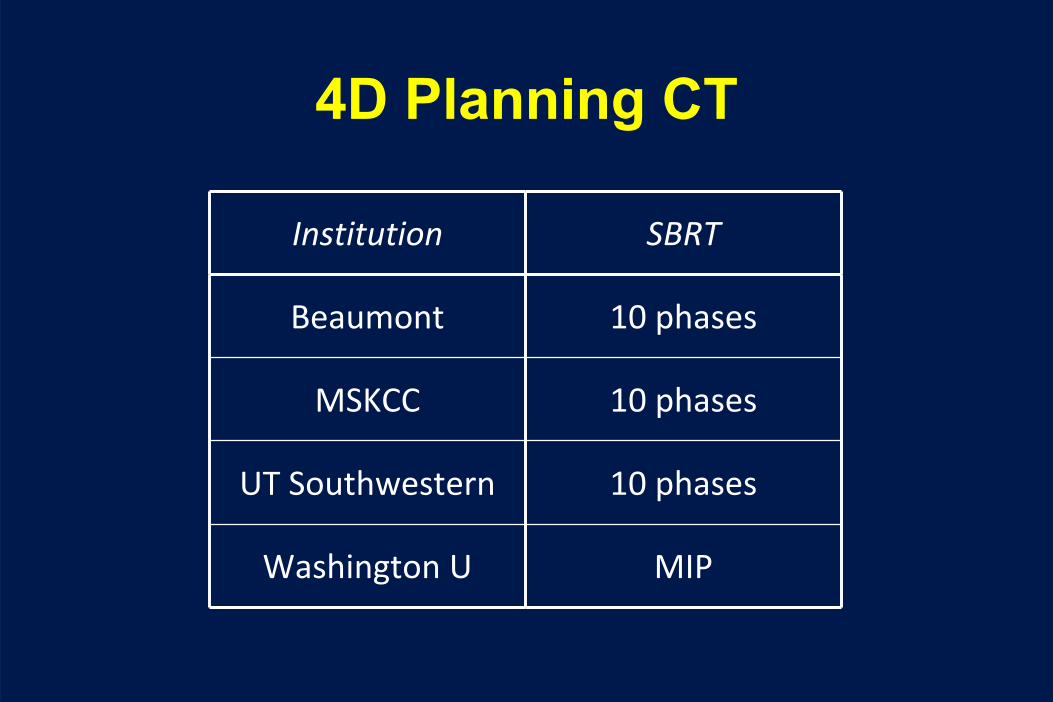
– Advanced patient immobilization– 4D Treatment planning CT– Consider PET scan for tumor delineation
– Accounts for tumor motion• CTV – clinical target volume
– Accounts for microscopic extension• PTV – planning target volume
– Accounts for set-up error, etc.
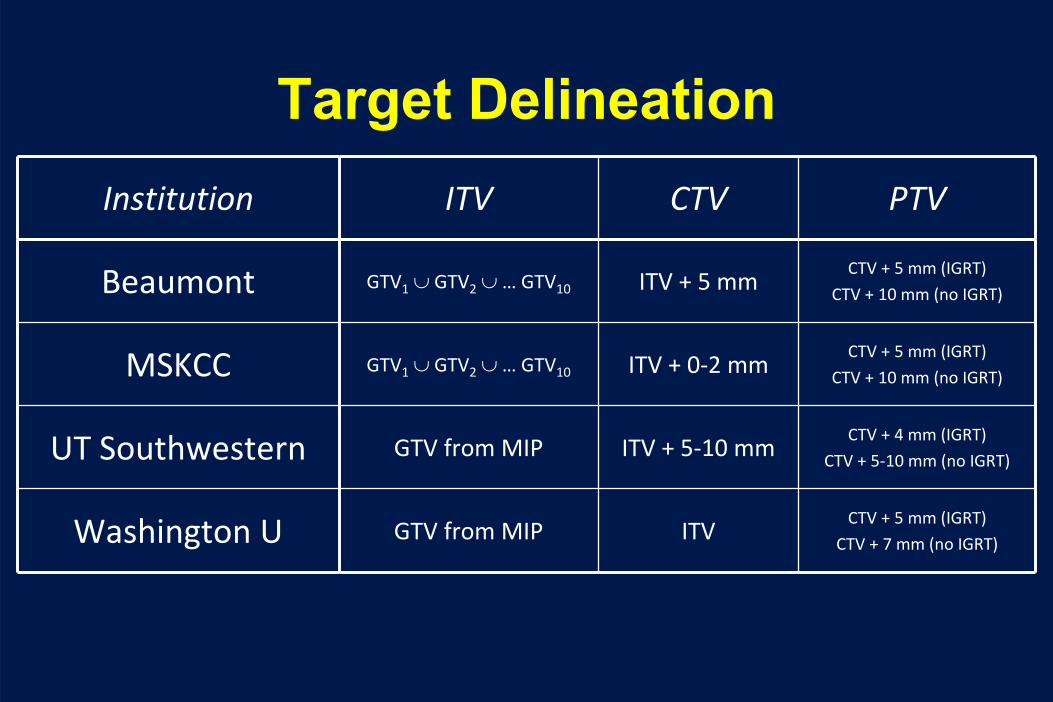
Target DelineationInstitution ITV CTV PTV
Beaumont GTV1
∪ GTV2
∪ … GTV10 ITV + 5 mmCTV + 5 mm (IGRT)
CTV + 10 mm (no IGRT)
MSKCC GTV1
∪ GTV2
∪ … GTV10 ITV + 0‐2 mmCTV + 5 mm (IGRT)
CTV + 10 mm (no IGRT)
UT Southwestern GTV from MIP ITV + 5‐10 mmCTV + 4 mm (IGRT)
CTV + 5‐10 mm (no IGRT)
Washington U GTV from MIP ITVCTV + 5 mm (IGRT)
CTV + 7 mm (no IGRT)
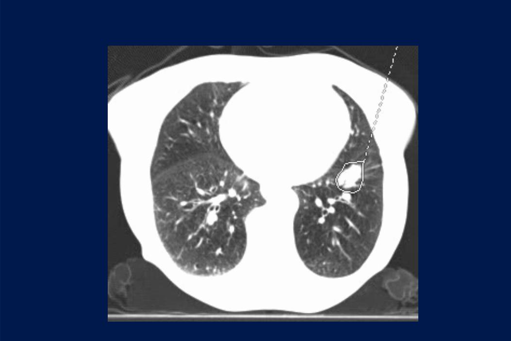
Determining the GTV
Determining the GTV
Determining the ITV
Determining the ITV
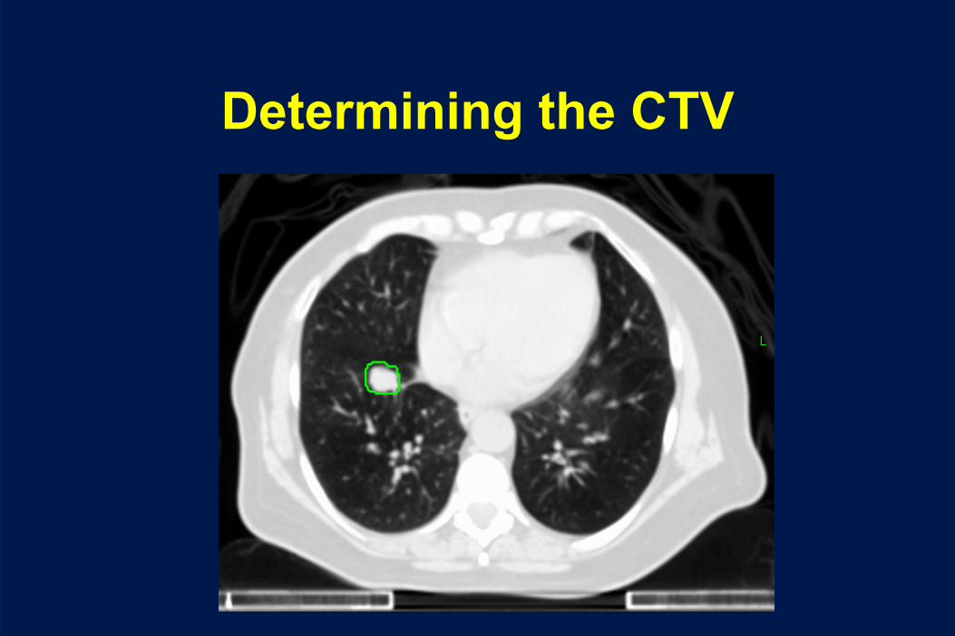
Determining the CTV
Determing the PTV
All Tumor Volumes
All Tumor Volumes
Normal Structure Constraints – SBRT
Institution Lungs Esophagus Spinal Cord
Beaumont4 fractions:
V20
≤
10%
V12.5
≤
15%
4 fractions:
Dmean
≤
30.5 Gy
4 fractions:
cord+3 mm Dmax
≤
20.5 Gy
MSKCC3 fractions:
both lungs V20
< 12%
ipsi lung V20
< 25%
3 fractions:
Dmax
≤
30 Gy
3 fractions:
Dmax
≤
24 Gy
UT Southwestern3 fractions:
D1000cc
< 12.4 Gy
D1500cc
< 11.6 GyV20
< 15%?
3 fractions:
Dmax
< 25.2 Gy
D5cc
< 17.7 Gy
3 fractions:
Dmax
< 21.9 Gy
D0.35cc
< 18.0 Gy
D1.2cc
< 12.3 Gy
Washington U3 fractions:
D1000cc
< 12.4 Gy
D1500cc
< 11.6 GyV20
< 15%?
3 fractions:
Dmax
≤
27 Gy
3 fractions:
Dmax
≤
18 Gy
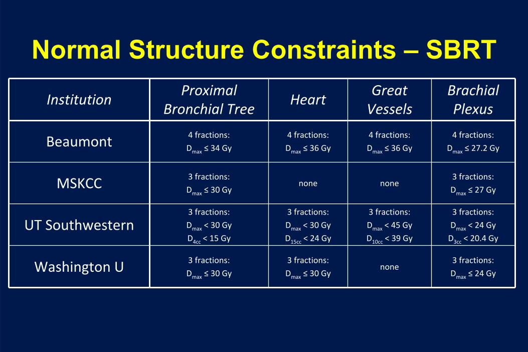
Normal Structure Constraints – SBRT
InstitutionProximal
Bronchial TreeHeart
Great
VesselsBrachial
Plexus
Beaumont 4 fractions:
Dmax
≤
34 Gy
4 fractions:
Dmax
≤
36 Gy
4 fractions:
Dmax
≤
36 Gy
4 fractions:
Dmax
≤
27.2 Gy
MSKCC 3 fractions:
Dmax
≤
30 Gynone none
3 fractions:
Dmax
≤
27 Gy
UT Southwestern3 fractions:
Dmax
< 30 Gy
D4cc
< 15 Gy
3 fractions:
Dmax
< 30 Gy
D15cc
< 24 Gy
3 fractions:
Dmax
< 45 Gy
D10cc
< 39 Gy
3 fractions:
Dmax
< 24 Gy
D3cc
< 20.4 Gy
Washington U 3 fractions:
Dmax
≤
30 Gy
3 fractions:
Dmax
≤
30 Gynone
3 fractions:
Dmax
≤
24 Gy
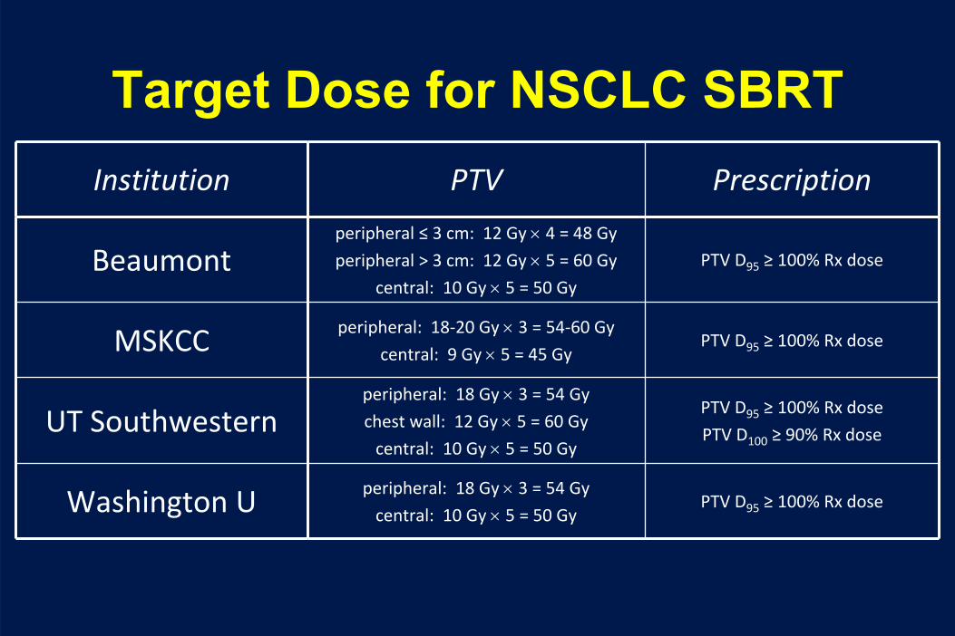
Target Dose for NSCLC SBRTInstitution PTV Prescription
Beaumontperipheral ≤
3 cm: 12 Gy ×
4 = 48 Gy
peripheral > 3 cm: 12 Gy ×
5 = 60 Gy
central: 10 Gy ×
5 = 50 Gy
PTV D95
≥
100% Rx dose
MSKCC peripheral: 18‐20 Gy ×
3 = 54‐60 Gy
central: 9 Gy ×
5 = 45 GyPTV D95
≥
100% Rx dose
UT Southwesternperipheral: 18 Gy ×
3 = 54 Gy
chest wall: 12 Gy ×
5 = 60 Gy
central: 10 Gy ×
5 = 50 Gy
PTV D95
≥
100% Rx dose
PTV D100
≥
90% Rx dose
Washington U peripheral: 18 Gy ×
3 = 54 Gy
central: 10 Gy ×
5 = 50 GyPTV D95
≥
100% Rx dose
Treatment Plan
Treatment Plan
Verifying Patient Position
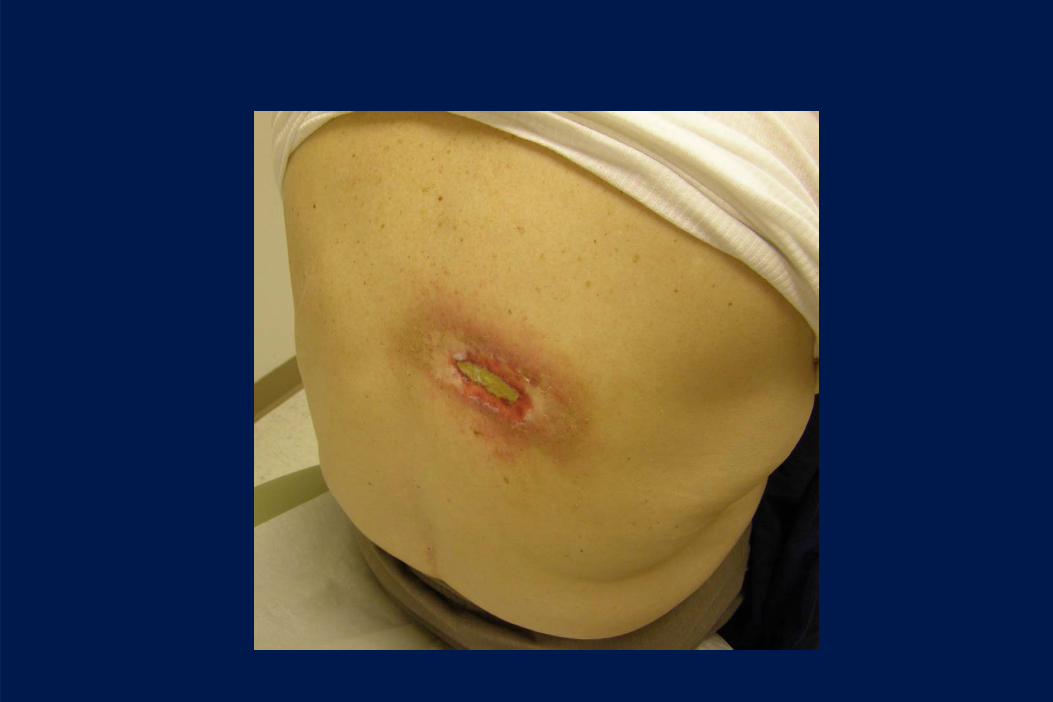
Toxicity of SBRT
• Fatigue• Skin reaction• Pneumonitis• Pain
Toxicity of Lung SBRT
• Timmerman, et al. JCO 2006
• 70 pts with Stage I NSCLC in a Phase II protocol• 20 Gy x 3 or 22 Gy x 3• Median overall survival 33 months, 2 yr OS 55%• 14 patients had Grade 3 to 5 toxicity
– 8 Grade 3/4 - ↓PFT’s, effusion, pneumonia– 6 toxic deaths – pneumonia, pericardial effusion, hemoptysis– Central tumors more likely to have toxicity
Limitations of SRS in the Lung “No Fly Zone”
Changes in Technique to Limit Skin Toxicity
• Use of Alpha cradle to allow lateralized beams
• Use more than 3 beams to prevent overlap
• Evaluate skin as an organ at risk
0 months 3 months 6 months 15 months
Pre-treatment 3 month 6 month
Pre-treatment 3 month 6 month 9 month
12 month 16 month 20 month 24 month
Differences between centers
• Use of 3D-CRT or IMRT• Variable use of inhomogeneity corrections• Use of more beams (8 – 10 in many
protocols)• Image guidance not always used• Tumor motion not evaluated• Variability in tumor margins
Why do all techniques work?
• The use of multiple beams and high doses is causing a “haze” of moderate dose radiation (~15 Gy per fraction) that is adequate to kill subclinical disease and account for tumor motion
SBRT – Future Directions
• Standardize CTV, PTV, inhomogeneity corrections, tumor motion control
• Identify best dose– Might need to dose de-escalate
• Figure out how to treat central tumors– Some centers (VU, Wash U.) have been reporting safe
early experience with 7. 5 – 10 Gy per fraction– RTOG 0813 to address this (currently at 10.5 Gy/fx)
• Test head to head against surgery– Japanese are doing this
Future in Early Stage
• Current RTOG protocol in operable patients– RTOG 0618 closed May 2010
• Future research needs:– Longer term results– Better ways to assess response– ? Need for a randomized trial vs. standard RT
Future Directions in Toxicity
• For the first time there is a possibility for long- term follow-up in a lung cancer population treated with RT
• Allows for better analysis for the causes of second tumors, specific toxicities (lung fibrosis), etc.