
Image Guided Radiation Therapy and Stereotactic Body
Radiation Therapy for Lung Cancer
Kenneth Rosenzweig, MDDepartment of Radiation OncologyMount Sinai School of MedicineSeptember 20, 2010

Small Problems
• Early stage tumors

Options for Early Stage NSCLC
• Surgery– Wedge, Lobectomy, etc.
• Radiation Therapy– Conventional Radiation– Stereotactic Body Radiotherapy (SBRT)
• Radiofrequency Ablation
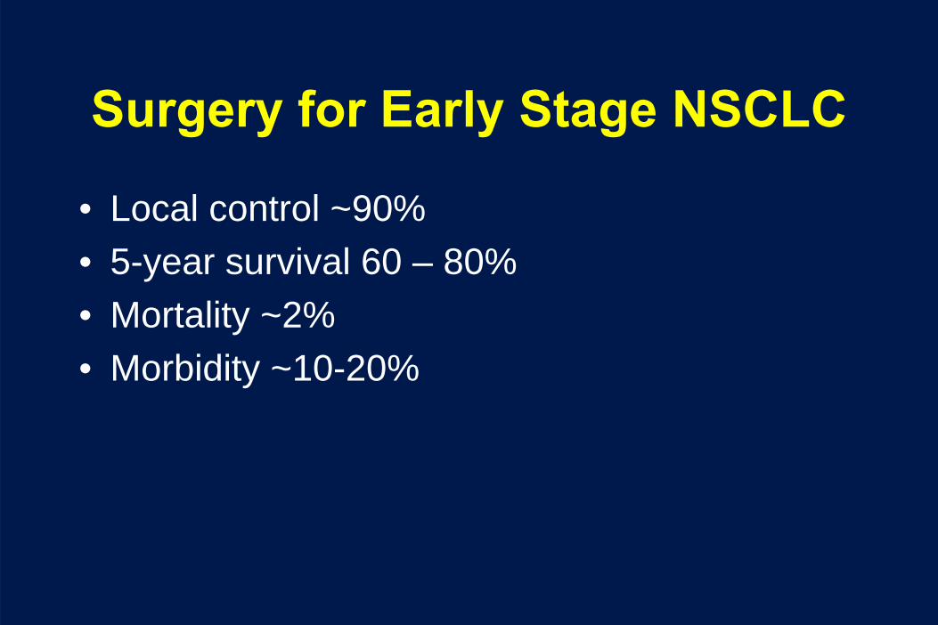
Surgery for Early Stage NSCLC
• Local control ~90%• 5-year survival 60 – 80%• Mortality ~2%• Morbidity ~10-20%

High Dose Conventional RT
No. patients
Median Survival (months)
5-year local control
5-year overall survival
Stage I/II 55 41 67% 36%
• 5% Grade 3+ acute pulmonary toxicity (2.5% grade 5)• 7% Grade 3+ late pulmonary toxicity
• 11% of long-term survivors on chronic oxygen

Lung SBRT Experience
• Onishi, Japan (Cancer October, 2004)
• Retrospective multi-institutional study• 273 patients with Stage I tumors• Dose was 18 – 75 Gy in 1 – 22 fractions
– BED ranged from 57 – 180 Gy• Complication rate 2.4%• Local failure in 12.5%
– Improved in good PS patients receiving > 100 Gy BED

# patients Median f/u (months)
Dose/fx Grade 3 toxicity
Local Control
Survival
Kyoto 45 30 4x12 Gy 0 94% 3 yr T1: 83%T2: 72%
Stanford 20 18 1x15-30 Gy 12.5% 92% 1 yr 85%
Aarhus, Denmark
40 29 3x15 Gy NA 85% 2 yr 48% 2 yr
Indiana 70 18 3x20-22 Gy 20% 95% 2 yr 55% 2 yr
Hedielberg 42 15 1x19-30 Gy NA 68% 3 yr 37% 3 yr
Tohuku 31 32 3x15 Gy8x7,5 Gy
3.2% T1: 78%T2: 40%
72% 3 yr
Karolinska (Sweden)
57* 23 3x15 Gy 21% 96% 65%
VU (Nether lands)
206* 12 3x20 Gy8x7.5 Gy
3% 93% 2 yr 64% 2yr
* not all biopsy proven

RTOG 0236

RTOG 0236 RT Specifications• No additional margin for microscopic
extension (i.e., no CTV)• PTV margin was:
– 5 mm axially– 10 mm craniocuadal
• 20 Gy x 3– 40 hours apart (max: 8 days)
• No tissue heterogeneity correction allowed– Later showed dose was closer to 18 Gy x 3

Copyright restrictions may apply.
Timmerman, R. et al. JAMA 2010;303:1070-1076.
Organ Tolerance Dose Limits for Radiation Therapy Oncology Group 0236

www.rtog.org 11
Loca
l Con
trol
(%
)
0
25
50
75
100
Months after Start of SBRT0 6 12 18 24 30 36
0
25
50
75
100
0 6 12 18 24 30 36Patientsat Risk 55 54 47 46 39 34 23
Fail: 1Total: 55
/ / / / / /// / / // / // / / / / / // / // //// //
Local ControlLocal Control•• 1 failure within PTV, 0 within 1 cm of PTV1 failure within PTV, 0 within 1 cm of PTV
36 monthlocal control = 98% (CI: 84-100%)

www.rtog.org 12
Dis
sem
inat
ed R
ecur
renc
e (%
)
0
25
50
75
100
Months after Start of SBRT0 6 12 18 24 30 36
0
25
50
75
100
0 6 12 18 24 30 36Patientsat Risk 55 51 44 43 38 33 21
Fail: 11Total: 55
// / // / / / / // / / / // / //
/// //
Disseminated RecurrenceDisseminated Recurrence
36 month disseminated recurrence = 22% (CI: 12-38%)
•• 6 patients (11%) disseminated within 1 year of Rx6 patients (11%) disseminated within 1 year of Rx

Copyright restrictions may apply.
Timmerman, R. et al. JAMA 2010;303:1070-1076.
RTOG 0236: Patient Course After Initiation of Stereotactic Body Radiation Therapy

Copyright restrictions may apply.
Timmerman, R. et al. JAMA 2010;303:1070-1076.
RTOG 0236: Adverse Events Related to Stereotactic Body Radiation Therapy

Copyright restrictions may apply.
Protocol-Specified Adverse Events Related to Stereotactic Body Radiation Therapy

Rationale of High Dose per Fraction RT
• By radiobiologic principles, the higher dose per fraction, the greater the damage to the tumor (and normal structures)– Biologic equivalent dose (BED)
• BED = nd (1 + d/(α/β))• So assuming α/β
= 10, then 20 Gy x 3 is
equivalent to 180 Gy given in conventional fractionation

Results and BED
•Onishi, et al., J Thor Onc, 2007

Typical Verification Film

Techniques for IGRT Imaging
• Two dimensional imaging– Fluoroscopy-type imaging, Cyber Knife– Usually need fiducial marker (gold seed)
• Mega Voltage Cone Beam Imaging– Uses the treatment machine as a CT scan
• Kilo Voltage Cone Beam Imaging– Adds an extra machine to the treatment machine
that functions as a CT scanner

Varian kV Imaging system (OBI)• kV source, kV detector, and MV detector all mounted on
robotic arms
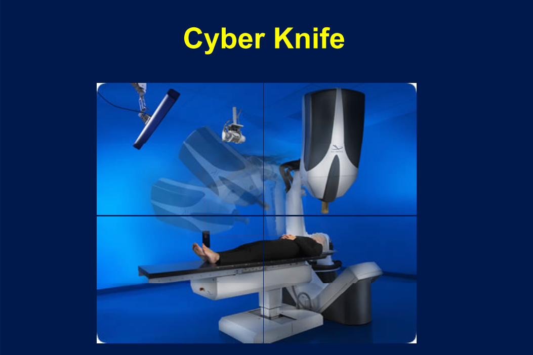
Cyber Knife

Technique for Lung SBRT• Simulation day
– Advanced patient immobilization– 4D Treatment planning CT– Consider PET scan for tumor delineation
• Treatment Planning– Five days
• Treatment day(s)– Advanced patient immobilization– Image guidance– Patient adjustment– Re-image– Treat

Immobilization
Institution SBRT
Beaumonthybrid α‐cradlewith BodyFix
MSKCC α‐cradle
UT Southwestern body frame
Washington U body frame or
BodyFix
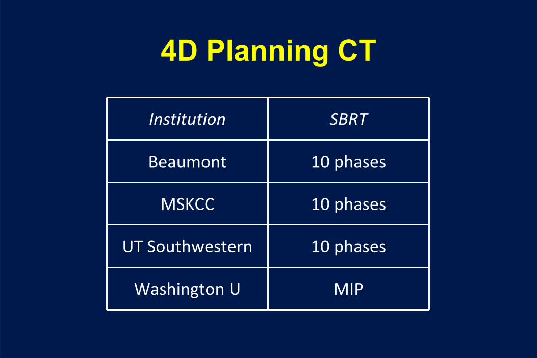
4D Planning CT
Institution SBRT
Beaumont 10 phases
MSKCC 10 phases
UT Southwestern 10 phases
Washington U MIP

PET Fusion
Institution SBRT
Beaumont 100%
MSKCC 0%
UT Southwestern sometimes
Washington U rarely

Determining Tumor Volumes
• GTV – gross tumor volume• ITV – internal target volume
– Accounts for tumor motion• CTV – clinical target volume
– Accounts for microscopic extension• PTV – planning target volume
– Accounts for set-up error, etc.
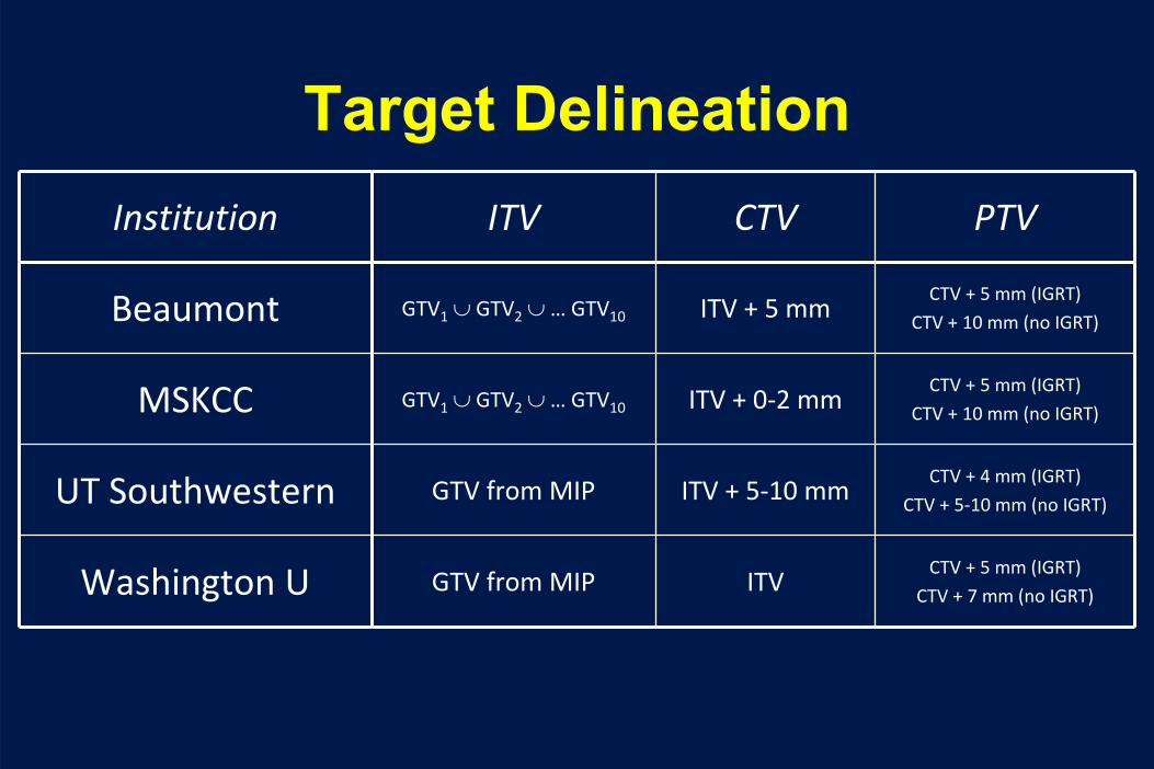
Target DelineationInstitution ITV CTV PTV
Beaumont GTV1
∪ GTV2
∪ … GTV10 ITV + 5 mmCTV + 5 mm (IGRT)
CTV + 10 mm (no IGRT)
MSKCC GTV1
∪ GTV2
∪ … GTV10 ITV + 0‐2 mmCTV + 5 mm (IGRT)
CTV + 10 mm (no IGRT)
UT Southwestern GTV from MIP ITV + 5‐10 mmCTV + 4 mm (IGRT)
CTV + 5‐10 mm (no IGRT)
Washington U GTV from MIP ITVCTV + 5 mm (IGRT)
CTV + 7 mm (no IGRT)

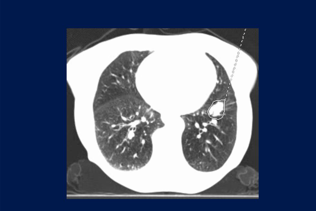
Determining the GTV

Determining the GTV


Determining the ITV

Determining the ITV
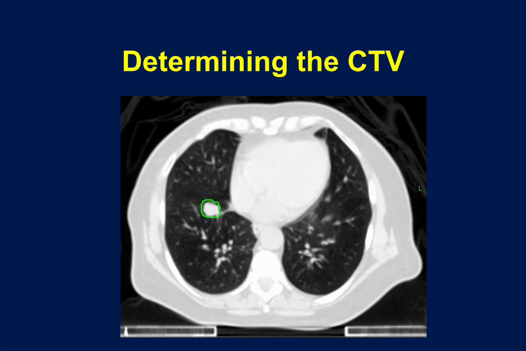
Determining the CTV

Determing the PTV

All Tumor Volumes

All Tumor Volumes

Normal Structure Constraints – SBRT
Institution Lungs Esophagus Spinal Cord
Beaumont4 fractions:
V20
≤
10%
V12.5
≤
15%
4 fractions:
Dmean
≤
30.5 Gy
4 fractions:
cord+3 mm Dmax
≤
20.5 Gy
MSKCC3 fractions:
both lungs V20
< 12%
ipsi lung V20
< 25%
3 fractions:
Dmax
≤
30 Gy
3 fractions:
Dmax
≤
24 Gy
UT Southwestern3 fractions:
D1000cc
< 12.4 Gy
D1500cc
< 11.6 GyV20
< 15%?
3 fractions:
Dmax
< 25.2 Gy
D5cc
< 17.7 Gy
3 fractions:
Dmax
< 21.9 Gy
D0.35cc
< 18.0 Gy
D1.2cc
< 12.3 Gy
Washington U3 fractions:
D1000cc
< 12.4 Gy
D1500cc
< 11.6 GyV20
< 15%?
3 fractions:
Dmax
≤
27 Gy
3 fractions:
Dmax
≤
18 Gy
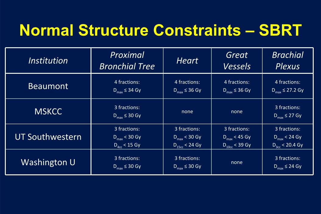
Normal Structure Constraints – SBRT
InstitutionProximal
Bronchial TreeHeart
Great
VesselsBrachial
Plexus
Beaumont 4 fractions:
Dmax
≤
34 Gy
4 fractions:
Dmax
≤
36 Gy
4 fractions:
Dmax
≤
36 Gy
4 fractions:
Dmax
≤
27.2 Gy
MSKCC 3 fractions:
Dmax
≤
30 Gynone none
3 fractions:
Dmax
≤
27 Gy
UT Southwestern3 fractions:
Dmax
< 30 Gy
D4cc
< 15 Gy
3 fractions:
Dmax
< 30 Gy
D15cc
< 24 Gy
3 fractions:
Dmax
< 45 Gy
D10cc
< 39 Gy
3 fractions:
Dmax
< 24 Gy
D3cc
< 20.4 Gy
Washington U 3 fractions:
Dmax
≤
30 Gy
3 fractions:
Dmax
≤
30 Gynone
3 fractions:
Dmax
≤
24 Gy
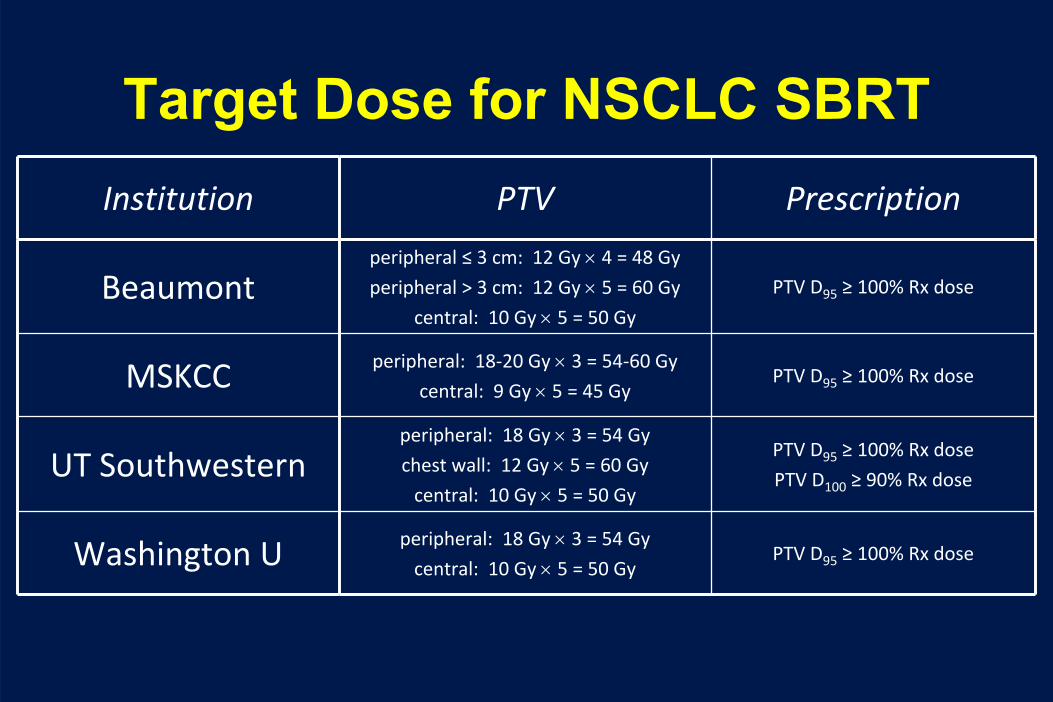
Target Dose for NSCLC SBRTInstitution PTV Prescription
Beaumontperipheral ≤
3 cm: 12 Gy ×
4 = 48 Gy
peripheral > 3 cm: 12 Gy ×
5 = 60 Gy
central: 10 Gy ×
5 = 50 Gy
PTV D95
≥
100% Rx dose
MSKCC peripheral: 18‐20 Gy ×
3 = 54‐60 Gy
central: 9 Gy ×
5 = 45 GyPTV D95
≥
100% Rx dose
UT Southwesternperipheral: 18 Gy ×
3 = 54 Gy
chest wall: 12 Gy ×
5 = 60 Gy
central: 10 Gy ×
5 = 50 Gy
PTV D95
≥
100% Rx dose
PTV D100
≥
90% Rx dose
Washington U peripheral: 18 Gy ×
3 = 54 Gy
central: 10 Gy ×
5 = 50 GyPTV D95
≥
100% Rx dose

Treatment Plan

Treatment Plan

Verifying Patient Position

Toxicity of SBRT
• Fatigue• Skin reaction• Pneumonitis• Pain

Toxicity of Lung SBRT
• Timmerman, et al. JCO 2006
• 70 pts with Stage I NSCLC in a Phase II protocol• 20 Gy x 3 or 22 Gy x 3• Median overall survival 33 months, 2 yr OS 55%• 14 patients had Grade 3 to 5 toxicity
– 8 Grade 3/4 - ↓PFT’s, effusion, pneumonia– 6 toxic deaths – pneumonia, pericardial effusion, hemoptysis– Central tumors more likely to have toxicity

Limitations of SRS in the Lung “No Fly Zone”


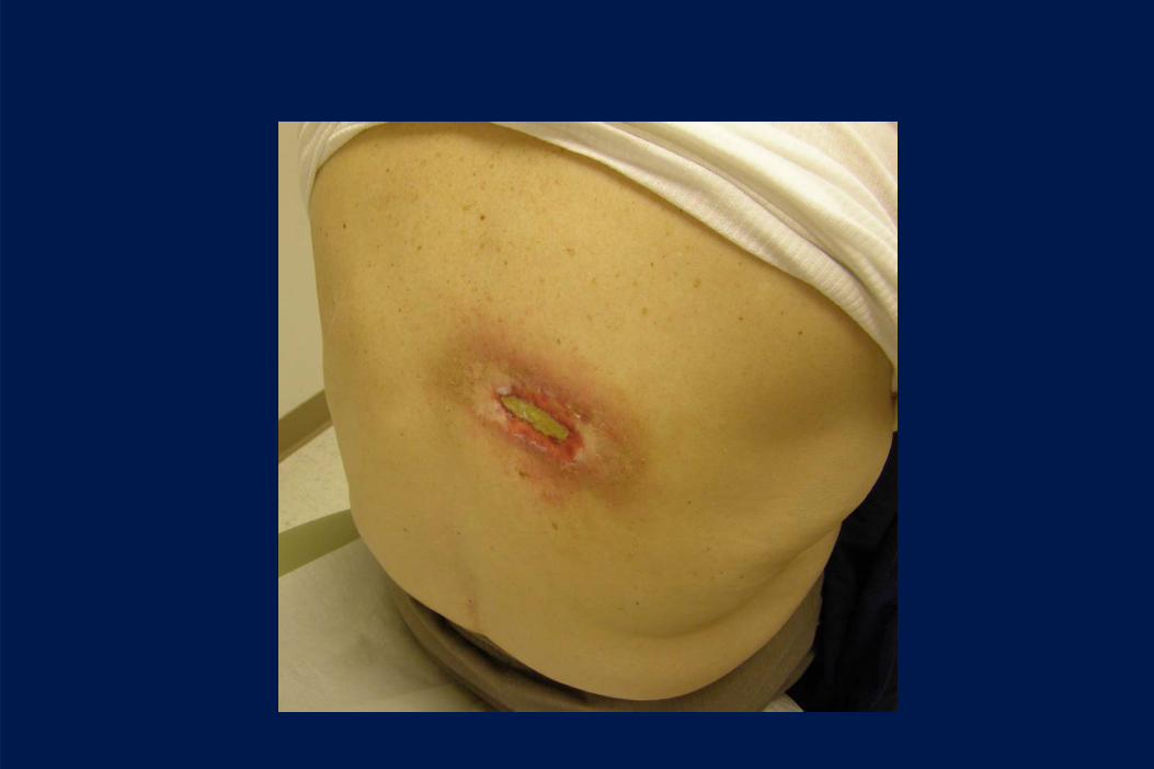


Changes in Technique to Limit Skin Toxicity
• Use of Alpha cradle to allow lateralized beams
• Use more than 3 beams to prevent overlap
• Evaluate skin as an organ at risk

0 months 3 months 6 months 15 months

Pre-treatment 3 month 6 month

Pre-treatment 3 month 6 month 9 month
12 month 16 month 20 month 24 month

Differences between centers
• Use of 3D-CRT or IMRT• Variable use of inhomogeneity corrections• Use of more beams (8 – 10 in many
protocols)• Image guidance not always used• Tumor motion not evaluated• Variability in tumor margins
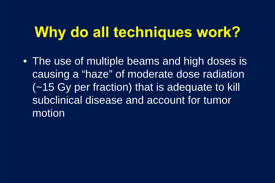
Why do all techniques work?
• The use of multiple beams and high doses is causing a “haze” of moderate dose radiation (~15 Gy per fraction) that is adequate to kill subclinical disease and account for tumor motion

SBRT – Future Directions
• Standardize CTV, PTV, inhomogeneity corrections, tumor motion control
• Identify best dose– Might need to dose de-escalate
• Figure out how to treat central tumors– Some centers (VU, Wash U.) have been reporting safe
early experience with 7. 5 – 10 Gy per fraction– RTOG 0813 to address this (currently at 10.5 Gy/fx)
• Test head to head against surgery– Japanese are doing this

Future in Early Stage
• Current RTOG protocol in operable patients– RTOG 0618 closed May 2010
• Future research needs:– Longer term results– Better ways to assess response– ? Need for a randomized trial vs. standard RT

Future Directions in Toxicity
• For the first time there is a possibility for long- term follow-up in a lung cancer population treated with RT
• Allows for better analysis for the causes of second tumors, specific toxicities (lung fibrosis), etc.


