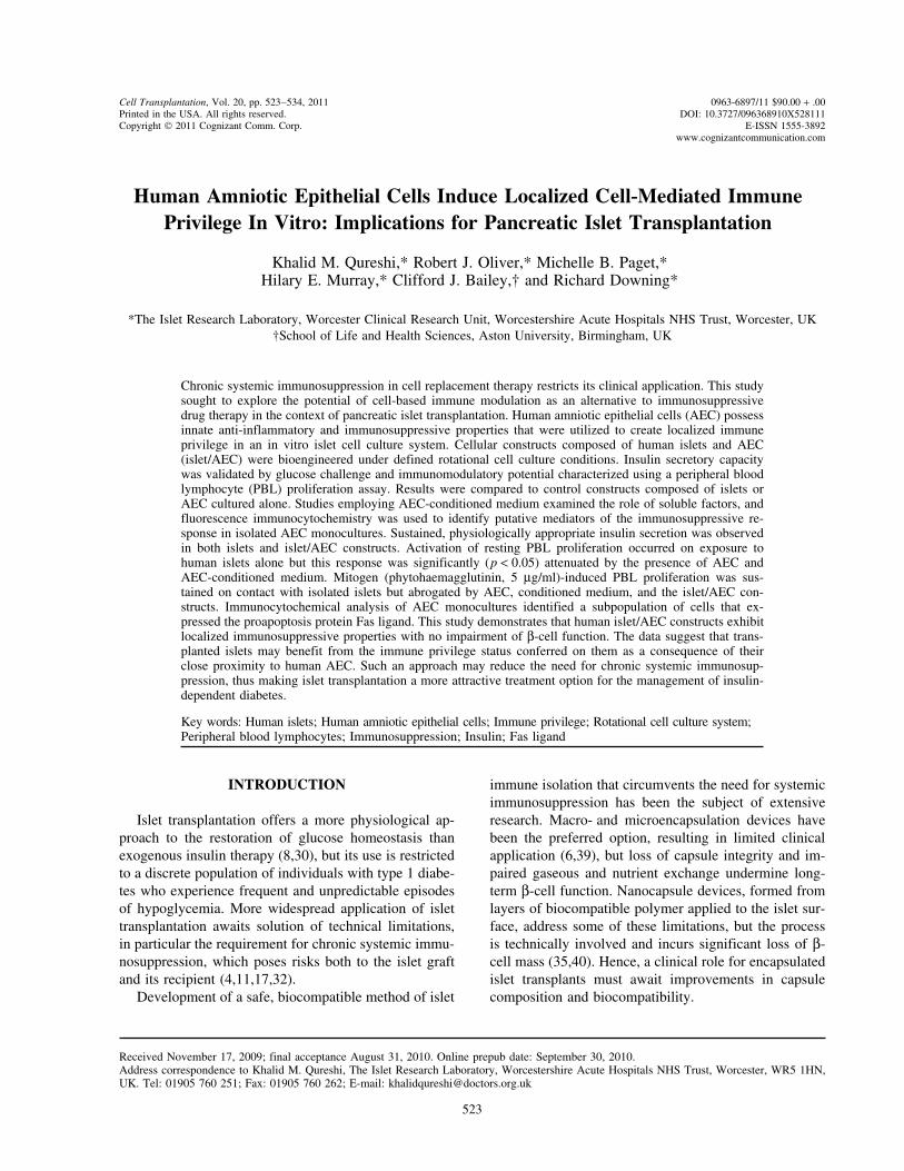
IMMUNE MODULATION WITH AMNIOTIC EPITHELIAL CELLS IN PANCREATIC ISLET TRANSPLANTATION by KHALID QURESHI A thesis submitted to the University of Birmingham for the degree of DOCTOR OF MEDICINE Department of Immunity and Infection University of Birmingham April 2012
Transcript
IMMUNE MODULATION WITH AMNIOTIC EPITHELIAL CELLS IN PANCREATIC ISLET
TRANSPLANTATION
by
KHALID QURESHI
A thesis submitted to the
University of Birmingham
for the degree of
DOCTOR OF MEDICINE
Department of Immunity and Infection
University of Birmingham
April 2012
University of Birmingham Research Archive
e-theses repository This unpublished thesis/dissertation is copyright of the author and/or third parties. The intellectual property rights of the author or third parties in respect of this work are as defined by The Copyright Designs and Patents Act 1988 or as modified by any successor legislation. Any use made of information contained in this thesis/dissertation must be in accordance with that legislation and must be properly acknowledged. Further distribution or reproduction in any format is prohibited without the permission of the copyright holder.
Abstract
Chronic systemic immunosuppression in pancreatic islet transplantation restricts its clinical
application. This study aims to explore the potential of cell-mediated immune-modulation as
an alternative to conventional immunosuppressive regimens; specifically investigating the
innate immunosuppressive properties of human amniotic epithelial cells (AEC).
Cell constructs composed of human islets and AEC (islet:AEC) were bio-engineered in
rotational culture. Insulin secretory capacity and immuno-modulatory potential were
characterised using appropriate in vitro assays. Fluorescence immunocytochemistry and
multiplex arrays was used to identify putative mediators of the immunosuppressive
response in isolated AEC monocultures.
Islets and islet:AEC constructs demonstrated sustained, physiologically-appropriate insulin
secretion. Resting peripheral blood mononuclear cells (PBMC) were activated on exposure to
human islets but this response was significantly (p<0.05) attenuated in islet:AEC constructs.
Phytohaemagglutinin (5g/ml)-induced PBMC proliferation was sustained on contact with
unmodified islets but abrogated in AEC and islet:AEC constructs. CD4+ and CD8+ T-cell
proliferation was responsive to AEC; their in vitro expansion both in response to CD3/CD28
activation and contact with human islets being suppressed by the presence of AEC.
Transplanted islets may thus benefit from an immune-privilege status conferred on them as
a consequence of their close proximity to human AEC. Such an approach may diminish the
requirement for generalised systemic immunosuppression in islet transplantation.
3.4. Results I ...................................................................................................................................... 77
3.4.1. Morphological characteristics of isolated AEC .................................................................... 77
4.3. Results I .................................................................................................................................... 102
4.3.1. Morphological analysis of islet:AEC constructs ................................................................. 102
4.4. Results II ................................................................................................................................... 108
4.4.1. Analysis of insulin secretory capacity of islet:AEC constructs ........................................... 108
4.4.2. Analysis of immunomodulatory potential of the islet:AEC constructs ............................. 110
and 10g/ml amphotericin B. Islets were cultured for 24 hours at 30°C in a humidified
atmosphere of 95%O2/5% CO2 to allow recovery and acclimatisation prior to functional
assessment as described below.
2.2.5. Functional Assessment
Islet functional viability was determined by assessing glucose-stimulated insulin secretion
(GSIS) 24 hours post-isolation. Static challenge studies were performed as follows:
Approximately 1000 IEQ were aspirated from the culture dish using a sterile pastette, placed
into a 15ml conical tube and centrifuged at 400g for 3 mins. The supernatant was discarded
and the resultant pellet re-suspended in a 2ml volume of 1.67mM glucose solution made up
in HBSS+0.2%BSA (basal glucose). Islets were then placed in a water bath at 37⁰C for 1 hour
(pre-incubation period). During this hour, an islet count as described above was performed
to ascertain the extent of -cell loss as a consequence of overnight culture. If necessary the
islet suspension was readjusted (by the addition of the 1.67mM solution) to a density of
400IEQ/ml and 50l aliquots (equivalent to 20 IEQ) and transferred to 12mm x 75mm
polypropylene tubes (NHS Logistics, Alfreton, UK). A total of 18 tubes were thus prepared, to
which 2ml of the appropriate secretagogue, diluted in HBSS+0.2% BSA, was added as
follows: 6 tubes received 1.67mM glucose solution to assess basal insulin secretion. To a
51
further 6 tubes a 16.7mM solution of glucose was added to determine stimulated insulin
secretion. The remaining tubes received 2ml of 16.7mM glucose supplemented with 10mM
theophylline; a potentiator of insulin secretion. The racked tubes were sealed with parafilm
and placed into the water bath at 37⁰C for 1 hour to allow insulin secretion (incubation
period). Following incubation the tubes were gently vortexed and then centrifuged at 400g
for 5 mins. The resultant supernatant was harvested for analysis of insulin content using an
enzyme-linked immunosorbent assay (ELISA; Mercodia, Diagenics UK), according to the
manufacturer’s instructions. Assessment of glucose-induced insulin secretion is a standard
method for estimating the functional viability of islets in the early post-isolation period and
is presented as the stimulation index (S.I; fold increase in insulin release in response to a
known secretagogue compared to basal secretion).
2.2.6. Statistical analysis
Statistical differences in response to insulin secretagogues were assessed by one way
analysis of variance (ANOVA) using insulin secretion under basal conditions as the control. In
all comparisons a p value of <0.05 was considered to be statistically significant. Statistical
analysis was performed using SigmaStat software version 3.5 (Systat Software Inc, Chicago,
USA).
52
2.3. Results
A total of seven human pancreata (five female and 2 male donors with a mean age of
47.14±3.35 years) were procured and successfully processed to obtain the islets used in the
present study. The mean cold ischemia time was 10.04±1.25 hours. An average of 70g of
pancreatic tissue was processed yielding a mean of approx 111,000 IEQ post-purification
(Table 2). In all instances sufficient numbers of viable islets were isolated for research
purposes.
2.3.1. Morphological characteristics of isolated islet preparation
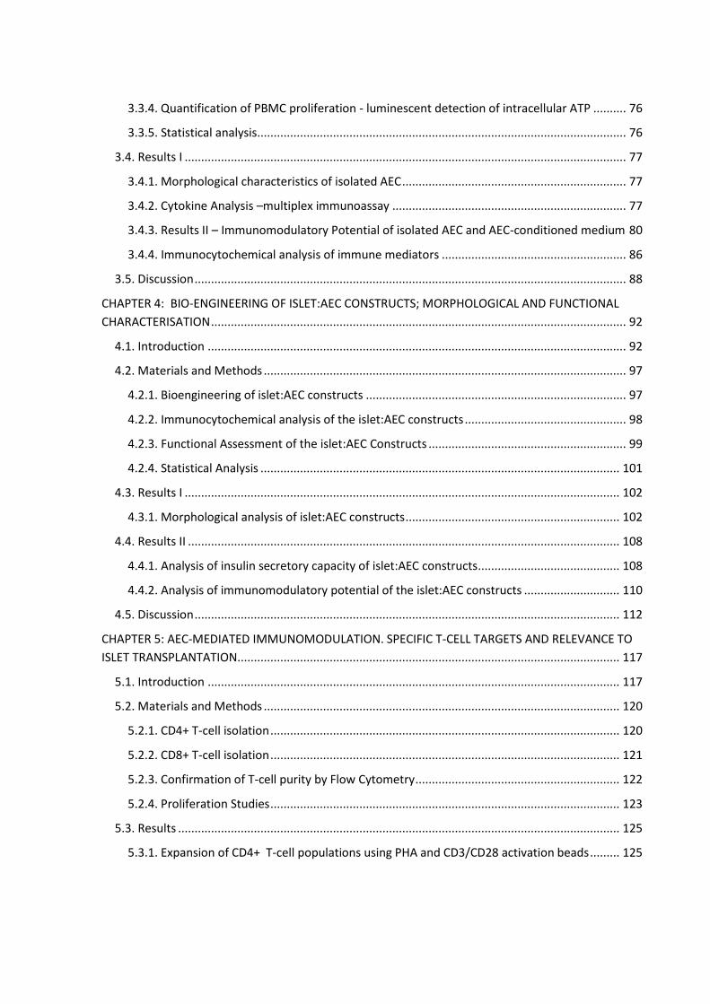
The human islet isolation protocol employed in the present study resulted in the harvest of
structurally intact islets which were well cleaved from the surrounding exocrine tissue, as
previously reported (Murray et al., 2009, Murray et al., 2005) (Fig.1). The purity of the islet
suspension following Ficoll gradient -assisted separation ranged from 70-85%, with islets
mostly sized between 100-500m. Trypan Blue exclusion served as an indicator of preserved
islet structural integrity.
53
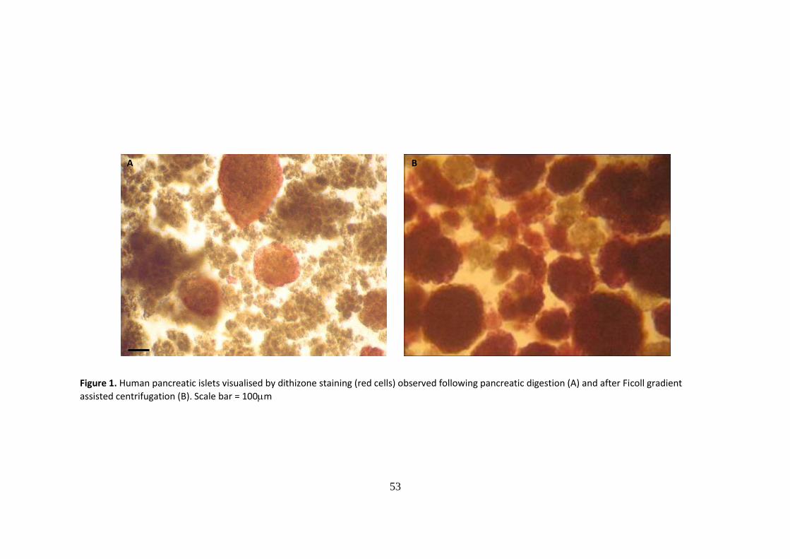
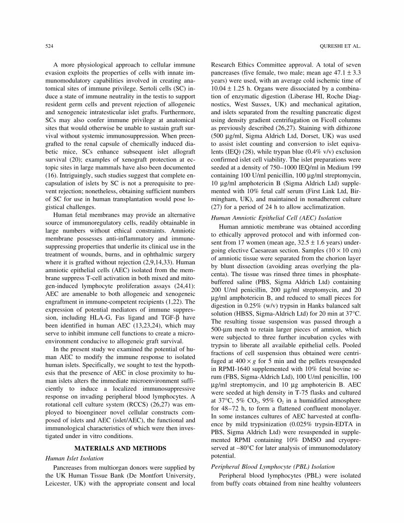
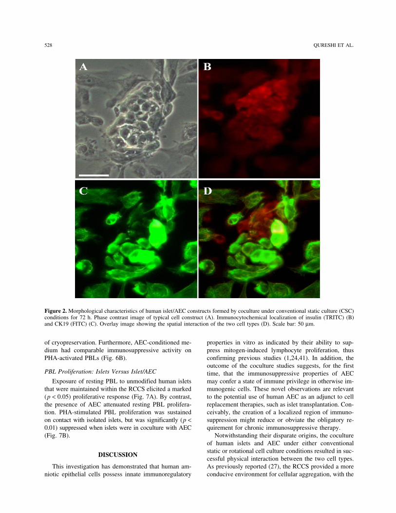
Figure 1. Human pancreatic islets visualised by dithizone staining (red cells) observed following pancreatic digestion (A) and after Ficoll gradient
assisted centrifugation (B). Scale bar = 100m
A B
54
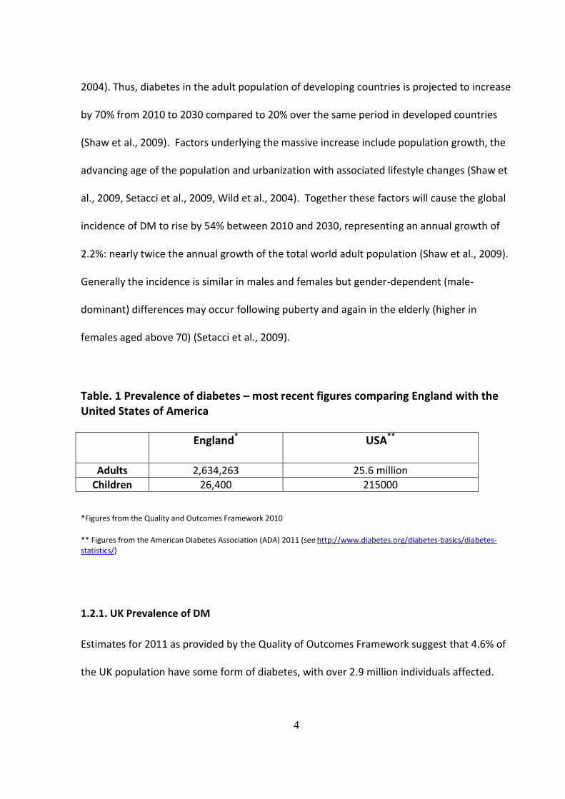
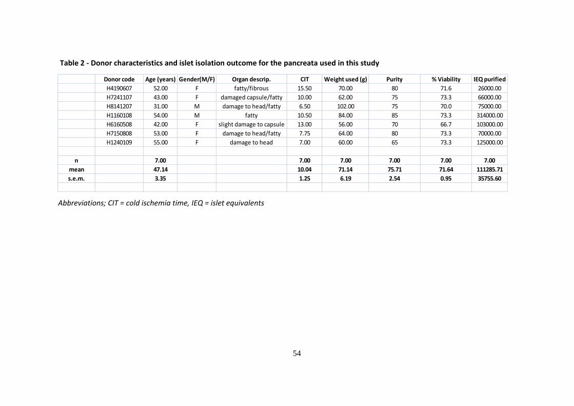
Table 2 - Donor characteristics and islet isolation outcome for the pancreata used in this study
Abbreviations; CIT = cold ischemia time, IEQ = islet equivalents
Donor code Age (years) Gender(M/F) Organ descrip. CIT Weight used (g) Purity % Viability IEQ purified
H4190607 52.00 F fatty/fibrous 15.50 70.00 80 71.6 26000.00
H7241107 43.00 F damaged capsule/fatty 10.00 62.00 75 73.3 66000.00
H8141207 31.00 M damage to head/fatty 6.50 102.00 75 70.0 75000.00
H1160108 54.00 M fatty 10.50 84.00 85 73.3 314000.00
H6160508 42.00 F slight damage to capsule 13.00 56.00 70 66.7 103000.00
H7150808 53.00 F damage to head/fatty 7.75 64.00 80 73.3 70000.00
H1240109 55.00 F damage to head 7.00 60.00 65 73.3 125000.00
n 7.00 7.00 7.00 7.00 7.00 7.00
mean 47.14 10.04 71.14 75.71 71.64 111285.71
s.e.m. 3.35 1.25 6.19 2.54 0.95 35755.60
55
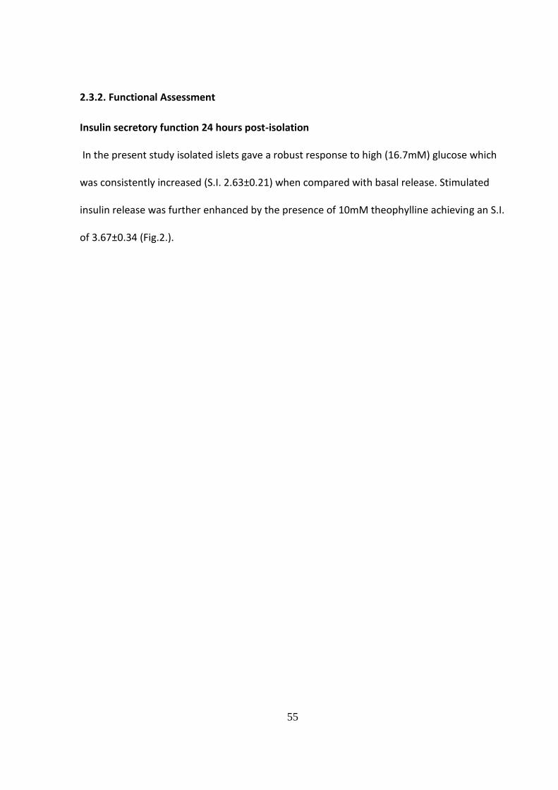
2.3.2. Functional Assessment
Insulin secretory function 24 hours post-isolation
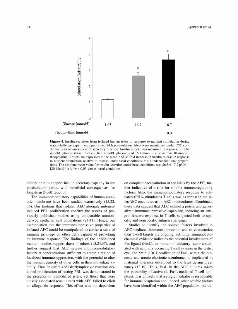
In the present study isolated islets gave a robust response to high (16.7mM) glucose which
was consistently increased (S.I. 2.63±0.21) when compared with basal release. Stimulated
insulin release was further enhanced by the presence of 10mM theophylline achieving an S.I.
of 3.67±0.34 (Fig.2.).
56
Figure 2. Insulin secretion from isolated human islets in response to nutrient stimulation during static challenge experiments performed 24 h post isolation. Islets were maintained under conventional static culture conditions prior to assessment of insulin secretory function. Insulin release was measured in response to 1.67 mmol/l glucose (basal release), 16.7 mmol/l glucose and 16.7 mmol/l glucose plus 10mmol/l theophylline. Results are expressed as the mean ± SEM fold increase in insulin release in response to nutrient stimulation relative to release under basal conditions (stimulation index S.I.). n=7 independent islet preparations. The absolute mean value for insulin secretion under basal conditions was 86.5±17.2 μUml-1 [20 islets]–1 h–1. * p<0.05 vs. basal conditions. (One way analysis of variance)
57
2.4. Discussion
The aim of the present chapter was to outline the process of procuring human pancreatic
tissue for research and the subsequent isolation of islets by use of an in-house manual
separation method.
Clinical islet transplant programmes require standardisation of islet isolation in order to
conform to strict regulatory guidelines for the manufacture of tissue for transplant purposes.
The semi-automated method of islet retrieval is amenable to validation with well-defined
standard operating procedures which may be readily adopted by multiple transplant centres
involved in clinical trials, ensuring that outcomes may be directly compared (Linetsky and
Ricordi, 2008). The trade-off arises from the resource-rich and labour-intensive nature of the
process, with the costs involved making islet isolation prohibitive to all but a few centres
across the UK, and indeed worldwide (Paget et al., 2007).
Whilst human islets used for research purposes are required to be of a similarly high
standard the method of islet procurement may differ in terms of the processes employed. In
contrast to the semi-automated-method where the procedures must be rigidly adhered to,
the manual method of islet retrieval may be subject to adaptation with changes made to the
protocol depending on the characteristics of the pancreas being processed. The ability to
make modifications during the procedure increases the likelihood of successful islet
isolation, despite the frequent use of marginal (sub-optimal) organs. This ensures optimum
use of the scarce resource of human pancreatic tissue for islet procurement.
58
In this study the use of a manual method of islet isolation was demonstrated to be an
effective means of obtaining human islets of suitable quality for research. The technique
may be readily mastered, with appropriate training, by researchers new to the field. The cost
of isolation is affordable, conducted by two personnel with fewer consumable resources
expended and no specialised equipment required in comparison to the semi-automated
process. Successful isolations were achieved with most organs procured and the yields
obtained were of an appropriate level (ranging from 25,000-300,000 IEQ per pancreas) for
the planned experiments.
The pancreata obtained through NHS Blood and Transplant had been declined for use in
whole organ or islet transplantation. In certain instances this may have been due to
anatomical incompatibility with the intended recipient or due to clinical service limitations.
However, on most occasions the organs were deemed unsuitable due to the period of cold
ischaemia, certain characteristic of the donor (age, BMI, medical history) or due to physical
damage to the organ. The manual method of islet isolation can be adapted to address these
shortcomings. For instance, islets from a donor with a high BMI may have fat infiltration
which would make it unsuitable for islet isolation using the semi-automated method, due to
the extended period of enzymatic digestion which is required. In the clinical isolation
process, increasing the digestion phase of the process is problematic and often leads to over
digestion of the preparation and subsequent islet fragmentation. However, using the manual
technique, the process of digestion is placed under greater surveillance with frequent
sampling of the digest (as frequently as every 5 mins) ensuring that islets are liberated
without risk of excessive digestion. Additionally, use of a discontinuous gradient (as opposed
59
to the COBE 2991) allows greater control of the purification process and conducting it at
room temperature with less -cell toxic gradients (McCall et al., 2011) provides the
opportunity to “rescue” poorly fractionated preparations and attempt further separation. As
a result it was possible to achieve purity of up to 70% in the batches of islets used in this
study; dithizone staining provided subjective evidence that that there was minimal exocrine
contamination and enabling us to exclude any compounding effects arising from the
presence of non-islet cellular components.
It has been argued that the use of manual methods of islet isolation increases the risk of
bacterial and viral infection (Goto et al., 2004). However, in the present study all islet
preparations were subject to microbiological analysis (performed by our Trust
microbiologists) and were found to be free of contaminating pathogens. The additional steps
involved in isolation are also thought to equate to a loss of morphological or functional
viability (Gurol et al., 2004). However, in this study, morphological analysis confirmed the
structural integrity of the cells. Furthermore, the results of the glucose challenge test
demonstrated the functional viability of the islets, notably their appropriate response to
nutrient stimulation as observed 24 hours after isolation. In clinical islet transplantation the
stimulation index is used, in part, to determine whether a batch of islets meets the criteria
for transplantation (D'Aleo et al., 2010). In most instances islet preparations with an S.I.
between 2.5 and 4.0 are transplanted although there is no clear correlation between pre-
transplant S.I. and long-term graft performance (Ryan et al., 2004). This, coupled with the
purity, suggests that the islets obtained would have been suitable for clinical use (Linetsky
60
and Ricordi, 2008) and were therefore an appropriate model for use in subsequent studies
to design low immunogenic tissue constructs for use in islet transplantation.
61
CHAPTER 3: HUMAN AMNIOTIC EPITHELIAL CELLS: ISOLATION, MORPHOLOGICAL AND FUNCTIONAL
ASSESSMENT
3.1. Introduction-Amnion-derived cells as candidates for transplantation therapies
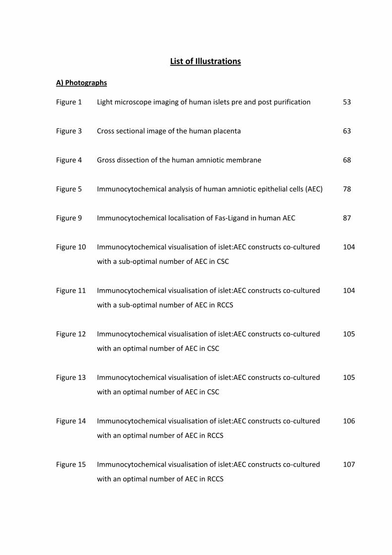
During gestation the developing foetus is surrounded by amniotic fluid, enclosed in a sac
lined by the amniochorionic membrane (Fig.3.). The innermost layer is composed of amniotic
epithelial cells (AEC), resting on a basement membrane and underlying avascular stromal
cells which collectively form the amniotic membrane (AM) (Hoyes, 1975). The amniotic
component of the foetal sac originates from the epiblast of the inner cell mass which
contrasts with the origin of the chorion, derived from extra-embryonic tissues (Benirschke
and Kaufmann, 2000).
The placenta gives rise to a number of distinct cell populations which have recently become
the subject of considerable interest in the fields of cell and tissue transplantation and
regenerative medicine (Parolini and Caruso, 2011, Parolini et al., 2008). Of note, the stem-
and progenitor-like characteristics of these cells, coupled with their relative immune-
inertness makes them strong candidates in the search for surrogates for use in cell
replacement therapy in a variety of clinical situations. The critical role played by the placenta
during pregnancy is understood to involve maintenance of feto-maternal tolerance,
preventing the partially allogeneic foetus from being rejected. As a consequence, the
placenta and its associated membranes are endowed with certain characteristics pertinent
to the modulation of the surrounding immune micro-environment. Of note, in extensive
62
studies the following human placental derived cell types have been shown to possess
immuno-modulatory potential with importance for the inhibition of inflammatory and/or
immune events.
i. chorionic trophoblastic cells (CTC) – isolated from the chorionic trophoblast of term
placenta
ii. chorionic mesenchymal stromal cells (CMSC) – obtained from the mesenchymal layer
of the chorionic membrane
iii. amniotic mesenchymal stromal cells (AMSC) – derived from the mesenchymal layer
of the amniotic membrane
iv. Amniotic epithelial cells (AEC) – sourced in large quantities from the epithelium of
the amniotic membrane
For the purpose of the present study this summary will focus on the cells derived from the
human amniotic membrane; i.e. amniotic epithelial cells whilst the others are
comprehensively reviewed elsewhere (Parolini and Caruso, 2011).
63
Figure 3. Illustration of the architecture of the human placenta and amniotic membranes
64
3.1.1. Clinical use of human amniotic membrane (AM)
The low-antigenicity and marked anti-inflammatory properties of human AM underlies its
importance in reconstructive and transplant medicine. Its’ most acknowledged use is in
ophthalmic surgery, where using AM as a basement membrane substitute or as a temporary
graft has become commonplace. AM has been shown to reduce inflammation and scarring,
prevent angiogenesis and fibrosis and is thought to be the source of certain growth factors
which encourage the re-epithelisation of the surface of the eye (Meller et al., 2011). Despite
the absence of controlled randomised clinical data directly supporting the role of AM
transplantation in ophthalmic surgery, the results of numerous case studies strongly suggest
that it serves as an effective approach to corneal and conjunctival reconstruction and has
been successfully applied to the treatment of burns (Meller et al., 2000), acute Stevens-
Johnson Syndrome (Gregory, 2011), intractable corneal ulcers (Nubile et al., 2011) and
persistent epithelial defects (Seitz et al., 2009).
In addition to its role in ophthalmic surgery AM has also been used in the management of
ulcers refractory to other treatments, and in venous leg ulcers AM grafts were shown to
encourage re-epithelisation from the edge of the wound inwards with a concomitant
reduction in fibrosis and associated pain (Mermet et al., 2007). The treatment of paediatric
burns using human AM as a temporary graft or for skin graft fixation has also been explored
clinically with promising results (Sheridan and Moreno, 2001, Mohammadi and Johari, 2010).
65
3.1.2. Clinical potential of human amniotic epithelial cells
The innate anti-inflammatory characteristics of AM have been attributed, at least in part, to
the cells lining the membrane surface, the amniotic epithelial cells (AEC) which, when
isolated, have been shown to exhibit immunomodulatory potential. In vitro studies
demonstrate the ability of human AEC to suppress T-cell activation in both mixed
lymphocyte and mitogen-induced proliferation assays (Wolbank et al., 2007, Li et al., 2005)
and AEC are amenable to both allogeneic and xenogeneic engraftment in immune-
competent recipients (Akle et al., 1981, Kong et al., 2008, Kubo et al., 2001, Sankar and
Muthusamy, 2003). Furthermore, the expression of several mediators of localised immune
suppression including HLA-G, Fas ligand and TGF have been characterised in isolated AEC or
culture supernatant (Li et al., 2005, Harirah et al., 2002, Hammer et al., 1997, Kubo et al.,
2001, Lefebvre et al., 2000). Such immuno-mediators have the capacity to counteract the
potentially harmful actions of immune cells; evidence suggests that AEC may be capable of
creating a microenvironment conducive to sustained allogeneic graft survival (Wolbank et
al., 2007, Li et al., 2005, Bailo et al., 2004, Kong et al., 2008, Sankar and Muthusamy, 2003).
Coupled with their relative immune-inertness, AEC also express a variety of stem cell
markers indicating multi-lineage differentiation potential. Indeed, under defined culture
conditions human AEC have the capacity to differentiate into cells from all three germ layers,
giving rise to bone, fat, liver, pancreas and neural cells (Murphy et al., 2010). As such, in
experimental models, human AEC have been shown to be beneficial in the treatment of
and liver fibrosis (Parolini and Caruso, 2011). There is limited evidence that differentiated
66
AEC are able to directly participate in tissue regeneration in vivo (Okawa et al., 2001).
However, the beneficial AEC-mediated effects observed are largely considered to be due to
the secretion of bioactive molecules that act on other cells and promote endogenous tissue
repair through paracrine effects (Parolini and Caruso, 2011).
A substantial body of evidence therefore exists in favour of a role for human AEC in
regenerative medicine. When compared with stem cells derived from an embryonic source,
the wide availability and relative lack of ethical constraints associated with procurement of
this tissue make AEC an ideal candidate for further exploration.
Aims of the Chapter
This study aims to provide evidence that AEC may be used as an adjunct to islet cell
transplantation, offering vital trophic support, whilst simultaneously protecting the islet
graft from immune assault. In the following sections we describe the methods of isolation
and subsequent characterisation of the AEC used in the present investigation, including their
immunomodulatory potential.
67
3.2. Materials and Methods: I: AEC Isolation and morphological characterisation
3.2.1. Donor recruitment and consent
All studies using human amniotic tissue were performed according to ethically approved
protocols (LREC: Q5/2801/70- Coventry and Warwickshire Ethics Committee) and with the
informed consent of the tissue donor. For amniotic membrane procurement, potential
participants were identified from an elective Caesarean section list. Women undergoing
normal vaginal delivery were excluded from the study to reduce the risk of microbial
contamination of the amnion sample. The prospective tissue donors were seen in the pre-
operative clerking clinic 24 hours beforehand and given information about the research
project. Following discussion of the proposed work, women were invited to take part in the
study and if appropriate gave informed, written consent prior to delivery.
3.2.2. AM harvest and dissociation
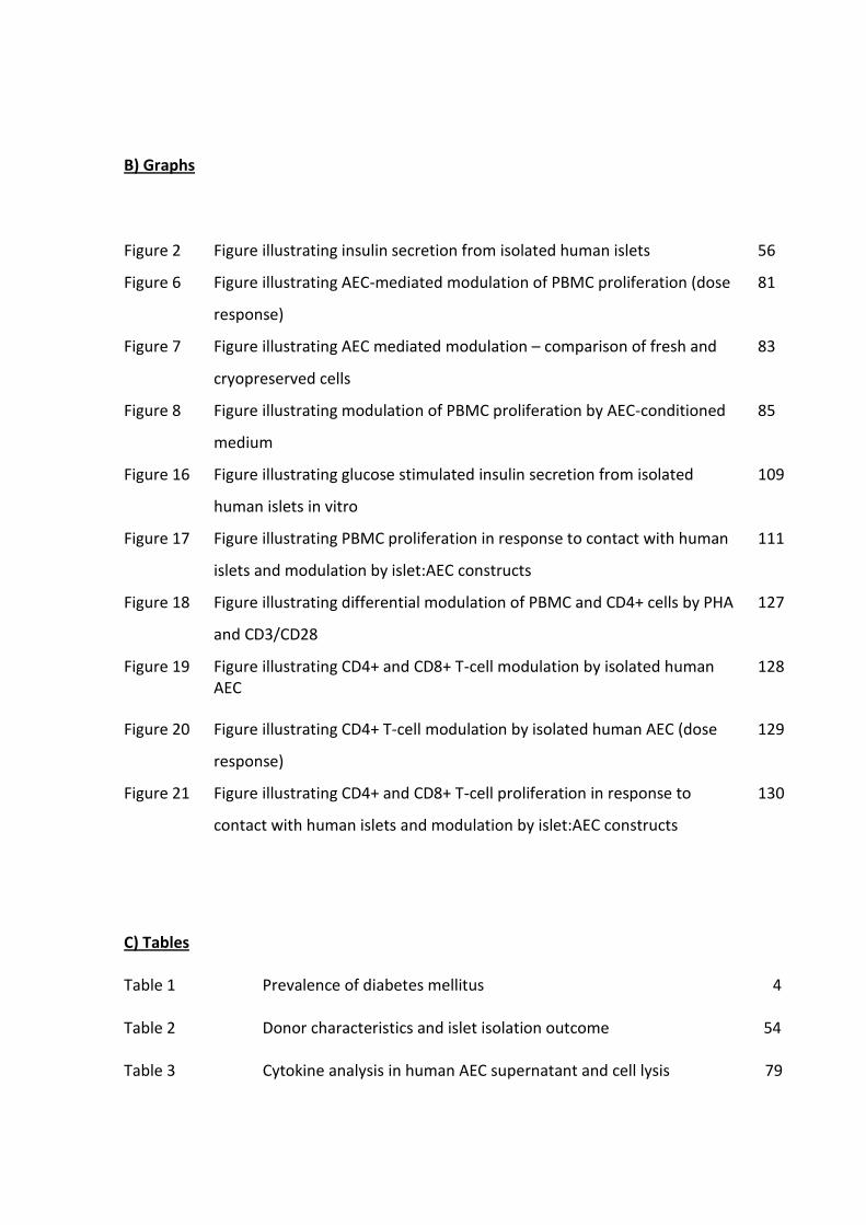
The amnion was harvested under aseptic conditions in the operating theatres. After the
placenta had been delivered and inspected by the midwife, the AM was mechanically
dissociated from the chorion layer. 2-3.5 cm2 pieces were stripped off and washed twice in
150ml of filter sterilised PBS containing 100U penicillin, 100g streptomycin and 10g
amphotericin B. The tissue was placed in a sterile pot containing 100ml of filter sterile PBS
supplemented with 200U penicillin, 200g streptomycin and 20g amphotericin B for
transport to the laboratory.
68
Figure 4. Human amniotic membrane mechanically separated from the chorion. Samples not in direct contact with the placenta were collected for processing.
amnion
chorion
placenta
69
The tissue was transferred to a Class II microbiological safety cabinet and the amnion was
cut into small pieces to increase its surface area. The AM fragments were placed in a conical
tube containing HBSS before being centrifuged at 400g for 5 mins (brake set to 5). The
supernatant was decanted leaving the pelleted amniotic tissue. A 0.25% solution of porcine
Trypsin (Sigma-Aldrich) was made up in HBSS in a sterile container and placed in a water
bath to reach a temperature of 37⁰C. Once achieved, 100ml of the trypsin solution was used
to re-suspend the amnion tissue and both were transferred to a glass beaker equipped with
a magnetic flea. The beaker was tightly sealed with a layer of parafilm and placed in a stirrer
oven at 37⁰C for 30 mins.
Following the first digestion period the dissociating tissue was passed through a 500m
mesh to harvest any detached cells; the tissue retained by the mesh was collected and
returned to the glass beaker, re-suspended in a fresh 100ml volume of 0.25% trypsin
solution and placed in the stirrer oven for a further 30 mins incubation period. The collected
filtrate (Fraction 1) was placed into 50ml conical tubes and centrifuged at 700g for 5 mins
(brake set on 5). The supernatant was decanted and the cell pellet re-suspended in 2mls
RPMI 1640 containing 10% FBS, 100U/ml penicillin, 100g/ml streptomycin and 10g/ml of
amphotericin B.
The above process was repeated until the amniotic epithelium was fully dissociated and the
resulting, dispersed AEC were collected in a total of 4 separate cell fractions (Fractions 1-4),
each of which was suspended in 2mls RPMI 1640 medium containing additives as described
above. In most instances fraction 1 was discarded due to the high number of contaminating
70
red blood cells. Fractions 2-4 were subsequently pooled and plated into T-75 flasks. Each
flask received 1ml of cell suspension (containing between 1-3 million AEC) and was made up
to 20ml total volume with RPMI 1640 + additives. The flasks were transferred to an
incubator for 48 hours at 37 °C in a humidified atmosphere of 95%O2/5% CO2 to allow cell
attachment. At the first medium change (48-72 hours) 15mls of medium was removed from
each flask and replaced with 10mls of fresh medium. Thereafter a full medium change
occurred at 2-3 day intervals. Flasks of dispersed amniotic epithelial cells reached confluence
at between 7-10 days post isolation. In this study AEC were routinely used at passage 1 and
at this time the cells were counted and assessed for viability.
3.2.3. Passaging of AEC – assessment of yield and viability
At confluence AEC cultures were passaged as follows:
The flasks containing the AEC monolayers were transferred to the hood and the medium was
gently aspirated using a sterile 10ml pipette. This was immediately replaced with 10mls of
filter sterilised PBS (Sigma-Aldrich) which was used to rinse the cells. Rinsing was performed
3 times to ensure all of the FBS-containing culture medium was removed (as this would
inhibit the subsequent trypsinisation process). 1ml of 0.025% Trypsin-EDTA in PBS (Sigma-
Aldrich) was added to each flask, gently swirling the flask to ensure that the entire
monolayer was covered by the enzyme. The flasks were then returned to the incubator for
20mins to assist mild dissociation of the monolayer. The AEC monolayers were viewed under
an inverted microscope to assess the level of disruption; AEC rolled up and become
detached from the base of the flask as the trypsin took effect. If necessary, cell detachment
71
was further assisted by gently tapping the flask against the palm of the hand. Once all the
AEC were free-floating the trypsin reaction was quenched by addition of 10ml of culture
medium contain 10% FBS. The AEC suspension was harvested into 50ml conical tubes for
centrifugation at 500g for 3 mins. Each AEC pellet was re-suspended in 1ml of culture RPMI
1640+ additives, (i.e. 1 ml of culture medium per flask of AEC) and all the pellets were
pooled for counting and viability assessment.
Samples of AEC (50-100l) were placed in an Eppendorf tube to which an equal volume of
0.4% Trypan Blue solution was added. Following mixing, a portion of the cell suspension was
used to fill the lower counting chamber of a haemocytometer – by capillary action. Cells
occupying the 4 quadrants (composed of 16 squares) of the chamber were counted and the
mean value used to determine the total cell number in one ml of suspension. From this total
AEC numbers were calculated. In addition, the non-viable cells (i.e. blue-stained) in each grid
were counted to give the percentage cell viability. The AEC suspension was adjusted to a
final density of 1x10⁶/cells per ml for subsequent use or for cryopreservation.
3.2.4. AEC Cryopreservation
As the ultimate aim of this study was to determine whether human AEC could be applied to
clinical cell transplantation we sought to determine whether these cells were amenable to
cryopreservation thus ensuring their ready availability. To this end studies were performed
to assess AEC function following a period in ultra-low temperature storage. AEC at passage 1
(P1) were counted as described above and adjusted to a final density of 1x10⁶ cells per ml in
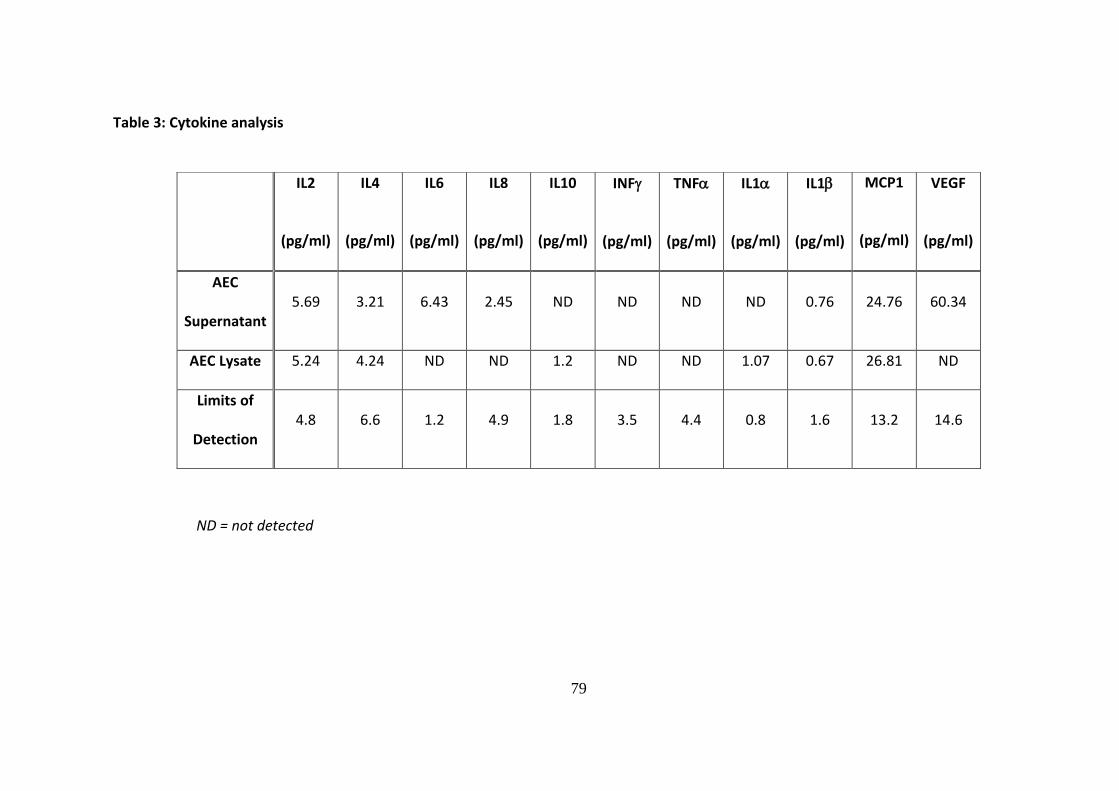
Chemotactic Protein-1 (MCP-1), Tumour Necrosis Factor-α (TNF-α), and Vascular Endothelial
Growth Factor (VEGF). Due to the combination of highly specific antibodies and advanced
chemistries the array enables all 12 cytokines and growth factors to be detected
simultaneously in a single sample. The limit of sensitivity for each analyte is presented in
table 3. Each sample was measured in duplicate and 2 samples from each group
(supernatant or lysate) were provided for analysis.
74
3.3. Material and Methods II: Assessment of Immunomodulatory potential
3.3.1. Isolation of peripheral blood mononuclear cells (PBMC)
Peripheral blood mononuclear cells (PBMC) were isolated from buffy coats obtained from
healthy volunteers through the National Blood Service (NBS, Birmingham, UK) with local
research ethics approval and with the consent of the donor. In all instances the buffy coats
came from regular blood donors who were screened for pathogens and who were free of
any illness which might adversely affect lymphocyte reactivity. To isolate the PMBC, buffy
coats were collected into 150 ml sterile pots and diluted in an equal volume of HBSS. This
was carefully layered onto 12ml of density gradient adjusted to 1077g/ml (Histopaque®-
1077 - Sigma-Aldrich), centrifuged at 700g for 30 mins (with no brake), and the resulting
leucocyte layer harvested from the interface using a sterile pastette. The isolated PBMC
were washed three times in HBSS, centrifuged at 500g for 10 mins, re-suspended in RPMI
1640 (supplemented as described above). The separated PBMC were counted using
haemocytometer and viability was assessed using trypan blue exclusion. The cells were then
cultured in uncoated plastic petri dishes at 37°C, 5%CO2, 95%O2 overnight. The PBMC
cultures were incubated overnight, half of them in the presence of the plant mitogen
phytohaemagglutinin (PHA, Sigma-Aldrich, 5g/ml), for 24 hours prior to use in proliferation
assays.
75
3.3.2. AEC vs. PBMC – Proliferation Assay
AEC at P1 were prepared as described in section 3.2.3 above. Once adjusted to a final
volume of 1x10⁶cells/ml in supplemented RPMI 1640, aliquots of the AEC suspension were
added to the appropriate wells of a 24-well plate. In preliminary studies a dose response
curve was performed plating AEC at 5,000, 50,000 and 500,000 AEC per well. Each well was
supplemented with medium up to a total volume of 1ml and the plates placed at 37°C,
5%CO2, 95%O2 for 72 hours to permit cell anchorage. Additionally, in selected experiments
cryopreserved AEC were rapidly thawed, rinsed in PBS and seeded as described above for
the fresh AEC prior to their use in PBMC assays as follows: AEC seeded plates were
processed by repeated washing in filter sterile PBS to ensure all unattached cells/cellular
debris was removed from the wells. Resting or PHA-activated PBMC were added at a density
of 50,000 cells/well either alone which served as a control or to wells pre-seeded with
50,000 firmly anchored AEC prior to co-incubation at 37°C, 5%CO2, 95%O2. Activated PBMC
continued to be cultured in the presence of 5g/ml PHA throughout the assay period. After
72 hours the PBMC were harvested, washed and assayed for intracellular ATP content as
described in section 3.3.4 (below).
3.3.3. AEC conditioned medium (CM) vs. PBMC
In additional selected experiments P1 AEC were re-plated into T-75 flasks at a density of
2x10⁵cells/ml (equivalent to 3x10⁶cells per flask) in 15mls of supplemented RPMI 1640
medium. The flasks were left for 72 hours without a medium change to allow concentration
of putative soluble factors released by the AEC. The resulting AEC-conditioned medium (CM)
76
was harvested and centrifuged at 1300g to ensure removal of all cells/cellular debris prior to
use in PBMC proliferation assays. 0.5ml of CM was dispensed to the appropriate wells of a
24-well plate and 5 x 10⁴ resting or PHA activated PBMC were added; adjusting the total
volume to 1.0ml using standard RPMI medium. Plates were incubated at 37°C, 5%CO2,
95%O2. After 72 hours the PBMC were harvested, rinsed and processed as described in
section 3.3.4 (below).
3.3.4. Quantification of PBMC proliferation - luminescent detection of intracellular ATP
After 72 hours the harvested PBMC were solubilised using cell lysis reagent (Vialight – Lonza
Ltd, Wokingham, UK) and analysed for ATP content using a commercial chemiluminescence
assay (Lonza Ltd) according to the manufacturer’s instructions. Concentration of ATP per
well, measured as relative light units (RLU) is directly proportional to cell number and thus
indicative of the proliferative activity of PBMC in culture (Sottong et al., 2000). Results were
expressed as the percentage increase in relative cell number compared to the control viz.
resting PBMC incubated in the absence of AEC.
3.3.5. Statistical analysis
Significant differences in PBMC proliferation in response to co-culture with AECs was
determined using Mann-Whitney U (by Rank) and Tukey’s multiple comparison tests, with
the response of resting PBMC serving as the control. In all comparisons a p value of <0.05
was considered to be statistically significant. Statistical analysis was performed using
SigmaStat software version 3.5 (Systat Software Inc, Chicago, USA).
77
3.4. Results I
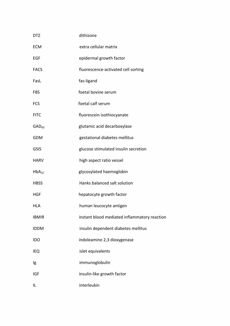
3.4.1. Morphological characteristics of isolated AEC
Human amniotic epithelial cells when isolated from the membrane and held in suspension
culture readily adopted a spherical morphology forming an apparently homogeneous
population (Fig.5.A). Once plated at high density in T75 flasks AEC readily attached and
flattened to form a monolayer (Fig.5.B), the vast majority of these cells staining positive for
the epithelial cell marker cytokeratin 19 (Fig.5.C). Additionally, a discreet sub-population of
cells stained positive for the intermediate filament marker vimentin (Fig.5.D).
3.4.2. Cytokine Analysis –multiplex immunoassay
AEC supernatants collected after 72 hours of culture as described in section 3.2.6., and cell
lysates prepared from AEC monocultures were processed by Randox Laboratories Ltd using a
cytokine array multiplex immunoassay.
Several cytokines relevant to immune-modulation were detected either in AEC cell
supernatant, lysate or both, as presented in Table 3. Despite using AEC at the same density
as that employed in the proliferation assays the concentration of cytokines detected were, in
many cases, below the level of sensitivity for the immunoassay.
78
Figure 5. Human amniotic epithelial cells (AEC) viewed by light microscopy immediately following isolation (A), viewed first under phase contrast (B) and using fluorescence immunocytochemistry for the localisation of the epithelial cell marker cytokeratin 19 (CK19) ( C) and the intermediate filament protein vimentin (D). A FITC-conjugated secondary antibody was used for visualisation. Vimentin expression by AEC maintained in monolayer culture differs from that of CK19 which is a cytoskeletal protein marker and is distributed throughout the cytoplasm. Most epithelial cell types co-express
vimentin. For A original magnification x40, B,C,D Scale bar = 100m.
3.4.3. Results II – Immunomodulatory Potential of isolated AEC and AEC-conditioned
medium
PBMC proliferation was evident following exposure to the plant-derived mitogen –
phytohaemagglutinin - 5g/ml (PHA) as demonstrated by a robust (20-fold) increase in
intracellular ATP concentration as measured by chemiluminescence detection. By contrast,
despite the fact that the two cell populations are derived from different donors and are
therefore allogeneic, PBMC grown in the presence of varying numbers of human AEC failed
to proliferate to a significant degree. At the highest concentrations of AEC (i.e. 50,000 and
500,000) there was a slight increase in PBMC numbers but this did not reach statistical
significance and was small in comparison to the magnitude of response seen to the non-
specific mitogen. PHA-mediated PBMC stimulation was significantly reduced by their co-
culture with AEC. In the dose response experiment AEC-induced inhibition of PBMC
proliferation was evident at an AEC:PBMC ratio of 1:10 (Fig.6) demonstrating a 60%
inhibition in cell numbers compared to the control viz. PBMC expansion in the absence of
AEC. Increasing numbers of AEC inhibited PBMC proliferation by a similar magnitude.
B
81
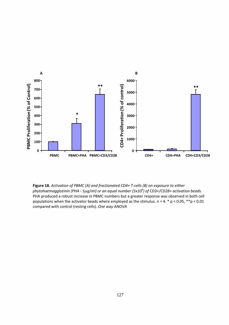
Figure 6. Modulation of peripheral blood mononuclear cell (PBMC) proliferation by co-incubation with varying numbers of human amniotic epithelial cells (AEC): AEC were plated at 5,000 (5K), 50,000 (50K) or 500,000 (500K) cells/ml into 24-well plates and allowed to firmly attach for up to 72 hours. PBMC were then added and
incubated either in the absence ( ) of presence ( ) of the plant mitogen phytohaemagglutinin (5g/ml) for a further 72 hours. The rate of PBMC proliferation following this period was measured using an ATP chemiluminescence assay. Data shows the percentage increase above control (resting PBMC) from 3 individual AEC preps and represents the typical observation. * p < 0.05, ** p < 0.01 PHA induced increase in PBMC numbers as compared to resting control. † p< 0.01 inhibition of PHA-activated PBMC proliferation in the presence vs. absence of AEC. (Mann-Whitney U (by Rank) and Tukey’s multiple comparison tests)
A
82
As this study seeks to provide evidence to justify clinical application of AEC in cell
transplantation therapy it was important to ascertain whether these cells were amenable to
cryopreservation. To this end in a sub-set of experiments AEC which had been frozen for
between 1 and 3 months were used to determine their immunomodulatory capacity in
relation to PBMC. When compared to fresh AEC, cryopreserved AEC elicited a mild
stimulation of PBMC on co-culture (Fig.7). However this response was small in comparison to
their response to PHA. A similar magnitude of inhibition (55%) of PHA-mediated proliferation
was observed when PBMC were co-incubated with either fresh or cryopreserved AEC.
83
Figure 7. Modulation of peripheral blood mononuclear cell (PBMC) proliferation by fresh (A) and cryopreserved human amniotic epithelial cells (AEC) (B). Resting ( ) or PHA-activated ( ) human PBMC were maintained in 24-well plates either alone, or in the presence of an equal number of human amniotic epithelial cells for a period of 72 hours. The rate of PBMC proliferation following this period was measured using an ATP chemiluminescence assay. Data shows the percentage increase above control (resting PBMC) from 6 individual AEC preps and represents the typical observation in fresh and cryopreserved AEC. * p < 0.05, ** p < 0.01 compared to control. † p< 0.01 for PHA-activated PBMC proliferation in the presence or absence of AEC. (Mann-Whitney U (by Rank) and Tukey’s multiple comparison tests)
84
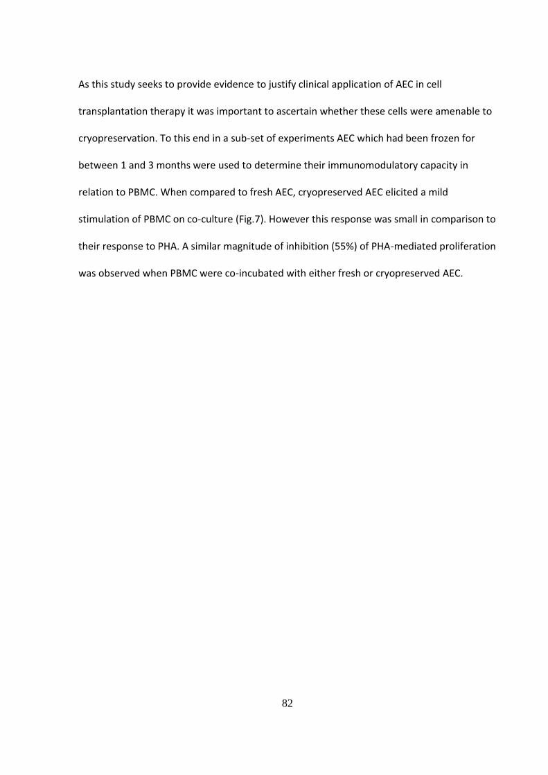
In a sub-set of experiments the effect of AEC- conditioned medium on PBMC proliferation
was also determined. In our study AEC- conditioned medium had comparable
immunosuppressive activity on PHA-activated PBMC as the AEC inhibiting proliferation by up
to 60% (Fig.8).
85
Figure 8. Modulation of peripheral blood mononuclear cell (PBMC) proliferation during exposure to AEC-conditioned medium (CM). Resting ( ) or PHA-activated ( ) human PBMC were maintained in 24-well plates either alone or in the presence of 0.5mls of AEC-conditioned medium for a period of 72 hours. The rate of PBMC proliferation following this period was measured using an ATP chemiluminescence assay. Data shows the percentage increase above control (resting PBMC) from 4 individual AEC-CM preps and represents the typical observation. * p < 0.05, ** p < 0.01 compared to control. † p< 0.01 for PHA-activated PBMC proliferation in the presence or absence of AEC-conditioned medium. (Mann-Whitney U (by Rank) and Tukey’s multiple comparison tests)
86
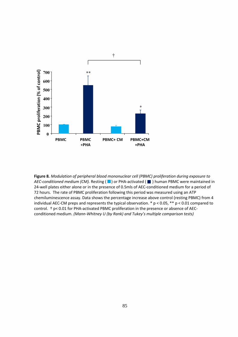
3.4.4. Immunocytochemical analysis of immune mediators
In an attempt to identify other potential immune modulators underlying the inhibitory
actions of human AEC, we sought to identify Fas Ligand expressing cells within P1 AEC
monocultures. The Fas/FasL pathway has been implicated in feto-maternal tolerance and
has previously been shown to be expressed in foetal membranes (Koenig and Chegini, 2000)
and amniotic cells (Li et al., 2005). The results of the immunocytochemical investigation
confirm that a small sub-population of AEC express FasL in culture. Whilst counterstaining
for cell nuclei was not performed in this study a comparison of the cell populations using
phase contrast and fluorescence microscopy suggests that approximately 30% of the AEC
population exhibited cytoplasmic localisation of FasL protein (Fig.9).
87
Figure 9. The images (right) show FasL expressed by AEC .FasL is a soluble membrane bound protein
expressed by approx. 30% of isolated AEC. Scale Bar = 100m B
A
88
3.5. Discussion
The results of this series of studies suggest that AEC are capable of eliciting a suppressive
effect on peripheral mononuclear cell proliferation, inducing effective inhibition even at a
ratio of 1:10 for AEC:PBMC. The immunomodulatory capabilities of human amniotic
membrane have been studied extensively (Hori et al., 2006, Kubo et al., 2001, Trelford et al.,
et al., 1994, Dheen et al., 1997, Robertson et al., 2008, Jiang and Harrison, 2005).
Additionally, components of the extracellular matrix are vital for appropriate pancreatic
development and several integrin receptors and their associated ligands including laminin,
fibronectin and collagen I are expressed by these cell types, notably epithelial cells (Jiang et
al., 1999, Cirulli et al., 2000, Jiang and Harrison, 2005).
We therefore propose that combining the use of bioreactor technology with tissue-
engineering to modify the function of transplantable therapeutic cells represents a novel
approach to improving clinical outcomes in islet replacement therapy. It is our hypothesis
that islet cells may be modified in vitro and adapted under defined culture conditions to
enhance survival in the post-transplant environment.
96
Aims of the Chapter
The specific aim of this section of the project was to construct heterotypic cell composite
grafts with the capacity to:
provide important paracrine regulatory and trophic support to native beta-cells by the
synthesis and release of appropriate growth factors
counteract islet-induced allo-immune responses by mediating localised suppression of
the innate and adaptive immune system
To this end, the work as outlined in Chapters 2 and 3 was extended to exploit the observed
immunomodulatory potential of human amniotic epithelial cells (AEC), employing a
rotational cell co-culture model to provide these beneficial characteristics to populations of
isolated, and purified human islets. As AEC are also reported to synthesise and secrete a
range of growth factors which may have relevance for the sustained functional viability of
islets (Fiaschi-Taesch et al., 2008, Movassat et al., 2003, Hanley and Rosenberg, 2007,
Koizumi et al., 2000, Kakishita et al., 2003, Scharfmann and Czernichow, 1996) we also
explored the impact of AEC co-culture on islet viability and functionality. The effectiveness of
this intervention was assessed using in vitro models of insulin-secretory function and
immunomodulation as detailed below.
97
4.2. Materials and Methods
4.2.1. Bioengineering of islet:AEC constructs
For co-culture studies islet suspensions obtained as described in section 2.2 were adjusted to
a density of 500-1000 IEQ per ml and placed under either conventional static culture (CSC)
conditions in 90mm culture plates (NHS Logistics, Alfreton, UK) or in a rotational cell culture
system (RCCS) in high aspect ratio vessels (HARVs, Cellon Ltd, Bereldange, Luxembourg). The
growth medium for both culture models was composed of Medium-199 (M199)
supplemented with 10% FBS, 100U/ml penicillin, 100g/ml streptomycin and 10g/ml of
amphotericin B – no additional trophic factors were added. The cultures were maintained at
30⁰C in a humidified atmosphere of 95%O2/5%CO2. Confluent AEC monolayers at passage 1
were disrupted by mild enzymatic digestion (0.025% trypsin-EDTA in PBS, Sigma-Aldrich Ltd)
and the resulting cell suspension was washed in PBS and introduced to the islet cultures
(both CSC and RCCS) at a final density ranging from 1 x 10⁴-1x10⁵ cells per ml. The islet:AEC
co-cultures were maintained under conditions as described above for 72 hours. The speed of
rotation of the HARV’s was initially set to 8 rpm and increased to a maximum of 15rpm as
the size of the islet:AEC aggregates increased. Control cultures consisted of islets seeded at
equal density (CSC and RCCS) in the absence of AEC.
98
4.2.2. Immunocytochemical analysis of the islet:AEC constructs
For immunocytochemistry islet:AEC co-cultures maintained for 72 hours either under CSC
conditions or within the RCCS were collected into separate centrifuge tubes and centrifuged
at 400g for 3 mins, the resulting cell pellets being re-suspended in 1ml of supplemented
RPMI-1640 as previously described for AEC culture. Autoclaved 13mm glass coverslips were
placed into each well of a 24-well plate and 0.5ml of supplemented RPMI-1640 was added
followed by an aliquot of cell suspension containing approximately 20 IEQ or islet:AEC
aggregates. The well volume was made up to 1 ml by addition of Medium-199 supplemented
as described above. The islet:AEC constructs were thus allowed to anchor to the glass
coverslips during a culture period of 48 hours at 37⁰C, 5%CO2, 95%O2. Adhered cell
aggregates were fixed in freshly prepared 4% paraformaldehyde during a 30 mins incubation
at RT (Sigma –Aldrich). Three 10 mins washes in PBS were followed by antigen-retrieval
(0.3% Triton-X-100, Sigma-Aldrich) and blocking using either 10% normal goat serum (NGS)
or 10% normal rabbit serum (NRS – both from Vector Laboratories Ltd, Peterborough, UK)
depending on the species in which the secondary antibody was raised. The constructs were
then incubated with the following primary antibodies: anti-human cytokeratin 19 (CK19),
anti-human vimentin (Dako UK Ltd, Cambridgeshire, UK– 1:100) or anti-human insulin (AbD
Serotec, Oxford, UK 1:10) for 1 hour at RT and at 4⁰C overnight. Secondary antibody (goat
anti-mouse IgG-FITC for CK19 and vimentin, goat anti-rabbit IgG-TRITC for insulin –
Cambridge Biosciences, Cambridge, UK, 1:100) was applied for 3 hours at RT. The coverslips
were rinsed and mounted in fluorescence mounting medium (Dako UK Ltd) before cell
imaging using a Zeiss Axioskop 40 fluorescence microscope equipped with an AxioCam MRC
99
colour camera and incorporating Axiovision imaging software (Carl Zeiss, Hertfordshire, UK).
Controls involved omission of the relevant primary antibody.
4.2.3. Functional Assessment of the islet:AEC Constructs
4.2.3.1. Estimation of glucose sensitivity - Static Challenge
Functional viability of islet:AEC constructs was determined by assessing glucose-stimulated
insulin secretion (GSIS). Static challenge studies were performed as described in Chapter 2
with the following modifications: Constructs (or unmodified islets - controls) were harvested
from the relevant culture systems by aspiration using a wide bore sterile pastette. The tissue
suspensions were placed into 15ml conical tubes and centrifuged at 400g for 3 mins, the
supernatants discarded and the resultant pellets re-suspended in a 2ml volume of 1.67mM
glucose solution made up in HBSS+0.2%BSA (basal glucose). The tubes containing constructs
(or unmodified islets) were then placed in a water bath at 37⁰C for 1 hour (pre-incubation
period). During this hour, a count was performed to determine the number of structurally
robust islet:AEC aggregates formed during the 72 hour co-culture period. If necessary the
islet/islet:AEC suspensions were re-adjusted (by the further addition of the 1.67mM glucose
solution) to achieve a density of 400 constructs/ml. After the pre-incubation period 50l
aliquots (equivalent to 20 IEQ/aggregates) of the cell suspension were transferred to 12mm
x 75mm polypropylene tubes (NHS Logistics, Alfreton, UK) using a pipette fitted with a “cell-
saver” tip to avoid damaging larger constructs. For each culture condition (i.e. CSC vs. RCCS
islets vs. islet:AEC) a total of 18 tubes were thus prepared, to which 2ml of the appropriate
secretagogue, diluted in HBSS+0.2% BSA, was added as follows: 6 tubes received 1.67mM
100
glucose solution to assess basal insulin secretion. To a further 6 tubes a 16.7mM solution of
glucose was added to determine stimulated insulin secretion. The remaining tubes received
2ml of 16.7mM glucose supplemented with 10mM theophylline. The racked tubes were
sealed with parafilm and placed into a water bath at 37⁰C for 1 hour to allow insulin
secretion (incubation period). Following incubation the tubes were gently vortexed and then
centrifuged at 400g for 5 mins. The resultant supernatant was harvested for analysis of
insulin content using an enzyme-linked immunosorbent assay (ELISA; Mercodia, Diagenics
UK), according to the manufacturer’s instructions.
4.2.3.2. Estimation of Immunomodulatory function – Mixed islet-lymphocyte reaction
(MILR)
Islet:AEC constructs were also assessed for immunomodulatory potential in comparison to
unmodified islets cultured in isolation for the same period. Once adjusted to a final volume
of approx. 1000 aggregates/ml in supplemented RPMI 1640, 20l of the islet or islet:AEC
suspension was added to the appropriate wells of a 24-well plate and made up to 1ml total
volume using the same medium. Thus, approximately 50 IEQ or 50 islet:AEC constructs were
added to each well and the plate placed at 37°C, 5%CO2, 95%O2 for 72 hours to permit stable
cell attachment. Islet: AEC seeded plates were processed by repeated washing in filter
sterile PBS to ensure all unattached cells/cellular debris was removed from the wells.
Thereafter, resting or PHA-activated PBMC were added at a density of 50,000 cells/well
either alone or to wells pre-seeded with firmly anchored islet:AEC constructs prior to co-
incubation at 37°C, 5%CO2, 95%O2. Activated PBMC continued to be cultured in the
101
presence of 5g/ml PHA throughout the assay period. After 72 hours the PBMC were
harvested, washed and assayed for intracellular ATP content as detailed in Chapter 3.
4.2.4. Statistical Analysis
Statistical differences in response to insulin secretagogues were assessed by one way
analysis of variance (ANOVA) using insulin secretion from control islets (islet alone in
maintained under CSC conditions). Significant differences in PBMC proliferation in response
to co-culture with islet:AEC constructs was determined using Mann-Whitney U (by Rank) and
Tukey’s multiple comparison tests, with the response of resting PBMC serving as the control.
In all comparisons a p value of <0.05 was considered to be statistically significant. Statistical
analysis was performed using SigmaStat software version 3.5 (Systat Software Inc, Chicago,
USA).
102
4.3. Results I
4.3.1. Morphological analysis of islet:AEC constructs
Constructs formed using sub-optimal numbers of AECs (CSC and RCCS)
In preliminary studies human islets were co-cultured under both CSC conditions and within
the RCCS employing AEC at low density (less that 5x10⁴/ml). Under these conditions the AEC
failed to adequately integrate with the islets with limited numbers of AEC attaching to the
islet surface (Figs. 10 and 11). This was observed in both CSC and RCCS cultures and mirrored
our observations in Chapter 3 suggesting that AEC require plating at high density to achieve
adequate attachment to the growing surface. In subsequent studies islet:AEC co-cultures
were initiated with a minimum of 1x10⁵ cells per ml to encourage optimal cellular
aggregation of the two cell types.
Islet:AEC constructs formed using optimal AEC density: CSC vs RCCS.
When a sufficient density of AEC was used in the co-culture system islets and AEC under
both CSC conditions and within the RCCS demonstrated a degree of cell association:
However, the extent of cellular integration differed between the two culture conditions.
When the constructs were formed using static cultures loose aggregates formed with AEC
overlying the surface of the islet; seemingly using the islet as a matrix (Figs. 12 and 13).
Robust, tightly formed cellular constructs exhibiting good integration of the two cell types
was achieved when islets and AEC were co-cultured for 72 hours within the RCCS. The vast
103
majority of islets within the RCCS became associated with AEC although, in most instances,
the AEC did not form a complete layer (Figs.14 and 15).
104
Constructs formed by islet co-culture with sub-optimal numbers of human AEC under
conventional static culture and within the rotational cell culture system.
Figures 10. Islet and AEC constructs with a sub-optimal number of AECs cultured under conventional static culture (CSC) conditions. Visualisation was achieved using TRITC (red, insulin) or FITC (green, CK19) conjugated secondary antibodies (A) phase contrast image of the constructs (B) insulin expression (C) CK19 expression (D) overlay image showing very poor cellular interaction between islets and AECs
Scale bar=20m
Figure 11. Islet and AEC constructs with a sub-optimal number of AECs maintained in a rotational cell culture system (RCCS). Visualisation was achieved using TRITC (insulin) or FITC (CK19) conjugated secondary antibodies (A) insulin expression (B) CK19 expression (C) overlay image showing very poor cellular interaction between islets and AECs
Scale bar= 50m
105
Constructs formed by islet co-culture with optimal numbers of human AEC under conventional static culture.
B
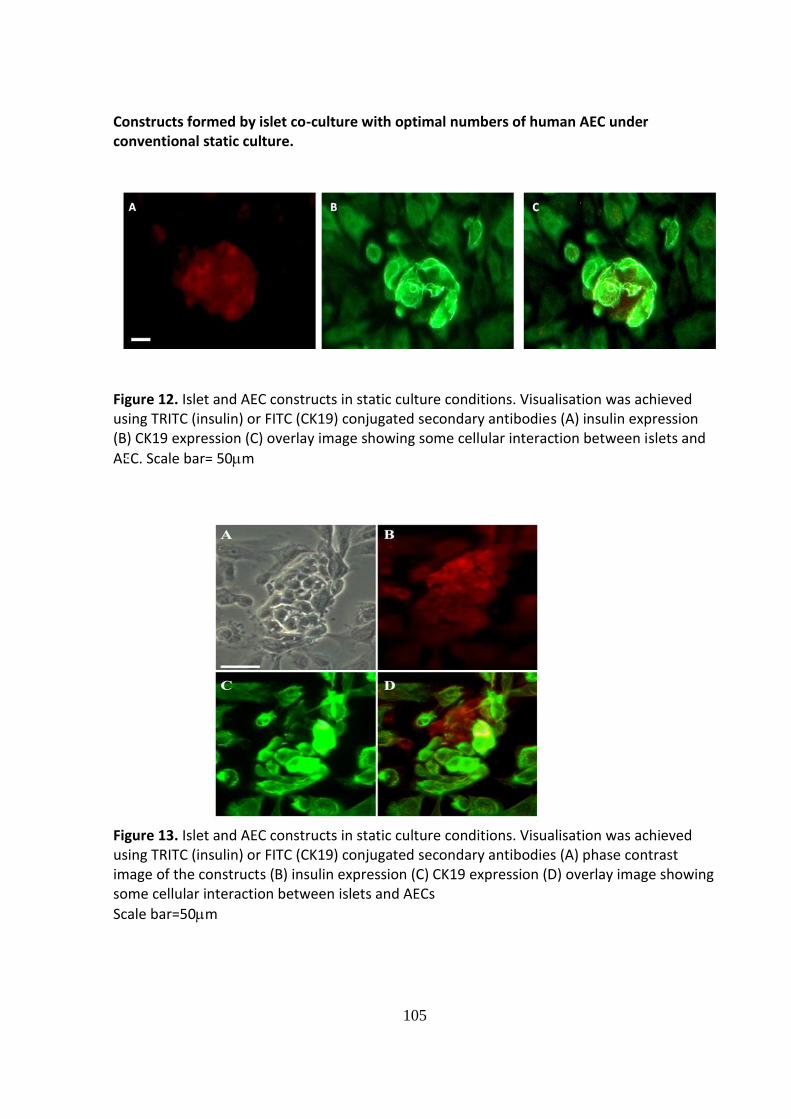
Figure 12. Islet and AEC constructs in static culture conditions. Visualisation was achieved using TRITC (insulin) or FITC (CK19) conjugated secondary antibodies (A) insulin expression (B) CK19 expression (C) overlay image showing some cellular interaction between islets and
AEC. Scale bar= 50m
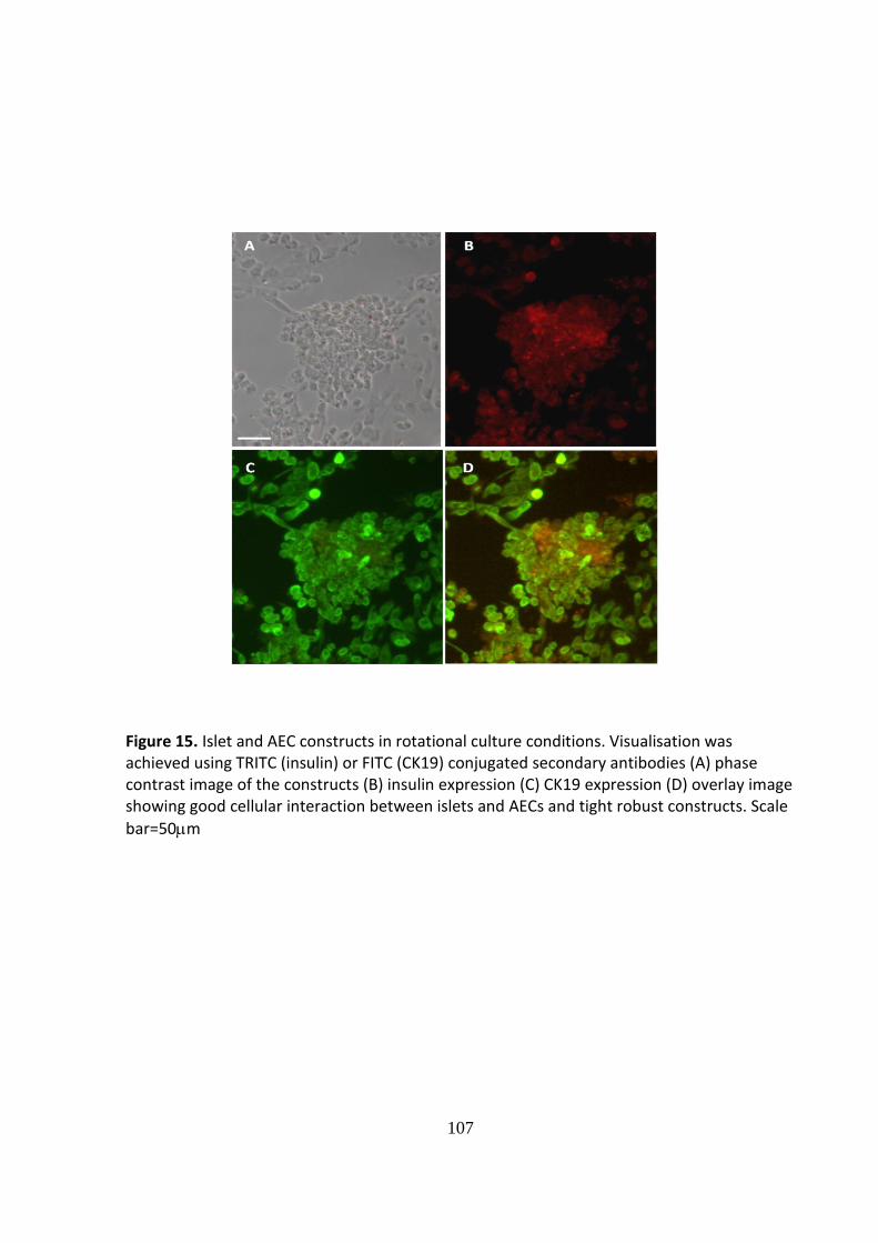
Figure 13. Islet and AEC constructs in static culture conditions. Visualisation was achieved using TRITC (insulin) or FITC (CK19) conjugated secondary antibodies (A) phase contrast image of the constructs (B) insulin expression (C) CK19 expression (D) overlay image showing some cellular interaction between islets and AECs
Scale bar=50m
A
C
106
Constructs formed by islet co-culture with optimal numbers of human AEC within the
rotational cell culture system.
Figure 14. Islet and AEC constructs in rotational culture conditions. Visualisation was achieved using TRITC (insulin) or FITC (CK19) conjugated secondary antibodies (A) phase contrast image of the constructs (B) insulin expression (C) CK19 expression (D) overlay image showing good cellular interaction between islets and AECs and tight robust constructs. Scale
bar=50m
107
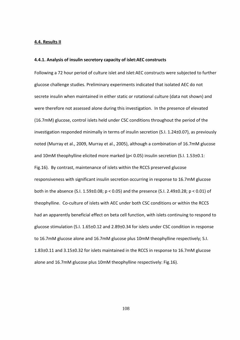
Figure 15. Islet and AEC constructs in rotational culture conditions. Visualisation was achieved using TRITC (insulin) or FITC (CK19) conjugated secondary antibodies (A) phase contrast image of the constructs (B) insulin expression (C) CK19 expression (D) overlay image showing good cellular interaction between islets and AECs and tight robust constructs. Scale
bar=50m
108
4.4. Results II
4.4.1. Analysis of insulin secretory capacity of islet:AEC constructs
Following a 72 hour period of culture islet and islet:AEC constructs were subjected to further
glucose challenge studies. Preliminary experiments indicated that isolated AEC do not
secrete insulin when maintained in either static or rotational culture (data not shown) and
were therefore not assessed alone during this investigation. In the presence of elevated
(16.7mM) glucose, control islets held under CSC conditions throughout the period of the
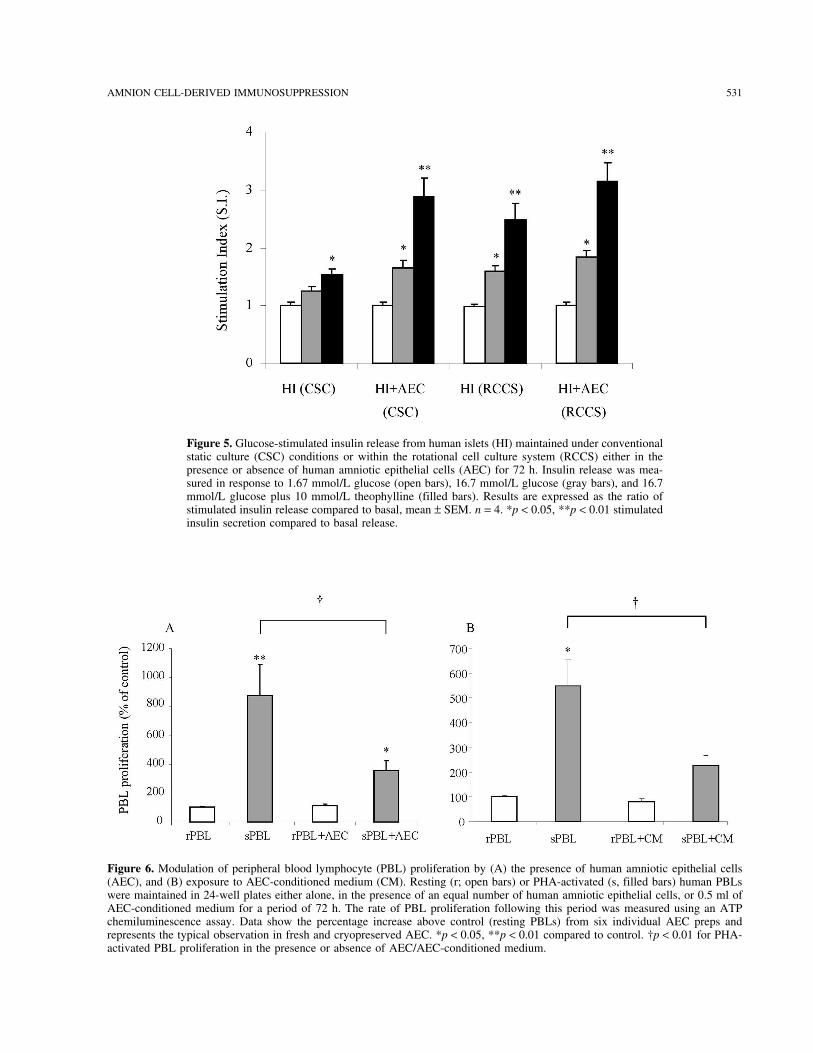
investigation responded minimally in terms of insulin secretion (S.I. 1.24±0.07), as previously
noted (Murray et al., 2009, Murray et al., 2005), although a combination of 16.7mM glucose
and 10mM theophylline elicited more marked (p< 0.05) insulin secretion (S.I. 1.53±0.1:
Fig.16). By contrast, maintenance of islets within the RCCS preserved glucose
responsiveness with significant insulin secretion occurring in response to 16.7mM glucose
both in the absence (S.I. 1.59±0.08; p < 0.05) and the presence (S.I. 2.49±0.28; p < 0.01) of
theophylline. Co-culture of islets with AEC under both CSC conditions or within the RCCS
had an apparently beneficial effect on beta cell function, with islets continuing to respond to
glucose stimulation (S.I. 1.65±0.12 and 2.89±0.34 for islets under CSC condition in response
to 16.7mM glucose alone and 16.7mM glucose plus 10mM theophylline respectively; S.I.
1.83±0.11 and 3.15±0.32 for islets maintained in the RCCS in response to 16.7mM glucose
alone and 16.7mM glucose plus 10mM theophylline respectively: Fig.16).
109
Figure 16.Glucose stimulated insulin release from human islets (HI) maintained under conventional static culture (CSC) conditions or within the rotational cell culture system (RCCS) either in the presence or absence of human amniotic epithelial cells (AEC) for 72 hours. Insulin release was measured in response to 1.67 mmol/l glucose ( ), 16.7 mmol/l glucose ( ), and 16.7 mmol/l glucose plus 10 mmol/l theophylline ( ). Results are expressed as the ratio of stimulated insulin release compared to basal, mean ± S.E.M. n=4. * p < 0.05 , ** p < 0.01 stimulated insulin secretion compared to basal release. † p < 0.01 for stimulated release in treatment groups compared to the control (ANOVA)
110
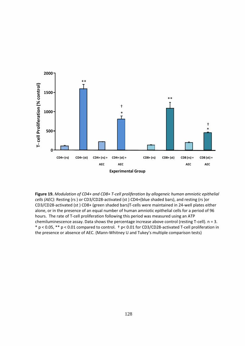
4.4.2. Analysis of immunomodulatory potential of the islet:AEC constructs
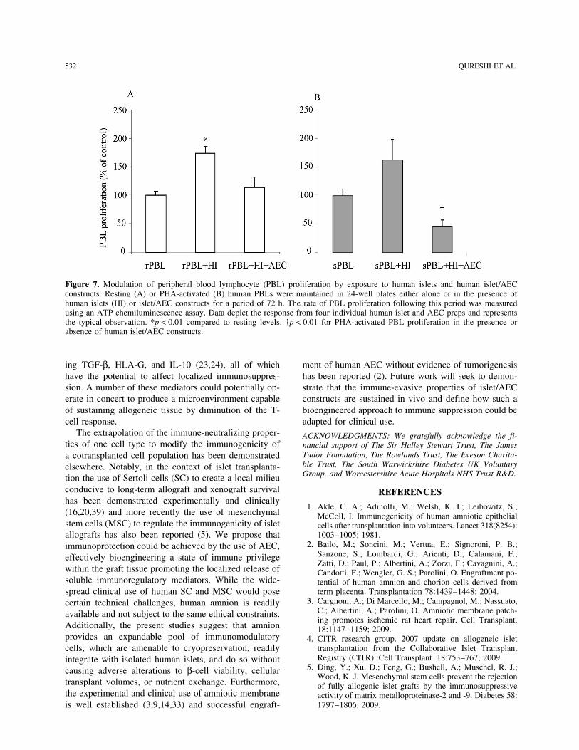
Exposure of resting PBMC to unmodified human islets which were maintained within the
RCCS elicited a marked (p <0.05) proliferative response (Fig.17.A). The presence of AEC
attenuated the resting PBMC proliferation elicited by human islets. PHA-stimulated PBMC
proliferation was increased on contact with isolated islets, but was significantly (p<0.01)
suppressed when islets were in co-culture with AEC (Fig.17.B).
111
Figures 17.A and 17.B. Modulation of peripheral blood mononuclear cell (PBMC) proliferation by exposure to human islets and human islet:AEC constructs: Resting (A) or PHA-activated (B) human PBMC were maintained in 24-well plates either alone or in the presence of human islets (HI) or islet:AEC constructs for a period of 72 hours. The rate of PBMC proliferation following this period was measured using an ATP chemiluminescence assay. Data depicts the response from 4 individual human islet and AEC preps and represents the typical observation. * p < 0.01 compared to PBMC alone (resting or activated). † p< 0.01 for PBMC proliferation in response to islet:AEC constructs compared to unmodified islets. (Mann-Whitney U)
*
†
*
†
112
4.5. Discussion
In previous chapters (2&3) the isolation, culture and morphological and functional
characterisation of human islets and AEC has been outlined in detail. The experiments
detailed in the present chapter sought to demonstrate that islet cells may be modified
through in vitro, pre-transplant interventions designed to enhance post-implant survival;
specifically, to provide evidence for our hypothesis that the co-culture of human islets with a
purified population of human amniotic epithelial cells modulates the immunogenic potential
of transplantable islet cells without impairment of -cell function. The results obtained
suggest that it is possible to bring these two cell types into close proximity whilst preserving
their respective insulin secretory function and immunomodulatory capabilities.
This chapter deals therefore, with the impact of co-culturing these two cell types and to
evaluate their combined function. Notwithstanding their disparate origins, the co-culture of
human islets and AEC under either conventional static or rotational cell culture conditions
resulted in successful physical interaction between the two cell types. The degree of
association was dependent on the density of AEC seeded with a minimum of 5x10⁴cells/ml
required before significant aggregation was observed. The observation correlates with our
earlier findings that AEC require plating at relatively high density in monolayer culture in
order to achieve good cell attachment and proliferation, which has been reported elsewhere
(Parolini et al., 2008).
The RCCS provided a more conducive environment for cellular aggregation, with the
formation of robust constructs exhibiting frequent spatial association of the insulin and CK19
113
expressing cells and a preserved islet-like morphology. The high aspect ratio vessels (HARVs)
are designed to create a microgravity environment with low shear forces permitting a
greater degree of cell-cell interaction (Unsworth and Lelkes, 1998) which may underlie the
efficient formation of stable islet:AEC constructs observed in the present study.
The close proximity of AEC to the human islets had no adverse effect on beta-cell function.
Indeed, the insulin-secretion data indicate preservation of glucose-sensitivity in human islets
maintained in co-culture with AEC. This may be compared with islets held alone under CSC
conditions which showed a diminution of glucose responsiveness. Previous studies
conducted in this laboratory indicate a beneficial impact of pancreatic ductal-epithelial cell
co-culture in preserving islet function (Murray et al., 2009), most likely due to their ability to
provide trophic support to neighbouring beta-cells (Rosenberg and Vinik, 1992). Similarly,
AEC are reported to synthesise and secrete a range of growth factors which may have
relevance for the sustained functional viability of islets seen in this novel co-culture model.
Of note, mRNA expression of TGF, EGF and KGF, known mediators of beta cell replication
(Fiaschi-Taesch et al., 2008, Movassat et al., 2003, Hanley and Rosenberg, 2007) have been
reported in intact human amniotic membrane and isolated amniotic epithelial cells (Koizumi
et al., 2000). Furthermore, dissociated AEC secrete biologically active neurotrophins
including brain derived neurotrophic factor (BDNF) (Kakishita et al., 2003) which have been
linked to -cell development and survival (Scharfmann and Czernichow, 1996). Other
studies suggest the trophic actions of AEC mediate repair processes in experimental models
of Parkinson’s Disease, stroke, spinal cord injury and liver fibrosis by encouraging
114
regeneration of host tissue or supporting the growth and engraftment of transplanted cells
(Parolini and Caruso, 2011) .
Isolated islets are known to release inflammatory cytokines (including IL-1, IL-6 and
TNFand pro-inflammatory molecules (including tissue factor and monocyte
chemoattractant protein-1) which have deleterious effects on -cell function, with
subsequent impairment of islet graft function (Marzorati et al., 2006, Matsuda et al., 2005).
Recent studies suggest that AEC exert anti-inflammatory properties as demonstrated in
animal models of lung and liver fibrosis (Manuelpillai et al., 2011, Manuelpillai et al., 2010a,
Murphy et al., 2011), reducing the tissue levels of pro-inflammatory cytokines with
concomitant release of IL-10. It is possible that in our co-culture model these anti-
function. Overall, it is likely that the close association of AEC to islets as provided by their co-
culture within the RCCS permits the paracrine release of soluble mediators able to support
insulin secretory capacity in the post isolation period with beneficial consequences in terms
of sustained islet graft function.
The proposition that the immunosuppressive properties of isolated AEC may be manipulated
to confer a state of immune-privilege on other cells capable of provoking an immune
response is confirmed by the mixed islet-lymphocyte reaction (MILR) study. Sustained
proliferation of resting PBMC was demonstrated in the presence of unmodified islets, yet
those which were closely associated (co-cultured) with AEC failed to elicit an allogeneic
response. This effect was not dependent on complete encapsulation of the islets by the
AEC; further indicative of a role for soluble immunoregulatory factors. Also, the
115
immunomodulatory response to activated (PHA-stimulated) T-cells was as robust in the
islet:AEC co-cultures as in AEC monocultures. Combined, these data suggest that AEC exhibit
a potent and generalised immunosuppressive capability, inducing an anti-proliferative
response in T-cells subjected both to mitogen and allo-antigen challenge.
This is the first study to demonstrate that the immunomodulatory capabilities of human
AEC, as observed in vitro, may be conferred on another, otherwise, immunogenic cell
population, provided that they are held in close proximity. The finding has relevance for the
wider use of islet cell replacement therapy as a treatment for Type 1 diabetes. The results
are analogous to contemporary studies where alternative immune-suppressing cell types
have been co-cultured/co-transplanted with islets, albeit in animal models. Notably, in the
context of islet transplantation the use of Sertoli cells (SC) to create a local milieu conducive
to long-term allograft and xenograft survival has been demonstrated experimentally and
clinically (Isaac et al., 2005, Kin et al., 2002, Valdes-Gonzalez et al., 2007) and more recently
the use of bone marrow-derived mesenchymal stem cells (MSC) to regulate the
immunogenicity of islet allografts has also been reported (Ding et al., 2009). We propose
that immuno-protection could be achieved by the use of AEC, effectively bio-engineering a
state of immune-privilege within the graft tissue promoting the localised release of soluble
immunoregulatory mediators. While the widespread clinical use of human SC and MSC
would pose certain technical challenges associated with accessibility and standardisation,
human amnion is readily available and not subject to the same ethical constraints.
Additionally, the present studies suggest that amnion provides an expandable pool of
immunomodulatory cells which are amenable to cryopreservation, readily integrate with
116
isolated human islets and do so without causing adverse alterations to beta cell viability,
cellular transplant volumes or nutrient exchange. Furthermore, the experimental and
clinical use of amniotic membrane is well established (Gomes et al., 2005, Hasegawa et al.,
2007, Sheridan and Moreno, 2001, Cargnoni et al., 2009) and successful engraftment of
human AEC without evidence of tumorigenesis has been reported (Bailo et al., 2004). Direct
application of this approach awaits “proof-of-concept” studies evaluating the function of
implanted islet:AEC constructs in immune-competent, diabetic animal models.
These findings raise the possibility of engineering insulin-secreting tissue constructs
applicable to cell-based therapies for diabetes, which are capable of restoring endogenous
insulin production without the need for adjuvant chronic systemic immunosuppression.
117
CHAPTER 5: AEC-MEDIATED IMMUNOMODULATION. SPECIFIC T-CELL TARGETS AND RELEVANCE TO ISLET
TRANSPLANTATION
5.1. Introduction
The previous chapters detailed a series of investigations to demonstrate the potential of
human AEC to modulate the actions of the immune system. The results suggest that AEC are
capable of subduing the proliferative activity of human PBMC in response to a known
mitogen as has been previously reported (Wolbank et al., 2007). Additionally, and for the
first time, this study provides in vitro evidence that AEC are able to confer their
immunomodulatory properties to other adjacent cell types with the overall effect of
reducing the immunogenic profile of, in this instance, human islet cells, and do so without
impairing function viz. physiological release of insulin. Such a property may have beneficial
implications for tissue/cell replacement therapy where localised immune-privilege mediated
by AEC may serve to shield co-transplanted therapeutic cells from immune rejection. At
present the precise mechanism(s) by which AEC restrict lymphocyte proliferation require
further elucidation; clinical application of the immunomodulatory actions of AEC would
benefit from a clearer understanding of the individual T-cell sub-populations targeted by AEC
and the role that such T-cells play in islet graft rejection.
Our understanding of the mechanism(s) underlying allo and auto-reactivity in islet-cell
replacement therapy is based on experimental and limited clinical transplantation data. The
presence of islet-specific autoreactive CD4+ T- cells at time of transplantation coupled with
118
increased frequencies of circulating CD8+ T- cells (insulin B10–18 reactive) is considered to
significantly influence clinical outcome of islet transplant recipients (Huurman et al., 2008,
Pinkse et al., 2005). The insulin-specific CD8+ T- cells show potential cytolytic activity,
producing granzyme B and IFN-γ and are therefore potentially able to destroy insulin-
producing -cells. Equally the absence of these markers, and therefore presumably of auto-
reactivity, correlates with good clinical outcome (Huurman et al., 2008, Pinkse et al., 2005).
Allo-rejection of transplanted islets is considered to be mediated by both CD4+ and CD8+ T-
cells and both populations are required to accomplish -cell death. The actions of a number
of other immune cells including macrophages, dendritic cells (DC) and B lymphocytes are co-
ordinated to induce and sustain the immune assault. Macrophages and DC act as antigen-
presenting cells and stimulate the migration and infiltration of grafted cells by peripheral
CD4+ and CD8+ T-cells. The islets are also targeted by natural killer cells (NK) and B
lymphocytes. Infiltrating macrophages serve to activate cytotoxic CD8+ cells which, in the
same manner as auto-reactive CD8+ T-cells cause -cell destruction by the release of
cytolytic agents. In allograft rejection CD4+ T-cells may also act indirectly through B-cell
activation and the generation of complement fixing antibodies. Pro-inflammatory cytokines
including interleukin (IL)-12 released by macrophages activate Th1-type CD4+ T-cells which
subsequently secrete IL-2, interferon-, and TNF- to further augment the CD8+ response.
119
Aims of the Chapter
As a means of gaining further understanding of the relevance of AEC- mediated
immunomodulation to islet graft protection the next series of studies sought to more closely
examine the specific immune cell targets involved. As populations of CD4+ and CD8+ T-cells
play a major role in both auto and allo-graft rejection this study sought to determine the
modulatory potential of AEC in regard to these two cell types.
120
5.2. Materials and Methods
5.2.1. CD4+ T-cell isolation
A Dynabead®-mediated negative selection system was employed to isolate CD4+ T-cell
populations (Dynabeads® Untouched™ Human CD4 T-cells, Life Technologies, Paisley, UK).
The isolation kit is designed to deplete B cells, NK cells, monocytes, platelets, dendritic cells,
CD8+ T-cells, granulocytes and erythrocytes from platelet-poor PBMC samples, leaving
isolated CD4+ T- cells free of bead and antibody, thus making them appropriate for use in
subsequent proliferation assays. In the present study PBMC were isolated from CD leucocyte
cones (leucocyte concentrates) obtained from healthy donors (NHS Blood and Transplant,
Birmingham). The cells were processed within 18 hours of blood collection and PBMC
isolation was performed as described in section 3.3.1. The PBMC preparation was subjected
to 3 washes in isolation buffer; Phosphate Buffered Saline (PBS) without Mg2+ and Ca2
+
supplemented with 0.6% sodium citrate and 0.1% BSA. PBMC were counted and adjusted to
a density of 1x10⁸cells/ml in isolation buffer. A 200l aliquot of the PMBC suspension was
transferred to a 15ml conical tube to which was added 40l of heat inactivated foetal calf
serum (FCS, Sigma-Aldrich Ltd). This was followed by addition of 40l of antibody mix
containing mouse IgG antibodies for CD8, CD14, CD16 (specific for CD16a and CD16b), CD19,
CD36, CD56, CDw123 and CD235a (GlycophorinA), being sure that the suspensions were
thoroughly mixed. The cell/antibody suspension was incubated for 20mins at 4°C prior to
thorough washing in 4mls of isolation buffer. Cells were harvested by centrifugation at 300g
for 8 mins at 4°C and re-suspended in 200l of isolation buffer. To this was added 200l of
pre-washed Depletion MyOne® Dynabeads at the same density as the cells, followed by
121
incubation for 15 mins at RT with gentle tilting and rotation using a Hulamixer®. The bead-
bound cells were re-suspended by vigorously triturating the sample through a 1000l
pipette tip (approx. 10 times) before addition of 2mls of isolation buffer. The tube containing
the cells was then placed into a magnet for 2 mins before transferring the supernatant to a
new tube. The original tube was washed with another 2mls of isolation buffer and returned
to the magnet. The supernatant was again collected into a fresh tube. Finally the
supernatants were pooled and placed in the magnet for a further 2 mins to remove any
remaining beads. The supernatants containing the free CD4+ cells were then washed in
isolation buffer and cells harvested by centrifugation at 300g for 5 mins at 4°C prior to
counting and assessment of viability as described in section 3.3.1.
5.2.2. CD8+ T-cell isolation
CD8+ T-cells were isolated using a Dynabead®-mediated negative selection system
(Dynabeads® Untouched™ Human CD8 T-cells, Life Technologies, Paisley, UK) as described
above for CD4+ T- cells but with the following modifications. 500l of PBMC suspension (at a
density of 1 x 10⁸cells/ml) were transferred to a 15ml conical tube. The cells were
supplemented first with 100l of FCS and then with 100l of antibody mix consisting of
biotinylated mouse IgG antibodies for CD4, CD14, CD16 (specific for CD16a and CD16b),
CD19, CD36, CD56, CDw123 and CD235a (Glycophorin A) ensuring that cell and antibody
solutions were well mixed. The cells were incubated for 20 mins at 4°C prior to thorough
washing in 10mls of isolation buffer. Cells were harvested by centrifugation at 350g for 8
mins at 4°C and re-suspended in 500l of isolation buffer. To this was added 500l of pre-
122
washed Depletion MyOne® Dynabeads followed by incubation for 15 mins at RT with gentle
tilting and rotation using a Hulamixer®. The bead-bound cells were re-suspended by
vigorously triturating the sample before addition of 5mls of isolation buffer. Magnetic
assisted separation of the bead-bound cells and the resulting purified CD8+ cells were
harvested, counted and assessed for viability.
5.2.3. Confirmation of T-cell purity by Flow Cytometry
Purity of the CD4+ T-cell subset was confirmed by cell surface marker labeling and flow cytometry.
Cell surface staining was achieved by use of a Brilliant Violet 421™conjugated anti-human
CD4+ antibody or the Isotype control (BioLegend, Supplied by Cambridge Bioscience,
Cambridge, UK). The antibodies were diluted 1:100 in Fluorescence activated cell sorting
(FACS) buffer and added to samples of the CD4+ T-cells prior to incubation on ice in the dark
(covered with foil) for 15 mins. 300l of FACS buffer was then added before centrifugation at
400g, 4°C, for 5 mins. The supernatant was removed and replaced with 100l of Fix Buffer
(made as per manufacturer’s instructions) to each tube, which were incubated on ice for 30
mins. CD4+ were harvested by centrifugation at 4°C, 400g for 6 mins and washed again with
300l of FACS Buffer. Following centrifugation at 400g, 4°C for 6 mins, the supernatant was
removed and the cells were washed 2 times with 200l of Perm Buffer (prepared according
to manufacturer’s instructions) pelleting between washes by centrifugation 400g, 4°C for 6
mins.
123
A Cyan 3 laser 9-colour flow cytometer equipped with Summit™ data software (Beckman Coulter,
High Wycombe, UK) was used to analyse the CD4+ T-cell population on the basis of the forward
scatter/side scatter profile of cells labelled with BV –anti-CD4+ as detailed above using an isotype-
matched negative control antibody to quantify the degree of positive staining.
5.2.4. Proliferation Studies
5.2.4.1. CD4+ T-cell proliferation: modulation with phytohaemagglutinin (PHA), human
amniotic epithelial cells (AEC)
In preliminary studies we used our existing protocol to determine whether CD4+ T-cell
proliferation was influenced by exposure to human amniotic epithelial cells (AEC). To this
end P1 AEC were plated at a density of 5x10⁴/ml into the wells of a 24-well plate and
incubated at 37°C for 72 hours to enable firm anchorage. CD4+ T-cells, processed as
described above, were plated at equal density either in the presence or absence of the
attached AEC. Additionally, further groups of CD4+ T-cells were plated in the presence of the
plant mitogen phytohaemagglutinin at a concentration of 5g/ml. Incubation was carried
out at 37°C for 72 hours before the CD4+ T-cells were harvested prior to intracellular ATP
analysis using chemiluminescence (see section 3.3.4).
124
5.2.4.2. PBMC, CD4+ and CD8+ T-cell proliferation: modulation with anti CD3/CD28, human
amniotic epithelial cells (AEC) and human islets
In light of the findings of the preliminary study (detailed in section 5.3.1. below) the next
series of experiments were performed using a more specific T-cell activation method.
Dynabeads® Human T-Activator CD3⁄CD28 beads (Invitrogen, Life Technologies, Paisley, UK)
were employed as an alternative to PHA. CD3/CD28 activator beads are reported to provide
physiological activation and expansion of human T-cells including CD4+ and CD8+ cells. The
cells once activated can be analysed or subjected to further differentiation protocols (e.g.
differentiation to T-helper cells). Proliferation assays were initiated using AEC at P1 plated at
a density of 5x10⁴cells/well, as described above. Unfractionated PBMC, purified CD4+ or
CD8+ T-cells were plated at the same density either in the presence or absence of AEC and a
further group of cells were plated in the presence of 5x10⁴ pre-washed CD3/CD28 activation
beads. Incubation was carried out at 37°C for 96 hours before the cells were harvested for
intracellular ATP analysis using chemiluminescence (see section 3.3.4). In a small series of
experiments mixed islet:lymphocyte proliferation studies were performed. For these studies
islets were cultured in the presence or absence of AEC in RCCS for 72 hours to allow
aggregates to form. Islets and islet:AEC constructs were seeded into 24-well plates
(50IEQ/aggregates per well) and allowed to anchor for up 72 hours. Proliferation assays
were then established as for the AEC monocultures as described above.
125
5.3. Results
5.3.1. Expansion of CD4+ T-cell populations using PHA and CD3/CD28 activation beads
Flow cytometry confirmed the purity of the CD4+ T-cells used in the present studies. In
preliminary investigations it was observed that isolated CD4+ T-cell populations were largely
unresponsive to the routinely used stimulator, phytohaemagglutinin (PHA). Whereas
unfractionated PBMC numbers increased in the presence of 5g/ml PHA, CD4+ T-cell
numbers did not change significantly. By contrast incubation of both unfractionated PBMC
and isolated CD4+ T-cells with CD3/CD28 stimulator beads produced a robust increase in
numbers of both cell populations (Fig 18). In subsequent proliferation studies CD3/CD28
beads were adopted as the stimulator of choice.
These results were used as the basis for subsequent proliferation assays using fractionated
CD4+ and CD8+ T-cells. In these studies it was observed that activation of both cell types are
modulated on co-culture with an equal number of allogeneic human AEC (Fig.19). Resting
CD4+ and CD8+ T-cells were largely unresponsive to co-culture with allogeneic AEC but
responded robustly to the presence of an equal number of CD3/CD28 activator beads. The
degree of T-cell expansion induced by the beads was significantly (p < 0.01) abrogated in the
presence of allogeneic AEC with a greater than 50% inhibition for both T-cell populations
(Fig.19).
126
5.3.2. Fractionated CD4+ and CD8+ T-cell proliferation assays. Impact of co-culture with
human AEC
This series of studies first sought to determine whether human AEC are able to modulate the
proliferative capacity of fractionated CD4+ and CD8+ T-cells. As observed with PBMC,
allogeneic AEC failed to provoke significant CD4+ T-cell proliferation when co-cultured at
varying densities ranging from 2.5x10³-5x10⁴. Employing CD3/CD28 activator beads as the
stimulus, AEC-mediated inhibition of CD4+ T-cell proliferation was observed at all ratios
tested from 1:20 – 1:1, exhibiting a degree of dose-dependency (Fig.20).
5.3.3. Response of CD4+ and CD8+ T-cell populations on co-culture with allogeneic human
islets and islet:AEC constructs
In a limited study human islets prepared as outlined in Section 2.2. were used in a mixed
islet:lymphocyte reaction. In vitro, allogeneic human islets provoked a moderate activation
of both CD4+ and CD8+ T-cells (Fig 21). Expansion of both T-cell populations was observed
during a 96 hour co-culture period with human islets. By contrast, when human islets were
pre-cultured with human AEC to form constructs as detailed section 4.2.1, the response of
CD4+ of CD8+ cells was significantly attenuated (Fig.21).
127
Figure 18. Activation of PBMC (A) and fractionated CD4+ T-cells (B) on exposure to either
phytohaemagglutinin (PHA - 5g/ml) or an equal number (5x10⁴) of CD3+/CD28+ activation beads. PHA produced a robust increase in PBMC numbers but a greater response was observed in both cell populations when the activator beads where employed as the stimulus. n = 4. * p < 0.05, **p < 0.01 compared with control (resting cells). One way ANOVA
0
100
200
300
400
500
600
700
800
PBMC PBMC+PHA PBMC+CD3/CD28
PB
MC
Pro
life
rati
on
(% o
f C
on
tro
l)
0
1000
2000
3000
4000
5000
6000
CD4+ CD4+PHA CD4+CD3/CD28
CD
4+P
rolif
era
tio
n (%
of
con
tro
l)
A B
*
** **
128
Figure 19. Modulation of CD4+ and CD8+ T-cell proliferation by allogeneic human amniotic epithelial cells (AEC): Resting (rs ) or CD3/CD28-activated (st ) CD4+(blue shaded bars), and resting (rs )or CD3/CD28-activated (st ) CD8+ (green shaded bars)T-cells were maintained in 24-well plates either alone, or in the presence of an equal number of human amniotic epithelial cells for a period of 96 hours. The rate of T-cell proliferation following this period was measured using an ATP chemiluminescence assay. Data shows the percentage increase above control (resting T-cell). n = 3. * p < 0.05, ** p < 0.01 compared to control. † p< 0.01 for CD3/CD28-activated T-cell proliferation in the presence or absence of AEC. (Mann-Whitney U and Tukey’s multiple comparison tests)
0
500
1000
1500
2000
CD4+ (rs) CD4+ (st) CD4+ (rs) +
AEC
CD4+ (st) +
AEC
CD8+ (rs) CD8+ (st) CD8 (rs) +
AEC
CD8 (st) +
AEC
T-
cell
Pro
life
rati
on
(% c
on
tro
l)
Experimental Group
**
**
*
*
†
†
129
Figure 20. Dose-dependent modulation of CD4+ T-cells on co-culture with human amniotic epithelial cells (AEC): AEC were pre-plated at densities ranging from 2.5x10³-5x10⁴ cells/well and co-cultured with 5x10⁴ CD4+ T-cells for 96 hours. Cell proliferation was measured by chemiluminescence assay. n = 3. * p < 0.05, ** p< 0.01 for CD4+ T-cell proliferation in the presence vs. absence of AEC. One way ANOVA
0
200
400
600
800
1000
1200
CD4 1:20 1:10 1:5 1:2.5 1:1
CD
4+
T c
ell
Pro
life
rati
on
(%
of
con
tro
l)
AEC:CD4+ ratio
***
**
130
Figure 21. Modulation of CD4+ and CD8+ T-cell proliferation by exposure to human islets and human islet:AEC constructs: Resting CD4+ T-cells (blue bars) and CD8+ T-cells (green bars) were maintained in 24-well plates either alone or in the presence of human islets (HI) or islet:AEC constructs (HI/AEC) for a period of 96 hours. The rate of T-cell proliferation following this period was measured using an ATP chemiluminescence assay. Data depicts the response from 2 individual human islet and AEC preps and represents the typical observation. * p < 0.01 compared to resting levels. † p< 0.01 for T-cell expansion in the presence of HI/AEC constructs compared to HI alone. (Mann-Whitney U and Tukey’s multiple comparison tests)
T c
ell
Pro
life
rati
on
(% o
f co
ntr
ol)
0
20
40
60
80
100
120
140
160
CD4+ CD4+/HI CD4+/HI/AEC CD8+ CD8+/HI CD8+/HI/AEC
Experimental Group
*
*
†
†
131
5.4. Discussion
This investigation provides further evidence that the immuno-modulatory potential of
human AEC may have relevance to the protection of islet allografts from immune assault.
The findings suggest that several of the targets of AEC-induced immunomodulation have
relevance to islet allograft rejection and as such indicate the potential use of AEC
populations as mediators of “bystander” immunosuppression in islet transplantation. The
data indicate that both CD4+ and CD8+ T-cell activation is responsive to AEC and that
bringing these immune cells into close proximity of AEC prevents their in vitro expansion in
response to a physiological stimulus viz. CD3/CD28 complex. As the activation and
proliferation of CD4+ and CD8+ T-cells is crucial to the appropriate and successful activation
of the innate and adaptive immune response, culminating in allograft rejection, suppression
of their function by AEC may have relevance for providing a localised means of providing
immune protection to transplanted islet cells and circumvent the need for long-term, non-
specific systemic immunosuppression.
The lack of responsiveness of fractionated CD4+ T-cells to the routinely used mitogen PHA
prompted modification to the protocol for assessing AEC-mediated modulation in the
present chapter. The failure of PHA to cause significant CD4+ T-cell expansion at a
concentration which effectively increased PBMC numbers indicates that accessory cell
populations (i.e. macrophages, dendritic cells) may be required for appropriate CD4+
stimulation in the presence of this mitogen. Indeed it has been reported that CD4+ T-cells
fail to express IL-2 receptor (CD25) or respond appropriately to IL-2 in the absence of cells
involved in antigen presentation and as a result, expansion of T-cell population is blunted
132
(Halvorsen et al., 1988, Leivestad et al., 1988). Hence it was necessary when conducting
studies with purified CD4+ and CD8+ populations to use a stimulus which would provide the
required antigen presentation and which also has relevance in terms of clinical application.
Immuno-magnetic anti-CD3 anti-CD28 coated beads are employed for in vitro T-cell
expansion prior to infusion in immunotherapy (Thompson et al., 2003). Commercially
available expansion beads seek to mimic physiological activation of CD4+ and CD8+ T-cells by
simulating in vivo T-cell proliferation in response to antigen-presenting cells. The system
utilizes the two activation signals CD3 and CD28, bound to a three-dimensional bead similar
in size to endogenous antigen-presenting cells. Thus this method provides appropriate T –
cell activation without the need to re-introduce macrophages/dendritic cells which may
themselves be directly or indirectly modulated by contact with human AEC.
Having made changes to the protocol it was possible to clearly demonstrate that CD4+ and
CD8+ T-cell proliferation, similarly to unfractionated PBMC, was abrogated by co-culture
with AEC. In contrast to the results with unsorted PBMC, we observed a relatively small
increase in CD4+ and CD8+ T-cell numbers under basal conditions which could be indicative
of a mild allogeneic response. However, as AEC are also a source of many trophic factors
(Parolini and Caruso, 2011) it is also possible that the increase in cell numbers under resting
conditions reflects the generalised cell-supportive characteristics of AEC/AEC-conditioned
medium which may serve to promote survival of purified resting T-cells and reduce the basal
rate of apoptosis.
133
The results of the initial mixed islet:lymphocyte study suggests that CD4+ and CD8+ cells are
activated by islet allo-antigens. The subdued nature of their response to islet co-culture may
be due to the decline of immunogenicity and depletion of carrier (host-derived) dendritic
cells which occurs in human islets during prolonged culture, especially within the RCCS
(Rutzky et al., 2002). Yet, elevated CD4+ and CD8+ T-cell numbers were observed during
T-cell:islet co-culture but this increase was abrogated in the islet:AEC constructs as
demonstrated in Chapter 4.
There is a general consensus, based on studies in experimental islet transplantation that
both CD4+ and CD8+ T-cells are involved islet allograft rejection with initial infiltration of
antigen-presenting cells, such as macrophages and DC mediating the response. The release
of cytokines notably IL-2 and IL-6 trigger lymphocyte activation and the migration of CD4+
and CD8+ T-cells to the site of the graft. Additional pro-inflammatory cytokines secreted by
CD4+ T-cells prompt the invasion of NK cells and B lymphocytes which on activation
generate complement fixing antibodies. CD8+ cytotoxic cells cause further -cell damage by
the release of chemicals granzymes and perforins which induce cell lysis. The allogeneic
response is exacerbated by pro-inflammatory cytokines released by activated Th1 type CD4+
T-cells which secrete IL-2, interferon-, and TNF- which maximise CD8+ activation.
To summarise, in combination the immunoregulatory properties of AEC have the capacity to
influence several aspects of immune-mediated destruction of the islet graft. Firstly they may
directly induce apoptosis of CD4+ and CD8+ T-cells by the actions of soluble FasL and HLA-G.
Whilst the Fas/FasL pathway is normally associated with CD8+ mediated cell destruction,
134
and has the propensity to activate neutrophil-mediated inflammation (Turvey et al., 2000)
the presence of FasL bearing AEC may become dominant in a microenvironment in which the
Th1/Th2 ratio is in favour of Th2 cells and therefore anti-inflammatory mediators
predominate (Pearl-Yafe et al., 2006). The secretion of TGF- and IL-10 by AEC may also
serve to prevent the activation and proliferation of T effector cells and inhibit the production
of pro-inflammatory cytokines. Additionally, AEC also secrete macrophage inhibitory factor,
reducing the magnitude of immune cell migration and therefore graft infiltration (Li et al.,
2005)
135
CHAPTER 6: FUTURE WORK, GENERAL DISCUSSION AND CONCLUSIONS
Diabetes mellitus is a metabolic disorder resulting in hyperglycaemia and the onset of
secondary complications, which are in turn associated with significant morbidity and
mortality. The pharmacological approach to its treatment (insulin, oral hypoglycaemics)
serves to manage the condition, but is not a cure and does not provide full protection from
the potentially life-threatening consequences of the disease. The definitive therapeutic
strategy for T1DM is the re-instatement of endogenous insulin production to restore
physiological glucose control. Both whole organ viz. pancreas and islet cell transplantation
offer the recipient the possibility to achieve and maintain euglycaemia and since the early
2000s and the introduction of the Edmonton protocol, islet transplantation outcomes have
improved significantly where 1- year insulin-independence rates have reached 70% to 90% in
experienced transplant centres (Shapiro et al., 2006). Yet whilst early results are good,
current protocols are still associated with a progressive and steady decrease in graft
function; up to 90% of recipients return to insulin therapy within 5 years of their first
infusion (Ryan et al., 2005).
As discussed in Chapter 1, the underlying cause of islet graft attrition is likely to be
multifactorial, involving recurrence of autoimmunity, allogeneic rejection, toxicity of the
immunosuppressive drug regimen, poor graft re-vascularisation and “metabolic fatigue” of
the islet graft. Determining the precise cause of graft failure in each clinical case is restricted
by the limitations in technology for monitoring transplanted islets although much research is
136
now concerned with graft imaging and surveillance for signs of immunologic events in the
peripheral blood (Berney and Toso, 2006). The impact of immune-mediated -cell loss is,
however, likely to be significant, with studies indicating an association between pre-
transplant cellular auto-reactivity and poor clinical outcome (Huurman et al., 2008).
Furthermore, in case studies graft failure is demonstrated to be preceded by the detection
of antibodies to HLA class II antigens (Kessler et al., 2009).
Immunosuppressive drugs fall short of producing long term graft protection and additionally,
expose the recipient to toxicity, increased vulnerability to infection and heightened
susceptibility to malignancy. These risks are factored into the pre-operative
evaluation/assessment of potential islet transplant recipients, being balanced against the
potential benefits of the procedure in terms of reducing insulin requirement, providing good
glucose control and limiting the frequency of hypoglycaemic events by restoring
hypoglycaemic awareness. Such considerations invariably result in only the most poorly
controlled diabetic individuals being selected for transplant, and then only after intensive
insulin therapies have been explored, including the use of insulin pumps. If islet
transplantation is to be made more readily available, becoming a routine treatment option
for diabetes, it is of paramount importance to develop more long term, effective and safe
methods of islet graft immuno-protection.
This thesis seeks to provide preliminary evidence in support of the clinical exploitation of
immune-privileged tissue to locally control the recipient immune response in cellular
transplantation. The unique features of the islet graft viz. discrete cell clusters transplanted
137
in a relatively small volume means that it may be possible to avoid chronic, systemic
immunosuppression by pre-transplant modification of the graft combined with manipulation
of the implantation site. Human amniotic epithelial cells (AEC) are chosen as a potential
candidate for this role due to certain key characteristics, notably their immune inertness on
transplantation and ability to markedly reduce T-cell proliferation as demonstrated in this
study (Chapter 3) and by others (Parolini et al., 2008). The present study is the first to seek
to capitalise on these features of AEC in the context of islet transplantation and to provide
direct (in vitro) evidence that AEC are capable of modulating the immunogenicity of human
islets to the T-cells directly implicated in graft rejection (Chapter 4&5).
Complete characterisation of the mechanisms underlying the immunosuppressive potential
of AEC will form the basis of future work. Our current understanding of the properties of AEC
will determine which potential mechanisms merit closer investigation. Special attention
should be paid to the possibility that the unique repertoire of soluble mediators secreted by
AEC create graft tolerance either by a direct cytotoxic effect on CD4+ and CD8+ T-cells
(potentially via soluble HLA-G) (Pratama et al., 2011, Hammer et al., 1997) or more
interestingly, by the induction of regulatory T-cells (T-regs). Increasingly, regulatory T-cells
(CD4+/CD25+/Foxp3 regulatory T-cells) are being implicated in the suppression of allogeneic
graft rejection and the development of immune-tolerance (Wood, 2011, Wood et al., 2003).
In the periphery T-regs are derived from naïve CD4+ cells and are thought to arise as a
consequence of appropriate exposure to antigen and co-stimulation in the presence of
cytokines which induce expression of Foxp3, a gene which, in turn, orchestrates T-reg
expansion. AEC secrete two cytokines considered to be implicated in the induction of Foxp3
138
namely transforming growth factor beta (TGF- and IL-10 (Manuelpillai et al., 2010b,
Pothoven et al., 2010, Chung et al., 2009, Li et al., 2005)and are therefore capable, at least
in theory, of creating a local environment conducive to the expansion of T-reg populations.
In the context of islet transplantation AEC could induce the expression of alloantigen-specific
T-regs able to counteract the deleterious effects of CD4+ and CD8+ T-cells and therefore
prevent graft rejection and mediate link unresponsiveness. To test this theory, future studies
using T-cell proliferation assays would be conducted to detect changes in the levels of Foxp3
expression, by flow cytometry, in CD4+ T-cells cultured in the presence or absence of
dispersed AEC and AEC supernatant, and also with islet:AEC constructs. Subsequent studies
would attempt to correlate changes in Foxp3 expression to the prevailing levels of TGF- and
IL-10 in the assay supernatant, and further validate their role by addition of TGF- and IL-10
antibodies to block Foxp3 induction.
Additional potential targets for AEC- mediated immunosuppression include restriction of
dendritic cell maturation (Li et al., 2006b) and inhibition of T-cell proliferation by
indoleamine 2,3 dioxygenase (IDO) via tryptophan depletion (Jones et al., 2007) which may
also be explored as part of ongoing studies.
The islet:AEC constructs bioengineered in the present study exhibit both insulin-secretory
and immunomodulatory capabilities (Chapters 4&5). These two characteristics make
islet:AEC constructs ideal for use in cell replacement therapy for the treatment of diabetes.
In future work an assessment of their ability to survive and perform under experimental,
physiological conditions must be undertaken using animal models, thus providing “proof-of-
139
concept”. A positive outcome would provide the necessary rationale for studies in higher
non-human primates prior to transferring the technology to a clinical setting.
The series of in vivo studies would include assessment of construct survival in both immune-
deficient and immune-competent murine models using the renal sub-capsular space as the
implant site and detection of circulating human C-peptide and insulin as indicators of graft
function. Additional studies in diabetic murine models (streptozotocin-treated and non
obese diabetic – NOD) will confirm the ability of the islet:AEC constructs to restore and
maintain normoglycaemia in the absence of chronic immunosuppression. Appropriate
histological examination of explanted tissue i.e. haematoxylin and eosinophil staining,
immunocytochemical localisation of human insulin, glucagon, E-cadherin, and infiltration of
murine CD4+, CD8+ and Foxp3 positive cells will further define the degree of graft survival
and increase our understanding of the mechanism(s) underlying graft immuno-protection.
It is envisaged that the clinical use of modified islets whether by co-aggregation with human
AEC, bone marrow-derived mesenchymal stem cells or other immunomodulatory cell
populations would ultimately operate in conjunction with other novel strategies of graft
immuno-protection. It is feasible to suggest that during the initial transplant period
additional methods of immune protection would be required, giving the AEC time to
establish and exert influence at the site of implantation. The putative mechanism(s)
underlying AEC-mediated immunosuppression may well be compatible with some of the
newer methods of graft protection currently being explored, including co-stimulation
blockade (notably the CD28/CD80/86 and CD40/CD154 pathways) and the use of anti-CD3 or
140
T-cell depleting antibodies (Bellin et al., 2012). Their use as induction therapy in conjunction
with islet:AEC co-grafting may circumvent the need for the use of calcineurin inhibitors
(tacrolimus) and other more conventional immunosuppressive agents (sirolimus) and
therefore eliminate their attendant toxic actions both on the graft and the recipient.
Whilst islet transplantation continues to be defined as a research or experimental treatment
it delivers real improvements both to the health status and quality of life of graft recipients.
Unquestionably, there is a need to refine the technique, reducing the risk:benefit ratio and
improving long term clinical outcome if islet transplantation is to become a serious rival to
exogenous insulin therapy. The work detailed in the present thesis provides a rationale to
examine novel cell-based strategies for islet graft immune protection, which may in time,
contribute to a more effective means of ensuring sustained islet graft survival.
141
REFERENCES ADA 2009. Diagnosis and classification of diabetes mellitus. Diabetes Care, 32 Suppl 1, S62-7.
ADAMS, G. A., WANG, X., LEE, L. K., PIERCY, C. E., ALFREY, E. J. & DAFOE, D. C. 1994. Insulin-like
growth factor-I promotes successful fetal pancreas transplantation in the intramuscular site.
Surgery, 116, 751-5.
ADINOLFI, M., AKLE, C. A., MCCOLL, I., FENSOM, A. H., TANSLEY, L., CONNOLLY, P., HSI, B. L., FAULK,
W. P., TRAVERS, P. & BODMER, W. F. 1982. Expression of HLA antigens, beta 2-microglobulin
and enzymes by human amniotic epithelial cells. Nature, 295, 325-7.
AKLE, C. A., ADINOLFI, M., WELSH, K. I., LEIBOWITZ, S. & MCCOLL, I. 1981. Immunogenicity of human
amniotic epithelial cells after transplantation into volunteers. Lancet, 2, 1003-5.
ALFADHLI, E., KOH, A., ALBAKER, W., BHARGAVA, R., ACKERMAN, T., MCDONALD, C., RYAN, E. A.,
SHAPIRO, A. M. & SENIOR, P. A. 2009. High prevalence of ovarian cysts in premenopausal
women receiving sirolimus and tacrolimus after clinical islet transplantation. Transpl Int, 22,
622-5.
BACHA, F., SAAD, R., GUNGOR, N. & ARSLANIAN, S. A. 2004. Adiponectin in youth: relationship to
visceral adiposity, insulin sensitivity, and beta-cell function. Diabetes Care, 27, 547-52.
BAILO, M., SONCINI, M., VERTUA, E., SIGNORONI, P. B., SANZONE, S., LOMBARDI, G., ARIENTI, D.,
CALAMANI, F., ZATTI, D., PAUL, P., ALBERTINI, A., ZORZI, F., CAVAGNINI, A., CANDOTTI, F.,
142
WENGLER, G. S. & PAROLINI, O. 2004. Engraftment potential of human amnion and chorion
cells derived from term placenta. Transplantation, 78, 1439-48.
BASSI, E. J., DE ALMEIDA, D. C., MORAES-VIEIRA, P. M. & CAMARA, N. O. 2011. Exploring the Role of
Soluble Factors Associated with Immune Regulatory Properties of Mesenchymal Stem Cells.
Stem Cell Rev.
BECK, J., ANGUS, R., MADSEN, B., BRITT, D., VERNON, B. & NGUYEN, K. T. 2007. Islet encapsulation:
strategies to enhance islet cell functions. Tissue Eng, 13, 589-99.
BELLIN, M. D., BARTON, F. B., HEITMAN, A., HARMON, J., BALAMURUGAN, A. N., KANDASWAMY, R.,
SUTHERLAND, D. E., ALEJANDRO, R. & HERING, B. J. 2012. Potent Induction Immunotherapy
Promotes Long-Term Insulin Independence After Islet Transplantation in Type 1 Diabetes. Am
J Transplant.
BEN-HAROUSH, A., YOGEV, Y. & FISCH, B. 2004a. Insulin resistance and metformin in polycystic ovary
ZHAO, M., MUIESAN, P., AMIEL, S. A., SRINIVASAN, P., ASARE-ANANE, H., FAIRBANKS, L., PERSAUD,
S., JONES, P., JONES, J., ASHRAF, S., LITTLEJOHN, W., RELA, M., HEATON, N. & HUANG, G. C.
175
2007. Human islets derived from donors after cardiac death are fully biofunctional. Am J
Transplant, 7, 2318-25.
ZUK, D. M., KOH, A., IMES, S., SHAPIRO, A. M. & SENIOR, P. A. 2011. Three cases of alopecia following
clinical islet transplantation. Am J Transplant, 11, 163-8.
APPENDIX
Publications arising from this work
Cell Transplantation, Vol. 20, pp. 523–534, 2011 0963-6897/11 $90.00 + .00Printed in the USA. All rights reserved. DOI: 10.3727/096368910X528111Copyright 2011 Cognizant Comm. Corp. E-ISSN 1555-3892
www.cognizantcommunication.com
Human Amniotic Epithelial Cells Induce Localized Cell-Mediated ImmunePrivilege In Vitro: Implications for Pancreatic Islet Transplantation
Khalid M. Qureshi,* Robert J. Oliver,* Michelle B. Paget,*Hilary E. Murray,* Clifford J. Bailey,† and Richard Downing*
*The Islet Research Laboratory, Worcester Clinical Research Unit, Worcestershire Acute Hospitals NHS Trust, Worcester, UK†School of Life and Health Sciences, Aston University, Birmingham, UK
Chronic systemic immunosuppression in cell replacement therapy restricts its clinical application. This studysought to explore the potential of cell-based immune modulation as an alternative to immunosuppressivedrug therapy in the context of pancreatic islet transplantation. Human amniotic epithelial cells (AEC) possessinnate anti-inflammatory and immunosuppressive properties that were utilized to create localized immuneprivilege in an in vitro islet cell culture system. Cellular constructs composed of human islets and AEC(islet/AEC) were bioengineered under defined rotational cell culture conditions. Insulin secretory capacitywas validated by glucose challenge and immunomodulatory potential characterized using a peripheral bloodlymphocyte (PBL) proliferation assay. Results were compared to control constructs composed of islets orAEC cultured alone. Studies employing AEC-conditioned medium examined the role of soluble factors, andfluorescence immunocytochemistry was used to identify putative mediators of the immunosuppressive re-sponse in isolated AEC monocultures. Sustained, physiologically appropriate insulin secretion was observedin both islets and islet/AEC constructs. Activation of resting PBL proliferation occurred on exposure tohuman islets alone but this response was significantly (p < 0.05) attenuated by the presence of AEC andAEC-conditioned medium. Mitogen (phytohaemagglutinin, 5 µg/ml)-induced PBL proliferation was sus-tained on contact with isolated islets but abrogated by AEC, conditioned medium, and the islet/AEC con-structs. Immunocytochemical analysis of AEC monocultures identified a subpopulation of cells that ex-pressed the proapoptosis protein Fas ligand. This study demonstrates that human islet/AEC constructs exhibitlocalized immunosuppressive properties with no impairment of β-cell function. The data suggest that trans-planted islets may benefit from the immune privilege status conferred on them as a consequence of theirclose proximity to human AEC. Such an approach may reduce the need for chronic systemic immunosup-pression, thus making islet transplantation a more attractive treatment option for the management of insulin-dependent diabetes.
Key words: Human islets; Human amniotic epithelial cells; Immune privilege; Rotational cell culture system;Peripheral blood lymphocytes; Immunosuppression; Insulin; Fas ligand
INTRODUCTION immune isolation that circumvents the need for systemicimmunosuppression has been the subject of extensiveresearch. Macro- and microencapsulation devices haveIslet transplantation offers a more physiological ap-
proach to the restoration of glucose homeostasis than been the preferred option, resulting in limited clinicalapplication (6,39), but loss of capsule integrity and im-exogenous insulin therapy (8,30), but its use is restricted
to a discrete population of individuals with type 1 diabe- paired gaseous and nutrient exchange undermine long-term β-cell function. Nanocapsule devices, formed fromtes who experience frequent and unpredictable episodes
of hypoglycemia. More widespread application of islet layers of biocompatible polymer applied to the islet sur-face, address some of these limitations, but the processtransplantation awaits solution of technical limitations,
in particular the requirement for chronic systemic immu- is technically involved and incurs significant loss of β-cell mass (35,40). Hence, a clinical role for encapsulatednosuppression, which poses risks both to the islet graft
and its recipient (4,11,17,32). islet transplants must await improvements in capsulecomposition and biocompatibility.Development of a safe, biocompatible method of islet
Received November 17, 2009; final acceptance August 31, 2010. Online prepub date: September 30, 2010.Address correspondence to Khalid M. Qureshi, The Islet Research Laboratory, Worcestershire Acute Hospitals NHS Trust, Worcester, WR5 1HN,UK. Tel: 01905 760 251; Fax: 01905 760 262; E-mail: [email protected]
523
524 QURESHI ET AL.
A more physiological approach to cellular immune Research Ethics Committee approval. A total of sevenpancreases (five female, two male; mean age 47.1 ± 3.3evasion exploits the properties of cells with innate im-
munomodulatory capabilities involved in creating ana- years) were used, with an average cold ischemic time of10.04 ± 1.25 h. Organs were dissociated by a combina-tomical sites of immune privilege. Sertoli cells (SC) in-
duce a state of immune neutrality in the testis to support tion of enzymatic digestion (Liberase HI, Roche Diag-nostics, West Sussex, UK) and mechanical agitation,resident germ cells and prevent rejection of allogeneic
and xenogeneic intratesticular islet grafts. Furthermore, and islets separated from the resulting pancreatic digestusing density gradient centrifugation on Ficoll columnsSCs may also confer immune privilege at anatomical
sites that would otherwise be unable to sustain graft sur- as previously described (26,27). Staining with dithizone(500 µg/ml, Sigma Aldrich Ltd, Dorset, UK) was usedvival without systemic immunosuppression. When preen-
grafted to the renal capsule of chemically induced dia- to assist islet counting and conversion to islet equiva-lents (IEQ) (28), while trypan blue (0.4% v/v) exclusionbetic mice, SCs enhance subsequent islet allograft
survival (20); examples of xenograft protection at ec- confirmed islet cell viability. The islet preparations wereseeded at a density of 750–1000 IEQ/ml in Medium 199topic sites in large mammals have also been documented
(16). Intriguingly, such studies suggest that complete en- containing 100 U/ml penicillin, 100 µg/ml streptomycin,10 µg/ml amphotericin B (Sigma Aldrich Ltd) supple-capsulation of islets by SC is not a prerequisite to pre-
vent rejection; nonetheless, obtaining sufficient numbers mented with 10% fetal calf serum (First Link Ltd, Bir-mingham, UK), and maintained in nonadherent cultureof SC for use in human transplantation would pose lo-
gistical challenges. (27) for a period of 24 h to allow acclimatization.Human fetal membranes may provide an alternative Human Amniotic Epithelial Cell (AEC) Isolation
source of immunoregulatory cells, readily obtainable inHuman amniotic membrane was obtained according
large numbers without ethical constraints. Amnioticto ethically approved protocol and with informed con-
membrane possesses anti-inflammatory and immune-sent from 17 women (mean age, 32.5 ± 1.6 years) under-
suppressing properties that underlie its clinical use in thegoing elective Caesarean section. Samples (10 × 10 cm)
treatment of wounds, burns, and in ophthalmic surgeryof amniotic tissue were separated from the chorion layer
where it is grafted without rejection (2,9,14,33). Humanby blunt dissection (avoiding areas overlying the pla-
amniotic epithelial cells (AEC) isolated from the mem-centa). The tissue was rinsed three times in phosphate-
brane suppress T-cell activation in both mixed and mito-buffered saline (PBS, Sigma Aldrich Ltd) containing
AEC are amenable to both allogeneic and xenogeneic µg/ml amphotericin B, and reduced to small pieces forengraftment in immune-competent recipients (1,22). The
digestion in 0.25% (w/v) trypsin in Hanks balanced saltexpression of potential mediators of immune suppres-
solution (HBSS, Sigma-Aldrich Ltd) for 20 min at 37°C.sion, including HLA-G, Fas ligand and TGF-β have
The resulting tissue suspension was passed through abeen identified in human AEC (13,23,24), which may
500-µm mesh to retain larger pieces of amnion, whichserve to inhibit immune cell functions to create a micro-
were subjected to three further incubation cycles withenvironment conducive to allogeneic graft survival.
trypsin to liberate all available epithelial cells. PooledIn the present study we examined the potential of hu-
fractions of cell suspension thus obtained were centri-man AEC to modify the immune response to isolated
fuged at 400 × g for 5 min and the pellets resuspendedhuman islets. Specifically, we sought to test the hypoth-
in RPMI-1640 supplemented with 10% fetal bovine se-esis that the presence of AEC in close proximity to hu-
rum (FBS, Sigma-Aldrich Ltd), 100 U/ml penicillin, 100man islets alters the immediate microenvironment suffi- µg/ml streptomycin, and 10 µg amphotericin B. AECciently to induce a localized immunosuppressive
were seeded at high density in T-75 flasks and culturedresponse on invading peripheral blood lymphocytes. A
at 37°C, 5% CO2, 95% O2 in a humidified atmosphererotational cell culture system (RCCS) (26,27) was em-
for 48–72 h, to form a flattened confluent monolayer.ployed to bioengineer novel cellular constructs com-
In some instances cultures of AEC harvested at conflu-posed of islets and AEC (islet/AEC), the functional and
ence by mild trypsinization (0.025% trypsin-EDTA inimmunological characteristics of which were then inves-
PBS, Sigma Aldrich Ltd) were resuspended in supple-tigated under in vitro conditions.
mented RPMI containing 10% DMSO and cryopre-served at −80°C for later analysis of immunomodulatoryMATERIALS AND METHODSpotential.Human Islet IsolationPeripheral Blood Lymphocyte (PBL) IsolationPancreases from multiorgan donors were supplied by
the UK Human Tissue Bank (De Montfort University, Peripheral blood lymphocytes (PBL) were isolatedfrom buffy coats obtained from nine healthy volunteersLeicester, UK) with the appropriate consent and local
AMNION CELL-DERIVED IMMUNOSUPPRESSION 525
through the National Blood Service (NBS, Birmingham, antibody (goat anti-mouse IgG-FITC for CK19, vimen-tin, and FasL, goat anti-rabbit IgG-TRITC for insulin;UK) with local research ethics approval. Briefly, buffy
coat fractions were resuspended in an equal volume of Cambridge Biosciences, Cambridge, UK, 1:100) was ap-plied for 3 h at RT. The coverslips were rinsed andHBSS and layered onto 12 ml of Histopaque-177
(Sigma-Aldrich Ltd), centrifuged at 700 × g for 30 min mounted in fluorescence mounting medium (Dako UKLtd) before cell imaging using a Zeiss Axioskop 40 fluo-(with no brake), and the resulting leucocyte layer har-
vested using a sterile pastette. The isolated PBLs were rescence microscope equipped with an AxioCam MRccolor camera and incorporating Axiovision imagingwashed three times in HBSS, centrifuged at 500 × g for
10 min, resuspended in RPMI-1640 (supplemented as software (Carl Zeiss, Hertfordshire, UK). Controls in-volved omission of the relevant primary antibody.described above), and cultured in uncoated plastic petri
dishes at 37°C, 5% CO2, 95% O2 overnight. A portionInsulin Secretory Capacity: Static Glucose Challengeof the isolated PBLs was incubated with the mitogen
Cultures of islets or cocultures consisting of isletsphytohemagglutinin (PHA, 5 µg/ml, Sigma Aldrich Ltd)and AEC maintained either under CSC conditions orfor 24 h prior to use in proliferation assays.within the RCCS as described above were assessed for
Islet/AEC Coculture: Conventional Static Culture preserved glucose responsiveness. The impact of cultureVersus a Rotational Cell Culture System (RCCS) condition on islet function was determined by measuring
insulin release in response to glucose under basal condi-For coculture studies islet suspensions were adjustedtions viz. in the presence of 1.67 mmol/L glucose into a density of 500–1000 IEQ/ml and placed under ei-modified HEPES-buffered HBSS comprised of (mmol/L):ther conventional static culture (CSC) conditions in 90-HEPES (9.9); NaCl (113.2); NaHCO3 (4.1); Na2HPO4mm culture plates (NHS Logistics, Alfreton, UK) or in(0.33); KCl (5.36); CaCl2 (0.95); MgSO4.7H2O (0.8);a rotational cell culture system (RCCS) in high aspectKH2PO4 (0.44), containing 0.2% BSA, pH 7.4, at 37°C,ratio vessels (HARVs, Cellon Ltd, Bereldange, Luxem-and subsequent to stimulation with high glucose (16.7mmol/bourg) as previously described (27). The cultures wereL) or a combination of 16.7 mmol/L glucose and 10maintained at 30°C in a humidified atmosphere of 95%mmol/L theophylline according to methods previouslyO2/5% CO2. Once confluent the AEC monolayers weredescribed (26,27). The secretory capacity demonstrateddisrupted by mild enzymatic digestion (0.025% trypsin-by islets maintained under CSC conditions was com-EDTA in PBS, Sigma Aldrich Ltd) and the resulting cellpared with that seen in islets held within the RCCS andsuspension washed in PBS and introduced to the isletto islets in coculture with AEC under both culture condi-cultures (both CSC and RCCS) at a final density of 1 ×tions. Response to glucose stimulation was quantified by105 cells/ml. The islet/AEC cocultures were maintainedmeasurement of insulin in the incubation medium usingunder conditions as described above for 72 h. Controla commercial ELISA (Diagenics Ltd, Milton Keynes,cultures consisted of islets seeded at equal density (CSCUK) and expressed as a ratio of insulin secretion underand RCCS) in the absence of AEC.basal conditions (stimulation index, SI). The islets were
Morphological Analysis of AEC Monocultures assessed for insulin secretory capacity at 24 h postisola-and Islet/AEC Cocultures Using tion and at 72 h after the initiation of the islet/AEC co-Fluorescence Immunocytochemistry cultures (viz. 5–7 days postisolation).
For immunocytochemistry isolated AEC and islet/Immunomodulation: PBL Proliferation AssayAEC cocultures maintained for 72 h either under CSC
conditions or within the RCCS were anchored to glass AEC Monocultures. Confluent monolayers of AECwere dispersed and transferred to 24-well plates at a fi-coverslips and fixed with 4% paraformaldehyde for 30
min at room temperature (RT). Three 10-min washes in nal density of 5 × 10⁴ cells/well in supplemented RPMIas described above. The cells were allowed to attach andPBS were followed by antigen retrieval (0.3% Triton X-
100, Sigma Aldrich Ltd) and blocking (10% normal goat flatten prior to the initiation of PBL proliferation assays.Resting or PHA-activated PBLs were added to each wellserum in PBS for CK19, vimentin, FasL; 10% normal
rabbit serum in PBS for insulin, Vector Laboratories at equal density (5 × 10⁴/well) for coincubation at 37°C,5% CO2, 95% O2. Activated PBLs in contact with AECLtd, Peterborough, UK). The AEC or islet/AEC con-
structs were then incubated with primary antibodies, continued to be cultured in the presence of 5 µg/mlPHA. After 72 h the PBLs were harvested, solubilizedanti-human cytokeratin 19 (CK19), anti-human vimentin
(Dako UK Ltd, Cambridgeshire, UK; 1:100), anti- (VialightPlus, cell lysis reagent, Lonza Ltd, Woking-ham, UK), and analyzed for ATP content using a com-human insulin (AbD Serotec, Oxford, UK; 1:10), or
anti-human Fas Ligand (FasL, CD95L, Sigma Aldrich mercial chemiluminescence assay (Lonza Ltd) accordingto the manufacturer’s instructions. Concentration ofLtd, 1:10) for 1 h at RT and at 4°C overnight. Secondary
526 QURESHI ET AL.
ATP per well, measured as relative light units (RLU), is statistically significant. Statistical analysis was per-formed using SigmaStat software version 3.5 (Systatdirectly proportional to cell number and thus indicative
of the proliferative activity of PBLs in culture (34). Re- Software Inc, Chicago, IL, USA).sults were expressed as a percentage of control (i.e.,
RESULTSresting PBLs incubated in the absence of AEC). In se-Morphological and Immunocytochemical Assessmentlected experiments cryopreserved AEC were rapidlyof Human Islet and Amniotic Epithelialthawed, rinsed in PBS, and seeded as described for theCells Postisolationfresh AEC prior to their use in PBL assays as detailed
above. The human islet isolation protocol employed in thepresent study resulted in the harvest of structurally intactAEC-Conditioned Medium. In a separate set of ex-islets, which were well cleaved from the surroundingperiments confluent monolayers of AEC were dispersedexocrine tissue, as previously reported (26,27). The pu-and replated in T75 flasks in supplemented RPMI me-rity of the islet suspension following Ficoll-assisted sep-dium as described above. The flasks were left for 72 haration ranged from 70% to 85%, with islets mostlywithout a medium change to allow concentration of pu-sized between 100 and 500 µm. Trypan blue exclusiontative soluble factors released by the AEC. The resultingserved as an indicator of preserved islet structural integ-AEC-conditioned medium was harvested and centri-rity.fuged at 1300 × g to ensure removal of all cells and cel-
AEC plated at high density in T75 flasks readilylular debris prior to use in PBL proliferation assays.attached and flattened to form a monolayer (Fig. 1A);Conditioned medium (0.5 ml) was dispensed to the ap-the vast majority of these cells stained positive for thepropriate wells of a 24-well plate and 5 × 10⁴ restingepithelial cell marker cytokeratin 19 (Fig. 1B). A dis-or PHA-activated PBLs were added, adjusting the totalcreet subpopulation of cells also stained positive for thevolume to 1.0 ml using standard RPMI medium. Platesintermediate filament marker vimentin (Fig. 1C) and awere incubated at 37°C, 5% CO2, 95% O2. After 72 h thesignificant number (�30%) expressed Fas ligand (Fig.PBLs were harvested and processed as described above.1D). Islets held in coculture with AEC under CSC con-Results were expressed as a percentage of control (i.e.,ditions demonstrated a degree of cell association: AECresting PBLs incubated in the absence of AEC-condi-were overlying islets in some instances (Fig. 2) andtioned medium).more robust, tightly formed cellular constructs exhibit-
Islet/AEC Cocultures. As the islets and islet/AECing good integration of the two cell types were achieved
cultures maintained within the RCCS demonstrated su-by 72-h coculture of islets and AEC within the RCCS
perior viability both with regard to morphology (islet/(Fig. 3). The vast majority of islets within the RCCS
AEC integration) and insulin secretory capacity com-became associated with AEC although, in most in-
pared to those held under CSC conditions, these culturesstances, the AEC did not form a complete layer.
were subjected to PBL proliferation studies. Cells weretransferred from the HARVs to 24-well plates (50–100 Islet Secretory Function at 24 h PostisolationIEQ or 50–100 islet/AEC aggregates/well). Following a Islets maintained under CSC conditions for a period48-h period to allow attachment the islet or islet/AEC of 24 h postisolation demonstrated functional viabilitycultures were exposed to either resting or PHA-activated as indicated by their response to a glucose challenge.PBLs (5 × 10⁴ cell per well) for a period of 72 h, after Insulin secretion was consistently increased by 16.7 mMwhich time the PBLs were harvested and analyzed for glucose (SI 2.63 ± 0.21) compared with basal release.ATP content as described above. This was further enhanced by the presence of 10 mM
Impact of islet/AEC Coculture ConditionStatistical differences between the culture conditionson β-Cell Functionin response to insulin secretagogues were assessed by
one-way analysis of variance (ANOVA) using islet mo- Following a 72-h period of culture islet and islet/AEC constructs were subjected to further glucose chal-nocultures maintained under CSC conditions as the con-
trol group. Significant differences in PBL proliferation lenge studies. Preliminary experiments indicated thatisolated AEC do not secrete insulin when maintained inin response to AEC, conditioned medium, islets or islet/
AECs were determined using Mann-Whitney U and Tu- either static or rotational culture (data not shown) andwere therefore not assessed during this investigation. Inkey’s multiple comparison tests (by Rank), with the re-
sponse of resting PBLs serving as the control. In all the presence of elevated (16.7 mM) glucose, control is-lets held under CSC conditions throughout the period ofcomparisons a value of p < 0.05 was considered to be
AMNION CELL-DERIVED IMMUNOSUPPRESSION 527
Figure 1. Morphological characteristics of human amniotic epithelial cells (AEC) in confluent monolayer culture. Phase contrastvisualization of isolated human AEC (A). Immunocytochemical localisation of cytokeratin 19 (CK-19) (B), vimentin (C), and FasLigand (FasL) (D). Scale bar: 100 µm.
the investigation responded minimally in terms of insu- tively; SI 1.83 ± 0.11 and 3.15 ± 0.32 for islets main-tained in the RCCS in response to 16.7 mM glucoselin secretion (SI 1.24 ± 0.07), as previously noted (26,
27), although a combination of 16.7 mM glucose and 10 alone and 16.7 mM glucose plus 10 mM theophylline,respectively) (Fig. 5).mM theophylline elicited more marked (p < 0.05) insu-
lin secretion (SI 1.53 ± 0.1) (Fig. 5). By contrast, main-PBL Proliferation: Influence of AECtenance of islets within the RCCS preserved glucose re-and AEC-Conditioned Mediumsponsiveness with significant insulin secretion occurring
in response to 16.7 mM glucose both in the absence (SI PBLs taken from healthy volunteers demonstrated asix- to ninefold stimulation in the presence of 5 µg/ml1.59 ± 0.08; p < 0.05) and the presence (SI 2.49 ± 0.28;
p < 0.01) of the potentiator. Coculture of islets with PHA for a period of 72 h (Fig. 6A, B). Resting PBLsfailed to respond on contact with an equal number ofAEC under both CSC conditions or within the RCCS
had an apparently beneficial effect on β-cell function, AEC or on exposure to AEC-conditioned medium overthe same time period. The proliferation of PHA-acti-with islets continuing to respond to glucose stimulation
(SI 1.65 ± 0.12 and 2.89 ± 0.34 for islets under CSC vated lymphocytes was abrogated by coculture withAEC (Fig. 6A). A similar inhibition to PHA-mediatedcondition in response to 16.7 mM glucose alone and
16.7 mM glucose plus 10 mM theophylline, respec- PBL proliferation was seen in AEC subjected to a period
528 QURESHI ET AL.
Figure 2. Morphological characteristics of human islet/AEC constructs formed by coculture under conventional static culture (CSC)conditions for 72 h. Phase contrast image of typical cell construct (A). Immunocytochemical localization of insulin (TRITC) (B)and CK19 (FITC) (C). Overlay image showing the spatial interaction of the two cell types (D). Scale bar: 50 µm.
of cryopreservation. Furthermore, AEC-conditioned me- properties in vitro as indicated by their ability to sup-press mitogen-induced lymphocyte proliferation, thusdium had comparable immunosuppressive activity on
PHA-activated PBLs (Fig. 6B). confirming previous studies (1,24,41). In addition, theoutcome of the coculture studies suggests, for the first
PBL Proliferation: Islets Versus Islet/AEC time, that the immunosuppressive properties of AECmay confer a state of immune privilege in otherwise im-Exposure of resting PBL to unmodified human islets
that were maintained within the RCCS elicited a marked munogenic cells. These novel observations are relevantto the potential use of human AEC as an adjunct to cell(p < 0.05) proliferative response (Fig. 7A). By contrast,
the presence of AEC attenuated resting PBL prolifera- replacement therapies, such as islet transplantation. Con-ceivably, the creation of a localized region of immuno-tion. PHA-stimulated PBL proliferation was sustained
on contact with isolated islets, but was significantly (p < suppression might reduce or obviate the obligatory re-quirement for chronic immunosuppressive therapy.0.01) suppressed when islets were in coculture with AEC
(Fig. 7B). Notwithstanding their disparate origins, the cocultureof human islets and AEC under either conventionalstatic or rotational cell culture conditions resulted in suc-
DISCUSSIONcessful physical interaction between the two cell types.As previously reported (27), the RCCS provided a moreThis investigation has demonstrated that human am-
niotic epithelial cells possess innate immunoregulatory conducive environment for cellular aggregation, with the
AMNION CELL-DERIVED IMMUNOSUPPRESSION 529
formation of robust constructs exhibiting frequent spa- a beneficial impact of ductal epithelial cell coculture inpreserving islet function (26), apparently due to theirtial association of the insulin- and CK19-expressing
cells and a preserved islet-like morphology. The high ability to provide trophic support to neighboring β-cells(29). AEC are also reported to synthesize and secrete aaspect ratio vessels (HARVs) are designed to create a
microgravity environment with low shear forces permit- range of growth factors that may have relevance for thesustained functional viability of islets seen in the cocul-ting a greater degree of cell–cell interaction (38), which
may underlie the efficient formation of stable islet/AEC ture model. Of note, mRNA expression of TGF-β, EGF,and KGF, known mediators of β-cell replication (7,12,constructs observed in the present study.
The close proximity of AEC to the human islets had 25) has been reported in intact human amniotic mem-brane and isolated amniotic epithelial cells (21). Further-no adverse effect on β-cell function. Indeed, the insulin
secretion data indicate preservation of glucose sensitiv- more, dissociated AEC secrete biologically active neuro-trophins including BDNF (18), which have been linkedity in human islets maintained in coculture with AEC.
This may be compared with islets held alone under CSC to β-cell development and survival (31). It is thus likelythat the close association of AEC to islets in this cocul-conditions, which showed a diminution of glucose re-
sponsiveness. In previous studies we have demonstrated ture model permits the paracrine release of soluble me-
Figure 3. Morphological characteristics of human islet/AEC constructs formed in rotational cell culture (RCCS) over 72 h. Phasecontrast image of typical cell construct (A). Immunocytochemical localisation of insulin (TRITC) (B) and CK19 (FITC) (C).Overlay image showing the spatial interaction of the two cell types (D). Scale bar: 100 µm.
530 QURESHI ET AL.
Figure 4. Insulin secretion from isolated human islets in response to nutrient stimulation duringstatic challenge experiments performed 24 h postisolation. Islets were maintained under CSC con-ditions prior to assessment of secretory function. Insulin release was measured in response to 1.67mmol/L glucose (basal release), 16.7 mmol/L glucose, and 16.7 mmol/L glucose plus 10 mmol/Ltheophylline. Results are expressed as the mean ± SEM fold increase in insulin release in responseto nutrient stimulation relative to release under basal conditions. n = 7 independent islet prepara-tions. The absolute mean value for insulin secretion under basal conditions was 86.5 ± 17.2 µUml−1
[20 islets]−1 h−1. *p < 0.05 versus basal conditions.
diators able to support insulin secretory capacity in the on complete encapsulation of the islets by the AEC, fur-ther indicative of a role for soluble immunoregulatorypostisolation period with beneficial consequences for
long-term β-cell function. factors. Also, the immunomodulatory response to acti-vated (PHA-stimulated) T cells was as robust in the is-The immunomodulatory capabilities of human amni-
otic membrane have been studied extensively (15,22, let/AEC cocultures as in AEC monocultures. Combined,these data suggest that AEC exhibit a potent and gener-36). Our findings that isolated AEC abrogate mitogen-
induced PBL proliferation confirm the results of pre- alized immunosuppressive capability, inducing an anti-proliferative response in T cells subjected both to spe-viously published studies using comparable amnion-
derived epithelial cell populations (24,41). Hence, our cific and nonspecific antigen challenge.Studies to identify the soluble factors involved inextrapolation that the immunosuppressive properties of
isolated AEC could be manipulated to confer a state of AEC-mediated immunosuppression and to characterizetheir T-cell targets are ongoing, yet initial immunocyto-immune privilege on other cells capable of provoking
an immune response. The findings of the conditioned chemical evidence indicates the potential involvement ofFas ligand (FasL), an immunomodulatory factor associ-medium studies support those of others (15,24,37), and
further suggest that AEC secrete immunomodulatory ated with naturally occurring T-cell evasion in the testis,eye, and brain (10). Localization of FasL within the pla-factors at concentrations sufficient to create a region of
localized immunosuppression, with the potential to alter centa and amnio-chorionic membranes is implicated inmaternal tolerance developed to the fetus during preg-the immunogenicity of other cells in their immediate vi-
cinity. Thus, in our mixed islet/lymphocyte reaction sus- nancy (13,19). Thus, FasL in the AEC cultures raisesthe possibility of activated, FasL-mediated T-cell apo-tained proliferation of resting PBL was demonstrated in
the presence of unmodified islets, yet those that were ptosis. It is unlikely that a single mediator is responsiblefor immune adaptation and, indeed, other soluble factorsclosely associated (cocultured) with AEC failed to elicit
an allogeneic response. This effect was not dependent have been identified within the AEC population, includ-
AMNION CELL-DERIVED IMMUNOSUPPRESSION 531
Figure 5. Glucose-stimulated insulin release from human islets (HI) maintained under conventionalstatic culture (CSC) conditions or within the rotational cell culture system (RCCS) either in thepresence or absence of human amniotic epithelial cells (AEC) for 72 h. Insulin release was mea-sured in response to 1.67 mmol/L glucose (open bars), 16.7 mmol/L glucose (gray bars), and 16.7mmol/L glucose plus 10 mmol/L theophylline (filled bars). Results are expressed as the ratio ofstimulated insulin release compared to basal, mean ± SEM. n = 4. *p < 0.05, **p < 0.01 stimulatedinsulin secretion compared to basal release.
Figure 6. Modulation of peripheral blood lymphocyte (PBL) proliferation by (A) the presence of human amniotic epithelial cells(AEC), and (B) exposure to AEC-conditioned medium (CM). Resting (r; open bars) or PHA-activated (s, filled bars) human PBLswere maintained in 24-well plates either alone, in the presence of an equal number of human amniotic epithelial cells, or 0.5 ml ofAEC-conditioned medium for a period of 72 h. The rate of PBL proliferation following this period was measured using an ATPchemiluminescence assay. Data show the percentage increase above control (resting PBLs) from six individual AEC preps andrepresents the typical observation in fresh and cryopreserved AEC. *p < 0.05, **p < 0.01 compared to control. †p < 0.01 for PHA-activated PBL proliferation in the presence or absence of AEC/AEC-conditioned medium.
532 QURESHI ET AL.
Figure 7. Modulation of peripheral blood lymphocyte (PBL) proliferation by exposure to human islets and human islet/AECconstructs. Resting (A) or PHA-activated (B) human PBLs were maintained in 24-well plates either alone or in the presence ofhuman islets (HI) or islet/AEC constructs for a period of 72 h. The rate of PBL proliferation following this period was measuredusing an ATP chemiluminescence assay. Data depict the response from four individual human islet and AEC preps and representsthe typical observation. *p < 0.01 compared to resting levels. †p < 0.01 for PHA-activated PBL proliferation in the presence orabsence of human islet/AEC constructs.
ing TGF-β, HLA-G, and IL-10 (23,24), all of which ment of human AEC without evidence of tumorigenesishas been reported (2). Future work will seek to demon-have the potential to affect localized immunosuppres-
sion. A number of these mediators could potentially op- strate that the immune-evasive properties of islet/AECconstructs are sustained in vivo and define how such aerate in concert to produce a microenvironment capable
of sustaining allogeneic tissue by diminution of the T- bioengineered approach to immune suppression could beadapted for clinical use.cell response.
The extrapolation of the immune-neutralizing proper- ACKNOWLEDGMENTS: We gratefully acknowledge the fi-ties of one cell type to modify the immunogenicity of nancial support of The Sir Halley Stewart Trust, The James
Tudor Foundation, The Rowlands Trust, The Eveson Charita-a cotransplanted cell population has been demonstratedble Trust, The South Warwickshire Diabetes UK Voluntaryelsewhere. Notably, in the context of islet transplanta-Group, and Worcestershire Acute Hospitals NHS Trust R&D.tion the use of Sertoli cells (SC) to create a local milieu
conducive to long-term allograft and xenograft survival REFERENCEShas been demonstrated experimentally and clinically
1. Akle, C. A.; Adinolfi, M.; Welsh, K. I.; Leibowitz, S.;(16,20,39) and more recently the use of mesenchymal McColl, I. Immunogenicity of human amniotic epithelialstem cells (MSC) to regulate the immunogenicity of islet cells after transplantation into volunteers. Lancet 318(8254):
1003–1005; 1981.allografts has also been reported (5). We propose that2. Bailo, M.; Soncini, M.; Vertua, E.; Signoroni, P. B.;immunoprotection could be achieved by the use of AEC,
Sanzone, S.; Lombardi, G.; Arienti, D.; Calamani, F.;effectively bioengineering a state of immune privilegeZatti, D.; Paul, P.; Albertini, A.; Zorzi, F.; Cavagnini, A.;
within the graft tissue promoting the localized release of Candotti, F.; Wengler, G. S.; Parolini, O. Engraftment po-soluble immunoregulatory mediators. While the wide- tential of human amnion and chorion cells derived from
term placenta. Transplantation 78:1439–1448; 2004.spread clinical use of human SC and MSC would pose3. Cargnoni, A.; Di Marcello, M.; Campagnol, M.; Nassuato,certain technical challenges, human amnion is readily
C.; Albertini, A.; Parolini, O. Amniotic membrane patch-available and not subject to the same ethical constraints.ing promotes ischemic rat heart repair. Cell Transplant.
Additionally, the present studies suggest that amnion 18:1147–1159; 2009.provides an expandable pool of immunomodulatory 4. CITR research group. 2007 update on allogeneic islet
transplantation from the Collaborative Islet Transplantcells, which are amenable to cryopreservation, readilyRegistry (CITR). Cell Transplant. 18:753–767; 2009.integrate with isolated human islets, and do so without
5. Ding, Y.; Xu, D.; Feng, G.; Bushell, A.; Muschel, R. J.;causing adverse alterations to β-cell viability, cellularWood, K. J. Mesenchymal stem cells prevent the rejection
transplant volumes, or nutrient exchange. Furthermore, of fully allogenic islet grafts by the immunosuppressivethe experimental and clinical use of amniotic membrane activity of matrix metalloproteinase-2 and -9. Diabetes 58:
1797–1806; 2009.is well established (3,9,14,33) and successful engraft-
AMNION CELL-DERIVED IMMUNOSUPPRESSION 533
6. Elliott, R. B.; Escobar, L.; Tan, P. L.; Muzina, M.; Zwain, N. J.; Quantock, A. J.; Kinoshita, S. Growth factor mRNAand protein in preserved human amniotic membrane. Curr.S.; Buchanan, C. Live encapsulated porcine islets from
a type 1 diabetic patient 9.5 yr after xenotransplantation. Eye Res. 20:173–177; 2000.22. Kubo, M.; Sonoda, Y.; Muramatsu, R.; Usui, M. Immuno-Xenotransplantation 14:157–161; 2007.
7. Fiaschi-Taesch, N. M.; Berman, D. M.; Sicari, B. M.; genicity of human amniotic membrane in experimentalxenotransplantation. Invest. Ophthalmol. Vis. Sci. 42:1539–Takane, K. K.; Garcia-Ocana, A.; Ricordi, C.; Kenyon,
N. S.; Stewart, A. F. Hepatocyte growth factor enhances 1546; 2001.23. Lefebvre, S.; Adrian, F.; Moreau, P.; Gourand, L.; Daus-engraftment and function of nonhuman primate islets. Di-
abetes 57:2745–2754; 2008. set, J.; Berrih-Aknin, S.; Carosella, E. D.; Paul, P. Modu-lation of HLA-G expression in human thymic and amni-8. Froud, T.; Ricordi, C.; Baidal, D. A.; Hafiz, M. M.; Ponte,
G.; Cure, P.; Pileggi, A.; Poggioli, R.; Ichii, H.; Khan, A.; otic epithelial cells. Hum. Immunol. 61:1095–1101; 2000.24. Li, H.; Niederkorn, J. Y.; Neelam, S.; Mayhew, E.; Word,Ferreira, J. V.; Pugliese, A.; Esquenazi, V. V.; Kenyon,
N. S.; Alejandro, R. Islet transplantation in type 1 diabetes R. A.; McCulley, J. P.; Alizadeh, H. Immunosuppressivefactors secreted by human amniotic epithelial cells. Invest.mellitus using cultured islets and steroid-free immunosup-
pression: Miami experience. Am. J. Transplant. 5:2037– Ophthalmol. Vis. Sci. 46:900–907; 2005.25. Movassat, J.; Beattie, G. M.; Lopez, A. D.; Portha, B.;2046; 2005.
9. Gomes, J. A.; Romano, A.; Santos, M. S.; Dua, H. S. Am- Hayek, A. Keratinocyte growth factor and beta-cell differ-entiation in human fetal pancreatic endocrine precursorniotic membrane use in ophthalmology. Curr. Opin. Oph-
thalmol. 16:233–240; 2005. cells. Diabetologia 46:822–829; 2003.26. Murray, H. E.; Paget, M. B.; Bailey, C. J.; Downing, R.10. Green, D. R.; Ferguson, T. A. The role of Fas ligand in
immune privilege. Nat. Rev. Mol. Cell. Biol. 2:917–924; Sustained insulin secretory response in human islets co-cultured with pancreatic duct-derived epithelial cells2001.
11. Hafiz, M. M.; Faradji, R. N.; Froud, T.; Pileggi, A.; Bai- within a rotational cell culture system. Diabetologia 52:477–485; 2009.dal, D. A.; Cure, P.; Ponte, G.; Poggioli, R.; Cornejo, A.;
Messinger, S.; Ricordi, C.; Alejandro, R. Immunosuppres- 27. Murray, H. E.; Paget, M. B.; Downing, R. Preservation ofglucose responsiveness in human islets maintained in asion and procedure-related complications in 26 patients
12. Hanley, S.; Rosenberg, L. Transforming growth factor 28. Ricordi, C.; Gray, D. W.; Hering, B. J.; Kaufman, D. B.;Warnock, G. L.; Kneteman, N. M.; Lake, S. P.; London,beta is a critical regulator of adult human islet plasticity.
Mol. Endocrinol. 21:1467–1477; 2007. N. J.; Socci, C.; Alejandro, R.; Zeng, Y.; Scharp, D. W.;Viviani, G.; Falqui, L.; Tzakis, A.; Bretzel, R. G.; Feder-13. Harirah, H. M.; Donia, S. E.; Parkash, V.; Jones, D. C.;
Hsu, C. D. Localization of the Fas-Fas ligand system in lin, K.; Pozza, G.; James, R. F. L.; Rajotte, R. V.; Carlo,V. D.; Morris, P. J.; Sutherland, D. E. R.; Starzl, T. E.;human fetal membranes. J. Reprod. Med. 47:611–616;
2002. Mintz, D. H.; Lacy, P. E. Islet isolation assessment in manand large animals. Acta Diabetol. Lat. 27:185–195; 1990.14. Hasegawa, T.; Mizoguchi, M.; Haruna, K.; Mizuno, Y.;
Muramatsu, S.; Suga, Y.; Ogawa, H.; Ikeda, S. Amnia for 29. Rosenberg, L.; Vinik, A. I. Trophic stimulation of the duc-tular-islet cell axis: A new approach to the treatment ofintractable skin ulcers with recessive dystrophic epider-
molysis bullosa: Report of three cases. J. Dermatol. 34: diabetes. Adv. Exp. Med. Biol. 321:95–104; 1992.30. Ryan, E. A.; Paty, B. W.; Senior, P. A.; Bigam, D.; Alfadhli,328–332; 2007.
15. Hori, J.; Wang, M.; Kamiya, K.; Takahashi, H.; Sakura- E.; Kneteman, N. M.; Lakey, J. R.; Shapiro, A. M. Five-year follow-up after clinical islet transplantation. Diabetesgawa, N. Immunological characteristics of amniotic epi-
thelium. Cornea 25:S53–58; 2006. 54:2060–2069; 2005.31. Scharfmann, R.; Czernichow, P. Differentiation and16. Isaac, J. R.; Skinner, S.; Elliot, R.; Salto-Tellez, M.;
Garkavenko, O.; Khoo, A.; Lee, K. O.; Calne, R.; Wang, growth of pancreatic beta cells. Diabetes Metab. 22:223–228; 1996.D. Z. Transplantation of neonatal porcine islets and sertoli
cells into nonimmunosuppressed nonhuman primates. 32. Senior, P. A.; Zeman, M.; Paty, B. W.; Ryan, E. A.; Shapiro,A. M. Changes in renal function after clinical islet trans-Transplant. Proc. 37:487–488; 2005.
17. Johnson, J. D.; Ao, Z.; Ao, P.; Li, H.; Dai, L. J.; He, Z.; plantation: Four-year observational study. Am. J. Trans-plant. 7:91–98; 2007.Tee, M.; Potter, K. J.; Klimek, A. M.; Meloche, R. M.;
Thompson, D. M.; Verchere, C. B.; Warnock, G. L. Dif- 33. Sheridan, R. L.; Moreno, C. Skin substitutes in burns.Burns 27:92; 2001.ferent effects of FK506, rapamycin, and mycophenolate
mofetil on glucose-stimulated insulin release and apopto- 34. Sottong, P. R.; Rosebrock, J. A.; Britz, J. A.; Kramer,T. R. Measurement of T-lymphocyte responses in whole-sis in human islets. Cell Transplant. 18:833–845; 2009.
18. Kakishita, K.; Nakao, N.; Sakuragawa, N.; Itakura, T. Im- blood cultures using newly synthesized DNA and ATP.Clin. Diagn. Lab. Immunol. 7:307–311; 2000.plantation of human amniotic epithelial cells prevents the
degeneration of nigral dopamine neurons in rats with 6- 35. Teramura, Y.; Kaneda, Y.; Iwata, H. Islet-encapsulation inultra-thin layer-by-layer membranes of poly(vinyl alcohol)hydroxydopamine lesions. Brain Res. 980:48–56; 2003.
19. Kauma, S. W.; Huff, T. F.; Hayes, N.; Nilkaeo, A. Placen- anchored to poly(ethylene glycol)-lipids in the cell mem-brane. Biomaterials 28:4818–4825; 2007.tal Fas ligand expression is a mechanism for maternal im-
mune tolerance to the fetus. J. Clin. Endocrinol. Metab. 36. Trelford, J. D.; Hanson, F. W.; Anderson, D. G.; Mendel,V. Implanted amniotic membrane as an autograft and as84:2188–2194; 1999.
20. Kin, T.; Rajotte, R. V.; Dufour, J. M.; Korbutt, G. S. De- an allograft. J. Med. 6:169–180; 1975.37. Ueta, M.; Kweon, M. N.; Sano, Y.; Sotozono, C.;velopment of an immunoprivileged site to prolong islet
allograft survival. Cell Transplant. 11:547–552; 2002. Yamada, J.; Koizumi, N.; Kiyono, H.; Kinoshita, S. Im-munosuppressive properties of human amniotic membrane21. Koizumi, N. J.; Inatomi, T. J.; Sotozono, C. J.; Fullwood,
534 QURESHI ET AL.
for mixed lymphocyte reaction. Clin. Exp. Immunol. 129: 40. Wilson, J. T.; Cui, W.; Chaikof, E. L. Layer-by-layer as-sembly of a conformal nanothin PEG coating for intrapor-464–470; 2002.
38. Unsworth, B. R.; Lelkes, P. I. Growing tissues in micro- tal islet transplantation. Nano Lett. 8:1940–1948; 2008.41. Wolbank, S.; Peterbauer, A.; Fahrner, M.; Hennerbichler,gravity. Nat. Med. 4:901–907; 1998.
39. Valdes-Gonzalez, R. A.; White, D. J.; Dorantes, L. M.; S.; van Griensven, M.; Stadler, G.; Redl, H.; Gabriel, C.Dose-dependent immunomodulatory effect of human stemTeran, L.; Garibay-Nieto, G. N.; Bracho-Blanchet, E.;
Davila-Perez, R.; Evia-Viscarra, L.; Ormsby, C. E.; cells from amniotic membrane: A comparison with humanmesenchymal stem cells from adipose tissue. Tissue Eng.Ayala-Sumuano, J. T.; Silva-Torres, M. L.; Ramirez-
Gonzalez, B. Three-yr follow-up of a type 1 diabetes mel- 13:1173–1183; 2007.litus patient with an islet xenotransplant. Clin. Transplant.21:352–357; 2007.