Implicit Memory for Complex Sounds in Higher AuditoryCortex of the Ferret
Kai Lu,1 Wanyi Liu,1 X Peng Zan,1 X Stephen V. David,2 X Jonathan B. Fritz,1 and X Shihab A. Shamma1,3
1Institute for Systems Research, University of Maryland, College Park, Maryland, 20740, 2Oregon Hearing Research Center, Oregon Health and ScienceUniversity, Portland, Oregon, 97239, and 3Department of Cognitive Studies, Ecole Normale Superieure, 75005 Paris, France
Responses of auditory cortical neurons encode sound features of incoming acoustic stimuli and also are shaped by stimulus context andhistory. Previous studies of mammalian auditory cortex have reported a variable time course for such contextual effects ranging frommilliseconds to minutes. However, in secondary auditory forebrain areas of songbirds, long-term stimulus-specific neuronal habituationto acoustic stimuli can persist for much longer periods of time, ranging from hours to days. Such long-term habituation in the songbirdis a form of long-term auditory memory that requires gene expression. Although such long-term habituation has been demonstrated inavian auditory forebrain, this phenomenon has not previously been described in the mammalian auditory system. Utilizing a similarversion of the avian habituation paradigm, we explored whether such long-term effects of stimulus history also occur in auditory cortexof a mammalian auditory generalist, the ferret. Following repetitive presentation of novel complex sounds, we observed significantresponse habituation in secondary auditory cortex, but not in primary auditory cortex. This long-term habituation appeared to beindependent for each novel stimulus and often lasted for at least 20 min. These effects could not be explained by simple neuronal fatiguein the auditory pathway, because time-reversed sounds induced undiminished responses similar to those elicited by completely novelsounds. A parallel set of pupillometric response measurements in the ferret revealed long-term habituation effects similar to observedlong-term neural habituation, supporting the hypothesis that habituation to passively presented stimuli is correlated with implicitlearning and long-term recognition of familiar sounds.
IntroductionNeural activity in the mammalian primary auditory cortex en-codes not only the acoustic features of sound but also stimulus
history and context (Ulanovsky et al., 2003, 2004; Nelken, 2014;Nieto-Diego and Malmierca, 2016; Parras et al., 2017). Most con-textual effects in the mammalian auditory system typically ex-hibit a fairly brief time-span, from 100s of milliseconds to 10s ofseconds (Brosch and Schreiner, 2000; Ulanovsky et al., 2003,2004; Bartlett and Wang, 2005). By contrast, a form of stimulus-specific habituation with a much longer time-span of hours, evendays, has been described in the secondary auditory forebrain[caudomedial neostriatum (NCM)] of songbirds (Chew et al.,1995, 1996; Phan et al., 2006), raising questions as to whetherthere are comparable forms of habituation in the mammalianauditory system. Although such stimulus long-term habituationwas observed in NCM for conspecific songs, it was not found in
Received Aug. 16, 2018; revised Sept. 14, 2018; accepted Sept. 19, 2018.Author contributions: S.A.S. edited the paper; K.L. designed research; K.L. and W.L. performed research; P.Z. and
S.V.D. contributed unpublished reagents/analytic tools; K.L., J.B.F., and S.A.S. analyzed data; K.L., J.B.F., and S.A.S.wrote the paper.
This work was supported by Grants from the National Institutes of Health (R01 DC005779) and an Advanced ERCGrant (ADAM) to S.A.S.
The authors declare no competing financial interests.Correspondence should be addressed to Dr. Shihab A. Shamma, 2202 A.V. Williams Building, College Park, MD
Long-term habituation in higher areas of songbird auditory forebrain is associated with gene expression and is correlated withrecognition memory. Similar long-term auditory habituation in mammals has not been previously described. We studied suchhabituation in single neurons in the auditory cortex of awake ferrets that were passively listening to repeated presentations ofvarious complex sounds. Responses exhibited long-lasting habituation (at least 20 min) in the secondary, but not primary audi-tory cortex. Habituation ceased when stimuli were played backward, despite having identical spectral content to the originalsound. This long-term neural habituation correlated with similar habituation of ferret pupillary responses to repeated presenta-tions of the same stimuli, suggesting that stimulus habituation is retained as a long-term behavioral memory.
The Journal of Neuroscience, November 14, 2018 • 38(46):9955–9966 • 9955
the primary auditory area of the songbird (Terleph et al., 2006).Avian long-term habituation has been shown to require RNAsynthesis, and is associated with expression of the immediateearly gene (ZENK), which is also necessary for long-term poten-tiation (Chew et al., 1995; Ribeiro et al., 2002; Phan et al., 2006).Such long-term habituation to conspecific songs is also known tocorrelate with behavioral responsiveness (Vicario, 2004), sug-gesting that it is a form of long-term auditory memory in song-birds that may be specialized for recalling the calls and songs ofconspecifics.
Some reports have suggested that there may be a comparablelong-term implicit auditory memory system in mammals. Forexample, monkeys implicitly learn acoustic presentations of sim-ple artificial grammars (AGs) after an exposure of 20 –30 min andshow memory for the AGs for at least 30 min to 1 d afterward(Fitch and Hauser, 2004; Wilson et al., 2013). Pre-verbal infantscan also process and recall different AGs and show implicit long-term statistical learning of words, auditory patterns, and musicalsequences for over 2 weeks (Saffran et al., 1996, 1999; Jusczyk andHohne, 1997). Adult humans show an extraordinarily robust andlong-lasting implicit auditory memory that can develop rapidlyeven for random noise or time patterns (Agus et al., 2010; Kang etal., 2017). However, the existence of long-term habituation ef-fects in the mammalian auditory system comparable to thoseobserved in songbirds has remained unexplored at the single-neuronal level. The experiments described here seek to determinewhether long-term stimulus habituation of neural responses alsoexists in ferret auditory cortex. We also wondered whether long-term stimulus habituation could be demonstrated in the ferret,and if so, whether such effects are only found in higher auditorycortical fields (as in songbirds), or are also present in the primaryauditory cortex (A1). To answer these questions, we recordedresponses in ferret A1, and also in two higher auditory corticalfields in the dorsal posterior ectosylvian gyrus (dPEG) of theferret (Bizley et al., 2015) adjacent to A1. These two fields, poste-rior pseudosylvian field (PPF) and posterior suprasylvian field(PSF), comprise an area in dPEG that has been shown to exhibitenhanced response plasticity and selectivity during behavior(Atiani et al., 2014). We presented a range of complex sounds,including speech, animal vocalizations, and music. Stimuli wererepeated in blocks, and then the habituated responses were testedfor their strength and persistence (for up to 20 min). A parallel setof pupillometric response measurements were also conducted inthe awake ferret to determine whether long-term behavioral ha-bituation effects are similar to the neuronal ones, testing the hy-pothesis that habituation to passively presented stimuli may becorrelated with long-term recognition of familiar sounds.
Materials and MethodsSubjects. Adult female ferrets, Mustela putorius (n � 6), were used for thisstudy. Three ferrets were used for electrophysiological recordings fromauditory cortex and three additional ferrets were used for pupillometrymeasurement. All ferrets used in this study had been previously trainedon auditory streaming instrumental tasks (Lu et al., 2017) that wereunrelated to this implicit habituation procedure. The ferrets were housedon a 12 h light/dark cycle. They were placed on a water-control protocolin which they received their daily liquid during experimental sessions on2 d per week and obtained ad libitum water on the other 5 d per week. Allexperimental procedures were in accordance with National Institutes ofHealth policy on animal care and use and conformed to a protocol ap-proved by the Institutional Animal Care and Use Committee of Univer-sity of Maryland.
Surgeries. A head-post was surgically implanted to stabilize the headfor neurophysiological and pupillary recordings. Animals were anesthe-
tized with isoflurane (1.5–2% in oxygen) and a customized stainless-steelhead post was surgically implanted on the ferret skull under aseptic con-ditions. The skull covering the auditory cortex was exposed and coveredby a thin layer of Heraeus Kulzer Charisma (1 mm thick) and surroundedby a thicker wall built with Charisma (3 mm thick). After recovery fromsurgery, the animals were habituated to head restraint in a customizedholder, which fixed the head post stably in place and restrained the bodyin a plastic tube. Two days before electrophysiological recording, a smallcraniotomy (1–3 mm diameter) was made above auditory cortex forelectrophysiological recordings. At the beginning and end of each re-cording session, the craniotomy was thoroughly rinsed with sterile saline.At the end of a recording session, the craniotomy and well was filled withtopical antibiotics (in saline solution) that were rotated on a weekly basis(either Baytril or cefazolin). After drying with sterile eye spears, the wellwas filled with silicone (sterile vinyl polysiloxane impression material,Examix NDS), which provided a tight, protective seal for the well. Thesilicone plug could be removed easily at the next recording session. Nec-essary steps were taken to maintain sterility in the craniotomy regionduring all procedures. After recordings were completed in the originalcraniotomy, it was gradually enlarged by removing adjacent small adja-cent bone sections of the skull over successive months of recording toeventually provide access for electrode recordings to the entire A1 anddPEG. The procedure for enlarging the craniotomy was identical to thatfollowed for drilling the original craniotomy.
Electrophysiological recording. Electrophysiological recordings weremade in a walk-in double-walled soundproof booth (IAC). The awakeanimal was immobilized in a comfortable tube and the implanted headpost was clamped to fix the head in place relative to a stereotaxic frame.Recordings were conducted in primary A1 and in dPEG of both left andright hemispheres. In each recording, 4�8 tungsten microelectrodes(2–3 M�, FHC) were introduced through the craniotomies and con-trolled by independently moveable drives (Electrode Positioning System,Alpha-Omega). Raw neural activity traces were amplified, filtered, anddigitally acquired by a data acquisition system (AlphaLab, Alpha-Omega). Multiunit neuronal responses were monitored online (includ-ing all spikes that rose above a threshold level of 3.5 SD of baseline noise).Single units were isolated online by searching for single-unit activity.Bandpass noise (0.2 s duration, 1 octave bandwidth) and pure tone (0.2 sduration) stimuli were presented to search for responsive sites. Afterrecordings, single units were isolated again by off-line customized spike-sorting software, which was based on a PCA and template-matchingalgorithm (Meska-PCA, NSL). As auditory responses were found, thefrequency tuning of neurons were determined by pure tone and tempo-rally orthogonal ripple combinations (Depireux et al., 2001). Followingcharacterization of receptive field properties and frequency tuning, stim-ulus playback experiments were performed.
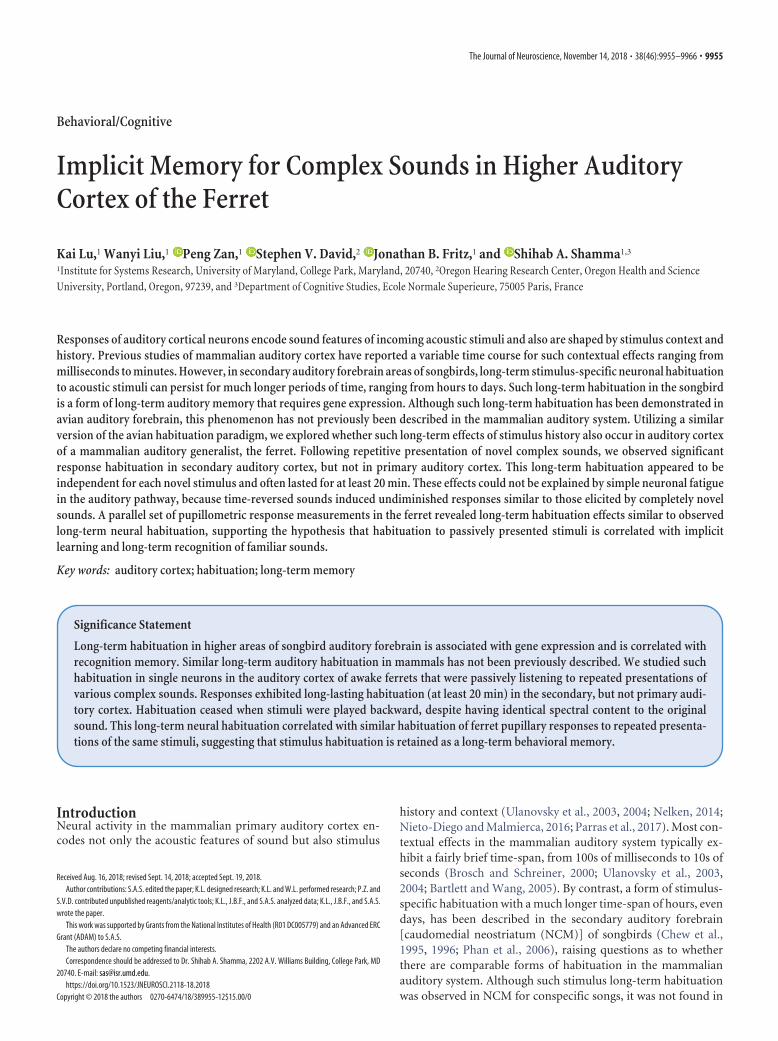
Auditory field localization. The approximate location of A1 was initiallydetermined by stereotaxic coordinates and then refined with neurophys-iological mapping of the tonotopic map in A1 (Bizley et al., 2005). Theposterior border of A1 (along the suprasylvian sulcus) was marked by thepresence of visual responses from neighboring visual cortex. The locationof dPEG was determined neurophysiologically by tonotopically mappingA1 and dPEG, which share a common low-frequency border (Fig, 1;Atiani et al., 2014; Bizley et al., 2005, 2015). The tonotopic gradient in A1goes from high- to low-frequency along a rostrally tilted dorsoventralaxis until the gradient reversed and best frequency increases again in themirror frequency maps in the two areas that comprise the dPEG-PPF andPSF. In addition to the A1/dPEG tonotopic gradient reversal, transitionto dPEG was also marked by somewhat longer latencies, greater sustainedresponses and weaker envelope phase-locking than in A1 (Atiani et al.,2014; Bizley et al., 2005). The ventral boundary of the dPEG was deter-mined by abrupt transition from high-frequency tuning to broad low-frequency tuning (Atiani et al., 2014; Bizley et al., 2005, 2015).
Auditory stimuli and experimental design. Acoustic stimuli were com-prised of 73 short samples of animal vocalizations, speech, vocal andinstrumental music, and sampled at 40 kHz. Animal vocalizations andhuman speech were 2– 4 s long (Fig. 2A). Instrumental music excerptswere 3–5 s long. All stimulus amplitudes were presented at 65 dB SPL
9956 • J. Neurosci., November 14, 2018 • 38(46):9955–9966 Lu et al. • Long-Term Implicit Auditory Memory in Higher Auditory Cortex
from a speaker placed 1 m in front of the animal in a large, walk-indouble-walled soundproof booth (IAC).
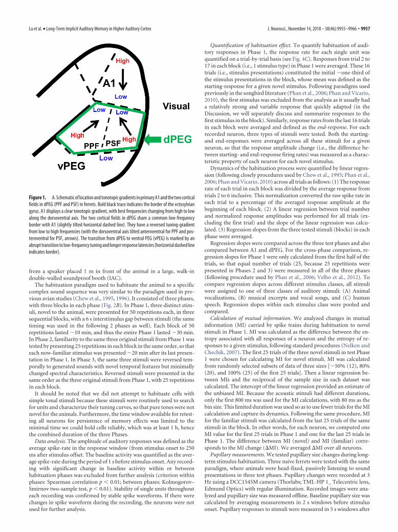
The habituation paradigm used to habituate the animal to a specificcomplex sound sequence was very similar to the paradigm used in pre-vious avian studies (Chew et al., 1995, 1996). It consisted of three phases,with three blocks in each phase (Fig. 2B). In Phase 1, three distinct stim-uli, novel to the animal, were presented for 50 repetitions each, in threesequential blocks, with a 6 s interstimulus gap between stimuli (the sametiming was used in the following 2 phases as well). Each block of 50repetitions lasted �10 min, and thus the entire Phase 1 lasted �30 min.In Phase 2, familiarity to the same three original stimuli from Phase 1 wastested by presenting 25 repetitions in each block in the same order, so thateach now-familiar stimulus was presented �20 min after its last presen-tation in Phase 1. In Phase 3, the same three stimuli were reversed tem-porally to generated sounds with novel temporal features but minimallychanged spectral characteristics. Reversed stimuli were presented in thesame order as the three original stimuli from Phase 1, with 25 repetitionsin each block.
It should be noted that we did not attempt to habituate cells withsimple tonal stimuli because these stimuli were routinely used to searchfor units and characterize their tuning curves, so that pure tones were notnovel for the animals. Furthermore, the time window available for retest-ing all neurons for persistence of memory effects was limited to theminimal time we could hold cells reliably, which was at least 1 h, hencethe combined duration of the three Phases.
Data analysis. The amplitude of auditory responses was defined as theaverage spike-rate in the response window (from stimulus onset to 250ms after stimulus offset. The baseline activity was quantified as the aver-age spike-rate during the period of 1 s before stimulus onset. Any record-ing with significant change in baseline activity within or betweenhabituation phases was excluded from further analysis (criterion withinphases: Spearman correlation p � 0.01; between phases: Kolmogorov–Smirnov two-sample test, p � 0.01). Stability of single units throughouteach recording was confirmed by stable spike waveforms. If there werechanges in spike waveform during the recording, the neurons were notused for further analysis.
Quantification of habituation effect. To quantify habituation of audi-tory responses in Phase 1, the response rate for each single unit wasquantified on a trial-by-trial basis (see Fig. 4C). Responses from trial 2 to17 in each block (i.e., 1 stimulus type) in Phase 1 were averaged. These 16trials (i.e., stimulus presentations) constituted the initial �one-third ofthe stimulus presentations in the block, whose mean was defined as thestarting-response for a given novel stimulus. Following paradigms usedpreviously in the songbird literature (Phan et al., 2006; Phan and Vicario,2010), the first stimulus was excluded from the analysis as it usually hada relatively strong and variable response that quickly adapted (in theDiscussion, we will separately discuss and summarize responses to thefirst stimulus in the block). Similarly, response rates from the last 16 trialsin each block were averaged and defined as the end-response. For eachrecorded neuron, three types of stimuli were tested. Both the starting-and end-responses were averaged across all these stimuli for a givenneuron, so that the response amplitude change (i.e., the difference be-tween starting- and end-response firing rates) was measured as a charac-teristic property of each neuron for each novel stimulus.
Dynamics of the habituation process were quantified by linear regres-sion (following closely procedures used by Chew et al., 1995; Phan et al.,2006; Phan and Vicario, 2010) across all trials as follows: (1) The responserate of each trial in each block was divided by the average response fromtrials 2 to 6 inclusive. This normalization converted the raw spike rate ineach trial to a percentage of the averaged response amplitude at thebeginning of each block. (2) A linear regression between trial numberand normalized response amplitudes was performed for all trials (ex-cluding the first trial) and the slope of the linear regression was calcu-lated. (3) Regression slopes from the three tested stimuli (blocks) in eachphase were averaged.
Regression slopes were compared across the three test phases and alsocompared between A1 and dPEG. For the cross-phase comparison, re-gression slopes for Phase 1 were only calculated from the first half of thetrials, so that equal number of trials (25, because 25 repetitions werepresented in Phases 2 and 3) were measured in all of the three phases(following procedure used by Phan et al., 2006; Velho et al., 2012). Tocompare regression slopes across different stimulus classes, all stimuliwere assigned to one of three classes of auditory stimuli: (A) Animalvocalizations, (B) musical excerpts and vocal songs, and (C) humanspeech. Regression slopes within each stimulus class were pooled andcompared.
Calculation of mutual information. We analyzed changes in mutualinformation (MI) carried by spike trains during habituation to novelstimuli in Phase 1. MI was calculated as the difference between the en-tropy associated with all responses of a neuron and the entropy of re-sponses to a given stimulus, following standard procedures (Nelken andChechik, 2007). The first 25 trials of the three novel stimuli in test Phase1 were chosen for calculating MI for novel stimuli. MI was calculatedfrom randomly selected subsets of data of three sizes [�50% (12), 80%(20), and 100% (25) of the first 25 trials]. Then a linear regression be-tween MIs and the reciprocal of the sample size in each dataset wascalculated. The intercept of the linear regression provided an estimate ofthe unbiased MI. Because the acoustic stimuli had different durations,only the first 800 ms was used for the MI calculations, with 80 ms as thebin size. This limited duration was used so as to use fewer trials for the MIcalculation and capture its dynamics. Following the same procedure, MIfor the familiar stimuli was calculated from the last 25 trials of the samestimuli in the block. In other words, for each neuron, we computed oneMI value for the first 25 trials in Phase 1 and one for the last 25 trials inPhase 1. The difference between MI (novel) and MI (familiar) corre-sponds to the MI change (�MI). We averaged �MI over all neurons.
Pupillary measurements. We tested pupillary size changes during long-term stimulus habituation. Three naive ferrets were tested with the sameparadigm, where animals were head-fixed, passively listening to soundpresentations in three test phases. Pupillary changes were recorded at 3Hz using a DCC1545M camera (Thorlabs; TML-HP 1_ Telecentric lens,Edmund Optics) with regular illumination. Recorded images were ana-lyzed and pupillary size was measured offline. Baseline pupillary size wascalculated by averaging measurements in 2 s windows before stimulusonset. Pupillary responses to stimuli were measured in 5 s windows after
Figure 1. A. Schematic of location and tonotopic gradients in primary A1 and the two corticalfields in dPEG (PPF and PSF) in ferrets. Bold black trace indicates the border of the ectosylviangyrus. A1 displays a clear tonotopic gradient, with best frequencies changing from high to lowalong the dorsoventral axis. The two cortical fields in dPEG share a common low-frequencyborder with A1 (slightly tilted horizontal dashed line). They have a reversed tuning-gradientfrom low to high frequencies (with the dorsoventral axis tilted anteroventral for PPF and pos-teroventral for PSF, arrows). The transition from dPEG to ventral PEG (vPEG) is marked by anabrupt transition to low-frequency tuning and longer response latencies (horizontal dashed lineindicates border).
Lu et al. • Long-Term Implicit Auditory Memory in Higher Auditory Cortex J. Neurosci., November 14, 2018 • 38(46):9955–9966 • 9957
stimulus onset. Then pupillary size measured in the response windowwas adjusted by subtracting the baseline pupillary measurement in eachtrial. To compare the pupillary responses to novel stimuli and familiarstimuli, pupillary responses, as a function of time, were calculated in thesame way as analysis of post-stimulus time histogram (PSTH) differences(except that more repetitions were used in the analysis because of themuch lower sampling rate of pupillary recording compared with electro-physiology). Pupillary responses to the first 15 trials of novel sounds werepooled together and compared with pupillary responses to the last 15trials of familiar sounds.
The latency of pupillary responses was determined in the same way asthe analysis of temporal profile of neural habituation. Habituation curvesof pupillary responses were estimated from trial-by-trial pupillary mea-surements as in the analysis of habituation curves of the neural data.
Statistical methods. Results were plotted as histograms or cumulativefrequency distributions (which reveal details of multiple distributions)and also as conventional box-and-whisker plots. Because sample distri-butions in some tests did not satisfy criteria for parametric tests, non-parametric statistics were used. Changes in response rates over repetitivestimulation and slopes of linear regression from the neuron populationwere tested by the Wilcoxon matched pairs test (the nonparametric ver-sion of one-sample t test). Differences between test phases were similarlytested. Friedman ANOVA tests were used to test multiple matched sam-ples. For non-matched samples, differences between the two brainregions (A1 and dPEG) were tested by the Kolmogorov–Smirnov two-sample test (the nonparametric version of two-sample t test). Mediantests (similar to one-factor ANOVA) were applied to multiple non-matched samples.
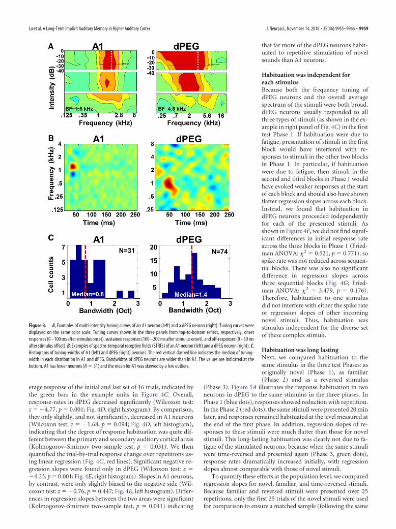
ResultsBasic response properties were measured in 83 single-units indPEG (pooled data from PPF and PSF), and 81 single units in A1,in the auditory cortices of three ferrets. A1 neurons showed clear,narrowly tuned receptive fields (Fig. 3A,B, left column), com-pared with the more broadly-tuned receptive fields of dPEG neu-rons (Fig. 3A,B, right column), consistent with earlier reports(Atiani et al., 2014). Distribution of bandwidths for A1 and dPEGreceptive field filters, as shown in Figure 3C, were significantlydifferent (A1: median � 0.8, dPEG: median � 1.4; Kolmogorov–Smirnov two-sample test, p � 0.023). We note that we groupedthe neurons in PPF and PSF together as one set in dPEG, becausethey showed similar properties in our experimental habituationparadigm.
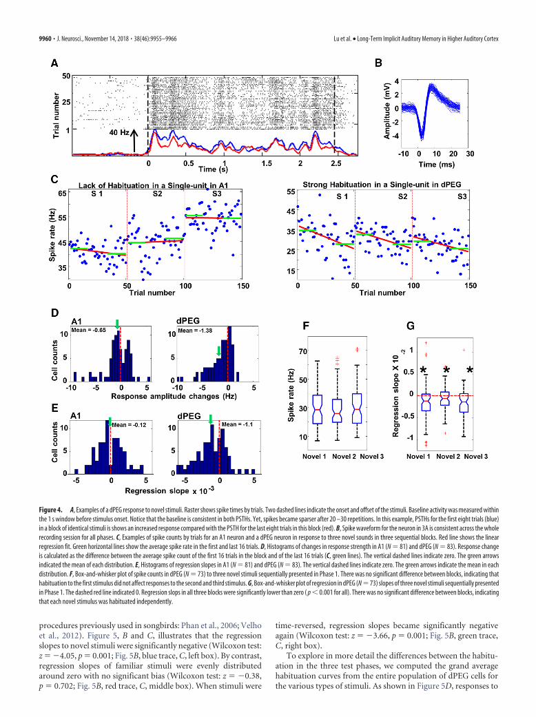
Habituation to repeated acoustic stimuliWe measured the average responses to repeated presentations ofnovel stimuli in Phase 1, as illustrated for one dPEG neuron bythe raster responses in Figure 4A. Both the temporal pattern ofresponses and spike waveforms remained consistent throughoutthe recording (Fig. 4A,B). Figure 4C showed spike-rate plots foran A1 neuron and a dPEG neuron. A typical pattern of responsechanges demonstrated in these plots is the habituation of re-sponses of dPEG cells (but not of A1) as the number of repetitionsincreases. This was quantified by the difference between the av-
Figure 2. A, Spectrograms of six diverse broadband stimuli used in the habituation study. B, Schematic illustration of how stimuli were presented. Blocks of three Novel stimuli (blue arrows) werepresented in Phase 1. In each block, there were 50 repetitions of one of the three novel stimuli. The now Familiar stimuli (red arrows) are then presented in blocks of 25 repetitions in Phase 2. TheReversed stimuli (green arrows) are then presented in blocks of 25 repetitions in Phase 3.
9958 • J. Neurosci., November 14, 2018 • 38(46):9955–9966 Lu et al. • Long-Term Implicit Auditory Memory in Higher Auditory Cortex
erage response of the initial and last set of 16 trials, indicated bythe green bars in the example units in Figure 4C. Overall,response-rates in dPEG decreased significantly (Wilcoxon test:z � �4.77, p � 0.001; Fig. 4D, right histogram). By comparison,they only slightly, and not significantly, decreased in A1 neurons(Wilcoxon test: z � �1.68, p � 0.094; Fig. 4D, left histogram),indicating that the degree of response habituation was quite dif-ferent between the primary and secondary auditory cortical areas(Kolmogorov–Smirnov two-sample test, p � 0.031). We thenquantified the trial-by-trial response change over repetitions us-ing linear regression (Fig. 4C, red lines). Significant negative re-gression slopes were found only in dPEG (Wilcoxon test: z ��4.23, p � 0.001; Fig. 4E, right histogram). Slopes in A1 neurons,by contrast, were only slightly biased to the negative side (Wil-coxon test: z � �0.76, p � 0.447; Fig. 4E, left histogram). Differ-ences in regression slopes between the two areas were significant(Kolmogorov–Smirnov two-sample test, p � 0.041) indicating
that far more of the dPEG neurons habit-uated to repetitive stimulation of novelsounds than A1 neurons.
Habituation was independent foreach stimulusBecause both the frequency tuning ofdPEG neurons and the overall averagespectrum of the stimuli were both broad,dPEG neurons usually responded to allthree types of stimuli (as shown in the ex-ample in right panel of Fig. 4C) in the firsttest Phase 1. If habituation were due tofatigue, presentation of stimuli in the firstblock would have interfered with re-sponses to stimuli in the other two blocksin Phase 1. In particular, if habituationwere due to fatigue, then stimuli in thesecond and third blocks in Phase 1 wouldhave evoked weaker responses at the startof each block and should also have shownflatter regression slopes across each block.Instead, we found that habituation indPEG neurons proceeded independentlyfor each of the presented stimuli. Asshown in Figure 4F, we did not find signif-icant differences in initial response rateacross the three blocks in Phase 1 (Fried-man ANOVA: � 2 � 0.521, p � 0.771), sospike rate was not reduced across sequen-tial blocks. There was also no significantdifference in regression slopes acrossthree sequential blocks (Fig. 4G; Fried-man ANOVA: � 2 � 3.479, p � 0.176).Therefore, habituation to one stimulusdid not interfere with either the spike rateor regression slopes of other incomingnovel stimuli. Thus, habituation wasstimulus independent for the diverse setof these complex stimuli.
Habituation was long lastingNext, we compared habituation to thesame stimulus in the three test Phases: asoriginally novel (Phase 1), as familiar(Phase 2) and as a reversed stimulus
(Phase 3). Figure 5A illustrates the response habituation in twoneurons in dPEG to the same stimulus in the three phases. InPhase 1 (blue dots), responses showed reduction with repetition.In the Phase 2 (red dots), the same stimuli were presented 20 minlater, and responses remained habituated at the level measured atthe end of the first phase. In addition, regression slopes of re-sponses to these stimuli were much flatter than those for novelstimuli. This long-lasting habituation was clearly not due to fa-tigue of the stimulated neurons, because when the same stimuliwere time-reversed and presented again (Phase 3, green dots),response rates dramatically increased initially, with regressionslopes almost comparable with those of novel stimuli.
To quantify these effects at the population level, we comparedregression slopes for novel, familiar, and time-reversed stimuli.Because familiar and reversed stimuli were presented over 25repetitions, only the first 25 trials of the novel stimuli were usedfor comparison to ensure a matched sample (following the same
Figure 3. A, Examples of multi-intensity tuning curves of an A1 neuron (left) and a dPEG neuron (right). Tuning curves weredisplayed on the same color scale. Tuning curves shown in the three panels from top-to-bottom reflect, respectively, onsetresponses (0 –100 ms after stimulus onset), sustained responses (100 –200 ms after stimulus onset), and off-responses (0 –50 msafter stimulus offset). B, Examples of spectro-temporal receptive fields (STRFs) of an A1 neuron (left) and a dPEG neuron (right). C,Histograms of tuning-widths of A1 (left) and dPEG (right) neurons. The red vertical dashed line indicates the median of tuning-width in each distribution in A1 and dPEG. Bandwidths of dPEG neurons are wider than in A1. The values are indicated at thebottom. A1 has fewer neurons (N � 31) and the mean for A1 was skewed by a few outliers.
Lu et al. • Long-Term Implicit Auditory Memory in Higher Auditory Cortex J. Neurosci., November 14, 2018 • 38(46):9955–9966 • 9959
procedures previously used in songbirds: Phan et al., 2006; Velhoet al., 2012). Figure 5, B and C, illustrates that the regressionslopes to novel stimuli were significantly negative (Wilcoxon test:z � �4.05, p � 0.001; Fig. 5B, blue trace, C, left box). By contrast,regression slopes of familiar stimuli were evenly distributedaround zero with no significant bias (Wilcoxon test: z � �0.38,p � 0.702; Fig. 5B, red trace, C, middle box). When stimuli were
time-reversed, regression slopes became significantly negativeagain (Wilcoxon test: z � �3.66, p � 0.001; Fig. 5B, green trace,C, right box).
To explore in more detail the differences between the habitu-ation in the three test phases, we computed the grand averagehabituation curves from the entire population of dPEG cells forthe various types of stimuli. As shown in Figure 5D, responses to
Figure 4. A, Examples of a dPEG response to novel stimuli. Raster shows spike times by trials. Two dashed lines indicate the onset and offset of the stimuli. Baseline activity was measured withinthe 1 s window before stimulus onset. Notice that the baseline is consistent in both PSTHs. Yet, spikes became sparser after 20 –30 repetitions. In this example, PSTHs for the first eight trials (blue)in a block of identical stimuli is shows an increased response compared with the PSTH for the last eight trials in this block (red). B, Spike waveform for the neuron in 3A is consistent across the wholerecording session for all phases. C, Examples of spike counts by trials for an A1 neuron and a dPEG neuron in response to three novel sounds in three sequential blocks. Red line shows the linearregression fit. Green horizontal lines show the average spike rate in the first and last 16 trials. D, Histograms of changes in response strength in A1 (N � 81) and dPEG (N � 83). Response changeis calculated as the difference between the average spike count of the first 16 trials in the block and of the last 16 trials (C, green lines). The vertical dashed lines indicate zero. The green arrowsindicated the mean of each distribution. E, Histograms of regression slopes in A1 (N � 81) and dPEG (N � 83). The vertical dashed lines indicate zero. The green arrows indicate the mean in eachdistribution. F, Box-and-whisker plot of spike counts in dPEG (N � 73) to three novel stimuli sequentially presented in Phase 1. There was no significant difference between blocks, indicating thathabituation to the first stimulus did not affect responses to the second and third stimulus. G, Box-and-whisker plot of regression in dPEG (N � 73) slopes of three novel stimuli sequentially presentedin Phase 1. The dashed red line indicated 0. Regression slops in all three blocks were significantly lower than zero ( p � 0.001 for all). There was no significant difference between blocks, indicatingthat each novel stimulus was habituated independently.
9960 • J. Neurosci., November 14, 2018 • 38(46):9955–9966 Lu et al. • Long-Term Implicit Auditory Memory in Higher Auditory Cortex
novel stimuli (blue dots) decreased by �20% in the first 25 trialsthat were used to calculate all regression slopes, reaching a pla-teau after �25–30 trials. Responses to familiar stimuli (red dots)were approximately habituated to the same level as this plateau, ex-cept (as discussed later) for the first two trials, which showed anincreased response. The regression slope for familiar stimuli wassignificantly flatter than the slope to novel stimuli. Finally, the regres-sion slope for reversed stimuli (green dots) was similar to the slopefor novel stimuli, suggesting that the long-lasting habituation to fa-miliar stimuli was not due to fatigue, but likely reflected a form oflong-term memory in dPEG (lasting longer than 20 min) analogousto that found in birds. The absence of long-lasting habituation in A1and its presence in dPEG distinguished it from stimulus-specificadaptation (SSA), which is observed in many earlier auditory pro-cessing stages such as inferior colliculus and auditory thalamus(Antunes et al., 2010; Duque et al., 2012; Nelken, 2014).
However, the response to the very first trial was clearly muchlarger than the responses in the rest of trials of the block. Wecompared its amplitude with the mean of trials 2–25 (used inlinear regression analysis). It was 23% larger than the rest of thetrials for novel stimuli, 13% larger than the rest of trials for famil-iar stimuli and 15% larger than the rest of trials for reversedstimuli, which indicated that a possible SSA-like effect may occurat the beginning of each block.
Finally, we also examined the effects of the habituation onthe temporal profile of the dPEG responses. The averagedPSTH of all dPEG responses to novel stimuli is shown in Fig-ure 5E (blue trace), together with the same response to thefamiliar version of the stimuli (red trace). As noted earlier, thetwo responses appear very similar except for an overall 18%attenuation of the habituated responses. In A1, a similar anal-ysis showed that there was no such attenuation.
Figure 5. A,Examplesofspikecountsbytrials intwodPEGneuronsinresponsetoastimuluswhenitwasnovel(blue), familiar(red),orreversed(green).NotethatthefirstpresentationofthefamiliarstimulusinPhase2occurred20minafterthelasttrialofthesamestimuluswhenitwasnovel inPhase1. B,Cumulativefrequencydistributionofregressionslopesfornovelstimuli (blue), familiarstimuli (red)andreversedstimuli (green) in dPEG. Horizontal dashed line on the right indicated that 68% regression slopes obtained from novel stimuli were lower than zero. Horizontal dashed line on the left indicated that only 50%(chance level) of the regression slopes obtained from familiar stimuli were lower than zero. C, Box-and-whisker plot of regression slopes in dPEG for novel (left), familiar (middle), and reversed (right) stimuli. D,Population habituation curve in PEG. Each circle indicates responses averaged from all neurons (N�83). Error bars on top of circles show SE. The first red line (across the blue circles) shows the linear regressionfit for the first 25 trials of novel stimuli. The blue line shows the linear regression fit for familiar stimuli. The second red line (across the green circles) shows the linear regression fit for reversed stimuli. The two redlines were clearly tilted. The blue line was flat. Notice that the very first trials in all three conditions were excluded from this analysis. E, The temporal profiles of auditory responses to novel stimuli in dPEG (red)and to familiar stimuli (blue), calculated from averaged PSTHs across all neurons and all stimuli. Red dashed line shows stimulus onset time. The Black dashed line is the threshold calculated from the mean in thebaseline plus SE (in 10 ms bins). The latency of the responses is marked by green vertical line. The peak of the PSTHs is indicated by the blue vertical line.
Lu et al. • Long-Term Implicit Auditory Memory in Higher Auditory Cortex J. Neurosci., November 14, 2018 • 38(46):9955–9966 • 9961
Habituation was similar for all stimulus typesAs described in Materials and Methods, three different categoriesof novel stimuli (speech, music, animal vocalizations) were usedin each recording. In the analysis discussed above, responses tothese three different types of stimuli were averaged in each testphase. To compare habituation effects among these three stimu-lus categories, we analyzed data for each stimulus-type separately.However, we found no significant differences in regression slopesacross the three classes, either in dPEG (Fig. 6A; Median test,� 2 � 5.10, df � 2, p � 0.078) or in A1 (Fig. 6B; Median test, � 2 �1.50, df � 2, p � 0.472). Stimuli from all three types of soundshabituated equally in dPEG: animal vocalizations (Wilcoxon test:z � �3.41, p � 0.001); musical pieces and vocal songs (Wilcoxontest: z � �2.31, p � 0.02); speech (Wilcoxon test: z � �4.13, p �0.001). Similarly, in A1, none of three types induced significanthabituation: animal vocalization (Wilcoxon test: z � �0.96, p �0.338); music pieces and vocal songs (Wilcoxon test: z � �0.35,p � 0.730); speech (Wilcoxon test: z � �0.966, p � 0.334).Therefore, habituation was independent of the type of soundpresented.
Habituation was not correlated with the tuning ofthe neuronsWe also tested to see whether habituation was related to neuronalfrequency tuning by correlating the bandwidth of each recordedneuron in A1 and dPEG with its regression slope for novel stimuli(Phase 1). We did not find any significant correlation between thetwo properties in either cortical field: A1 (� � �0.03, p � 0.858)and dPEG (� � �0.14, p � 0.222). However, there is a slightnegative trend for correlations calculated in dPEG neurons (Fig.6C), indicating that neurons with wider tuning bandwidths mayhabituate slightly faster than narrowly tuned neurons.
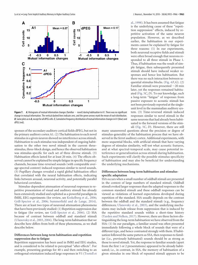
Mutual information increased after habituationSo far, habituation of neural responses was quantified as thespike-rate change during stimulus presentations. We also exam-ined the spike pattern changes during habituation. We hypothe-sized that as the spike rates decreased with habituation, responsenoise would also decrease such that the MI between spike-trainsand stimuli would increase, as found in works with songbirds (Luand Vicario, 2014). Thus, we calculated the MIs for the first halfof trials of all three stimuli in test Phase 1 and compared themwith the MIs for the second half of trials of the same stimuli in oneblock in Phase 1, when they were habituated. Consistent with thehypothesis, our analysis revealed that the MI did not significantlychange in A1 (Fig. 7A, C, blue trace; Wilcoxon test: z � �1.34,p � 0.181), whereas it significantly increased in dPEG (Fig. 7B, C,red trace; Wilcoxon test: z � �4.11, p � 0.001).
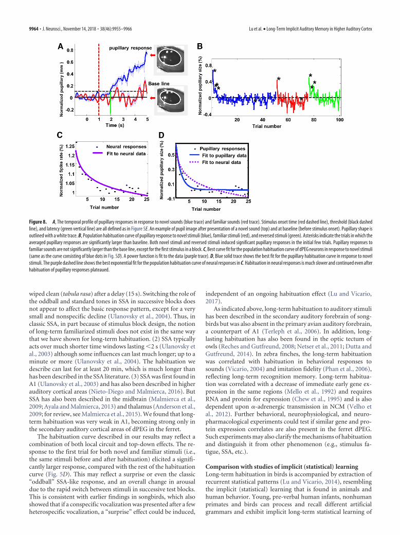
Pupillary measurement was consistent with neuralhabituation resultsBased on analogous findings in birds, the results thus far openedup the possibility that the long-lasting habituation of neural re-sponses observed in dPEG might reflect a long-term auditoryrecognition memory for complex sounds that could be measuredat a behavioral level. To confirm the presence of memory, wesought a behavioral measure that could demonstrate the animals’recognition of familiar stimuli compared with the novel ones. Aspupillary size-changes in response to sound have been shown tobe an indicator of arousal (Reimer et al., 2016) we therefore mea-sured pupillary size-changes in three awake, quietly listening an-imals presented with the same sequence of stimuli used for theelectrophysiology experiments. Animals’ pupillary size increasedsignificantly (13% increase relative to the baseline, Wilcoxon test:z � �17.9, p � 0.001) after presentation of novel sounds (Fig. 8A,blue trace) with a latency of 0.9 s. By comparison, pupillary re-sponses to familiar sounds (same sounds presented �20 minlater in Phase 2) were much smaller and not significantly differentfrom the baseline (Wilcoxon test: z � �0.04, p � 0.968; Fig. 8A,red trace), as a result of habituation. Therefore, habituation ofpupillary responses was consistent with the pattern of long-termhabituation of neural responses in dPEG. Trial-by-trial analysisof pupillary responses revealed a similar picture: pupillary re-sponses to novel stimuli (blue trace) showed a clear reduction inthe initial few trials; whereas pupillary size did not change signif-icantly to familiar stimuli (red trace), except for the first trial. Thehabituation curve to the time-reversed stimuli (green trace) wassimilar to that of the novel stimuli, exhibiting recovered re-sponses to those stimuli in the initial few trials.
However, one difference between pupillary and neural habit-uation was that habituation in pupillary responses to novel stim-uli was much faster than neuronal habituation to the samestimuli. Many fewer trials were needed to induce pupillary habit-uation, which reached plateau after only the first five trials. Bycomparison, neural habituation to novel stimuli in dPEG did notplateau until 25�30 trials (Fig. 5D). This difference is highlightedby the difference between the fitted power-functions to the neu-ronal habituation (Fig. 8C, solid purple curve, replotted as D,dashed curve) and its pupillary habituation counterpart (Fig. 8D,solid blue curve). The dramatic difference between the two curvesshows that the global habituation effect (as reflected by the pupil-lary modulation) occurred much faster, within only a few trials.
DiscussionHabituation experiments described in this report yielded severalsignificant findings: (1) Long-term habituation occurred in re-
Figure 6. A, Box-and-whisker plots of regression slopes for animal vocalizations, music andspeech, measured in dPEG. All are significantly lower than zero, and there are no significantdifferences among them. Animal: p � 0.001; Music: p � 0.02; Speech: p � 0.001. B, Box-and-whisker plot of regression slopes for animal vocalizations, music, and speech, measured in A1.None of them is significantly lower than zero. C, There was a marginal (not significant) negativerelationship between regression slopes and tuning-width in dPEG neurons.
9962 • J. Neurosci., November 14, 2018 • 38(46):9955–9966 Lu et al. • Long-Term Implicit Auditory Memory in Higher Auditory Cortex
sponses of the secondary auditory cortical fields dPEG, but not inthe primary auditory cortex A1. (2) The habituation to each novelstimulus in a given neuron showed no interference across stimuli.Habituation to each stimulus was independent of ongoing habit-uation to the other two novel stimuli in the current three-stimulus, three-block design, and hence the observed habituationwas stimulus-specific for each set of three diverse stimuli. (3)Habituation effects lasted for at least 20 min. (4) The effects ob-served cannot be explained by simple fatigue in specific frequencychannels, because time-reversed sounds (with comparable aver-age spectral content) induced responses similar to novel sounds.(5) Pupillary changes revealed a rapid global habituation effectthat correlated with the neural habituation effects, indicatinglinks between arousal, neuronal activity, and potentially parallelbehavioral correlates.
Stimulus-dependent attenuation of neuronal responses to re-petitive presentation of visual and auditory stimuli has alreadybeen extensively studied and exploited as a powerful paradigm infMRI, EEG experiments (for review, see Krekelberg et al., 2006;Grill-Spector et al., 2006; Summerfield and de Lange, 2014).There are at least two types of neuronal attenuation phenomenathat have been previously studied: (1) Repetition suppression dueto fatigue (for review, see Grill-Spector et al., 2006). (2) SSAbecause of contrast between oddball and standard stimuli(Ulanovsky et al., 2003, 2004). The long-term habituation that wedescribe here differs from both of these phenomena, as we shalldescribe below.
Differences between long-term habituation and repetitionsuppression due to fatigueRepetition suppression has been used in fMRI and EEG studies,and is considered to be related to perceptual “after-effects”. Forexample, presenting gratings for 40 s and then switching to anorthogonal orientation induced large responses in V1 (Tootell et
al., 1998). It has been assumed that fatigueis the underlying cause of these “repeti-tion suppression” effects, induced by re-petitive activation of the same neuronpopulation. However, as we describedearlier, the habituation in our experi-ments cannot be explained by fatigue forthree reasons: (1) In our experiments,both neuronal receptive fields and stimuliwere often broad enough that neurons re-sponded to all three stimuli in Phase 1.Thus, if habituation was the result of sim-ple fatigue, then subsequently presentedstimuli should have induced weaker re-sponses and hence less habituation. Butthere was no such interaction between se-quential stimulus blocks. (Fig. 4F,G). (2)Familiar stimuli were presented �20 minlater, yet the responses remained habitu-ated (Fig. 5C, D). To our knowledge, sucha long-term “fatigue” of responses frompassive exposure to acoustic stimuli hasnot been previously reported at the single-unit level in the mammalian auditory sys-tem. (3) Time-reversed stimuli inducedresponses similar to novel stimuli in thesame neurons that had already been habit-uated to the forward versions of the stim-uli (Fig. 5C, D). However, there are still
many unanswered questions about the precision or degree ofstimulus-generality of the habituation process that we have ob-served in the ferret auditory cortex. Additional experiments, withmore sequential blocks, with artificial stimuli that have varyingdegrees of stimulus similarity, will test what acoustic features,and at what spectral-temporal scale, may cause potential in-terference or generalization across stimuli during habituation.Such experiments will clarify the possible stimulus specificityof habituation and may also be beneficial for understandingthe underlying mechanisms.
Differences between long-term habituation and stimulus-specific adaptationSSA occurs when a small number of oddball stimuli are presentedin the context of large numbers of standard stimuli. Oddballstimuli evoked larger responses than the adapted responses to thecommon standard stimuli and these oddball responses can beviewed as violations of learned expectations developed duringrepetition of the standard. SSA usually depends on the contrastbetween the oddball and the standard stimuli (e.g., frequencydifferences; Ulanovsky et al., 2003), and the underlying mecha-nisms may include release from suppression that is induced bythe repetitive standard sounds within a short-time history(Yarden and Nelken, 2017). However, there are three factors dis-tinguishing the long-term habituation we have observed from theSSA: (1) In our paradigm, a familiar sound was often presentedimmediately following a whole block of sounds that were of adifferent type, and hence contrasted strongly with them. If habit-uation followed the same pattern as SSA, then responses to famil-iar (i.e., previously habituated) sounds should be as strong asthose to novel stimuli. Yet, the response to familiar sounds (apartfrom the first 1 or 2 presentations) appeared to be already habit-uated; i.e., in contrast, in SSA, the memory of habituation of agiven stimulus in one block of repeated stimuli appears to be
Figure 7. A, Histograms of mutual information changes (familiar � novel) during habituation in A1. There was no significantchange in mutual information. The vertical dashed lines indicate zero, and the green arrows mark the mean of each distribution.B, Same plots as in A, except for all dPEG cells. C, Cumulative frequency distribution of mutual information changes in A1 (blue) anddPEG (red).
Lu et al. • Long-Term Implicit Auditory Memory in Higher Auditory Cortex J. Neurosci., November 14, 2018 • 38(46):9955–9966 • 9963
wiped clean (tabula rasa) after a delay (15 s). Switching the role ofthe oddball and standard tones in SSA in successive blocks doesnot appear to affect the basic response pattern, except for a verysmall and nonspecific decline (Ulanovsky et al., 2004). Thus, inclassic SSA, in part because of stimulus block design, the notionof long-term familiarized stimuli does not exist in the same waythat we have shown for long-term habituation. (2) SSA typicallyacts over much shorter time windows lasting �2 s (Ulanovsky etal., 2003) although some influences can last much longer; up to aminute or more (Ulanovsky et al., 2004). The habituation wedescribe can last for at least 20 min, which is much longer thanhas been described in the SSA literature. (3) SSA was first found inA1 (Ulanovsky et al., 2003) and has also been described in higherauditory cortical areas (Nieto-Diego and Malmierca, 2016). ButSSA has also been described in the midbrain (Malmierca et al.,2009; Ayala and Malmierca, 2013) and thalamus (Anderson et al.,2009; for review, see Malmierca et al., 2015). We found that long-term habituation was very weak in A1, becoming strong only inthe secondary auditory cortical areas of dPEG in the ferret.
The habituation curve described in our results may reflect acombination of both local circuit and top-down effects. The re-sponse to the first trial for both novel and familiar stimuli (i.e.,the same stimuli before and after habituation) elicited a signifi-cantly larger response, compared with the rest of the habituationcurve (Fig. 5D). This may reflect a surprise or even the classic“oddball” SSA-like response, and an overall change in arousaldue to the rapid switch between stimuli in successive test blocks.This is consistent with earlier findings in songbirds, which alsoshowed that if a conspecific vocalization was presented after a fewheterospecific vocalization, a “surprise” effect could be induced,
independent of an ongoing habituation effect (Lu and Vicario,2017).
As indicated above, long-term habituation to auditory stimulihas been described in the secondary auditory forebrain of song-birds but was also absent in the primary avian auditory forebrain,a counterpart of A1 (Terleph et al., 2006). In addition, long-lasting habituation has also been found in the optic tectum ofowls (Reches and Gutfreund, 2008; Netser et al., 2011; Dutta andGutfreund, 2014). In zebra finches, the long-term habituationwas correlated with habituation in behavioral responses tosounds (Vicario, 2004) and imitation fidelity (Phan et al., 2006),reflecting long-term recognition memory. Long-term habitua-tion was correlated with a decrease of immediate early gene ex-pression in the same regions (Mello et al., 1992) and requiresRNA and protein for expression (Chew et al., 1995) and is alsodependent upon �-adrenergic transmission in NCM (Velho etal., 2012). Further behavioral, neurophysiological, and neuro-pharmacological experiments could test if similar gene and pro-tein expression correlates are also present in the ferret dPEG.Such experiments may also clarify the mechanisms of habituationand distinguish it from other phenomenon (e.g., stimulus fa-tigue, SSA, etc.).
Comparison with studies of implicit (statistical) learningLong-term habituation in birds is accompanied by extraction ofrecurrent statistical patterns (Lu and Vicario, 2014), resemblingthe implicit (statistical) learning that is found in animals andhuman behavior. Young, pre-verbal human infants, nonhumanprimates and birds can process and recall different artificialgrammars and exhibit implicit long-term statistical learning of
Figure 8. A, The temporal profile of pupillary responses in response to novel sounds (blue trace) and familiar sounds (red trace). Stimulus onset time (red dashed line), threshold (black dashedline), and latency (green vertical line) are all defined as in Figure 5E. An example of pupil image after presentation of a novel sound (top) and at baseline (before stimulus onset). Pupillary shape isoutlined with a white trace. B, Population habituation curve of pupillary response to novel stimuli (blue), familiar stimuli (red), and reversed stimuli (green). Asterisks indicate the trials in which theaveraged pupillary responses are significantly larger than baseline. Both novel stimuli and reversed stimuli induced significant pupillary responses in the initial few trials. Pupillary responses tofamiliar sounds are not significantly larger than the base line, except for the first stimulus in a block. C, Best curve fit for the population habituation curve of dPEG neurons in response to novel stimuli(same as the curve consisting of blue dots in Fig. 5D). A power function is fit to the data (purple trace). D, Blue solid trace shows the best fit for the pupillary habituation curve in response to novelstimuli. The purple dashed line shows the best exponential fit for the population habituation curve of neural responses in C. Habituation in neural responses is much slower and continued even afterhabituation of pupillary responses plateaued.
9964 • J. Neurosci., November 14, 2018 • 38(46):9955–9966 Lu et al. • Long-Term Implicit Auditory Memory in Higher Auditory Cortex
auditory patterns and sequences, or even random noise patterns(Saffran et al., 1996, 1999; Hauser et al., 2001; Fitch and Hauser,2004; Newport et al., 2004; Agus et al., 2010; Abe and Watanabe,2011; Wilson et al., 2013; Kang et al., 2017; Milne et al., 2018). Theexperiments we describe in this report may constitute the neuro-nal underpinnings of such long-term implicit learning phenom-ena, which have hitherto been unexplored at a cellular level in themammalian auditory system.
Habituation in the visual systemThe basic phenomenon of stimulus-dependent auditory habitu-ation is likely related to various forms of plasticity observed inother modalities, such as the visual system. For example, a fewdays of exposure to sequences of visual patterns led to long-termpotentiation in responses in V1 (Gavornik and Bear, 2014).Given our results, and the bird literature on habituation, both ofwhich demonstrate the importance of higher auditory areas inlong-term habituation, it is surprising and intriguing that suchhabituation can occur in the mouse primary visual cortex; al-though we speculate that V1 in the mouse may be more of anassociative cortical area. In support of the importance of highervisual cortical areas in habituation, repetitive presentation of avisual object image led to habituation in neurons in higher visualcortical areas such as inferior temporal cortex in the macaque (Liet al., 1993; Miller and Desimone, 1994; Sobotka and Ringo,1994; Ringo, 1996), a habituation that is long-lasting (5 min)even after 150 intervening stimuli (Li et al., 1993). However, theselatter studies in the monkey are quite different from our para-digm in that they experimented with behaving (as opposed topassively viewing) monkeys performing delayed match-to-sample tasks, and hence the effects were dependent on the rewardstructure in the task. When a given visual object did not match thesample (irrelevant to the reward), the responses to repetition ofthis object image habituated. By contrast, if a given object was amatch (led to reward), the responses increased with repetition.Finally, similar effects of long-lasting visual memories have beendescribed in fMRI/EEG studies with attentive human subjectspresented with repetitive or even single instance face or objectstimuli (Henson et al., 2000; Jiang et al., 2000; van Turennout etal., 2000; Doniger et al., 2001; Schendan and Kutas, 2003).
Contribution of habituation to complementary processes ofimplicit acoustic scene analysisWe speculate that the combination of SSA and habituation torepetitive, behaviorally neutral stimuli may be an effective blendof implicit mechanisms for allocating attention to incomingstimuli in the environmental soundscape, each with their owntime window. Although SSA detects violation of expectation inshort time window, habituation avoids over-responsiveness tofamiliar sounds over a longer history. Thus, when a novel stimu-lus arrives, its biological relevance to the animal is initially un-known and hence it is beneficial to turn attention to the novelevent with an enhanced oddball alerting response. However, ifthe same stimulus is repeated again and again without significantbehavioral consequences, its immediate behavioral relevance isjudged to be low and hence responses to this specific stimulus arehabituated (filtered out), and stored in long-term memory as asignal that can be safely ignored, facilitating the processing ofother potentially more relevant incoming signals. These implicitmechanisms are possibly complemented by processes during ac-tive engagement and attention where (as described earlier) asso-ciation with a reward may potentiate the response.
ReferencesAbe K, Watanabe D (2011) Songbirds possess the spontaneous ability to
Agus TR, Thorpe SJ, Pressnitzer D (2010) Rapid formation of robust audi-tory memories: insights from noise. Neuron 66:610 – 618. CrossRefMedline
Anderson LA, Christianson GB, Linden JF (2009) Stimulus-specific adapta-tion occurs in the auditory thalamus. J Neurosci 29:7359 –7363. CrossRefMedline
Antunes FM, Nelken I, Covey E, Malmierca MS (2010) Stimulus-specificadaptation in the auditory thalamus of the anesthetized rat. PLoS One5:e14071. CrossRef Medline
Atiani S, David SV, Elgueda D, Locastro M, Radtke-Schuller S, Shamma SA,Fritz JB (2014) Emergent selectivity for task-relevant stimuli in higher-order auditory cortex. Neuron 82:486 – 499. CrossRef Medline
Ayala YA, Malmierca MS (2013) Stimulus-specific adaptation and deviancedetection in the inferior colliculus. Front Neural Circuits 6:89. CrossRefMedline
Bartlett EL, Wang X (2005) Long-lasting modulation by stimulus context inprimate auditory cortex. J Neurophysiol 94:83–104. CrossRef Medline
Bizley JK, Bajo VM, Nodal FR, King AJ (2015) Cortico-cortical connectivitywithin ferret auditory cortex. J Comp Neurol 523:2187–2210. CrossRefMedline
Bizley JK, Nodal FR, Nelken I, King AJ (2005) Functional organization offerret auditory cortex. Cereb Cortex 15:1637–1653. CrossRef Medline
Brosch M, Schreiner CE (2000) Sequence sensitivity of neurons in cat pri-mary auditory cortex. Cereb Cortex 10:1155–1167. CrossRef Medline
Chew SJ, Mello C, Nottebohm F, Jarvis E, Vicario DS (1995) Decrements inauditory responses to a repeated conspecific song are long-lasting andrequire two periods of protein synthesis in the songbird forebrain. ProcNatl Acad Sci U S A 92:3406 –3410. CrossRef Medline
Chew SJ, Vicario DS, Nottebohm F (1996) A large-capacity memory systemthat recognizes the calls and songs of individual birds. Proc Natl Acad SciU S A 93:1950 –1955. CrossRef Medline
Depireux DA, Simon JZ, Klein DJ, Shamma SA (2001) Spectro-temporalresponse field characterization with dynamic ripples in ferret primaryauditory cortex. J Neurophysiol 85:1220 –1234. CrossRef Medline
Doniger GM, Foxe JJ, Schroeder CE, Murray MM, Higgins BA, Javitt DC(2001) Visual perceptual learning in human object recognition areas: arepetition priming study using high-density electrical mapping. Neuro-image 13:305–313. CrossRef Medline
Duque D, Perez-Gonzalez D, Ayala YA, Palmer AR, Malmierca MS (2012)Topographic distribution, frequency, and intensity dependence ofstimulus-specific adaptation in the inferior colliculus of the rat. J Neuro-sci 32:17762–17774. CrossRef Medline
Dutta A, Gutfreund Y (2014) Saliency mapping in the optic tectum and itsrelationship to habituation. Front Integr Neurosci 8:1. CrossRef Medline
Fitch WT, Hauser MD (2004) Computational constraints on syntactic pro-cessing in a nonhuman primate. Science 303:377–380. CrossRef Medline
Grill-Spector K, Henson R, Martin A (2006) Repetition and the brain: neu-ral models of stimulus-specific effects. Trends Cogn Sci 10:14 –23.CrossRef Medline
Hauser MD, Newport EL, Aslin RN (2001) Segmentation of the speechstream in a nonhuman primate: statistical learning in cotton-top tama-rins. Cognition 78:B53–B64. CrossRef Medline
Henson R, Shallice T, Dolan R (2000) Neuroimaging evidence for disso-ciable forms of repetition priming. Science 287:1269 –1272. CrossRefMedline
Jiang Y, Haxby JV, Martin A, Ungerleider LG, Parasuraman R (2000) Com-plementary neural mechanisms for tracking items in human workingmemory. Science 287:643– 646. CrossRef Medline
Jusczyk PW, Hohne EA (1997) Infants’ memory for spoken words. Science277:1984 –1986. Medline
Kang H, Agus TR, Pressnitzer D (2017) Auditory memory for random timepatterns. J Acoust Soc Am 142:2219. CrossRef Medline
Krekelberg B, Boynton GM, van Wezel RJ (2006) Adaptation: from singlecells to BOLD signals. Trends Neurosci 29:250 –256. CrossRef Medline
Li L, Miller EK, Desimone R (1993) The representation of stimulus famil-
Lu et al. • Long-Term Implicit Auditory Memory in Higher Auditory Cortex J. Neurosci., November 14, 2018 • 38(46):9955–9966 • 9965
Lu K, Vicario DS (2014) Statistical learning of recurring sound patternsencodes auditory objects in songbird forebrain. Proc Natl Acad Sci U S A111:14553–14558. CrossRef Medline
Lu K, Vicario DS (2017) Familiar but unexpected: effects of sound contextstatistics on auditory responses in the songbird forebrain. J Neurosci 37:12006 –12017. CrossRef Medline
Lu K, Xu Y, Yin P, Oxenham AJ, Fritz JB, Shamma SA (2017) Temporalcoherence structure rapidly shapes neuronal interactions. Nat Commun8:13900. CrossRef Medline
Malmierca MS, Anderson LA, Antunes FM (2015) The cortical modulationof stimulus-specific adaptation in the auditory midbrain and thalamus: apotential neuronal correlate for predictive coding. Front Syst Neurosci9:19. CrossRef Medline
Malmierca MS, Cristaudo S, Perez-Gonzalez D, Covey E (2009) Stimulus-specific adaptation in the inferior colliculus of the anesthetized rat. J Neu-rosci 29:5483–5493. CrossRef Medline
Mello CV, Vicario DS, Clayton DF (1992) Song presentation induces geneexpression in the songbird forebrain. Proc Natl Acad Sci U S A 89:6818 –6822. CrossRef Medline
Miller EK, Desimone R (1994) Parallel neuronal mechanisms for short-term memory. Science 28; 263:520 –522. Medline
Milne AE, Petkov CI, Wilson B (2018) Auditory and visual sequencelearning in humans and monkeys using an artificial grammar learningparadigm. Neuroscience. 389:104 –117. CrossRef Medline
Nelken I (2014) Stimulus-specific adaptation and deviance detection in theauditory system: experiments and models. Biol Cybern 108:655– 663.CrossRef Medline
Nelken I, Chechik G (2007) Information theory in auditory research. HearRes 229:94 –105. CrossRef Medline
Netser S, Zahar Y, Gutfreund Y (2011) Stimulus-specific adaptation: can itbe a neural correlate of behavioral habituation? J Neurosci 31:17811–17820. CrossRef Medline
Newport EL, Hauser MD, Spaepen G, Aslin RN (2004) Learning at a dis-tance: II. Statistical learning of non-adjacent dependencies in a non-human primate. Cogn Psychol 49:85–117. CrossRef Medline
Nieto-Diego J, Malmierca MS (2016) Topographic distribution ofstimulus-specific adaptation across auditory cortical fields in the anesthe-tized rat. PLoS Biol 14:e1002397. CrossRef Medline
Parras GG, Nieto-Diego J, Carbajal GV, Valdes-Baizabal C, Escera C, Malm-ierca MS (2017) Neurons along the auditory pathway exhibit a hierar-chical organization of prediction error. Nat Commun 8:2148. CrossRefMedline
Phan ML, Pytte CL, Vicario DS (2006) Early auditory experience generateslong-lasting memories that may subserve vocal learning in songbirds.Proc Natl Acad Sci U S A 103:1088 –1093. CrossRef Medline
Phan ML, Vicario DS (2010) Hemispheric differences in processing of vo-calizations depend on early experience. Proc Natl Acad Sci U S A 107:2301–2306. CrossRef Medline
Reches A, Gutfreund Y (2008) Stimulus-specific adaptations in the gazecontrol system of the barn owl. J Neurosci 28:1523–1533. CrossRefMedline
Reimer J, McGinley MJ, Liu Y, Rodenkirch C, Wang Q, McCormick DA,Tolias AS (2016) Pupil fluctuations track rapid changes in adrenergicand cholinergic activity in cortex. Nat Commun 7:13289. CrossRefMedline
Ribeiro S, Mello CV, Velho T, Gardner TJ, Jarvis ED, Pavlides C (2002)Induction of hippocampal long-term potentiation during waking leads toincreased extrahippocampal zif-268 expression during ensuing rapid-eye-movement sleep. J Neurosci 22:10914 –10923. CrossRef Medline
Ringo JL (1996) Stimulus specific adaptation in inferior temporal andmedial temporal cortex of the monkey. Behav Brain Res 76:191–197.CrossRef Medline
Saffran JR, Aslin RN, Newport EL (1996) Statistical learning by 8-month-old infants. Science 274:1926 –1928. CrossRef Medline
Saffran JR, Johnson EK, Aslin RN, Newport EL (1999) Statistical learning oftone sequences by human infants and adults. Cognition 70:27–52.CrossRef Medline
Schendan HE, Kutas M (2003) Time course of processes and representa-tions supporting visual object identification and memory. J Cogn Neuro-sci 15:111–135. CrossRef Medline
Sobotka S, Ringo JL (1994) Stimulus specific adaptation in excited but notin inhibited cells in inferotemporal cortex of macaque. Brain Res 646:95–99. CrossRef Medline
Summerfield C, de Lange FP (2014) Expectation in perceptual decisionmaking: neural and computational mechanisms. Nat Rev Neurosci 15:745–756. CrossRef Medline
Terleph TA, Mello CV, Vicario DS (2006) Auditory topography and tem-poral response dynamics of canary caudal telencephalon. J Neurobiol66:281–292. CrossRef Medline
Tootell RB, Hadjikhani NK, Vanduffel W, Liu AK, Mendola JD, Sereno MI,Dale AM (1998) Functional analysis of primary visual cortex (V1) inhumans. Proc Natl Acad Sci U S A 95:811– 817. CrossRef Medline
Ulanovsky N, Las L, Nelken I (2003) Processing of low-probability soundsby cortical neurons. Nat Neurosci 6:391–398. CrossRef Medline
Ulanovsky N, Las L, Farkas D, Nelken I (2004) Multiple time scalesof adaptation in auditory cortex neurons. J Neurosci 24:10440 –10453.CrossRef Medline
van Turennout M, Ellmore T, Martin A (2000) Long-lasting cortical plas-ticity in the object naming system. Nat Neurosci 3:1329 –1334. CrossRefMedline
Velho TA, Lu K, Ribeiro S, Pinaud R, Vicario D, Mello CV (2012) Norad-renergic control of gene expression and long-term neuronal adaptationevoked by learned vocalizations in songbirds. PLoS One 7:e36276.CrossRef Medline
Vicario DS (2004) Using learned calls to study sensory-motor integration insongbirds. Ann N Y Acad Sci 1016:246 –262. CrossRef Medline
Wilson B, Slater H, Kikuchi Y, Milne AE, Marslen-Wilson WD, Smith K,Petkov CI (2013) Auditory artificial grammar learning in macaque andmarmoset monkeys. J Neurosci 33:18825–18835. CrossRef Medline
Yarden TS, Nelken I (2017) Stimulus-specific adaptation in a recurrent net-work model of primary auditory cortex. PLoS Comput Biol 13:e1005437.CrossRef Medline
9966 • J. Neurosci., November 14, 2018 • 38(46):9955–9966 Lu et al. • Long-Term Implicit Auditory Memory in Higher Auditory Cortex