+ All Categories
Home > Documents > Improvement of Target Registration Accuracy with Deeply Seated

Improvement of Target Registration Accuracy with Deeply Seated

Date post: 09-Feb-2022
Category:
Upload: others
View: 2 times
Download: 0 times
Share this document with a friend
4
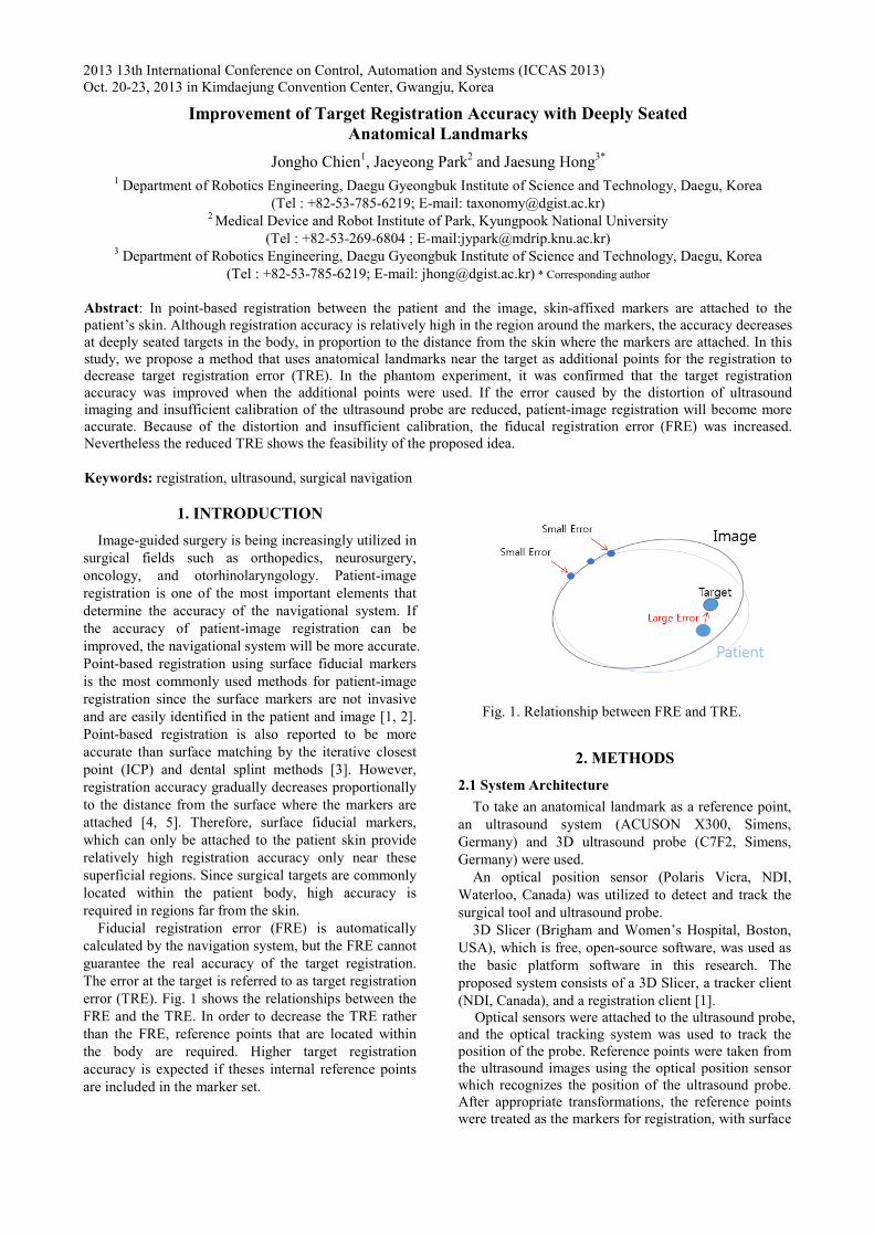
2013 13th International Conference on Control, Automation and Systems (ICCAS 2013) Oct. 20-23, 2013 in Kimdaejung Convention Center, Gwangju, Korea 1. INTRODUCTION Image-guided surgery is being increasingly utilized in surgical fields such as orthopedics, neurosurgery, oncology, and otorhinolaryngology. Patient-image registration is one of the most important elements that determine the accuracy of the navigational system. If the accuracy of patient-image registration can be improved, the navigational system will be more accurate. Point-based registration using surface fiducial markers is the most commonly used methods for patient-image registration since the surface markers are not invasive and are easily identified in the patient and image [1, 2]. Point-based registration is also reported to be more accurate than surface matching by the iterative closest point (ICP) and dental splint methods [3]. However, registration accuracy gradually decreases proportionally to the distance from the surface where the markers are attached [4, 5]. Therefore, surface fiducial markers, which can only be attached to the patient skin provide relatively high registration accuracy only near these superficial regions. Since surgical targets are commonly located within the patient body, high accuracy is required in regions far from the skin. Fiducial registration error (FRE) is automatically calculated by the navigation system, but the FRE cannot guarantee the real accuracy of the target registration. The error at the target is referred to as target registration error (TRE). Fig. 1 shows the relationships between the FRE and the TRE. In order to decrease the TRE rather than the FRE, reference points that are located within the body are required. Higher target registration accuracy is expected if theses internal reference points are included in the marker set. Fig. 1. Relationship between FRE and TRE. 2. METHODS 2.1 System Architecture To take an anatomical landmark as a reference point, an ultrasound system (ACUSON X300, Simens, Germany) and 3D ultrasound probe (C7F2, Simens, Germany) were used. An optical position sensor (Polaris Vicra, NDI, Waterloo, Canada) was utilized to detect and track the surgical tool and ultrasound probe. 3D Slicer (Brigham and Women’s Hospital, Boston, USA), which is free, open-source software, was used as the basic platform software in this research. The proposed system consists of a 3D Slicer, a tracker client (NDI, Canada), and a registration client [1]. Optical sensors were attached to the ultrasound probe, and the optical tracking system was used to track the position of the probe. Reference points were taken from the ultrasound images using the optical position sensor which recognizes the position of the ultrasound probe. After appropriate transformations, the reference points were treated as the markers for registration, with surface Improvement of Target Registration Accuracy with Deeply Seated Anatomical Landmarks Jongho Chien 1 , Jaeyeong Park 2 and Jaesung Hong 3* 1 Department of Robotics Engineering, Daegu Gyeongbuk Institute of Science and Technology, Daegu, Korea (Tel : +82-53-785-6219; E-mail: [email protected]) 2 Medical Device and Robot Institute of Park, Kyungpook National University (Tel : +82-53-269-6804 ; E-mail:[email protected]) 3 Department of Robotics Engineering, Daegu Gyeongbuk Institute of Science and Technology, Daegu, Korea (Tel : +82-53-785-6219; E-mail: [email protected]) * Corresponding author Abstract: In point-based registration between the patient and the image, skin-affixed markers are attached to the patient’s skin. Although registration accuracy is relatively high in the region around the markers, the accuracy decreases at deeply seated targets in the body, in proportion to the distance from the skin where the markers are attached. In this study, we propose a method that uses anatomical landmarks near the target as additional points for the registration to decrease target registration error (TRE). In the phantom experiment, it was confirmed that the target registration accuracy was improved when the additional points were used. If the error caused by the distortion of ultrasound imaging and insufficient calibration of the ultrasound probe are reduced, patient-image registration will become more accurate. Because of the distortion and insufficient calibration, the fiducal registration error (FRE) was increased. Nevertheless the reduced TRE shows the feasibility of the proposed idea. Keywords: registration, ultrasound, surgical navigation
Transcript
bhlee
입력 텍스트
978-89-93215-05-2 95560/13/$15 ⓒICROS

Fig. 6. Experimental configurat

An acrylic box filled with pure watethe ultrasound system. The tracking systhe phantom and the ultrasound probultrasound probe detected landmarks (Fphantom. After appropriate transformaticalculated during calibration, landmarkfiducial markers. The paired point regwas performed between a reconstructecomputed tomography (CT) volumephantom.

Two experiments were performed. depth mark, seven fiducial makers surface of the phantom were used Second, with one depth marker, seven attached to the surface of the phantomregistration. We measured the FRE anexperiment. TREs were measured usinthe inner landmarks, except the point tthe depth fiducial marker.

3. RESULTS

The registration accuracy withou

anatomical landmark was examined. Umarkers and additional one anatomicanumber of trials was 10 for each target.

tion.

er was used with stem tracked both be (Fig. 6). The Fig. 5) inside the ions, which were

ks were used like gistration method ed image of the e data and the

First, without a attached to the for registration. fiducial markers

m were used for nd TRE in every ng three points of that was used for

ut and with an Use seven surface al landmark. The

Fig. 7. Registration erroanatomical landmark

Fig. 7 shows the TREs and

with an anatomical landmark 2.13 ± 0.37 mm. Likewiseanatomical landmark, TRE1s 1.27 ± 0.39 mm, TRE2s were± 0.73 mm, TRE3s were 1.20.47 mm, TRE4s were 1.65 ±mm. Except TRE2, the use ocaused an increase in FRE, TRE.

4. CONCL

In this study, a point-baseincludes an anatomical landmusing an ultrasound systeregistration accuracy with anlandmark was examined. In twas confirmed that the targetimproved when the referenceerror caused by the distortioninsufficient calibration of thereduced, patient-image regaccurate.

ACKNOWLED

This work was supported development program (MOtorhinolaryngology, and invasive multi-DOF surgicaMinistry of Trade, Industry(10040097). It was also suppand Convergence Science DGIST.

REFERE [1] J. Hong, M. Hashizume,

registration tool for suEndosc, Vol. 24, pp. 944-9

[2] J. Hong, N. Matsumoto, RHashizume, “Medical navsurgery based on hybridintraoperative computed tBiomed Eng, Vol. 56, No.

[3] M. C. Metzger, A. Rafii, Pahm, B. Strong, “Comstrategies for computer-aidOtolaryngol Head Neck S2007.

[4] J. M. Fitzpatrick, J. B“Predicting error in registration”, IEEE Trans pp. 694-702, 1998.

[5] W. Zhang, G. Shen, C. Fiducial Configuration onin Image-Guided SurgerBiomed Eng Inform, Vol. 4

or with and without an

d FREs. FREs without and were 0.76 ± 0.12 mm and

e, with and without the were 1.46 ± 0.31 mm and

e 1.69 ± 0.41 mm and 1.89 24 ± 0.47 mm and 0.95 ± ± 0.74 mm and 1.04 ± 0.53 of the anatomical landmark

and a slight decrease in

USIONS d registration method that mark as a reference mark em was proposed. The nd without an anatomical the phantom experiment, it t registration accuracy was e points were used. If the n of ultrasound image and e ultrasound probe can be gistration will be more

DGEMENTS

in part by a research and Medical Image- guided

neurosurgical minimally al robot systems) of the y and Energy of Korea ported by the MIREBrain Center (12-BD-0402) of

ENCES

“An effective point-based urgical navigation”, Surg 948, 2010. R. Ouchida, S. Komune, M. vigation system for otologic d registration and virtual tomography”, IEEE Trans 2, pp. 426-432, 2009. B. Holhaeg-Majert, A. M.

mparison of 4 registration ded maxillofacial surgery”, Surg, Vol. 137, pp. 93-99,

B. West, C. R. Maurer, rigid-body point-based

Med Imag, Vol. 17, No. 5,

Wang, Y. Liu, “Effect of n Target Registration Error ry-A Experiment Study”, 4, pp. 1477-1481, 2010.

283

[6] K. S. Arun, S. D. Blostein, “Least-square fitting of two 3-D point sets”, IEEE Trans Pattern Anal, Vol. 9, pp. 698–700, 1987.

[7] I. M. Germano, “Advanced techniques in image-guided brain and spine surgery”, New York: Thieme, 2001.

[8] E. W. Nottmeier, T. L. Crosby, “Timing of paired points and surface matching registration in three-dimensional (3-D) image-guided spinal surgery”, J Spinal Disord Tech, Vol. 20, No. 4, pp. 268-270, 2007.

[9] J. B. West, J. M. Fitzpatrick, S. A. Toms, C. R. Maurer, Jr., R. J. Maciunas, “Fiducial point placement and the accuracy of point-based, rigid body registration”, Neurosurgery, Vol. 48, No. 4, pp. 810-816; discussion 816-817, 2001.

284


Recommended