Incretins: Their physiology and application in the treatment of diabetes mellitus Salih Sanlioglu VMD, PhD Human Gene and Cell Therapy Center of Akdeniz University, Antalya, Türkiye, 07058 1
Transcript
Incretins: Their physiology and application in the treatment of diabetes mellitus
Salih Sanlioglu VMD, PhDHuman Gene and Cell Therapy Center of Akdeniz University, Antalya, Türkiye, 07058
1
2
Currently, 382 million people have been reported to have diabetes in the world, and 90% of them has type 2 diabetes mellitus (T2DM).
Increase in obesity rates has been correlated with an increase in the prevalence of diabetes. Diabetes is now considered to be the world’s biggest pandemic disease with a prevalence of 8%.
Five million people have died in 2013 because of the secondary complications of diabetes including coronary heart disease and peripheral vascular diseases.
Furthermore, diabetes has been reported to be the most important cause of blindness and renal failure in developed countries.
Although diabetes is primarily managed by lifestyle changes and dietary modifications, administration of a pharmacological agent is required especially when treatment goals are not achieved.
İntroduction
3
These conventional treatment agents include but not limited to biguanides, sulfonyl- ureas, thiazolidinediones, meglitinides, alpha-glucosidase inhibitors and insulin along with a recently developed amylin analogue pramlintide.
Current guidelines recommend biguanide metformin as a first-line treatment, with subsequent addition of other agents when this monotherapy is no longer effective.
Despite intensive therapy, glycaemic control can still be lost, leading to an increase in HbA1c levels of diabetic patients. Moreover, current therapies are often associated with weight gain and hypoglycaemia.
Other adverse events include but not limited to gastrointestinal discomfort with the use of biguanides, and possible oedema, cardiac failure or fractures due to the use of thiazolidinediones.
İntroduction
4
• a defect of insulin action in skeletal muscle and liver (insulin resistance);
• and a bifunctional defect in pancreatic hormone secretion characterized by:
◦ an incapacity of beta cells to increase insulin secretion to compensate for tissue insulin resistance,
◦ and an increase in glucagon secretion by alpha cells despite hyperglycaemia.
Type 2 diabetes is characterized by two important defects:
5
In addition, the kinetics of insulin secretion are also markedly altered in the post-absorptive period. Indeed, the early phase of insulin release is blunted in type 2 diabetes and leads to a decreased capacity of insulin to inhibit hepatic glucose production in the postprandial period.
As glucagon secretion is not normally inhibited by hyperglycaemia in the post-absorptive state, increased glucagon secretion sustains liver glucose production, which explains the hyperglycaemic excursions observed after a meal.
The incapacity to compensate for insulin resistance is the result of the loss of beta-cell mass in type 2 diabetes mainly due to apoptosis of beta cells induced by glucolipotoxicity.
Type 2 diabetes
6
The entero-insular axis. Postprandially, insulin secretion is directly stimulated by substrates and by the strong endocrine stimulation through incretin hormones. Modified according to Creutzfeldt.1 GIP, gastric inhibitory polypeptide; AA, amino acids; CHO, carbohydrates; FA, fatty acids.
Gallwitz B Diabetes Technol Ther. 2005 Aug;7(4):651-7
• The term ‘Entero-insular Axis’ was coined by Unger & Eisentraut (1969) to include all those gut factors which contribute to enhanced insulin secretion fol lowing ingestion of a meal.
• It is now apparent that the Entero-insular Axis possesses an important Neural , an Endocrine, and Metabolic component.
7
The idea that certain factors produced by the intestinal mucosa in response to nutrient ingestion are capable of stimulating the release of substances from the endocrine pancreas, thereby reducing blood glucose levels, was first introduced in the early 1900s.
It was shown that oral glucose administration is associated with a much greater increase in plasma insulin compared with the same amount of glucose given intravenously.
In the 1930s, Dr Jean La Barre from Belgium introduced the term “incretin” to describe a substance in the gut mucosa that produces hypoglycaemia when injected into normal animals, but not in those who had undergone pancreatectomy.
Dr Hans Heller from Austria made a similar suggestion a few years later, although he suggested the term “duodenin” for this tentative substance.
The incretin concept
8
The incretin theory was elucidated by Creutzfeld in 1979 as endocrine signals produced in the gastrointestinal tract, released by nutrients to stimulate insulin secretion in the presence of glucose.
This means that incretins act as amplifiers of glucose signals. This phenomenon was estimated to involve 50–70% of the total insulin secreted following oral glucose administration.
Today, incretin therapy is an established glucose-lowering strategy in type 2 diabetes and uses both GLP-1 receptor agonists and dipeptidyl peptidase (DPP)-4 inhibitors.
İntroduction
B. Ahrén / Diabetes & Metabolism 39 (2013) 195–201
9
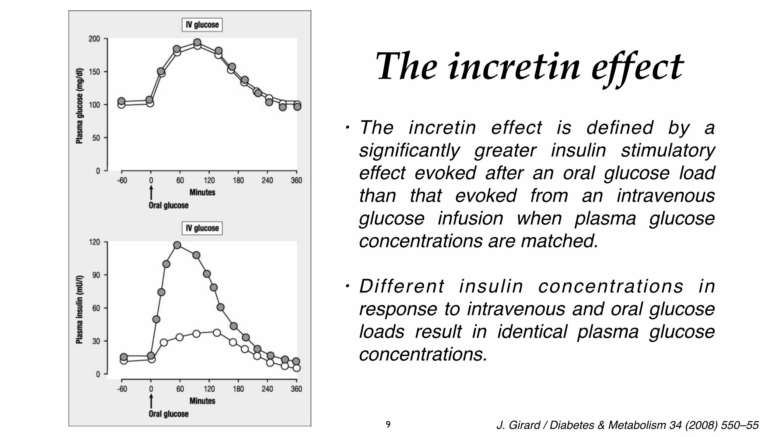
• The incretin effect is defined by a significantly greater insulin stimulatory effect evoked after an oral glucose load than that evoked from an intravenous glucose infusion when plasma glucose concentrations are matched.
• Different insulin concentrations in response to intravenous and oral glucose loads result in identical plasma glucose concentrations.
The incretin effect
J. Girard / Diabetes & Metabolism 34 (2008) 550–55
10
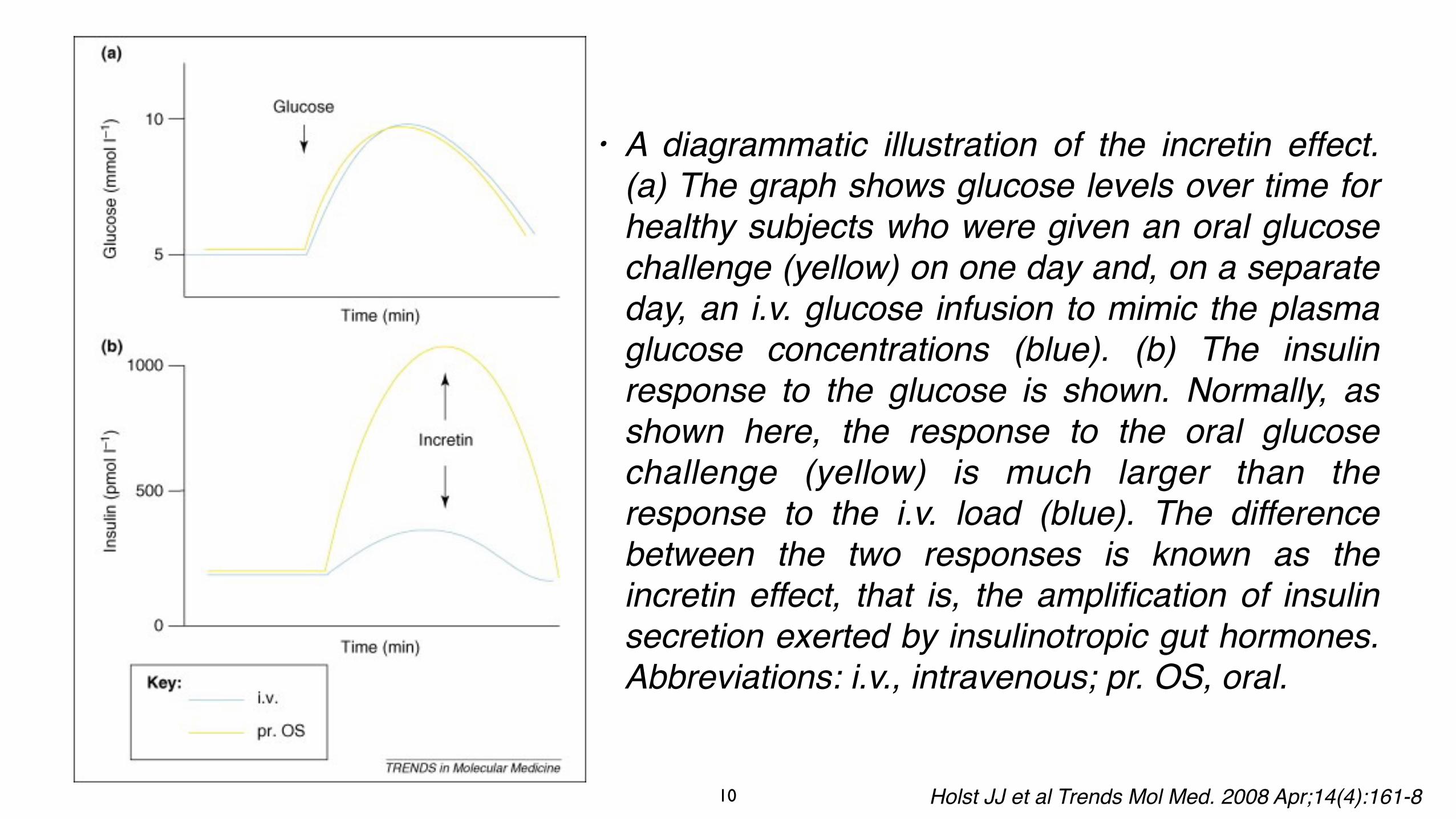
• A diagrammatic illustration of the incretin effect. (a) The graph shows glucose levels over time for healthy subjects who were given an oral glucose challenge (yellow) on one day and, on a separate day, an i.v. glucose infusion to mimic the plasma glucose concentrations (blue). (b) The insulin response to the glucose is shown. Normally, as shown here, the response to the oral glucose challenge (yellow) is much larger than the response to the i.v. load (blue). The difference between the two responses is known as the incretin effect, that is, the amplification of insulin secretion exerted by insulinotropic gut hormones. Abbreviations: i.v., intravenous; pr. OS, oral.
Holst JJ et al Trends Mol Med. 2008 Apr;14(4):161-8
11
Although carbohydrates, protein and fat all contribute to the secretion of incretins to some degree, carbohydrates is the most effective agent in causing incretin secretion. This is because carbohydrate absorption is the only way to increase glucose levels in circulation, and incretins stimulate insulin secretion only when blood glucose is high.
Although incretins are crucial in the maintenance of normoglycaemia by way of facilitating glucose transport into peripheral tissues, T2DM patients displayed insulin resistance and reduced incretin secretion, resulting in ineffective glucose clearance from circulation.
Because reduced incretin response to food ingestion is one of the primary defects associated with glucose intolerance and hyperglycaemia in T2DM, incretin-based treatment strategies recently gained a significant momentum as a novel class of medications with anti-diabetic potential.
İntroduction
12
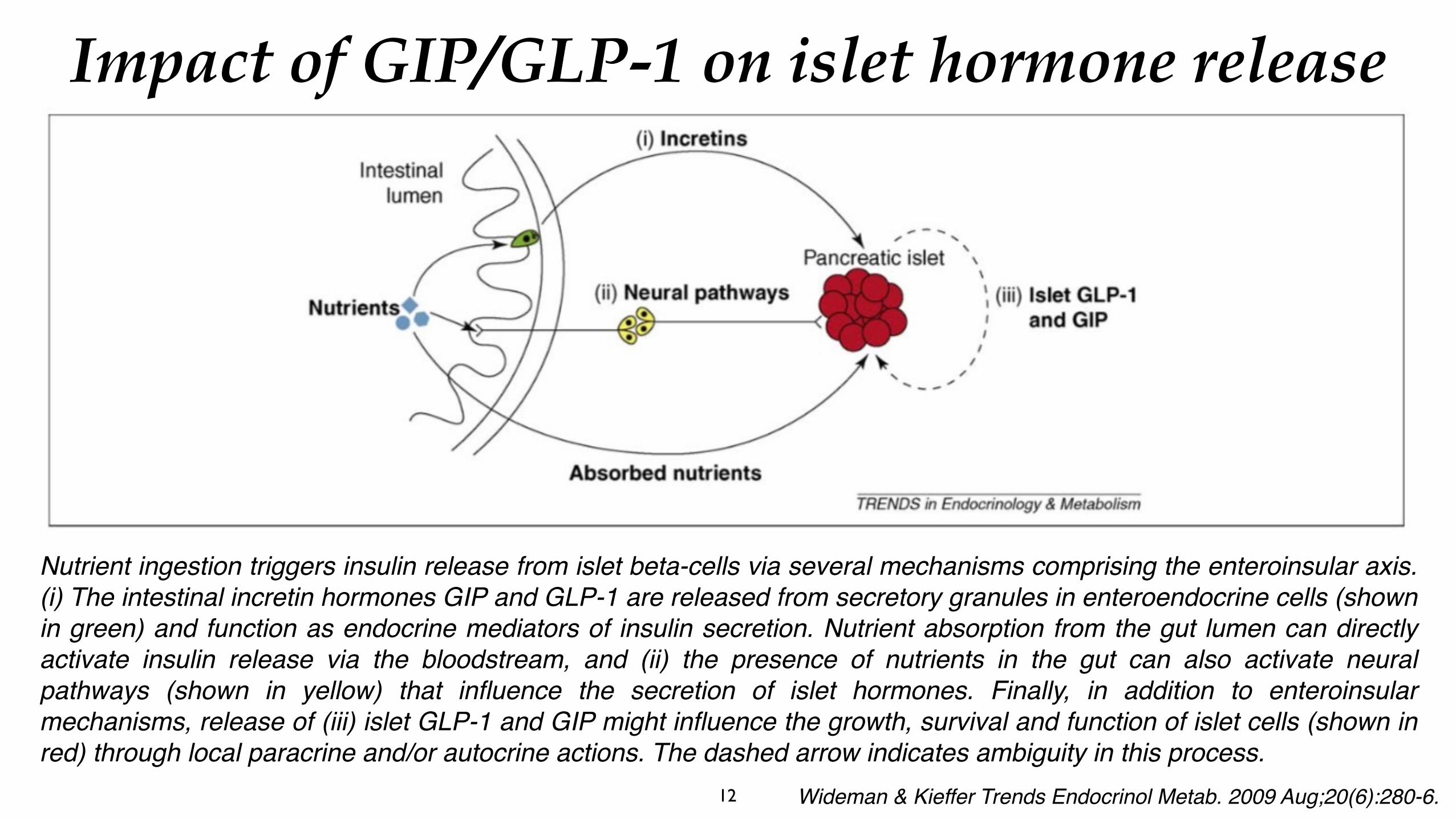
Nutrient ingestion triggers insulin release from islet beta-cells via several mechanisms comprising the enteroinsular axis. (i) The intestinal incretin hormones GIP and GLP-1 are released from secretory granules in enteroendocrine cells (shown in green) and function as endocrine mediators of insulin secretion. Nutrient absorption from the gut lumen can directly activate insulin release via the bloodstream, and (ii) the presence of nutrients in the gut can also activate neural pathways (shown in yellow) that influence the secretion of islet hormones. Finally, in addition to enteroinsular mechanisms, release of (iii) islet GLP-1 and GIP might influence the growth, survival and function of islet cells (shown in red) through local paracrine and/or autocrine actions. The dashed arrow indicates ambiguity in this process.
The first hormone identified was initially called ‘gastric inhibitory polypeptide’ (GIP) because of its ability to inhibit gastric secretion at pharmacological concentrations, but was later renamed ‘glucose-dependent insulinotropic polypeptide’ to reflect its incretin action at physiological concentrations.
GIP is released by duodenal K cells in response to glucose and fat absorption, but it was soon recognized that GIP alone could not fully account for the incretin effect in vivo.
The discovery of a second incretin hormone, glucagon-like peptide-1 (GLP-1), was made after the cloning and sequencing of the proglucagon gene. GLP-1 is synthesized and released by enteroendocrine L cells in the distal ileum and colon in response to glucose, fat and protein absorption. It enhances glucose-stimulated insulin secretion.
Molecular structure and secretion of incretins
14
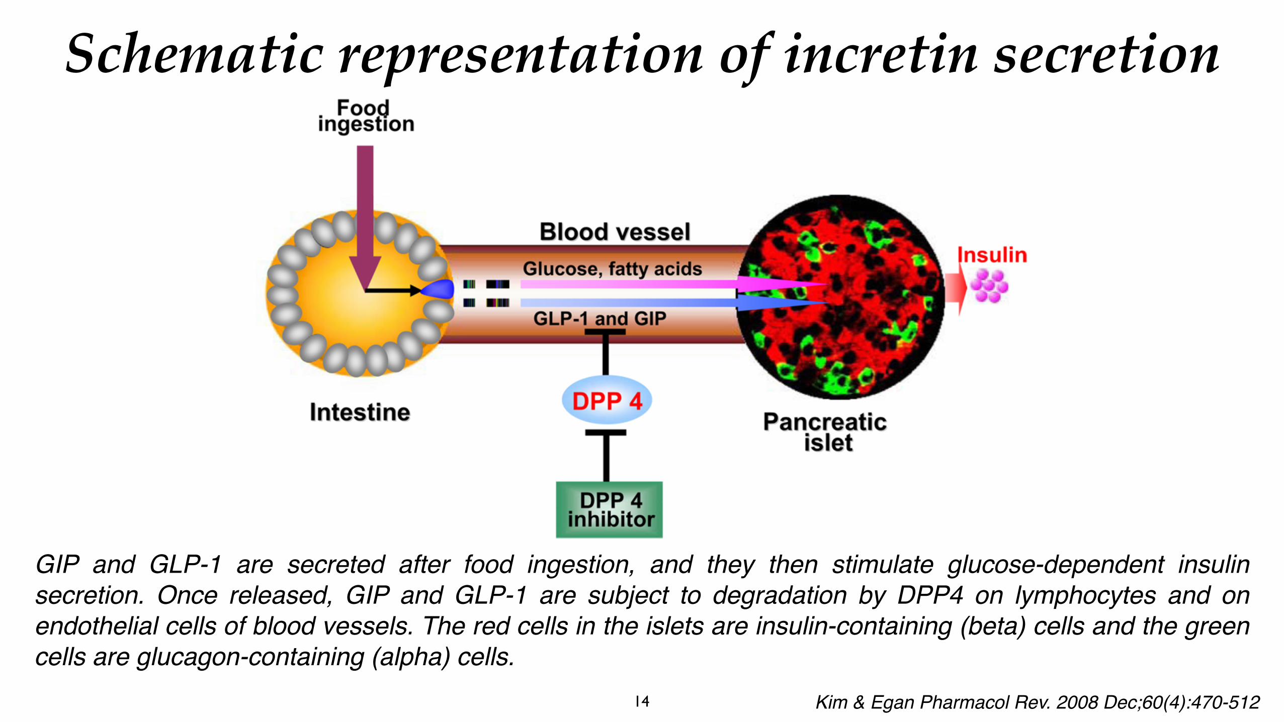
GIP and GLP-1 are secreted after food ingestion, and they then stimulate glucose-dependent insulin secretion. Once released, GIP and GLP-1 are subject to degradation by DPP4 on lymphocytes and on endothelial cells of blood vessels. The red cells in the islets are insulin-containing (beta) cells and the green cells are glucagon-containing (alpha) cells.
Schematic representation of incretin secretion
Kim & Egan Pharmacol Rev. 2008 Dec;60(4):470-512
15
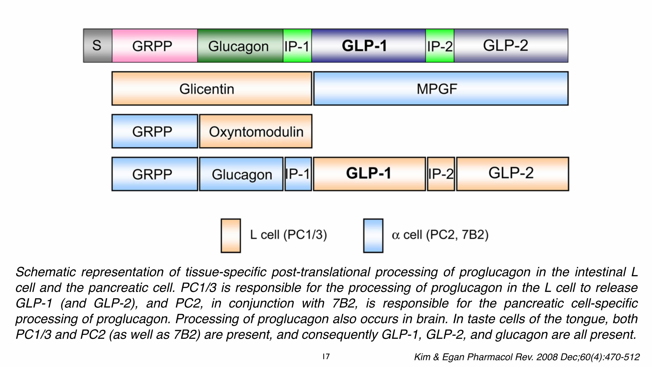
Schematic representation of proGIP and proglucagon. GIP is a single 42-amino acid peptide derived from the post-translational processing of proGIP by PC1/3 in enteroendocrine K cells. It is the only functional proGIP product in all species examined to date, and there is a greater than 90% amino acid identity between human, rat, murine, porcine, and bovine sequences.
GLP-1 is a post-translational cleavage product of the proglucagon gene by PC1/3 in enteroendocrine L cells and GLP-1 (7–36) amide is a major form of circulating biologically active GLP-1 in humans. In mammals, proglucagon is expressed in pancreas, enteroendocrine L cells, brain, and taste cells with an identical mRNA transcript in each tissue.
Schematic representation of proGIP and proglucagon
Kim & Egan Pharmacol Rev. 2008 Dec;60(4):470-512
16
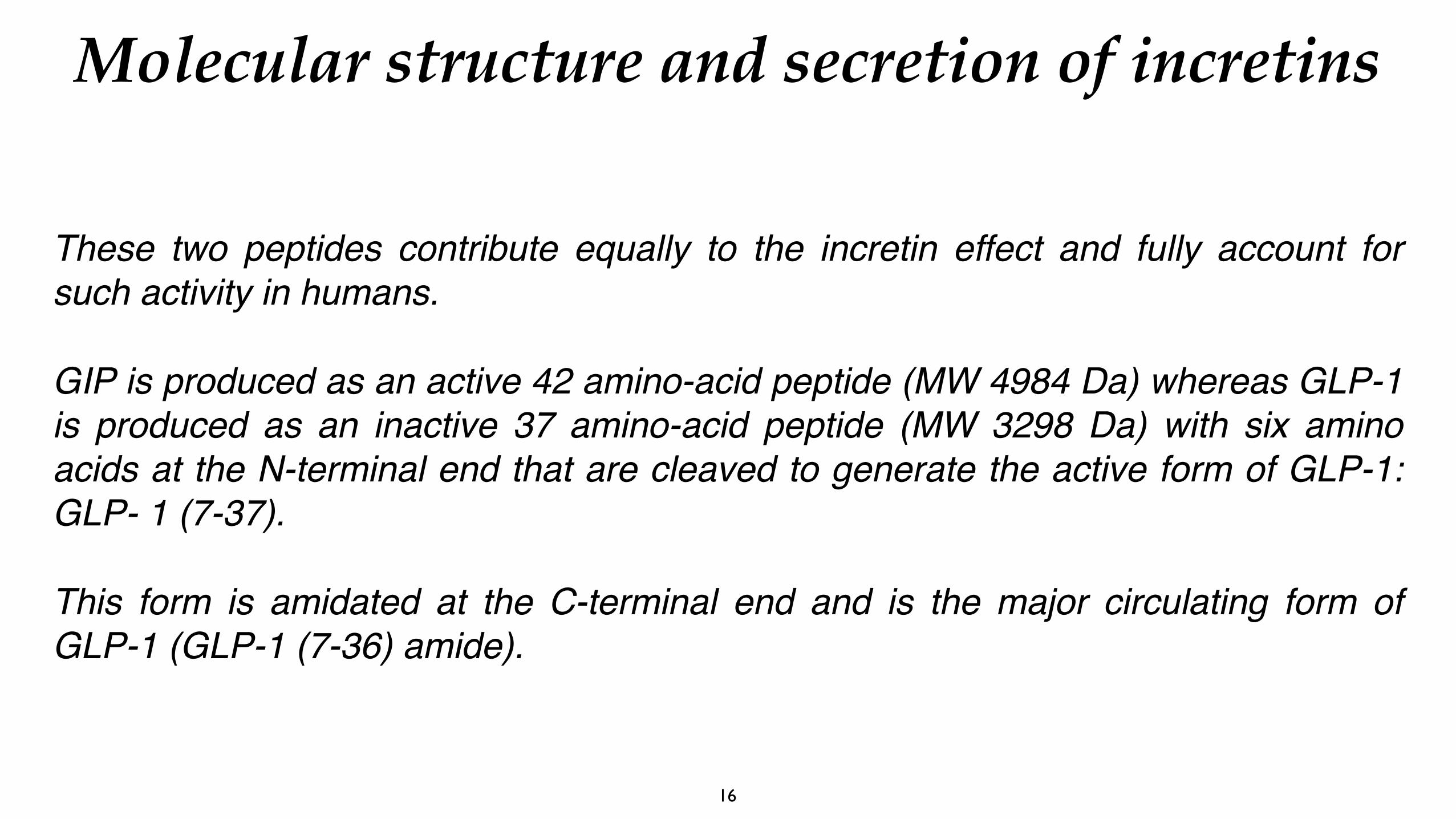
These two peptides contribute equally to the incretin effect and fully account for such activity in humans.
GIP is produced as an active 42 amino-acid peptide (MW 4984 Da) whereas GLP-1 is produced as an inactive 37 amino-acid peptide (MW 3298 Da) with six amino acids at the N-terminal end that are cleaved to generate the active form of GLP-1: GLP- 1 (7-37).
This form is amidated at the C-terminal end and is the major circulating form of GLP-1 (GLP-1 (7-36) amide).
Molecular structure and secretion of incretins
17
Schematic representation of tissue-specific post-translational processing of proglucagon in the intestinal L cell and the pancreatic cell. PC1/3 is responsible for the processing of proglucagon in the L cell to release GLP-1 (and GLP-2), and PC2, in conjunction with 7B2, is responsible for the pancreatic cell-specific processing of proglucagon. Processing of proglucagon also occurs in brain. In taste cells of the tongue, both PC1/3 and PC2 (as well as 7B2) are present, and consequently GLP-1, GLP-2, and glucagon are all present.
Kim & Egan Pharmacol Rev. 2008 Dec;60(4):470-512
18
Plasma concentrations of both incretins are low in the fasting state (5–10 pmol/L) and increase within 5–15 min of a meal (15–50pmol/L). GLP-1 has two circulating molecular forms—GLP-1(7-37) and GLP-1(7-36) amide—with the latter being more abundant in the circulation.
GIP and GLP-1 are secreted in response to nutrient ingestion (carbohydrates, proteins, fats), and it is the rate of nutrient absorption rather than its mere presence in the intestine that stimulates incretin release. It is important to note that only GIP and GLP-1 secretion in response to carbohydrate absorption leads to insulin secretion.
Circulating levels of GIP and GLP-1 decrease rapidly as both are quickly inactivated in plasma by the enzyme dipeptidyl peptidase IV (DPP-IV) and by renal clearance. Both are also excellent substrates for DPP-IV. The circulation half-lives of the two incretins are 1–2 min for GLP-1 and 5–7 min for GIP.
Molecular structure and secretion of incretins
19
Electrogenic transport of glucose via the apical ly expressed sodium-coupled glucose transporter SGLT1 generates a sufficient inward current to depolarise the cell membrane (DC) and trigger action potentials, leading to the opening of voltage-gated Ca2+ channels. The consequent rise in intracellular Ca2+ is the pr imary t r igger of exocytosis and glucagon-like peptide 1 (GLP-1) secretion. In addition, glucose metabolism results in the closure of ATP-sensitive KATP channels, contributing to the excitability of the cell and GLP-1 secretion.
Parker et al. Expert Rev Mol Med. 2010 Jan 5;12:e1Proposed glucose-sensing mechanisms in primary L-cells.
20
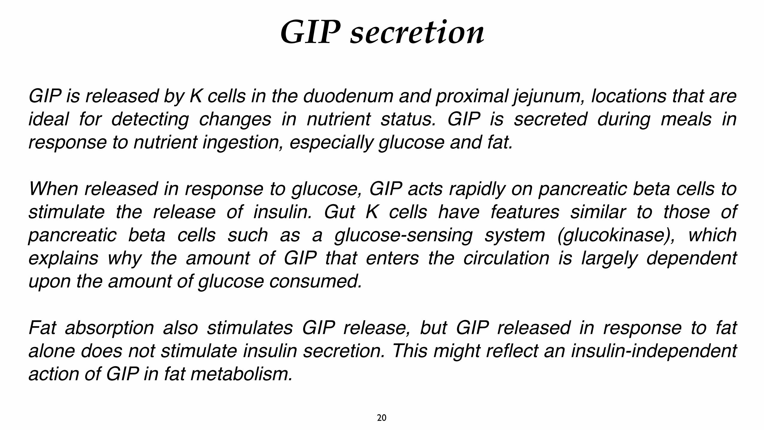
GIP is released by K cells in the duodenum and proximal jejunum, locations that are ideal for detecting changes in nutrient status. GIP is secreted during meals in response to nutrient ingestion, especially glucose and fat.
When released in response to glucose, GIP acts rapidly on pancreatic beta cells to stimulate the release of insulin. Gut K cells have features similar to those of pancreatic beta cells such as a glucose-sensing system (glucokinase), which explains why the amount of GIP that enters the circulation is largely dependent upon the amount of glucose consumed.
Fat absorption also stimulates GIP release, but GIP released in response to fat alone does not stimulate insulin secretion. This might reflect an insulin-independent action of GIP in fat metabolism.
GIP secretion
21
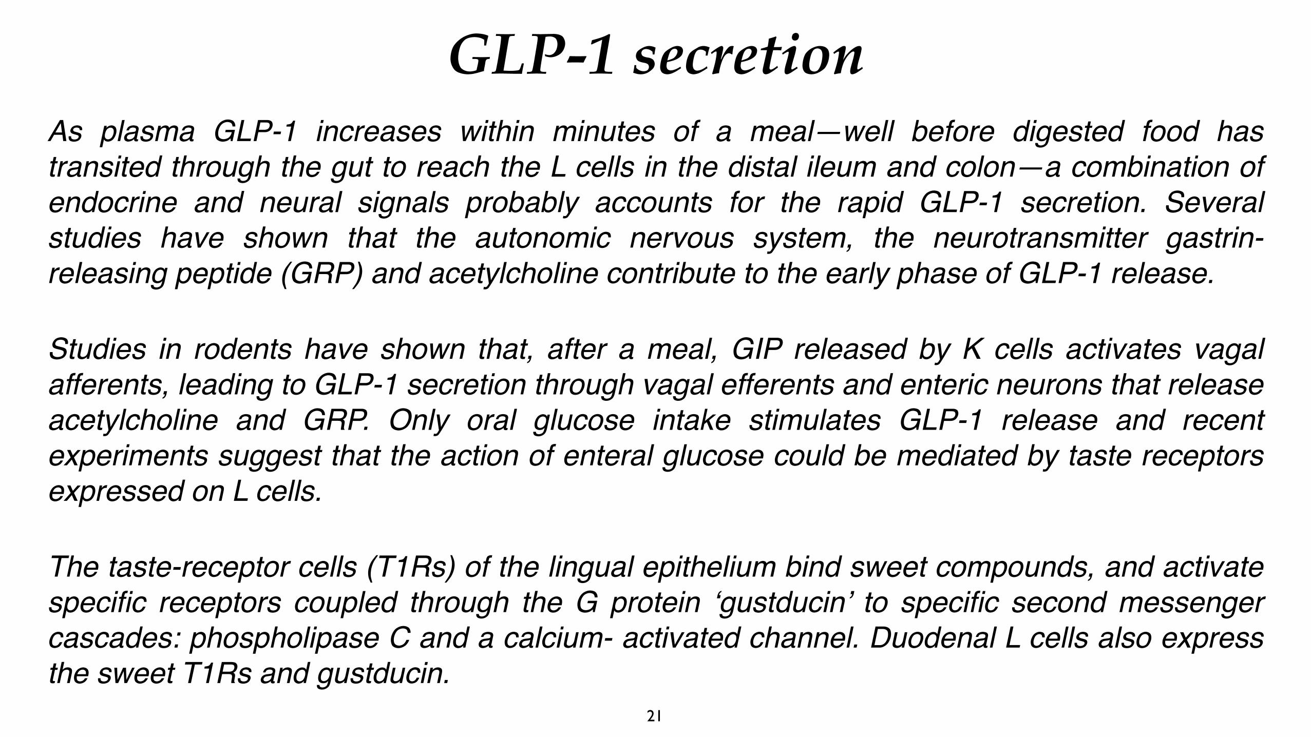
As plasma GLP-1 increases within minutes of a meal—well before digested food has transited through the gut to reach the L cells in the distal ileum and colon—a combination of endocrine and neural signals probably accounts for the rapid GLP-1 secretion. Several studies have shown that the autonomic nervous system, the neurotransmitter gastrin-releasing peptide (GRP) and acetylcholine contribute to the early phase of GLP-1 release.
Studies in rodents have shown that, after a meal, GIP released by K cells activates vagal afferents, leading to GLP-1 secretion through vagal efferents and enteric neurons that release acetylcholine and GRP. Only oral glucose intake stimulates GLP-1 release and recent experiments suggest that the action of enteral glucose could be mediated by taste receptors expressed on L cells.
The taste-receptor cells (T1Rs) of the lingual epithelium bind sweet compounds, and activate specific receptors coupled through the G protein ‘gustducin’ to specific second messenger cascades: phospholipase C and a calcium- activated channel. Duodenal L cells also express the sweet T1Rs and gustducin.
GLP-1 secretion
22
• Regulation of GLP-1 secretion by ingested nutrients. After a meal, nutrients in the duodenum activate a proximal neuroendocrine loop, which stimulates GLP-1 secretion from L cells in the ileum.
• In rodents, GIP released by K cells activates vagal afferent fibres, which, in turn, trigger GLP-1 secretion through vagal efferent fibres and enteric neurons that release acetylcholine (Ach) and gastrin-releasing peptide (GRP).
Secretion of incretins
J. Girard / Diabetes & Metabolism 34 (2008) 550–55
23
The endocrine pathway for the actions of GLP-1. GLP-1 secretion is stimulated by nutrients in the gut lumen (a magnified intestinal villus with an open-type L-cell is shown at the lower left). GLP-1 diffuses across the basal lamina into the lamina propria and is taken up by a capillary, only to be broken down by dipeptidyl peptidase IV (DPP-IV), located on the luminal surface of the endothelial cells (white cells lining the capillary) so that only 25% of the secreted amount reaches the portal circulation. In the liver, a further 40 –50% is destroyed so that only 10–15% enters the systemic circulation and may reach the pancreas and the brain via the endocrine pathway (perhaps even less because of the continued proteolytic activity of soluble DPP-IV present in plasma).
Holst JJ Physiol Rev. 2007 Oct;87(4):1409-39
24
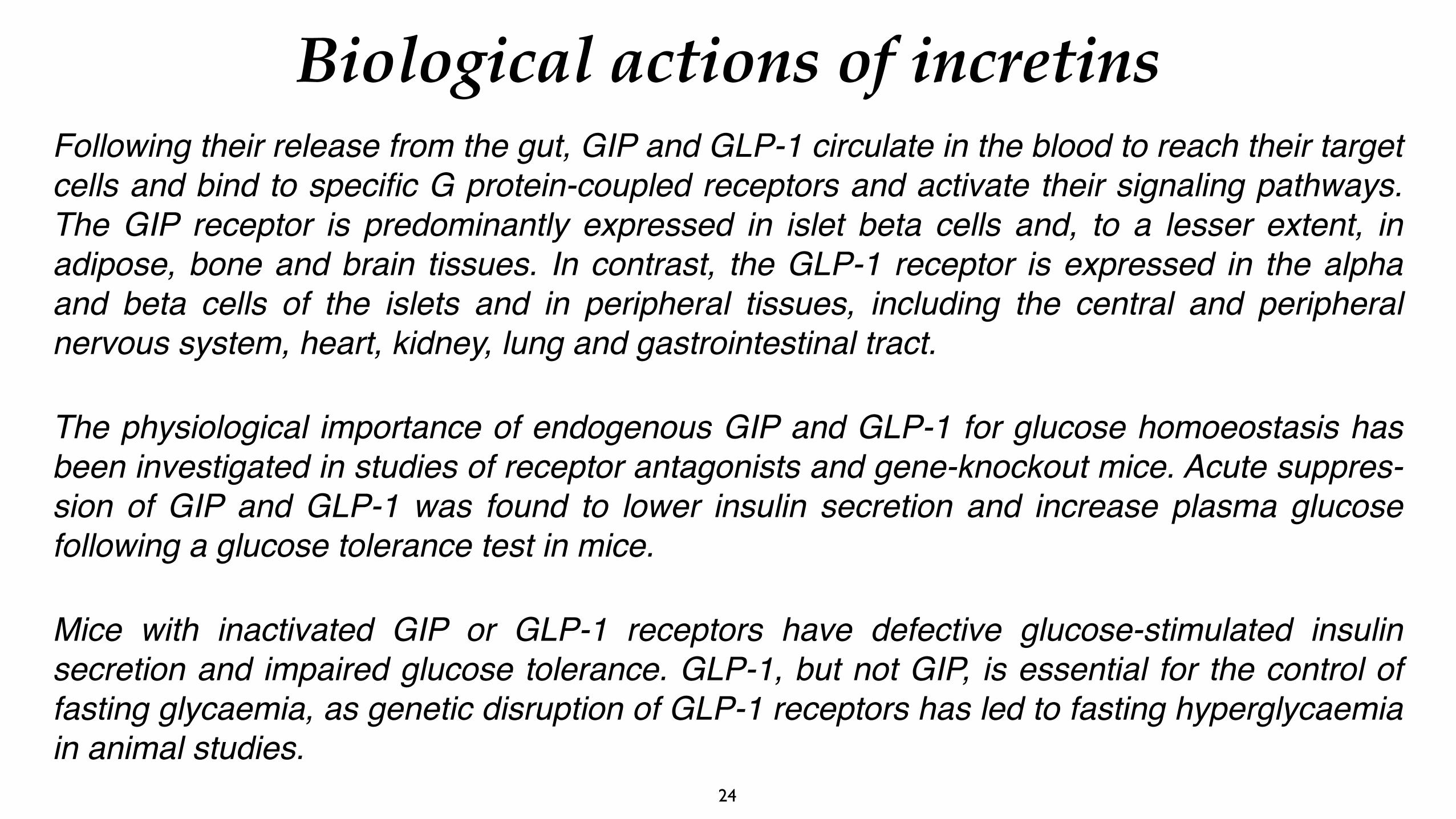
Following their release from the gut, GIP and GLP-1 circulate in the blood to reach their target cells and bind to specific G protein-coupled receptors and activate their signaling pathways. The GIP receptor is predominantly expressed in islet beta cells and, to a lesser extent, in adipose, bone and brain tissues. In contrast, the GLP-1 receptor is expressed in the alpha and beta cells of the islets and in peripheral tissues, including the central and peripheral nervous system, heart, kidney, lung and gastrointestinal tract.
The physiological importance of endogenous GIP and GLP-1 for glucose homoeostasis has been investigated in studies of receptor antagonists and gene-knockout mice. Acute suppres- sion of GIP and GLP-1 was found to lower insulin secretion and increase plasma glucose following a glucose tolerance test in mice.
Mice with inactivated GIP or GLP-1 receptors have defective glucose-stimulated insulin secretion and impaired glucose tolerance. GLP-1, but not GIP, is essential for the control of fasting glycaemia, as genetic disruption of GLP-1 receptors has led to fasting hyperglycaemia in animal studies.
Biological actions of incretins
25
Biological actions of incretins Actions of gastrointestinal hormones o n k e y t i s s u e s i n g l u c o s e homoeostasis. Both GIP and GLP-1 promote insulin biosynthesis, insulin secretion and islet beta-cell survival. GLP-1 exerts additional actions, including inhibition of glucagon secretion and gastric-emptying, and induction of food intake. GIP has a direct effect on adipocytes coupled to energy storage. In contrast, CCK and gastrin do not regulate plasma glucose levels, but could be important for stimulation of islet neogenesis, with permission from the American Society for Clinical Investigation).
J. Girard / Diabetes & Metabolism 34 (2008) 550–55
26
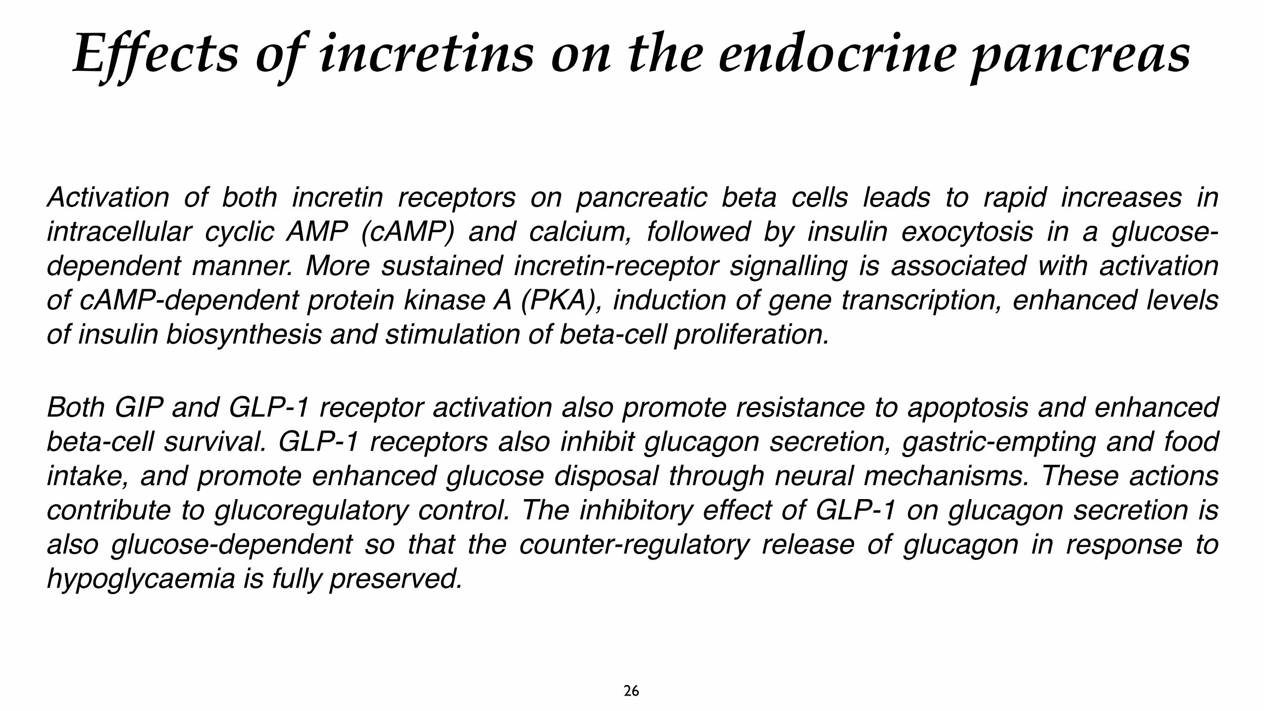
Activation of both incretin receptors on pancreatic beta cells leads to rapid increases in intracellular cyclic AMP (cAMP) and calcium, followed by insulin exocytosis in a glucose- dependent manner. More sustained incretin-receptor signalling is associated with activation of cAMP-dependent protein kinase A (PKA), induction of gene transcription, enhanced levels of insulin biosynthesis and stimulation of beta-cell proliferation.
Both GIP and GLP-1 receptor activation also promote resistance to apoptosis and enhanced beta-cell survival. GLP-1 receptors also inhibit glucagon secretion, gastric-empting and food intake, and promote enhanced glucose disposal through neural mechanisms. These actions contribute to glucoregulatory control. The inhibitory effect of GLP-1 on glucagon secretion is also glucose-dependent so that the counter-regulatory release of glucagon in response to hypoglycaemia is fully preserved.
Effects of incretins on the endocrine pancreas
27
The main molecular events during incretin-induced insulin secretion from beta-cell
Kim & Egan Pharmacol Rev. 2008 Dec;60(4):470-512
The binding of incretin ligands or agonists to the incretin receptors results in production of cAMP via adenylyl cyclase (AC) activation and subsequent activation of PKA and the Epac family of cAMP-regulated guanine nucleotide exchange factors (cAMP-GEFs), which leads to elevation of intracellular Ca2 levels via a depolarization of plasma membrane by inhibition of KATP and KV channels after ATP generation from glucose and consequent opening of voltage-gated L-type Ca2 channels. Intracellular Ca2 levels are further increased via stimulation of IP3R and RyR on the ER. Long-term GLP-1 treatment also stimulates the expression of GLUT2 transporter and glucokinase (the beta-cell glucose sensor), which lead to increased mitochondrial ATP synthesis. In addition, L-type Ca2 channels are phosphorylated by PKA, resulting in an increase of their open probability and thus facilitation of enhanced Ca2 influx. The changes in intracellular Ca2 concentrations lead to fusion of insulin-containing vesicles to the plasma membrane and subsequent rapid exocytosis of insulin from cells. In addition, the exocytosis of insulin-containing vesicles is directly regulated by PKA and Epac2 via interaction with the regulators of exocytosis and ATP.
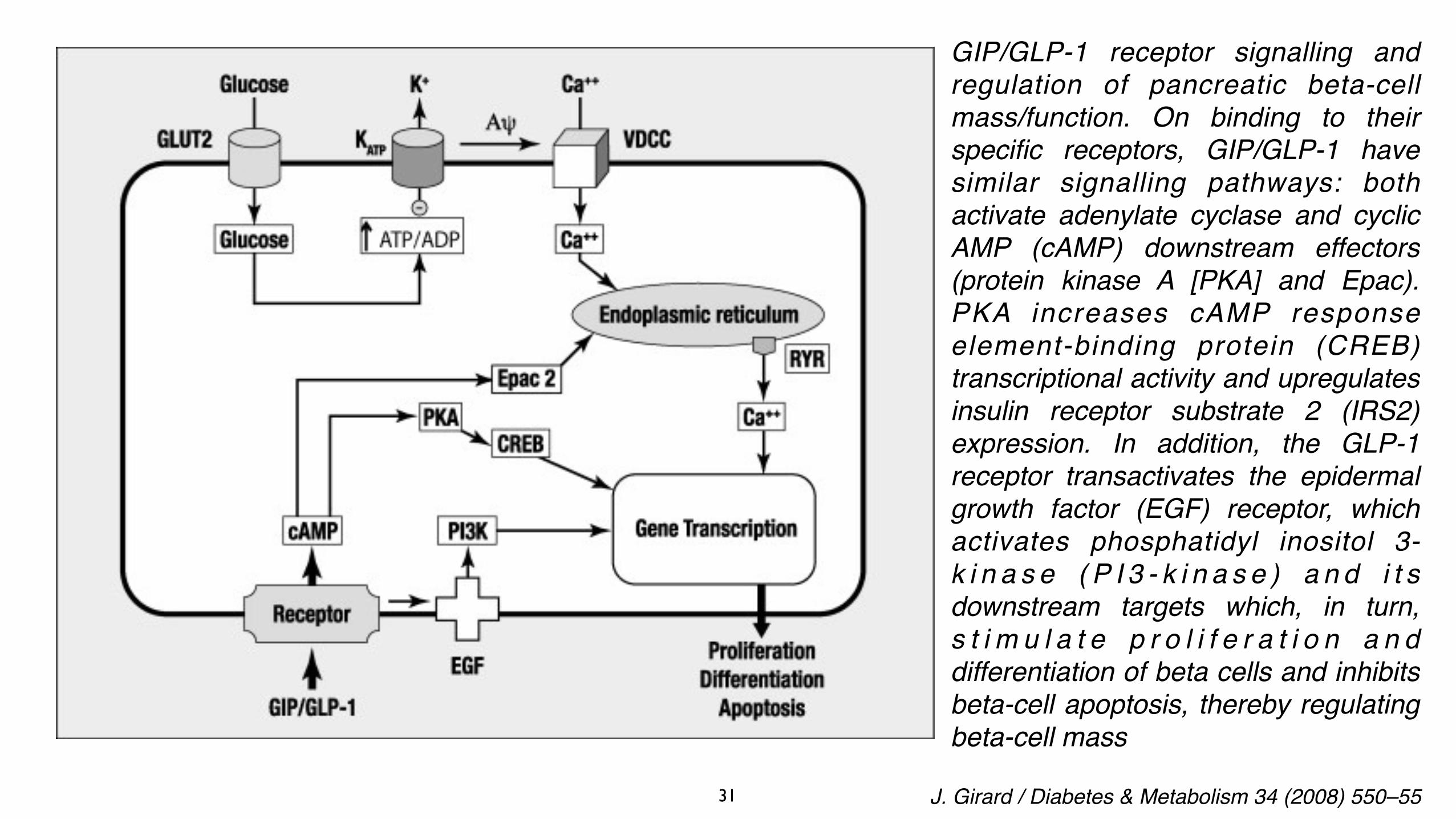
28
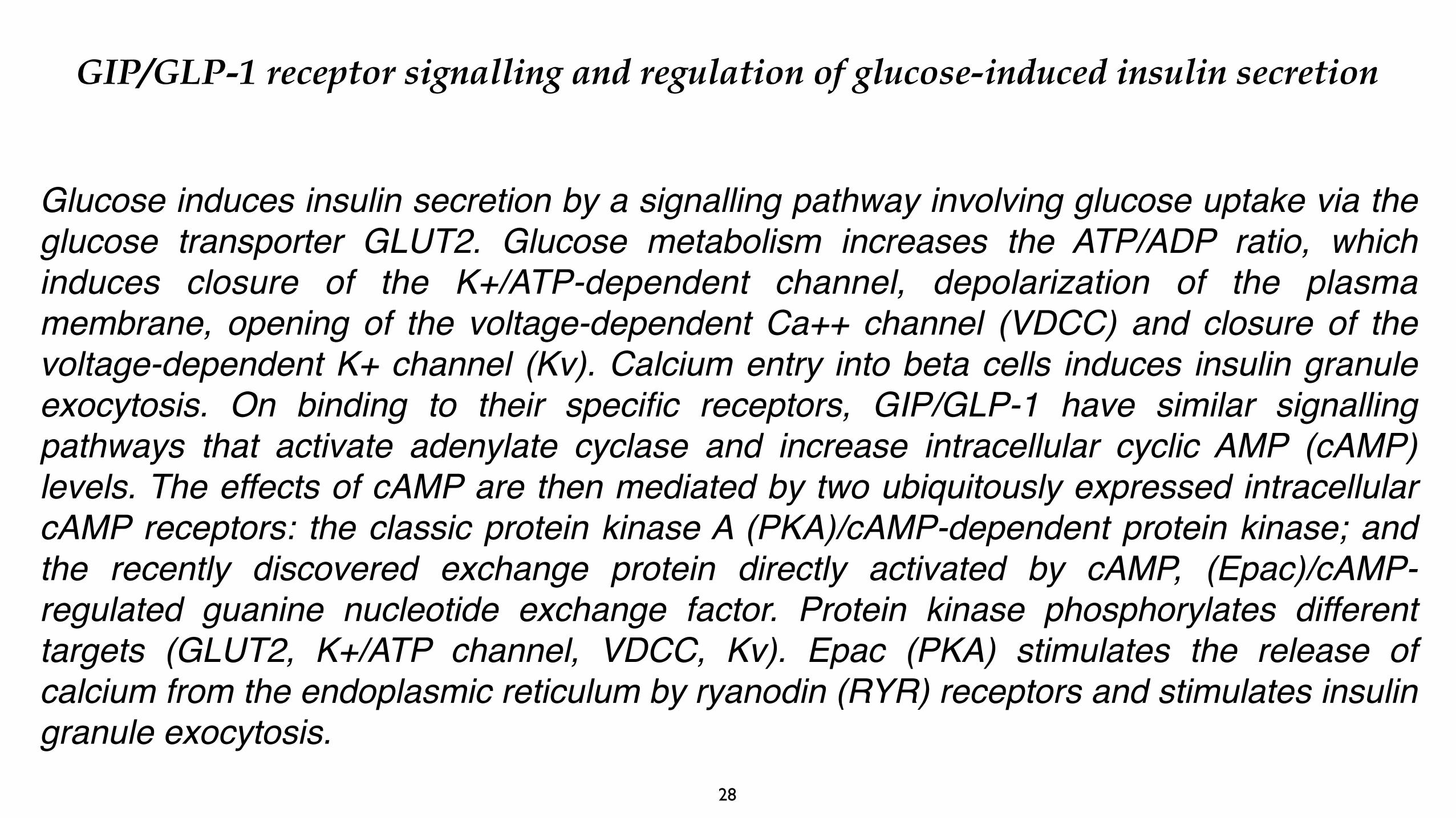
Glucose induces insulin secretion by a signalling pathway involving glucose uptake via the glucose transporter GLUT2. Glucose metabolism increases the ATP/ADP ratio, which induces closure of the K+/ATP-dependent channel, depolarization of the plasma membrane, opening of the voltage-dependent Ca++ channel (VDCC) and closure of the voltage-dependent K+ channel (Kv). Calcium entry into beta cells induces insulin granule exocytosis. On binding to their specific receptors, GIP/GLP-1 have similar signalling pathways that activate adenylate cyclase and increase intracellular cyclic AMP (cAMP) levels. The effects of cAMP are then mediated by two ubiquitously expressed intracellular cAMP receptors: the classic protein kinase A (PKA)/cAMP-dependent protein kinase; and the recently discovered exchange protein directly activated by cAMP, (Epac)/cAMP-regulated guanine nucleotide exchange factor. Protein kinase phosphorylates different targets (GLUT2, K+/ATP channel, VDCC, Kv). Epac (PKA) stimulates the release of calcium from the endoplasmic reticulum by ryanodin (RYR) receptors and stimulates insulin granule exocytosis.
GIP/GLP-1 receptor signalling and regulation of glucose-induced insulin secretion
29
The primary function of GIP and GLP-1 is to enhance glucose-dependent insulin secretion. The mechanisms involved are overlapping and include an increase in intracellular cAMP, inhibition of ATP-dependent potassium (K-ATP) channels, an increase in intracellular calcium and stimulation of exocytosis.
Incretins also upregulate insulin-gene transcription and biosyn- thesis.
Effects of incretins on insulin biosynthesis and secretion
30
GLP-1 could inhibit glucagon secretion via stimulation of somatostatin secretion as delta cells contain specific GLP-1 receptors. The cellular mechanisms are believed to involve an increase in cAMP/PKA, closure of K-ATP channels, membrane depolarization, inactivation of ion channels and reduction of intracellular calcium.
Effects of GLP-1 on glucagon secretion
Effects of GIP on glucagon secretion
GIP can induce glucagon secretion from pancreatic alpha cells thus may worsen (hyperglycaemia) prognosis of T2DM patients.
31
GIP/GLP-1 receptor signalling and regulation of pancreatic beta-cell mass/function. On binding to their specific receptors, GIP/GLP-1 have similar signalling pathways: both activate adenylate cyclase and cyclic AMP (cAMP) downstream effectors (protein kinase A [PKA] and Epac). PKA increases cAMP response element-binding protein (CREB) transcriptional activity and upregulates insulin receptor substrate 2 (IRS2) expression. In addition, the GLP-1 receptor transactivates the epidermal growth factor (EGF) receptor, which activates phosphatidyl inositol 3-k i n a s e ( P I 3 - k i n a s e ) a n d i t s downstream targets which, in turn, s t i m u l a t e p r o l i f e r a t i o n a n d differentiation of beta cells and inhibits beta-cell apoptosis, thereby regulating beta-cell mass
J. Girard / Diabetes & Metabolism 34 (2008) 550–55
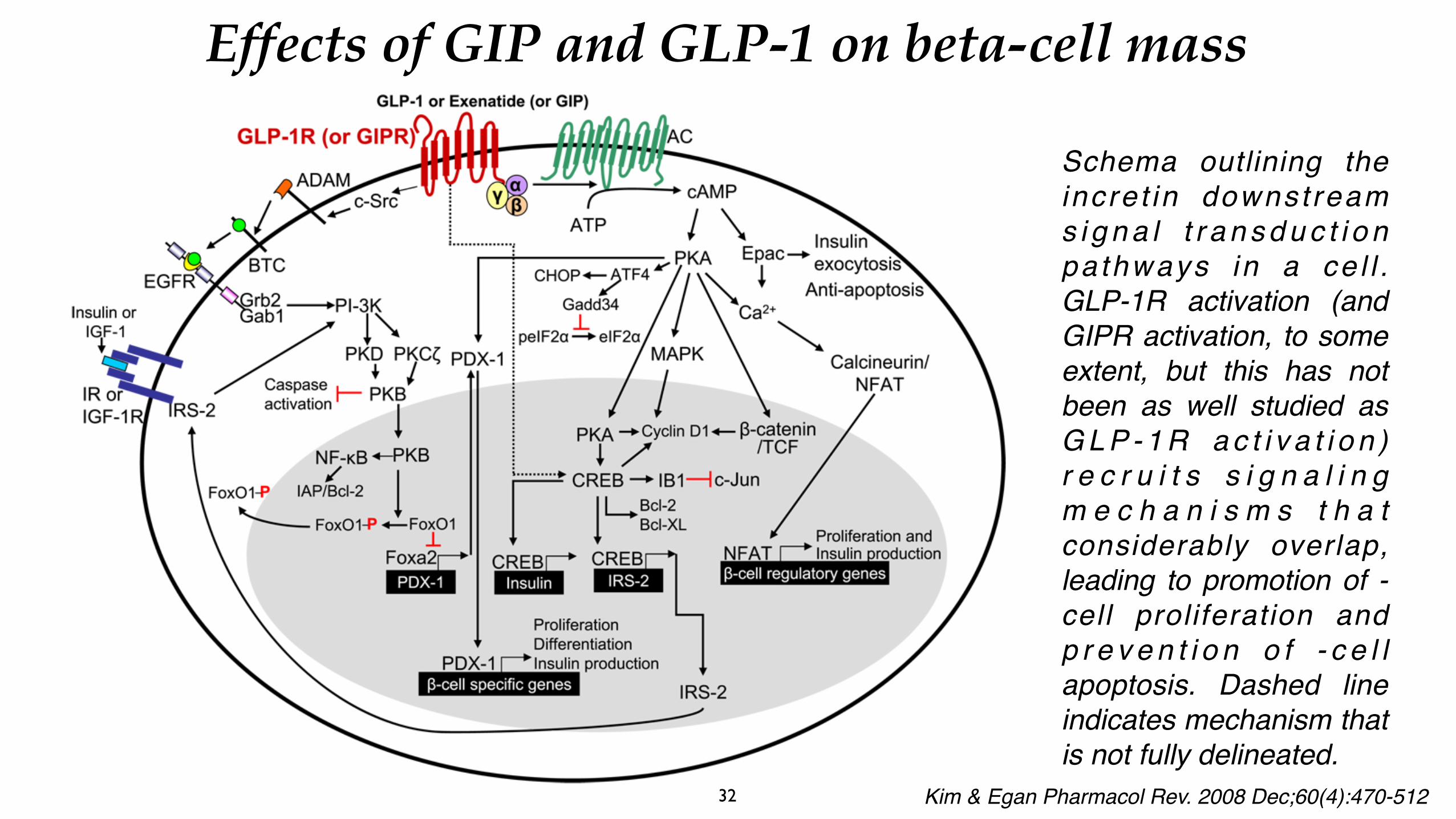
Schema outlining the incre t in downst ream s i g n a l t r a n s d u c t i o n pa thways in a ce l l . GLP-1R activation (and GIPR activation, to some extent, but this has not been as well studied as G L P - 1 R a c t i v a t i o n ) r e c r u i t s s i g n a l i n g m e c h a n i s m s t h a t considerably overlap, leading to promotion of -cell proliferation and p r e v e n t i o n o f - c e l l apoptosis. Dashed line indicates mechanism that is not fully delineated.
32 Kim & Egan Pharmacol Rev. 2008 Dec;60(4):470-512
Effects of GIP and GLP-1 on beta-cell mass
33
GIP works in synergy with glucose to stimulate beta-cell proliferation and improve survival of pancreatic beta cells. The proliferative action of GIP includes activation of cAMP/PKA, PKA/CREB and MAP kinase pathways. GIP also activates antiapoptotic pathways via the phosphatidylinositol 3-kinase (PI3-kinase)/protein kinase B (PKB) pathway. GIP activates phosphorylation of PKB and nuclear exclusion of FOXO1, resulting in decreased expression of the proapoptotic gene BAX and upregulation of the antiapoptotic gene BCL2.
In animal studies, GLP-1 promotes the proliferation and neogenesis of pancreatic beta cells, and reduces beta-cell apoptosis. The signal transduction pathway whereby GLP-1 mediates its proliferative effects involves PI3-kinase, EGF receptor transactivation, MAP kinase and PKCz. GLP-1 also activates a transcriptional programme that is critical for cell survival. PDX1, FOXO1 and IRS2 have been identified as downstream targets for GLP-1-dependent cytoprotection of beta cells. GLP-1 also reduces the expression of proapoptotic genes and prevents glucotoxicity and lipotoxicity through a mechanism involving PKB/Akt.
Effects of GIP and GLP-1 on beta-cell mass
34
GIP receptors are expressed on adipocytes and can mediate a wide variety of actions on adipocyte biology. Studies in knockout mice genetically deprived of GIP receptors strongly implicate GIP in the regulation of body weight. GIP receptor-knockout mice are resistant to the development of diet-induced obesity. Furthermore, GIP receptor-deficient ob/ob mice have a 40% reduction in body weight, and plasma lipids such as triglycerides, free fatty acids (FFA) and cholesterol. Moreover, energy expenditure was increased following high-fat feeding in GIP receptor-deficient mice, indicating that inhibition of the GIP signal results in reduction of obesity, obesity-related hyperglycaemia and dyslipidaemia.
GIP released in response to fat intake could have an effect on fat metabolism. GIP receptors have been found in adipocytes, and it has been reported that GIP promotes chylomicron-triglyceride clearance from the circulation by increasing the activity of lipoprotein lipase. The FFA released by triglyceride hydrolysis can be taken up by adipose tissue to be stored. GIP stimulates fatty-acid synthesis and their incorporation into triglycerides. Therefore, GIP facilitates the efficient disposal of absorbed fat.
Effects of GIP on adipose tissue
35
GLP-1 exerts inhibitory effects on gastrointestinal (GI) secretion and motility and, in particular, on gastric-emptying. Administration of GLP-1 at physiological doses in healthy volunteers results in a dose-dependent slowing of gastric-emptying and glucose absorption, both of which participate in the subsequent reduction of postprandial plasma glucose concentrations.
Under physiological conditions, it is likely that the GI effects of GLP-1—such as decreased gastric secretion and slowing of gastric-emptying—are more important than its insulinotropic action. In pathological conditions such as diabetes, the inhibitory effects of GLP-1 on GI motility, particularly gastric-emptying, are of special interest because they may potentially reduce postprandial glucose excursions.
It has been clearly demonstrated that slowing of gastric-emptying by the amylin analogue pramlintide reduces postprandial entry of glucose into the systemic circulation and, thus, blunts postprandial plasma glucose excursions. This highlights the importance of gastric-empting in determining postprandial plasma glucose excursions.
Effects of GLP-1 on the gastrointestinal system
36
In normal subjects, the intravenous administration of GLP-1 to above physiological levels induced increased feelings of satiety as well as a reduction of food intake.
Similar effects were observed in obese subjects as well as in patients with type 2 diabetes. In type 2 diabetic patients treated with a subcutaneous infusion of GLP-1 for up to six weeks, the reduction of food intake was sustained and associated with weight loss.
The exact mechanism by which peripheral GLP-1 is able to modulate food intake has yet to be completely elucidated. One possibility is that peripheral GLP-1 acts on vagal afferent fibres, permitting modulation of GLP-1 neuronal transmission in the central nervous system (CNS).
It is also likely that inhibition of gastric-emptying mediated by GLP-1 increases the sensation of fullness and leads to stopping eating, thereby participating in the regulation of food intake.
Effects of GLP-1 on food intake
37
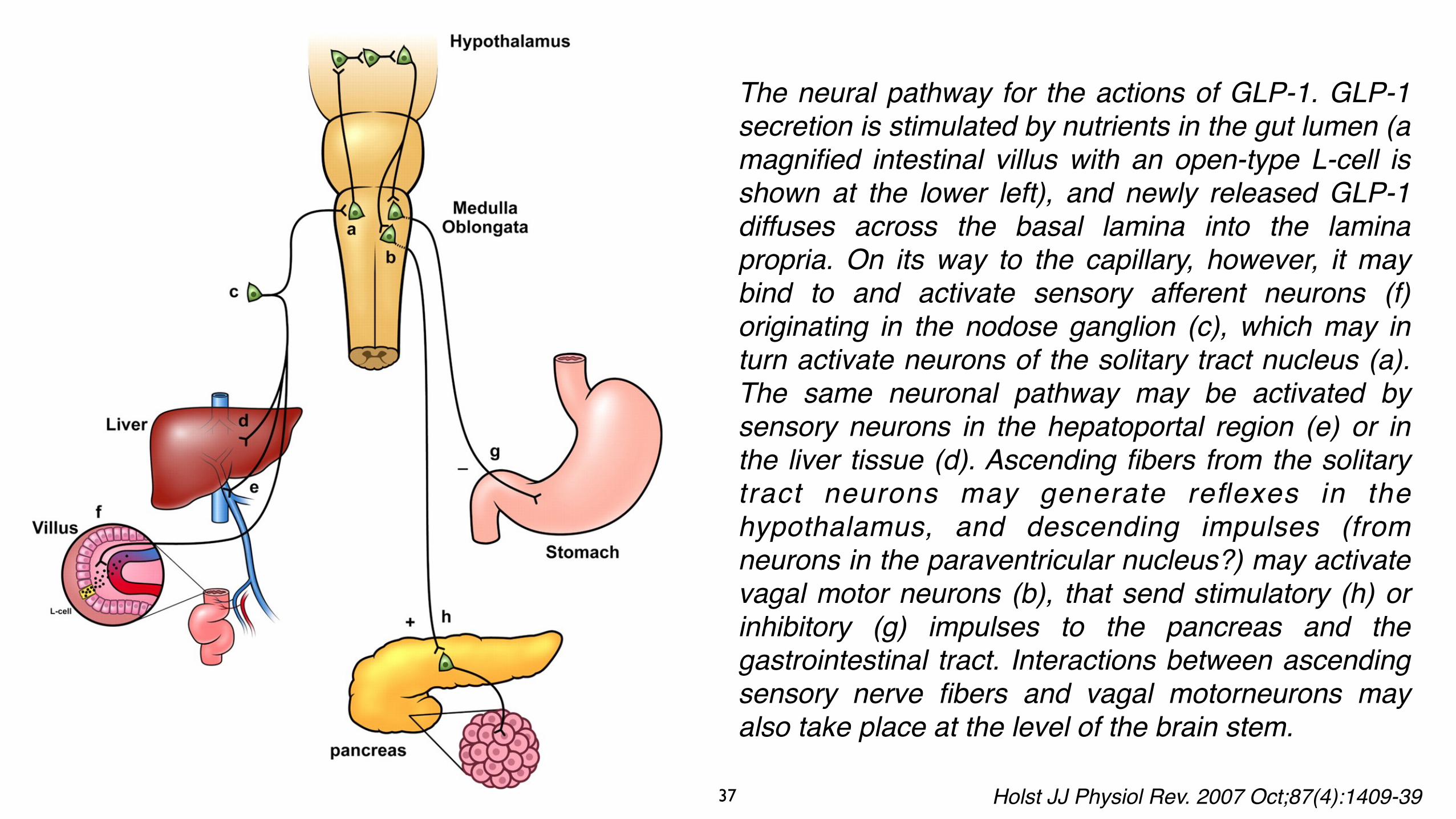
The neural pathway for the actions of GLP-1. GLP-1 secretion is stimulated by nutrients in the gut lumen (a magnified intestinal villus with an open-type L-cell is shown at the lower left), and newly released GLP-1 diffuses across the basal lamina into the lamina propria. On its way to the capillary, however, it may bind to and activate sensory afferent neurons (f) originating in the nodose ganglion (c), which may in turn activate neurons of the solitary tract nucleus (a). The same neuronal pathway may be activated by sensory neurons in the hepatoportal region (e) or in the liver tissue (d). Ascending fibers from the solitary tract neurons may generate reflexes in the hypothalamus, and descending impulses (from neurons in the paraventricular nucleus?) may activate vagal motor neurons (b), that send stimulatory (h) or inhibitory (g) impulses to the pancreas and the gastrointestinal tract. Interactions between ascending sensory nerve fibers and vagal motorneurons may also take place at the level of the brain stem.
Holst JJ Physiol Rev. 2007 Oct;87(4):1409-39
38
Increasing evidence suggests that GLP-1 has beneficial effects on myocardial function. GLP-1 infusion has been reported to reduce infarct size in rodent models of myocardial ischaemia, and to improve myocardial contractility and glucose uptake in normal and post-ischaemic hearts.
GLP-1 can also improve left ventricular performance and cardiac output in dilated cardiomyopathy. In humans with type 2 diabetes and congestive heart failure, GLP-1 improves myocardial function.
Cardiovascular effects of GLP-1
39
A 50% reduction in incretin response was detected in T2DM patients in comparison with healthy individuals as revealed by isoglycaemic glucose tolerance tests. This suggested that a defect in GIP and/or GLP-1 secretion or impaired activation of relevant signalling pathways might lead to a diminished incretin response in T2DM patients.
However, GIP-based therapeutic strategies presented mechanistic problems in the treatment of T2DM.
Mice fed with a high-fat diet exhibited GIP overexpression and insulin resistance associated with an extreme visceral and subcutaneous fat deposition. On the contrary, GIPR-knockout mice displayed insulin sensitivity and resistance to diet-induced obesity. Because GIP overexpression was directly linked to diet-induced obesity, GIP-induced signalling initially represented a potential target for anti-obesity drugs.
Furthermore, GIP treatment was not effective in T2DM patients because of the lack of GIPR expression in pancreatic beta cells. Despite the fact that normalization of blood glucose could restore GIPR expression leading to insulin secretion from beta cells in T2DM patients, GIP also induced glucagon secretion, worsening hyperglycaemia in T2DM patients. As a result, GIP treatment is not advised for T2DM patients.
Pathophysiology in type 2 diabetes mellitus
40
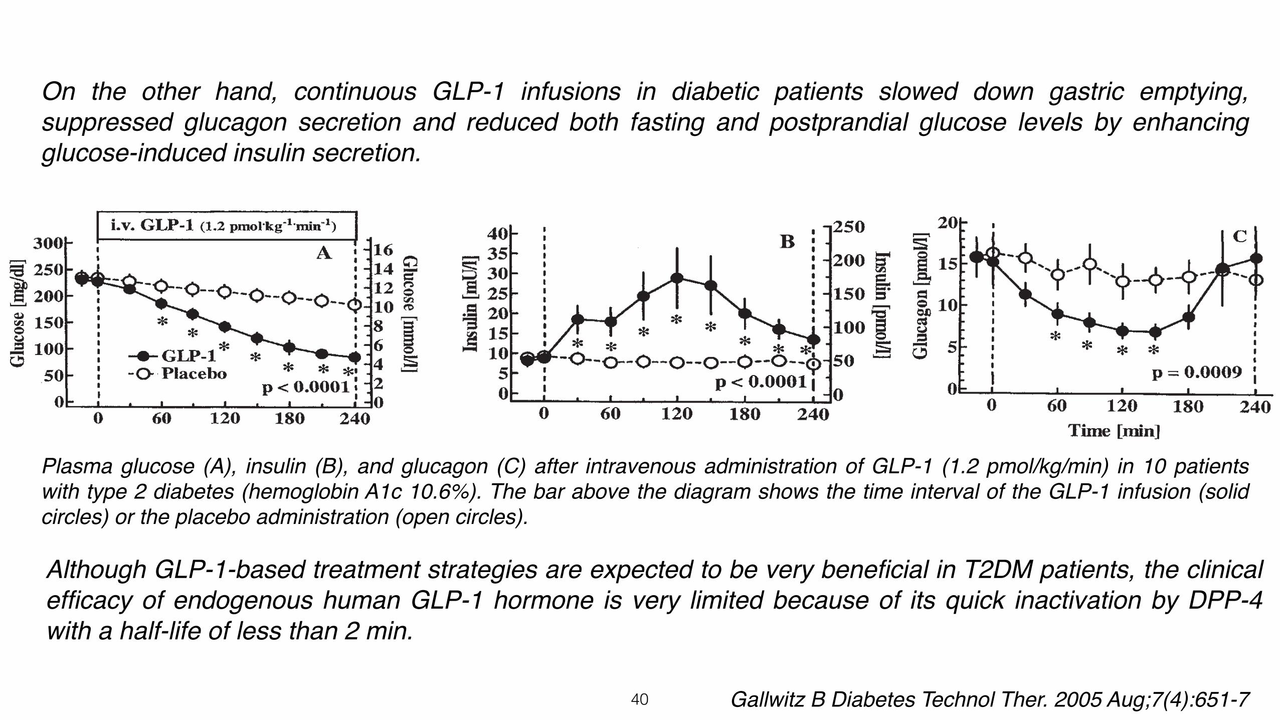
Plasma glucose (A), insulin (B), and glucagon (C) after intravenous administration of GLP-1 (1.2 pmol/kg/min) in 10 patients with type 2 diabetes (hemoglobin A1c 10.6%). The bar above the diagram shows the time interval of the GLP-1 infusion (solid circles) or the placebo administration (open circles).
Gallwitz B Diabetes Technol Ther. 2005 Aug;7(4):651-7
On the other hand, continuous GLP-1 infusions in diabetic patients slowed down gastric emptying, suppressed glucagon secretion and reduced both fasting and postprandial glucose levels by enhancing glucose-induced insulin secretion.
Although GLP-1-based treatment strategies are expected to be very beneficial in T2DM patients, the clinical efficacy of endogenous human GLP-1 hormone is very limited because of its quick inactivation by DPP-4 with a half-life of less than 2 min.
41
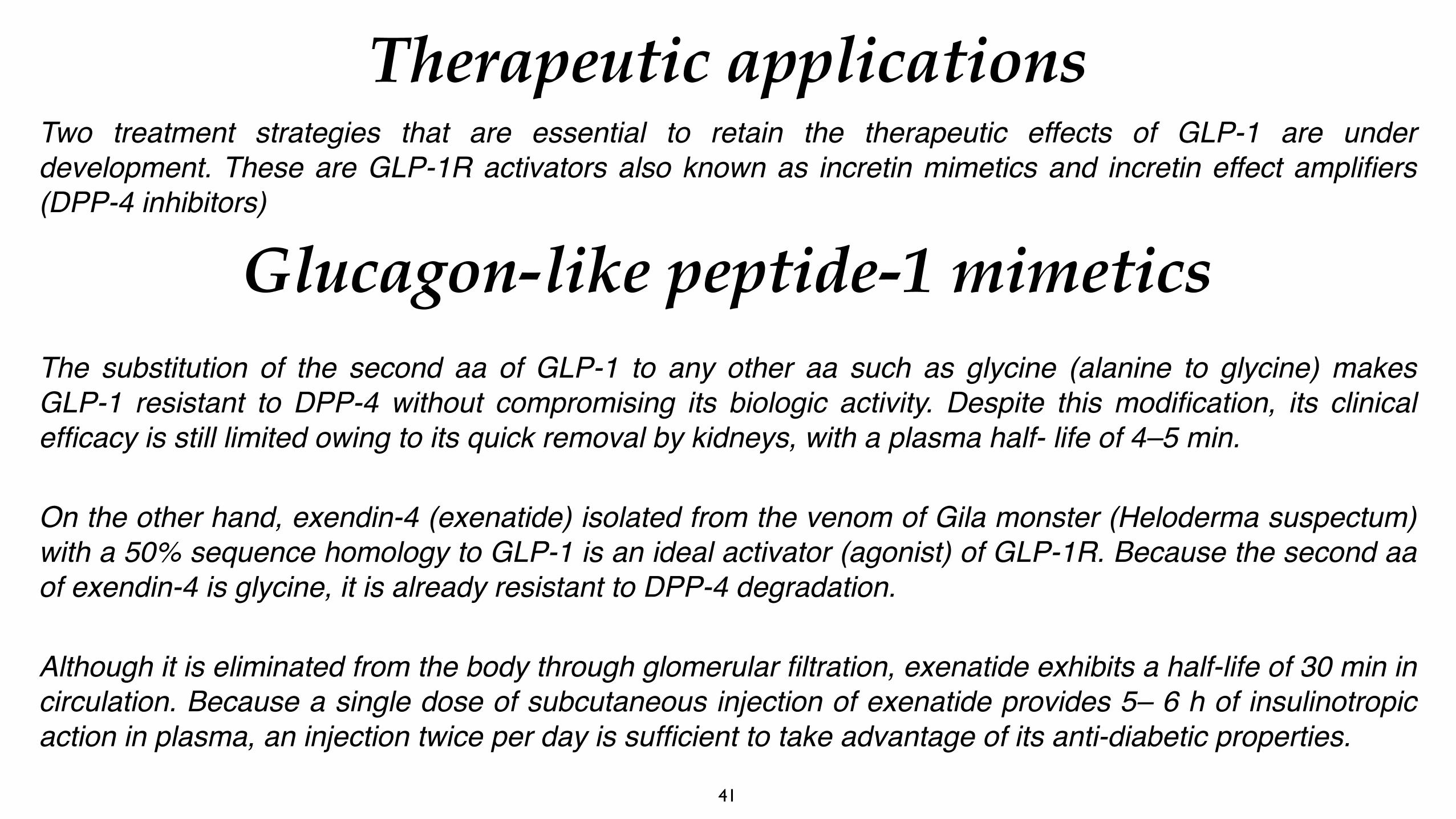
Two treatment strategies that are essential to retain the therapeutic effects of GLP-1 are under development. These are GLP-1R activators also known as incretin mimetics and incretin effect amplifiers (DPP-4 inhibitors)
Therapeutic applications
Glucagon-like peptide-1 mimetics The substitution of the second aa of GLP-1 to any other aa such as glycine (alanine to glycine) makes GLP-1 resistant to DPP-4 without compromising its biologic activity. Despite this modification, its clinical efficacy is still limited owing to its quick removal by kidneys, with a plasma half- life of 4–5 min.
On the other hand, exendin-4 (exenatide) isolated from the venom of Gila monster (Heloderma suspectum) with a 50% sequence homology to GLP-1 is an ideal activator (agonist) of GLP-1R. Because the second aa of exendin-4 is glycine, it is already resistant to DPP-4 degradation.
Although it is eliminated from the body through glomerular filtration, exenatide exhibits a half-life of 30 min in circulation. Because a single dose of subcutaneous injection of exenatide provides 5– 6 h of insulinotropic action in plasma, an injection twice per day is sufficient to take advantage of its anti-diabetic properties.
42
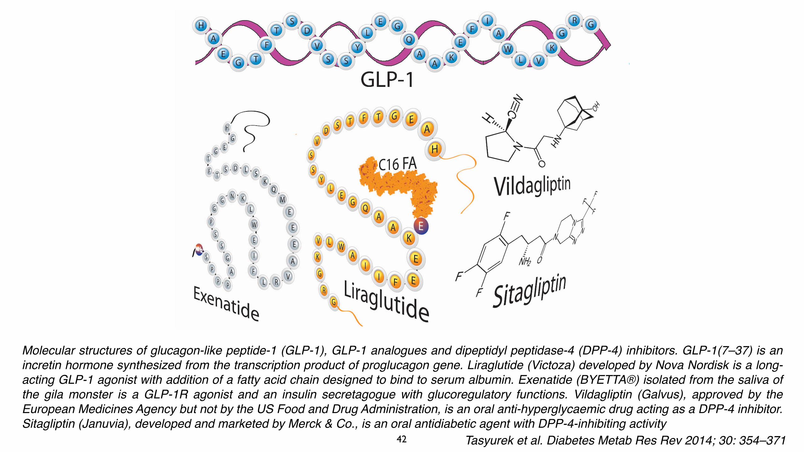
Molecular structures of glucagon-like peptide-1 (GLP-1), GLP-1 analogues and dipeptidyl peptidase-4 (DPP-4) inhibitors. GLP-1(7–37) is an incretin hormone synthesized from the transcription product of proglucagon gene. Liraglutide (Victoza) developed by Nova Nordisk is a long-acting GLP-1 agonist with addition of a fatty acid chain designed to bind to serum albumin. Exenatide (BYETTA®) isolated from the saliva of the gila monster is a GLP-1R agonist and an insulin secretagogue with glucoregulatory functions. Vildagliptin (Galvus), approved by the European Medicines Agency but not by the US Food and Drug Administration, is an oral anti-hyperglycaemic drug acting as a DPP-4 inhibitor. Sitagliptin (Januvia), developed and marketed by Merck & Co., is an oral antidiabetic agent with DPP-4-inhibiting activity
Tasyurek et al. Diabetes Metab Res Rev 2014; 30: 354–371
43
Structure of native GLP-1, exenatide, liraglutide, sitagliptin, and vildagliptin. Green letters indicate changes introduced in derivatives or occur naturally in exendin-4 (and replicated in the synthetic version, exenatide). The N-terminal dipeptide “HA” of GLP-1 and liraglutide is the site of proteolytic cleavage of DPP4. A broken red arrow indicates absent DPP4 activity, and a dotted red arrow indicates reduced DPP4 activity.
Kim & Egan Pharmacol Rev. 2008 Dec;60(4):470-512
44
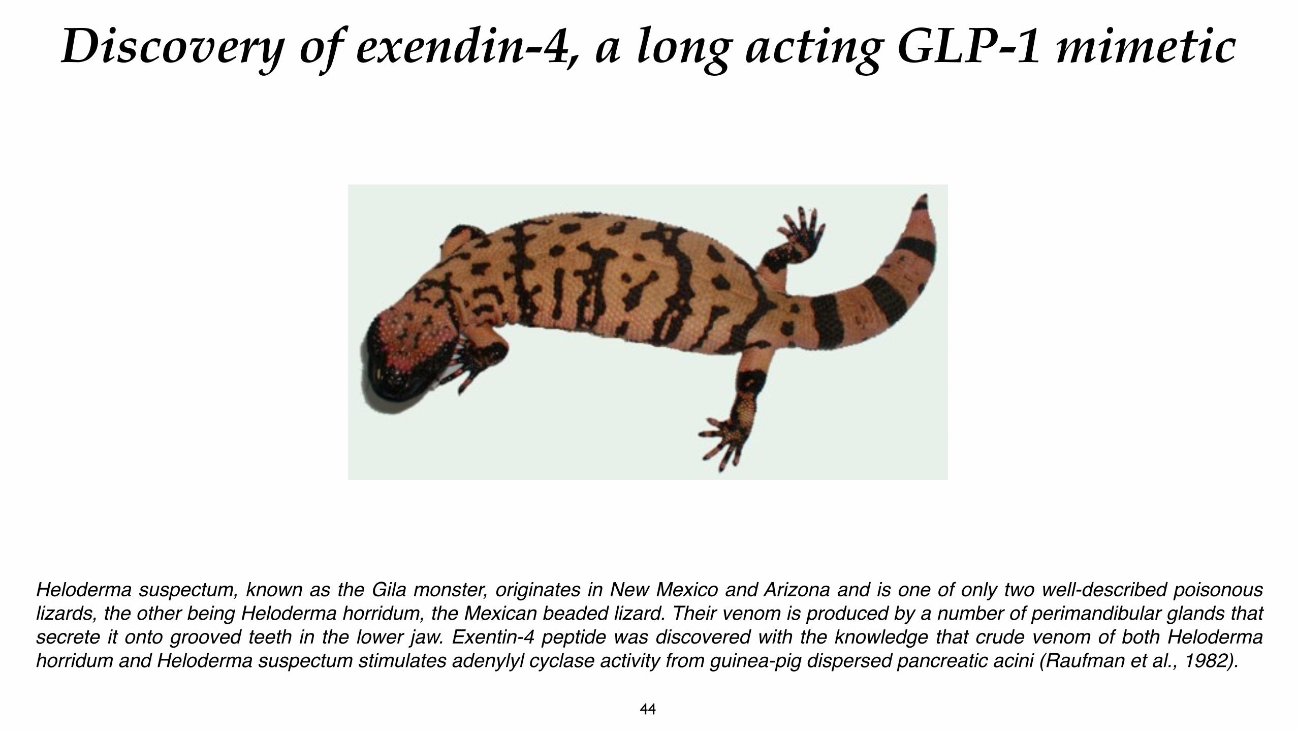
Heloderma suspectum, known as the Gila monster, originates in New Mexico and Arizona and is one of only two well-described poisonous lizards, the other being Heloderma horridum, the Mexican beaded lizard. Their venom is produced by a number of perimandibular glands that secrete it onto grooved teeth in the lower jaw. Exentin-4 peptide was discovered with the knowledge that crude venom of both Heloderma horridum and Heloderma suspectum stimulates adenylyl cyclase activity from guinea-pig dispersed pancreatic acini (Raufman et al., 1982).
Discovery of exendin-4, a long acting GLP-1 mimetic
45
Therapeutic applications
Mean(s.e.)post-prandial plasma glucose concentration during a standard meal at baseline and after treatment with exenatide or sitagliptin. Exenatide was administered at T = −15 min. Sitagliptin was administered at T=−30 min. Standardized meal was given at T = 0 min.
Fineman et al. Diabetes, Obesity and Metabolism 14: 675–688, 2012
The effects of exenatide and sitagliptin on postprandial glucose metabolism were compared in a randomized 2-week crossover study in patients with type 2 diabetes. Both treatments resulted in reductions in PPG and glucagon compared to baseline, although the magnitude of effect was significantly greater for exenatide compared to sitagliptin. It is noteworthy that with exenatide treatment, the PPG rise is almost completely blunted following the meal with concentrations falling below the preprandial concentration within 1 h. This suggests that the effects on gastric emptying, glucagon secretion and insulin secretion are all contributing to the overall glucose profile.
46
Liraglutide (Victoza) is another long-acting incretin mimetic (GLP-1 agonist) developed by Novo Nordisk. The European Medicines Agency (EMEA) approved its use on 3 July 2009, and the US Food and Drug Administration (FDA) on 25 January 2010.
Liraglutide is a DPP-4-resistant GLP-1 analogue that achieves slowed absorption and increased half-life through the substitution of arginine for lysine at position 34 and the addition of a C16 fatty acid chain at position 26 allowing for reversible binding to albumin.
The half-life of liraglutide is 11–15h resulting in continuous exposure with once-daily (QD) administration. Although the potency of liraglutide is reduced 100-fold relative to GLP-1 (albumin binding is 98–99%), it retains the basic actions of GLP-1.
Despite the fact that 57% of the patients displayed nausea and 17% vomiting, gastrointestinal side effects were mainly dose dependent.
Glucagon-like peptide-1 mimetics
47
Dipeptidyl peptidase-4 is a serine peptidase present in plasma, kidneys, intestinal mucosa, hepatocytes and vascular endothelial cells. DPP-4 removes two aa from the amino terminus of the target peptides including GLP-1 and GIP, resulting in their inactivation. Sitagliptin was the very first marketed DPP-4 inhibitor (Januvia, Merck). Entrance of the second DPP-4 inhibitor, Vildagliptin (Galvus, Novartis), to European market took place in the spring of 2008. These two DPP-4 inhibitors are taken as oral tablets and provide 70–90% inhibition in DPP-4 activity lasting 24 h when administered as a single dose.
Intriguingly, vildagliptin, similar to other GLP-1 mimetics, induced peripheral insulin sensitivity, enhanced glucose-induced insulin secretion from beta cells and suppressed glucagon secretion. Because a single 100-mg dose of vildagliptin caused an increase in hepatic transaminases, 50-mg vildagliptin twice-a-day formulation was recommended for use. However, unlike incretin mimetics, DPP-4 inhibitors do not generate weight loss. Because metformin enhanced GLP-1 biosynthesis and secretion, DPP-4 inhibitors were more effective when they were used in combination with metformin. In addition, DPP-4 inhibitors could also be used in conjunction with insulin therapy.
In conclusion, despite the fact that sitagliptin and vildagliptin monotherapies exhibited significant antidiabetic properties, they were more effective in reducing hyperglycaemia when they were used in combination with other anti-diabetic agents such as metformin, sulfonylurea and thiazolidinediones.
Incretin effect amplifiers (DPP-4 inhibitors)
48
Dose–response relationships for the effects of GLP-1. The figure illustrates the relationship between the plasma concentrations of GLP-1 or GLP-1 mimetics and their clinical effects and side effects, as observed during treatment with GLP-1 mimetics or incretin enhancers. With modestly elevated GLP-1 levels, as obtained with both mimetics and enhancers, there are significant effects on the pancreatic islets. Higher concentrations (which might not be reached during therapy with enhancers) are needed to slow down gastric emptying and to reduce appetite and food intake. At even higher concentrations, which can be reached with GLP-1 mimetics, side effects such as nausea, diarrhea and vomiting might result. Holst JJ et al Trends Mol Med. 2008 Apr;14(4):161-8
49
The incretin effect depends largely on the two main incretin hormones, glucose-dependent insulinotropic polypeptide (GIP) and GLP-1, both of which are released after oral glucose ingestion and potentiate glucose-stimulated insulin secretion.
There are two major components in the incretin effect: release of incretin hormones from the gut; and the effects of incretin hormones on beta-cells, leading to insulin secretion. This suggests that the impaired incretin effect in type 2 diabetes could be due to either impaired incretin hormone secretion (incretin hormone deficiency) and/or defective insulinotropic action of the incretin hormones (incretin hormone resistance). Several studies have explored both these possibilities.
In patients with type 2 diabetes the incretin effect is markedly reduced, primarily because of a defect in beta cell sensitivity to GIP, while the insulinotropic effect of GLP-1 is preserved, although reduced. Moreover, several studies have shown reduced secretion of the incretin hormones in patients with type 2 diabetes.
Treatment targets in incretin-based therapy
B. Ahrén / Diabetes & Metabolism 39 (2013) 195–201
50
Reduced incretin effect
Toft-
Nie
lsen
et a
l. Th
e J
of C
linic
al E
ndo
& M
et, A
ugus
t 200
1, 8
6(8)
:371
7–37
23
To elucidate the mechanism of reduced incretin effect, the secretion of incretin h o r m o n e s G L P - 1 a n d G I P w a s investigated during a 4-h mixed-meal test in 54 T2DM patients, 33 matched control subjects with normal glucose tolerance (NGT) and 15 unmatched subjects with impaired glucose tolerance (IGT).
As patients with impaired glucose tolerance were hyperinsulinaemic and generally showed similar metabolic abnormalities to diabetic patients, the meal-related GLP-1 secretion, but not GIP response, was severely reduced in T2DM patients.
51
Reduced incretin effect: As reported previously, incretin effect is reduced i n T2DM. The p lasma concentrations of intact biologically active GLP-1 and GIP were measured after a mixed breakfast meal in 12 T2DM patients (body mass index of 31 kg/m2 and HbA1c of 9.2%) and 12 healthy controls.
The late GLP-1 response, but not GIP secretion, was strongly reduced in T2DM pa t i en t s , suppo r t i ng t he hypothesis that an impaired GLP-1 function contributed to the ineffective insulin secretion.
Treatment targets in incretin-based therapy
B. Ahrén / Diabetes & Metabolism 39 (2013) 195–201
52
Glucose tolerance is usually lost long before the actual appearance of T2DM. The UK Prospective Diabetes Study Group stated that a 50% decrease in beta cell function and 40% loss in islet cell mass have been observed in newly diagnosed T2DM patients. Hyperglycaemia-induced glucotoxicity and lipotoxicity associated with an increase in fatty acids in circulation resulted in the functional loss of beta cell. Therefore, beta cell function and mass must be restored to prevent progression of diabetes.
Treatment targets in incretin-based therapy
Impaired beta cell function and beta cell loss:
Patients with T2DM are generally overweight, and most of the anti-hyperglycaemic agents except biguanides are not effective in causing weight loss. Instead, anti-hyperglycaemic agents such as sulfonylurea and insulin contribute to weight gain in diabetic patients, complicating the treatment efficacy or even worsening the prognosis of diabetic patients.
Weight gain :
53
Rapid gastric emptying led to the inability to control blood glucose in T2DM patients as demonstrated in nine newly diagnosed T2DM patients and nine sex-matched and age-matched non-diabetic control subjects. Therefore, deceleration of gastric emptying is helpful to manage glucose excursions after feeding, reducing hyperglycaemia. By this token, amylin (pramlintide) and incretin mimetics (GLP-1) represent two options with beneficial effects in T2DM.
Treatment targets in incretin-based therapy
Rapid gastric emptying
Patients with T2DM generally manifest high glucagon levels. On top of that, high glucagon is the sign of impaired glucose tolerance. Consequently, high glucagon in T2DM patients stimulates glucose release from liver, enhancing hyperglycaemia.
Hyperglucagonaemia
54
Although incretins have been first proposed for the treatment of T2DM in 1992, the first incretin hormone (GLP-1) for commercial use was approved in 2005. One of the main reasons for the delayed approval was the short half-life of GLP-1, which is less than 2 min, requiring constant infusion or frequent injection to maintain its insulinotropic activity.
Intriguingly, less than 10% of the administrated GLP-1 is intact and biologically active only minutes after the injection. To overcome this problem, two strategies have been proposed concerning the development of either DPP-4 resistant GLP-1 mimetics or DPP-4 inhibitors.
Incretins enhance glucose-induced insulin secretion through interaction with GPCRs on pancreatic beta cells. However, incretins cannot exhibit their insulinotropic effect at low glucose concentration under 4 mM, necessary to prevent development of hypoglycaemia.
Effects of incretin treatment including side effects
55
In addition, GLP-1 stimulates both gene expression and biosynthesis of insulin. Stimulation of beta cell proliferation and differentiation and inhibition of beta cell apoptosis are the other beneficial effects of GLP-1. Apart from causing weight loss through inhibition of appetite and food intake and deceleration of gastric emptying, GLP-1 enhanced myocardial performance, reduced infarct area and restored endothelial functions in T2DM patients.
Lastly, GLP-1 mimetics increased plasma GLP-1 concentration better than what was achieved with DPP-4 inhibitors alone. The GLP-1 analogue exenatide can be injected twice daily before meals or once weekly when given within dissolvable poly-(D,L-lactide-co-glycolide) microspheres. Despite all these beneficial effects of GLP-1, there are considerable numbers of concerns relating to the side effects of incretin-based therapy.
Effects of incretin treatment including side effects
56
For example, exenatide and liraglutide have been reported to cause significant gastrointestinal discomfort in T2DM patients. Because exendin-4 (exenatide)-based treatment strategies are antigenic, it is not clear if this would limit the clinical efficacy of GLP-1 mimetics.
The other side effects of GLP-1 mimetics include but not limited to nausea, vomiting and hypoglycaemia. Severe side effects leading to circulatory collapse, cardiovascular complications or even renal problems have also been reported.
Although liraglutide and exenatide treatments have been claimed to cause acute pancreatitis in humans, no evidence of pancreatitis was observed when three different animal species including mice, rats or monkeys were injected with liraglutide at a dose 60 times higher than what was recommended for humans.
side effects: nausea, vomiting and hypoglycaemia
57
On the contrary, exenatide treatment, instead of evoking pancreatitis, attenuated chemically induced pancreatitis in control and diabetic rodents. Thus, examination of autopsy materials from liraglutide-treated or exenatide-treated patients with T2DM has been advised to settle this dispute.
Although it was suggested that patients should be made aware of the potential side effect of the incretin treatment, it is necessary to keep in mind that diabetes itself is associated with a two-fold increase in the incidence of acute pancreatitis.
Despite this, FDA required addition of warnings about acute pancreatitis risk for the entire class of incretin-based therapies to drug labels. Current labelling of incretin-based therapies includes warnings about the use in patients with a history of pancreatic disease and recommendations to discontinue the treatment in patients who develop pancreatitis.
side effects: Pancreatitis
58
Examination of the US FDA’s reported adverse event database indicated that pancreatic cancer was more commonly reported among patients who took sitagliptin or exenatide as compared with those who were subjected to other therapies.
Butler and his colleagues have recently reported a marked expansion of exocrine and endocrine pancreas along with exocrine pancreas dysplasia in patients treated with incretin therapy. This autopsy study reporting abnormal pancreatic findings from T2DM patients treated with sitagliptin or exenatide suggested that incretin therapy might be associated with a potential risk of pancreatic cancer.
Because of this controversy, on 12–13 June 2013, the National Institute of Diabetes, Digestive and Kidney Diseases in association with the National Cancer Institute (NIDDK-NCI) held a workshop on pancreatitis, diabetes and pancreatic cancer in Lister Hill Auditorium of the NIH Campus, Bethesda, MD. One of the purposes of the gathering (out of many) was to review the effects of anti-diabetic therapy on the development of pancreatic ductal adenocarcinoma (PDAC).
side effects: pancreatic cancer
59
Epidemiologic studies revealed that diabetes itself is associated with an 82% increased risk of pancreatic cancer, independent of therapy. Consequently, the increase in the prevalence of T2DM is somehow connected to the increase in the inci- dence of PDAC. In addition, cohort studies suggested that 1–2% of new-onset adult diabetes mellitus patients would develop PDAC.
The fact that Dr Butler’s findings either came from genetically manipulated animal models or a very restricted set of human autopsy data raised substantial concerns about the interpretation of his data and out-come of his research findings.
On 28 June 2013, The American Diabetes Association, the European Association for the Study of Diabetes and the International Diabetes Federation issued a joint statement declaring that no alteration of current treatment recommendations was necessary concerning the use of incretin therapy and pancreatic disease for patients with diabetes. Accordingly, the EMEA also issued a similar statement on 26 July 2013.
side effects: pancreatic cancer
60
In addition, some concerns were also raised about GLP-1R activation inducing C-cell hyperplasia of thyroid glands. Animal safety studies conducted on rodents suggested that liraglutide or exenatide treatment might cause C-cell adenocarcinoma. Upon long-term exposure, constant stimulation of GLP-1R induced C-cell proliferation leading to the formation of C-cell adenomas and medullary thyroid carcinomas (MTC) in mice and rats. Hypothetically, long-term exposure to GLP-1R agonists might also induce C-cell neoplasia in human thyroid glands.
Despite C-cell neoplasia and especially MTC being very rare in humans, rodents such as mice and rats spontaneously develop C-cell abnormalities ranging from C-cell hyperplasia to C-cell adenoma and MTC. Not surprisingly, daily injection of liraglutide enhanced C-cell abnormalities, generating C-cell carcinomas in both mice and rats.The presence of high levels of GLP-1R on thyroid C cells in rodents and absence or low levels of GLP-1R expression on human and/or cynomolgus monkeys’ C cells could account for the differences observed between species. Thus, there appeared to be species-specific differences in GLP-1R expression in thyroid.
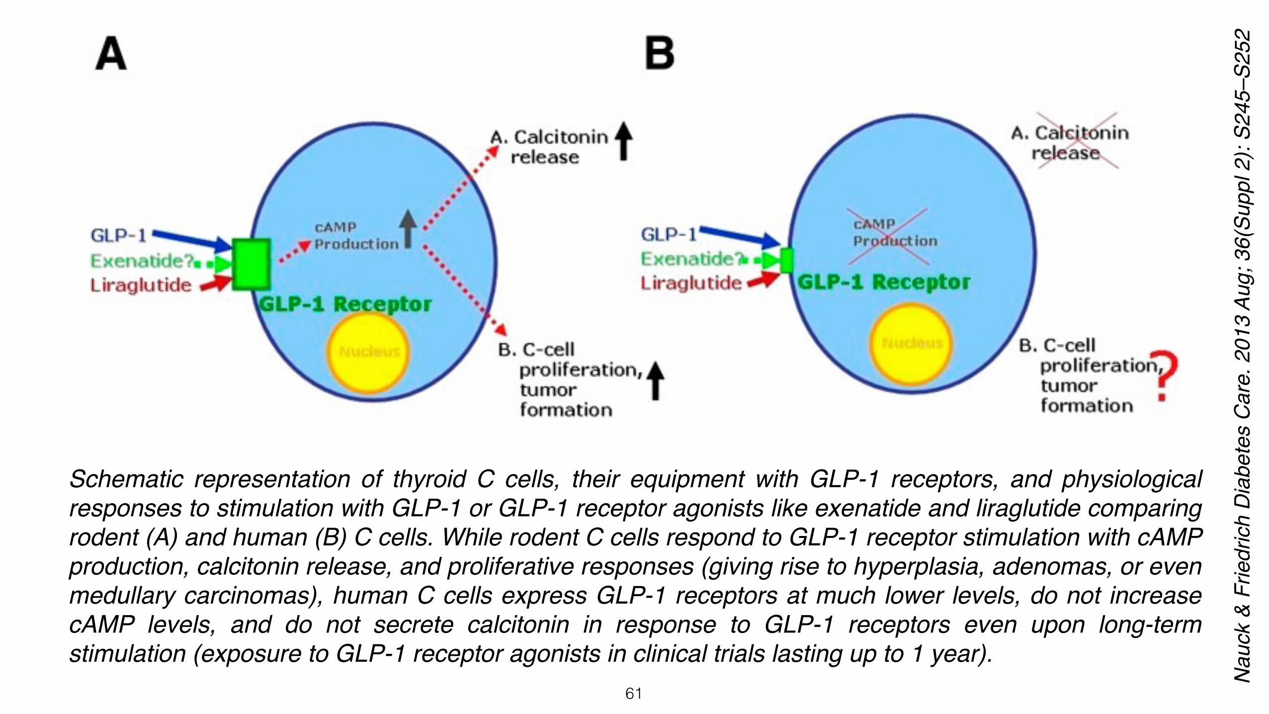
Nevertheless, according to the US FDA adverse event reporting system database, there is indeed an increased risk for thyroid cancer associated with exenatide. Consequently, it is essential to carefully follow up diabetic patients exposed to long-term GLP-1 analogues for any incidence of thyroid cancer.
side effects: thyroid cancer
61
Schematic representation of thyroid C cells, their equipment with GLP-1 receptors, and physiological responses to stimulation with GLP-1 or GLP-1 receptor agonists like exenatide and liraglutide comparing rodent (A) and human (B) C cells. While rodent C cells respond to GLP-1 receptor stimulation with cAMP production, calcitonin release, and proliferative responses (giving rise to hyperplasia, adenomas, or even medullary carcinomas), human C cells express GLP-1 receptors at much lower levels, do not increase cAMP levels, and do not secrete calcitonin in response to GLP-1 receptors even upon long-term stimulation (exposure to GLP-1 receptor agonists in clinical trials lasting up to 1 year).
Nau
ck &
Frie
dric
h D
iabe
tes
Car
e. 2
013
Aug;
36(
Supp
l 2):
S245
–S25
2
62
In addition, beta cell growth-promoting properties of incretins have not been replicated in human studies as demonstrated in animal models. The dose of GLP-1, which is used to promote beta cell growth in rodents, typically ranges between 50 and 100 μg/kg of body weight. Considering the tolerable dose of GLP-1 in humans is <2 μg/kg body weight and GLP-1 mimetic exenatide (Byetta) is currently prescribed at a dose of 5 μg (injected twice daily) to treat T2DM patients, systemic injections may not be the route of drug delivery to achieve beta cell growth-promoting actions of GLP-1 mimetics in diabetic patients.
Contrary to GLP-1 mimetics, DPP-4 inhibitors have a neutral effect on body weight. This is an intriguing finding because some weight gain would be expected as a result of glucosuria resulting in glucose retention (as fat) in the body. Because DPP-4 has a wide range of substrates such as chemokines, hormones and neuropeptides, side effects and the outcome of long-term DPP-4 inhibition are not known. Although preclinical and clinical trial data concerning sitagliptin or vildagliptin treatments did not indicate an increased risk of pancreatitis in patients with T2DM, common side effects included headache, nasopharyngitis, upper respiratory infections, urinary system infections, severe allergic reactions and hypoglycaemia.
Beta cell growth-promoting properties of incretins
side effects of DPP-4 inhibitors
63
The first incretin mimetic (exenatide) was approved by the US FDA in April 2005. Soon after that, in October 2006, the US FDA approved the first oral incretin effect amplifier, DPP-4 inhibitor (sitagliptin). So far, several other incretin mimetics have reached the market, and there are even more incretin-based drugs under development awaiting marketing approval.
Clinical trials of incretin-based therapies demonstrated that incretins are as effective as other anti-diabetic drugs (sulfonylureas, thiazolidinediones and long-acting insulin therapies), if not superior for improving blood glucose control and achieving weight loss. Although some concerns were raised against incretin-based therapies regarding pancreatitis or pancreatic cancer, the US FDA review of preclinical and some limited clinical data from all currently available incretin therapies revealed no concern for pancreatic disease.
The fact that no clinical incretin-based treatment study has been suspended for safety concerns further supported this notion. Consequently, regulatory agencies advised no change to current treatment protocols of patients with diabetes treated with incretin-based therapies.
Conclusions
64
• Incretins: Their Physiology and Application in the Treatment of Diabetes Mellitus. Tasyurek HM, Altunbas HA, Balci MK, and Sanlioglu S. Diabetes Metab Res Rev. 2014 Jul;30(5):354-71. doi: 10.1002/dmrr.2501
• GLP-1-mediated gene therapy approaches for diabetes treatment. Tasyurek MH, Altunbas HA, Canatan H, Griffith TS, Sanlioglu S. Expert Rev Mol Med. 2014 Mar 26;16:e7. doi: 10.1017/erm.2014.7.
• Clinical utility of insulin and insulin analogs. Sanlioglu AD, Altunbas HA, Balci MK, Griffith TS, Sanlioglu S. Islets. 2013 Mar-Apr;5(2):67-78. PMID: 23584214
• Insulin Gene Therapy From Design to Beta Cell Generation. Sanlioglu AD, Altunbas HA, Balci MK, Griffith TS and Sanlioglu S. Expert Rev Mol Med. 2012 Oct 15;14:e18. doi: 10.1017/erm.2012.12.PMID: 23062285
• Therapeutic Potential of VIP versus PACAP in Diabetes. Sanlioglu AD, Karacay B, Balci MK, Griffith TS and Sanlioglu S. J Mol Endocrinol 2012 Oct 12;49(3):R157-67. doi: 10.1530/JME-12-0156. Print 2012 Dec. PMID: 22991228
• Inkretin Tabanlı Tedavi Stratejilerinde Komplikasyon Kaygıları ve Senaryoları. Sanlioglu S. Klinik Tıp Bilimleri Dergisi, Clinical Medical Sciences, Diyabet özel sayısı. Nisan 2014 2(1):19-24
• Tip 1 diyabetli hastalarda insulin tedavisinde ve insulin gen terapisinde güncel gelişmeler. Klinik Tıp Bilimleri Dergisi, Clinical Medical Sciences, Diyabet özel sayısı. Nisan 2012 1-5


![Combination Use of Insulin and Incretins in Type 2 ... Update - Incretins- Insulin v… · Combination Use of Insulin and Incretins in Type 2 Diabetes [DRAFT] 1 1 BACKGROUND Type](https://static.documents.pub/doc/80x56/5f2de9130649e313ed28bd46/combination-use-of-insulin-and-incretins-in-type-2-update-incretins-insulin.jpg)