34
Inflammation Dr. Raid Jastania
| Date post: | 22-Dec-2015 |
| Category: |
Documents |
| View: | 217 times |
| Download: | 1 times |

Inflammation
Dr. Raid Jastania

Stress
Injury
Overload
Cell Death
Response
Adaptation
Inflammation
Insult Results

Causes of Inflammation
Injury and cell Death
Inflammation
PhysicalChemicalInfectionsIschemiaImmuneMetabolic

Types of Inflammation
Injury and cell Death
Inflammation
PhysicalChemicalInfectionsIschemiaImmuneMetabolic
Acute
Chronic
Granuloma

Immune Reaction
Injury and cell Death
Inflammation
PhysicalChemicalInfectionsIschemiaImmuneMetabolic
Acute
Chronic
Granuloma
Immune

Chronic Inflammatory Diseases
Injury and cell Death
Inflammation
PhysicalChemicalInfectionsIschemiaImmuneMetabolic
Acute
Chronic
Granuloma
ImmuneChronic
Inflammatory Diseases

Acute Inflammation
Chronic Inflammation
Chemical Mediators of Inflammation

• Intended Learning Outcomes:1. Students should be able to define inflammation and
understand clinical features of inflammation and its systemic effect.
2. Students should know the vascular and cellular events in acute inflammation and understand its morphology.
3. Students should know the cellular events in chronic inflammation.
4. Students should be able to define granulomatous inflammation and know its causes.
5. Students should be able to apply the rules of acute and chronic inflammation to predict the features of inflammation in the different organs of the body.
Inflammation
• Inflammation is a protective response of connective tissue to injury.
• Aim: to eliminate the injury and start the process of repair.
• Inflammation starts with activation of endothelial cells and white blood cells. – Changes in vessels– Cellular events. – Chemical mediators


Inflammation
• Inflammation is divided into acute and chronic type.
• Acute inflammation is the immediate response to injury, and neutrophils are the main cell type.
• Chronic inflammation is mediated by mononuclear cells (macrophages, lymphocytes, plasma cell…)

Clinical Features:
• Exercise:
– During a foot ball game, a player was injured and had twist in ankle joint.
– What are the signs/symptoms that he would have?

Clinical Features:
• Exercise:
– 19 year old boy has inflammation of the appendix, Appendicitis.
– What are the signs/symptoms that he would have?

Clinical Features:
• Localized or systemic.
– The localized features are: Redness, Swelling, Heat, Pain and Loss of function.
– The systemic features include: Fever, elevated WBC, malaise, anorexia, and hypotension, Acute phase reaction

Acute Inflammation

Acute Inflammation:
• It is the immediate early response to injury. It is characterized by neutorphil infiltrate and fluid exudates.
• The changes in acute inflammation may be divided to: vascular changes and cellular events.


Vascular changes:1. Change in the vascular caliber and flow• initial transient vasoconstriction of the arterioles
followed by vasodilatation. The end result is blood stasis.
2. Increase in vascular permeability• increase in the hydrostatic pressure and leakage
of fluid to the extravascualr space (Transudate).• increase in the osmotic pressure of the
interstitium leading to leakage of protein-rich fluid (Exudate).
• The end result is Edema.

Mechanisms of increased vascular permeability:
1. Endothelial contraction: histamine, PG, Immediate transient response
2. Endothelial retraction: 4-6 hours after injury
3. Direct endothelial damage: Immediate sustained Response
4. Delayed prolonged leakage:
Mechanisms of increased vascular permeability:
5. Leukocyte-dependent endothelial injury
6. Increased Transcytosis: Through intracellular vesicular pathway, and occurs after exposure to VEGF.
7. Leakage from new blood vessels (angiogenesis)
Cellular Events


Cellular Events:
1. Margination and Rolling:• WBC slow down and are pushed to the
side of the vessel near endothelial cells. This process is “Margination”
• WBC’s transiently stick to endothelial cells. This process is “Rolling”
• The adhesion is facilitated by the action of adhesion molecules called “Selectins”.

Cellular Events:
1. Margination and Rolling:• Selections are present on WBC, endothelial cells
and platelets. • E-Selectin: on endothelial cell• P-Selectin: on Platelets and endothelial cells• L-Selectin: on WBC’s• Selectins are up regulated by IL-1, and TNF.• Selectins bind to sugar molecules. Example:
Sialyl-Lewis X
Cellular Events:
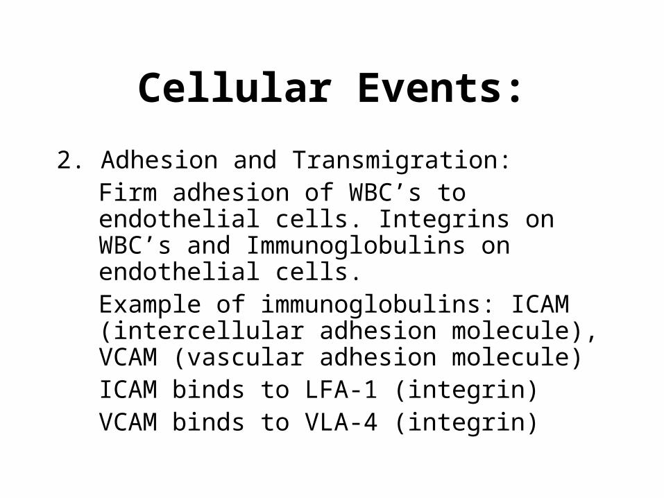
2. Adhesion and Transmigration:Firm adhesion of WBC’s to endothelial cells. Integrins on WBC’s and Immunoglobulins on endothelial cells. Example of immunoglobulins: ICAM (intercellular adhesion molecule), VCAM (vascular adhesion molecule)ICAM binds to LFA-1 (integrin)VCAM binds to VLA-4 (integrin)
Cellular Events:
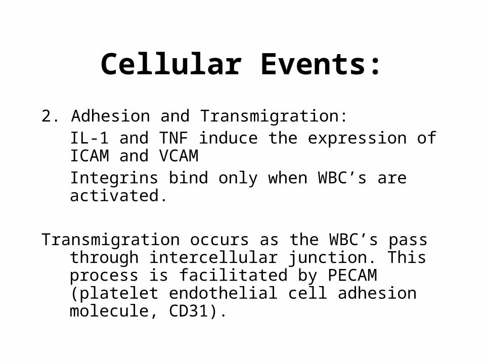
2. Adhesion and Transmigration:IL-1 and TNF induce the expression of ICAM and VCAMIntegrins bind only when WBC’s are activated.
Transmigration occurs as the WBC’s pass through intercellular junction. This process is facilitated by PECAM (platelet endothelial cell adhesion molecule, CD31).

Cellular Events:
3. Migration in interstitium: ChemotaxisMigration of WBC’s is facilitated by chemotactic agents. These are molecules that attract WBC’s. They include:
a. Bacterial productsb. Complement system, C5ac. Leukotriene B4 (LTB4)d. Cytokines (IL-8)

Leukocyte Activation:
• by G-protein activation
• WBC activation is characterized by:a.Degranulation of WBC granules and formation
of oxidative burst
b.Secretion of arachidonic acid metabolites (Leukotrienes and prostaglandins)
c.Expression of adhesion molecules.
Phagocytosis
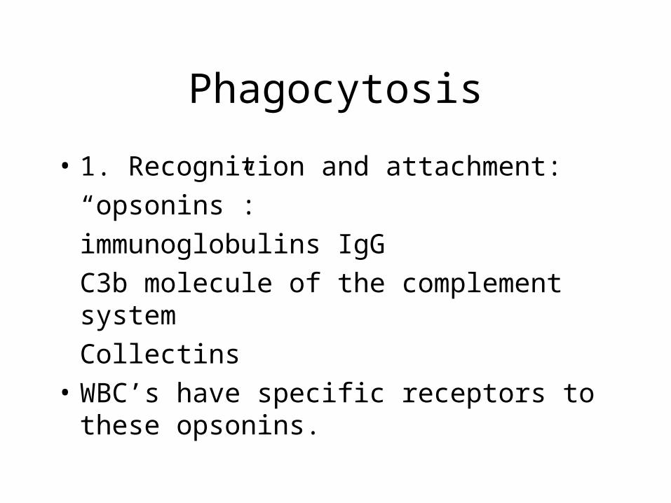
• 1. Recognition and attachment:
“opsonins”:
immunoglobulins IgG
C3b molecule of the complement system
Collectins• WBC’s have specific receptors to these
opsonins.

Phagocytosis
• 2. Engulfment in phagocytic vacuole:
phagosome.• 3. Killing and degradation:
Phagosome fuses to lysosome to form phagolysosome.
Killing is facilitated by:• a. Oxygen free radicals (oxidative burst)• b. Lysosomal enzymes (myeloperoxidase)

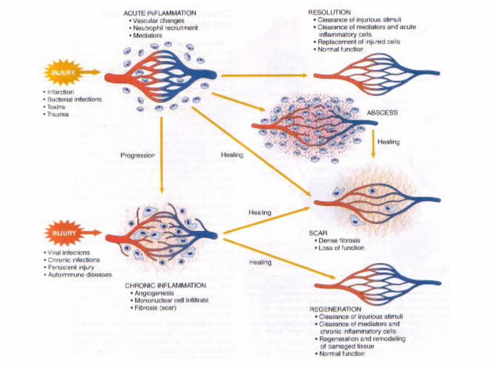
Outcome of Acute Inflammation:
1. Resolution
2. Acute persists with complications: Abscess
3. Progression to chronic inflammation
4. Scarring and Fibrosis
organization and fibrosis

Inflammation-Induced Tissue Injury


