+ All Categories
Home > Documents > Inflammation is a process carried out in response to either physical or immunological tissue insult....

Inflammation is a process carried out in response to either physical or immunological tissue insult....

Date post: 29-Dec-2015
Category:
Upload: magnus-blake
View: 216 times
Download: 1 times
Share this document with a friend
Popular Tags:
36
Inflammation is a process carried out in response to either physical or immunological tissue insult. It consists of a destructive process to remove the inflammatory trigger and damaged tissue, followed by repair and replacement. The main actors in the destructive phase are neutrophils and macrophages. Inflammation is signaled by heat, pain, redness, and swelling (calor, dolor, rubor, tumor). Inflammatory conditions are named with the suffix itis. Chapter 10 Inflammation
Transcript

Inflammation is a process carried out in response to either physical or immunological tissue insult.

It consists of a destructive process to remove the inflammatory trigger and damaged tissue, followed by repair and replacement.

The main actors in the destructive phase are neutrophils and macrophages.

Inflammation is signaled by heat, pain, redness, and swelling (calor, dolor, rubor, tumor).

Inflammatory conditions are named with the suffix itis.

Chapter 10 Inflammation

Macrophages are highly complex cells engaged in normal tissue maintenance and turnover.

Among their varied functions, they are sentinels against infection, both through the use of receptors for common molecules carried by pathogens, and through opsonization.

Macrophages exist in various levels of activation, wherein they become more aggressive at destroying macromolecules, and sending distress signals to recruit other components of the inflammatory response.

The most aggressively activated macrophages are derived from blood monocytes in response to inflammatory signals. These are called “infiltrating macrophages”.

Names of Macrophages According to Tissue Locations

Location

Connective tissue

Serous cavity

Liver

Bone tissue

Lung

Nervous system

Spleen

Skin

Inflamed tissue

Name

Histiocyte

Peritoneal macrophage

Kupffer cell

Osteoclast

Alveolar macrophage (dust cell)

Microglial cell

Sinusoidal lining cell

Langerhans cell

Infiltrating macrophage

Macrophage activation:

Toll-like receptors (TLR1-10) & C-type lectin receptors:

Moieties found in cell walls of Gram positive and Gram negative bacteria, Mycobacteria, Trypanosomes, Treponema, Neisseria; bacterial flagellin; viral glycoproteins; dsRNA; virus single stranded RNA; unmethylated CpG DNA; malarial pigment haemozoin; Toxoplasma gondi profilin-like protein; HIVgp120; fungus -glucans; zymosan.

Opsonization:

Immune complexes with IgG, IgA, activated complement on bacterial cell wall.

T lymphocyte signaling

Interferon

Homing

lymphocyte selectin binds endothelial addressin

cellular integrins bind endothelial ICAM/VCAM

Cells involved in inflammatory response

Neutrophils

Monocytes -> Infiltrating Macrophages

T lymphocytes

Resolution phase:

Epithelial cells

Fibroblasts

Leukocytes and % of Each Type in Blood

Granular

Neutrophils

Eosinophils

Basophils

%

59.0

2.7

0.3

Nongranular

Monocytes

Lymphocytes

%

4.0

34.0

Some cytokines involved in inflammatory responses

IL1 Macrophage distress signal; recruits and activates CD4 and CD8 T cells; systematically causes fever.

INF, IL2 Drives TH1 response.

IL4,5 Drives TH2 response; suppresses TH1 response.

TGF Deactivate macrophages; stimulate resolution; promote matrix synthesis; switch B cells from IgG to IgA.

IL3 Stimulates production of more blood cells.

IL8 Chemotactic factor for neutrophils.

INF, Shuts down translation; initiates virus-resistant state.

Mediators of inflammatory responses

Histamine

Kinins

Serotonin

Kinins are produced from kininogens during the coagulation cascade.

Serotonin is released from platelets during platelet activation.

Neutrophil/ macrophage interaction

Activated neutrophils secrete oxygen radicals, antimicrobial peptides, lysosomal granule contents, and signals to attract monocytes.

Monocytes differentiate to macrophages which will remove dead neutrophils and their secreted products.

Kinin (bradykinin) receptors

Acute inflammation Chronic inflammation

B2 receptor B1 receptor

Constitutively expressed Induced by chronic stimulation

Rapidly desensitized Not desensitized

When phagocytes fail to remove the inflammatory trigger:

Purulent exudate (pus) formation in an abscess – typically means that a bacterial pathogen is resisting killing by macrophages.

Fibrosis – formation of abnormal scar tissue because repair cells can not properly access the still-inflamed area.

Chronic inflammation – a long term state of inflammation in which there will be loss of tissue.

Deepening Periodontal Pocket

Periodontal probe used to measure depth of periodontal pocket

Steps and inhibitors of hemostasis

1. Vascular spasm

2. Platelet plugging

aspirin (acetylsalicylic acid), plavix (colpidogrel)

3. Coagulation

coumadin (warfarin), dicoumerol (active form of coumarin)

Vascular Spasm

Contraction of smooth muscle cells surrounding the injured vessel. Upstream constriction reduces blood loss. Can last 30 minutes.

Signaled by:

Thromboxane A2

Thrombin

Platelet activation

Receptors:

Glycoprotein Ib/V/IX --> von Willebrand Factor

Glycoprotein Ia --> collagen

Glycoprotein VI --> collagen

Glycoprotein IIb/IIIa --> fibronectin, other platelets

Signals:

Thromboxane A2 (COX pathway) [inhibited by aspirin]

ADP [inhibited by plavix]

(Other N.S.A.I.D.s inhibit COX, but are reversible inhibitors).

True or False?

http://biochem.uthscsa.edu/hardies-bin/survey.pl

The glycoproteins of this kind are generically called “adhesion proteins”

Three of the proteins are of a class called “integrins”. Integrins exhibit “inside out” signaling. That means:

a) When activated, the cell turns inside out.

b) When it binds its ligand, a signal is passed into the cell.

c) When the cell is activated, it shifts the integrin from an inactive to an active binding conformation.

d) They cause platelets to become “sticky”.

NSAIDs (non steroidal anti inflammatory drugs) include:

True or False?

http://biochem.uthscsa.edu/hardies-bin/survey.pl

a) aspirin

b) ibuprofin

c) tylenol

d) naproxin

e) prednisone

Coagulation Pathways

COO- Vitamin K /Protein-CH2-CH2-COO- + HCO3- ---------> Protein-CH2-CH \ COO-

Glutamic acid Carboxylase -carboxy glutamic acid

-carboxy glutamic acid modification in liver

coumadin (warfarin)

vitamin K vitamin K epoxide

vitamin K epoxide reductase

dicoumerol

carboxylaseinactive factor gamma carboxylated factor

O

Membrane binding domain of prothrombin

Coagulation Complexes

neg. surface

XII HMW kininogen

preKallikrein Kallikrein

kinin

XIIa

platelet surfaceVIIIa

IXa X

Ca+2

Ca+2

Xa

tissue cellTF

X

Ca+2

VIIa

platelet surfaceVa

Xa II

Ca+2IIa

Ca+2 Ca+2

Coagulation Pathways

vWF+

PTT+

PT/INR+

Coumadin

Coumadin

Coumadin

Coumadin

platelets

aspirinplavix

hemophilia A

hemophilia B

kininogenkinin Inflammation

Tissue cell

collagen fibers

+

++

+

(platelet count; platelet function assay)

ristocetin +vWFaggregation

True or False?

http://biochem.uthscsa.edu/hardies-bin/survey.pl

a) The lab test for coagulation integrity in a patient taking coumadin is PTT.

b) von Willebrand Disease can look like a mild factor VIII deficiency.

c) A lab test for adequate compensation for classic hemophilia is INR.

d) A specific test for von Willebrand Disease is the ristocetin agglutination assay.

Regulation of coagulation

heparin: chopped up glycosaminoglycan released from mast cells, or administered clinically. Stimulates antithrombin III, which inhibits factors IIa, IXa, Xa, XIa, and XIIa. Reversed by protamine sulfate.

heparan sulfate: on surface of endothelial cells acts like heparin.

thrombomodulin: on surface of endothelial cells; activates Protein C & S; Protein C cleaves and inactivates factors VIIIa and Va.

prostaglandin I2 (PGI2): secreted by endothelial cells inhibits platelet activation.

Fibrinogen

fibrin

Thrombin removes N ter. propeptides

half staggered array
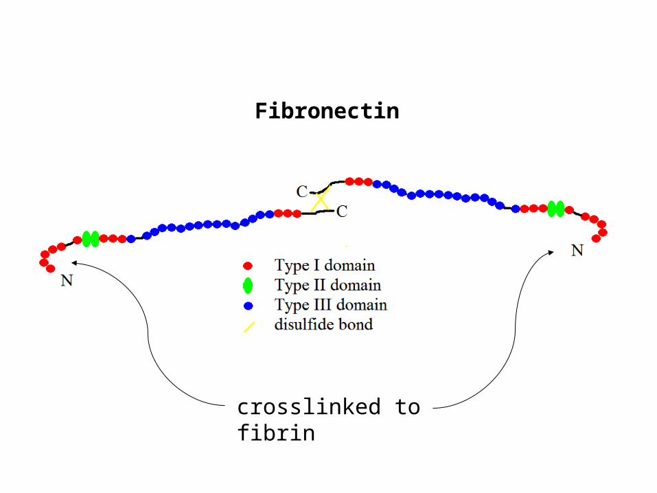
Fibronectin

crosslinked to fibrin

Fibrinolysis

plasminogen plasmin

tissue plasminogen activator

TPA resolving a clot in a coronary artery

Causes of abnormal bleeding

Genetic

hemophilia. A (F-VIII),

B (F-IX). X-linked

von Willebrand Diseaseup to 1%, men or women

Drugs

coumadin, plavix, aspirin,

Vascular fragilityvitamin C deficiency, connective tissue disorders

Diseases affecting platelets

leukemia, AIDS

Diseases affecting the liver

cirrhosis

chemotherapy, alcohol,

broad spectrum antibiotics

Laboratory Tests

Platelet count

Prothrombin time (PT/INR)

plasma from patient plus thromboplastin (contains triggers of extrinsic pathway)

Measures 3 Vit K dependent factors. VII has shortest half life of vitamin K-dependent factors.

Time to clot normalized by normal controls and adjusted for the potency of the thromboplastin is called INR (0.8 – 1.2 is normal).

Partial thromboplastin time (PTT)

plasma from patient plus thromboplastin (without TF) plus trigger of intrinsic pathway.

Functions of the clot

Stop bleeding

Seal against infection

Scaffold for epithelial cells, and for deposition of granulation tissue


Recommended