104
Ingegneria delle tecnologie per la salute Fondamenti di anatomia e istologia Lezione 4.a.b.c aa. 2019-20

Ingegneria delle tecnologie per la salute
Fondamenti di anatomia e istologia
Lezione 4.a.b.c
aa. 2019-20

Ingegneria delle tecnologie per la salute
Fondamenti di anatomia e istologia
aa. 2019-20
Sistema locomotore • Ossa 4.a • Articolazioni 4.b • Muscoli 4.c

BONES 4.a
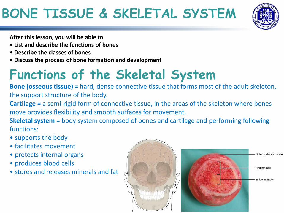
After this lesson, you will be able to: • List and describe the functions of bones • Describe the classes of bones • Discuss the process of bone formation and development
BONE TISSUE & SKELETAL SYSTEM
Functions of the Skeletal System Bone (osseous tissue) = hard, dense connective tissue that forms most of the adult skeleton, the support structure of the body. Cartilage = a semi-rigid form of connective tissue, in the areas of the skeleton where bones move provides flexibility and smooth surfaces for movement. Skeletal system = body system composed of bones and cartilage and performing following functions: • supports the body • facilitates movement • protects internal organs • produces blood cells • stores and releases minerals and fat

206 bones composing skeleton, divided
into 5 categories
based on their shapes ( distinct function)
Bone Classification

Bone Classification

Bone tissue differs greatly from other tissues in the body: is hard (many of its
functions depend on this hardness) and also dynamic (its shape adjusts to
accommodate stresses).
Bone Structure
histology
gross anatomy

Gross Anatomy of Bone
structure of a LONG BONE, 2 parts:
1. diaphysis: tubular shaft that runs
between the proximal and distal ends of the bone, where the hollow region is called medullary cavity (filled with yellow marrow) and the walls are composed of dense and hard compact bone
2. epiphysis: wider section at each end
of the bone, filled with spongy bone (its spaces filled with red marrow) epiphyseal plate (growth plate): where epiphysis meets diaphysis at the metaphysis, narrow area containing a layer of hyaline (transparent) cartilage in a growing bone (in early adulthood, appr. 18-21 yrs, cartilage replaced by osseous tissue).

endosteum = delicate membranous lining where bone growth, repair, and remodeling occur. periosteum = fibrous membrane of outer surface containing blood vessels, nerves, and lymphatic vessels that nourish compact bone and where tendons and ligaments also attach (periosteum covers the entire outer surface except where epiphyses meet other bones to form joints (covered with articular cartilage, a thin layer of cartilage that reduces friction and acts as a shock absorber).
Gross Anatomy of Bone

endosteum & periosteum
Gross Anatomy of Bone

Flat bones = layer of diploe (spongy
bone), lined on either side by a layer of
compact bone, working together to
protect the internal organs
Gross Anatomy of Bone

Gross Anatomy of Bone

Gross Anatomy of Bone Bone Markings
Bony surface features vary considerably, depending on the function and location in the body 3 general classes of bone markings: (1)articulation: where two bone surfaces come together, conforming to one another, to facilitate the function of joint (2)projection: area of a bone that projects above the surface of the bone, where are attachment points for tendons and ligaments, being an indication of the forces exerted through the attachment to the bone (3)hole: opening or groove in the bone, allowing blood vessels and nerves to enter the bone.

Gross Anatomy of Bone Bone Markings

Gross Anatomy of Bone
Bone Markings

• hydroxyapatite crystals = hardness and strength
• collagen fibers = flexibility
• cells = small amount, but crucial to the function of bones
• 4 types of cells: 1. osteoblasts, 2. osteocytes, 3. osteogenic cells, 4. osteoclasts
= a relatively small number of cells entrenched in a matrix of collagen fibers that provide a surface for inorganic salt crystals (= calcium phosphate and calcium carbonate combine to create hydroxyapatite, which incorporates other inorganic salts like magnesium hydroxide, fluoride, and sulfate as it crystallizes, or calcifies, on the collagen fibers) to adhere.
Bone Cells and Tissue HISTOLOGY

Bone Cells and Tissue HISTOLOGY
1. osteoblast = responsible for forming new bone, found in the growing portions (periosteum and endosteum), not dividing, but synthesizing collagen matrix and calcium salts, that trap them, becoming 2. osteocyte = primary cell of mature bone, located in a space called a lacuna, maintain the mineral concentration of the matrix, lacking mitotic activity and communicating with each other via long cytoplasmic processes extending in canaliculi 3. osteogenic cell = undifferentiated with high mitotic activity, found in the deep layers of the periosteum and the marrow.

Bone Cells and Tissue HISTOLOGY
4: osteoclast = responsible for bone resorption, found on bone surfaces, multinucleated, originating from monocytes and macrophages Bone dynamic nature = new tissue is constantly formed (osteoblasts), and old, injured, or unnecessary bone is dissolved (osteoclasts)

Bone Cells and Tissue HISTOLOGY

Bone Cells and Tissue HISTOLOGY
Compact bone = denser, stronger, found under the periosteum and in the diaphyses of long bones, where it provides support and protection, being its microscopic structural unit the osteon (= concentric rings of calcified matrix called lamellae), its blood vessels, nerves, and lymphatic vessels in the central canal, or Haversian canal, + perforating canal, Volkmann’s canals, to extend to the periosteum and endosteum (osteocytes located inside lacunae, found at the borders of adjacent lamellae).

Bone Cells and Tissue HISTOLOGY
osteon (=
concentric rings of
calcified matrix
called lamellae)

Bone Cells and Tissue HISTOLOGY

Bone Cells and Tissue HISTOLOGY

Bone Cells and Tissue HISTOLOGY

Bone Cells and Tissue HISTOLOGY

Bone Cells and Tissue HISTOLOGY
Spongy (Cancellous) Bone contains osteocytes housed in lacunae, but arranged in a lattice-like network of matrix spikes called trabeculae (forms along lines of stress to provide strength to the bone), not in concentric circles.

Bone Cells and Tissue HISTOLOGY . Compact Bone Compact bone is the

Bone Cells and Tissue HISTOLOGY . Compact Bone Compact bone is the

Bone Cells and Tissue HISTOLOGY
Blood and Nerve supply The spongy bone and medullary cavity receive nourishment from arteries that pass through the compact bone = nutrient foramen (small openings in the diaphysis). The osteocytes in spongy bone are nourished by blood vessels of the periosteum that penetrate spongy bone and blood that circulates in the marrow cavities. As the blood passes through the marrow cavities, it is collected by veins, which then pass out of the bone through the foramina. In addition to the blood vessels, nerves follow the same paths into the bone where they tend to concentrate in the more metabolically active regions of the bone.

skeletal system = all of the bones, cartilages, and ligaments of the body that support and give shape to the body. skeleton = bones of the body (adults = 206 bones), subdivided into 2 major divisions: axial (80) and appendicular (126).
Skeletal System

Skeletal System
Axial (80 bones) Appendicular (126 bones)
Head Trunk UpperExtremities Lower Extremities
(29 bones) (51bones) (64 bones) (62 bones)
Cranial (8) Frontal—1 Parietal—2Occipital—1Temporal—2Sphenoid—1Ethmoid—1Facial (14)Maxilla—2Mandible—1Zygoma—2Lacrimal—2Nasal—2Turbinate—2Vomer—1Palatine—2 Hyoid(1) Auditoryossicles (6)Malleus—2 Incus—2 Stapes—2
Vertebrae(26)Cervical—7Thoracic—12Lumbar—5 Sacrum—1Coccyx—1 Ribs(24) Truerib—14False rib—6Floatingrib—4Sternum(1)
Arms andshoulders (10)Clavicle—2Scapula—2Humerus—2Radius—2 Ulna—2 Wrists (16)Scaphoid—2Lunate—2Triquetrum—2Pisiform—2Trapezium—2Trapezoid—2Capitate—2Hamate—2Hands (38)Metacarpal 10Phalanx (fingerbones)—28
Legs and hips (10)Innominate or hip bone(fusion of the ilium,ischium, and pubis)—2Femur—2 Tibia—2Fibula—2 Patella(kneecap)—2 Ankles(14) Talus—2Calcaneus (heel bone)—2 Navicular—2Cuboid—2 Cuneiform,internal—2 Cuneiform,middle—2 Cuneiform,external—2 Feet (38)Metatarsal—10Phalanx (toe bones)—28

Skeletal System
axial skeleton = forms the vertical axis of the body, consisting of: skull, vertebral column (including the sacrum and coccyx), and the thoracic cage, formed by the ribs and sternum. appendicular skeleton = all bones of the upper and lower limbs.

Skull
• facial bones underlie facial structures, form the nasal cavity, enclose the eyeballs, and support the teeth of the upper and lower jaws.
• rounded brain case surrounds and protects the brain and houses the middle and inner ear structures
consists of 22 individual bones [21 immobile and united into a single unit + the 22nd bone, mandible (lower jaw), only moveable bone of the skull]
cranium (skull) = skeletal structure of the head, supporting face and protecting brain, subdivided into facial bones + brain case, or cranial vault

Anterior View of Skull
anterior skull = facial bones, providing bony support for eyes and structures of face (this view is dominated by the openings of the orbits and the nasal cavity; also seen the upper and lower jaws, with their respective teeth).

Anterior View of Skull
orbit = bony socket housing eyeball and muscles moving the eyeball or opening upper eyelid (upper margin of ant. orbit = supraorbital margin; located near the midpoint of supraorbital margin is a small opening called supraorbital foramen, providing passage of a sensory nerve to the skin of the forehead; below orbit = infraorbital foramen, being point of emergence for a sensory nerve that supplies the ant. face below orbit).

Anterior View of Skull
nasal cavity = divided into halves by nasal septum (upper portion formed by perpendicular plate of ethmoid bone and lower portion being vomer bone), each side of nasal cavity is triangular in shape, with a broad inf. space that narrows sup. (looking into nasal cavity, 3 bony plates projecting from each lateral wall = larger of these being inf. nasal concha, an independent bone of the skull + located just above inf. concha, middle nasal concha, which is part of ethmoid bone + third bony plate, also part of ethmoid bone, sup. nasal concha, much smaller and out of sight, above middle concha, located just lateral to perpendicular plate, in upper nasal cavity).

Anterior View of Skull
conchae Anatomy of the nasal cavity demonstrated in a 13-year-old child.A coronal computed tomography scan shows middle and inferior turbinates (asterisks) and vertical lamella of the middle turbinate attached to the cribriform plate (arrow).
Lateral View of Skull
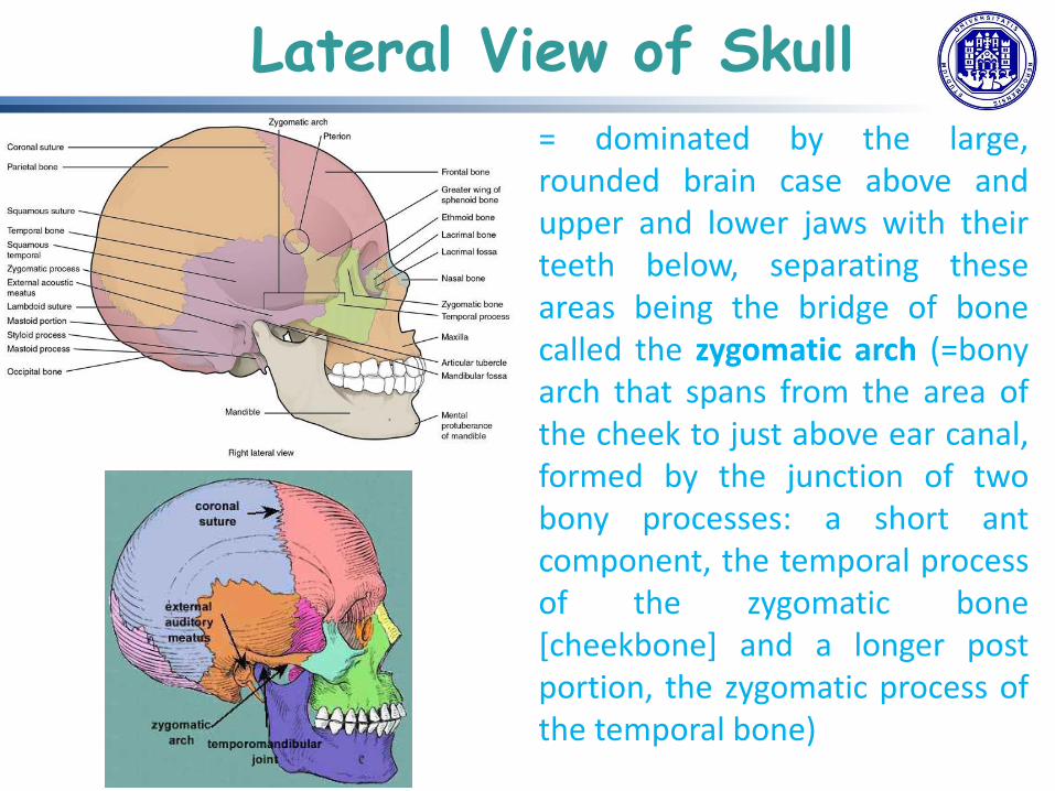
= dominated by the large, rounded brain case above and upper and lower jaws with their teeth below, separating these areas being the bridge of bone called the zygomatic arch (=bony arch that spans from the area of the cheek to just above ear canal, formed by the junction of two bony processes: a short ant component, the temporal process of the zygomatic bone [cheekbone] and a longer post portion, the zygomatic process of the temporal bone)
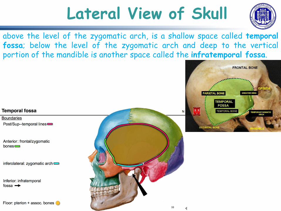
Lateral View of Skull above the level of the zygomatic arch, is a shallow space called temporal fossa; below the level of the zygomatic arch and deep to the vertical portion of the mandible is another space called the infratemporal fossa.

Bones of the Brain Case
• = contains and protects the brain (interior space, almost completely occupied by the brain, called cranial cavity, bounded sup by rounded top of the skull, called calvaria (skullcap), and the lateral and posterior sides of the skull)
• bones that form the top and sides of the brain case are usually referred to as the “flat” bones of the skull.

Bones of the Brain Case
• inside the skull, base is subdivided into 3 large spaces, called ant cranial fossa, middle cranial fossa, and post cranial fossa (fossa = “trench or ditch”): from ant to post, the fossae increase in depth.
• floor of brain case, referred to as base of the skull = complex area varying in depth and having numerous openings for the passage of cranial nerves, blood vessels, and the spinal cord.

Bones of the Brain Case brain case consists of 8 bones, including paired parietal and temporal bones, plus unpaired frontal, occipital, sphenoid, and ethmoid bones. Parietal Bone = forms most of the upper lateral side of the skull, being paired bones, with the right and left parietal bones joining together at the top of the skull. Each parietal bone is also bounded anteriorly by the frontal bone, inferiorly by the temporal bone, and posteriorly by the occipital bone.

Bones of the Brain Case Temporal Bone • = forms lower lateral side of the skull, so
named because this area of the head (the temple) is where hair typically first turns gray, indicating the passage of time.
• subdivided into 4 regions: 1 flattened, upper portion is the squamous portion, 2 below this area, projecting ant is zygomatic process forming post portion of the zygomatic arch, 3 posteriorly is the mastoid portion of the temporal bone (projecting inf from this region is a large prominence, mastoid process, which serves as a muscle attachment site, can easily be felt on the side of the head just behind earlobe), 4 on the interior of the skull, the petrous portion of each temporal bone forms the prominent, diagonally oriented petrous ridge in the floor of the cranial cavity (located inside small cavities that house the structures of the middle and inner ears)

Bones of the Brain Case Important landmarks of the temporal bone: • External acoustic meatus (ear canal)—large opening on the lateral side of the skull that is associated with the ear. • Internal acoustic meatus—opening located inside the cranial cavity, on the medial side of the petrous ridge. It connects to the middle and inner ear cavities of the temporal bone. • Mandibular fossa—deep, oval-shaped depression located on the external base of the skull, just in front of the external acoustic meatus. The mandible (lower jaw) joins with the skull at this site as part of the temporomandibular joint, which allows for movements of the mandible during opening and closing of the mouth. • Articular tubercle—smooth ridge located immediately anterior to the mandibular fossa. Both the articular tubercle and mandibular fossa contribute to the temporomandibular joint, the joint that provides for movements between the temporal bone of the skull and the mandible

Bones of the Brain Case • Styloid process—Post to mandibular fossa on the external base of the skull is an elongated, downward bony projection, so named because of its resemblance to a stylus (a pen or writing tool), serving as an attachment site for several small muscles and for a ligament that supports the hyoid bone of the neck • Stylomastoid foramen—small opening located between the styloid process and mastoid process. This is the point of exit for the cranial nerve that supplies the facial muscles. • Carotid canal—The carotid canal is a zig-zag shaped tunnel, providing passage through the base of the skull for one of the major arteries that supplies the brain.

Bones of the Brain Case Frontal Bone = single bone forming the forehead: in ant midline, between eyebrows, slight depression called glabella • also forms supraorbital margin
of the orbit: near the middle of this margin, supraorbital foramen (= opening providing passage for a sensory nerve to the forehead); thickened just above each supraorbital margin, forming rounded brow ridges, located just behind eyebrows and generally larger in males.
• inside cranial cavity, extends posteriorly in a flattened region forming both roof of the orbit below and floor of ant cranial cavity above

Bones of the Brain Case Occipital Bone = single bone forming post. skull and post base of cranial cavity (on outside surface, at post midline, a small protrusion called external occipital protuberance, serving as an attachment site for a ligament of the posterior neck) • on the base contains large
opening of the foramen magnum, which allows for passage of the spinal cord
• on either side of the foramen magnum is an oval-shaped occipital condyle. These condyles form joints with the first cervical vertebra and thus support the skull on top of the vertebral column.

Bones of the Brain Case Sphenoid Bone = single, complex bone of central skull: “keystone” bone, joining with almost every other bone of the skull and forming much of the base of central skull + extending laterally to contribute to the sides

Bones of the Brain Case Sphenoid Bone • inside cranial cavity, right and lesser wings of
sphenoid bone, which resemble the wings of a flying bird, form the lip of a prominent ridge that marks the boundary between the anterior and middle cranial fossae.
• sella turcica (“Turkish saddle”) located at midline of middle cranial fossa: rounded depression in the floor of the sella turcica is the hypophyseal (pituitary) fossa, which houses the pea-sized pituitary (hypophyseal) gland.

Bones of the Brain Case • greater wings of sphenoid bone
extend laterally to either side away from the sella turcica, where they form the anterior floor of the middle cranial fossa and best seen on the outside of lateral skull, where it forms a rectangular area immediately anterior to the squamous portion of the temporal bone.
• On the inf aspect of skull, each half of the sphenoid bone forms two thin, vertically oriented bony plates: medial and lateral pterygoid plate (pterygoid = “wing-shaped”), forming posterior, lateral walls of nasal cavity and serving as attachment sites for chewing muscles that fill the infratemporal space and act on the mandible.

Bones of the Brain Case Ethmoid Bone =single, midline bone forming the roof and lateral walls of the upper nasal cavity, the upper portion of the nasal septum, and contributing to medial wall of the orbit • On the interior of the skull, also
forms a portion of floor of ant cranial cavity
• Within the nasal cavity, the perpendicular plate of the ethmoid bone forms the upper portion of the nasal septum. The ethmoid bone also forms the lateral walls of the upper nasal cavity. Extending from each lateral wall are the superior nasal concha and middle nasal concha, which are thin, curved projections that extend into the nasal cavity

Bones of the Brain Case • In the cranial cavity, forms a small area
at the midline in the floor of the anterior cranial fossa (also forms the narrow roof of the underlying nasal cavity) consisting of two parts, the crista galli (“rooster’s comb or crest”) is a small upward bony projection located at the midline) and cribriform plates (cribrum = “sieve”, a small, flattened area with numerous small openings termed olfactory foramina, small nerve branches from the olfactory areas of the nasal cavity pass through these openings to enter the brain.)
• lateral portions are located between the orbit and upper nasal cavity, and thus form the lateral nasal cavity wall and a portion of the medial orbit wall.

Bones of the Brain Case Sutures of the Skull suture = immobile joint between adjacent bones of the skull (narrow gap filled with dense, fibrous connective tissue), are not straight, but instead follow irregular, tightly twisting paths. 2 suture lines seen on the top of the skull are the coronal (running from side to side across the skull, within the coronal plane of section) and sagittal sutures (extending post from coronal suture, running along the midline at the top of the skull in the sagittal plane of section lambdoid suture extends downward and laterally to either side away from its junction with sagittal suture (named for its shape, resembling the Greek letter lambda (Λ). squamous suture, located on lateral skull, unites the squamous portion of the temporal bone with the parietal bone Pterion = small, capital-H-shaped suture line region at the intersection of four bones

Facial Bones of the Skull = 14 bones (six paired bones: maxilla, palatine, zygomatic, nasal, lacrimal, and inferior nasal conchae and two unpaired bones: vomer and mandible) forming the upper and lower jaws, the nose, nasal cavity and nasal septum, and the orbit. Maxillary Bone = (maxilla, plural = maxillae), form the upper jaw, much of the hard palate, the medial floor of the orbit, and the lateral base of the nose. The curved, inferior margin of the maxillary bone that forms the upper jaw and contains the upper teeth is the alveolar process of the maxilla. On the anterior maxilla, just below the orbit, is the infraorbital foramen. This is the point of exit for a sensory nerve that supplies the nose, upper lip, and anterior cheek. On the inferior skull, the palatine process from each maxillary bone can be seen joining together at the midline to form the anterior three-quarters of the hard palate. The hard palate is the bony plate that forms the roof of the mouth and floor of the nasal cavity, separating the oral and nasal cavities.

Facial Bones of the Skull Palatine Bone = pair of irregularly shaped bones, contributing to the lateral walls of the nasal cavity and the medial wall of each orbit and the hard palate. Zygomatic Bone = paired, forming much of the lateral wall of orbit and lateral-inferior margins of ant orbital opening Nasal Bone = two small bones that articulate (join) with each other to form the bony base (bridge) of the nose, also supporting cartilages of the nose Lacrimal Bone = small, rectangular bone, forming the ant., medial wall of the orbit, forming a shallow depression called the lacrimal fossa Inferior Nasal Conchae = curved bony plate that projects into the nasal cavity space from the lower lateral wall Vomer Bone = unpaired triangular-shaped, forming posterior-inferior part of nasal septum

Facial Bones of the Skull Mandible = lower jaw and only moveable bone of the skull. Each side of the mandible consists of a horizontal body and posteriorly, a vertically oriented ramus of the mandible (ramus = “branch”). The ramus on each side of the mandible has 2 upward-going bony projections. The more ant projection is the flattened coronoid process of the mandible, which provides attachment for one of the biting muscles. The post projection is the condylar process of the mandible, which is topped by the oval-shaped condyle, that articulates (joins) with the mandibular fossa and articular tubercle of the temporal bone. The broad U-shaped curve located between the coronoid and condylar processes is the mandibular notch.

Facial Bones of the Skull landmarks for the mandible: • Alveolar process of the mandible = upper border of the mandibular body, serving to anchor lower teeth. • Mental protuberance = forward projection from the inf margin of the ant mandible that forms the chin (mental = “chin”). • Mental foramen = opening located on each side of the ant-lat mandible, which is exit site for a sensory nerve that supplies the chin. • Mylohyoid line = bony ridge extending along the inner aspect of the mandibular body. The muscle that forms the floor of the oral cavity attaches to the mylohyoid lines on both sides of the mandible. • Mandibular foramen = opening located on the medial side of the ramus of the mandible, leading into a tunnel that runs down the length of the mandibular body. The sensory nerve and blood vessels that supply the lower teeth enter the mandibular foramen and then follow this tunnel. • Lingula = located immediately next to the mandibular foramen, on the medial side of the ramus.

Orbit

Nasal Septum

Cranial Fossae

Hyoid Bone = independent bone that does not contact any other bone and thus is not part of the skull. • It is a small U-shaped bone
located in the upper neck near the level of the inf mandible, with the tips of the “U” pointing post.
• serves as the base for the tongue above, and is attached to the larynx below and the pharynx post.
• is held in position by a series of small muscles that attach to it either from above or below.

Vertebral Column
vertebrae + intervertebral discs = VC [flexible column supporting head, neck, and body, allowing for their movements & also protecting spinal cord, which passes down the back through openings in the vertebrae]
VC, also known as spinal column or spine = consists of a sequence of vertebrae (singular = vertebra), each of which is separated and united by an intervertebral disc.

Regions of VC
• subdivided into 5 regions [vertebrae in each area named for that region and numbered in descending order]: in the neck, 7 cervical vertebrae [designated with the letter “C” followed by its number; sup C1 vertebra articulates (forms a joint) with the occipital condyles of the skull, inf C1 articulates with the C2 vertebra, and so on] + 12 thoracic vertebrae [T1–T12] + 5 lumbar vertebrae [L1–L5] + single sacrum [also part of the pelvis & formed by the fusion of 5 sacral vertebrae] + coccyx [tailbone, resulting from the fusion of 4 small coccygeal vertebrae].
• VC originally develops as a series of 33 vertebrae, but this number is eventually reduced to 24 vertebrae, plus the sacrum and coccyx.
• sacral and coccygeal fusions do not start until age 20 and are not completed until middle age.

Curvatures of VC
adult VC does not form a straight line, but instead has 4 curvatures along its length, to increase VC’s strength, flexibility, and ability to absorb shock [when load on the spine is increased, by carrying a heavy backpack for example, curvatures increase in depth (become more curved) to accommodate the extra weight; they then spring back when the weight is removed]
4 adult curvatures are classified as either primary or secondary curvatures: primary curves are retained from the original fetal curvature, while secondary curvatures develop after birth.

Curvatures of VC During fetal development, body is flexed ant into fetal position, giving entire VC a single curvature that is concave ant. In the adult, this fetal curvature is retained in 2 regions of the VC, as thoracic curve, which involves thoracic vertebrae, and sacrococcygeal curve, formed by sacrum and coccyx. Each of these is thus called a primary curve because they are retained from original fetal curvature of VC. secondary curve develops gradually after birth as the child learns to sit upright, stand, and walk. Secondary curves are concave post, opposite in direction to the original fetal curvature: cervical curve of the neck region develops as the infant begins to hold their head upright when sitting & later, as the child begins to stand and then to walk, lumbar curve of lower back develops.
In adults, the lumbar curve is generally deeper in females.

General Structure of a Vertebra
Within the different regions of the VC, vertebrae vary in size and shape, but they all follow a similar structural pattern. A typical vertebra will consist of: 1. body 2. vertebral arch 3. 7 processes

General Structure of a Vertebra
1. body is the ant portion of each vertebra, supporting the body weight, so progressively increase in size and thickness going down the VC: bodies of adjacent vertebrae are separated and strongly united by an intervertebral disc.
2. vertebral arch forms the post portion of each vertebra, consisting of 4 parts, right and left pedicles + right and left laminae. Each pedicle forms one of the lateral sides of the vertebral arch and are anchored to the post side of the vertebral body. Each lamina forms part of the post roof of the vertebral arch.

General Structure of a Vertebra
The large opening between the vertebral arch and body is the vertebral foramen, which contains the spinal cord. In the intact vertebral column, the vertebral foramina of all of the vertebrae align to form the vertebral (spinal) canal, which serves as the bony protection and passageway for the spinal cord down the back. When the vertebrae are aligned together in the vertebral column, notches in the margins of the pedicles of adjacent vertebrae together form an intervertebral foramen, the opening through which a spinal nerve exits from the vertebral column

General Structure of a Vertebra
7 processes arise from vertebral arch. (2) each paired transverse process projects laterally and arises from the junction point between the pedicle and lamina. (1) single spinous process (vertebral spine) projects post at the midline of the
back. transverse and spinous processes serve as important muscle attachment sites (2) sup articular process extends or faces upward (2) inf articular process faces or projects downward
paired sup articular processes of one vertebra join with the corresponding paired inf articular processes from the next higher vertebra. These junctions form slightly moveable joints between the adjacent vertebrae. The shape and orientation of the articular processes vary in different regions of the VC and play a major role in determining type and range of motion available in each region.

General Structure of a Vertebra
bodies of adjacent vertebrae are separated and united by an intervertebral disc, which provides padding and allows for movements between adjacent vertebrae. disc consists of a fibrous outer layer called the anulus fibrosus and a gel-like center called the nucleus pulposus. The intervertebral foramen is the opening formed between adjacent vertebrae for the exit of a spinal nerve.
Regional Modifications of Vertebrae
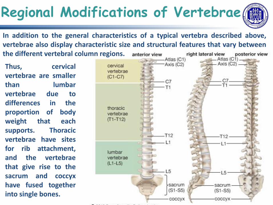
In addition to the general characteristics of a typical vertebra described above, vertebrae also display characteristic size and structural features that vary between the different vertebral column regions.
Thus, cervical vertebrae are smaller than lumbar vertebrae due to differences in the proportion of body weight that each supports. Thoracic vertebrae have sites for rib attachment, and the vertebrae that give rise to the sacrum and coccyx have fused together into single bones.

Cervical Vertebrae
Typical C vertebrae, such as C4 or C5, have several characteristic features that differentiate them from T or L vertebrae: a small body [reflecting the fact that they carry the least amount of body weight], a bifid (Y-shaped) spinous process [spinous processes of C3–C6 are short, but C7 is much longer], transverse processes sharply curved (U-shaped) to allow for passage of the cervical spinal nerves] and also having an opening called transverse foramen [important artery that supplies brain ascends up the neck by passing through these openings], sup and inf articular processes flattened and largely face upward or downward, respectively.

Cervical Vertebrae
first and second C vertebrae further modified, giving each a distinctive appearance: C1 also called atlas, supporting skull on top of VC (in Greek mythology, Atlas was the god who supported the heavens on his shoulders), does not have a body or spinous process, instead, being ring-shaped, consisting of an ant arch and a post arch, transverse processes longer and extending more laterally than do the transverse processes of any other C vertebrae; sup articular processes face upward and deeply curved for articulation with the occipital condyles on the base of the skull; inf articular processes flat and facing downward to join with the sup articular processes of C2.

C2 called axis, serving as axis for rotation when turning head toward right or left, resembling typical C vertebrae in most respects, but easily distinguished by dens (odontoid process = bony projection extending upward from vertebral body and joining with inner aspect of ant arch of atlas, where is held in place by transverse ligament)
Cervical Vertebrae
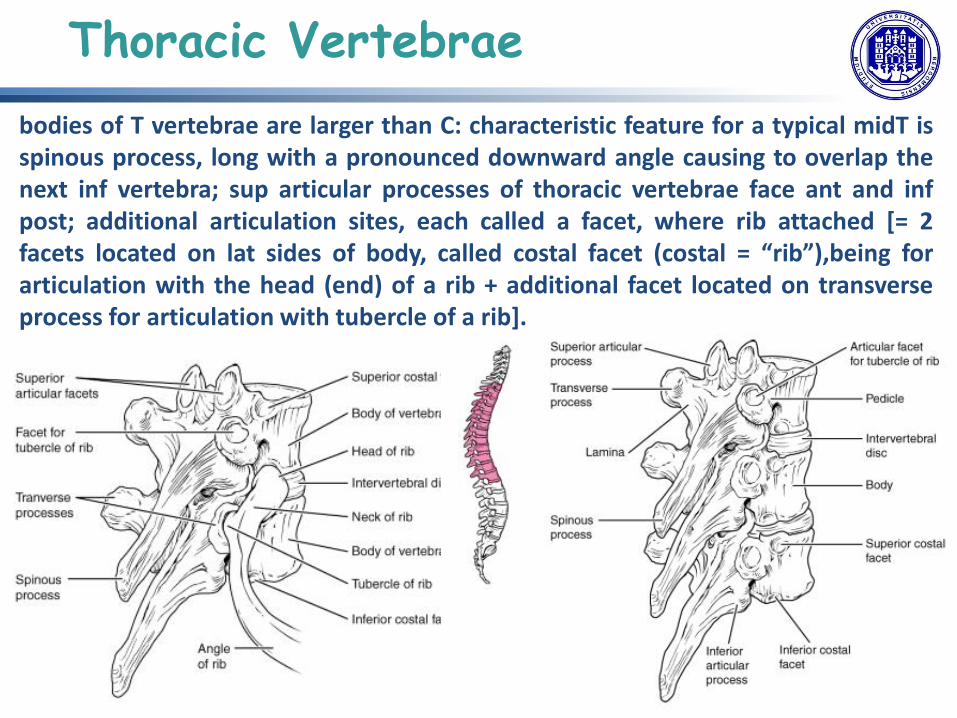
bodies of T vertebrae are larger than C: characteristic feature for a typical midT is spinous process, long with a pronounced downward angle causing to overlap the next inf vertebra; sup articular processes of thoracic vertebrae face ant and inf post; additional articulation sites, each called a facet, where rib attached [= 2 facets located on lat sides of body, called costal facet (costal = “rib”),being for articulation with the head (end) of a rib + additional facet located on transverse process for articulation with tubercle of a rib].
Thoracic Vertebrae

L vertebrae carry greatest amount of body weight and are thus characterized by the large size and thickness of vertebral body: short transverse processes and a short, blunt spinous process that projects posteriorly; articular processes are large, with the sup facing backward and the inf facing forward.
Lumbar Vertebrae

sacrum = triangular-shaped bone, thick and wide across its sup base where it is weight bearing and then tapers down to an inf, non-weight bearing apex, formed by fusion of 5 S vertebrae [process beginning after age of 20, on ant surface lines of vertebral fusion can be seen as 4 transverse ridges; on post surface, running down the midline, median sacral crest, a bumpy ridge = remnant of the fused spinous processes; similarly, fused transverse processes form lateral sacral crest]; sacral promontory = ant lip of sup base of sacrum and lateral to this a roughened auricular surface, which joins with ilium portion of hipbone to form the immobile sacroiliac joints of the pelvis.
Sacrum and Coccyx

Passing inf through sacrum is a bony tunnel called sacral canal, which terminates at the sacral hiatus near the inf tip of S; ant and post surfaces have a series of paired openings called sacral foramina (singular = foramen) that connect to the sacral canal. Each of these openings is called a post (dorsal) sacral foramen or ant (ventral) sacral foramen. These openings allow for the ant and post branches of sacral spinal nerves to exit sacrum; sup articular process of sacrum, one of which is found on either side of the sup opening of sacral canal, articulates with the inf articular processes from the L5.
Sacrum and Coccyx
coccyx, or tailbone, = derived from fusion of 4 very small coccygeal vertebrae, articulates with inf tip of sacrum, not weight bearing in standing position, but may receive some body weight when sitting.

bodies of adjacent vertebrae strongly anchored to each other by an intervertebral disc [= structure providing padding between bones during weight bearing, and because it can change shape, also allowing for movement between vertebrae]
Although total amount of movement available between any 2 adjacent vertebrae is small, when these movements summed together along entire length of VC, large body movements can be produced].
Ligaments that extend along the length of the VC also contribute to its overall support and stability.
Intervertebral Discs and Ligaments of the VC

ID = fibrocartilaginous pad filling the gap between adjacent vertebral bodies , anchored to the bodies of its adjacent vertebrae, thus strongly uniting them and also providing padding between vertebrae during weight bearing. Because of this, ID thin in the C region and thickest in the L region, which carries the most body weight: in total, IDs account for approximately 25% of body height between top of pelvis and base of the skull; are also flexible and can change shape to allow for movements of the VC.
each ID consists of 2 parts: 1 anulus fibrosus is the tough, fibrous outer layer of the disc, forming a circle (anulus = “ring” or “circle”) firmly anchored to the outer margins of the adjacent vertebral bodies; 2 inside nucleus pulposus, consisting of a softer, more gel-like material, with a high water content serving to resist compression and thus being important for weight bearing: with increasing age, the water content of the nucleus pulposus gradually declines, causing disc to become thinner, decreasing total body height somewhat, and reduces the flexibility and range of motion of the disc, making bending more difficult.
Intervertebral Discs

gel-like nature of the nucleus pulposus also allows the ID to change shape as one vertebra rocks side to side or forward and back in relation to its neighbors during movements of VC. Thus, bending forward causes compression of ant. portion of the disc but expansion of the posterior disc. If the posterior anulus fibrosus is weakened due to injury or increasing age, the pressure exerted on the disc when bending forward and lifting a heavy object can cause the nucleus pulposus to protrude posteriorly through the anulus fibrosus, resulting in a herniated disc (“ruptured” or “slipped” disc) and compression of a spinal nerve, resulting in pain and/or muscle weakness in the body regions supplied by that nerve.
Intervertebral Discs

Thoracic cage (rib cage)
= thorax (chest) portion of the body, 12 pairs of ribs (anchored posteriorly to 12 thoracic vertebrae, T1–T12) with their costal cartilages and sternum, protecting heart and lungs.

Sternum
3 parts: manubrium, body, xiphoid process. jugular (suprasternal) notch =shallow, U-shaped border at the top of the manubrium + clavicular notch = shallow depression located on either side at the superior-lateral margins of the manubrium; first ribs also attach to the manubrium. Ribs 2–7 attach to the sternal body.

= 12 pairs of curved, flattened bone that contributes to the wall of the thorax, articulating posteriorly with the T1–T12 thoracic vertebrae, and most attach ant via their costal cartilages to the sternum.
Ribs

appendicular skeleton = all of the limb bones + bones that unite each limb with the axial skeleton pectoral girdle (shoulder girdle) = bones that attach each upper limb to the axial skeleton = 2 bones, scapula and clavicle.
Pectoral Girdle
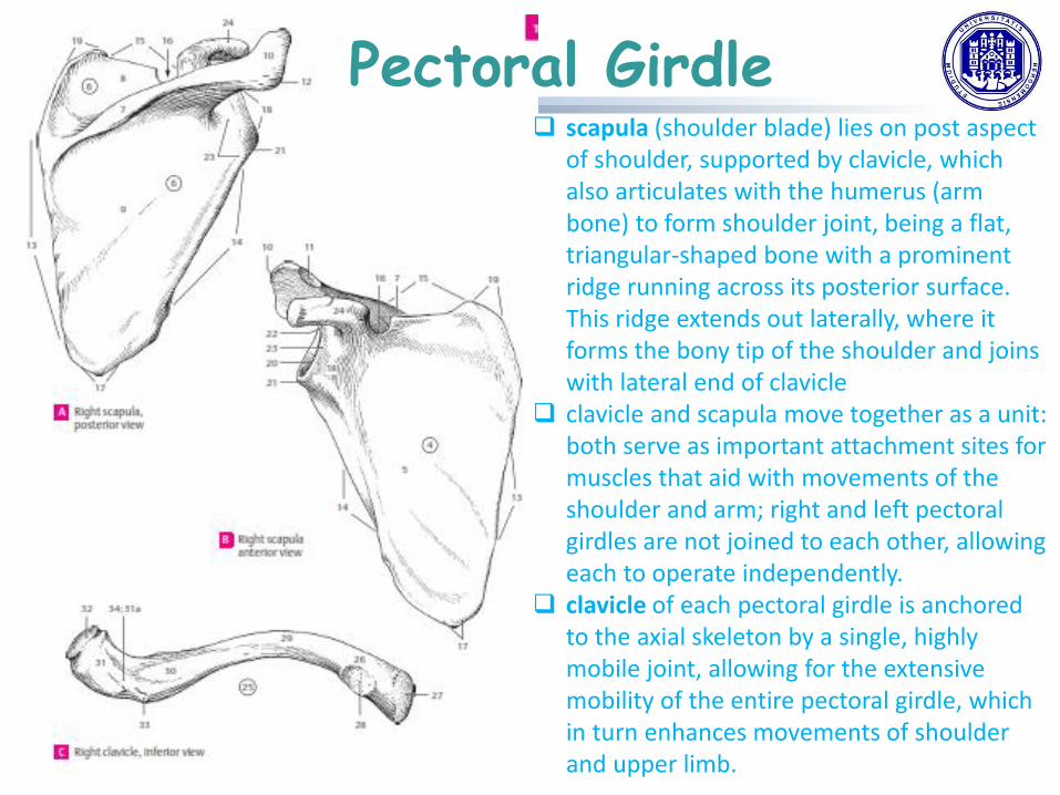
Pectoral Girdle scapula (shoulder blade) lies on post aspect
of shoulder, supported by clavicle, which also articulates with the humerus (arm bone) to form shoulder joint, being a flat, triangular-shaped bone with a prominent ridge running across its posterior surface. This ridge extends out laterally, where it forms the bony tip of the shoulder and joins with lateral end of clavicle
clavicle and scapula move together as a unit: both serve as important attachment sites for muscles that aid with movements of the shoulder and arm; right and left pectoral girdles are not joined to each other, allowing each to operate independently.
clavicle of each pectoral girdle is anchored to the axial skeleton by a single, highly mobile joint, allowing for the extensive mobility of the entire pectoral girdle, which in turn enhances movements of shoulder and upper limb.

Pectoral Girdle

• upper limb divided into 3 regions: arm, located between shoulder and elbow joints; forearm, between elbow and wrist joints; hand, distal to the wrist.
• 30 bones in each upper limb: humerus = single bone of the upper arm + ulna (medially) and radius (laterally) = paired bones of forearm + base of the hand contains 8 bones, each called a carpal bone, + palm of the hand is formed by 5 bones, each called a metacarpal bone + fingers and thumb contain a total of 14 bones, each of which is a phalanx bone of the hand.
Bones of the Upper Limb

Humerus

Ulna + Radius

Carpal Bones, Metacarpal Bones, Phalanx Bones

Carpal Bones, Metacarpal Bones, Phalanx Bones

Carpal Tunnel

The Pelvic Girdle and Pelvis
pelvic girdle (hip girdle) = single bone, the hip bone or coxal bone (coxal = “hip”), which serves as the attachment point for each lower limb. Each hip bone, in turn, is firmly joined to the axial skeleton via its attachment to the sacrum of the vertebral column. The right and left hip bones also converge anteriorly to attach to each other. pelvis = entire structure formed by the two hip bones, the sacrum, and, attached inferiorly to the sacrum, the coccyx

The Pelvic Girdle and Pelvis

Hip Bone

Hip Bone

Pelvis

Pelvis

Bones of the Lower Limb divided into 3 regions: thigh is that portion of the
lower limb located between hip joint and knee joint; leg is specifically the region between knee joint and ankle joint. Distal to the ankle is the foot. The lower limb contains 30 bones: femur, patella, tibia, fibula, tarsal bones, metatarsal bones, and phalanges
femur single bone of the thigh + patella is the kneecap and articulates with the distal femur + tibia is the larger, weight-bearing bone located on the medial side of the leg + fibula is the thin bone of the lateral leg. + 7 tarsal bone (post portion of the foot ) + 5 metatarsal bone (mid-foot elongated bones) + 14 small bones of toes

Femur + Patella

Tibia + Fibula

Tarsal + Metatarsal bones + Phalanges


