81
Integrated Diagnostic Approach to the Classification of Myeloid Neoplasms Daniel A. Arber, MD Stanford University

Integrated Diagnostic Approach to the Classification of Myeloid
Neoplasms
Daniel A. Arber, MDStanford University

What is an integrated approach?

What is an integrated approach?
• Incorporating all diagnostic data related to a single patient sample into a single report

What is an integrated approach?
• Incorporating all diagnostic data related to a single patient sample into a single report
• Models– Surgical pathology– Lymph node pathology

The Lymph Node Model
• 68 year-old woman with skin rashes and lymphadenopathy. A cervical lymph node is biopsied.

The Lymph Node Model
Images courtesy of Dr. Roger Warnke

The Lymph Node Model
Images courtesy of Dr. Roger Warnke

The Lymph Node Model
CD3 LAM
KAP
Images courtesy of Dr. Roger Warnke
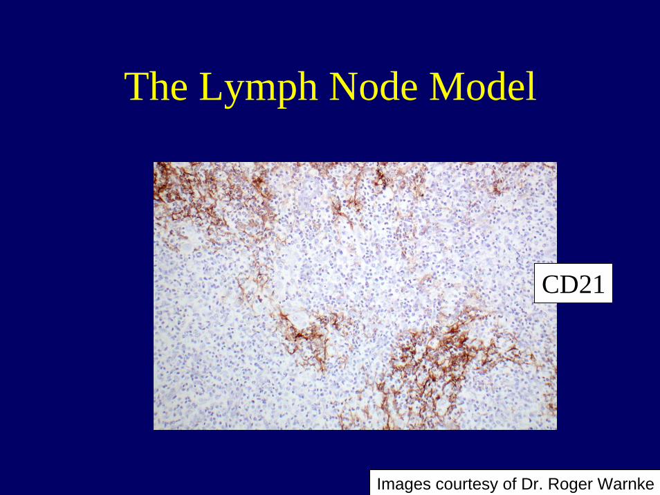
The Lymph Node Model
CD21
Images courtesy of Dr. Roger Warnke

The Lymph Node Model
CD10Images courtesy of Dr. Roger Warnke

The Lymph Node Model
• Additional studies– TCRG PCR clonal– IGH PCR clonal– EBV EBER1 ISH negative
The Lymph Node Model
• Diagnosis– Angioimmunoblastic T cell lymphoma
complicated by a monotypic plasmacytoid B cell population

The Old Myeloid Disorder Model

The Old Myeloid Disorder Model
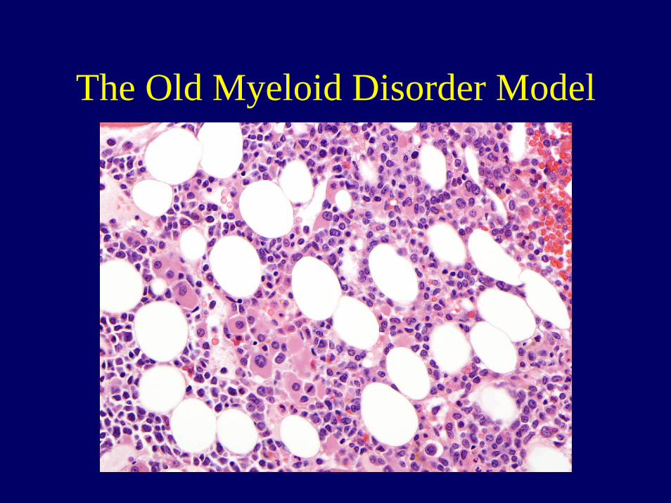
The Old Myeloid Disorder Model
The Old Myeloid Disorder Model
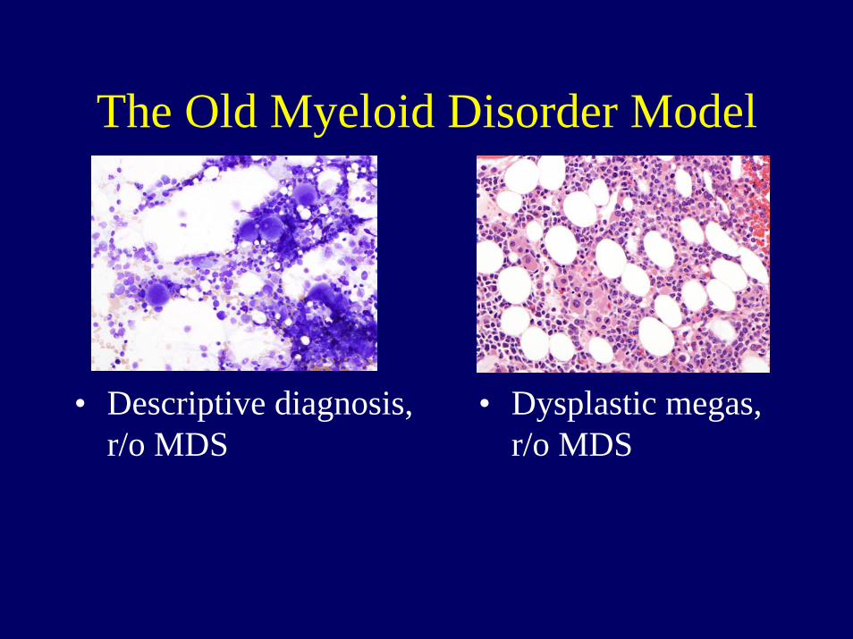
• Descriptive diagnosis, r/o MDS
• Dysplastic megas, r/o MDS

The Old Myeloid Disorder Model
• Descriptive diagnosis, r/o MDS
• del(5q) as sole abnormality
• Dysplastic megas, r/o MDS
• inv(3)

Why do we need an integrated approach to myeloid disorders?

Myeloproliferative Neoplasms• Chronic myelogenous leukemia, BCR-ABL1+• Chronic neutrophilic leukemia• Polycythemia vera• Primary myelofibrosis• Essential thrombocythemia• Chronic eosinophilic leukemia• Myeloid neoplasms associated with PDGFRA, PDGFRB
and FGFR1 rearrangements• Mast cell disease• Myeloproliferative neoplasms, unclassified

From Tefferi and Vardiman. Curr Opin Hematol, 14:115, 2007

Diagnostic Algorithm for Suspected Polycythemia Vera
From Tefferi and Vardiman. Leukemia, Sep 20 Epub, 2007

Diagnostic Algorithm for Primary Eosinophilia
From Tefferi and Vardiman Leukemia, Sep 20 Epub, 2007

Myelodysplastic Syndromes
• Refractory cytopenia• Refractory anemia with ring sideroblasts• Refractory cytopenia with multilineage dysplasia• Refractory anemia with excess blasts• Myelodysplastic syndrome with isolated del(5q)• Myelodysplastic syndrome, unclassifiable

Diagnostic Algorithm for Myelodysplastic Syndromes
From Komrokji & Bennett. Curr Opin Hematol, 14:98, 2007

International Prognostic Scoring System for MDS
KaryotypeGood= normal, del(5q),
del(20q) or –YIntermediate= othersPoor= complex, -7
Risk Group ScoreLow 0Int-1 0.5-1Int-2 1.5-2High >2.5

Myelodysplastic/Myeloproliferative Neoplasms
• Chronic myelomonocytic leukemia• Atypical chronic myeloid leukemia• Juvenile myelomonocytic leukemia• Myelodysplastic/myeloproliferative
neoplasms, unclassified

Key Elements for a Bone Marrow Diagnosis
• Peripheral blood findings
• Marrow aspirate• Marrow trephine/clot
biopsy• Immunophenotyping
– Flow cytometry– Immunohisto-
chemistry
• Karyotype analysis• Molecular analysis
– FISH• Translocations• Chromosome gains or
loss– PCR
• Translocations• Mutations

Who are the players?
• Peripheral blood -Instruments, MT/CLS
• Marrow aspirate
• Marrow trephine/clot biopsy
• Immunophenotyping
• Karyotype analysis
• Molecular analysis

Who are the players?
• Peripheral blood -Instruments, MT/CLS
• Marrow aspirate –Hematologist and/or Pathologist
• Marrow trephine/clot biopsy
• Immunophenotyping
• Karyotype analysis
• Molecular analysis

Who are the players?
• Peripheral blood -Instruments, MT/CLS
• Marrow aspirate -Hematologist and/or Pathologist
• Marrow trephine/clot biopsy - Pathologist
• Immunophenotyping
• Karyotype analysis
• Molecular analysis

Who are the players?
• Peripheral blood -Instruments, MT/CLS
• Marrow aspirate -Hematologist and/or Pathologist
• Marrow trephine/clot biopsy - Pathologist
• Immunophenotyping –Flow lab/Reference lab
• Karyotype analysis
• Molecular analysis

Who are the players?
• Peripheral blood -Instruments, MT/CLS
• Marrow aspirate -Hematologist and/or Pathologist
• Marrow trephine/clot biopsy - Pathologist
• Immunophenotyping –Flow lab/Reference lab
• Karyotype analysis –Cytogenetics lab/ Reference lab
• Molecular analysis

Who are the players?
• Peripheral blood -Instruments, MT/CLS
• Marrow aspirate -Hematologist and/or Pathologist
• Marrow trephine/clot biopsy - Pathologist
• Immunophenotyping –Flow lab/Reference lab
• Karyotype analysis –Cytogenetics lab/ Reference lab
• Molecular analysis –Molecular lab/ Reference lab

Who is best qualified to put it all together?

Who is best qualified to put it all together?
The clinician?
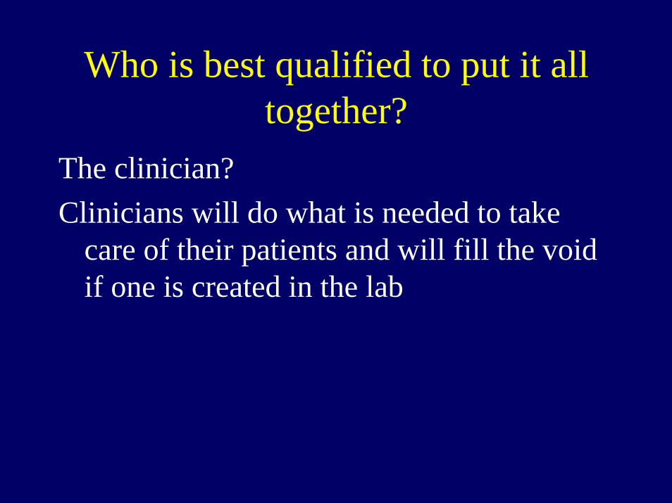
Who is best qualified to put it all together?
The clinician?Clinicians will do what is needed to take
care of their patients and will fill the void if one is created in the lab

Who is best qualified to put it all together?
The clinician?The pathologist?

Who is best qualified to put it all together?
The clinician?The pathologist?When pathologists correlate findings, they
become a critical and visible part of the care giving team

Who is best qualified to put it all together?
The clinician?The pathologist?The pathologist and the clinician together?

Who is best qualified to put it all together?
The clinician?The pathologist?The pathologist and the clinician together?The pathologist can assist the clinician in
ordering the appropriate tests, can make sense of different results, and even explain the reason for discrepancies

The Pathologist Role
• Consult with clinician– Recommend tests, explain specimen requirements,
etc• Spot inappropriate orders
– i.e. Flow for Hodgkin lymphoma staging• Recommend and implement additional tests
– i.e. Adding FISH to a PB with flow cytometry suggestive of mantle cell lymphoma
• Address all testing in a single report

Case 1
• 48 year old woman with diarrhea. A CBC is performed.– A peripheral blood smear review is performed
due to instrument flag for white cell abnormalities


Case 1
• CLS Hematology Specialist notes increase basophils and some dysplastic neutrophils. Suggests accelerated phase of CML and correlation with studies for t(9;22).

Case 1
• CLS Hematology Specialist notes increase basophils and some dysplastic neutrophils. Suggests accelerated phase of CML and correlation with studies for t(9;22).– Bone marrow, flow cytometry, karyotype and
PCR for BCR-ABL1 are ordered.


CD117 TRYPTASE
CD2CD25

CD2
CD25

Case 1
• D816V mutation of KIT detected by PCR
• BCR-ABL1 negative
From Gotlib J. Immunol Allergy Clin N Am, 26:575, 2006

Case 1
• Mast cell leukemia• Mast cells with aberrant expression of
CD25 detected• D816V KIT mutation positive

Case 2
• 28 year old man with history of bipolar disorder
• Presents to psychiatrist with worsening of psychiatric symptoms, mild fatigue and night sweats
Courtesy Dita Gratzinger

Case 2
• Physical exam reveals marked splenomegaly, no lymphadenopathy
• CT scan shows 24 cm spleen, no other significant findings
Courtesy Dita Gratzinger

Courtesy Dita Gratzinger

Courtesy Dita Gratzinger

Courtesy Dita Gratzinger

Courtesy Dita Gratzinger

Case 2
• BCR-ABL1 negative• No KIT mutation detected• FISH for CHIC2 deletion (surrogate for
FIP1L1-PDGFRA fusion) positive

Case 2

Case 2
• Chronic eosinophilic leukemia (Myeloid neoplasm associated with PDGFRArearrangement)
• CHIC2 deletion positive• TCRG gene rearrangement of unknown
significance detected

The Bone Marrow Report
• Standardized reporting?• Synoptic reporting?
– Improves report completeness– A training tool?
• College of American Pathologists Cancer Protocols and Checklists for Bone Marrow– http://www.cap.org/apps/docs/cancer_
protocols/2005/bonemarrow05_ckw.pdf

The Bone Marrow Report• Clinical information• Aspirate and biopsy sites• Peripheral blood• Marrow aspirate/touch preps• Marrow biopsy/clot• Immunophenotyping• Cytogenetics• Molecular genetics• Other ancillary tests• Diagnosis
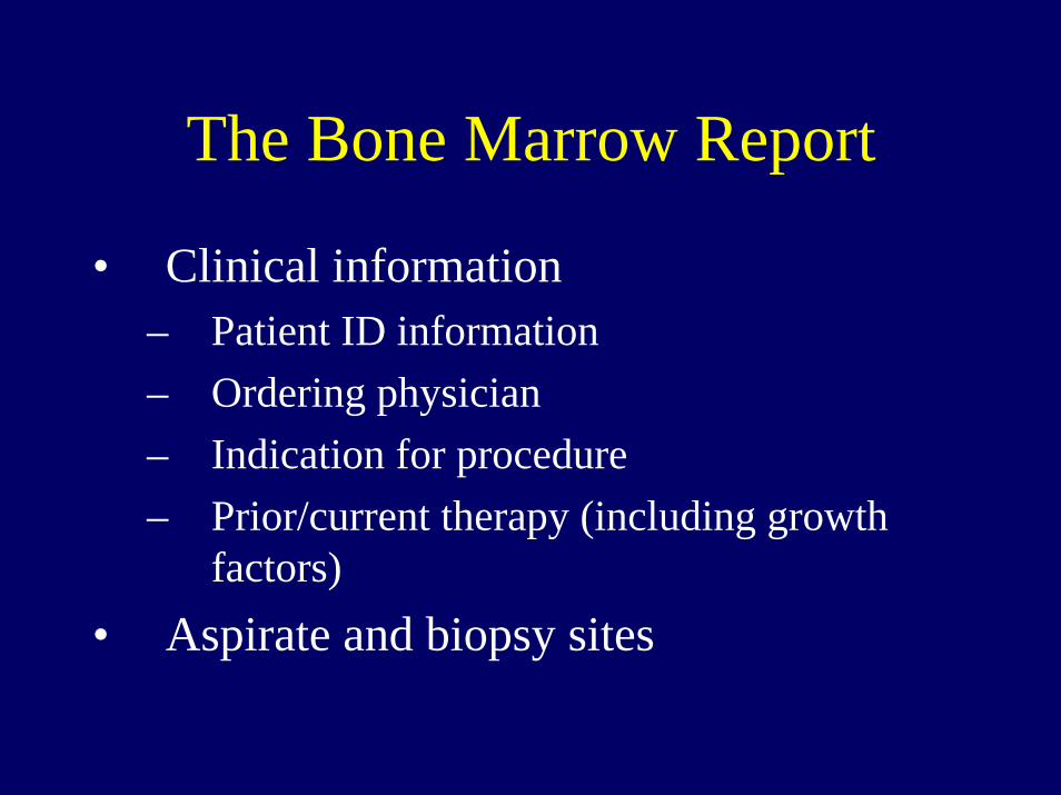
The Bone Marrow Report
• Clinical information– Patient ID information– Ordering physician– Indication for procedure– Prior/current therapy (including growth
factors)• Aspirate and biopsy sites

The Bone Marrow Report
• Peripheral Blood– CBC data, including units and reference
ranges– Morphologic description

The Bone Marrow Report
• Marrow aspirate/touch preps– Adequacy– Differential cell count– Morphologic findings

The Bone Marrow Report
• Marrow biopsy/clot– Adequacy for the questioned asked– Morphologic description

The Bone Marrow Report
• Immunophenotyping– Method(s) used– Where studies performed– Antibodies used– Interpretation

The Bone Marrow Report
• Cytogenetics– Testing performed– Where studies were performed– Interpretation

The Bone Marrow Report
• Molecular genetics– Testing performed– Where studies were performed– Interpretation

The Bone Marrow Report
• Other ancillary tests– Iron stain– Other cytochemistry– AFB, GMS– Reticulin, etc

The Bone Marrow Report
• Diagnosis– WHO classification where appropriate– Address the question asked in the clinical
information in a comment if only a descriptive diagnosis is rendered

Pathologists as the Gate-Keepers
• Stop unnecessary tests• Suggest more appropriate tests to get to
the correct diagnosis• Put all the information together

Do we really need to do everything on every case?

Do we really need to do everything on every case?
• PB, BM aspirate and biopsy should be reviewed together

Do we really need to do everything on every case?
• PB, BM aspirate and biopsy should be reviewed together
• Increased utility of karyotype and molecular genetic testing in myeloid disorders

Do we really need to do everything on every case?
• PB, BM aspirate and biopsy should be reviewed together
• Increased utility of karyotype and molecular genetic testing in myeloid disorders
• Does every case of CML need karyotype and BCR-ABL1?
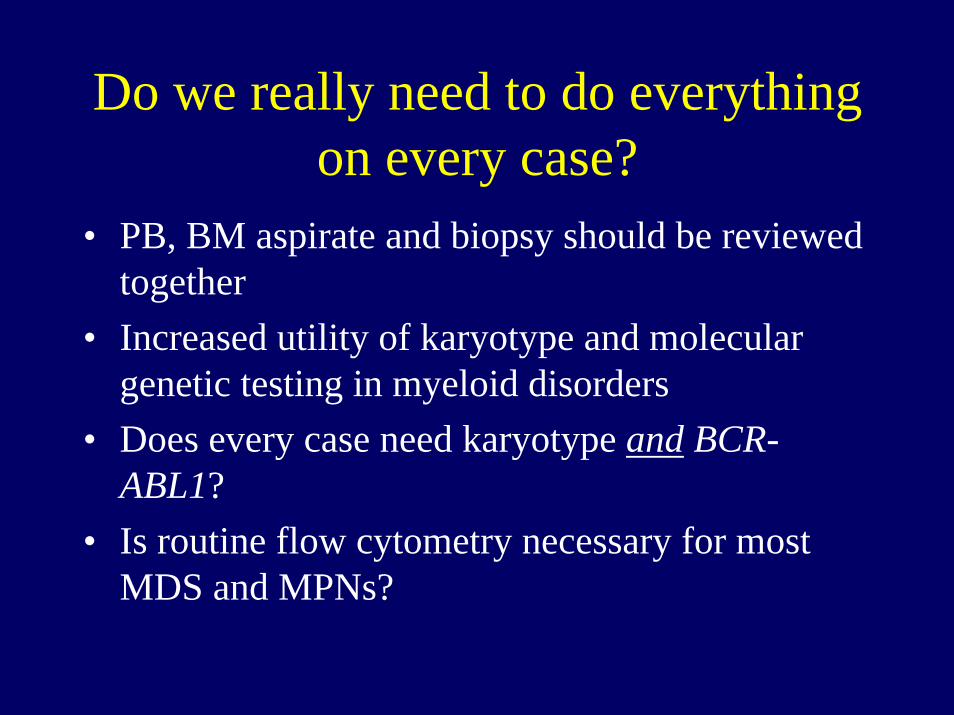
Do we really need to do everything on every case?
• PB, BM aspirate and biopsy should be reviewed together
• Increased utility of karyotype and molecular genetic testing in myeloid disorders
• Does every case need karyotype and BCR-ABL1?
• Is routine flow cytometry necessary for most MDS and MPNs?

Do we really need to do everything on every case?
• PB, BM aspirate and biopsy should be reviewed together
• Increased utility of karyotype and molecular genetic testing in myeloid disorders
• Does every case need karyotype and BCR-ABL1?• Is routine flow cytometry necessary for most MDS and
MPNs?• When do you perform both immunohistochemistry and
flow cytometry immunophenotyping?

• “In life there are drivers and passengers….”

• “In life there are drivers and passengers. Everyone else is just roadkill.”


