Interactions Crucial for Three-Dimensional Domain Swapping in the HP-RNase Variant PM8 Pere Tubert, † Douglas V. Laurents, ‡ Marc Ribo ´, † Marta Bruix, ‡ Maria Vilanova, † * and Antoni Benito † * † Laboratori d’Enginyeria de Proteı ¨nes, Departament de Biologia, Facultat de Cie ` ncies, Universitat de Girona, Campus de Montilivi, Maria Aure `lia Campmany, 69 E-17071 Girona, Spain and Institut d’Investigacio ´ Biome ` dica de Girona (IdIBGi), Girona, Spain; and ‡ Instituto de Quı ´mica Fı ´sica ‘‘Rocasolano’’, Consejo Superior de Investigaciones Cientı ´ficas, Serrano 119, E-28006 Madrid, Spain ABSTRACT The structural determinants that are responsible for the formation of higher order associations of folded proteins remain unknown. We have investigated the role on the dimerization process of different residues of a domain-swapped dimer human pancreatic ribonuclease variant. This variant is a good model to study the dimerization and swapping processes because dimer and monomer forms interconvert, are easily isolated, and only one dimeric species is produced. Thus, simple models for the swapping process can be proposed. The dimerization (dissociation constant) and swapping propensity have been studied using different variants with changes in residues that belong to different putative molecular determinants of dimerization. Using NMR spectroscopy, we show that these mutations do not substantially alter the overall conformation and flexibility, but affect the residue level stability. Overall, the most critical residues for the swapping process are those of one subunit that interact with the hinge loop of another one-subunit residue, stabilizing it in a conformation that favors the interchange. Tyr 25 , Gln 101 , and Pro 19 , with Asn 17 , Ser 21 , and Ser 23 , are found to be the most significant; notably, Glu 103 and Arg 104 , which were postulated to form salt bridges that would stabilize the dimer, are not critical for dimerization. INTRODUCTION ‘‘Three-dimensional domain swapping’’ is a term used to describe the process that usually leads to the formation of an intertwined oligomer wherein two or more identical protein chains exchange the same structural element that is usually referred to as a ‘‘domain’’ (1). The swapped domain may correspond to an entire tertiary globular domain or simply to a single element of secondary structure (2) and usually is at the N- or C-terminus, although an internal domain has been described in at least one case (3). The exchangeable domain is usually appended to a flex- ible hinge region or loop and this hinge can swing to allow the swapped region to pack correctly in both the intramolec- ular and intermolecular structures. The resulting oligomers are composed of subunits that have the same structure as the original monomer, with the exception of this hinge loop. The closed interface is the intramolecular interface found in the monomer structure that is recreated by two polypeptide chains in the domain-swapped structure. This interface consti- tutes the main adhesive force allowing the domain swapped oligomer to form. In a domain-swapped dimer, a new inter- face, the open interface, is formed when nonswapped regions of the subunits contact each other. When this interface is present, any favorable interactions found there stabilize the oligomeric state and compensate for the loss of entropy which is probably the main factor opposing oligomerization (4,5). Different members of the pancreatic ribonuclease (RNase) family provide interesting models for the study of the transition from a monomer to a three-dimensional domain-swapped dimer. Bovine seminal RNase (BS-RNase) exists in nature as a dimer. The two subunits are crosslinked by two disulfide bonds between Cys 31 of each chain and Cys 32 of the other one (6). The dimer has the unique feature that it consists of an equilibrium mixture of two functionally active forms: one of which has swapped N-terminal domains (the M M form) whereas the second one is not swapped (M ¼ M) (7). Unlike BS-RNase, other RNases, such as bovine pancre- atic RNase (RNase A) and PM8, a human pancreatic RNase (HP-RNase) variant (8), are generally found as monomers, but they are able to form noncovalent domain swapped dimers in vitro (9,10), and the latter forms some dimer in vivo (11). These proteins share between 70 and 80% sequence identity and can swap the homologous N-terminal helix, although the resulting relative orientation of the subunits is different (for a review, see (12)). In addition, RNase A is the only known protein that can alternatively interchange a domain located at the C-terminus (2). The crystal structure of PM8 is constituted by a new kind of N-terminal domain-swapped dimer (10). The oligomeric structure was unexpected because, in solution, most PM8 molecules exist as monomers. Nevertheless, the presence of a few dimers and oligomers was confirmed by nondenaturing electrophoresis (10) and by size-exclusion chromatography in the presence of 20% ethanol (13). Thus, the equilibrium between the monomer and dimer is displaced toward the monomer in aqueous solutions. The analysis of the structure indicated that the electrostatic interactions found along the open interface of the PM8 dimer were too weak to stabilize it in aqueous solution even at the highest protein concentra- tion. These interactions could be more favored in the crystal Submitted May 17, 2011, and accepted for publication June 13, 2011. *Correspondence: [email protected]or [email protected]Editor: Patrick Loria. Ó 2011 by the Biophysical Society 0006-3495/11/07/0459/9 $2.00 doi: 10.1016/j.bpj.2011.06.013 Biophysical Journal Volume 101 July 2011 459–467 459
Transcript
Biophysical Journal Volume 101 July 2011 459–467 459
Interactions Crucial for Three-Dimensional Domain Swappingin the HP-RNase Variant PM8
Pere Tubert,† Douglas V. Laurents,‡ Marc Ribo,† Marta Bruix,‡ Maria Vilanova,†* and Antoni Benito†*†Laboratori d’Enginyeria de Proteınes, Departament de Biologia, Facultat de Ciencies, Universitat de Girona, Campus de Montilivi, MariaAurelia Campmany, 69 E-17071 Girona, Spain and Institut d’Investigacio Biomedica de Girona (IdIBGi), Girona, Spain; and ‡Instituto deQuımica Fısica ‘‘Rocasolano’’, Consejo Superior de Investigaciones Cientıficas, Serrano 119, E-28006 Madrid, Spain
ABSTRACT The structural determinants that are responsible for the formation of higher order associations of folded proteinsremain unknown. We have investigated the role on the dimerization process of different residues of a domain-swapped dimerhuman pancreatic ribonuclease variant. This variant is a good model to study the dimerization and swapping processes becausedimer and monomer forms interconvert, are easily isolated, and only one dimeric species is produced. Thus, simple models forthe swapping process can be proposed. The dimerization (dissociation constant) and swapping propensity have been studiedusing different variants with changes in residues that belong to different putative molecular determinants of dimerization. UsingNMR spectroscopy, we show that these mutations do not substantially alter the overall conformation and flexibility, but affect theresidue level stability. Overall, the most critical residues for the swapping process are those of one subunit that interact with thehinge loop of another one-subunit residue, stabilizing it in a conformation that favors the interchange. Tyr25, Gln101, and Pro19,with Asn17, Ser21, and Ser23, are found to be the most significant; notably, Glu103 and Arg104, which were postulated to form saltbridges that would stabilize the dimer, are not critical for dimerization.
INTRODUCTION
‘‘Three-dimensional domain swapping’’ is a term used todescribe the process that usually leads to the formation ofan intertwined oligomer wherein two or more identicalprotein chains exchange the same structural element thatis usually referred to as a ‘‘domain’’ (1). The swappeddomain may correspond to an entire tertiary globulardomain or simply to a single element of secondary structure(2) and usually is at the N- or C-terminus, although aninternal domain has been described in at least one case(3). The exchangeable domain is usually appended to a flex-ible hinge region or loop and this hinge can swing to allowthe swapped region to pack correctly in both the intramolec-ular and intermolecular structures. The resulting oligomersare composed of subunits that have the same structure asthe original monomer, with the exception of this hinge loop.
The closed interface is the intramolecular interface foundin the monomer structure that is recreated by two polypeptidechains in the domain-swapped structure. This interface consti-tutes the main adhesive force allowing the domain swappedoligomer to form. In a domain-swapped dimer, a new inter-face, the open interface, is formed when nonswapped regionsof the subunits contact each other. When this interface ispresent, any favorable interactions found there stabilize theoligomeric state and compensate for the loss of entropy whichis probably the main factor opposing oligomerization (4,5).
Different members of the pancreatic ribonuclease(RNase) family provide interesting models for the study ofthe transition from a monomer to a three-dimensional
Submitted May 17, 2011, and accepted for publication June 13, 2011.
domain-swapped dimer. Bovine seminal RNase (BS-RNase)exists in nature as a dimer. The two subunits are crosslinkedby two disulfide bonds between Cys31 of each chain andCys32 of the other one (6). The dimer has the unique featurethat it consists of an equilibrium mixture of two functionallyactive forms: one of which has swapped N-terminal domains(the M � M form) whereas the second one is not swapped(M ¼ M) (7).
Unlike BS-RNase, other RNases, such as bovine pancre-atic RNase (RNase A) and PM8, a human pancreatic RNase(HP-RNase) variant (8), are generally found as monomers,but they are able to form noncovalent domain swappeddimers in vitro (9,10), and the latter forms some dimerin vivo (11). These proteins share between 70 and 80%sequence identity and can swap the homologous N-terminalhelix, although the resulting relative orientation of thesubunits is different (for a review, see (12)). In addition,RNase A is the only known protein that can alternativelyinterchange a domain located at the C-terminus (2).
The crystal structure of PM8 is constituted by a newkind ofN-terminal domain-swapped dimer (10). The oligomericstructure was unexpected because, in solution, most PM8molecules exist as monomers. Nevertheless, the presence ofa few dimers and oligomers was confirmed by nondenaturingelectrophoresis (10) and by size-exclusion chromatographyin the presence of 20% ethanol (13). Thus, the equilibriumbetween the monomer and dimer is displaced toward themonomer in aqueous solutions. The analysis of the structureindicated that the electrostatic interactions found along theopen interface of the PM8 dimer were too weak to stabilizeit in aqueous solution even at the highest protein concentra-tion. These interactions could be more favored in the crystal
due to the low dielectric constant of the precipitant solutionand the high effective protein concentration.
We have proposed amodel to explain the domain swappingprocess of PM8 (12,13). This model involves two steps: First,two monomers of PM8, which have a highly dynamic hingeloop, interact to produce an open interface. Second, afterthe formation of this new interface, additional residues ofone monomer would interact with the hinge loop of the othermonomer and stabilize it in an ordered conformation. (Thisconformational change would fix the highly dynamic stretchin a conformation that would promote the domain-swapping,and therefore, relative positions of the two subunits in thedimer would prepare the molecule for the interchange.)
In this work, we have characterized the molecular deter-minants contributing to three-dimensional domain swappingin this HP-RNase variant. Our results show that residuesGln101 and Tyr25 are very critical for the dimerization andthe swapping process. In contrast, we show that Glu103
and Arg104, which were postulated to form salt bridgesthat would stabilize the dimer, do not do so. Mutationalanalysis of the polar residues Asn17, Ser21, and Ser23 ofthe hinge loop reveals that their intermolecular hydrogenbonds contribute to domain swapping by modifying thedynamic behavior of the hinge loop.
MATERIALS AND METHODS
Construction of variants of PM8 and PM8E103C
Construction of PM8 and PM8E103C has been previously described in
Canals et al. (10) and Rodrıguez et al. (13). PM8 is an HP-RNase variant
carrying five substitutions at the N-terminus: Arg4Ala, Lys6Ala, Gln9Glu,
and Asp16Gly, and Ser17Asn together with the replacement Pro101Gln.
PM8E103C is a variant of PM8 carrying the substitution Glu103Cys and
has been used to create covalent dimers of PM8 (13). Variants of PM8
and PM8E103C were constructed using the QuikChange kit (Stratagene,
La Jolla, CA) following the manufacturer’s instructions.
Ribonuclease expression and purification
HP-RNase variants were produced and purified essentially as described in
Ribo et al. (14). The molecular mass of each variant was confirmed by
matrix-assisted laser desorption/ionization-time of flight (MALDI-TOF)
mass spectrometry using Bruker-Biflex (Bruker BioSpin, Billerica, MA)
equipment in the Biocomputation and Protein Sequencing Facility of the
Institut de Biotecnologia i Biomedicina of the Universitat Autonoma de
Barcelona (Spain). In some cases, MALDI-TOF was also used to identify
a glutathione moiety, probably to Cys103.
The protein concentration of the variants was determined by UV spectros-
copy using the extinction coefficients of 7,950 M�1 cm�1 and 16,025 M�1
cm�1 for monomers and covalent dimers, respectively, except for
PM8_Y25A and PM8E103C_Y25A. For the latter variants, the extinction coef-
ficients, 6,460 M�1 cm�1 and 13,045 M�1 cm�1 for monomers and covalent
dimers, respectively,were calculated according to themethodof Pace et al. (15).
Production of dimeric PM8E103C variants
Purified monomeric PM8E103C variants presented one moiety of gluta-
thione bound to Cys103. Each protein was dissolved in one ml of
Biophysical Journal 101(2) 459–467
100 mM Tris/acetate, 1.7 mM DTT, pH 8.5 (final monomer concentration
2.5 mg/ml) and incubated for 30 min at 25�C. In these conditions, only
the intermolecular disulfide bond with glutathione is reduced. The dimer
was then purified by size exclusion chromatography at a flow of 0.4 ml/min
using a G75 HR 10/30 column (Amersham Biosciences, Piscataway, NJ)
equilibrated with 200 mM sodium acetate pH 5.0 as described in Rodrıguez
et al. (13).
Production of labeled PM8E103C variantsfor NMR studies
The double uniformly labeled 13C/15N PM8E103C variant was produced
following the protocol previously described. Escherichia coli BL21(DE3)
cells were grown in a M9 minimal medium with 15NH4Cl (1 g/L) and13C6-glucose (4 g/L) (Cambridge Isotope Labs, Andover, MA) as sole
nitrogen and carbon sources, respectively. Protein purification was achieved
as described above for the nonlabeled variants. The purity of the protein
samples was confirmed by MALDI_TOF analysis. Following a similar
protocol using a M9 minimal medium with 15NH4Cl (1 g/L) as sole
nitrogen source (Cambridge Isotope Labs), 15N-labeled samples of
PM8E103C_H80S and PM8E103C_3A variants were also produced.
Labeled PM8E103C dimer was obtained as described above for the nonla-
beled variants.
Assessment of the extentof the domain-swapping
The degree of N-terminal domain swapping was investigated in crosslink-
ing experiments with divinyl sulfone (DVS) using the protocol described by
Ciglic et al. (16). Briefly, PM8E103C (14 mg, 1 nM of subunit) in 100 mM
sodium acetate, pH 5.0 (100 mL) and DVS (1 mL 10% solution in ethanol,
1 mM) were incubated at 30�C. This is an ~1000-fold excess of sulfone per
subunit of the protein. Aliquots were withdrawn over a period of 150–250 h
depending on the variant, and the reaction was quenched by adding 2-mer-
captoethanol (final concentration 200 mM) and incubating for 15–30 min at
room temperature. The samples were analyzed by reducing sodium dodecyl
sulfate-polyacrylamide gel electrophoresis (SDS-PAGE) and bands were
revealed by Coomassie Blue staining, and quantified by densitometry using
the Quantity One software (Bio-Rad Laboratories, Hercules, CA).
NMR spectroscopy
Typically, NMR samples contained up to 0.5 mM of protein and were
prepared in 90% H2O/10% D2O and D2O at pH 4.5. All NMR spectra
were recorded on a model No. AV-800 spectrometer (Bruker BioSpin)
equipped with a cryoprobe at 25�C. For labeled 13C/15N PM8E103C mono-
mer, assignment of 1H, 13C, and 15N resonances was achieved using
a standard suite of heteronuclear two-dimensional and triple resonance
three-dimensional spectra: 1H-15N-heteronuclear single quantum coherence
and 1H-15N-HSQC-nuclear Overhauser enhancement spectroscopy (NO-
ESY) spectra. The spectra of the dimeric form of PM8E103C were assigned
by comparison with the corresponding monomer on the bases of three-
dimensional 1H-15N-HSQC-NOESY experiment recorded in the same
conditions.
NMR relaxation experiments were carried out at the same conditions
described above. Conventional 15N heteronuclear nuclear Overhauser effect
(NOE) data were determined for the PM8E103C, PM8E103C_H80S, and
3D Domain Swapping in Human Ribonuclease 461
PM8E103C_3A monomeric variants. The experiments with and without
proton saturation were acquired simultaneously in an interleaved manner
with a recycling delay of 5 s, and were split during processing into separate
spectra for analysis. The values for the heteronuclear NOEs were obtained
from the ratio intensities of the resonances according to Isat/Iref. The uncer-
tainty was estimated to be ~5%.
Finally, NMR was also used to study the conformational stability on the
residue level by hydrogen-deuterium exchange experiments with
PM8E103C, PM8E103C_3A, and PM8E103C_H80S variants. The
exchange of amide protons with solvent deuterons was started by dissolving
lyophilized, protonated 15N variant samples into deuterated solvent. The
temperature and pH* values of the exchange experiments were the same
as those used in the assignment process (see above). The hydrogen
exchange rates were determined by integrating the volume of 1H-15N amide
crosspeaks in a series of heteronuclear single quantum coherence (HSQC)
spectra recorded consecutively after a dead time of ~30 min. A single-expo-
nential decay function was fit to the data to determine the observed
exchange rate, kex. The intrinsic exchange rates for unprotected HN groups,
krc, were calculated using the parameters reported by Bai et al. (17)
corrected for pH* 4.5 and temperature. Using equations described therein,
the protection factors, i.e., the ratio of the intrinsic and observed exchange
rates, of the individual groups were determined. When exchange is
governed by the EX2 regime, as commonly occurs for RNases under similar
conditions of temperature and pH (18,19), the conformational stability,
DGHX, of each amide group can be calculated from the protection factor
DGHX ¼ �RT lnðkex=krcÞ ¼ �RT ln Kop
¼ �RT lnð1=PFÞ;where Kop is the local or global equilibrium constant for the opening reac-
tion and PF the protection factor. Spectra were processed with Topspin
(Bruker BioSpin, Karlsruhe, Germany) and analyzed and integrated with
SPARKY (T. D. Goddard and D. G. Kneller, SPARKY, University of Cal-
ifornia at San Francisco, San Francisco, CA).
Kinetics of dimerization of PM8 variantsand Kd calculations
Monomeric PM8 variants were incubated at 29�C in 50 mM MOPS
(3-(n-morpholino)propanesulfonic acid), 50 mM NaCl, and 20% ethanol,
pH 6.7 at concentrations ranging from 0.1 to 1.5 mM. After 100 h of incu-
bation, aliquots were withdrawn, and the mixtures were immediately chro-
matographed at a flow rate of 0.4 ml/min on a Sephadex G75 HR 10/30
column (Pharmacia, GE Lifesciences, Waukesha, WI). Because no protein
aggregation or higher oligomers were observed in these experiments, the
concentrations of monomer and dimer could be quantified by integrating
their peaks. Only when the samples reached equilibrium were they were
used for Kd calculations. Given the equilibrium M þ M 4 D, the Kd can
be calculated from the slope of a plot of M2 concentration versus D concen-
tration (13,20). The dimer was rechromatographed immediately after the
recovery of the peak at 72 h later, and the amount of RNase reeluted as
a dimer was of 100% and 70%, respectively (not shown). This indicated
that the exchange between dimer and monomer was slow on the timescale
of the chromatography.
Determination of thermal stability
The conformational stability of the different variants was determined by
circular dichroism (CD). CD spectra were recorded using a J-810 spectro-
polarimeter (JASCO, Oklahoma City, OK) equipped with a thermostated
cell holder. The protein was dissolved at 20 mM in 100 mM KH2PO4
10% D2O. A quartz cell of 0.2-cm optical pathlength was used to record
the molar ellipticity (Em) at 218 nm. The temperature was raised from 20
to 85�C and the signal was recorded at 0.5�C. The solvent contribution
was subtracted using KaleidaGraph software (Synergy Software, Bangkok,
Thailand). Temperature-unfolding transitions curves were fitted to a two-
state thermodynamic model combined with sloping linear functions for
the native and denatured states, as described elsewhere (21).
RESULTS
Protein design
To characterize their role on the three-dimensional domainswapping and dimerization of PM8, we replaced by site-directed mutagenesis different residues of this RNase. Threekinds of variants were envisaged, as follows.
Variants to study the role of residues postulatedto be important for forming the initial open interface
In the crystal structure of PM8 (10), the open interface ismainly stabilized by two electrostatic interactions betweenresidues Arg104 and Glu103 of both subunits (Fig. 1 A). Totest the possibility that residues Arg104 and Glu103 wereimportant for the formation of the nascent nonswappedPM8 dimer, we constructed a PM8_E103S_R104S variantin which both charged residues were replaced by Ser. TheseSer residues are too far apart at the interface to establishinterchain hydrogen bonds, and we have not detected otheralternative interactions in the PM8 structure.
Variants to study the role in the dimerization and on thedomain swapping of residues postulated to interact with thehinge loop of the other subunit, stabilizing it in a conformationthat favors the interchange
In the structure of the PM8 dimer, the side chains of Gln101
and Tyr25 of each subunit create a cavity in which the Pro19
of the other subunit is sandwiched (Fig. 1 B), fixing thehinge loop in a conformation that extents to the othersubunit (10). In addition, Gln101 establishes three hydrogenbonds with the Ser20 residues from both subunits whichfurther stabilize the extended conformation of the hingeloop in the dimer. To study the role of these two residueson the dimerization and on the domain-swapping process,we constructed variants of PM8 in which Gln101 and Tyr25
were substituted by Ala (variants PM8_Q101A andPM8_Y25A, respectively).
Variants to study the role of the hinge loop stabilityon dimerization
We previously postulated that the stability of the hinge loopupon dimerization could be very important in determiningthe propensity of the dimer to swap (13). Analysis of thecrystal structure of dimeric PM8 (10) reveals the presenceof a hydrogen-bond network within the hinge loop thatstabilizes this stretch on a helix 310 conformation and thatis absent in a closely related monomeric variant (PM7(22)). We have studied the effect of removing specific side-chain-sidechain hydrogen bonds inside the hinge loop thatstabilize this stretch in a 310 helical conformation (Fig. 1 C),
Biophysical Journal 101(2) 459–467
FIGURE 1 Details of the molecular determi-
nants investigated in the PM8 dimer. (A) General
view of the dimeric structure of PM8. Residues
modified in this work are indicated in only one of
the two subunits. One of the two electrostatic inter-
actions found between Glu103 and Arg104 is also
indicated by minus and plus signs. (B) Detail of
the stacking between Pro19 (red) from one subunit
with Tyr25 and Gln101 (blue) from the other subunit
in the dimeric structure of PM8. The van der Waals
surfaces of these residues are shown as dotted
surfaces. (C) Stabilization of the hinge loop in
a helical conformation by multiple-centered
hydrogen bonds (discontinuous red lines)
belonging to the same chain residues. (D) General
view of the PM7 human pancreatic RNase mono-
meric variant (Protein DataBank accession code:
1DZA). The residues 1, 18–22, and 126–128
were not solved in the crystal structure and have
been modeled (22). The figure was drawn using
the software PyMol (DeLano Scientific, http://
www.pymol.sourceforge.net/).
462 Tubert et al.
which is present only in the dimer. We selected residuesAsn17, Ser21, and Ser23 to create variants PM8_3A.
Additionally, in an attempt to stabilize the hinge peptideof monomeric PM8, we took advantage of previous resultswith BS-RNase and RNase A to design a variant that couldpossess a less dynamic hinge loop. It has been described thatthe hinge loop of RNase A is mainly stabilized bya hydrogen bond established only in the monomer betweenSer18 and Ser80 and that the substitution of Ser80 by Argproduces a variant with a highly dynamic hinge peptide(23). Furthermore, it has been shown for hA-mBS (a mono-meric BS-RNase variant in which the hinge peptidesequence has been substituted by that of RNase A, togetherwith the replacement of Asn67 by Asp) that the substitutionof Arg80 by Ser reduces both the flexibility of the hingepeptide region and the swapping propensity from 70% to30% (24).
The decrease in swapping propensity was attributed to thegain of a stabilizing interaction between Ser80 and Ser18 inthe monomeric form (24) and to the loss of favorable inter-actions afforded by this positive charge in the dimer (25).We postulated that replacing His80 by Ser in PM8 couldallow the formation of a hydrogen bond between Ser18
Biophysical Journal 101(2) 459–467
and the incorporated Ser80, resulting in a stabilization ofthe hinge peptide in the monomer (Fig. 1 D). Consequently,we constructed a variant of PM8 in which His80 wassubstituted by Ser (variant PM8_H80S) to study the effectthat this mutation has on dimerization and domain swap-ping. Different replacements performed in each variantcan be found in Table S1 in the Supporting Material.
Assessment of the global fold
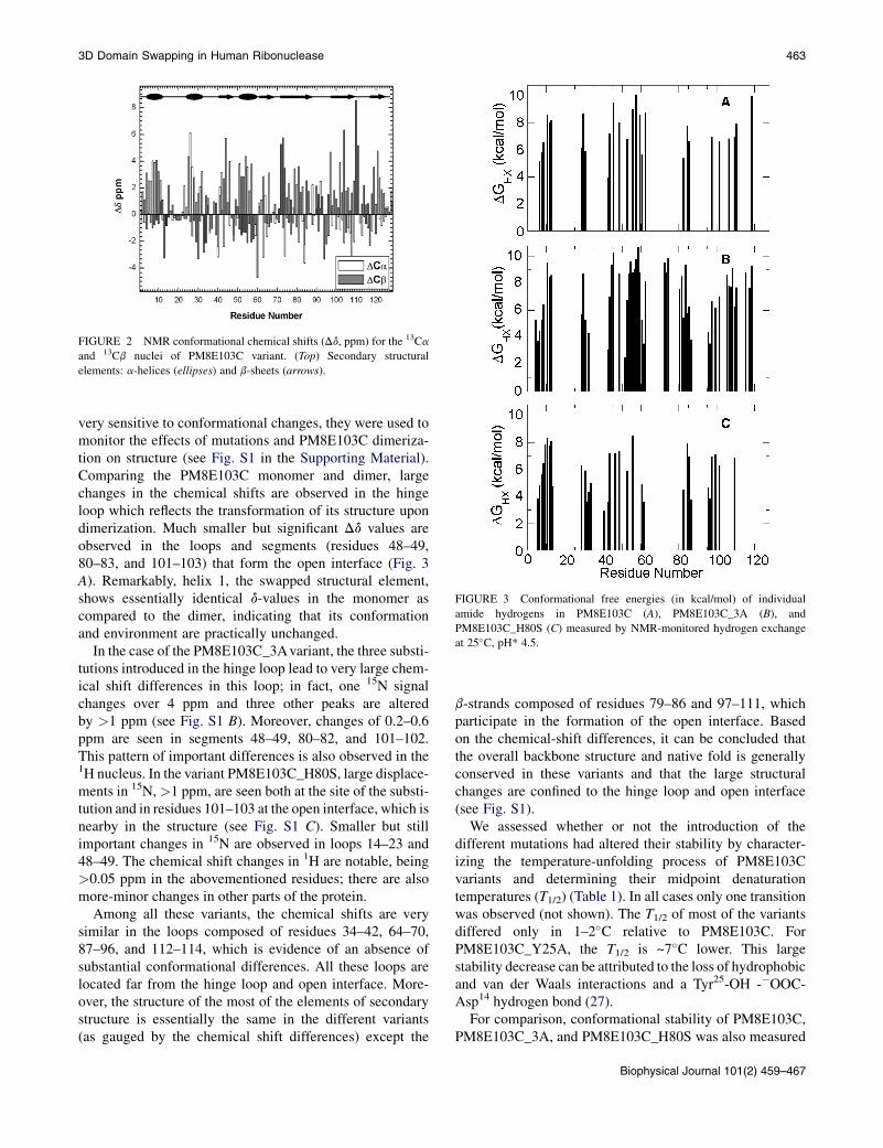
Before performing the dimerization studies, the structure ofsome variants used in this work was characterized by NMR.To begin, the 1H, 15N, and 13C NMR spectral resonances ofPM8E103C were assigned by following the three-dimen-sional strategy (26) and the conformational chemical shifts(Dd ¼ dexperimental � drandom coil) for Ca and Cb nuclei arerepresented in Fig. 2. The analysis of these data confirmsthat this variant is correctly folded and has the expectedsecondary structure.
The NMR spectra of the monomeric variants,PM8E103C_H80S and PM8E103C_3A, and of thePM8E103C dimer, were assigned by comparison with thoseof the monomeric PM8E103C. Because chemical shifts are
FIGURE 2 NMR conformational chemical shifts (Dd, ppm) for the 13Ca
and 13Cb nuclei of PM8E103C variant. (Top) Secondary structural
elements: a-helices (ellipses) and b-sheets (arrows).
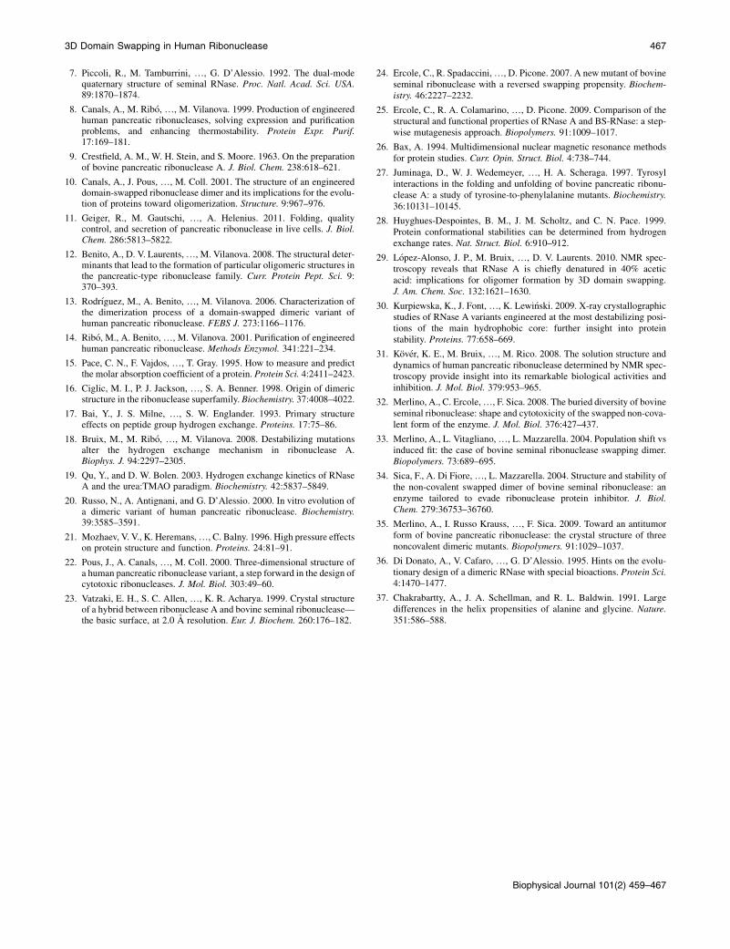
FIGURE 3 Conformational free energies (in kcal/mol) of individual
amide hydrogens in PM8E103C (A), PM8E103C_3A (B), and
PM8E103C_H80S (C) measured by NMR-monitored hydrogen exchange
at 25�C, pH* 4.5.
3D Domain Swapping in Human Ribonuclease 463
very sensitive to conformational changes, they were used tomonitor the effects of mutations and PM8E103C dimeriza-tion on structure (see Fig. S1 in the Supporting Material).Comparing the PM8E103C monomer and dimer, largechanges in the chemical shifts are observed in the hingeloop which reflects the transformation of its structure upondimerization. Much smaller but significant Dd values areobserved in the loops and segments (residues 48–49,80–83, and 101–103) that form the open interface (Fig. 3A). Remarkably, helix 1, the swapped structural element,shows essentially identical d-values in the monomer ascompared to the dimer, indicating that its conformationand environment are practically unchanged.
In the case of the PM8E103C_3Avariant, the three substi-tutions introduced in the hinge loop lead to very large chem-ical shift differences in this loop; in fact, one 15N signalchanges over 4 ppm and three other peaks are alteredby >1 ppm (see Fig. S1 B). Moreover, changes of 0.2–0.6ppm are seen in segments 48–49, 80–82, and 101–102.This pattern of important differences is also observed in the1H nucleus. In the variant PM8E103C_H80S, large displace-ments in 15N,>1 ppm, are seen both at the site of the substi-tution and in residues 101–103 at the open interface, which isnearby in the structure (see Fig. S1 C). Smaller but stillimportant changes in 15N are observed in loops 14–23 and48–49. The chemical shift changes in 1H are notable, being>0.05 ppm in the abovementioned residues; there are alsomore-minor changes in other parts of the protein.
Among all these variants, the chemical shifts are verysimilar in the loops composed of residues 34–42, 64–70,87–96, and 112–114, which is evidence of an absence ofsubstantial conformational differences. All these loops arelocated far from the hinge loop and open interface. More-over, the structure of the most of the elements of secondarystructure is essentially the same in the different variants(as gauged by the chemical shift differences) except the
b-strands composed of residues 79–86 and 97–111, whichparticipate in the formation of the open interface. Basedon the chemical-shift differences, it can be concluded thatthe overall backbone structure and native fold is generallyconserved in these variants and that the large structuralchanges are confined to the hinge loop and open interface(see Fig. S1).
We assessed whether or not the introduction of thedifferent mutations had altered their stability by character-izing the temperature-unfolding process of PM8E103Cvariants and determining their midpoint denaturationtemperatures (T1/2) (Table 1). In all cases only one transitionwas observed (not shown). The T1/2 of most of the variantsdiffered only in 1–2�C relative to PM8E103C. ForPM8E103C_Y25A, the T1/2 is ~7�C lower. This largestability decrease can be attributed to the loss of hydrophobicand van der Waals interactions and a Tyr25-OH -�OOC-Asp14 hydrogen bond (27).
For comparison, conformational stability of PM8E103C,PM8E103C_3A, and PM8E103C_H80S was also measured
Biophysical Journal 101(2) 459–467
TABLE 1 Biophysical characterization of the different HP-RNase variants analyzed
PM8_3A H-bonds in swapped hinge loop on dimerization. ND ND >104k ND
PM8E103C_3A H-bonds in hinge loop on swapping. 62.6 10.3 Covalent dimer 50%
PM8_Y25A Y25, P19 ring stacking on dimerization. ND ND >104k ND
PM8E103C_Y25A Y25, P19 ring stacking on swapping. 54.8 ND Covalent dimer 0%
PM8_Q101A H-bonds to S20, P19 packing on dimerization. ND ND >104k ND
PM8E103C_Q101A H-bonds to S20, P19 packing on swapping. 61.1 ND Covalent dimer 55%
PM8_H80S H-bond to hinge loop on dimerization. ND ND 0.50 ND
PM8E103C_H80S H-bond to hinge loop on swapping. 60.0 8.3 Covalent dimer 100%
PM8_E103S_R104S Salt bridges at open interface on dimerization. 60.0 ND 4.5 ND
ND, not determined.
*Values for monomeric forms calculated from temperature denaturation curves followed by CD. Experimental uncertainty: 0.1–0.4�C.yValues for monomeric forms calculated from hydrogen exchange experiments by NMR. Experimental uncertainty: 0.2 kcal/mol.zValues calculated from the slope of a plot of M2 concentration versus D concentration. Experimental uncertainty: 0.04 mM (H80S), 0.4–0.7 mM (other
variants).xValue calculated at pH 8.0. All the other Kd values were calculated at pH 5.0.{Data from Canals et al. (8).kKd limit calculated based on the minimal amount of dimer detectable during chromatographic analysis.
464 Tubert et al.
by hydrogen exchange (Fig. 3 and Table 1) (28). ForPM8E103C_3A, this value is 10.3 kcal/mol, slightly largerthan the value obtained for PM8E103C, 9.9 kcal/mol, whichis in line with the rank order of the stabilities defined bythermal denaturation. On the other hand, the internaldynamics as measured by the 15N-1H NOE shows that theNOE ratio, which gauges the backbone flexibility on thepicosecond-to-nanosecond timescale, is very similar tothe other variants as shown in Fig. S2.
For PM8E103C_H80S, the conformational stabilitymeasuredbyhydrogenexchange is slightly reduced (Table1).Interestingly, the decrease in the conformational stabilityshows variation on the level of individual residues (Fig. 3).All the secondary structural elements are less stable than inthe other variants, except for the first a-helix, which is themost stable element of structure in this variant. Takentogether, the data indicate that the mutations do not dramat-ically decrease the variant’s global stability.
Effects on the propensity to swap
The degree of swapping was investigated by crosslinkingHis12 and His119 with divinyl sulfone (DVS) (16), whichcrosslinks both of the active site His. If the active site ofthe dimer is composite, with His12 coming from one subunitand His119 coming from the other, the crosslink should cova-lently join the two subunits, which remain bound even underreducing conditions. Otherwise, crosslinking connects twoHis from the same subunit, yielding monomers underreducing conditions. Because the disposition of both Hisresidues is equal in monomeric and composite active sites,the reaction rates with DVS are equivalent in both cases.Determination of the swapping propensity of PM8 has thelimitation that the formation of the open interface and theprocess of swapping both occur simultaneously, which
Biophysical Journal 101(2) 459–467
makes it difficult to tweeze apart the interactions that governthese processes.
To overcome this limitation, we previously constructeda PM8 variant in which Glu103 was replaced by a Cys (13)to stabilize the open interface of the dimer by means of a di-sulfide bond (variant PM8E103C). This position was chosenbecause in the structure of the PM8 dimer (10) the Ca atomsof the two Glu103 side chains are facing each other (Fig. 1)and are separated by a distance similar to that foundbetween the equivalent atoms of two Cys in longer disulfidebonds. Because dissociation does not occur in this variantdimer, we could specifically study its degree of swapping.We took advantage of this variant to construct a new setof variants designed to investigate the role of the residuesdescribed above on the domain-swapping process. Variantswere PM8E103C_Y25A, in which Tyr25 was substituted byAla; PM8E103C_Q101A, in which Gln101 was substitutedby Ala; PM8E103C_3A, in which Asn17, Ser21, and Ser23
were substituted by Ala; and PM8E103C_H80S in whichHis80 was substituted by Ser.
Different incubation times with DVS were assayed tooptimize the reaction. After 150 h of incubation, theproportion of dimer and monomer in a reductive SDS-PAGE remained unchanged for all the variants, savePM8E103C_H80S (Fig. 4), indicating that the reactionwas completed. The proportion of swapped molecules atequilibrium is shown in Table 1. In agreement with previousresults (13), all the subunits of dimeric PM8E103C swappedthe N-terminal a-helix. For PM8E103C_Q101A, a cleardecrease of 50% relative to that of the parental variant inthe swapping propensity was observed. A more drasticeffect was observed for PM8E103C_Y25A in which noswapping could be detected. Interestingly, the swappingpropensity of PM8E103C_3A decreased to one-half thatof the parental variant and the replacement of His80 by
FIGURE 4 Degree of swapping in PM8E103C variants. PM8E103C
(shaded circles), PM8E103C_Q101A (solid circles), and PM8E103C_3A
(open circles). Percentages shown in Table 1 were obtained when the reac-
tion reached the equilibrium. (Inset) SDS-PAGE analysis of the DVS cross-
linking reaction for PM8E103C. Incubations times (h) are indicated over
each lane. Molecular mass markers correspond to 15, 20, 25, 37, and
50 kDa.
3D Domain Swapping in Human Ribonuclease 465
Ser affected neither the propensity of the resulting variant toswap its N-terminal domain (Table 1) or the backbone flex-ibility of the hinge loop (see Fig. S2).
Effects on the Kd of the dimers
We also sought to analyze which of the replacements intro-duced in PM8 altered the propensity of the resulting variantsto dimerize. The amount of dimer and monomer at equilib-rium was measured from the areas of the correspondingpeaks in molecular exchange chromatograms. For variantsPM8_Q101A, PM8_Y25A, and PM8_3A, no dimer couldbe detected even at the highest concentrations assayed(Table 1). Taking into account an extinction coefficient of13,045 M�1 cm�1 for PM8_Y25A, of 13,045 M�1 cm�1
for PM8_3A, and of 15,900 M�1 cm�1 for PM8_Q101Afor these variants, considering the highest protein concentra-tion used (1.5 mM) and that the lower detection threshold ofthe dimer peak in the HPLC is 1 mAU, the estimated lowerlimit on the Kd value is >36 M for PM8_Y25A andPM8_3A, and >29 M for PM8_Q101A.
These mutations produce a 103-fold increase of the Kd
compared to the parental variant (see Table 1), which corre-sponds to a free energy difference of roughly 5 kcal/mol.This value has been estimated at 29�C, the temperature atwhich the Kd values have been calculated. The Kd ofPM8_H80S is significantly lower compared to that ofPM8 (Table 1), thus the PM8_H80S dimer is more stable.Notably, the PM8 dimer is somewhat less stable at pH 8.0,where His80 is mainly neutral as compared to pH 5.0 whereHis80 is chiefly in the charged state (Table 1). This suggeststhat the stabilization of the dimer by the His80 to Ser substi-tution is not due to the removal of the positive charge ofHis80. Finally, we found that the replacement of Glu103
and Arg104 by Ser decreases the Kd of the resulting dimer(Table 1) (that is, it makes the dimer slightly more stable).
DISCUSSION
In contrast to RNase A which must unfold to attain
a domain-swapped dimer (29), the PM8 and BS-RNase
can interchange its N-terminal domains while maintaining
the native structure in solution. Site-directed mutagenesis
has allowed us to investigate the role of different residues
in both the swapping and dimerization of PM8. The analysis
of the structure, dynamics, and conformational stability of
the variants have provided evidence that the mutations intro-
duced in this work do not significantly affect the stability
and overall tertiary structure, although considerable effects
on the conformation of segments near the mutated site are
observed. These results are not surprising because the
tertiary structure is preserved even in more severely destabi-
lized RNase A variants in which structural changes are
limited to the neighborhood of the mutation site (18,30).Concerning the early events of the PM8 dimerization
when the swapping event has not still occurred, the relativepositions of the subunits would prepare the molecule for theswapping of the N-terminal domains. Because charge-charge interactions act over long distances, it was feasiblethat favorable electrostatic interactions between Glu103
and Arg104 from different monomers (that are also foundin the swapped dimer (Fig. 1 A)) aid the approach andcorrect orientation of the monomers during the earlieststages of dimerization. Nevertheless, here we have shownby substituting Glu103 and Arg104 by Ser that their electro-static interactions are not critical to form this initial dimer.Afterwards, the side chain of Arg104, which is mobile andsamples a wide variety of conformations in HP-RNasemonomer (31), would necessarily become fixed and loseconformational entropy to adopt the fixed, salt-bridgedstructure seen in the PM8 crystal structure. The observedKd value for the PM8_E103S_R104S variant may suggestthat this unfavorable loss of conformational entropy mayslightly outweigh the favorable contribution of the charge-charge interactions. It is possible that, in the inchoate non-covalent dimer, alternative open interfaces could formupon the removal of these charges residues from whichdomain-swapping could be produced. Accordingly, verydifferent open interfaces have been described for highlyrelated BS-RNase variants (32).
Regarding the role of residues Gln101 and Tyr25, ourresults show that hydrogen bonds established by Gln101
and, more crucially, the sandwiching of the Pro19 ringbetween the Gln101 side chain and the Tyr25 ring, play a vitalrole in the stabilization of the hinge loop in its swappedconformation and on the capacity of the molecule todimerize. Our results indicate that for PM8 these interac-tions are established by residues Gln101 and Tyr25, whichproduce a more fixed conformation of the hinge loop thatcould help to drive the process of swapping. These findingsare consistent with previous structural evidence (10,22) andresults from the noncovalent and covalent swapped dimers
Biophysical Journal 101(2) 459–467
466 Tubert et al.
of BS-RNase in which Pro19 is also clamped between theside chains of residues Tyr25 and Gln101 (33–35), suggestingthat this interaction may be also an important determinantfor BS-RNase domain swapping.
We previously postulated that the propensity of swappingcould depend on the relative stability of the hinge loopconformations in the monomer and dimers (12). For RNaseA, the dynamic behavior of the hinge loop is restrained bydifferent hydrogen bonds, but this is not the case for eitherBS-RNase or PM8. For RNase A, the restricted mobilityof the hinge loop could explain why more severely destabi-lizing conditions are needed to form RNase A N-dimers.The importance of the hinge loop stability differencesbetween monomeric and dimeric forms on swapping ishighlighted by the yield of swapped molecules of the cova-lent dimeric RNase variants CC-RNase A (RNase Acarrying Cys residues at position 31 and 32) (36), whereBS-RNase and PM8E103C is compared. The former variantforms only low amounts of swapped molecules (15%) (25).On the other hand, the BS-RNase and the PM8E103CHP-RNase dimers are 70% and ~100% of swapped, respec-tively. The difference of the swapping efficiency betweenthe human variant and BS-RNase has been attributed toa major stabilization of the hinge loop in the human variant(12). Therefore, we focused our interest to determine theeffect of replacing Ser17, Ser21, and Ser23 by Ala.In the case of PM8E103C_3A, removal of the polar resi-
dues that form intramolecular hydrogen bonds within thehinge loop in dimeric form of PM8 leads to a weaker dimerwith less swapping. Interestingly, the hydrogen exchangemeasurements reveal that the stability differences are notuniform along the sequence. The stability of the first a-helixis highest in the PM8E103C_3A variant, with respect toPM8E103C, which might reflect the introduction of threeAla residues in the nearby hinge loop as Ala has the highestintrinsic helix-forming propensity (37). This confirms thehypothesis that this network of hydrogen bonds stabilizesthe swapped, dimeric conformation of this loop (10). Thisvariant is more stable than the parental PM8 and it ispossible to advance the idea that this might be related toan increase of stability of the hinge loop in the monomericform. The Kd experiments also reinforce the importance ofthese positions together with Tyr25 and Gln101 on the dimer-ization process. In all these cases, the mutations producea 103-fold increase of the Kd compared to the parentalvariant, which corresponds roughly to a difference in freeenergy of 5 kcal/mol.
Regarding the role of His80, it was expected that theHis80Ser mutation might stabilize the monomer relative tothe dimer, and to increase the fraction of monomer. Surpris-ingly, our results show that the PM8_H80S dimer is signif-icantly stabilized, i.e., its Kd is lower. The comparison of thestability of individual amide hydrogens (Fig. 3) and back-bone dynamics (see Fig. S2) between the hinge loop ofmonomeric PM8E103C and PM8E103C_H80S indicates
Biophysical Journal 101(2) 459–467
that in this case the introduction of Ser80 does not alter thedynamic behavior of the hinge loop. We have also shownthat the increase of dimer stability cannot be attributed tothe removal of the positive charge of His that could be estab-lishing unfavorable interactions with residues of the othermonomer (i.e., repulsive charge-charge interactions). His80
protrudes somewhat from the open interface (Fig. 1 A)and it would be feasible that the removal of this residuecould produce a new and more stable open interface. Infact, for PM8E103C_H80S, the larger displacement of 15Nand 1H chemical shifts (see Fig. S1 C) were observed in resi-dues located at the open interface (Gln101 to Cys103)together with the site of the substitution, favoring thehypothesis of a local rearrangement of this region.
CONCLUSION
The results presented in this work indicate that the most crit-ical residues involved in the dimerization process of PM8are Tyr25 and Gln101 which interact with the Pro19 residueof the hinge loop of the other subunit. They also indicatethat different primary open interfaces can lead to the forma-tion of a swapped structure. Finally, it is derived that thepredisposition to swap of a protein domain can be modu-lated by changing the stability of the hinge loop structuresin the monomer or dimer.
Overall, this work gives clues for understanding theformation of oligomers in other proteins through three-dimensional domain swapping.
SUPPORTING MATERIAL
Two figures and one table are available at http://www.biophysj.org/
biophysj/supplemental/S0006-3495(11)00710-7.
P.T. acknowledges his fellowship from Ministerio de Educacion y Ciencia,
Spain. Work was supported by grants No. BFU2009-06935/BMC, No.
CTQ2010-21567-C02-02, and No. CTQ2008-00080/BQU from Ministerio
de Ciencia e Innovacion (Spain).
REFERENCES
1. Bennett, M. J., M. P. Schlunegger, and D. Eisenberg. 1995. 3D domainswapping: a mechanism for oligomer assembly. Protein Sci. 4:2455–2468.
2. Liu, Y., and D. Eisenberg. 2002. 3D domain swapping: as domainscontinue to swap. Protein Sci. 11:1285–1299.
3. Mizuno, H., Z. Fujimoto,., T. Morita. 1997. Structure of coagulationfactors IX/X-binding protein, a heterodimer of C-type lectin domains.Nat. Struct. Biol. 4:438–441.
4. Tamura, A., and P. L. Privalov. 1997. The entropy cost of protein asso-ciation. J. Mol. Biol. 273:1048–1060.
5. Erickson, H. P. 1989. Co-operativity in protein-protein association. Thestructure and stability of the actin filament. J. Mol. Biol. 206:465–474.
6. Mazzarella, L., S. Capasso, ., A. Zagari. 1993. Bovine seminal ribo-nuclease: structure at 1.9 A resolution. Acta Crystallogr. D Biol. Crys-tallogr. 49:389–402.
7. Piccoli, R., M. Tamburrini, ., G. D’Alessio. 1992. The dual-modequaternary structure of seminal RNase. Proc. Natl. Acad. Sci. USA.89:1870–1874.
8. Canals, A., M. Ribo, ., M. Vilanova. 1999. Production of engineeredhuman pancreatic ribonucleases, solving expression and purificationproblems, and enhancing thermostability. Protein Expr. Purif.17:169–181.
9. Crestfield, A. M., W. H. Stein, and S. Moore. 1963. On the preparationof bovine pancreatic ribonuclease A. J. Biol. Chem. 238:618–621.
10. Canals, A., J. Pous, ., M. Coll. 2001. The structure of an engineereddomain-swapped ribonuclease dimer and its implications for the evolu-tion of proteins toward oligomerization. Structure. 9:967–976.
11. Geiger, R., M. Gautschi, ., A. Helenius. 2011. Folding, qualitycontrol, and secretion of pancreatic ribonuclease in live cells. J. Biol.Chem. 286:5813–5822.
12. Benito, A., D. V. Laurents,., M. Vilanova. 2008. The structural deter-minants that lead to the formation of particular oligomeric structures inthe pancreatic-type ribonuclease family. Curr. Protein Pept. Sci. 9:370–393.
13. Rodrıguez, M., A. Benito, ., M. Vilanova. 2006. Characterization ofthe dimerization process of a domain-swapped dimeric variant ofhuman pancreatic ribonuclease. FEBS J. 273:1166–1176.
14. Ribo, M., A. Benito,., M. Vilanova. 2001. Purification of engineeredhuman pancreatic ribonuclease. Methods Enzymol. 341:221–234.
15. Pace, C. N., F. Vajdos, ., T. Gray. 1995. How to measure and predictthe molar absorption coefficient of a protein. Protein Sci. 4:2411–2423.
16. Ciglic, M. I., P. J. Jackson, ., S. A. Benner. 1998. Origin of dimericstructure in the ribonuclease superfamily. Biochemistry. 37:4008–4022.
17. Bai, Y., J. S. Milne, ., S. W. Englander. 1993. Primary structureeffects on peptide group hydrogen exchange. Proteins. 17:75–86.
18. Bruix, M., M. Ribo, ., M. Vilanova. 2008. Destabilizing mutationsalter the hydrogen exchange mechanism in ribonuclease A.Biophys. J. 94:2297–2305.
19. Qu, Y., and D. W. Bolen. 2003. Hydrogen exchange kinetics of RNaseA and the urea:TMAO paradigm. Biochemistry. 42:5837–5849.
20. Russo, N., A. Antignani, and G. D’Alessio. 2000. In vitro evolution ofa dimeric variant of human pancreatic ribonuclease. Biochemistry.39:3585–3591.
21. Mozhaev, V. V., K. Heremans,., C. Balny. 1996. High pressure effectson protein structure and function. Proteins. 24:81–91.
22. Pous, J., A. Canals, ., M. Coll. 2000. Three-dimensional structure ofa human pancreatic ribonuclease variant, a step forward in the design ofcytotoxic ribonucleases. J. Mol. Biol. 303:49–60.
23. Vatzaki, E. H., S. C. Allen, ., K. R. Acharya. 1999. Crystal structureof a hybrid between ribonuclease A and bovine seminal ribonuclease—the basic surface, at 2.0 A resolution. Eur. J. Biochem. 260:176–182.
24. Ercole, C., R. Spadaccini,., D. Picone. 2007. A new mutant of bovineseminal ribonuclease with a reversed swapping propensity. Biochem-istry. 46:2227–2232.
25. Ercole, C., R. A. Colamarino, ., D. Picone. 2009. Comparison of thestructural and functional properties of RNase A and BS-RNase: a step-wise mutagenesis approach. Biopolymers. 91:1009–1017.
26. Bax, A. 1994. Multidimensional nuclear magnetic resonance methodsfor protein studies. Curr. Opin. Struct. Biol. 4:738–744.
27. Juminaga, D., W. J. Wedemeyer, ., H. A. Scheraga. 1997. Tyrosylinteractions in the folding and unfolding of bovine pancreatic ribonu-clease A: a study of tyrosine-to-phenylalanine mutants. Biochemistry.36:10131–10145.
28. Huyghues-Despointes, B. M., J. M. Scholtz, and C. N. Pace. 1999.Protein conformational stabilities can be determined from hydrogenexchange rates. Nat. Struct. Biol. 6:910–912.
29. Lopez-Alonso, J. P., M. Bruix, ., D. V. Laurents. 2010. NMR spec-troscopy reveals that RNase A is chiefly denatured in 40% aceticacid: implications for oligomer formation by 3D domain swapping.J. Am. Chem. Soc. 132:1621–1630.
30. Kurpiewska, K., J. Font,., K. Lewi�nski. 2009. X-ray crystallographicstudies of RNase A variants engineered at the most destabilizing posi-tions of the main hydrophobic core: further insight into proteinstability. Proteins. 77:658–669.
31. Kover, K. E., M. Bruix, ., M. Rico. 2008. The solution structure anddynamics of human pancreatic ribonuclease determined by NMR spec-troscopy provide insight into its remarkable biological activities andinhibition. J. Mol. Biol. 379:953–965.
32. Merlino, A., C. Ercole,., F. Sica. 2008. The buried diversity of bovineseminal ribonuclease: shape and cytotoxicity of the swapped non-cova-lent form of the enzyme. J. Mol. Biol. 376:427–437.
33. Merlino, A., L. Vitagliano,., L. Mazzarella. 2004. Population shift vsinduced fit: the case of bovine seminal ribonuclease swapping dimer.Biopolymers. 73:689–695.
34. Sica, F., A. Di Fiore,., L. Mazzarella. 2004. Structure and stability ofthe non-covalent swapped dimer of bovine seminal ribonuclease: anenzyme tailored to evade ribonuclease protein inhibitor. J. Biol.Chem. 279:36753–36760.
35. Merlino, A., I. Russo Krauss, ., F. Sica. 2009. Toward an antitumorform of bovine pancreatic ribonuclease: the crystal structure of threenoncovalent dimeric mutants. Biopolymers. 91:1029–1037.
36. Di Donato, A., V. Cafaro, ., G. D’Alessio. 1995. Hints on the evolu-tionary design of a dimeric RNase with special bioactions. Protein Sci.4:1470–1477.
37. Chakrabartty, A., J. A. Schellman, and R. L. Baldwin. 1991. Largedifferences in the helix propensities of alanine and glycine. Nature.351:586–588.