Page 1
van Baal, J.O.A.M., Van de Vijver, K.K., Coffelt, S.B., van der Noort, V.,
van Driel, W.J., Kenter, G.G., Buist, M.R., and Lok, C.A.R. (2017)
Incidence of lymph node metastases in clinical early-stage mucinous and
seromucinous ovarian carcinoma: a retrospective cohort study. BJOG: An
International Journal of Obstetrics and Gynaecology, 124(3), pp. 486-494.
There may be differences between this version and the published version.
You are advised to consult the publisher’s version if you wish to cite from
it.
van Baal, J.O.A.M., Van de Vijver, K.K., Coffelt, S.B., van der Noort, V.,
van Driel, W.J., Kenter, G.G., Buist, M.R., and Lok, C.A.R. (2017)
Incidence of lymph node metastases in clinical early-stage mucinous and
seromucinous ovarian carcinoma: a retrospective cohort study. BJOG: An
International Journal of Obstetrics and Gynaecology, 124(3), pp. 486-
494. (doi:10.1111/1471-0528.14425) This article may be used for non-
commercial purposes in accordance with Wiley Terms and Conditions for
Self-Archiving.
http://eprints.gla.ac.uk/138089/
Deposited on: 09 March 2017
Page 2
Enlighten – Research publications by members of the University of Glasgow
http://eprints.gla.ac.uk
Page 3
INCIDENCE OF LYMPH NODE METASTASES IN CLINICAL EARLY STAGE LOW GRADE MU-
CINOUS OVARIAN CARCINOMA
J.O.A.M. van Baal1, K.K. Van de Vijver2, S.B. Coffelt3, V. van der Noort4, W.J. van Driel1, G.G.
Kenter1, M.R. Buist1, PALGA-group, C.A.R. Lok1
1 Department of Gynecologic Oncology, Centre for Gynecologic Oncology Amsterdam, The Neth-
erlands
2 Division of Diagnostic Oncology & Molecular Pathology, The Netherlands Cancer Institute, Antoni
van Leeuwenhoek Hospital, Amsterdam, The Netherlands
3 Division of Immunology, The Netherlands Cancer Institute, Antoni van Leeuwenhoek Hospital,
Amsterdam, The Netherlands
4 Department of Biometrics, The Netherlands Cancer Institute, Amsterdam, The Netherlands.
Abstract
Background: The use of lymph node sampling during staging procedures in clinical early stage
mucinous ovarian carcinoma (MOC) is an ongoing matter of debate. The incidence of lymph node
metastases in MOC in relation to tumor grade is unknown. If lymph node metastases in clinical
early stage G1 MOC would be non-existent, lymph node sampling might be safely omitted. We
aimed to determine the incidence of lymph node metastases in clinical early stage MOC per tumor
grade.
Materials & Methods: Histology report summaries from patients with MOC between 2002 and 2012
were obtained from the Dutch National Pathology Registry (PALGA). All reports were reviewed to
confirm diagnosis, tumor grade and presence of lymph node metastases. Clinical data, surgery
reports and radiology reports of patients with lymphadenopathy, were retrieved from hospital files.
Results: In the Netherlands, 915 patients with MOC were diagnosed and 426 underwent lymph
node sampling. The other 489 patients had either cytoreductive surgery or were staged without
lymph node sampling. In 7 patients, lymph node metastases were discovered by lymph node sam-
pling. In 4 of 190 (2.1%) G1 MOC patients, lymph node metastases were present, compared to 1
of 115 (0.9%) G2 MOC patients and 3 of 22 (13.6%) G3 MOC patients. Tumor grade was not spec-
ified in 99 patients. No recurrence-free survival benefit from lymph node sampling was observed in
patients with clinical early stage MOC.
Conclusion: These data indicate that lymph node sampling can be safely omitted in patients with
G1 and G2 MOC without clinical suspicion of metastases.
Page 4
Introduction
Epithelial ovarian cancer (EOC) is the cancer with the highest mortality of all gynecological
malignancies. In the Netherlands, each year approximately 1300 patients are diagnosed with this
disease. EOC is a term that encompasses serous, mucinous, seromucinous, endometrioid,
clearcell and undifferentiated adenocarcinomas. The majority of patients with EOC are diagnosed
with serous adenocarcinoma. Mucinous ovarian carcinoma (MOC) is a relatively rare subgroup of
these ovarian malignancies. Due to revised criteria for the diagnosis of MOC, its incidence has
even further declined over the past decades and is now estimated to be 3-5% of all EOC [1, 2].
Patients with MOC often present with a large unilateral ovarian mass without metastasized
disease. In these patients, prognosis is relatively good with a 5-year disease free survival of 90.8%
[3]. However, the course of advanced stage MOC is less favorable with fast progression and low
response rates to chemotherapy. Seromucinous carcinoma is a rare ovarian malignancy that has
been identified as a separate entity in the revised World Health Organization Classification of
Tumors of the Female Reproductive Organs [4]. This tumor type is characterized by an admixture
of serous, mucinous and endometrioid cell types. Moreover, patients with a seromucinous
carcinoma are primarily diagnosed with disease confined to the ovary. Therefore, their prognosis is
relatively good [5].
In patients with clinical early stage EOC, a complete staging procedure including lymph
node sampling is recommended to exclude the presence of microscopic metastases. During the
staging procedure, bilateral salpingo-oophorectomy, hysterectomy and (infracolic) omentectomy
are performed and peritoneal biopsies are taken. Furthermore, a lymph node sampling of at least
10 lymph nodes from the para-aortic and pelvic region is advised. In many patients with MOC, this
procedure is also performed; although, several studies have demonstrated a low incidence of
lymph node metastases of 0.0-6.7% [6, 7].
Apart from histological classification, EOC can be divided by tumor grade. Internationally,
the Silverberg/Shimizu criteria, by which the tumor is rated for dominating architectural pattern,
nuclear atypia and mitotic activity rate, are often used. However, a two-tier system dividing the
tumor into either low or high-grade tumors is preferred by more and more pathologists [8, 9]. These
systems are primarily developed for serous and endometrioid carcinomas, but are less applicable
to MOC because of their overall low-grade architectural appearance. No histological grading
system has been universally accepted for MOC. As a result, most pathologists use the three-tier
grading system for MOC, in the absence of a better alternative. Histological grading is important
because of its prognostic relation, but can also influence the choice of treatment. In several
histotypes of EOC, low-grade tumors have a more indolent course of disease with a favorable
progression-free and overall survival, compared to high-grade tumors [10]. It can be expected that
patients with low-grade tumors demonstrate a much lower incidence of lymph node metastases
compared to high-grade tumors. Indeed, Kleppe et al. demonstrated that 4.0% of serous and
endometrioid EOC patients with grade (G) 1 tumors and apparent FIGO stages I and II disease
Page 5
had lymph node metastases, compared to 20.0% in G3 tumors. [6]. In MOC, the correlation
between tumor grade and lymph node metastases is unknown. We hypothesized that lymph node
metastases in clinical early stage G1 MOC have a low incidence or are non-existent. The aim of
this study was to evaluate the need for a complete staging procedure, including lymph node
sampling in patients with clinical early stage MOC of different tumor grades.
Materials and methods
Patient selection
The Dutch Pathology Registry (PALGA) [11], a nationwide network and registry that records all
histopathology and cytopathology since 1991, was searched after approval of the privacy
committee of PALGA. All patients diagnosed with MOC between January 2002 and December
2012 were selected based on pathology reports comprising the terms: “ovary” OR “tube”, AND
“mucinous carcinoma” OR “mucinous adenocarcinoma” OR “mucin”. The pathology reports during
treatment of MOC, as well as all reports before and after treatment with a follow-up period of at
least 24 months, were obtained. A database of 18,465 pathology reports from a total of 1828
patients was built. For privacy reasons, all patients from the registry are itemized by a specific
PALGA-code. Under this code, excerpts containing anonymized pathology reports, dates of tissue
collection and age at time of tissue collection are registered. Researchers had no access to patient
names or private information. Therefore, no patient informed consent and no additional approval of
the Institutional Review Board was required.
All excerpts were initially scrutinized by the principal author (JVB). Ovarian tumors with a
histological diagnosis other than MOC, tumors with insufficient criteria of invasive malignancy (i.e.
borderline tumor, carcinoma in situ), ovarian metastasis of tumors from a different primary origin
and reports with inconclusive data were excluded (for exclusion criteria, see Figure 1). In case of
ambiguity concerning diagnosis, reports were discussed with an expert in gynecologic pathology
(KVV). FIGO stage was determined for each tumor. Of each case, the primary diagnosis and
official revision of histopathology were documented.
Seromucinous, endocervical-type mucinous and Müllerian mucinous carcinomas or mixed
cell types with a mixed endometrioid or serous and mucinous aspect were collectively grouped as
seromucinous carcinoma, according to the new WHO-guidelines [12].
Information including clinical data, surgery reports, radiology reports and follow-up data of
all patients with lymph node metastases was retrieved from the hospital files via an intermediate
procedure of PALGA. Anonymized clinical data and patient characteristics were requested from the
treating physicians to maintain absolute privacy of the patient. No histology was required for this
study. Therefore, no informed consent was needed to collect these additional clinical data.
Statistical analyses
The incidence rate of lymph node metastases in patients with MOC was calculated by dividing the
Page 6
patients with metastases by the total number of patients. Additionally, to gain insight into changes
in incidence of MOC over the study period, incidence rates per year and per 100,000 women were
calculated with the average yearly female population numbers in the Netherlands [13]. Recurrence-
free survival (RFS) was calculated for all patients that had recurrent disease, confirmed by
histological examination. Normally distributed data was described as mean values with standard
deviations. In case data was not normally distributed, median values and ranges (0-100%) were
reported. Statistical analyses were performed using IBM SPSS (Statistical Package for the Social
Sciences) version 22.0 (SPSS Inc., Chicago, Illinois). The two-sided Chi-square test was used for
categorical variables and the student’s t-test was used to evaluate differences in normally
distributed, continuous variables. Kaplan-Meier survival curves were generated for FIGO stage I
patients with and without lymph node sampling to determine RFS. Patients that were lost to follow-
up, were right-censored in the survival curves. Equality of RFS between these groups was
calculated with Log Rank (Mantel-Cox) tests.
Results
Mucinous ovarian cancer
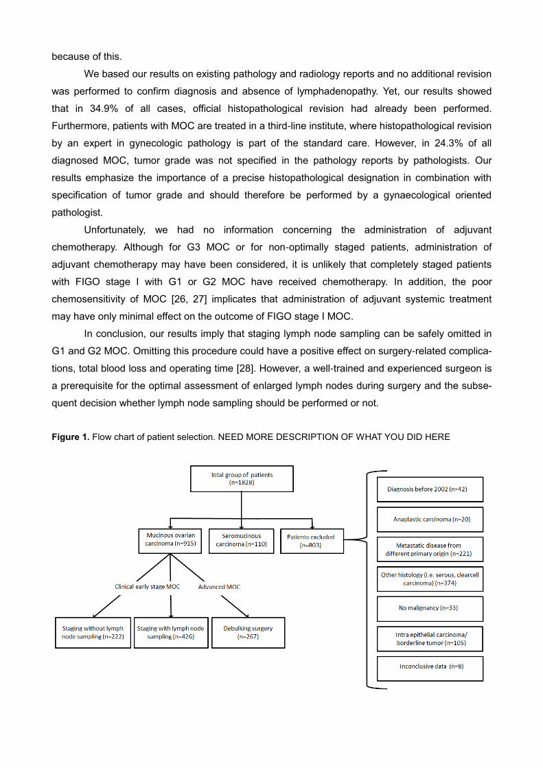
From a search of PALGA, the Dutch Pathology Registry, a total of 1828 patients with possible
MOC were identified between January 2002 and December 2012. After detailed examination of the
reports from the PALGA registry, 803 patients were excluded for reasons such as ovarian
metastases of a different primary origin or lack of malignancy, leaving 1025 patients eligible for our
study (Figure 1). Of these 1025, patients diagnosed with a primary MOC totalled 915 individuals
and 110 patients with a seromucinous carcinoma. Clinical characteristics of these patients with
MOC are shown in Table 1.
The majority of patients were diagnosed with a G1 or G2 MOC. In the total group of patients
with MOC, 17 (1.9%) patients were diagnosed with lymph node metastases. In 9 patients with
nodal disease, lymph node metastases were removed during cytoreductive surgery for advanced
disease or after histological biopsy of an enlarged lymph node. A significantly lower incidence of
lymph node metastases was seen in G1 MOC (1.4%) compared to G3 MOC (5.7%, p = 0.03). No
significant difference was seen between G1 and G2 MOC (1.4% vs. 2.6%, p = 0.35), or between
G2 and G3 MOC (p = 0.19) (Table 1). During the 11 years included in this study, the incidence
rates of MOC per 100,000 Dutch women per year declined (Figure 2).
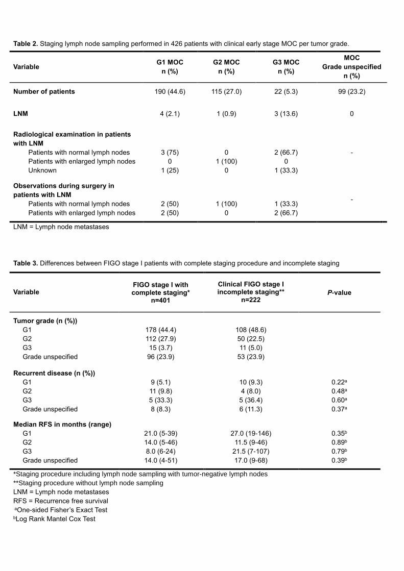
To understand whether tumor grade influences the chance of lymph node metastases in
clinical early stage MOC, we focussed on the patients who received staging procedures (Table 2).
Complete staging procedures including lymph node sampling, were performed in 426 patients,
revealing 8 patients with lymph nodes metastases. Patients with G1 and G2 disease showed
significantly less lymph node metastases compared to G3 MOC (G1 versus G3 p = 0.03; G2
versus G3 p = 0.01). These data indicate that lymph node metastases in clinical early stage MOC
of G1 and G2 disease are rare.
Page 7
To examine whether the patients with lymph node metastases (n=8) had apparent evidence
of lymphadenopathy on preoperative radiological imaging or during the staging procedures, we
examined the clinical data of these patients (Table 3). Interestingly, in 5 out of 8 (62.3%) of patients
with metastases identified during staging lymph node sampling, enlarged lymph nodes were
already present on radiological examination or enlarged by palpation during the staging
procedures. Thus, in patients with G1 MOC without clinical suspicion of metastatic disease, only 2
(95%CI 0.13-3.75%) patients had unexpected lymph node metastases. Patients with G2 MOC
without signs of clinical metastases had no lymph node metastases found with staging procedures
(95%CI 0-0.03%). However, 4.5% (95%CI 0.12-22.84%) of patients with G3 MOC, without pre-
operative evidence of metastatic disease in the lymph nodes were shown to have (microscopic)
lymphadenopathy.
Next, to evaluate whether tumor grade is correlated with RFS, Kaplan-Meier survival
analyses were performed of all FIGO stage I patients with MOC (Figure 3a). A more favorable RFS
was observed for patients with G1 and G2 MOC than for those with G3 MOC (p < 0.0001).
In 6.6% of patients with G1 MOC with FIGO stage I, recurrent disease was diagnosed after
a median of 16 months (range 5-39), whereas 11.0% of patients with G2 MOC and 33.3% of
patients with G3 developed recurrent disease after 15.5 and 8.0 months, respectively. RFS of
clinical FIGO stage I patients without lymph node sampling and patients who underwent staging
lymph node sampling were comparable for G1, G2 and G3 (Figure 3b). Table 3 demonstrates
numbers of patients with recurrent disease and RFS per tumor grade. Taken together, these results
demonstrate that performing lymph node sampling in the absence of clinical evidence of
metastases does not favor RFS.
Seromucinous ovarian cancer
Of the 1828 patients who presented with EOC between January 2002 and December 2012, 110
patients were diagnosed with a seromucinous (or endocervical-type mucinous) carcinoma (Figure
1). During this time, the incidence of seromucinous carcinoma was stable (Figure 2). The
characteristics of these patients are presented in Table 4. The mean age of this cohort was 56.1
years and there was no difference in age when the group was subdivided by grade. Most patients
(58.3%) were diagnosed with G1 disease. Among the entire seromucinous carcinoma cohort, 5
(4.5%) patients had lymph node metastases. Strikingly, none of these lymph node metastases
were found in patients with G1 seromucinous carcinoma. In 3 (60%) patients with
lymphadenopathy, axillar or supraclavicular lymph node metastases were found, which was not
found in the MOC patients. Staging procedures were performed in 46 out of 110 patients, but did
not reveal any additional lymph node metastases. Recurrent disease occurred in 25 patients
(22.7%) with seromucinous carcinoma. In patients with FIGO stage I disease with G1, G2 and G3
seromucinous tumors, recurrent disease occurred in 4 (12.9%), 3 (16.7%) and 2 (50%) patients,
respectively. In conclusion, lymph node metastases in G1 seromucinous carcinoma could not be
Page 8
found. However, the number of patients that received lymph node samplings in the present study is
too small to draw solid conclusions from these results.
Discussion
The current study shows that the incidence of lymph node metastases in patients with clinical early
stage G1 or G2 MOC is very low. In case of absence of enlarged lymph nodes on radiological
examination or on palpation during staging procedures, only 0.7% of the patients with G1 and G2
MOC together had lymph node metastases. In addition, no RFS benefit from lymph node sampling
was observed in patients with clinical FIGO stage I MOC.
This is the first study that reports lymph node metastases in MOC per tumor grade.
Previous studies demonstrated a low overall incidence of lymph node metastases in clinical early
stage MOC (0.0-6.7%) [6, 7]. Our study demonstrates that G3 MOC is associated with a higher
incidence of lymph node metastases discovered during staging procedures, compared to G1 and
G2 MOC. Staging lymph node samplings were performed in only 22 patients with G3 MOC.
However, our findings suggests that G3 MOC has another clinical behavior leading to a more
advanced stage disease at presentation and a higher incidence of lymph node metastases.
Patients with G1 and G2 MOC presented with a similar course of disease, with equal FIGO stages
at diagnosis, equal incidences of lymph node metastases and comparable RFS. This tumor grade
specific behavior can also be seen in other histotypes of EOC, such as low grade and high grade
serous carcinoma [14, 15]. Therefore, different tumor grades of MOC should not be regarded as
one group, as G1 and G2 MOC represent a different course of disease than G3 MOC. The
differences in clinical behavior may be explained by differences in genetic drivers. Several mutated
genes have been identified for MOC, including KRAS, BRAF, CDKN2A and TP53 genes [16-19].
Recently, a study of Ryland et al. investigated the variances of the genomic landscapes between
the tumor grades of MOC [16]. However, in this study, demonstrating evident differences between
the tumor grades was hindered by the heterogeneity of MOC and the small study populations.
Defining tumor grade in MOC remains a matter of debate amongst pathologists, as the
current classification system is suboptimal for MOC and a specific grading system does not exist. A
new classification system of grading MOC is needed to optimize separating patients with poor
prognosis from patients with more favorable prognosis. Our results implicate that possibly a two-
tier classification system should be developed in which G1, G2 and G3 MOC are subdivided into a
low grade and a high grade group. Identification of the molecular differences between tumor
grades of MOC will also create a valuable contribution to the distinction of the different prognostic
groups.
Lymph node sampling in early stage EOC has been the subject of debate for the past
years. Previously, some studies demonstrated a survival benefit for patients with early stage EOC
who received complete staging procedures [19, 20]. However, in these studies, different histotypes
of EOC are taken together and none of these studies investigated MOC with focus on its separate
Page 9
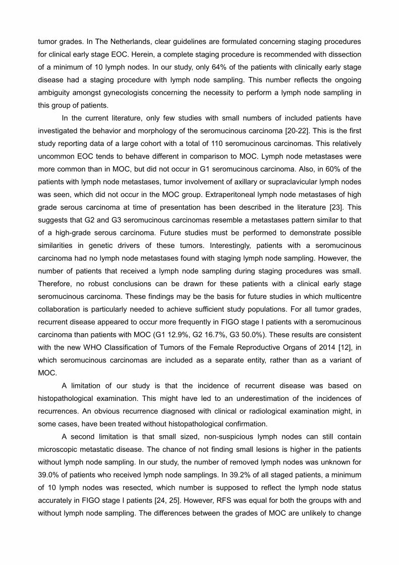
tumor grades. In The Netherlands, clear guidelines are formulated concerning staging procedures
for clinical early stage EOC. Herein, a complete staging procedure is recommended with dissection
of a minimum of 10 lymph nodes. In our study, only 64% of the patients with clinically early stage
disease had a staging procedure with lymph node sampling. This number reflects the ongoing
ambiguity amongst gynecologists concerning the necessity to perform a lymph node sampling in
this group of patients.
In the current literature, only few studies with small numbers of included patients have
investigated the behavior and morphology of the seromucinous carcinoma [20-22]. This is the first
study reporting data of a large cohort with a total of 110 seromucinous carcinomas. This relatively
uncommon EOC tends to behave different in comparison to MOC. Lymph node metastases were
more common than in MOC, but did not occur in G1 seromucinous carcinoma. Also, in 60% of the
patients with lymph node metastases, tumor involvement of axillary or supraclavicular lymph nodes
was seen, which did not occur in the MOC group. Extraperitoneal lymph node metastases of high
grade serous carcinoma at time of presentation has been described in the literature [23]. This
suggests that G2 and G3 seromucinous carcinomas resemble a metastases pattern similar to that
of a high-grade serous carcinoma. Future studies must be performed to demonstrate possible
similarities in genetic drivers of these tumors. Interestingly, patients with a seromucinous
carcinoma had no lymph node metastases found with staging lymph node sampling. However, the
number of patients that received a lymph node sampling during staging procedures was small.
Therefore, no robust conclusions can be drawn for these patients with a clinical early stage
seromucinous carcinoma. These findings may be the basis for future studies in which multicentre
collaboration is particularly needed to achieve sufficient study populations. For all tumor grades,
recurrent disease appeared to occur more frequently in FIGO stage I patients with a seromucinous
carcinoma than patients with MOC (G1 12.9%, G2 16.7%, G3 50.0%). These results are consistent
with the new WHO Classification of Tumors of the Female Reproductive Organs of 2014 [12], in
which seromucinous carcinomas are included as a separate entity, rather than as a variant of
MOC.
A limitation of our study is that the incidence of recurrent disease was based on
histopathological examination. This might have led to an underestimation of the incidences of
recurrences. An obvious recurrence diagnosed with clinical or radiological examination might, in
some cases, have been treated without histopathological confirmation.
A second limitation is that small sized, non-suspicious lymph nodes can still contain
microscopic metastatic disease. The chance of not finding small lesions is higher in the patients
without lymph node sampling. In our study, the number of removed lymph nodes was unknown for
39.0% of patients who received lymph node samplings. In 39.2% of all staged patients, a minimum
of 10 lymph nodes was resected, which number is supposed to reflect the lymph node status
accurately in FIGO stage I patients [24, 25]. However, RFS was equal for both the groups with and
without lymph node sampling. The differences between the grades of MOC are unlikely to change
Page 10
because of this.
We based our results on existing pathology and radiology reports and no additional revision
was performed to confirm diagnosis and absence of lymphadenopathy. Yet, our results showed
that in 34.9% of all cases, official histopathological revision had already been performed.
Furthermore, patients with MOC are treated in a third-line institute, where histopathological revision
by an expert in gynecologic pathology is part of the standard care. However, in 24.3% of all
diagnosed MOC, tumor grade was not specified in the pathology reports by pathologists. Our
results emphasize the importance of a precise histopathological designation in combination with
specification of tumor grade and should therefore be performed by a gynaecological oriented
pathologist.
Unfortunately, we had no information concerning the administration of adjuvant
chemotherapy. Although for G3 MOC or for non-optimally staged patients, administration of
adjuvant chemotherapy may have been considered, it is unlikely that completely staged patients
with FIGO stage I with G1 or G2 MOC have received chemotherapy. In addition, the poor
chemosensitivity of MOC [26, 27] implicates that administration of adjuvant systemic treatment
may have only minimal effect on the outcome of FIGO stage I MOC.
In conclusion, our results imply that staging lymph node sampling can be safely omitted in
G1 and G2 MOC. Omitting this procedure could have a positive effect on surgery-related complica-
tions, total blood loss and operating time [28]. However, a well-trained and experienced surgeon is
a prerequisite for the optimal assessment of enlarged lymph nodes during surgery and the subse-
quent decision whether lymph node sampling should be performed or not.
Figure 1. Flow chart of patient selection. NEED MORE DESCRIPTION OF WHAT YOU DID HERE
Page 11
Figure 2. Incidence of mucinous and seromucinous ovarian cancer in The Netherlands between January 2002 and December 2012.
Page 12
Table 1. Characteristics of total group of 915 patients with MOC per tumor grade.
Variable All MOC
G1 MOC G2 MOC G3 MOC Grade
unspecified P-value
n (%) 915 (100) 369 (40.3) 229 (25.0) 88 (9.6) 229 (25.0)
Mean age (yrs
(95%CI)) 55.7 (54.7-56.7) 54.0 (52.4-55.6) 55.4 (53.4-57.5) 56.8 (53.6-60.0) 58.1 (56.1-60.0) 0.02a
FIGO stage (n (%))
I
II
III
IV
Unknown
623
46
159
29
58
286 (77.5)
17 (4.6)
42 (11.4)
4 (1.1)
20 (5.4)
162 (70.7)
8 (3.5)
41 (17.9)
8 (3.5)
10 (4.4)
26 (29.5)
14 (15.9)
32 (36.4)
9 (10.2)
7 (8.0)
149 (65.1)
7 (3.1)
44 (19.2)
8 (3.5)
21 (9.2)
0.04b
Histopathological
revision (n (%)) 319 (34.9) 151 (40.9) 75 (32.8) 30 (34.1) 63 (27.5) 0.01c
Tumor
characteristics
(n (%))
Intestinal type
Infiltrative growth
Expansive growth
84 (9.2)
6 (0.7)
42 (4.6)
38 (10.3)
3 (0.8)
15 (4.1)
20 (8.7)
1 (0.4)
8 (3.5)
4 (4.5)
0 (0)
4 (4.5)
22 (9.6)
2 (0.9)
15 (6.6)
0.40c
0.09b
LNM (n (%))
Yes
No
Unknown
17 (1.9)
428 (46.8)
470 (51.4)
5 (1.4)
188 (50.9)
176 (47.7)
6 (2.6)
117 (51.1)
106 (46.3)
5 (5.7)
21 (23.9)
62 (70.5)
1 (0.4)
102 (44.5)
126 (55.0)
<0.001c
aOne-Way ANOVA test bLinear-by-Linear Association test cPearson Chi-Square test
LNM = Lymph node metastases
Page 13
Table 2. Staging lymph node sampling performed in 426 patients with clinical early stage MOC per tumor grade.
Variable G1 MOC
n (%)
G2 MOC
n (%)
G3 MOC
n (%)
MOC
Grade unspecified
n (%)
Number of patients 190 (44.6) 115 (27.0) 22 (5.3) 99 (23.2)
LNM 4 (2.1) 1 (0.9) 3 (13.6) 0
Radiological examination in patients
with LNM
Patients with normal lymph nodes
Patients with enlarged lymph nodes
Unknown
3 (75)
0
1 (25)
0
1 (100)
0
2 (66.7)
0
1 (33.3)
-
Observations during surgery in
patients with LNM
Patients with normal lymph nodes
Patients with enlarged lymph nodes
2 (50)
2 (50)
1 (100)
0
1 (33.3)
2 (66.7)
-
LNM = Lymph node metastases
Table 3. Differences between FIGO stage I patients with complete staging procedure and incomplete staging
Variable FIGO stage I with complete staging*
n=401
Clinical FIGO stage I incomplete staging**
n=222 P-value
Tumor grade (n (%))
G1
G2
G3
Grade unspecified
178 (44.4)
112 (27.9)
15 (3.7)
96 (23.9)
108 (48.6)
50 (22.5)
11 (5.0)
53 (23.9)
Recurrent disease (n (%))
G1
G2
G3
Grade unspecified
9 (5.1)
11 (9.8)
5 (33.3)
8 (8.3)
10 (9.3)
4 (8.0)
5 (36.4)
6 (11.3)
0.22a
0.48a
0.60a
0.37a
Median RFS in months (range)
G1
G2
G3
Grade unspecified
21.0 (5-39)
14.0 (5-46)
8.0 (6-24)
14.0 (4-51)
27.0 (19-146)
11.5 (9-46)
21.5 (7-107)
17.0 (9-68)
0.35b
0.89b
0.79b
0.39b
*Staging procedure including lymph node sampling with tumor-negative lymph nodes
**Staging procedure without lymph node sampling
LNM = Lymph node metastases
RFS = Recurrence free survival aOne-sided Fisher’s Exact Test bLog Rank Mantel Cox Test
Page 14
Table 4. Characteristics of 110 patients with seromucinous ovarian carcinoma per tumor grade
Variable
All
Seromucinous
carcinoma
Seromucinous
carcinoma
G1
Seromucinous
carcinoma
G2
Seromucinous
carcinoma
G3
Seromucinous
carcinoma
Grade
unspecified
P-value
n (%) 110 (100) 43 (39.1) 30 (27.3) 16 (14.5) 21 (19.1)
Mean age (yrs
(95%CI))
56.1 (53.5-
58.8)
58.3 (54.0-
62.5)
58.3 (53.4-
63.1)
51.6 (43.6-
59.6)
52.1 (45.4-
58.7) 0.17a
FIGO stage (n
(%))
I
II
III
IV
Unknown
66 (60.0)
13 (11.8)
21 (19.1)
7 (6.4)
3 (2.7)
31 (72.1)
4 (9.3)
4 (9.3)
2 (4.7)
2 (4.7)
18 (60.0)
5 (16.7)
5 (16.7)
2 (6.7)
0 (0)
4 (25.0)
2 (12.5)
7 (43.8)
3 (18.8)
0 (0)
13 (61.9)
2 (9.5)
5 (23.8)
0 (0)
1 (4.8)
0.56b
Histopathological
revision
(n (%))
38 (34.5) 21 (48.8) 8 (26.7) 4 (25.0) 5 (23.8) 0.09c
LNM (n (%))
Yes
No
Unknown
5 (4.5)
48 (43.6)
57 (51.8)
0 (0)
20 (46.5)
23 (53.5)
2 (6.7)
16 (53.3)
12 (40.0)
2 (12.5)
4 (25.0)
10 (62.5)
1 (4.8)
8 (38.1)
12 (57.1)
0.10c
Staging lymph
node sampling
(n (%))
46 (41.8) 19 (44.2) 16 (53.3) 3 (18.8) 8 (38.1)
Recurrent
disease (n (%)) 25 (22.7) 7 (16.3) 9 (30.0) 4 (25.0) 5 (23.8) 0.58c
aOne-Way ANOVA test bLinear-by-Linear Association test cPearson Chi-Square test
Page 15
Figure 4a. Kaplan Meijer curves of survival of all FIGO stage I patients with MOC per tumor grade.
Survival curves of all patients with clinically FIGO stage I MOC, with a favorable RFS for G1 MOC and a
poorer RFS for G3 MOC (Log Rank 18.30, p < 0.0001).
Figure 4b. Kaplan Meijer curves demonstrating RFS of patients with FIGO stage I disease, G1 and G2 MOC, with and without lymph node sampling.
Survival curves are demonstrated of patients with G1 and G2 combined, who received staging procedures
either with or without lymph node sampling. No survival benefit was observed in the patients who had lymph
node sampling (Log Rank 0.183, p = 0.67).
Page 16
References
1. Seidman, J.D., et al., The histologic type and stage distribution of ovarian carcinomas of
surface epithelial origin. Int J Gynecol Pathol, 2004. 23(1): p. 41-4.
2. Shimada, M., et al., Clinicopathological characteristics of mucinous adenocarcinoma of the
ovary. Gynecol Oncol, 2009. 113(3): p. 331-4.
3. Vergote, I., et al., Prognostic importance of degree of differentiation and cyst rupture in
stage I invasive epithelial ovarian carcinoma. Lancet, 2001. 357(9251): p. 176-82.
4. Kurman RJ, C.M., Herrington CS, et al, WHO Classification of Tumours of Female
Reproductive Organs. Lyon: International Agency for Research on Cancer, 2014.
5. Taylor, J. and W.G. McCluggage, Ovarian seromucinous carcinoma: report of a series of a
newly categorized and uncommon neoplasm. Am J Surg Pathol, 2015. 39(7): p. 983-92.
6. Kleppe, M., et al., Lymph node metastasis in stages I and II ovarian cancer: a review.
Gynecol Oncol, 2011. 123(3): p. 610-4.
7. Schmeler, K.M., et al., Prevalence of lymph node metastasis in primary mucinous carcinoma
of the ovary. Obstet Gynecol, 2010. 116(2 Pt 1): p. 269-73.
8. Anais Malpica, M., * Michael T.Deavers,MD,* Karen Lu,MD,† Diane
C.Bodurka,MD,†Edward N.Atkinson,MD,‡ David M.Gershenson,MD,† and Elvio
G.Silva,MD*, Grading Ovarian Serous Carcinoma Using a Two-Tier System. Am J Surg
Pathol, 2004.
9. Zeppernick, F. and I. Meinhold-Heerlein, The new FIGO staging system for ovarian,
fallopian tube, and primary peritoneal cancer. Arch Gynecol Obstet, 2014. 290(5): p. 839-
42.
10. Bodurka, D.C., et al., Reclassification of serous ovarian carcinoma by a 2-tier system: a
Gynecologic Oncology Group Study. Cancer, 2012. 118(12): p. 3087-94.
11. Casparie, M., et al., Pathology databanking and biobanking in The Netherlands, a central
role for PALGA, the nationwide histopathology and cytopathology data network and
archive. Cell Oncol, 2007. 29(1): p. 19-24.
12. Kurman, R.J., Carcangiu M.L., Herrington C.S., et al., WHO classification of tumours of
female reproductive organs. Lyon: IARC Press, 2014.
13. Statistics, Available at www.cbs.nl. Accessed March 2015
The Netherlands.
14. Chen, M., et al., A survival analysis comparing women with ovarian low-grade serous
carcinoma to those with high-grade histology. Onco Targets Ther, 2014. 7: p. 1891-9.
15. Groen, R.S., D.M. Gershenson, and A.N. Fader, Updates and emerging therapies for rare
epithelial ovarian cancers: One size no longer fits all. Gynecol Oncol, 2014.
16. Ryland, G.L., et al., Mutational landscape of mucinous ovarian carcinoma and its neoplastic
precursors. Genome Med, 2015. 7(1): p. 87.
17. Gemignani, M.L., et al., Role of KRAS and BRAF gene mutations in mucinous ovarian
carcinoma. Gynecol Oncol, 2003. 90(2): p. 378-81.
18. Anglesio, M.S., et al., Molecular characterization of mucinous ovarian tumours supports a
stratified treatment approach with HER2 targeting in 19% of carcinomas. J Pathol, 2013.
229(1): p. 111-20.
19. Mackenzie, R., et al., Targeted deep sequencing of mucinous ovarian tumors reveals
multiple overlapping RAS-pathway activating mutations in borderline and cancerous
neoplasms. BMC Cancer, 2015. 15: p. 415.
20. Taylor, J. and W.G. McCluggage, Ovarian Seromucinous Carcinoma: Report of a Series of a
Newly Categorized and Uncommon Neoplasm. Am J Surg Pathol, 2015.
21. Shappell, H.W., et al., Diagnostic criteria and behavior of ovarian seromucinous
(endocervical-type mucinous and mixed cell-type) tumors: atypical proliferative (borderline)
tumors, intraepithelial, microinvasive, and invasive carcinomas. Am J Surg Pathol, 2002.
26(12): p. 1529-41.
Page 17
22. Dube, V., et al., Mucinous ovarian tumors of Mullerian-type: an analysis of 17 cases
including borderline tumors and intraepithelial, microinvasive, and invasive carcinomas. Int
J Gynecol Pathol, 2005. 24(2): p. 138-46.
23. Euscher, E.D., et al., Serous carcinoma of the ovary, fallopian tube, or peritoneum
presenting as lymphadenopathy. Am J Surg Pathol, 2004. 28(9): p. 1217-23.
24. Trimbos, J.B., Staging of early ovarian cancer and the impact of lymph node sampling. Int J
Gynecol Cancer, 2000. 10(S1): p. 8-11.
25. Maggioni, A., et al., Randomised study of systematic lymphadenectomy in patients with
epithelial ovarian cancer macroscopically confined to the pelvis. Br J Cancer, 2006. 95(6):
p. 699-704.
26. Alexandre, J., et al., Mucinous advanced epithelial ovarian carcinoma: clinical presentation
and sensitivity to platinum-paclitaxel-based chemotherapy, the GINECO experience. Ann
Oncol, 2010. 21(12): p. 2377-81.
27. Pisano, C., et al., Activity of chemotherapy in mucinous epithelial ovarian cancer: a
retrospective study. Anticancer Res, 2005. 25(5): p. 3501-5.
28. Cho, Y.H., et al., Is complete surgical staging necessary in patients with stage I mucinous
epithelial ovarian tumors? Gynecol Oncol, 2006. 103(3): p. 878-82.