Page 1
Interplay between BDNF/TrkB signalling
and GABAergic inhibition in the visual
cortex of mice
Dissertation
Zur Erlangung des Grades
Doktor der Naturwissenschaften
Am Fachbereich Biologie
Der Johannes Gutenberg-Universität Mainz
vorgelegt von
Daniele Marongiu
geb. am 16.07.1985 in Italien
Mainz, 2013
Page 2
Tag der mündlichen Prüfung: 24.September.2013
Page 3
Table of Contents
1
Table of Contents
Table of Contents ....................................................................................................................... 1
List of Figures ............................................................................................................................ 4
Abstract ...................................................................................................................................... 6
Zusammenfassung...................................................................................................................... 8
Abbreviations .......................................................................................................................... 10
1. Introduction ........................................................................................................................ 13
1.1. The mouse visual system ............................................................................................ 13
1.1.1. Structure and circuits of the primary visual cortex .......................................... 15
1.2. Plasticity in the visual cortex ...................................................................................... 16
1.3. The GABAergic system .............................................................................................. 18
1.3.1. GABA (γ-amminobutyric acid) ...................................................................... 18
1.3.2. GABA receptors............................................................................................... 19
1.3.3. The role of Chloride Transporters on GABAergic function ............................ 20
1.4. Brain Derived Neurotrophic Factor (BDNF) ............................................................. 22
1.4.1. BDNF receptors and signaling pathways ......................................................... 23
1.4.2. Effect of BDNF on GABAergic and glutamatergic synaptic transmission ..... 26
1.4.3. BDNF and long term plasticity ........................................................................ 27
1.4.4. TrkB receptor agonists and antagonists ........................................................... 28
1.5. Flavonoids and 7,8-Dihydroxyflavone ....................................................................... 28
1.6. Interplay between BDNF and GABAergic inhibition (in the regulation of visual
cortical plasticity) ...................................................................................................... 30
1.7. Focal laser lesion in the visual cortex ......................................................................... 33
1.8. Objectives of the study ............................................................................................... 34
2. Materials and Methods ....................................................................................................... 35
2.1. Animals ....................................................................................................................... 35
2.2. In-vivo laser lesion in the mouse ................................................................................ 35
2.3. Electrophysiology ....................................................................................................... 36
2.3.1. The patch clamp technique .............................................................................. 36
2.3.2. Acute slices preparation ................................................................................... 39
2.3.3. The patch clamp setup ..................................................................................... 39
2.3.4. Glass micropipettes production........................................................................ 40
2.3.5. Intracellular solutions....................................................................................... 40
2.3.6. Performing a patch clamp recording ................................................................ 40
2.3.7. Data acquisition ............................................................................................... 41
2.3.8. Design of the experiments ............................................................................... 42
Page 4
Table of Contents
2
2.3.9. Isolation of excitatory and inhibitory postsynaptic currents ............................ 42
2.3.10. Stimulation protocols ....................................................................................... 43
2.3.11. Analysis of electrophysiological data .............................................................. 43
2.3.12. Drugs ................................................................................................................ 44
2.3.13. 7,8 DHF in vitro and in vivo treatment ............................................................ 44
2.4. Histology..................................................................................................................... 44
2.4.1. Nissl staining .................................................................................................... 44
2.5. BDNF quantification .................................................................................................. 45
2.6. Statistical analysis ....................................................................................................... 45
3. Results ................................................................................................................................ 47
3.1. Modulatory effects of the novel TrkB receptor agonist 7,8-Dihydroxyflavone on
synaptic transmission and intrinsic neuronal excitability in vitro .............................. 47
3.1.1. 7,8-DHF depresses GABAergic transmission ................................................ 47
3.1.2. 7,8-DHF does not affect the glutamatergic transmission................................. 52
3.1.3. 7,8-DHF alters layer 2/3-pyramidal neurons intrinsic properties .................... 56
3.2. Modulatory effects of the novel TrkB receptor agonist 7,8-Dihydroxyflavone on
synaptic transmission and intrinsic neuronal excitability in vivo ............................... 58
3.2.1. Chronic administration of 7,8-DHF reduces GABAA receptors-mediated
mIPSCs frequency ............................................................................................... 58
3.2.2. Chronic administration of 7,8-DHF does not rescue the impairment of
GABAergic transmission in BDNF (+/−) mice ................................................... 59
3.3. Lesion-induced alterations of GABAergic transmission in the visual cortex of WT
and BDNF (+/−) mice ................................................................................................. 61
3.3.1. Histology of the lesion ..................................................................................... 61
3.3.2. Unaltered intrinsic properties of pyramidal neurons in layers 2/3 post-lesion 62
3.3.3. Lesion-induced effects on basal GABAergic synaptic transmission ............... 64
3.3.4. Increased Paired-Pulse ratio (PPR) post-lesion ............................................... 66
3.3.5. Effects of the lesion on eIPSCs ........................................................................ 68
3.3.6. The effects of the lesion on the GABAergic neurotransmission are mimicked
by an acute block of the TrkB receptor ............................................................... 71
3.3.7. The lesion doe not reduce BDNF protein levels .............................................. 73
4. Discussion .......................................................................................................................... 75
4.1. Characterization of the pharmacological profile of the new TrkB receptor agonist
7,8-Dihydroxyflavone ................................................................................................. 75
4.1.1. In vitro 7,8-DHF effects on synaptic transmission and intrinsic neuronal
excitability ........................................................................................................... 75
4.1.2. In vivo 7,8-DHF effects on GABAergic transmission in WT and BDNF (+/−)
mice ..................................................................................................................... 79
4.2. Lesion-induced alterations of the GABAergic transmission in the visual cortex of
WT and BDNF (+/−) mice .......................................................................................... 82
4.2.1. Lesion-induced effects on layer 2/3-pyramidal neurons intrinsic properties... 83
Page 5
Table of Contents
3
4.2.2. Presynaptic functional properties of GABAergic synapses in shamand lesion-
treated WT and BDNF (+/−) mice....................................................................... 83
4.2.3. Postsynaptic properties of GABAergic in sham and lesion-treated WT and
BDNF (+/−) mice ................................................................................................ 85
4.2.4. Implication of the BDNF-TrkB signaling in the lesion-induced alterations of
GABAergic transmission ..................................................................................... 89
5. Concluding remarks ........................................................................................................... 90
Reference list ........................................................................................................................... 91
Page 6
List of Figures
4
List of figures
Fig. 1.1 Basic architecture of mouse visual cortex ............................................................ 14
Fig. 1.2 Ocular dominance (OD) plasticity in mouse visual cortex .................................. 17
Fig. 1.3 GABAA receptor structure and neuronal location ................................................ 20
Fig. 1.4 Intracellular signal cascades triggered by BDNF receptors ................................. 25
Fig. 1.5 7,8-Dihydroxyflavone chemical structure ............................................................ 30
Fig. 1.6 Time course of the critical period (CP) for ocular dominance (OD) plasticity in
response to molecular deprivation in rodents ...................................................... 32
Fig. 2.1 Schematic representations of patch clamp configurations ................................... 38
Fig. 3.1 7,8-DHF reduced the amplitude of GABAA receptor-mediated eIPSCs .............. 48
Fig. 3.2 7,8-DHF decreased the frequency of mIPSCs ..................................................... 50
Fig. 3.3 Effect of 7,8 DHF on evoked GABA-A receptor mediated currents ................... 51
Fig. 3.4 No effect of 7,8 DHF on AMPAR mediated mEPSCs ........................................ 53
Fig. 3.5 7,8 DHF did not alter evoked AMPAR-mediated EPSCs ................................... 54
Fig. 3.6 Lack of effect of 7,8 DHF on NMDAR-mediated currents ................................. 55
Fig. 3.7 Effect of 7,8 DHF on intrinsic properties of layer 2/3 pyramidal neurons .......... 57
Fig. 3.8 Chronic administration of 7,8 DHF slightly decreased the mIPSCs frequency in
WT Mice .............................................................................................................. 59
Fig. 3.9 Chronic 7,8 DHF decreased the mIPSCs frequency in BDNF (+/−) mice .......... 60
Fig. 3.10 Histology of the focal laser lesion in the visual cortex ........................................ 61
Fig. 3.11 Unaltered intrinsic properties of the layer 2/3 pyramidal neurons post-lesion .... 63
Fig. 3.12 Effects of the lesion on the basal GABAergic synaptic transmission .................. 65
Fig. 3.13 Increased PPR of eIPSCs post-lesion ................................................................... 67
Fig. 3.14 The lesion reduced the overall strength of the GABAergic inhibition ................ 69
Fig. 3.15 Lesion-induced changes in the kinetics of eIPSCs .............................................. 70
Fig. 3.16 Acute TrkB block with the anatagonist k252a mimicked the lesion-induced
Page 7
List of Figures
5
alterations of the basal GABAergic transmission ................................................ 72
Fig. 3.17 Unaltered BDNF expression post-lesion .............................................................. 74
Page 8
Abstract
6
Abstract
The visual cortex represents one of the most attractive model systems for studying the
molecular mechanisms underlying synaptic plasticity in the brain. It has been shown that the
neurotrophin Brain-Derived-Neurotrophic-Factor (BDNF) and the GABAergic inhibition
play a pivotal role in controlling the timing of the visual cortical plasticity during normal
brain development. BDNF signalling is mediated by the activation of TrkB receptors. Besides
BDNF, there are not many TrkB agonists available. The flavonoid 7,8-Dihydroxyflavone
(7,8-DHF) is a new recently identified molecule, which possesses a potent neurotrophic
activity. In the first part of the present thesis, the effects of this agent on synaptic
transmission and intrinsic neuronal properties were examined, by performing whole-cell
patch clamp recordings from layer 2/3 pyramidal neurons in the mouse visual cortex. It was
found that incubation of acute cortical slices for 30 minutes in 20 µM of 7,8 DHF attenuates
the strength of the inhibition, without affecting the glutamatergic transmission. Moreover,
7,8-DHF was also able to alter the intrinsic neuronal excitability, by increasing spike
frequency and input resistance. The selective action of 7,8-DHF on the GABAergic system
was also confirmed after a chronic in vivo administration.
In the context of a lesion, the functional interplay between the BDNF-TrkB signalling and the
GABAergic inhibition is less understood. One of the most frequently reported
electrophysiological alterations post-lesion is the suppression of the GABAergic
transmission. In order to investigate the contribution of the BDNF/TrkB signalling to this
partial disinhibition, in the second part of the present thesis a well established “ex vivo-in
vitro” model of focal laser lesion in the visual cortex of WT and BDNF (+/−) mice was
employed. In vitro electrophysiological recordings revealed that the impact of a laser lesion
on the GABAergic transmission in the mouse visual cortex is dependent on the initial
concentration of BDNF. Indeed, in WT animals, the lesion caused a reduction in the
frequency of mIPSCs, accompanied by an increase in the Paired-Pulse Ratio (PPR),
indicating a presynaptic mechanism of action. Conversely, in BDNF (+/−) mice, an increased
amplitude of mIPSCs and a prolonged decay time of eIPSCs strongly suggest that the lesion
affected the function of the postsynaptic GABAA receptors. Moreover, it was shown that an
acute pharmacological block of the TrkB receptor completely reproduced the lesion-induced
alterations of GABAergic transmission, and that these alterations are not mediated by a
reduction in the expression of BDNF.
Page 9
Abstract
7
Taken together these findings demonstrate that the activity of the BDNF/TrkB signalling
seems to be fundamental in the functional reorganization of the GABAergic transmission
post-lesion. The possibility to pharmacologically modulate this pathway could be important
for the development of future therapeutic strategies of recovery from an injury.
Page 10
Zusammenfassung
8
Zusammenfassung
Der visuelle Kortex ist eine der attraktivsten Modellsysteme zur Untersuchung der
molekularen Mechanismen der synaptischen Plastizität im Gehirn. Es hat sich gezeigt, dass
der Wachstumsfaktor brain-derived-neurotrophic-factor (BDNF) und die GABAerge
Hemmung während der Entwicklung eine essentielle Funktion in der Regulierung der
synaptischen Plastizität im visuellen Kortex besitzen. BDNF bindet u.a. an TrkB Rezeptoren,
die das Signal intrazellular an unterschiedliche Effektormoleküle weiter vermitteln. Außer
BDNF sind auch andere TrkB-Rezeptor Agonisten in der Literatur beschrieben. Einer davon
ist das kürzlich identifizierte Flavonoid 7,8-Dihydroxyflavone (7,8-DHF), welchem eine
neurotrophe Wirkung zugeschrieben wird. Im ersten Abschnitt der vorliegenden Doktorarbeit
wurde der Effekt dieses Agonisten auf die synaptische Übertragung und intrinsischen
Zelleigenschaften im visuellen Kortex der Maus untersucht. Dies wurde mit Hilfe der whole-
cell patch clamp Methode durchgeführt, wobei die synaptischen Eingänge der
Pyramidalzellen der kortikalen Schicht 2/3 von besonderem Interesse waren.
Eine 30 minütige Inkubationszeit der kortikalen Schnitte mit 7,8 DHF (20µM) erzielte eine
signifikante Reduktion der GABAergen Hemmung, während die glutamaterge synaptische
Übertragung unverändert blieb. Des weiteren konnte in Gegenwart von 7,8 DHF eine
Veränderung der intrinsischen neuronalen Zellmembraneigenschaften beobachtet werden.
Dies wurde deutlich in der Erhöhung des Eingangwiderstandes und der Frequenz der
induzierten Aktionspotentiale. Die chronische Applikation von 7,8 DHF in vivo bestätigte die
selektive Wirkung von 7,8 DHF auf das GABAerge System.
Die Rolle des BDNF-TrkB-Signalweges in der GABAergen Hemmung nach kortikalen
Verletzungen ist bisher wenig verstanden. Eine häufig beschriebene elektrophysiologische
Veränderung nach kortikaler Verletzung ist eine Reduktion in der GABAergen Hemmung.
Im zweiten Abschnitt dieser Doktorarbeit wurde hierzu die Funktion des BDNF-TrkB-
Signalweges auf die GABAerge Hemmung nach kortikaler Verletzung untersucht. Es wurde
ein "ex-vivo/in-vitro“ Laser-Läsions Modell verwendet, wobei mittels eines Lasers im
visuellen Kortex von WT und heterozygoten BDNF (+/−) Mäusen eine definierte,
reproduzierbare Läsion induziert wurde. Nachfolgende elektrophysiologische Messungen
ergaben, dass die Auswirkung einer Verletzung des visuellen Kortex auf die GABAerge
Funktion signifikant von der basalen BDNF Konzentration im Kortex abhängt. Des weiteren
konnte beobachtet werden, dass nach kortikaler Verletzung in WT Mäusen sowohl die
Page 11
Zusammenfassung
9
Frequenz der basalen inhibitorischen, postsynaptischen Potentiale (mIPSCs) reduziert war,
als auch ein erhöhtes Paired-Pulse Verhältnis vorlag. Diese Ergebnisse deuten auf
Veränderungen der präsynaptischen Funktion inhibitorischer Synapsen auf Pyramidalneurone
hin. Im Gegensatz dazu konnte in BDNF (+/−) mice Mäusen eine erhöhte und gleichzeitig
verlängerte mIPSC-Amplitude beobachtet werden, induziert durch Reizung afferenter
Nervenfasern. Hieraus lässt sich schließen, dass kortikale Verletzungen in BDNF (+/−) mice
Mäusen Auswirkungen auf die Eigenschaften von postsynaptischen GABAA-Rezeptoren
haben. Die nachfolgende Gabe eines TrkB-Rezeptor Antagonisten bestätigte diese Ergebnisse
für das GABAerge System post-Läsion. Dies zeigt auch, dass die Änderungen der
synaptischen Hemmung nicht auf eine Reduktion der BDNF-Konzentration zurückzuführen
sind. Zusammengefasst zeigen die Ergebnisse der vorliegenden Arbeit, dass der BDNF-TrkB
Signalweg eine wichtige Rolle in der Reorganisation der GABAergen Hemmung nach
kortikalen Verletzungen spielt. So könnte ein TrkB-Rezeptor Agonist, wie das kürzlich
entdeckte 7,8-DHF, über eine Modulation der BDNF-TrB Signalkaskade pharmakologisch
die funktionelle Reorganisation des Kortex nach einer fokalen Gehirnverletzung fördern.
Page 12
Abbreviations
10
Abbreviations
7,8-DHF 7,8-Dihydroxyflavone
ACSF artificial cerebrospinal fluid
AMPA alpha-amino-3-hydroxy-5-methyl-4-isoxazole propionate
AMPARs AMPA receptors
AP action potential
CA1/CA hippocampal subfields 1-3 of the ammon horn
cAMP cyclic adenosine monophosphate
CCC cation chloride cotransporters
cGMP cyclic guanosine monophosphate
CNS central nervous system
CREB cAMP response element binding protein
DAG diacylglycerol
D-AP5 D-(-)-2-amino-5-phosphonopentanoic acid
DMSO Dimethylsulfoxide
DNQX 6,7-dinitroquinoxaline-2,3-(1H,4H)-dione
eEPSCs evoked excitatory postsynaptic currents
EGTA ethylene glycol tetraacetic acid
eIPSCs evoked inhibitory postsynaptic currents
Erk extracellular signal regulated kinase
GABA γ-aminobutyric acid
GABAARs γ-aminobutyric acid receptors type A
GABAB Rs γ-aminobutyric acid receptors type B
GABAT GABA transaminase
GAD 65/67 glutamic acid decarboxylase 65/67
GAT GABA transporter
GFAP glial fibrillary acidic protein
HEPES 4-(2-hydroxyethyl)-1-piperazineethanesulfonic acid
Page 13
Abbreviations
11
IP3 inositol 1,4,5 triphosphate
ISI inter-stimulus interval
K252a tyrosine kinase inhibitor
LGN lateral geniculate nucleus
LTD long-term depression
LTP long-term potentiation
MAPK mitogen-activated protein kinase
mEPSC miniature excitatory postsynaptic current
mIPSC miniature inhibitory postsynaptic current
NFkB nuclear factor kB
NGF nerve growth factor
NMDA N-methyl-D-aspartate
NMDARs NMDA receptors
NT neurotrophin
p75NTR pan neurotrophin receptor
PBS phosphate buffer saline
PCR polymerase chain reaction
PI3K phosphatidylinositol-3-kinase
PIP2 phosphatidylinositide
PKC protein kinase C
PLC γ phosholipase Cγ
PPR paired-pulse ratio
PTX picrotoxin
QX-314 N-(2,6-Dimethylphenylcarbamoylmethyl)-triethylammonium bromide
Ras GTP binding protein
RGCs retinal ganglion cells
Ri input resistance
SEM standard error of the mean
SH2 src homology domain 2
Page 14
Abbreviations
12
TrkA/B/C tropomyosin-related kinase A/B/C
TTX tetrodotoxin
VGAT vescicular GABA transporter
Vm resting membrane potential
WT wild type
Page 15
Introduction
13
1. Introduction
1.1 The mouse visual system
Over the last few decades, the majority of vision studies were carried out in primates. In
recent years, however, more and more groups started using mice as animal model. Despite
their poor visual acuity, mice have a highly developed visual system, which has most of the
elements found in primates (for review see: Huberman and Niell, 2011) (Fig. 1.1).
As in primates, the light photons activate particular photopigments (opsines) located in the
photoreceptors (rods and cones) of the retina. The mouse retina is rod-dominated (only 3%
are cones), and thereby specialized for vision under low light (scotopic) conditions. The
activation of the opsines in the photoreceptors is responsible for the conversion of the
photons into electrical signals, which contain the visual information. After photoreceptors
convert light into electrical impulses, this information is filtered through the three major
classes of retinal interneurons: horizontal, bipolar and amacrine cells. These interneurons
filter and transmit the visual information to the retinal ganglion cells (RGCs) (for review see:
Huberman and Niell, 2011). The axons of RGCs project from the retina of both eyes via two
optic nerves, which cross at the optic chiasm, located at the base of the hypothalamus. After
they passed the optic chiasm, the axons continue as the optic tract. The information for the
left visual field is carried in the right optic tract, while information from the right visual field
is carried in the left optic tract.
The majority (90%) of the retinal ganglion cells project to the dorsal lateral geniculate
nucleus of the thalamus (dLGN), which is the visual part of the thalamus, and sends
information directly to the primary visual cortex. The role of the dLGN is to process and to
relay visual information to the cortex for conscious visual perception. The right LGN receives
information from the left visual field, while the left LGN receives information from the right
visual field. A minority of the RGCs send their axons to a midbrain structure, the superior
colliculus (SC), primary involved in the control of eye movements (saccades), to the
pretectum (which lies between the thalamus and the midbrain), for the pupillary light reflex
and to the suprachiasmatic nucleus (SCN) in the hypothalamus, implicated in the control of
circadian rhythms and sleep (for review see: Huberman and Niell, 2011).
From the LGN, visual information is relayed to the visual cortex. Most of the axons from the
LGN form the optic radiation, which terminates in the visual cortical areas. The visual cortex
Page 16
Introduction
14
is located in the occipital lobe of the cerebral cortex in the posterior part of the brain, and it is
specialized in processing the visual stimuli. It can be subdivided in primary visual cortex
(also known as V1) and extrastriate visual cortical areas such as V2, V3, V4 and V5, which
further integrate the visual information and contribute to the final perception of the visual
scene. The primary visual cortex corresponds to the Brodmann area 17 (for review see:
Huebener, 2003). There is a visual cortex in each cortical hemisphere. The left hemisphere
receives signals from the right visual field and the right visual cortex from the left visual
field. Moreover each V1’s hemisphere receives information directly from its ipsilateral LGN.
Fig. 1.1 Basic architecture of mouse visual system. Schematic diagram of the mouse visual pathways, showing direct retinal projections to the dorsal
lateral geniculate nucleus (dLGN) and to the superior colliculus (SC) as well as geniculocortical
pathways from the dLGN to the visual cortex (from Huberman and Niell, 2011).
Page 17
Introduction
15
1.1.1 Structure and circuits of the primary visual cortex
In the rodents visual cortex there are two main categories of neurons, distinguishable by the
shape of their soma and by the appearance of their dendritic tree: pyramidal and non-
pyramidal neurons, including spiny stellate cells and smooth or sparsely spinous interneurons
(Coogan and Burkhalter, 1993).
The primary visual cortex consists of six distinct horizontal layers, normally identified with
Roman numbers: VI, V, IV, III, II, I (from the white matter to the pia mater) (Coogan and
Burkhalter, 1993).
Layer I is located just below the cortical surface and consists of small non-pyramidal
neurons, which possess short dendrites and axons that arborize only locally.
Layer II and III are hardly separable, and usually are considered a unique entity: the layer
II/III. They contain many small soma and dendrites of excitatory pyramidal cells. The
dendrites of most pyramidal cells are thick and have several spines. The axons of some
pyramidal cells project to the white matter, while collateral branches reach the layer I and
layer V. Others are confined to the layer where they originate, and establish horizontal
connections with adiacent cortical columns. In this layer there are also few non-pyramidal
cells, which present short dendrites and locally arborizing axons.
Layer IV contains both pyramidal and non-pyramidal neurons. The apical dendrites of
pyramidal neurons reach layer I without branching. Star pyramidal cells and spiny stellate
neurons represent the main excitatory cells in this layer. The axons of star pyramidal cells
reach layer V, giving off a small number of terminal branches. The spiny stellate cells
dendrites are instead confined in layer 4 and their short axons cover almost exclusively the
region around the dendritic tree.
Layer V has several large and small excitatory pyramidal neurons, which innervate the LGN
and provide feedback to this relay area. The cell body of these cells has a broad base with
prominent basal dendrites extending horizontally and vertically. The most part of their axons
reach layer I. A variety of non-pyramidal cells are also present throughout the layer.
Layer VI consists of small and medium sized pyramidal neurons. Some of these cells project
to the ipsilateral LGN. Non-pyramidal cells are also present in this layer (Hubel and Wiesel,
1962; Coogan and Burkhalter, 1993).
The cortical layers are strongly interconnected. Layer IV receives the most part of the visual
inputs coming from the LGN. From layer IV precise afferents project to layer II/III, and then
Page 18
Introduction
16
the information is sent to extrastriate areas and to the controlateral hemisphere via the corpus
callosum. Cells in the layer IV project also sparsely to layer V and VI. Layer VI neurons
establish synapses with layer IV neurons through their distal dendrites. The main output of
layers II/III is layer V. Layer V project to layer VI and back to layer I and II/III (for review
see: Khan et al, 2011). Visual inputs undergo different levels of signal integration and
processing through these interlaminar vertical connections (Burkhalter, 1989).
1.2 Plasticity in the visual cortex
The term “plasticity” refers to the ability of the nervous system to structurally and
functionally reorganize its connections in response to changes in environmental experience
(for review see: Spolidoro et al, 2009).
The capability of the visual cortex to undergo plastic changes, made it one of the most
attractive model systems for studying the molecular mechanisms underlying synaptic
plasticity in the brain. In rodents, the majority of the plastic changes occur in a limited time
window in the early postnatal life called “critical period” (absent at eye opening (P14-P15),
peaks at P28-P35 and decline over weeks). During the critical period, the neural circuits
display an increased synaptic plasticity in response to the external stimuli, that are essential
for the normal brain development. After the end of the critical period, neural plasticity
dramatically wanes. In the adult brain in fact, the plasticity is extremely limited (for review
see: Levelt and Hubener, 2012).
The first detailed investigation of the critical period at the neuronal level was conducted by
Wiesel and Hubel in the early 1960s (Hubel and Wiesel, 1962). They discovered that in the
visual cortex the information coming from both eyes converges at the level of single neurons,
and many neurons in this area can be driven by stimulation through either eye. Typically, a
neuron fires more action potentials when identical visual stimuli are presented to one eye
versus the other. This preference for inputs coming from one eye is called “ocular dominance
(OD)” (for review see: Levelt and Hubener, 2012). Since then, ocular dominance plasticity
served as an important model system to study how cortical circuits are shaped by experience.
In the visual cortex of cats, ferrets and primates, neurons are clustered together according to
the eye by which they are preferentially driven, forming the so called “OD columns”. The
rodents visual cortex lacks OD columns but individual neurons in the binocular part show OD
(Espinosa and Stryker, 2012) (Fig. 1.2a).
Page 19
Introduction
17
OD can change during the development, for example, due to refractive error, cataract or
misalignment of the eyes, or under experimental conditions during so called “monocular
deprivation (MD)”. MD, as one prominent type of experience dependent plasticity, implies
the closure of one eye for a limited period early in life. This transient closure alters the OD in
the visual cortex, resulting in the temporarily closed eye becoming less effective in driving
the cortical neurons, whereas inputs from the nondeprived eye become more dominant. At
the level of OD columns, this is reflected in an expansion of the open eye’s columns at the
expense of the columns associated with the closed eye. In other words the MD causes an OD
shift towards the open eye, which is accompanied by an extensive rewiring of thalamocortical
and intracortical synaptic connections. Similar to higher mammals, a shift in the OD
distribution can also be detected in rodents after monocular deprivation. Taken together, the
above-described changes are termed OD plasticity (Fig. 1.2b) (for review see: Hensch, 2005).
Emerging body of evidence points to the neurotrophin BDNF and the GABAergic inhibition
as key mediators of experience-dependent plasticity in the visual cortex (for review see:
Berardi et al, 2003).
Page 20
Introduction
18
Fig. 1.2 Ocular dominance (OD) plasticity in mouse visual cortex
a) Scheme showing the mouse visual system. The major part of the visual cortex (V1) receives input
only from the contralateral retina (light blue projections). The third lateral of V1 is innervated in
addition by ipsilateral projections (red). Although neurons in the binocular zone are dominated by
contralateral eye input, most neurons respond to both eyes. Thalamocortical axons from the dorsal
lateral geniculate nucleus (dLGN) reach not only layer 4 (L4) but also the superficial layers (L1-3).
b) 4 days of monocular deprivation (MD) in young mice ( around P28) lead to changes in binocular
cortical responses: the OD distribution of neurons shifts toward the open eye (ipsilateral eye). OD
classes from 1-7 indicate the relative responsiveness of neurons to contralateral and ipsilateral eye
stimulation (1 or 7, cells respond only to controlateral or ipsilateral eye, respectively; 4, equal
response to both eyes). The OD shifts are caused by a strong weakening of responsiveness and a
partial strengthening of non-deprived eye responsiveness, as measurement of population response
strength with visually-evoked potentials and intrinsic signal imaging (from Hofer et al, 2006).
1.3 The GABAergic system
1.3.1 GABA (γ-amminobutyric acid)
GABA (γ-amminobutyric acid) is the main inhibitory neurotransmitter in the CNS. It is
implicated in many neuronal processes: neuronal excitability, generation of oscillations,
propagation of neuronal activity within neuronal networks and neuronal plasticity. At the
cellular level, it mediates, by hyperpolarizing the membrane and thereby decreasing the
probability to elicit an action potential, an inhibition in the adult brain. It also plays an
important role in cognition, regulation of sleep states, control of anxiety and executive
functions (for review see: Kilb, 2012).
GABA does not penetrate the Blood Brain Barrier (BBB); it is synthesized directly in the
brain from glutamate by the enzyme glutamic acid decarboxylase (GAD), which exists in two
different isoforms, GAD65 and GAD67. These two GADs are products of two separate
genes, and exhibit different subcellular localizations and functions. GAD65 is concentrated in
the nerve terminals, where it synthesizes GABA for neurotransmission purposes, while
GAD67 catalyzes mainly cytoplasmatic GABA pools (for review see: Soghomonian and
Martin, 1998). After being synthesized, GABA is accumulated in synaptic vesicles by the
vesicular inhibitory amino acid transporter (VGAT). It is released into the synaptic cleft by
Ca2+ dependent exocytosis, diffuses out of the cleft, and it is afterwards reuptaken by a
selective Na/Cl dependent GABA transporters (GAT) into nerve terminals and glial cells.
After the reuptake GABA is metabolized by the enzyme GABA transaminase (GABAT) in
both neurons and glia (for review see: Farrant and Kaila, 2007).
Page 21
Introduction
19
1.3.2 GABA receptors
GABA predominantly binds to two different types of receptors: the ionotropic GABAA and
the metabotropic GABAB receptors.
GABAA receptors are ligand-gated ion channels, permeable to chloride and to a less extent to
bicarbonate (HCO3). They are members of Cys loop superfamily (so called for a conserved
motif in the amino-terminal domain in which a pair of cysteines forms a disulphide bridge),
which also includes Ach, glycine and 5HT3 receptors (Fig. 1.3a). The receptor consists of
distinct subunits (each with four transmembrane domains; M1–4) which form a pentamer. To
date, 19 different subunits, grouped in 8 families ( α, β, γ, δ, ε, θ, π ) have been cloned. The
subunits composition determines the functional properties and pharmacological modulation
of the receptors. The majority of the receptors generally contain 3 different subunits.
However, combinations of α and β subunits are sufficient to have functional GABAA
receptors. In the brain the most abundant GABAA receptor subtype is α1β2γ2. Binding of two
GABA molecules at the extracellular interface between α and β subunits is required to trigger
the conformational change, which opens the internal pore of the channel, allowing the ions to
pass through the receptor pore (for review see: Farrant and Kaila, 2007) (Fig. 1.3b).
GABAB receptors are G-protein coupled receptors which are localized on both pre- and
postsynaptic membranes. Postsynaptic GABAB activation causes the activation of inwardly
rectifying K channels (GIRK or Kir3) that hyperpolarize the cell. Presynaptic GABAB
receptors act as autoreceptors, diminishing the release of neurotransmitter through the
inhibition of voltage-gated Ca2+ channels. Activation of GABAB receptors can also modulate
the production of cAMP. Fully functional GABAB receptors are heterodimers, that require the
assembly of the 2 subunits GABAB1 and GABAB2 (Ben-Ari et al, 2007).
A third subclass of ligand-gated ionotropic GABA receptors, insensitive to typical allosteric
modulators of GABAA receptor channels such as benzodiazepines and barbiturates, has been
recently identified. These receptors are known as GABAC or GABAA-rho receptors (for
review see: Chebib, 2004). They belong to the GABAA receptors family and are entirely
composed of rho (ρ) subunits. These receptors are expressed in many areas of the brain, but
are especially expressed in the retina. They consist of 5 subunits, forming a central pore
permeable to chloride ions. Binding of GABA to the receptor results in opening of the
channels and in a hyperpolarization of the cell (for review see: Chebib, 2004).
Page 22
Introduction
20
Fig. 1.3 GABAA receptor structure and neuronal location
a) GABAA receptors are members of the ligand-gated ion channel superfamily. They consist of 4
hydrophobic transmembrane domains (TM1-4). TM2 is considered the pore of the channel. The large
extracellular amino terminus is the site of GABA binding and also contains the binding site of
psychoactive drug , such as benzodiazepine (BZ). Each subunit also has a large intracellular domain
between TM3 and TM4, which is the site for protein interactions as well as for post-translational
modifications that modulate receptor activity. b) 5 subunits from 7 different subfamilies (α, β, γ, δ, ε,
θ and π) assemble to form a heteropentameric Cl- permeable channel. The majority of the receptors
consist of 2 α, 2 β and 1 γ subunits. GABA binding site is located at the interface between α and β
subunits. Binding of the neurotransmitter triggers a rapid Cl- influx. BZ bind at the interface between
α and γ subunits and potentiate GABA-induced Cl- influx (from Jacob et al, 2008).
1.3.3 The role of Chloride Transporters on GABAergic function
Activation of GABAA receptors can also result in opposite effects, depending on the
maturation state of the GABAergic system and on the distribution of the Cl- ions across the
plasma membrane. In adult neurons, the intracellular Cl- concentration is low due to active
Cl- extrusion. Therefore, after the opening of GABAA receptors, Cl
- ions tend to flow into the
cell, thereby hyperpolarizing the membrane. Contrarily, in immature neurons the intracellular
Cl-
concentration is much higher than in adult neurons due to an inefficient Cl-
extrusion
system (Rivera et al, 2005). For this reason in immature neurons, upon GABAA receptor
activation, Cl-
ions tend to flow out of the cell, thereby eliciting depolarizing IPSPs. The
GABAergic system during its development undergoes a “developmental switch”: the
Page 23
Introduction
21
depolarizing action of GABA in developing neurons turns to hyperpolarizing in adult neurons
(For review see: Ben-Ari, 2002).
Slight changes in the intracellular Cl- concentration can have profound consequences on the
normal physiology of the cell (i.e.: alterations in the resting membrane potential, lowering the
threshold for action potential generation etc.). Thus, the maintainance of the Cl- homeostasis
is crucial. Neuronal Cl-
homeostasis is chiefly controlled by two cation-chloride co-
transporter molecules (CCC): KCC2 and NKCC1.
The CCCs are glycoproteins of 120-200 kDa. They consist of 12 transmembrane segments,
flanked on one side by a small intracellular amino-terminus and by a large intracellular
carboxy-terminus on the other. They do not consume ATP, but rather obtain the energy for
Cl- translocation from the Na+ and K+ gradients generated by the Na-K-ATPase (for review
see: Blaesse et al., 2009).
KCC2 is a neuron-specific plasma membrane protein that extrudes 1 Cl- ion together with 1
K+ driven by the K+ gradient. It exists as mono- and oligomers. It is the major CCC in
mature central neurons and the only one expressed exclusively in the CNS. It has been found
in the cell body and in the dendrites but not in the axons (Gulyas et al, 2005). The GABA
developmental switch is based on a decrease in the intracellular Cl- concentration. The
developmental upregulation of KCC2 seems to be the mechanism underlying this change.
BDNF is an important modulator of KCC2 expression. In developing neurons BDNF
upregulates KCC2 (Aguado et al, 2003), whereas in mature neurons BDNF, through the TrkB
receptor downregulate KCC2 expression (Rivera et al, 2002). KCC2 activity can be inhibited
by Zn2+ (for review see: Blaesse et al., 2009; Ben-Ari et al., 2012).
NKCC1 is driven by Na+ and K+ gradients and it is responsible for the intracellular Cl-
accumulation by transporting 1 Na+, 1 K+ and 2 Cl-. NKCC1 is predominantly expressed in
immature neurons, where it keeps Cl- concentration high. In adult neurons KCC2 takes over
and overcomes NKCC1 activity, thus promoting the GABA shift. In immature neurons
NKCC1 is located in somata, whereas in mature neurons in mainly dendritic. NKCC1 can be
selectively blocked by diuretic drug bumetanide, which does not affect KCC2 (for review
see: Kahle et al, 2008; Blaesse et al., 2009).
Page 24
Introduction
22
1.4 Brain Derived Neurotrophic Factor (BDNF)
Brain Derived Neurotrophic Factor (BDNF) is the most studied and well-known member of
the Neurotrophin family, which also includes the Nerve Growth Factor (NGF), Neurotrophin
3 (NT-3) and Neurotrophin 4/5 (NT-4/5). The Neurotrophins are small homodimeric
polypeptides ( ~ 120 aa), all structurally related, sharing approximately 50% of the amino
acidic sequence (for review see: Huang and Reichardt, 2001).
BDNF was isolated and sequenced for the first time from the pig brain by Ives Barde and his
colleagues (Barde et al., 1982). It is implicated in several aspects of the CNS function: cell
differentiation, neuronal survival, migration, dendritic arborization, synaptogenesis,
neurotransmitter release, myelination, activity-dependent forms of synaptic plasticity (for
review see: Gottmann et al, 2009).
The BDNF gene consists of multiple 5’ untranslated exons (10 in humans, 8 in rodents, 6 in
lower vertebrates) and one protein coding 3’ exon, which are under the control of distinct
promoters. The transcription of the gene results in BDNF mRNA transcripts containing one
of the eight 5’ exons spliced to the protein coding exon and in a transcript containing only 5’
extended protein coding exon. Through the use of alternative promoters, splicing and
polyadenylation sites, up to 24 transcripts can be produced and remarkably each transcript
encodes an identical initial BDNF protein product. Some promoters maintain basal levels of
BDNF expression necessary for neuronal survival and differentiation, whereas others drive
developmental, tissue specific and activity-dependent BDNF expression. BDNF is expressed
in many regions of the CNS, including the spinal cord, substantia nigra, amygdala,
hypothalamus, cerebellum, hippocampus and cortex.
BDNF is synthesized as 32 k-Dalton precursor, the Pro-BDNF (containing a prodomain and a
mature domain), which undergoes an enzymatic cleavage to release the mature 14 k-Dalton
protein (mBDNF). ProBDNF is either proteolytically cleaved intracellularly by enzymes such
as furin or pro-convertases and secreted as the 14 kDa mature BDNF (mBDNF), or secreted
as proBDNF and then further cleaved by extracellular proteases, such as metalloproteinases
and plasmin, to mBDNF (for review see: Lu et al, 2005).
Both proBDNF and mBDNF are sorted and packaged in vesicles belonging either to the
constitutive or to the regulated pathway, and transported to the appropriate cellular
compartment to be secreted. BDNF is present in pre- and postsynaptic compartments and it
can undergo both retrograde and anterograde transport. Moreover, BDNF can act via
autocrine and paracrine mechanisms (Greenberg et al, 2009).
Page 25
Introduction
23
Histological studies showed that BDNF is produced in hippocampal and neocortical
pyramidal neurons, as well as the its receptor TrkB, whereas interneurons express TrkB but
not BDNF (Marty et al., 1996; Gorba and Wahle, 1999; Aid et al, 2007).
Many non-neuronal cells, such as smooth muscle cells, fibroblasts and astrocytes, may not
express molecular components of the regulated secretory pathway and, therefore, secrete
neurotrophins only constitutively. Regulated secretion is prevalent in neurons. Neurotrophin-
containing secretory granules are transported to dendrites and spines, and are secreted
postsynaptically. On the other hand, neurotrophin containing large dense core vesicles
(LDCVs) undergo anterograde transport to axonal terminals (for review see: Lu et al, 2005,
Park and Poo, 2013).
The BDNF release is regulated by the level of neuronal activity: an increase in the neuronal
activity leads to a massive Ca2+ influx into the cell, that ultimately triggers the release of
BDNF (for review see: Lu et al, 2005; Park and Poo, 2013).
BDNF can exert its physiological actions via two different receptors types: the tropomyosin
related kinase TrkB receptor and the pan neurotrophin receptor p75NTR (for review see: Lu
et al, 2005).
1.4.1 BDNF receptors and signalling pathways
In the mammalian brain the four neurotrophins act via 4 different receptors: p75, TrkA, TrkB
and TrkC. The p75 receptor binds with low affinity all neurotrophins , whereas each of the
Trk receptors is activated with high affinity by a particular neurotrophin: NGF binds to TrkA,
BDNF and NT4 bind TrkB and NT-3 (for review see: Reichardt, 2006)
The pan-neurotrophin receptor p75NTR is a member of the tumour necrosis superfamily
(TNFR-SF), characterized by ligand binding domains consisting of four repeats of cystein-
rich domains (CRD). Crucial for the binding of the neurotrophins and for the activation of the
downstream signalling pathway is the presence of a disulfide linked p75NTR dimer, formed
via cisteinyl residues within the transmembrane domains. Neurotrophin-dependent p75 NTR
activation involves association of a neurotrophin dimer with CRDs 2-4 of the two
extracellular domains of a p75 NTR dimer. It was demonstrated that proBDNF binds p75
NTR preferentially (Yang et al, 2009). The activation of p75NTR by BDNF initiates both
pro-survival NF-kB and pro-apoptotic Jun kinase signalling cascades. p75NTR-mediated
survival involves the activation of the NF-kB pathway via the association of p75NTR with
Page 26
Introduction
24
tumour necrosis factor receptor associated factor 4/6 (TRAF4/6) and receptor interacting
protein-2 (for review see: Teng and Hempstead, 2004).
The Trks receptors are receptor tyrosine kinases. Their extracellular domain consists of a
cysteine-rich cluster, followed by three leucine rich repeats, another cystein-rich cluster and
two immunoglobulin-like domains. Each receptor has a transmembrane domain and
terminates with a cytoplasmatic domain consisting of a tyrosin kinase domain surrounded by
many tyrosine residues, which serve as a phosphorylation dependent docking sites for
cytoplasmatics downstream signalling proteins like the SRC homologous and collagen like
(Shc) adaptor protein and Phospholipase C-ϒ. The binding of the neurotrophins to the Trks
receptors induces ligand-receptor dimerization and autophosphorylation of tyrosin residues in
the intracellular domain of the receptor. Shc and PLC-ϒ together with other adaptors proteins
such as Grb2, APS and Frs2 are phosphorylated by the activated Trk (Huang and Reichardt,
2003; for review see: Yoshii and Constantin-Paton, 2010).
Until now, it is known that virtually all BDNF effects are mediated by the TrkB receptor
(Squinto et al, 1991). TrkB activation by BDNF triggers three different signalling pathways:
phospholipase C (PLC-ϒ), phosphatidylinositol 3 kinase (PI3K) and mitogen activated
protein kinase (MAPK) pathway (for review see: Schecterson and Bothwell, 2010) (Fig. 1.4).
In the MAP kinase pathway activation of Shc recruits the G-protein RAS, that, through
adaptors Grb2 and SOS, starts a sequential activation by Phosphorilation of the MAP kinase
cascade; whose elements are Raf (a serine threonine kinase), Mek (a mixed specificity
kinase) and Erk (a MAP kinase). ERK is translocated into the nucleus, where it activates
some transcription factor such as cAMP-response-element binding protein (CREB). The
MAP pathway is fundamental for the normal neuronal development, neuronal survival and
neurogenesis (for review see: Chao, 2003).
In the PLC-ϒ pathway, PLC-ϒ catalyzes the breakdown of phosphatidylinositol 4,5-
biphosphate (PIP2) to diacyl glycerol (DAG) and inositol (1,4,5) triphosphate (IP3). DAG
activates several protein kinase C (PKC) isoforms. IP3 signaling through specific receptors
promotes the release of Ca2+ from the intracellular stores. The rise in the intracellular Ca2+
concentration activates Ca2+ sensitive enzymes including Ca-calmoduline kinases II
(CaMKII) and phosphatases, MAPKs and PI3K. PLC-ϒ pathway is important for many
brain functions like NT-dependent regulation of synaptic plasticity and learning and memory.
Moreover through the CaMKII signalling the BDNF regulates its own expression (Segal,
2003).
Page 27
Introduction
25
In the PI3 kinase pathway, activated Shc recruits PI3K via adaptor proteins Grb2 and Gab1;
otherwise, PI3K can be activated by Trk receptors through adaptor protein IRS. PI3K
phosporylates phosphoinositol (PI) lipids, generating 3,4,5-triphosphates (PIP3). PIP3 recruits
two proteins to the plasma membrane by binding to pleckstrin homology (PH) domains.
These proteins are Akt (PKB) and PDK-1. PI3K pathway is essential for NT-mediated
neuronal survival (Kaplan and Miller, 2000).
Fig. 1.4 Intracellular signal cascades triggered by BDNF receptors
BDNF binds to TrkB receptor with high affinity and induce its dimerization and autophosphorilation
of tyrosine residues in the cytoplasmatic domain, which serve as docking sites for effector molecules,
which trigger the activation of the 3 main signalling pathways: PLCγ, PI3K and ERK. Downstream in
the nucleus, the ultimate goal is the phosphorylation and activation of the transcription factor CREB
that mediates the transcription of genes, important for neuronal survival and differentiation.
Recruitment of PLCγ increases the intracellular Ca2+
concentration that, leads to the activation of
protein CaMKII, which phosphorylates CREB. PI3K can be activated via the Shc/Grb2/SOS complex
through Gab1 and by IRS1/2. Lipid products generated by the activated PI3K, the
phosphatidylinositides, bind and activate protein kinase Akt, upstream of CREB. The ERK cascade
can be activated both by the Shc/Grb2/SOS complex and by PI3K. ERK phosphorylation leads
directly to CREB phosphorylation. Both Akt and ERK activate mTOR, responsible for enhanced
translation initiation. ProBDNF binds the p75 receptor and promotes apoptosis (from Cunha et al,
2010)
Page 28
Introduction
26
1.4.2 Effect of BDNF on GABAergic and glutamatergic synaptic transmission
BDNF exerts a pleiotropic effect on synaptic transmission in different brain regions (for
review see: Gottmann et al., 2009). It is well accepted that BDNF acutely (i.e. within
minutes) enhances the glutamatergic transmission. Several studies, performed in cultured
neurons and acute slices, have demonstrated that this enhancement of the glutamatergic
currents is mainly due to an increase in the presynaptic glutamate release (Lessmann et al,
1994; Akaneya et al, 1997; Carmignoto et al, 1997). Interestingly, the BDNF presynaptic
effects are dependent on the maturation state of the glutamatergic synapses. Indeed immature
synapses are more likely potentiated by BDNF than mature synapses (Berninger et al, 1999).
Apart from these presynaptic effects, BDNF also acts postsynaptically. It has been reported
that BDNF induces a potentiation of the NMDA receptors activity through
BDNF/TrkB/MAPK dependent phosphorylation of NMDAR subunit 2B, which leads to
prolonged openings of NMDA receptor channels (Levine et al, 1998). In addition to the well
known BDNF-induced Ca2+
release from internal stores via TrkB/PLC gamma signalling
(Berninger et al, 1993), BDNF can also activate postsynaptic Na+
ascribable to the gating of
the Nav 1.9 channels (Blum et al., 2002), and Ca2+
currents through the activaction of the
TRPC channels (Li et al 1999; Amaral and Pozzo-Miller, 2007).
Conversely, acute application of BDNF has opposite effects on the GABAergic transmission,
leading to suppression of the inhibition (Tanaka et al., 1997; Frerking et al, 1998). The
majority of the effects on the GABAergic system are mediated by BDNF/TrkB dependent
activation of PKC, which induces internalization of the postsynaptic GABAA receptors
(Brunig et al, 2001). It has also been reported that BDNF attenuates the GABAergic
inhibition by triggering postsynaptically the synthesis and the release of endocannabinoids,
that acting retrogradely at presynaptic CB1 cannabinoids receptors, reduce the presynaptic
GABA release (Lemtiri-Chlieh and Levine, 2010).
Furthermore, chronic administration of BDNF enhances the formation and functional
maturation of both glutamategic and GABAergic synapses (for review see: Gottmann et al.,
2009)
Page 29
Introduction
27
1.4.3 BDNF and long term plasticity
In the adult brain, the BDNF-TrkB signalling pathway plays an important role in synaptic
plasticity. Synaptic plasticity can be defined as a series of changes in synaptic strength in
response to a defined stimulus. The most studied form of synaptic plasticity is the long-term
potentiation (LTP). LTP is typically induced by a high-frequency stimulation (HFS) of
excitatory input, which results in an increase in the magnitude of the postsynaptic response
(Lisman, 2003). The maintenance of LTP consists at least of two phases: early and late LTP.
The early LTP (E-LTP), which lasts 1-2 h, does not require new protein synthesis but only
modification of existing proteins and protein trafficking at the synapses. The development of
late LTP (L-LTP), which could last several days, depends on de novo mRNA and protein
synthesis. LTP is unanimously considered the neuronal correlate of learning and memory (for
review see: Minichiello, 2009).
It has been reported that acute application of mature BDNF (mBDNF) facilitates LTP in the
hippocampus (Figurov et al., 1996) and in the visual cortex (Akaneya et al, 1997).
Conversely inhibition of BDNF activity by gene knockout (Korte et al, 1995), by application
of TrkB-IgG (a molecular scavenger of endogenous BDNF) or by TrkB receptor blockade
with K252a (a non specific TrkB receptor blocker), attenuates the LTP (Figurov et al, 1996;
Meis et al, 2012).
BDNF regulates, via the TrkB receptor, both E-LTP and L-LTP through different
mechanisms. During the induction of E-LTP BDNF stimulates the release of existing
presynaptic BDNF-containing vesicles. By contrast L-LTP requires synthesis and release of
ProBDNF and extracellular conversion of proBDNF to mature BDNF (Bramham and
Messaoudi, 2005). It has also been demonstrated that the uncleaved proBDNF, through the
p75NTR, promotes another form of synaptic plasticity: long-term depression (LTD) (Woo et
al, 2005). Conversely the mature BDNF has been shown to impair the LTD induction
(Akaneya et al, 1996).
Long-term changes in synaptic plasticity like LTP and LTD are always accompanied by
structural and morphological alterations of synapses, particularly in the structure and density
of dendritic spines. Many studies suggest that structural modifications of the synapses are
associated with the bidirectional expression of long-term synaptic plasticity: LTP provokes
new synaptic formation, whereas LTD is accompanied by synapse retraction (for review see:
Lu et al, 2005). It appears that these morphological changes are mediated, at least in part, by
BDNF. BDNF-TrkB signalling activation induces LTP and leads to axonal branching,
Page 30
Introduction
28
dendritic growth, increase in density and number of spines. Instead, proBDNF-p75NTR
signalling pathway leads to spines retraction and synapses shrinkage, and induces LTD (Woo
et al, 2005)
1.4.4 TrkB receptor agonists and antagonists
One commonly used TrkB receptor antagonist is K252a. It is an alkaloid isolated from of a
fungus of the Nocurdiopsis species. It is an unspecific kinase inhibitor (Knuesel and Hefti,
1992) and a selective inhibitor of the tyrosine kinase activity of the TrkB receptor (Tapley,
1992).
Besides BDNF, there are not many molecules available, that are capable of activating the
TrkB receptor. The ones which have been reported until now are: Amitriptyline, N-
acetylserotonin (NAS) and 7,8 Dihydroxyflavone (7,8 DHF).
Amitriptyline is a tricyclic antidepressant. It acts as TrkA and TrkB agonist, promotes the
heterodimerization of these proteins in the absence of NGF and has a potent neurotrophic
activity (Jang et al., 2009).
N-Acetylserotonin, also known as normelatonin, is a chemical intermediate in the
endogenous production of melatonin from serotonin. It produces robust antidepressant,
neuoprotective and and neurotrophic effects, that are TrkB-mediated (Jang et al, 2010).
7,8 DHF (Fig. 1.5) is a new recently identified flavonoid, which binds and activates the TrkB
receptor both in vitro and in vivo, and has shown several interesting biological effects (Jang et
al, 2010). This makes it a good candidate for follow-up clinical investigations.
1.5 Flavonoids and 7,8-Dihydroxyflavone
The Flavonoids are the most common and abundant polyphenolic compounds (ie, several
hydroxyl groups on aromatic rings) in the human diet. So far, over 6000 Flavonoids have
been reported. The main sources of Flavonoids include a variety of fruits, vegetables, cereals,
tea, wine, chocolate and fruit juices (for review see: Manach et al, 2004).
Flavonoids consist of two aromatic carbon rings, benzopyran (A and C rings) and benzene (B
ring), and may be divided into six subgroups based on the degree of the oxidation of the C-
ring, the hydroxylation pattern of the ring structure and the substitution of the 3-position: (1)
flavonols (2) flavones (3) isoflavones (4) flavanones (5) flavanols (6) anthocyanidins.
Initially, they have attracted much attention as free radical scavengers with antioxidant
Page 31
Introduction
29
activity. They are believed to play a significant role in reducing the risks of age- and lifestyle-
related diseases such as cancer or cardiovascular disease (Spencer, 2008).
Due to their lipophilic structure they can cross the Blood Brain Barrier (BBB) and exert their
effects directly on the brain, by modulating the activity of enzymes, receptors and signalling
pathways (for review see: Youdim et al, 2004;).
They happened to be beneficial in counteracting functional age-related cognitive deficits by
improving learning and memory; and they also appear to be neuroprotective due to their
ability to suppress neuroinflammation, to protect neurons against stress-induced injury and to
prevent neurodegeneration associated with Alzheimer’s and Parkinson’s diseases. Flavonoids
of different classes are inhibitors of monoamine oxidase A or B, thereby working as
antidepressants, while others have anxiolitic, sedative and anticonvulsant activities, by way of
their interaction with the GABAA receptors (for review see: Jäger and Saaby, 2011, Rendeiro
et al., 2012).
Recently Jang et al. (2010), screening a library of new chemical compounds, identified a
small molecule, 7,8-Dihydroxyflavone (7,8 DHF). It belongs to the Flavonoids family and
possesses a potent neurotrophic activity. This flavone can be obtained from the plant
Godmania aesculifolia or through chemical synthesis.
The existing literature shows that 7,8-DHF shares many biological properties with the BDNF.
In vitro, 7,8-DHF binds TrkB receptors with high affinity and provokes its dimerization and
autophosphorilation, leading to the activation of the downstream intracellular signalling
cascades (Jang et al., 2010). When administered systemically in vivo, 7,8-DHF causes a
substantial activation of TrkB receptors in the entire brain (Jang et al., 2010). Furthermore,
like BDNF, it has been shown to be neuroprotective against excytotoxic and ischemic
damage, and to influence the emotional behaviour as well as rescuing memory and cognitive
deficits in aged animals (Andero et al., 2011; Chen et al, 2011; Choi et al., 2010; Devi and
Ohno, 2011). Despite all these findings, surprisingly almost nothing is known about the
effects of this molecule on synaptic transmission and intrinsic neuronal excitability. Thus, the
present thesis has addressed this point.
Page 32
Introduction
30
Fig. 1.5 7,8 dihydroxyflavone chemical structure (from Liu et al., 2010)
1.6 Interplay between BDNF and GABAergic inhibition (in the regulation
of visual cortical plasticity)
Many studies have shown that BDNF and GABAergic inhibition play a pivotal role in
controlling the timing of plasticity in the visual cortex (for review see: Hensch, 2004). Their
mutual interaction is crucial in setting the time course of the critical period and in shaping the
cortical neuronal circuits during development.
The initial evidence for the involvement of the GABAergic inhibition in regulating visual
plasticity came from experiments in mice, in which GABA synthesis was reduced due to a
deletion of the glutamic acid decarboxylase GAD 65. These animals failed to exhibit OD
plasticity and the critical period could not be induced. A treatment with the benzodiazepine
diazepam (a GABAA receptor agonist) rescued OD plasticity. Moreover, administration of
benzodiazepines in normal animals accelerated both the onset and the closure of the critical
period. (for review see: Baroncelli et al, 2011) (Fig. 1.6).
Compatible observations have been made in transgenic animals overexpressing BDNF. In
these animals maturation of the GABAergic system was enhanced, leading to a premature
closure of the critical period (Huang et al, 1999). BDNF is a protein, whose expression and
release are regulated by neuronal activity. When wild-type mice are dark reared (housed in
absence of light), BDNF expression is downregulated (due to a lack of stimuli coming from
the external environment), the development of inhibitory innervation and critical-period onset
are delayed (Morales et al, 2002). Furthermore, in BDNF knockout mice, lacking one copy of
the BDNF gene, the GABAergic system appears immature and functionally impaired (Abidin
et al, 2008). Surprisingly, in this animal model the OD plasticity during or after the critical
period was unaffected (Bartoletti et al, 2002).
Page 33
Introduction
31
Taken together these findings clearly indicate that BDNF is an important factor in regulating
the visual plasticity and highlights the functional interplay between the BDNF and the
GABAergic inhibition in the developing visual cortex.
The GABAergic inhibition in the cortex is mediated by a heterogeneous population of
interneurons. Interestingly not all GABA circuits are involved in the regulation of the critical
period. Most evidence point to a subset of interneurons, the Parvalbumin (PV) expressing
basket cells, as a primary candidate (Fagiolini et al, 2004). Blockade of the activity of a
specific potassium channels (Kv3.1), important for the efficiency of the fast spiking
behaviour and the release of GABA, slows down OD plasticity. Similar effect can be
achieved by knocking down BDNF, which is crucial for the development of the PV
interneurons. Furthermore, it has been shown that a particular GABAA receptor subtype
containing the α1 subunits, located on the PV cells, plays an important role in the initiation of
the critical period (Sale et al, 2010).
One long-lasting dogma in neuroscience claimed that brain plasticity is a phenomenon
restricted to the early postnatal life. Recent studies have begun to challenge this dogma,
demonstrating that the OD plasticity can also be induced, although to a lesser extent, in the
adult cortex (Spolidoro et al, 2009).
It has been demonstrated that it is possible to restore OD plasticity by pharmacologically
reducing the intracortical inhibition through infusion of either MPA (an inhibitor of GABA
synthesis) or picrotoxin (GABAA receptor antagonist) directly into the visual cortex
(Harauzov et al, 2010), or via a chronic treatment with the antidepressant Fluoxetine (Maya
Vetencourt et al, 2008). An alternative strategy, reducing the GABAergic inhibition and
enhancing plasticity in adulthood would be the environmental enrichment paradigm (EE). EE
is a combination of complex sensory-motor stimulation, obtained by rearing large groups of
animals in wide stimulating environments, where a variety of objects are used to promote
exploration, cognitive activity, social interaction and physical exercise. EE has been shown to
increase the BDNF levels. Interestingly, while during development environmental enrichment
increases BDNF and accelerates the maturation of inhibition in the visual cortex, in adult
animals housed in an enriched environment increased levels of BDNF are associated with
reduced GABAergic inhibition (Baroncelli et al, 2011).
The reactivation of OD plasticity in the adult brain is associated with a structural rewiring of
the GABAergic circuits. With age, the PV interneurons are progressively enwrapped in a
perineuronal network (PNN) of the extracellular matrix (ECM). Disruption of PNN by using
Page 34
Introduction
32
for example the enzyme chondroitinase weakens the perisomatic inhibition, thus allowing the
induction of the OD plasticity through MD (Pizzorusso et al, 2002).
At the synaptic level a recovery of plasticity in adult animals is accompanied by a restoration
of the white matter-induced LTP in the layer 2/3 (WM-LTP), a form of plasticity which can
be elicited only during the critical period (Harauzov et al, 2010).
Cortical plasticity has also been discussed in studies of functional recovery from brain injury.
A deep understanding of the pathophysiology of the damaged tissue is crucial for the
development of effective rehabilitation strategies post-injury. In the last few decades, a
variety of lesion models have been established, in the attempt to reproduce different aspects
of this pathological condition. In this study, a laser lesion model has been used, to investigate
the cellular correlates of lesion-induced alterations in the mouse visual cortex.
Fig. 1.6 Time course of the critical period (CP) for ocular dominance (OD) plasticity in response
to monocular deprivation in rodents. OD plasticity is normalize to the CP peak’s level.
CP is anticipated by increasing intracortical inhibition through Benzodiazepine treatment or
Perineuronal network removal. Conversely, the end of the CP can be delayed by preventing the
maturation of the GABAergic inhibition through dark rearing from birth. The effects of dark rearing
can be counteracted by concomitant environmental enrichment. BDNF overexpression promotes a
faster maturation of GABAergic interneurons, acting both on the onset and closure of the critical
period, shifting leftward the plasticity curve (from Sale et al, 2010).
Page 35
Introduction
33
1.7 Focal laser lesion in the visual cortex
Lasers energy is commonly used in humans as treatment for injuries (Jallo et al, 2002). They
have also been employed extensively in basic research to induce brain lesions, in order to
study the reorganization of the neural circuits post-injury.
In the present study an “ex vivo in vitro” laser lesion model has been used in the visual cortex
of mice. The laser lesion of the cortex, offers considerable advantages in comparison to other
injury models: it is highly reproducible; it is a non invasive and highly precise procedure: the
experimenter can deliver a circumscribed lesion of defined energy into the cortex through the
intact dura mater and a thinly drilled skull. Moreover the border of the lesion is sharp and the
gliosis reaction following the injury is spatially limited (for review see: Roll et al, 2012).
Different studies based on this lesion model have reported that laser-induced lesions in the rat
visual cortex altered the synaptic plasticity in the vicinity of the injury (Imbrosci et al, 2010).
These alterations in synaptic plasticity have been revealed mainly through in vitro
electrophysiological techniques. The main findings were a facilitated long term potentiation
(LTP) (Mittmann et Eysel, 2001) , associated with a modest increase in the intracellular Ca2+
concentration (Barmashenko et al, 2003), and an impaired long term depression (LTD)
(Imbrosci et al, 2010). These plasticity changes seem to be, at least in part, mediated by the
activity of NMDA receptor containing the NR2B subunits (Huemmeke et al 2004, Yan et al,
2012). Laser lesions were originally performed in cats. These experiments revealed a
reorganization of receptive fields. A receptive field of a cell in the visual system is defined as
the region of retina (or visual field), over which one can influence the firing of that cell. It is
suggested that a structural reorganization of the cortical connections takes place after lesion
(Eysel and Schweigart, 1999). Furthermore, it was demonstrated that laser lesions can trigger
neurogenesis in the adult brain: An increase in proliferation neural stem progenitor cells
(NSPCs) was detected within the lesion area (Sirko et al, 2009).
To date all lesions have been performed in cats and rats. However, this laser lesion model can
be also transferred to the mouse cortex. Mouse models offer more advantages over the rat
model due to the greater availability of genetically modified mouse models, which allow
scientists to study the selective role of a single gene in response to lesions.
In the present study, the laser lesion has been perfomed for the first time in the mouse visual
cortex.
Page 36
Objectives of the study
34
1.8 Objectives of the study
The aim of the present thesis was to investigate the interplay between the BDNF-TrkB
pathway and the GABAergic inhibition under physiological and pathophysiological
conditions in the mouse visual cortex.
In the first part, the effects of the new recently identified TrkB receptor agonist, 7,8
Dihydroxyflavone (7,8-DHF), on synaptic transmission and on the intrinsic neuronal
excitability were tested.
In the second part of this thesis, the lesion-induced alterations of the GABAergic
transmission in WT and BDNF (+/−) mice were studied.
In particular, the following questions have been addressed:
What is the contribution of BDNF/TrkB signalling to the functional reorganization of
the GABAergic circuits post-lesion?
Can the laser lesion model previously used in rats be transferred to mice?
The results could pave the way for the development of new therapeutical strategies for the
recovery from a brain injury.
Page 37
Materials and Methods
35
2. Materials and Methods
2.1 Animals
All experiments of the present study were performed in the mouse visual cortex.
C57BL/6 mice (n=62) at the age of P20-P24 days were used to investigate the effects of 7,8-
DHF on GABAergic and glutamatergic synaptic transmission, and on the intrinsic neuronal
excitability.
BDNF heterozygous (+/−) (n=57) and WT littermates (n=52) (P21- P27) were used to study
lesion-induced alterations in GABAergic transmission.
Generation and genotyping of the BDNF (+/−) mice were described previously (Abidin et al,
2008, Korte et al, 1995). Briefly, in heterozygous knock-out mice one allele of the BDNF
coding region is replaced by a neomycine-resistance gene (BDNF (+/−)) resulting in fully
viable and fertile animals. Wild type littermates served as controls. The presence of the
transgene was verified in each experiment by polymerase chain reaction (PCR) from tail
tissue. PCR primers were used to recognize BDNF (5`-ACC ATA AGG ACG CGG ACT
TGT AC-3`) and neomycine (5`GAT TCG CAG CGC ATC GCC TT-3`), while 5`-GAA
GTG TCT ATC CTT ATG AAT CGC-3` was used as a reverse primer.
Since BDNF homozygous (−/−) mice usually die within the first 2 postnatal weeks, only
BDNF heterozygous (+/−) mice of either sex were used for the experiments. BDNF (+/−)
mice display deficits in feeding behaviour, aggressiveness and spatial learning (Kernie et al.,
2000, Linnarsson et al., 1997; Lyons et al., 1999; Montkowski and Holsboer, 1997;), and
show an impaired hippocampal and cortical LTP (Korte et al., 1995; Patterson et al., 1996).
Despite these slight abnormalities they have normal growth, fertility and survival.
Mice were group-housed under standard conditions, with food and water available ad libitum
and were kept on a 12-h day/night cycle.
In the lesion experiments the animals were divided in 4 groups: WT sham, WT lesion, BDNF
sham, BDNF lesion.
2.2 In-vivo laser lesion in the mouse visual cortex
Infrared laser lesions were performed in vivo in the visual cortex of juvenile mice. BDNF
(+/−) and WT littermates at the age of 21 days were anaesthetized by intraperitoneal injection
Page 38
Materials and Methods
36
comprising a mixture of Ketamine (100 mg/kg) and Xilazine (8 mg/Kg). Subsequently,
animals were fixed in a stereotaxic apparatus, and the skull was exposed and cautiously
drilled above the right visual cortex parallel to the midline in a rectangular area 1 mm width
beginning right anterior at the lambda suture and extending 3 mm towards the bregma
without penetrating the dura mater. Cortical lesions were made through the translucent wet
bone under visual control with a 810-nm infrared diode laser (OcuLight SLX, Iris Medical)
attached to a binocular operating microscope (Leica, Germany). Multiple, partially
overlapping round lesions (each 0.5 mm in diameter, 2 W intensity, ca. 4 s duration) were
performed about 2 to 2.5 mm lateral from the midline in order to form an elongated lesion of
0.5 mm mediolateral width and 1 mm anteroposterior length starting anterior to the lambda
suture in the visual cortex (areas V1M, V2ML V2MM). After surgery, the skin above the
lesion was closed with histoacryl blue glue (Braun, Melsungen, Germany). The animals were
carefully observed until they woke up, and post-surgery analgesia was performed if
necessary. Age-matched sham-operated animals were treated similarly and served as control.
In these animals after the skull opening no laser lesions were induced. The animals were
sacrificed and the electrophysiological recordings were performed 3-6 days after lesion.
2.3 Electrophysiology
2.3.1 The patch clamp technique
The patch clamp technique is the method to studying the electrophysiological properties of
biological membranes. It was established in the early 1980s by Erwin Neher and Bert
Sakmann, who were awarded the Nobel Prize for it in 1991.
Patch clamp recordings are performed by employing a small glass micropipette, whose tip
has a diameter of about 1 μm, permitting isolation of a small membrane surface area
(“patch”) and measurement of the ionic currents flowing through the channels contained in
the patch. The patch pipette is tipically filled with a specific intracellular solution and a
chloride silver wire is placed in contact with this solution to conduct electric current to the
amplifier.
Using this method, one can work either in voltage-clamp mode or in current-clamp mode.
The voltage-clamp mode allows measurements of currents across the membrane by
maintaining the transmembrane voltage constantly “clamped” to a command (or holding)
Page 39
Materials and Methods
37
potential set by the experimenter. Alternatively, current-clamp mode allows to monitor
changes in the membrane potential in response to defined current injection.
In order to record currents in the picoampere range with low background noise, it is necessary
to form a high resistance seal (“Gigaseal”, since the electrical resistance is in excess of 109
Ω)
between the pipette and the cell membrane. This is achieved by pressing the patch pipette
against the cell membrane and by applying a gentle suction (negative pressure) to facilitate
the adhesion of the pipette tip to the membrane. The result is the so called “cell attached”,
which is the precursor of all other patch clamp configurations. “Inside-out” and “Outside-
out” configurations are called “excised patch techniques”, because the membrane patch is
excised (removed) from the main body of the cell. In general cell-attached and both excised
patch techniques are employed to study the behaviour of individual ion channels in the
section of membrane attached to the pipette. Whole-cell allows studying the electrical
behaviour of the entire cell, instead of only single channels conductances.
In cell-attached configuration (Fig. 2.1a), the pipette is sealed to the patch of the membrane
and the cell maintains its integrity. This allows the recordings of currents through single ion
channels present in that particular patch, without disrupting the interior of the cell. The
inside-out configuration (Fig. 2.1c) can be obtained by swiftly withdrawing the pipette from
the cell, thus detaching the patch from the cell body, leaving it attached to the pipette and
exposing the intracellular side of the membrane to the external bath solution. This
configuration can be useful for example to study ion channels, which are activated by
intracellular ligands. As an alternative to excising the patch, one can simply break the patch
by applying a pulse of suction, thereby creating a hole in the membrane. The electrode is left
in place on the cell and makes contact with the interior of the cell. This configuration is called
“whole-cell” (Fig. 2.1b) and it is characterized by low access resistance (Ra= it is the
resistance through the pipette to ground, which is affected by the geometry of the pipette in
addition to anything that impedes current flow through the pipette tip, such as air bubbles,
poorly conducting solution, debris in the bath, cellular debris, etc.), which enables voltage-
clamping the whole cell. From the whole-cell configuration the pipette may be slowly
withdrawn from the cell. This usually results in resealing of both the membrane of the cell
and the patch at the pipette tip. After the patch excision the outside of the membrane will be
facing the bath solution, leading to the “outside-out” configuration (Fig. 2.1d). This is usually
used to examine the properties of an ion channel when it is isolated from the cell, and
exposed to different solutions on the extracellular surface of the membrane.
Page 40
Materials and Methods
38
All patch clamp recordings in the present study were performed in the whole cell
configuration.
Fig 2.1 Schematic representations of patch clamp configurations
a) Cell –attached configuration: the electrode is tightly sealed to the cell membrane. A gentle suction
facilitates the adhesion of the electrode tip to the membrane, allowing the establishment of the
Gigaseal. Cell-attached is the precursor of all other patch clamp configurations. b) Whole-cell
configuration: this configuration is achieved by applying a suction through the pipette, which leads to
a rupturing of the plasma membrane, putting the pipette in direct contact with the interior of the cell.
c) Inside-out configuration: After the formation of the Gigaseal, the pipette is quickly withdrawn from
the cell, detaching the patch from the cell body, which remains attached to the pipette, and thus
exposing the intracellular side of the membrane to the external bath solution. d) Outside-out
configuration: from the Whole-cell configuration the pipette can be slowly withdrawn from the cell,
which allows the outside of the membrane to face the external bath solution (from Yajuan et al.,
2012).
Page 41
Materials and Methods
39
2.3.2 Acute slices preparation
Mice were deeply anesthetized with isoflurane and decapitated. The brains were quickly
removed and transferred into ice-cold artificial CerebroSpinal Fluid (aCSF) containing (in
mM): 125 NaCl, 25 NaHCO3, 2.5 KCl, 1.5 MgCl2, 2 CaCl2, 1.25 NaH2PO4 and 25 D-glucose
(PH 7.4) and bubbled with 95% O2 and 5% CO2. Coronal slices of 300 µm thickness
containing the visual cortex were prepared using a vibratome (VT 1000 S; Leica, Germany).
The slices were allowed to recover in a submerged holding chamber with oxygenated aCSF at
room temperature for at least 1h. Subsequently, single slices were transferred into a standard
submerged recording chamber, superfused with oxygenated aCSF at 32 ± 2 ºC. The recording
chamber was mounted on an upright microscope (Olympus-BX51WI, Olympus, Japan),
equipped with 4X and 40X water immersion type objectives.
2.3.3 The patch clamp setup
Patch clamp experiments in this study were carried out using an upright microscope
(Olympus BX51-WI, Olympus, Japan), placed on a vibration isolation table and enclosed in a
Faraday cage. The microscope was equipped with differential interference contrast (DIC) and
with 4X dry- and 40X water immersion objectives (Olympus) to visualize the brain slices.
Video microscopy was performed with a CCD camera (Pco., Pixelfly), and the images were
displayed on a connected monitor (Dell). The 40X water immersion objective allowed the
identification of neurons in the tissue and the pyramidal shaped-soma, characteristic of
pyramidal neurons, could easily be recognized.
The position of the recording pipette was controlled with the micromanipulator (MP-285,
Sutter instrument). An amplifier (Multiclamp 700A, axon instrument) was used to record the
electrophysiological signal. The aCSF was perfused into a submerged recording chamber
through a tube pump system (minipulse 3, Gilson company, France). The aCSF was
continuously bubbled with 95% oxygen and 5% carbon dioxide. The temperature of the aCSF
was controlled by a waterbath (Lauda, ET20, Germany). During the experiments the
temperature was kept constant at 32 ± 2 °C. After 1h incubation, single brain slices were
transferred from the storage chamber to the recording chamber, placed on a nylon net and
fixed with a thin silver wire.
The electrical signals were monitored with an oscilloscope (54602B, Hawlett Packard),
digitized and filtered by an AD/DA board (Digidata 1322A, Axon Instruments) and stored
Page 42
Materials and Methods
40
on a PC. pClamp9 (Axon Instruments) software was used for data acquisition and analysis.
Synaptic stimulation was delivered through an extracellular glass electrode (resistance 3-5
MΩ), connected to a stimulation unit (Stimulus isolator A365, WPI, USA).
2.3.4 Glass micropipettes production
Patch pipettes were pulled from borosilicate glass capillaries (GB 150F-8P; Science Products,
Germany), using a horizontal micropipette puller (DMZ-Universal puller, Zeitz Instr.
Germany). The tips of the pipettes were heat polished, with an additional step, to improve the
seal formation. The resistance of the pipettes for patch clamp recordings was in the range of
4-6 MΩ. The same procedure was used to produce glass electrodes for extracellular
stimulations.
2.3.5 Intracellular solutions
Patch pipettes were filled with chemical solutions resembling the intracellular ionic
composition. This is a crucial factor because after the cell opening the intracellular solution
diffuses through the citosol diluting the intracellular content. Different intracellular solutions
were used, depending upon the experimental purpose. For recordings performed in current-
clamp mode the pipette solution contained (in mM): 140 K-gluconate, 8 KCl, 2 MgCl2, 4
Na2-ATP, 0.3 Na2-GTP, 10 Na-phosphocreatin and 10 HEPES. The intracellular solution
used to measure miniature inhibitory postsynaptic currents (mIPSCs) contained (in mM): 120
CsCl, 2 MgCl2, 2 CaCl2, 2 Mg-ATP, 0.3 Na-GTP, 10 Hepes, 10 EGTA. Miniature excitatory
postsynaptic currents (mEPSCs) and evoked postsynaptic currents were recorded by use of an
intracellular solution containing (in mM): 125 Csgluconate, 5 CsCl, 10 EGTA, 2 MgCl2, 2
Na2-ATP, 0.4 Na2-GTP, 10 HEPES and 5 QX-314. The pH was always adjusted to 7.3 with
either KOH or CsOH. QX-314 (a Na+
channels blocker) was added to the intracellular
solution to prevent the induction of action potentials upon electrical stimulation.
2.3.6 Performing a patch clamp recording
After optical identification of a cell of interest, a patch pipette was filled with the appropriate
intracellular solution, fixed on the headstage and lowered under visual control into the bath.
Positive pressure was continuously applied to the patch pipette, in order to keep the pipette
tip clean. A chloride silver wire, immersed in the solution of the recording chamber, was
Page 43
Materials and Methods
41
connected to the headstage and served as a ground electrode. Another chloride silver wire, in
contact with the intracellular solution, transmitted the electric signals from and to the
amplifier (Multiclamp 700A, axon instrument). This electrode was used as a voltage sensor
as well as current-passing electrode.
A patch clamp amplifier operates as a “differential amplifier”: it monitors if the cell
membrane potential (Vm), detected by the recording electrode (relative to the ground
electrode) is equal to the command (holding) potential (Vc) set by the experimenter. The
current, flowing through the membrane ion channels, causes a change in the cell membrane
potential (Vm), generating a potential difference (Vm- Vc) across the cell membrane. To
compensate this potential shift, and to bring the membrane potential (Vm) to the command
potential (Vc), the amplifier sends an output current through the recording electrode (which
now works as current source) into the cell. This is the current measured during a patch clamp
experiment in voltage clamp mode.
When the pipette entered the bath solution the pipette offset was set to zero to eliminate
liquid junction potentials between the bath solution and solution inside the pipette. The
resistance of the pipette was monitored by applying repetitive 3 mV pulses. As soon as the
patch pipette reached the surface of the cell of interest, the positive pressure was quickly
removed and replaced with a small negative pressure to establish a high resistance seal
(Gigaseal). Pipette capacitance transients were erased using the fast compensation adjustment
of the amplifier, and the holding potential was set. Disruption of the cell membrane, by
applying a strong and short negative pressure into the pipette, led to the whole-cell
configuration. After compensation of the whole-cell capacitance and the series resistance, the
recordings started.
In this study, intrinsic membrane properties were investigated in current clamp mode. All
other electrophysiological recordings were conducted in voltage clamp mode.
2.3.7 Data acquisition
Electrophysiological current signals are converted in the headstage to voltage and amplified
via the amplifier Multiclamp 700A amplifier (Axon Instruments, USA) and transmitted as
analogue voltage-signals. Initially the electrical signals were visualized with an oscilloscope
(54602B, Hawlett Packard). The data were filtered at 2 kHz and digitized at 4 kHz using an
analogue-to-digitial/digital-to analogue transducer (AD/ DA transducer; Digidata-1322A
system, Axon Instruments). The Digidata was connected to a computer, where the raw data
were stored and displayed via the PClamp 9.2 software (Molecular Devices, Sunnyvale, CA,
Page 44
Materials and Methods
42
USA). This software was also exploited to control intracellular current injections and
extracellular stimulations. The digital signals were inversely converted into analogue signals
by the Digidata and then sent to either the patch electrode for intracellular stimulation or to a
stimulation unit finally connected to an extracellular stimulating electrode placed in the
region of interest on the brain tissue.
2.3.8 Design of the experiments
Whole-cell patch-clamp recordings were performed from layer 2/3 pyramidal neurons in the
visual cortex under visual guidance with DIC optics and a 40X objective (Olympus 40X).
The recordings were always started five minutes after establishing the whole cell
configuration. Passive membrane properties and firing behaviour of neurons were studied in
current-clamp mode by injecting hyperpolarizing and depolarizing current steps through the
recording electrode, starting with a current step of -20 pA, then gradually increased the
magnitude in steps of 20 pA.
All other experiments were performed in voltage-clamp mode. Voltage-clamp recordings
were not corrected for liquid junction potentials and series resistance. Only cells with high
seal resistance of > 1 GΩ and a series resistance of < 25MΩ were included in the analysis.
Series- and input-resistance were controlled before and after each recording, and the cells
were discarded if one or both parameters changed more than 20% throughout the experiment.
It was recorded from one single neuron in each brain slice, 3 to 5 slices per animal and at
least 3 animals were used for each recording parameter and each experimental group. Finally,
the mean of all recorded neurons was calculated.
2.3.9 Isolation of excitatory and inhibitory postsynaptic currents
Type-A γ-aminobutyric acid receptor (GABAAR)-mediated IPSCs were pharmacologically
isolated by bath application of the N-methyl-D-aspartic acid (NMDA) receptor blocker D-2-
amino-5-phosphonopentanoic acid (DAP-5) (25 μM) and α-amino-3-hydroxy-5-methyl-4-
isoxazolepropionic acid (AMPA) receptor antagonist 6,7-dinitroquinoxaline-2,3-dione
(DNQX) (20 μM). AMPA-mediated currents were pharmacologically isolated by bath
application of the GABAA receptor antagonist picrotoxin (PTX) (100 μM) and D-AP5 (25
μM). NMDA-mediated currents were isolated by bath application of PTX (100 μM), and
DNQX (20 μM). GABAAR and AMPAR mediated currents were acquired at the holding
Page 45
Materials and Methods
43
potential of -80 mV, while NMDAR mediated currents were measured at the holding
potential of +40 mV. To measure mIPSCs and mEPSCs, Tetrodotoxin (TTX) (1 μM) was
added to the aCSF solution and recordings were performed for at least 5 minutes.
2.3.10 Stimulation protocols
Evoked postsynaptic currents were elicited by stimuli of 100 µs duration through an
extracellular glass electrode (resistance 3-5 MΩ). The stimulation electrode was placed in
layer 4 to activate afferent axonal fibres projecting onto the recorded pyramidal neuron in
layer 2/3.
To precisely compare the evoked signals between different experimental groups, the stimulus
intensity was adjusted to yield a response with similar amplitude (100 ± 20 pA). Paired-pulse
ratios (PPRs) were derived from two consecutively evoked responses with interstimulus
intervals (ISIs) of 30, 50 and 100 ms. An input–output (I/O) curve was created by firstly
using a stimulation intensity, which induced a maximal amplitude response. Next, the
stimulus duration was gradually reduced in steps of 20 µs from 200 to 20 µs to reveal the I/O-
curve.
2.3.11 Analysis of electrophysiological data
PClamp 9.2 software was used for off-line analysis.
Miniature IPSCs and EPSCs (mIPSCs, mEPSCs): mIPSCs and mEPSCs were semi-
automatically identified by Clampfit 9.2 and further validated through a careful visual
inspection. Frequency and amplitude of mISPCs were calculated as the median of 600 events
for each cell. Only those events with a clear signal to noise ratio, a defined baseline and
smooth rising phase were used for the analysis.
Paired Pulse Ratio (PPR): The PPR of evoked IPSCs and EPSCs was calculated as the ratio
between the peak amplitude of the second response to the first one. Here, averages of six to
eight consecutive responses were used , which were repeated every 10 s. PPR represents a
useful parameter to estimate the neurotransmitter release probability.
Kinetics of postsynaptic currents: The rise time of postsynaptic currents was calculated
between 10% and 90% of the peak amplitude onset, while the decay time constant was
obtained by fitting the decaying current to the following monoexponential function: f(t) =
Aie-t/τi
+ C.
Page 46
Materials and Methods
44
2.3.12 Drugs
Picrotoxin (PTX) (100 µM), d-(–)-2-amino-5-phosphonopentanoic acid (D-AP5) (25 µM),
6,7-dinitroquinoxaline-2,3-dione (DNQX) (20 µM), 7,8-Dihydroxyflavone (7,8-DHF),
K252a (200nM) were obtained from Tocris Biozol (Eching, Germany); Tetrodotoxin (TTX)
(1 µM) was from (ICS Munich, Germany), N-(2,6-Dimethylphenylcarbamoylmethyl)
triethylammonium bromide (QX-314) (5 mM) was from Sigma-Aldrich (Munich, Germany).
The specific concentrations of PTX, D-AP5, DNQX and K252a were chosen based on the
fact that they selectively and completely blocked GABAA, NMDA, AMPA or TrkB receptors
activity. 7,8-DHF was dissolved in the organic solvent Dimethylsulfoxide (DMSO) and
diluted with aCSF before application. When slices were incubated in 20 µM 7,8-DHF, the
final (highest) DMSO concentration reached 0.1 %. aCSF containing a similar concentration
of DMSO alone served as control for potential unspecific effects of the organic solvent.
2.3.13 7,8 DHF in vitro and in vivo treatment
The effects of 7,8 DHF on synaptic transmission and intrinsic neuronal excitability were
investigated both in vitro and in vivo.
For in vitro experiments slices were incubated for 30’ in 20 µM 7,8-DHF before every
electrophysiological recording.
For in vivo experiments 7,8-DHF was dissolved in phosphate-buffered saline (PBS)
containing 17% dimethylsulfoxide (DMSO). Both WT (P14) and BDNF (P 21) received
repeated intraperitoneal injections of 5 mg/kg 7,8-DHF or 17% DMSO vehicle once daily for
7 consecutive days. The last administration was performed 1h before the preparation of acute
slices.
2.4 Histology
2.4.1 Nissl staining
Nissl staining is a method established by the German neuropathologist Franz Nissl (1860-
1919). It is used to visualize morphological features of neurons. The method exploits the
properties of some basic dyes such as cresyl violet, toluidinblue, thionine or aniline, which
Page 47
Materials and Methods
45
bind and stain in blue/violet the negatively charged RNA and DNA present in the nuclei and
in the ribosoms of neurons. Nissl stainings were performed in a population of slices for the
morphological characterization of the lesion and to control the quality of the tissue.
The Nissl staining procedure consists of the following steps:
Prepare acute slices (300 µm thickness) with a vibratome (VT 1000 S; Leica,
Germany).
Put the slices in fixative solution 4% PFA (Paraformaldehyde) overnight.
Transfer the slices into a crioprotective solution, sucrose 30% (in phosphate buffer
saline solution, PBS 0.1 M, pH 7.4).
Coronal sections containing the visual cortex (30 μm thickness) were cut on a
freezing microtome and collected in PBS, 0.1M.
Wash slices 2X in PBS
Mount slices on gelatine coated objective glass holders and let them completely dry
Put objective containing the slices in alcohol gradient solutions: 30% EtOH-5 min,
50% EtOH-5min, 70% EtOH-5min, 100% EtOH-5min.
Put the objectives in 0.1 % cresyl violet dye (Sigma Aldrich) based solution for 5-10
min
Wash 3-5x the slices with distillate water till the tissue colour lightens.
Let the objectives dry and store them at 4 ˚C in the refrigerator.
Visualize the slices under the microscope
2.5 BDNF quantification
The visual cortex of BDNF (+/−) (P24) and WT littermates (P24) 3 days postlesion were
excised, shock frozen in liquid nitrogen and stored at -80 °C. The amount of BDNF protein
was determined by using the BDNF Quantikine ELISA kit (R&D Systems, Wiesbaden,
Germany). The samples were processed according to the kit instructions.
2.6 Statistical analysis
Parametric Student’s t test was performed for statistical evaluation of the data between
control and 7,8 DHF-treated groups. Quantitative differences between the WT sham, WT
lesion, BDNF sham and BDNF lesion were evaluated statistically by one-way variance
Page 48
Materials and Methods
46
(ANOVA), followed by post-hoc Fischer LSD test using SPSS software (version 15.0.0,
Chicago USA). Results are presented as mean ± standard error of the mean (S.E.M.). The
level of significance (*, P <0.05; **, P< 0.01; ***, P<0.001) is indicated by asterisks in each
figure.
Page 49
Results
47
3. Results
3.1 Modulatory effects of the novel TrkB receptor agonist 7,8-
Dihydroxyflavone on synaptic transmission and intrinsic neuronal
excitability in vitro
3.1.1 7,8-DHF depresses GABAergic transmission
7,8-DHF attracted much attention for its ability to induce a robust activation of TrkB
receptors and their downstream signalling pathways (Jang et al, 2010). Over the last few
years this molecule has been the object of extensive studies, which have demonstrated its
great therapeutic potential. Surprisingly, so far there is only little experimental data on the
effects of 7,8 DHF on neurotransmission (Zeng et al., 2011; 2012). To bridge this gap, patch
clamp recordings from pyramidal neurons in the layer 2/3 of the mouse visual cortex were
performed. In line with previous studies (Jang et al., 2010; Mantilla and Ermilov, 2011; Zeng
et al., 2011), acute slices were pre-incubated for 30 minutes in presence of 7,8 DHF before
starting the experiment. Untreated slices were used as controls.
Initially the impact of 7,8 DHF on the GABAergic transmission was investigated. In order to
quantify GABAergic inputs converging onto pyramidal neurons, the amplitude of the
maximal evoked inhibitory postsynaptic currents (eIPSCs), elicited by layer 4 stimulation,
was determined using an I/O-relationship. It represents a reliable index of the overall strength
of the GABAergic system. In control slices a maximal eIPSCs amplitude of 500.2 ± 48 pA
was yielded (Fig. 3.1A and 3.1B).
Various differing 7,8-DHF concentrations were tested. As shown in Fig. 3.1C, 7,8-DHF (1-40
µM) reduced the maximal amplitude of eIPSCs in a dosage-dependent manner. At
concentrations of 1-10 µM no significant changes were observed (1 µM: 515.8 ± 55 pA,
n=10 from 3 animals; 2 µM: 473.4 ± 40.3 pA, n=10 from 3 animals; 5 µM: 395.9 ± 11.1 pA,
n=10 from 3 animals; 10 µM: 382 ± 43.4 pA, n=13 from 3 animals; P > 0.05). A significant
reduction in eIPSCs amplitude was detected both at 20 µM (% of the control) 48.2 ± 8.9
(241.1 ± 21 pA, n = 12 from 4 animals, P = 0.0001) and at 40 µM, 50 ± 13.7 (250.2 ± 34 pA,
n = 10 from 4 animals, P = 0.001). Since the concentration of 40 µM produced no further
Page 50
Results
48
reduction in the eIPSC amplitude, the concentration of 20 µM was used for all following
experiments.
Fig. 3.1: 7,8 DHF reduced the amplitude of GABAA receptor-mediated eIPSCs.
a) Representative traces of maximal eIPSCs recorded in normal aCSF (black traces) or in presence of
7,8 DHF (20 µM, grey traces) at different stimulus durations (40, 80, 120, 160, 200 µs). b) Input-
output curves of eIPSCs recorded in neurons in normal aCSF (control), 7,8 DHF or in 7,8 DHF +
K252a treated neurons. c) Summary bar plot showing the concentration dependent inhibitory effect of
7,8 DHF on eIPSCs.
Page 51
Results
49
To ascertain if the effect of 7,8-DHF on the amplitude of eIPSCs was mediated by TrkB
receptors, the slices were co-incubated with the TrkB receptor antagonist K252a (200 nM). In
the presence of K252a, the effect of 7,8-DHF on eIPSCs was completely eliminated. The
amplitude of the eIPSCs was 100.1 ± 9.8 (% of the control) (500.8 ± 49 pA, n = 12 from 4
animals, P > 0.05). As an additional control, the slices were incubated in the isolated presence
of the vehicle DMSO or K252a, in order to exclude any possible modulatory effects of these
two compounds on synaptic transmission. Neither DMSO (96.5 ± 12 % of the control; 488.8
± 43 pA, n = 12 from 4 animals, P > 0.05) nor K252a (94.7 ± 8.8 %; n = 10 from 3 animals, P
> 0.05) exerted a significant effect on the eIPSCs amplitude (Fig. 3.1C). These results
indicate a direct involvement of TrkB receptors in the action of 7,8 DHF.
To gain more insights into the effects of 7,8-DHF on GABAergic synaptic transmission,
mIPSCs were recorded (Fig 3.2A). The mIPSCs amplitude was unaltered (control 18.0 ± 0.9
pA, n = 20; 7,8-DHF 18.4 ± 1.1 pA, n = 17; P >0.05) (Fig. 3.2B, 3.2D), whereas the mIPSCs
frequency was significantly decreased in 7,8-DHF-treated slices in comparison with controls.
(10.7 ± 0.7 Hz; n = 20 from 5 animals) (Fig. 3.2C). This finding was confirmed by the
analysis of the cumulative probability of inter-event intervals (Fig. 3.2E), which revealed a
rightward shift in the curve for the slices treated with 7,8-DHF. When slices were co-
incubated with K252a, no difference in the frequency of mIPSCs could be observed (10.0 ±
0.5; n = 13 from 3 animals, P > 0.05). These results indicate that (1) 7,8-DHF reduces the
basal GABAergic transmission, and (2) changes in frequency, but not in amplitude, suggest
that this reduction is most likely due to changes in the presynaptic GABA release.
To further investigate potential changes in presynaptic GABAergic function, the paired-pulse
ratio (PPR) of two consecutive eIPSCs, evoked by two successive stimuli at given interval
(30, 50, 100 ms) was analysed (Fig. 3.3A). In agreement with my hypothesis, the PPR was
significantly larger in presence of 7,8-DHF (30 ms: control 0.64 ± 0.05, n = 19 from 5
animals; 7,8-DHF 0.98 ± 0.07, n = 14 from 4 animals, P = 0.001; 50 ms: control 0.69 ± 0.05,
n = 19; 7,8-DHF 0.98 ± 0.08, n = 14, P = 0.006; 100 ms: control 0.69 ± 0.05, n = 19; 7,8-
DHF 0.98 ± 0.08, n = 14, P = 0.015). Once again, co-incubation with K252a completely
blocked the effect of 7,8-DHF. The PPR in slices co-treated with K252a and 7,8-DHF was
indeed indistinguishable from control values (30 ms: 0.69 ± 0.04, n = 16 from 4 animals; 50
ms: 0.71 ± 0.04, n = 16; 100 ms: 0.65 ± 0.02; P > 0.05) (Fig. 3.3B). These results corroborate
the idea of a 7,8-DHF-induced suppression of presynaptic GABA release.
Page 52
Results
50
Subsequently, I tested whether 7,8-DHF affects the functional properties of GABAA
receptors, by examining the kinetics of eIPSCs (Fig. 3.3C). The rise time of the currents was
significantly increased (control: 2.11 ± 0.2 ms, n = 16 from 4 animals; 7,8-DHF: 3.12 ± 0.3
ms, n = 16 from 4 animals; P = 0.02) (Fig 3.3D). Moreover, a prolonged decay-time constant
of eIPSCs in 7,8-DHF treated slices was detected (control: 14.3 ± 1 ms, n = 17; 7,8-DHF:
21.9 ± 2.4 ms, n = 16; P = 0.009) (Fig. 3.3E). The effects were not detected in slices co-
incubated with K252a (rise time: 2.03 ± 0.2 ms, n = 13; decay time: 14.9 ± 1 ms, n = 13; P>
0.05) (Marongiu et al, 2013).
Fig. 3.2: 7,8 DHF decreased the frequency of mIPSCs a) Representative traces of mIPSCs recorded from cells in normal aCSF (control) and in presence of 7,8 DHF or 7,8 DHF+K252a. b, c) Bar graphs showing mean amplitude and frequency of mIPSCs. d,e) Cumulative probability plots showing peak amplitude and inter-event intervals of mIPSCs.
Page 53
Results
51
Fig. 3.3: Effect of 7,8 DHF on evoked GABA-A receptor mediated currents
a) Representative traces of GABAAR-mediated currents evoked by pairs of synaptic stimulation with
an inter-stimulus interval (ISI) of 50 ms in control and in 7,8 DHF-treated neurons. b) Summary
diagram of the mean paired-pulse ratio (PPR) at different ISIs (30 ms, 50 ms and 100 ms) c)
Superimposed traces of single eIPSCs recorded under control conditions and in presence of 7,8 DHF.
d,e) Bar plots showing the means values of rise-time and decay-time constants of eIPSCs.
Page 54
Results
52
3.1.2 7,8-DHF does not affect the glutamatergic transmission
Activation of TrkB receptors also plays an important role in modulating the function of
glutamatergic synapses (Carvalho et. al, 2008). In order to investigate whether 7,8-DHF
affects the basal glutamatergic transmission, AMPAR-mediated mEPSCs were recorded (Fig
3.4A). Application of 7,8-DHF (20 µM) altered neither the amplitude (control 10.68 ± 0.3, n
= 16 from 4 animals; 7,8-DHF 11.20 ± 0.3, n = 14 from 4 animals; P > 0.05) (Fig. 3.4B,
3.4D) nor the frequency of mEPSCs (control 11.48 ± 0.7, n = 15; 7,8-DHF 12.29 v 0.7, n=
13; p> 0.05) (Fig. 3.4C, 3.4E).
For the next step AMPAR-mediated evoked EPSCs (eEPSCs) were analyzed (Fig. 3.5A). The
PPR (Fig. 3.5B), calculated from two consecutive evoked EPSCs (eEPSCs) at different
interstimulus intervals (ISIs), (30 ms: control 1.41 ± 0.04, n = 16 from 5 animals ; 7,8-DHF
1.49 ± 0.05, n = 14 from 4 animals; 50 ms: control 1.48 ± 0.08, n = 16; 7,8-DHF 1.44 ± 0.08,
n = 14, 100 ms: control 1.34 ± 0.06, n = 16; 7,8-DHF 1.35 ± 0.08, n = 14, P > 0.05), as well
as the I/O-curve of eEPSCs were also unaltered. The maximal amplitude of the eEPSC in the
control group was not different from the amplitude recorded in the 7,8-DHF-treated neurons
(control: 1302.7 ± 143.8 pA, n = 11 from 4 animals; 7,8-DHF: 1334 ± 161.7 n=12 from 4
animals; P > 0.05; Fig. 3.5F, 3.5G). Moreover, no changes could be detected in the kinetics of
the AMPA receptors (Fig. 3.5C). Both, rise time (control: 2.57 ± 0.2 ms, n = 20 from 5
animals, 7,8-DHF: 2.58 ± 0.2 ms, n = 20 from 4 animals; P > 0.05) (Fig. 3.5D) and decay-
time constant (control: 7.18 ± 0.4 ms, n = 20, 7,8-DHF: 7.14 ± 0.3 ms, n = 20; P > 0.05) (Fig.
3.5E) were similar to controls values. These results strongly indicate, that 7,8-DHF does not
exert any effect on presynaptic glutamate release and the basal function of AMPA receptors.
Several studies (Carmignoto et al., 1997; Kim et al., 2006; Kolb et al., 2005; Legrand et al.,
2005; Michel et al., 2006) have reported that NMDA receptors represent another important
downstream target of the TrkB signalling pathway. To shed light on the action of 7,8-DHF on
NMDA receptor function, an input-output relationship of evoked NMDA-mediated currents
was recorded. The peak amplitude of evoked NMDA-mediated currents in control slices was
not significantly different from the peak magnitude measured in 7,8-DHF-treated slices
(control: 391.9 ± 44.7 pA, n = 13 from 4 animals; 7,8-DHF: 399.5 ± 56.5, n = 10 from 3
animals; P > 0.05) (Fig. 3.6 C-D). The kinetics of evoked NMDA-mediated currents was also
unaffected (control: 151.7 ± 13 ms, n = 15 from 4 animals, 7,8-DHF: 159.1 ± 14.4 ms, n = 10
from 4 animals; P > 0.05) (Fig. 3.6A, 3.6B). Taken together, these data suggest that 7,8-DHF
does not affect the function of the glutamatergic neurotransmission (Marongiu et al, 2013).
Page 55
Results
53
Fig. 3.4: No effect of 7,8 DHF on AMPAR mediated mEPSCs a) Sample traces of AMPAR-mediated mEPSCs recorded in normal aCSF (control) and in presence of
7,8 DHF. (b,c) Bar charts showing amplitude and frequency of mEPSCs in control and in presence of
7,8 DHF d,e) Cumulative probability plots showing the peak amplitude and inter-event interval of the
mEPSCs.
Page 56
Results
54
Fig. 3.5. 7,8 DHF did not alter evoked AMPAR-mediated EPSCs
a) Representative traces of AMPAR-mediated eIPSCs from control and 7,8 DHF-treated neurons,
obtained in response to 2 consecutive stimulations separated by 50 ms. b) Bar plot showing PPR at
different ISIs (30 ms, 50 ms, 100 ms) after 7,8 DHF treatment. c) Superimposed traces of AMPAR-
mediated eEPSCs from control and 7,8 DHF- treated neurons. d,e) Diagram showing the kinetics of
evoked AMPAR-mediated currents. f) Representative traces of maximal AMPA-mediated eEPSCs
measured at different stimulus durations (40, 80, 120, 160, 200 µs) in control and 7,8 DHF treated
neurons. g) The input-output relationship of AMPAR-mediated eEPSCs in control and in presence of
7,8 DHF.
Page 57
Results
55
Fig. 3.6. Lack of effect of 7,8 DHF on NMDAR-mediated currents
a) Representative traces illustrating NMDAR-mediated eEPSCs recorded at a holding potential of +40
mV from control and 7,8 DHF treated neurons. b) Histogram displaying decay time constants from
control and 7,8 DHF treated neurons. c) Examples traces of maximal NMDAR-mediated eEPSCs
measured at different stimulus durations (40, 80, 120, 160, 200 µs) in control and 7,8 DHF treated
neurons d) The input-output relationship of NMDAR-mediated eEPSCs in control and in presence of
7,8 DHF.
Page 58
Results
56
3.1.3. 7,8-DHF alters layers 2/3-pyramidal neurons intrinsic properties
Next, the effect of 7,8-DHF on the intrinsic membrane properties in cortical pyramidal
neurons was investigated, by performing whole-cell patch-clamp recordings in current-clamp
mode in the presence of blockers of synaptic transmission (PTX, 100 µM, D-AP5, 25 µM and
DNQX, 20 µM). Typical membrane potential responses to a similar hyperpolarizing or
depolarizing current step (+/- 20 pA, duration 1 s) are presented in Fig. 3.7A.
A significant increase in the spike frequency upon current injection was observed in slices
treated with 7,8-DHF. As shown in Fig. 3.7B, this increment was significant up to a current
injection of 100 pA (control: 6 ± 1.3 Hz , n = 15 from 4 animals, 7,8-DHF: 10 ± 1.2 Hz, n =
15 from 4 animals; P = 0.04). Once again, this effect was reversed when co-incubating slices
with 7,8-DHF and K252a (6.4 ± 1.1 Hz, n = 15 from 4 animals; P > 0.05). While no changes
in the resting membrane potential of the recorded neurons were observed (Fig. 3.7C) (control:
-75.1 ± 1.3 mv, n = 17 from 4 animals, 7,8-DHF: -74.1 ± 1.2 mv, n = 19 from 4 animals; P >
0.05), 7,8-DHF caused a significant increase in the input resistance (Fig. 3.7D) (control:
243.75 ± 23.4 MΩ, n = 16 from 4 animals, 7,8-DHF: 338.5 ± 25.1 MΩ, n = 15 from 4
animals; P = 0.011). Again, this effect was absent in presence of TrkB antagonist K252a (235
± 26.6 MΩ, n = 15 from 4 animals). Taken together, our findings suggest that 7,8-DHF
modifies the intrinsic excitability of neocortical pyramidal neurons (Marongiu et al, 2013).
Page 59
Results
57
Fig. 3.7: Effect of 7,8 DHF on intrinsic properties of layer 2/3 pyramidal neurons
a) Membrane voltage deflection and spike trains in response to a small negative (-20 pA) or a
suprathreshold positive somatic current injection (+ 140 pA) in control and 7,8-DHF-treated slices. b)
Summary plot showing spike frequency of the recorded neurons in response to gradually increasing
current injection, measured in control neurons, in 7,8-DHF and in 7,8-DHF+K252a treated pyramidal
neurons. c, d) Mean resting membrane potential and input resistance in control and in presence of 7,8-
DHF or 7,8-DHF + K252a.
Page 60
Results
58
3.2 Modulatory effects of the novel TrkB receptor agonist 7,8-
Dihydroxyflavone on GABAergic transmission in vivo
3.2.1 Chronic administration of 7,8 DHF reduces GABAA receptors-mediated mIPSCs
frequency
The BDNF-TrkB signalling pathway turned out to be involved in the pathogenesis of several
psychiatric and neurodegenerative diseases such as Parkinson’s disease, Alzheimer’s disease,
schizophrenia and depression (Balu and Coyle, 2011; Dumas and Monteggia, 2006; Zuccato
and Cattaneo, 2009). The possibility to pharmacologically manipulate this pathway would be
a crucial breakthrough in the treatment of these disorders. BDNF has a poor pharmacokinetic
profile, which limits its therapeutic usage. To date, 7,8-DHF represents the most promising
candidate for the development of a new class of compounds capable of mimicking BDNF
action in vivo, for its ability to penetrate the blood brain barrier (BBB) after systemic
administration and to strongly activate TrkB receptors in different brain areas, such as the
hippocampus, amigdala and cortex.
In vitro experiments revealed that acutely applied 7,8 DHF (30 minutes incubation) is able to
selectively inhibit the GABAergic transmission in the visual cortex of WT animals. It is
however still unknown whether the 7,8 DHF exerts similar effects also in vivo, and what is
the chronical impact of this molecule.
In order to address these points, an in vivo treatment with 7,8 DHF was carried out, and
mIPSCs were recorded to assess the consequences of 7,8 DHF administration on the basal
GABAergic synaptic transmission (Fig. 3.8A).
WT mice (P14) received repeated intraperitoneal injections of 5 mg/kg 7,8-DHF or 17%
DMSO vehicle once daily for 7 consecutive days. The last injection was performed 1h before
preparation of acute slices. The dosage was chosen based on previous studies (Jang et al.,
2010, Choi et al, 2010, Andero et al., 2011), which demonstrated that 7,8-DHF at this
concentration exhibits the highest efficacy.
Chronic 7,8-DHF administration didn’t have any noticeable effect on mIPSCs amplitude
(control: 27.12 ± 2.1, n = 16 from 3 animals; 7,8-DHF: 26.4 ± 1.7, n= 15 from 3 animals; p >
0.05) (Fig. 3.8B). Also mIPSCs were not significantly, reduced in frequency in 7,8 DHF-
treated animals (control: 6.98 ± 0.8, n = 16 from 3 animals; 7,8-DHF: 5.23 ± 0.5, n= 15 from
3 animals; p= 0.105) (Fig. 3.8C).
Page 61
Results
59
These data suggest that the 7,8-DHF in vitro acute effects are not reproducible in WT mice
after a chronic in vivo treatment.
Fig. 3.8: Chronic administration of 7,8 DHF slightly decreased the mIPSCs frequency in WT
mice
a) Representative traces of mIPSCs recorded from cells in control and 7,8 DHF animals. b, c) Bar
graphs showing mean amplitude and frequency of mIPSCs.
3.2.2 Chronic administration of 7,8 DHF does not rescue the impairment of
GABAergic transmission in BDNF (+/−) mice
Accumulating evidence shows that BDNF-TrkB pathway profoundly regulates the maturation
and the function of the GABAergic system. The link between these two systems emerged
from studies employing BDNF transgenic animals. BDNF overexpressing mice display an
Page 62
Results
60
acceleration in the maturation of the GABAergic system (Huang et al., 1999), while in BDNF
(+/−) mice the inhibitory system is impaired (Abidin et al, 2008).
A key objective of this study was the attempt to rescue the impaired inhibitory synaptic
strength in BDNF (+/−) mice, by chronically administering a TrkB agonist.
BDNF (+/−) mice (P21) underwent the same treatment performed in WT mice, and mIPSCs
were recorded (Fig. 3.8A). Administration of 5 mg/kg 7,8-DHF once per day for 1 week
failed to enhance the GABAergic transmission. Surprisingly, 7,8-DHF decreased mIPSCs
frequency in BDNF (+/−) mice (control: 8.94 ± 0.9, n = 10 from 4 animals; 7,8-DHF: 4.38 ±
0.4, n= 14 from 4 animals; p<0.001) (Fig. 3.9C). mIPSCs amplitude was unaltered (control:
39.3 ± 2, n = 10 from 4 animals; 7,8-DHF: 38.4 ± 2.7, n= 14 from 4 animals; p>0.05) (Fig.
3.9 B).
These results clearly indicate that GABAergic transmission in BDNF (+/−) mice cannot be
rescued to WT level through chronic 7,8 DHF application.
Fig. 3.9: Chronic 7,8 DHF decreased the mIPSCs frequency in BDNF (+/−) mice
a) Representative traces of mIPSCs recorded from cells in control and 7,8 DHF animals. b, c) Bar
graphs showing mean amplitude and frequency of mIPSCs.
Page 63
Results
61
3.3 Lesion-induced alterations in GABAergic transmission in the visual
cortex of WT and BDNF (+/−) mice
3.3.1 Histology of the lesion
To study the location and the morphological alterations of the tissue at the border of the
lesion, Nissl stainings were performed between 3 and 6 days after lesion (n=3).
Nissl stainings revealed that the lesion was located in the primary and secondary visual
cortices (V1M, V2MM, V2ML), in cortical slices prepared from the region Bregma -2.75
mm to Bregma -4.6 mm (Fig 3.10A). Slices containing a lesion within these areas were
chosen for the experimental investigations.
In this part of this study the alterations in the GABAergic transmission post-lesion in WT and
BDNF (+/−) mice were investigated.
All recordings were made from visually identified pyramidal neurons in the layer 2/3 of the
visual cortex, located in a region 1 to 2 mm away from the lesion border.
To confirm that the recordings were carried out from layer 2/3 pyramidal cells, some of the
patched neurons were labelled with lucyferase yellow (Fig. 3.10B). Pyramidal neurons could
be easily recognized due to the typical pyramidal shape of their soma.
Fig. 3.10: Histology of the focal laser lesion in the visual cortex:
a) Nissl stained visual cortex slice from a 24 days old rat, 3 days post lesion. b) Intracellularly
lucyferase yellow-labelled pyramidal neuron in layer 2/3 of mouse visual cortex
Page 64
Results
62
3.3.2 Unaltered intrinsic properties of pyramidal neurons in layers 2/3 post-lesion
BDNF (+/−) mice (n=57) and their WT littermates (n=52) (P21-P27) were used for all
electrophysiological investigations. Each of these two groups was subdivided in SHAM and
LESION animals. Thus a total of four experimental groups (WT SHAM, WT LESION,
BDNF SHAM, BDNF) were employed in this study.
In the first set of experiments, the intrinsic properties of layer 2/3 pyramidal neurons post-
lesion were analyzed by performing patch clamp recordings under current clamp conditions,
in presence of the synaptic transmission blockers (PTX, 100 µM, D-AP5, 25 µM and DNQX,
20 µM) (Fig. 3.11A).
No significant differences were observed either in the resting membrane potential (Vm) (WT
SHAM: -78.75 ± 1 mv, n= 20 from 4 animals; WT LESION: -77 ± 0.9 mv, n= 21 from 4
animals; BDNF SHAM: -78.4 ± 1.3 mv, n=23 from 4 animals; BDNF LESION: -78.4 ± 1.1
mv, n=21 from 4 animals; p > 0.05) (Fig. 3.11C) or in the input resistance (Ri) (WT SHAM:
162.5 ± 14.5 MΩ , n= 20 from 4 animals; WT LESION: 155.9 ± 8.9 MΩ, n=21 from 4
animals; BDNF SHAM: 150 ± 12.2 MΩ, n=23 from 4 animals; BDNF LESION: 153.6 ± 18.4
MΩ, n=21 from 4 animals; p > 0.05) (Fig. 3.11D).
To assess the intrinsic membrane excitability a voltage-current relationship was determined
by measuring membrane potential changes in response to a series of depolarizing steps of 20
pA. The action potential frequency was plotted as a function of the amplitude of the injected
current. Neurons from all 4 groups exhibited a regular and adapting spiking profile, which is
characteristic of the pyramidal neurons (McCormick et al., 1995).
As shown in Fig. 3.11B the spike frequency did not differ significantly among the 4
experimental groups (WT SHAM: 29.65 ± 1.8 Hz , n= 20 from 4 animals; WT LESION: 29.4
± 1.8 Hz, n=21 from 4 animals; BDNF SHAM: 28.6 ± 2.2 Hz, n=23 from 4 animals; BDNF
LESION: 28.42 ± 2.4 Hz, n=21 from 4 animals; p > 0.05).
Taken together these data suggest that the lesion does not alter the intrinsic properties of the
pyramidal neurons in the layer 2/3 of the visual cortex.
Page 65
Results
63
Fig. 3.11 Unaltered intrinsic properties of the layer 2/3 pyramidal neurons post-lesion
a) Voltage membrane responses to hyper- and depolarizing (increments 40 pA) current pulses from
WT SHAM, WT LESION, BDNF SHAM and BDNF LESION animals. b) Graph showing the
voltage-current obtained by plotting the action potential frequency as a function of the amplitude of
the injected current. c, d) Histogram summarizing the mean values of resting membrane potential (vm)
and input resistance (Ri)
Page 66
Results
64
3.3.3 Lesion-induced effects on basal GABAergic synaptic transmission
In order to study the effects of the lesion on the basal GABAergic synaptic transmission,
miniature inhibitory postsynaptic currents (mIPSCs) were recorded in presence of 1 µM TTX
(Fig. 3.12A).
In a previous report Abidin et al. (2008) have shown that the frequency of mIPSCs in BDNF
(+/−) mice was significantly reduced in comparison to the values measured in WT animals,
due to a chronic lack of BDNF during development.
This finding was confirmed in the present study. Indeed the frequency of mIPSCs was
significantly smaller in the BDNF SHAM group (4.9 ± 0.4 Hz, n= 14 from 5 animals; P <
0.001), when compared with the frequency of the neurons recorded from the WT SHAM
group (10.2 ± 0.6 Hz; n= 14 from 5 animals) (Fig. 3.12C). On the contrary, the amplitude of
mIPSCs was not significantly different between the two groups (WT SHAM: 24.3 ± 1.9 pA,
n= 14 from 5 animals; BDNF SHAM: 22.6 ± 1.25 pA, n= 14 from 5 animals; P > 0.05) (Fig.
3.12B).
Imbrosci et al (2013) have demonstrated that a similar focal laser lesion in rats weakens the
GABAergic inhibition in the visual cortex by reducing the phasic GABA release. To verify if
this result could also be reproducible in mice, mIPSCs in WT lesioned mice was analyzed.
The lesion dramatically reduced the mIPSCs frequency (WT lesion: 7.3 ± 0.7 Hz, n=15 from
5 animals; P = 0.001) (Fig. 3.12C) with no changes in the amplitude (WT lesion: 23.1 ± 2.1
pA, n=15 from 5 animals; P > 0.05) (Fig. 3.12B).
As revealed by the statistical analysis, the impact of a chronic lack of BDNF on GABAergic
transmission was stronger than the impact of a lesion. mIPSCs frequency in the BDNF
SHAM group was significantly lower than the frequency in the WT lesion group (WT
LESION: 7.3 ± 0.7 Hz, n=15 from 5 animals; BDNF SHAM: 4.9 ± 0.4 Hz, n= 14 from 5
animals; P= 0.005) (Fig. 3.12C). Once again the amplitude was not significantly different
(WT LESION: 23.1 ± 2.1 pA; BDNF SHAM: 22.6 ± 1.25 pA; P > 0.05) (Fig. 3.12B).
The lesion did not affect mIPSCs frequency in the BDNF (+/−) mice. In the BDNF LESION
group mIPSCs frequency was slightly but not significantly increased in comparison with the
BDNF SHAM group (BDNF SHAM: 4.9 ± 0.4 Hz, n= 14 from 5 animals; BDNF LESION: 6
± 0.5 Hz, n= 13 from 4 animals; p > 0.05) (Fig. 3.12C).
Surprisingly the amplitude of mIPSCs was significantly larger in the BDNF LESION group
(31.3 ± 2 pA, n= 13 from 4 animals) when compared to the amplitude of all other
Page 67
Results
65
experimental groups (WT SHAM: 24.3 ± 1.9 pA; P= 0.017; WT lesion: 23.1 ± 2.1 pA; P=
0.005; BDNF SHAM: 22.6 ± 1.25 pA; P= 0.003) (Fig. 3.12B).
Fig. 3.12 Effects of the lesion on the basal GABAergic synaptic transmission a) Sample traces
representing mIPSCs recorded in WT SHAM, WT LESION, BDNF SHAM and BDNF LESION
animals. b, c) Bar plots showing the effects of a lesion on amplitude and frequency of mIPSCs from
all 4 experimental groups. d, e) Bar plots showing the lesion effects on rise and decay time of
mIPSCs.
Page 68
Results
66
Moreover no difference was found in rise time (WT SHAM: 0.67 ± 0.02 ms; WT LESION:
0.69 ± 0.05 ms; BDNF SHAM: 0.64 ± 0.02 ms; BDNF LESION: 0.7 ± 0.02 ms; P > 0.05)
(Fig. 3.12D) and decay time constants of mIPSCs (WT SHAM: 6.7 ± 0.3 ms; WT LESION: 7
± 0.5 ms; BDNF SHAM: 7.4 ± 0.5 ms; BDNF LESION: 6.5 ± 0.4 ms; P > 0.05) (Fig.
3.12E).
Taken together these data suggest that both the lesion and a lack of BDNF from birth
weakened the GABAergic neurotransmission, most probably through alterations in the
presynaptic terminals, whereas performing a lesion in BDNF (+/−) mice, which already have
a functionally immature GABAergic system, rather triggered homeostatic compensatory
mechanisms on the postsynaptic site.
3.3.4 Increased Paired Pulse ratio (PPR) post-lesion
To further explore the functional properties of the GABAergic synapses post-lesion and to
gain more insights into the effects of the lesion on the presynaptic GABA release, a paired-
pulse ratio protocol was applied (Fig. 3.13A). Two consecutive eIPSCs were elicited by two
stimuli, delivered with the interevent interval (ISI) of 30 (Fig. 3.13B) and 50 ms (Fig. 3.13C).
From these recordings the paired-pulse ratio (PPR) was calculated.
In the WT SHAM group the PPR was 0.68 ± 0.05 (n= 20 from 5 animals) at the ISI of 30 ms
and 0.66 ± 0.04 (n=17 from 5 animals) at the ISI of 50 ms respectively.
In agreement with Abidin et al. (2008) the PPR was significantly increased at both ISI (30
ms: 0.85 ± 0.05, n= 15 from 5 animals; P= 0.016; 50 ms: 0.86 ± 0.07; n= 17 from 5 animals;
P= 0.003) in BDNF SHAM animals in comparison with WT SHAM mice. The PPR was also
significantly larger in WT LESION animals (30 ms: 0.81 ± 0.04, n=18 from 5 animals; P=
0.039; 50 ms: 0.84 ± 0.05, n= 19 from 5 animals; P= 0.017).
These findings substantiate the result of a decreased frequency of mIPSCs, as observed in the
WT LESION and BDNF SHAM groups, supporting the hypothesis of a reduced GABA
release.
The lesion further increased the PPR in BDNF LESION animals (30 ms: 0.95 ± 0.04, n=14
from 5 animals; 50 ms: 0.97 ± 0.04, n=14 from 5 animals). Despite this increment, the PPR in
the BDNF LESION group was not significantly different from the PPR in the BDNF SHAM
group (P= 0.384).
Page 69
Results
67
Taken together these data suggest that both lesion and BDNF deficiency reduced GABA
release probability.
Fig. 3.13 Increased PPR of eIPSCs post-lesion
a) Examples traces of evoked pairs of eIPSCs at the interstimulus interval of 30 and 50 ms. b, c) Bar
plots of the mean PPR.
Page 70
Results
68
3.3.5 Effects of the lesion on eIPSCs
The maximal amplitude of evoked PSCs is a very reliable criterion to estimate the synaptic
strength in an in vitro brain slice preparation. To assess the overall strength of the
GABAergic inhibition in the visual cortex, an input-output relationship of eIPSCs was
determined (Fig. 3.14A). Ascending afferent fibers in layer IV were electrically stimulated.
The stimulation intensity was adjusted to elicit a maximal response, and the eIPSCs
amplitude was plotted against the gradually decreasing stimulus duration expressed (in µs)
(Fig. 3.14B).
The maximal eIPSCs amplitude in the WT SHAM group was 529.7 ± 27.8 pA (n=12 from 4
animals). The lesion significantly reduced the eIPSCs amplitude in WT animals. In the WT
LESION group eIPSCs amplitude was 384.5 ± 27.3 pA (n= 15 from 5 animals; P= 0.003).
Furthermore, the eIPSCs amplitude recorded in the BDNF SHAM group was significantly
lower than in the WT SHAM group (362.9 ± 33 pA, n=15 from 5 animals; P= 0.001).
Moreover the strength of inhibition was further diminished in the BDNF LESION group (306
± 32.2 pA, n= 17 from 6 animals; P < 0.001). There was, however, no significant difference
between the BDNF SHAM and BDNF LESION groups (Fig. 3.14C).
These results indicate that the lesion impairs the GABAergic inhibition both in WT and
(although to a lesser extent) in BDNF mice.
Next, in order to investigate in more detail the functional properties of the postsynaptic
GABAA receptors, the kinetics of eIPSCs was analyzed (Fig. 3.15A).
The rise time was unaltered (WT SHAM: 3 ± 0.5 ms, n=16 from 5 animals; WT LESION: 2.7
± 0.3 ms, n=18 from 6 animals; BDNF SHAM: 2.7 ± 0.3 ms, n=15 from 5 animals; BDNF
LESION: 2.95 ± 0.5 ms, n=13 from 4 animals; P > 0.05) in all 4 groups (Fig. 3.15B).
Conversely, the decay-time constant of eIPSCs revealed significantly prolonged in the BDNF
SHAM group (24.5 ± 2.6 ms) in comparison with all other groups (WT SHAM: 17.8 ± 1.63
ms; P= 0.012; WT LESION: 15.9 ± 0.9 ms; P= 0.001; BDNF LESION: 17.3 ± 1.8 ms; P=
0.011) (Fig. 3.15C).
These data indicate that the lesion induced changes in the function of postsynaptic GABAA
receptors in the BDNF (+/−) mice.
Page 71
Results
69
Fig. 3.14 The lesion reduced the overall strength of the GABAergic inhibition.
a) Representative traces of maximal eIPSCs recorded from WT SHAM, WT LESION, BDNF SHAM
and BDNF LESION animals at different stimulus durations (40, 80, 120, 160, 200 µs). b) Input-output
curves of eIPSCs c) Bar plot showing the the maximal amplitude of eIPSCs in the 4 experimental
groups
Page 72
Results
70
Fig. 3.15 Lesion-induced changes in the kinetics of eIPSCs
a) Representative traces of postsynaptic GABAAR-mediated currents b, c ) bar plots showing the
rise-time and the decay-time constant of eIPSCs from WT SHAM, WT LESION, BDNF SHAM and
BDNF LESION animals
Page 73
Results
71
3.3.6 The effects of the lesion on the basal GABAergic neurotransmission are mimicked
by an acute block of the TrkB receptor
The analysis of the mIPSCs revealed that the lesion caused a significant reduction in the
mIPSCs frequency in WT LESION animals. A similar scenario was observed in the BDNF
SHAM groups, in which a chronic absence of BDNF impaired the presynaptic GABA
release. For these reasons, it is conceivable to hypothesize that the lesion might exert its
effects on the GABAergic system through a direct modulation of the BDNF-TrkB signalling
pathway.
The analysis of the mIPSCs also revealed a strong increase in the mIPSCs amplitude in the
BDNF LESION group. This result was reported in a previous study (Hennenberger et al,
2002), in which it was documented that, in superior collicular slices deriving from P15 mice,
an acute block of the TrkB receptor with the TrkB receptor antagonist K252a could mimic
the observed increase in mIPSCs amplitude.
This is further confirmation of the hypothesis that BDNF-TrkB signalling might be involved
in the lesion-induced alterations of the cortical inhibition.
To test this in our model the same protocol from Hennenberger et al (2002) was employed in
my experiments.
Slices from all 4 experimental groups were preincubated for 1.5 h in 200 nM K252a. All the
following electrophysiological recordings were performed in presence of K252a.
Surprisingly, K252a completely reproduced the lesion effects on the mIPSCs amplitude.
The mIPSCs amplitude was significantly larger in the WT SHAM (control: 24.3 ± 1.9 pA, n=
14 from 5 animals; K252a: 34.3 ± 1.8 pA, n=15 from 5 animals; P= 0.001), WT LESION
(control: 23.1 ± 2.1 pA, n= 15 from 5 animals; K252a: 30.8 ± 0.8 pA, n=16 from 5 animals;
P= 0.003) and BDNF SHAM (control: 22.6 ± 1.2 pA, n= 14 from 5 animals; K252a: 34.4 ±
1.9 pA, n=14 from 5 animals; P < 0.001) groups. No additional effect on the amplitude of
mIPSCs could be observed in the BDNF LESION group. The amplitude of mIPSCs recorded
in control and K252a-treated neurons was indistinguishable (control: 31.3 ± 1.9 pA, n= 14
from 5 animals; K252a: 34.3 ± 1.8 pA, n=15 from 5 animals; P= 0.001) (Fig 3.16A).
Furthermore, incubation with K252a was able to significantly reduce the mIPSCs frequency
in WT animals (control: 10.2 ± 0.6 Hz, n=14 from 5 animals; K252a: 7.2 ± 1.1 Hz, n=15
from 5 animals; P= 0.032) like in the lesion-treated group. Remarkably K252a did not induce
any changes in the frequency of all other groups: WT LESION (control: 7.3 ± 0.6 Hz, n=15
from 5 animals; K252a: 7.4 ± 0.7 Hz, n=16 from 5 animals; P > 0.05), BDNF SHAM
Page 74
Results
72
(control: 4.9 ± 0.4 Hz, n=14 from 5 animals; K252a: 5.4 ± 0.7 Hz, n=14 from 5 animals; P >
0.05) and BDNF
Fig. 3.16 Acute TrkB block with the anatagonist k252a mimicked the lesion-induced alterations
of the basal GABAergic transmission
Diagram showing the mean a) amplitude and b) frequency of mIPSCs after K252a treatment.
Page 75
Results
73
LESION (control: 6 ± 0.5 Hz, n=14 from 5 animals; K252a: 6.6 ± 0.9 Hz, n=15 from 5
animals; P > 0.05) (Fig 3.16B).
Taken together these results clearly show that an acute pharmacological downregulation of
the activity of BDNF/TrkB pathway, through TrkB receptor block, can reproduce the lesion
effects.
3.3.7 The lesion does not reduce BDNF protein levels
The observed decrease in the BDNF/TrkB signalling could be mediated by a lesion-induced
reduction in the levels of the upstream generated BDNF protein.
In several studies it has been reported that different brain injury models led to a reduction of
the BDNF post-lesion (for review see: Li et al, 2011; Schober et al, 2009, Fumagalli et al
2012).
To test whether the laser lesion can affect the expression of BDNF, the levels of BDNF were
quantified by sandwich ELISA. The visual cortices were excised on ice and processed
according to a standard protocol. Five animals per group were used for the statistical analysis.
As expected, the ELISA revealed a ~50% BDNF reduction in the BDNF SHAM group in
comparison with the WT SHAM group. In the WT sham group the concentration of BDNF
was 228.2 ± 13.2 pg/mg, while in the BDNF SHAM group, the BDNF concentration was
125.6 ± 8.7 pg/mg (P < 0.001). Surprisingly, the lesion did not reduce the BDNF expression
either in the WT LESION (243.4 ± 20.3 pg/mg; P > 0.05) or in the BDNF LESION (122.8 ±
8.1 pg/mg; P > 0.05) group (Fig 3.17).
These data indicate that a reduction in BDNF expression cannot account for the changes in
the inhibitory transmission in the visual cortex post-lesion.
Page 76
Results
74
Fig. 3.17 Unaltered BDNF expression post-lesion
Bar plot showing the BDNF concentration estimated by ELISA sandwich in the 4 experimental
conditions
Page 77
Discussion
75
4. Discussion
4.1 Characterization of the pharmacological profile of the new TrkB
receptor agonist 7,8 Dihydroxyflavone
4.1.1 In vitro 7,8-DHF effects on synaptic transmission and intrinsic neuronal
excitability
The lack of TrkB agonists and the fact that the BDNF is a relatively expensive
pharmacological compound with a poor pharmacokinetic profile, prompted scientists to look
for new molecules, which are able to reproduce BDNF effects. The identification of the
flavonoid 7,8-DHF triggered a flurry of experimental activity to disclose its pharmacological
profile.
In the initial part of this study the effects of 7,8-DHF on the electrophysiological properties
of neocortical pyramidal neurons were investigated. Whole-cell patch-clamp recordings from
layer 2/3 pyramidal neurons revealed that incubation of acute cortical slices for 30 min in 20
µM of 7,8-DHF caused a selective reduction in the strength of GABAergic inhibition, while
the glutamatergic transmission was unaffected. Moreover, 7,8-DHF was able to increase
both, input resistance and intrinsic excitability of neurons, leaving the resting membrane
potential unaltered. Remarkably, all these effects were abolished in presence of K252a (a
TrkB receptor antagonist), indicating a direct involvement of TrkB receptors in the action of
7,8-DHF (Marongiu et al., 2013).
There is an extensive number of evidence demonstrating that flavonoids interfere with the
activity of receptors, ion channels or entire signaling pathways, inducing profound effects in
the central nervous system (for review see: Jager and Saaby, 2011). In the most part of the in
vitro studies flavonoids displayed significant physiological effects in the µM range (Losi et
al, 2004, Ren et al, 2011, or review see: Hanrahan et al, 2011). Until now, 7,8-DHF was used
on brain slices only in another study, where the authors reported that LTP in adult animals
can be rescued by preincubation of the slices with 10 µM (Zeng et al, 2011). These findings
are not much different from the concentration of 20 µM, which was employed in the present
study.
Page 78
Discussion
76
Jang et al (2010) demonstrated that 7,8-DHF binds the same region of the TrkB receptor
targeted by the BDNF, and like BDNF is able to induce the dimerization of the receptor and
activation of the downstream signalling pathway.
Attenuation of the GABAergic transmission after acute administration of BDNF was
observed and reproduced in several neuronal preparations (Bardoni et al., 2007; Cheng and
Yeh, 2003; Frerking et al., 1998; Mizoguchi et al., 2003; Ohbuchi et al., 2009; Tanaka et al.,
1997). However, due to the complexity of the downstream signaling pathways triggered by
TrkB receptors activation and to the great number of targets subject to phosphorylation, the
precise mechanism underlying 7,8-DHF effect is still a matter of debate. The effect was
shown to depend most probably on BDNF/TrkB-dependent activation of PKC, which appears
to specifically target the GABAA receptors (for review see: Gottmann et al, 2009).
Nonetheless, the observed reduction in mIPSCs frequency together with the increase in PPR
of eIPSCs strongly suggests that, 7,8-DHF is causing a reduction in presynaptic release of
GABA.
A presynaptic effect of 7,8-DHF was also proposed in another study, in which 7,8 DHF
enhanced neuromuscular transmission presynaptically (Mantilla, 2012).
Upon TrkB receptor activation, two mechanims might, at least in part, account for the
reduced GABA release: a reduced GABA reuptake (Canas et al, 2004) and/or the production
of the endocannabinoids (Lemtiri-Chlieh and Levine, 2010).
A reduced GABA transporters activity slows down the clearance of the neurotransmitter from
the synaptic cleft. This usually increases ambient GABA levels, leading to activation of
presynaptic GABAB receptors and to a consequent suppression of GABA release from the
presynaptic terminals (Kirmse et al 2008).
Lemtiri-Clieh et al. (2010) have demonstrated that in layer 2/3 of the neocortex the effects of
BDNF at inhibitory synapses are mediated by the release of endocannabinoids. These lipid
compounds are synthesized by the postsynaptic pyramidal neurons following TrkB receptor
activation, and, acting as retrograde messengers, suppress the GABA release via activation of
CB1 cannabinoids receptors expressed at presynaptic inhibitory terminals.
In vitro experiments have also disclosed that 7,8-DHF can interfere with the kinetics of
GABAA receptors, prolonging both rise time and decay time of the eIPSCs transients. Many
factors contribute to the regulation of GABAA receptor kinetics (for review see: Farrant and
Kaila, 2007). It can be influenced by the GABAA receptor subunits composition, as well as
by GABA transporters.
Page 79
Discussion
77
It seems unlikely that it can originate from changes in the composition of GABAA receptor
subunits. It is hard to believe that protein synthesis and the subunit exchange will be complete
within 30 minutes, the incubation time used in the present study.
Another mechanism, which has to be taken in account, is the activity of the GABA
transporters. The activity of these transporters determines the amount of neurotransmitter
present in the synaptic cleft, controlling thus the timing of the GABA action. It is well known
that Tyrosine Kinases regulate the function of GABA transporters (Law et al, 2000), and in
particular, acute BDNF application has been shown to rapidly inhibit GABA uptake, leading
to accumulation of GABA at the synapses (Vaz et al, 2008). This could, at least in part,
explain the observed eIPSCs prolonged decay time, and it may represent a strategy to
compensate for a reduction in the GABA release.
A change in the phosphorylation state of the receptors can also be responsible of the altered
kinetics. Phosphorylation mechanisms have indeed been hypothesized to underlie the rapid
modulation of the GABAA receptors function by BDNF. It has been reported that the
administration of BDNF on a relatively short time scale leads to the activaction of several
specific kinases and phosphatases, which target the GABAA receptors (Jovanovic et al,
2004). Phosphorylation and dephosphorylation of specific internal domains of the receptors
concur to modulate their function, control receptor endocytosis and to shape the postsynaptic
GABAergic responses (for review see: Brandon et al, 2002; Kittler and Moss, 2003).
Surprisingly, the glutamatergic transmission, mediated by AMPA and NMDA receptors, was
unaltered. No significant changes following 7,8-DHF incubation were detected in either
evoked EPSCs or mEPSCs. A similar scenario after acute application of BDNF was
described by other groups in the CA1 region of the hippocampus (Frekling et al., 1998;
Tanaka et al., 1997). They found that the BDNF exerts an effect exclusively on the
GABAergic transmission, with no effects on the excitatory system.
Several other studies however, have reported that AMPA and NMDA receptors mediated
currents are acutely potentiated by BDNF (Carmignoto et al., 1997; Lessmann et al., 1994;
Levine et al., 1998; Madara and Levine, 2008; Taniguchi et al., 2000; for review see:
Carvahlo et al, 2008; Gottmann et al, 2009; Poo, 2001).
The reason for this discrepancy is not clear. Many factors can have an impact on the
pharmacological effects of 7,8-DHF. Crucial is for example the expression pattern and the
synaptic location of TrkB receptors, which differ greatly among brain areas and during
development and it is also strongly dependent on the state of neuronal activity (for review
Page 80
Discussion
78
see: McAllister et al, 1999). Another explanation for this apparent discrepancy might lie in
the intracellular events, which are triggered by the binding of the 7,8-DHF to the TrKB
receptors. This is followed by the activation of the three major downstream signalling
cascades: the phosphatidylinositol 3-kinase (PI3K)/ Akt, phospholipase Cγ (PLCγ)/ protein
kinase C (PKC) and mitogen-activated protein kinase (MAP kinase). Interestingly, each of
these pathways is in charge of different effects of BDNF. This implies that a differential
activation of these pathways might therefore mediate different physiological neuronal
properties for example by altering the phosphorylation level of target molecules and by
modulating ion channels and receptor function (for review see: Minichiello, 2009; Reichardt,
2006; Yoshii and Constantine-Paton, 2010).
Recently, Zeng et al. (2011) have shown that 7,8-DHF, like BDNF, is able to rescue Long
Term Potentiation (LTP) in aged animals.
The observed reduction of the GABAergic tone, following 7,8-DHF application, might
constitute one of the cellular mechanisms underlying the restoration of synaptic plasticity,
typical of old animals. In support of this hypothesis, emerging evidence indicates that the
strength of GABAergic inhibition is a crucial factor in determining the level of synaptic
plasticity in many cortical and subcortical structures (for review see: Jiang et al, 2005). In this
regard, it has been demonstrated that LTP can be pharmacologically enhanced in vitro
(Kirkwood and Bear, 1994; Meredith et al., 2003; Steele and Mauk, 1999; Wigström and
Gustafsson, 1983) as well as in vivo (Harauzov et al., 2010) by a partial GABAAR blockade.
In addition to the effects on synaptic transmission, incubation of cortical slices in 7,8-DHF
induced an increase in neuronal excitability, most likely caused by an increased input
resistance.
The intrinsic neuronal excitability is normally governed by the differential activation or
distribution of some ion channels on the plasma membrane (for review see: Daoudal and
Debanne, 2003).
In line with the findings of this study, it is well known that the BDNF-TrkB signaling plays
an important role in regulating the excitability of cortical neurons (Desai et al., 1999). Recent
reports pointed out that BDNF can exert a modulatory action on different types of ion
channels. This modulation is mediated not only by activation of intracellular signalling
cascades, but also by a direct and rapid (within milliseconds) TrkB-receptor dependent
alteration of the gating properties of such channels (for review see: Rose et al., 2004).
Page 81
Discussion
79
Future studies should disclose, which channel subtypes are involved in the alterations
induced by 7,8-DHF.
4.1.2 In vivo 7,8-DHF effects on GABAergic transmission in WT and BDNF (+/−) mice
The ability of 7,8-DHF to cross the BBB and to exert its pharmacological effects directly into
the brain has been already exploited in several studies. Intraperitoneal injections of 7,8 DHF
produced in mice and rats a great variety of effects. For example, 7,8 DHF turned out to be
neuroprotective against Kainate-induced apoptosis and excitotoxicity (Jang et al, 2010). It
also possesses strong antidepressant effects (Liu et al, 2010), the capability to modulate the
emotional behaviour (Andero et al, 2011; 2012) and to enhance memory and cognition in
aged animals (Zeng et al, 2012) as well as in animal models of Alzheimer disease (Devi and
Ohno, 2011).
On the basis of these reports and of the in vitro data collected in this study, which disclosed a
selective 7,8-DHF on the GABAergic transmission, the in vivo 7,8-DHF effects were
investigated to answer the following questions: 1) are the in vitro 7,8-DHF effects
reproducible in vivo? 2) what are the effects of chronic 7,8-DHF administration on
GABAergic transmission? 3) is it possible to rescue the deficits in the GABAergic
transmission in BDNF (+/−) mice through a chronic TrkB receptor activation?
The route of administration and concentration of the drug as well as duration of the treatment
and interval between last injection and beginning of the in vitro recordings were chosen based
upon previously published studies.
In the present study an intraperitoneal injection was used. This approach, which was
employed also in almost all other studies, is the easiest way to administer drugs to small
rodents. The only exception was presented in a recent study (Liu et al, 2013), in which an oral
administration was preferred.
All mice received a dose of 5 mg/Kg once daily for 7 consecutive days and the last injection
was performed 1h before preparation of acute slices.
Andero et al (2010) screened a range of differing dosages (1, 5, 25 mg/Kg) and demonstrated
that 5 mg/Kg may be the optimal dose for animals treatment. Indeed the drug displayed its
maximal effects at this concentration. In support of this finding another study (Liu et al,
2013) showed that chronic treatment with 7,8 DHF revealed no demonstrable toxicity at 5
mg/Kg.
Page 82
Discussion
80
The duration of the treatment in all other reports was longer than 1 week. It ranges from 10
days (Devi and Ohno, 2011) up to 5 weeks (Zeng et al, 2012). In this study the treatment was
limited to 7 days in order to study specifically the modulatory effects of 7,8-DHF in the third-
fourth postnatal week of young mice.
Furthermore the mice were sacrified 1h after the last injection, as 7,8-DHF exhibited its peaks
of activity after 1-2 h, although its metabolites can be detected both in plasma and in the brain
up to 8h after administration (Liu et al, 2013).
Seven consecutive days of treatment induced a slight, but not significant, reduction in the
mIPSCs frequency. This result clearly shows that the effects of a 30 minutes incubation of
7,8 DHF on GABAergic inhibition in vitro incubation with 7,8-DHF are not comparable in
WT mice after a chronical application in in vivo.
This is surprising, since it is well accepted that long-lasting TrkB activation enhances the
formation and maturation of GABAergic synapses, whereas acute BDNF supply induces a
fast reduction of GABA inhibition (for review see: Gottmann et al, 2009). In this regard one
might expect different effect upon TrkB receptor activation.
So far mainly in vitro experimental strategies (e.g. cell cultures) have been employed to study
the chronic effects of BDNF application (for review see: Gottmann et al, 2009). On the
contrary, due to the challenging nature of this approach, there is only one study in which the
consequences of an in vivo chronic exposure to BDNF on synaptic transmission have been
studied. In this study the authors infused BDNF directly into the brain through a
microcannulae connected with an osmotic pump (Celada et al, 1996).
In the present study the effects of a chronic systemic administration of a TrkB agonist on the
GABAergic transmission were analyzed for the first time: 7,8-DHF failed to chronically
potentiate the GABAergic transmission chronically.
Many parameters can influence the drug efficacy. Perhaps the duration of the treatment
should be extended, or the number of daily injections increased. Another reason could be
that, the compound does not reach an adequate concentration in the brain, after having passed
the BBB, to significantly alter the GABA circuits.
Many finer details of information are still missing. For example it is unknown which
transport mechanism 7,8-DHF uses to pass through the BBB after an in vivo injection.
Furthermore no data are available on the drug half-life and on the activity of enzymes in
charge of metabolizing 7,8-DHF. For these reasons additional pharmacokinetic investigations
are necessary to address these points.
Page 83
Discussion
81
Another aim of this study was to investigate whether a chronic application of 7,8 DHF is able
to rescue the deficits in GABAergic synaptic transmission in BDNF (+/−) mice.
Abidin et al (2008) documented that the GABAergic system in these animals is functionally
impaired, most probably due to a chronic lack of BDNF during development, which prevents
the emergence of mature properties of GABAergic synapses.
Rescue experiments in BDNF (+/−) mice have been successfully performed only in vitro by
means of exogenous BDNF supply or adenoviral vectors in the attempt to reinstate the LTP,
that in these animals turned out to be impaired (Korte et al,1996; Patterson et al, 1996).
Rescue of deficits in the GABAergic system has been attempted only once in vitro, in the
prefrontal cortex of mice carrying a genetic mutation, the BDNF Val66Met polymorphism,
which leads to a reduction in BDNF release and ultimately to the suppression of the
inhibition (Patwell et al., 2012). In this report, the deficits in the GABAergic transmission
could not be reversed simply by activating the BDNF-TrkB pathway.
A similar scenario was disclosed in the visual cortex of BDNF (+/−) mice, where the
GABAergic transmission was not restored subsequent to a chronic 7,8-DHF treatment to WT
level.
It is plausible that morphological alterations, related to a delay in the maturation of the
GABAergic circuitry, may contribute to the deficits in inhibitory system observed in BDNF
(+/−) mice and to the impossibility of reversing them.
Most unexpectedly, in BDNF (+/−) mice a reduction of the mIPSCs frequency was observed.
Surprisingly once again the results obtained in vivo perfectly matched the findings of the in
vitro experiments. This fact, on one side corroborates and further validates the mechanism of
action of the 7,8-DHF, and on the other side brings to the conclusion that most likely the
exposure of the neurons to the compound in the brain after an in vivo injection is only
transient and comparable to the in vitro incubation. One can speculate that this is due to a
short 7,8-DHF half life as a consequence of a fast enzymatic degradation.
However, further studies will be needed to clarify the precise mechanisms of 7,8-DHF-
induced suppression of GABA receptor transmission in the cortex, and to define the right
protocol allowing a complete restoration of the GABAergic inhibition in BDNF (+/−) mice.
In several studies (for review see: Andero and Ressler, 2012; Choi et al, 2010; Andero et al.,
2011; Choi et al, 2012; Zeng et al 2012) 7,8-DHF was successfully employed to reverse
deficits in fear memory and emotional learning, resulting from BDNF depletion in both
transgenic and aged animals. These studies highlighted the importance of the BDNF-TrkB
Page 84
Discussion
82
signalling pathway in the function of crucial brain areas such as amigdala, hippocampus,
prefrontal cortex and nucleus accumbens, which are strongly involved in the regulation of the
emotional behaviour. 7,8-DHF upon in vivo administration, not only rescued abnormalities in
acquisition consolidation and extinction of fear memory, but also lowered the threshold of
LTP induction and induced a long-lasting enhancement of synaptic plasticity.
Another interesting finding is that administration of GABAA receptor agonists disrupts the
fear memory, whereas antagonists facilitate the formation of fear memories (for review see:
Makkar et al., 2010).
In light of all these experimental evidence, it is conceivable that attenuation of the
GABAergic inhibition is one of the mechanisms underlying 7,8-DHF effects, not only in the
visual cortex but also in other brain areas.
4.2 Lesion-induced alterations of the GABAergic transmission in the visual
cortex of WT and BDNF (+/−) mice
Brain injuries may lead to impairments and disabilities, which can vary greatly in severity. In
the attempt to improve the prognosis of the patients post-lesion and to better understand
pathophysiological dynamics of the damaged tissue at a cellular level, many different injury
models have been established (for review see: Xiong et al. 2013).
In the second part of this thesis a well established and highly reproducible “ex vivo in vitro”
lesion model was employed. So far, neocortical laser lesions have been performed only in
cats and rats. In the present study this model was for the first time transferred into the visual
cortex of the mouse (Marongiu et al, in preparation).
One of the most common and frequently observed alterations post-lesion is the attenuation of
the GABAergic inhibition, which has profound consequences on network excitability and is
presumed to cause epilepsy (for review see: Li et al, 2011).
It is well known that in physiological conditions the neurotrophin BDNF/TrkB signalling
strongly regulates the function of the GABAergic system (Gottmann et al, 2009). Less is
known about the role of BDNF in the functional reorganization of the GABAergic system in
pathophysiological conditions such as after a brain injuries. To my knowledge, only one
study addressing this issue have been published (Shulga et al. 2008). This study has
highlighted the existence of a strong interaction between these two systems in injured
Page 85
Discussion
83
neurons: the post-injury changes in the GABAergic transmission make these neurons become
dependent on BDNF/TrkB signalling for survival, and the BDNF itself promotes their
recovery.
Therefore a laser lesion was performed in the visual cortex of WT and BDNF (+/−) mice to
address these points.
4.2.1 Lesion-induced effects on layer 2/3 pyramidal neurons intrinsic properties
In WT SHAM and LESION mice, as well as in BDNF SHAM and LESION mice, the
intrinsic properties of the visual cortical layer 2/3 pyramidal neurons were not altered. This is
in line with previous studies from our lab.
It has been reported by Abidin et al. (2006) that the genetic deletion of one BDNF gene does
not alter the excitability of the neurons.
In another study, Imbrosci et al. (2010) documented that the laser lesion did not induce
significant changes in the passive properties of the pyramidal neurons.
All the following experiments in the present study focused on possible changes in the
GABAergic transmission in the visual cortical network.
4.2.2 Presynaptic functional properties of GABAergic synapses in sham and lesion-
treated WT and BDNF (+/−) mice
Electrophysiological recordings from layer 2/3 pyramidal neurons in BDNF SHAM animals
revealed a decreased mIPSCs frequency in comparison with WT SHAM animals. These
findings are in agreement with previous studies (Abidin et al. 2008; Laudes et al, 2012),
which documented that a chronic absence of BDNF impaired the GABAergic transmission in
different brain areas.
In contrast to these reports, a reduction in mIPSCs amplitude could not be observed in the
present experiments. This difference can be probably attributed to the experimental
procedures (i.e animal housing, anaesthesia, fixation at the stereotaxic apparatus etc), that,
the BDNF SHAM mice underwent before the recordings. These factors may strongly
influence the neurotransmission (Biggio et al., 1987).
A reduction in the mIPSCs frequency normally indicates a decrease in presynaptic GABA
release. Two potential mechanisms may account for this effect: reduction in the number of
Page 86
Discussion
84
functional GABAergic synapses, which contact the postsynaptic neurons, and/or decrease
presynaptic release probability.
It is well known that BDNF regulates the maturation of the GABAergic system (Huang et al,
1999). One could expect that a long-term BDNF deficit would lead to a delay in the structural
development of the GABAergic synapses. However, Korte et al (1995) reported that BDNF
(+/−) mice did not display any remarkable morphological abnormality in the hippocampus. A
similar observation was also made by Genoud et al (2004) which showed that, the synaptic
structure of the barrel cortex was indistinguishable between WT and BDNF KO animals.
These studies suggest that during circuit formation, the BDNF levels in the transgenic
animals should be near a threshold and are apparently still sufficient to guarantee a normal
brain development.
For these reasons a reduced anatomical arborization cannot be considered responsible of a
impairment in GABA release, which most probably has a functional nature.
Reduction in the release probability may be a likely mechanism. This hypothesis is further
supported by the observed increase in the paired pulse ratio, a parameter that is inversely
correlated with the release probability.
The release probability at the GABAergic synapses is normally linked to the activity of both
Ca2+
channels (Baldelli et al, 2002; 2005) and Glutamate Decarboxilase (GAD, the enzyme in
charge of GABA synthesis), which are influenced by the BDNF concentrations (Henneberger
et al, 2005). These two elements were not investigated in detail in this study.
According to Abidin et al (2008), BDNF KO animals have a slower vesicles turnover than
WT animals. This can contribute to the reduction in GABA release.
The lesion caused similar effects in WT animals. Indeed, the mIPSCs frequency was
significantly reduced in WT LESION animals in comparison with WT SHAM mice, although
to a lesser extent, in respect to BDNF SHAM group, whereas the amplitude was unaffected.
The reduction in the frequency was accompanied by a larger PPR. These two findings are
once again a clear sign of impairments in the presynaptic neurotransmitter release.
Many lesion models are known to provoke alterations in the presynaptic terminals. In the
“undercut cortex”, a well established model of posttraumatic epilepsy, for example, the
presynaptic terminals are functionally abnormal (Faria and Prince, 2010). One reason for
these abnormalities appears to be a dysfunction and a decrease density of perisomatic of N-
type calcium channels. This in turn reduces the calcium influx in presynaptic GABAergic
terminals contributing to decreased inhibitory input onto pyramidal cells (Faria et al, 2012).
Page 87
Discussion
85
Moreover structural abnormalities, such as reduction in axonal length, decreased axonal
volume, were noted (for review see: Prince et al, 2009).
Using the same laser model lesion in rats, Imbrosci et al. (2012) failed to detect any structural
deficit in the GABAergic interneurons, which are the major source of inhibition in the
mammalian brain and are known to degenerate following a brain damage (for review see:
Prince et al, 2009). The main outcome of this study was the finding that the laser lesion
induces a downregulation of the phasic GABAergic inhibition due to a suppression of the
GABA release, and induces a potentiation of the tonic component. Imbrosci et al proposed
that this shift from phasic to tonic inhibition should preserve a basal level of inhibition post-
injury to maintain the brain homeostasis.
The results of Imbrosci and al (2012) concerning the phasic GABAergic transmission
obtained in the rat visual cortex, are perfectly in line with the findings of the present study,
which have been obtained in mice. This speaks in favour of the reproducibility of the laser
lesion model, which guarantees the same results in different animal species.
Impairments in the GABA release most probably account for the reduction in the overall
strength of the GABAergic inhibition in the layer 2/3 of the visual cortex, as resulted in the
decreased maximal eIPSCs amplitude in the Input-Output curve of WT LESION and BDNF
SHAM animals.
A slight, but not significant, reduction in the I/O curve was detected also in the BDNF
LESION group.
Interestingly, in this group the lesion did not affect mIPSCs frequency. No significant
changes could be observed in the PPR either. These results indicate absence of presynaptic
modulation by the lesion.
Surprisingly, the lesion caused a significant increase in mIPSCs amplitude. This is a strong
evidence of a postsynaptic action of the lesion.
4.2.3 Postsynaptic properties of GABAergic synapses in sham and lesion-treated WT
and BDNF (+/−) mice
The level of expression of BDNF strongly influence the GABAergic transmission:
overexpression of BDNF enhances the inhibition, whereas a depletion of BDNF
downregulates it (Huang et al, 1999; Abidin et al, 2008).
The BDNF concentration in the brain seems also to be important in the context of an injury.
Page 88
Discussion
86
Indeed, according to the electrophysiological data collected in the present thesis, the impact
of the lesion appears different in WT and BDNF (+/−) mice.
In WT animals, as described above, the lesion acted presynaptically by reducing the release
of GABA.
Conversely, in BDNF KO animals, which contain ~50% less BDNF, the lesion seems to exert
a postsynaptic action.
This assumption is corroborated by the fact that, in BDNF LESION animals (but not in WT
animals) the injury induced a significant increase in mIPSCs amplitude and altered the
kinetics of eIPSCs, which rely on postsynaptic mechanisms, with no alteration in the
frequency and PPR, and which are normally related to presynaptic changes.
The study by Abidin et al. (2008) disclosed a prolonged decay time of eIPSCs in BDNF (+/−)
mice, which was confirmed in the present work. Moreover, I have also shown that the lesion
in BDNF LESION animals shortened the decay time constants of eIPSCs.
A similar, not significant, trend was also noted in the kinetics of mIPSCs. The decay time
was slightly prolonged in BDNF SHAM animals in comparison with all other 3 experimental
groups, and appeared shorter postlesion. On the contrary the rise time of both mIPSCs and
eIPSCs was unaffected.
Many factors can contribute to the regulation of the kinetics of the GABAergic currents.
Barberis et al. (2011) have shown that the GABA concentration in the synaptic cleft may
have an impact on the IPSCs kinetics. At the synapses, the levels of GABA are kept constant
by the activity of the GABA transporters, GAT1 and GAT 2/3, which re-uptake the
neurotransmitter into nerve terminals. It has been demonstrated that the GABA transporters
function is impaired post-lesion (Clarkson et al., 2010). However, according to the study by
Imbrosci et al. (2012), in the present lesion model variations in eIPSCs kinetics are unlikely
to be dependent on dysfunctional GABA transporters.
Another possible explanation may lie in the subunits composition of the GABAA receptors,
which is known to strongly affect the rise and decay time of the IPSCs transients (Eyre et al,
2012). It is well established for example that, GABAA receptors containing α1 and α6
subunits give rise to IPSCs characterized by faster kinetics, than the kinetics of currents
generated by the activation of receptors containing α2 and α3 subunits (Eyre et al, 2012).
It has been demonstrated that cortical lesions profoundly modify the pattern of expression of
the GABAA receptor subunits (Redecker et al, 2000). Therefore, this mechanism must be
taken into account.
Page 89
Discussion
87
Additional cellular mechanisms, which can affect IPSCs kinetics, are for example the
differential phosphorilation state of the GABAA receptors (Jones and Westbrook, 1997) or a
rearrangement in the location of the receptors inside the synapses (which is in part
determined by the receptor subunits composition), in relation to the neurotransmitter release
site (Farrant and Kaila, 2007).
As mentioned before, besides the kinetics of eIPSCs, also the mIPSCs amplitude turned out
to be altered post-lesion in BDNF (+/−) mice.
Generally amplitude of mIPSCs primarily depends on the number and activity of postsynaptic
receptors (Perrais and Ropert 2000).
The selective increase in mean mIPSC amplitude without effects on mIPSC kinetics observed
in the BDNF LESION group, supports the idea that the lesion induced an increase in the total
number of cell functional GABAA receptors on the surface of visual cortical pyramidal
neurons.
Another factor, which can influence the mIPSCs amplitude, might be the amount of
neurotransmitter in individual vesicles (Rannals and Kapur, 2011). The production of GABA
is strictly regulated by the enzyme glutamic acid decarboxylase (GAD), which exists in two
distinct isoforms: GAD65 and GAD67.
In a recent study (Imbrosci et al., 2012) employing the same lesion model, the Western blot
analysis did not reveal changes in the expression of these two enzymes. Therefore it is
unlikely that these enzymes play a role in the increase of mIPSCs amplitude.
It is noteworthy to mention that a larger mIPSCs amplitude did not correspond to an increase
in eIPSCs amplitude post-lesion.
This indicates that in BDNF animals the lesion only modulated the basal synaptic
GABAergic transmission at a single synapse level, rather than the action potential-dependent
transmission in absence of TTX.
The maximal amplitude of eIPSCs in BDNF LESION mice was not further suppressed by the
lesion in comparison with BDNF SHAM mice, which already have a reduced efficiency of
the inhibitory system from birth (Abidin et al, 2008).
It could be speculated that this is a result of a series of compensatory homeostatic
mechanisms triggered by the lesion, which take place in the already impaired GABAergic
system.
The aim of these compensatory adjustments in the inhibitory synaptic strength may be to
keep the activity of the affected neurons within a physiological range, in order to guarantee a
Page 90
Discussion
88
minimal level of inhibition for the normal brain function in response to external
perturbations.
The increase in mIPSCs amplitude, also called “up-scaling”, might indeed be one of the
above-mentioned compensatory mechanisms taking place post-lesion. A recent study
reported the occurrence of a similar lesion-induced homeostatic synaptic scaling at the
excitatory synapses in the entorhinal cortex (Vlachos et al, 2012).
Homeostatic changes of mIPSCs amplitude are mostly mediated by changes in the number of
synaptic GABAA receptors (Rannals and Kapur, 2011). This was shown by quantitative
immunohistochemistry. This issue was not addressed in detail in the present thesis.
Several factors are implicated in the homeostatic regulation of inhibition. BDNF and TrkB
receptors seem to play a pivotal role in this phenomenon (for review see: Wenner, 2011).
For these reasons the contribution of the BDNF-TrkB signalling in the lesion-induced
alteration in the GABAergic transmission were investigated. The results are discussed in the
next paragraph.
4.2.4 Implication of the BDNF-TrkB signalling in the lesion-induced alterations of
GABAergic transmission
A pioneering study by Rutherford et al. (1997) has demonstrated that pharmacological
manipulation of the BDNF/TrkB signalling can modify the quantal amplitude of pyramidal
neurons.
Moreover, an up-scaling of IPSCs, similar to the one observed in the present work, was
described in another study, performed in the superior colliculus of BDNF (-/-) mice
(Henneberger et al., 2002). Interestingly this effect could be mimicked by acute blocking of
the TrkB receptors, by pre-incubating the slices with the TrkB antagonists K252a.
Surprisingly, the same experimental protocol was able to reproduce the lesion effects in the
visual cortex: in the WT SHAM group K252a reduces the presynaptic GABA release. In WT
SHAM, WT LESION, BDNF SHAM animals the amplitude was significantly larger.
Remarkably no further increment in the already increased mIPSCs amplitude could be
observed in BDNF lesion animals.
This may be an occlusion effect due to an overlap in the lesion and K252a action: most
probably, both the pharmacological block of the TrkB receptor and the lesion activate the
same signalling pathways and no cumulative effects can be observed.
Page 91
Discussion
89
The pre- and postsynaptic action of K252a can be explained by the ubiquitous location of the
TrkB receptors. A study by Drake et al (1999) pointed out that they can be found both in the
presynaptic terminals and in the postsynaptic densities of every synapse in the central
nervous system.
The physiological significance of a downregulation of the BDNF/TrkB signalling post-lesion
is unknown.
A recent review (Boulle et al, 2012) showed that in many pathophysiological conditions, such
as brain injury, epilepsy, addiction, eating disorders etc., a prolonged TrkB receptors
activation may underlie the course and the development of clinical symptoms. For this
reason, decreasing the TrkB activation appears to be a promising therapeutic landscape in the
treatment of various diseases.
In our preparation this could be a neuroprotective strategy, put in practice by the brain, to
dampen the deleterious consequences of the injury.
Downregulation of the BDNF /TrkB signalling can be the result of a reduction in the levels of
BDNF, which selectively binds and activates the TrkB receptors.
Several studies have reported that the BDNF expression is reduced post-lesion (Fumagalli et
al, 2009; Prince et al, 2009; Schober et al, 2012).
However, the ELISA test revealed that, the BDNF levels after a laser lesion were unaffected.
The tissues of lesioned animals have been collected 3 days post-lesion. Possibly, the BDNF
downregulation occurs at a different time point.
BDNF is not the only endogenous ligand of TrkB receptors: the neurotrophin NT4 is also
able to activate the TrkB receptors with comparable efficacy (for review see: Chao, 2003).
The expression levels of NT4 mRNA post-lesion was investigated. It turned out that NT4
mRNA level were not affected by this laser lesion model (Mittmann and Wahle, unpublished
observations).
The p75 receptor represents another important key player in pathophysiological conditions.
This receptor is normally upregulated after an injury. It mediates opposite effects to the TrkB
receptor, although it has been demonstrated that there is a mutual interaction between the
TrkB and the p75 signalling.
Moreover a recent study (Shulga et al., 2012) reported that, the p75 receptor is strongly
implicated in the post-traumatic changes in the GABAergic transmission.
Further investigations are necessary to address these issues.
Page 92
Discussion
90
5. Concluding remarks
The data of the present PhD-thesis indicate, that the impact of a laser lesion on GABAergic
transmission in the mouse visual cortex is dependent on the initial concentration of BDNF.
Indeed, in WT mice the lesion acts presynaptically, by reducing the GABA release from the
presynaptic terminals, whereas in BDNF (+/−) mice the lesion affected the function of the
postsynaptic GABAA receptors. Furthermore, a block of the TrkB receptors completely
reproduced the lesion effects both in WT and BDNF (+/−) animals. In light of these results,
the activity of the BDNF/TrkB signalling cascade seems to play a fundamental role in the
functional reorganization of the GABAergic circuits post-lesion, and the possibility to
pharmacologically modulate this pathway could be important for the development of future
therapeutic strategies.
The new TrkB receptor agonist 7,8-DHF represents a pharmacological tool, which might be
used to this purpose. In the present study, its pharmacological profile at the cellular level has
been characterized. 7,8-DHF acts selectively on the GABAergic system, by attenuating the
strength of the inhibition. These findings could be a starting point for the development and
test of other new compounds “BDNF mimetics”, which might be employed to treat human
patients in the future.
Page 93
Reference list
91
Reference list
Abidin, I., Eysel, U.T., Lessmann, V., Mittmann, T., 2008. Impaired GABAergic inhibition in
the visual cortex of brain-derived neurotrophic factor heterozygous knockout mice. J Physiol.
586, 1885-901.
Abidin, I., Köhler, T., Weiler, E., Zoidl, G., Eyse,l U.T., Lessmann, V., Mittmann, T., 2006.
Reduced presynaptic efficiency of excitatory synaptic transmission impairs LTP in the visual
cortex of BDNF-heterozygous mice. Eur J Neurosci. 24, 3519-31.
Aguado, F., Carmona, M.A., Pozas, E., Aguiló, A., Martínez-Guijarro, F.J., Alcantara, S.,
Borrell, V., Yuste, R., Ibañez, C.F., Soriano, E., 2003. BDNF regulates spontaneous
correlated activity at early developmental stages by increasing synaptogenesis and expression
of the K+/Cl- co-transporter KCC2. Development. 130, 1267-80.
Aid, T., Kazantseva, A., Piirsoo, M., Palm, K., Timmusk, T., 2007. Mouse and rat BDNF
gene structure and expression revisited. J Neurosci Res. 85, 525-35.
Akaneya, Y., Tsumoto, T., Hatanaka, H., 1996. Brain-derived neurotrophic factor blocks
long-term depression in rat visual cortex. J Neurophysiol. 76, 4198-201.
Akaneya, Y., Tsumoto, T., Kinoshita, S., Hatanaka, H., 1997. Brain-derived neurotrophic
factor enhances long-term potentiation in rat visual cortex. J Neurosci. 17, 6707-16.
Amaral, M.D., Pozzo-Miller, L., 2007. TRPC3 channels are necessary for brain-derived
neurotrophic factor to activate a nonselective cationic current and to induce dendritic spine
formation. J Neurosci 27:5179–5189
Andero, R., Heldt, S.A., Ye, K., Liu, X., Armario, A., Ressler, K.J., 2011. Effect of 7,8-
dihydroxyflavone, a small-molecule TrkB agonist, on emotional learning. Am. J. Psychiatry
168, 163-72.
Andero, R., Ressler, K.J., 2012. Fear extinction and BDNF: translating animal models of
PTSD to the clinic. Genes Brain Behav. 11, 503-12.
Baldelli, P., Novara, M., Carabelli, V., Hernández-Guijo, J.M., Carbone, E., 2002. BDNF up-
regulates evoked GABAergic transmission in developing hippocampus by potentiating
presynaptic N- and P/Q-type Ca2+ channels signalling. Eur J Neurosci. 16, 2297-310.
Baldelli, P., Hernandez-Guijo, J.M., Carabelli, V., Carbone, E., 2005. Brain-derived
neurotrophic factor enhances GABA release probability and nonuniform distribution of N-
and P/Q-type channels on release sites of hippocampal inhibitory synapses. J Neurosci. 25,
3358-68.
Balu, D.T., Coyle, J.T., 2011. Neuroplasticity signaling pathways linked to the
pathophysiology of schizophrenia. Neurosci. Biobehav. Rev. 35, 848-870.
Page 94
Reference list
92
Barberis, A., Petrini, E.M., Mozrzymas, J.W., 2011. Impact of synaptic neurotransmitter
concentration time course on the kinetics and pharmacological modulation of inhibitory
synaptic currents. Front Cell Neurosci. 5:6. doi: 10.3389/fncel.2011.00006.
Barde, Y.A., Edgar, D., Thoenen, H., 1982. Purification of a new neurotrophic factor from
mammalian brain. EMBO J. 1, 549-53.
Bardoni, R., Ghirri, A., Salio, C., Prandini, M., Merighi, A., 2007. BDNF-mediated
modulation of GABA and glycine release in dorsal horn lamina II from postnatal rats. Dev.
Neurobiol. 67, 960-75.
Barmashenko, G., Eysel, U.T., Mittmann, T., 2003. Changes in intracellular calcium
transients and LTP in the surround of visual cortex lesions in rats. Brain Res. 990, 120-8.
Baroncelli, L., Braschi, C., Spolidoro, M., Begenisic, T., Maffei, L., Sale, A., 2011. Brain
plasticity and disease: a matter of inhibition. Neural Plast. 2011, doi: 10.1155/2011/286073.
Bartoletti, A., Cancedda, L., Reid, S.W., Tessarollo, L., Porciatti, V., Pizzorusso, T., Maffei,
L., 2002. Heterozygous knock-out mice for brain-derived neurotrophic factor show a
pathway-specific impairment of long-term potentiation but normal critical period for
monocular deprivation. J Neurosci. 22, 10072-7.
Ben-Ari Y., 2002. Excitatory actions of gaba during development: the nature of the nurture.
Nat Rev Neurosci. 3, 728-39.
Ben-Ari, Y., Gaiarsa, J.L., Tyzio, R., Khazipov, R., 2007. GABA: a pioneer transmitter that
excites immature neurons and generates primitive oscillations. Physiol Rev. 87, 1215-84.
Ben-Ari, Y., Khalilov, I., Kahle, K.T., Cherubini, E., 2012. The GABA excitatory/inhibitory
shift in brain maturation and neurological disorders. Neuroscientist. 18, 467-86
Berardi, N., Pizzorusso, T., Ratto, G.M., Maffei, L., 2003. Molecular basis of plasticity in the
visual cortex. Trends Neurosci. 26, 369-78.
Berninger, B., Garcia, D.E., Inagaki, N., Hahnel, C., Lindholm, D., 1993. BDNF and NT-3
induce intracellular Ca2+ elevation in hippocampal neurones. NeuroReport 4,1303–1306
Berninger, B., Schinder, A.F., Poo, M.M., 1999. Synaptic reliability correlates with reduced
susceptibility to synaptic potentiation by brain-derived neurotrophic factor. Learn Mem. 6,
232-42.
Biggio, G., Concas, A., Mele, S., Corda, M.G., 1987. Changes in GABAergic transmission
induced by stress, anxiogenic and anxiolytic beta-carbolines. Brain Res Bull. 19, 301-8.
Blaesse, P., Airaksinen, M.S., Rivera, C., Kaila, K., 2009. Cation-chloride cotransporters and
neuronal function. Neuron. 61, 820-38.
Blum, R., Kafitz, K.W., Konnerth, A., 2002. Neurotrophin-evoked depolarization requires the
sodium channel Na(V)1.9. Nature 419, 687–693
Page 95
Reference list
93
Boulle, F., Kenis, G., Cazorla, M., Hamon, M., Steinbusch, H.W., Lanfumey, L., van den
Hove D.L., 2012. TrkB inhibition as a therapeutic target for CNS-related disorders. Prog
Neurobiol. 98, 197-206.
Bramham, C.R., Messaoudi, E., 2005. BDNF function in adult synaptic plasticity: the
synaptic consolidation hypothesis. Prog Neurobiol. 76, 99-125.
Brandon, N., Jovanovic, J., Moss, S., 2002. Multiple roles of protein kinases in the
modulation of gamma-aminobutyric acid(A) receptor function and cell surface expression.
Pharmacol. Ther. 94, 113-122.
Brunig I, Penschuck S, Berninger B, Benson J, Fritschy JM (2001) BDNF reduces miniature
inhibitory postsynaptic currents by rapid downregulation of GABA(A) receptor surface
expression. Eur J Neurosci 13:1320–1328
Burkhalter, A., 1989. Intrinsic connections of rat primary visual cortex: laminar organization
of axonal projections. J Comp Neurol. 279, 171-86.
Canas, N., Pereira, I.T., Ribeiro, J.A., Sebastião, A.M., 2004. Brain-derived neurotrophic
factor facilitates glutamate and inhibits GABA release from hippocampal synaptosomes
through different mechanisms. Brain. Res. 1016, 72-78.
Carmignoto, G., Pizzorusso, T., Tia, S., Vicini, S., 1997. Brain-derived neurotrophic factor
and nerve growth factor potentiate excitatory synaptic transmission in the rat visual cortex. J.
Physiol. 498, 153-164.
Carvalho, A.L., Caldeira, M.V., Santos, S.D., Duarte, C.B., 2008. Role of the brain-derived
neurotrophic factor at glutamatergic synapses. Br. J. Pharmacol. 153, 310-24.
Celada, P., Siuciak, J.A., Tran, T.M., Altar, C.A., Tepper, J.M., 1996. Local infusion of
brain-derived neurotrophic factor modifies the firing pattern of dorsal raphé serotonergic
neurons. Brain Res. 712, 293-8.
Chao, M.V., 2003. Neurotrophins and their receptors: a convergence point for many
signalling pathways. Nat Rev Neurosci. 4, 299-309.
Chebib, M., 2004. GABAC receptor ion channels. Clin Exp Pharmacol Physiol. 31, 800-4.
Chen, J., Chua, K.W., Chua, C.C., Yu, H., Pei, A., Chua, B.H., Hamdy, R.C., Xu, X., Liu,
C.F., 2011. Antioxidant activity of 7,8-dihydroxyflavone provides neuroprotection against
glutamate-induced toxicity. Neurosci. Lett. 499,181-5.
Cheng, Q., Yeh, H.H., 2003. Brain-derived neurotrophic factor attenuates mouse cerebellar
granule cell GABA(A) receptor-mediated responses via postsynaptic mechanisms. J. Physiol.
548, 711-21.
Choi, D.C., Maguschak, K.A., Ye, K., Jang, S.W., Myers, K.M., Ressler, K.J., 2010.
Prelimbic cortical BDNF is required for memory of learned fear but not extinction or innate
fear. Proc. Natl. Acad. Sci. U S A 107, 2675-80.
Page 96
Reference list
94
Choi, D.C., Gourley, S.L., Ressler, K.J., 2012. Prelimbic BDNF and TrkB signaling regulates
consolidation of both appetitive and aversive emotional learning. Transl Psychiatry. doi:
10.1038/tp.2012.128.
Clarkson, A.N., Huang, B.S., Macisaac, S.E., Mody, I., Carmichael, S.T., 2010. Reducing
excessive GABA-mediated tonic inhibition promotes functional recovery after stroke. Nature.
11;468, 305-9.
Coogan, T.A., Burkhalter, A., 1993. Hierarchical organization of areas in rat visual cortex. J
Neurosci. 13, 3749-72.
Cunha, C., Brambilla, R., Thomas, K.L., 2010. A simple role for BDNF in learning and
memory? Front Mol Neurosci. 3:1.
Daoudal, G., Debanne, D., 2003. Long-term plasticity of intrinsic excitability: learning rules
and mechanisms. Learn Mem. 10, 456-65.
Desai, N.S., Rutherford, L.C., Turrigiano, G.G., 1999. BDNF regulates the intrinsic
excitability of cortical neurons. Learn Mem. 6, 284-91.
Devi, L., Ohno, M., 2011. 7,8-dihydroxyflavone, a small-molecule TrkB agonist, reverses
memory deficits and BACE1 elevation in a mouse model of Alzheimer's disease.
Neuropsychopharmacology 37, 434-44.
Drake, C.T., Milner, T.A., Patterson, S.L., 1999. Ultrastructural localization of full-length
trkB immunoreactivity in rat hippocampus suggests multiple roles in modulating activity-
dependent synaptic plasticity. J Neurosci. 19, 8009-26.
Dumas, R.S., Monteggia, L.M., 2006. A neurotrophic model for stress-related mood
disorders. Biol. Psychiatry 59, 1116-1127.
Espinosa, J.S., Stryker, M.P., 2012. Development and plasticity of the primary visual cortex.
Neuron. 75, 230-49.
Eyre, M.D., Renzi, M., Farrant M., Nusser, Z., 2012. Setting the time course of inhibitory
synaptic currents by mixing multiple GABA(A) receptor α subunit isoforms. J Neurosci. 32,
5853-67.
Eysel, U.T., Schweigart, G., 1999. Increased receptive field size in the surround of chronic
lesions in the adult cat visual cortex. Cereb Cortex 9, 101–109.
Fagiolini, M., Fritschy, J.M., Löw, K., Möhler, H., Rudolph, U., Hensch, T.K., 2004. Specific
GABAA circuits for visual cortical plasticity. Science. 303, 1681-3.
Faria, L.C., Parada, I., Prince, D.A., 2012. Interneuronal calcium channel abnormalities in
posttraumatic epileptogenic neocortex. Neurobiol Dis. 45, 821-8.
Faria, L.C., Prince, D.A., 2010. Presynaptic inhibitory terminals are functionally abnormal in
a rat model of posttraumatic epilepsy. J Neurophysiol. 104, 280-90.
Page 97
Reference list
95
Farrant, M., Kaila, K., 2007. The cellular, molecular and ionic basis of GABA(A) receptor
signalling. Prog Brain Res. 160, 59-87.
Figurov, A., Pozzo-Miller, L.D., Olafsson, P., Wang, T., Lu, B., 1996. Regulation of synaptic
responses to high-frequency stimulation and LTP by neurotrophins in the hippocampus.
Nature. 381, 706-9.
Frerking, M., Malenka, R.C., Nicoll, R.A., 1998. Brain-derived neurotrophic factor (BDNF)
modulates inhibitory, but not excitatory, transmission in the CA1 region of the hippocampus.
J. Neurophysiol. 80, 3383-6.
Fumagalli, F., Madasch,i L., Caffino, L., Marfia, G., Di Giulio, A.M., Racagni, G., Gorio, A.,
2009. Acute spinal cord injury reduces brain derived neurotrohic factor expression in rat
hippocampus. Neuroscience.159, 936-9.
Genoud, C., Knott, G.W., Sakata, K., Lu, B., Welker, E., 2004. Altered synapse formation in
the adult somatosensory cortex of brain-derived neurotrophic factor heterozygote mice. J
Neurosci. 24, 2394-400.
Gorba, T., Wahle, P., 1999. Expression of TrkB and TrkC but not BDNF mRNA in
neurochemically identified interneurons in rat visual cortex in vivo and in organotypic
cultures. Eur J Neurosci. 11, 1179-90.
Gottmann, K., Mittmann, T., Lessmann, V., 2009. BDNF signaling in the formation,
maturation and plasticity of glutamatergic and GABAergic synapses. Exp Brain Res. 199,
203-34.
Greenberg, M.E., Xu, B., Lu, B., Hempstead, B.L., 2009. New insights in the biology of
BDNF synthesis and release: implications in CNS function. J Neurosci. 29, 12764-7.
Gulyás, A.I., Sík, A., Payne, J.A., Kaila, K., Freund, T.F., 2001. The KCl cotransporter,
KCC2, is highly expressed in the vicinity of excitatory synapses in the rat hippocampus. Eur
J Neurosci. 13, 2205-17.
Hanrahan, J.R., Chebib, M., Johnston, G.A., 2011. Flavonoid modulation of GABA(A)
receptors. Br J Pharmacol. 163, 234-45.
Harauzov, A., Spolidoro, M., DiCristo, G., De Pasquale, R., Cancedda, L., Pizzorusso, T.,
Viegi, A., Berardi, N., Maffei, L., 2010. Reducing intracortical inhibition in the adult visual
cortex promotes ocular dominance plasticity. J Neurosci. 30, 361-71.
Henneberger, C., Jüttner, R., Rothe, T., Grantyn, R., 2002. Postsynaptic action of BDNF on
GABAergic synaptic transmission in the superficial layers of the mouse superior colliculus. J
Neurophysiol. 88, 595-603.
Henneberger, C., Kirischuk, S., Grantyn, R., 2005. Brain-derived neurotrophic factor
modulates GABAergic synaptic transmission by enhancing presynaptic glutamic acid
decarboxylase 65 levels, promoting asynchronous release and reducing the number of
activated postsynaptic receptors. Neuroscience 135, 749-63.
Page 98
Reference list
96
Hensch, T.K., 2004. Critical period regulation. Annu Rev Neurosci. 27, 549-79.
Hensch, T.K., 2005. Critical period plasticity in local cortical circuits. Nat Rev Neurosci. 6,
877-88.
Hofer, S.B., Mrsic-Flogel, T.D., Bonhoeffer, T., Hübener, M., 2006. Lifelong learning: ocular
dominance plasticity in mouse visual cortex. Curr Opin Neurobiol. 16, 451-9.
Huang, Z.J., Kirkwood, A., Pizzorusso, T., Porciatti, V., Morales, B., Bear, M.F., Maffei ,L.,
Tonegawa, S., 1999. BDNF regulates the maturation of inhibition and the critical period of
plasticity in mouse visual cortex. Cell. 98, 739-55.
Huang, E.J., Reichardt, L.F., 2001. Neurotrophins: roles in neuronal development and
function. Annu Rev Neurosci. 24, 677-736.
Huang, E.J., Reichardt, L.F., 2003. Trk receptors: roles in neuronal signal transduction. Annu
Rev Biochem. 72, 609-42.
Hubel, D.H, Wiesel, T.N., 1962. Receptive fields, binocular interaction and functional
architecture in the cat's visual cortex. J Physiol. 160, 106-54.
Hübener, M., 2003. Mouse visual cortex. Curr Opin Neurobiol. 13, 413-20.
Huberman, A.D., Niell, C.M., 2011. What can mice tell us about how vision works? Trends
Neurosci. 34, 464-73.
Huemmeke, M., Eysel, U.T., Mittmann, T., 2004. Lesion-induced enhancement of LTP in rat
visual cortex is mediated by NMDA receptors containing the NR2B subunit. J Physiol. 559,
875-82.
Imbrosci, B., Eysel, U.T., Mittmann, T., 2010. Metaplasticity of horizontal connections in the
vicinity of focal laser lesions in rat visual cortex. J Physiol. 588, 4695-703.
Imbrosci, B., Mittmann, T., 2011. Functional consequences of the disturbances in the GABA-
mediated inhibition induced by injuries in the cerebral cortex. Neural Plast. doi:
10.1155/2011/614329.
Imbrosci, B., Neubacher, U., White, R., Eysel, U.T., Mittmann, T., 2013 Shift from phasic to
tonic GABAergic transmission following laser-lesions in the rat visual cortex. Pflugers Arch.
465, 879-93
Jacob, T.C., Moss, S.J., Jurd, R., 2008. GABA(A) receptor trafficking and its role in the
dynamic modulation of neuronal inhibition. Nat Rev Neurosci. 9, 331-43.
Jäger, A.K., Saaby, L., 2011. Flavonoids and the CNS. Molecules 16, 1471-85.
Jallo, G.I., Kothbauer, K.F., Epstein, F.J., 2002. Contact laser microsurgery. Childs Nerv
Syst. 18, 333–336.
Page 99
Reference list
97
Jang, S.W., Liu, X., Chan, C.B., Weinshenker, D., Hall, R.A., Xiao, G., Ye, K., 2009.
Amitriptyline is a TrkA and TrkB receptor agonist that promotes TrkA/TrkB
heterodimerization and has potent neurotrophic activity. Chem Biol. 16, 644-56.
Jang, S.W., Liu, X., Pradoldej, S., Tosini, G., Chang, Q., Iuvone, P.M., Ye, K., 2010. N-
acetylserotonin activates TrkB receptor in a circadian rhythm. Proc Natl Acad Sci U S A.
107,3876-81.
Jang, S.W., Liu, X., Yepes, M., Shepherd, K.R., Miller, G.W., Liu, Y., Wilson, W.D., Xiao,
G., Blanchi, B., Sun, Y.E., Ye, K., 2010. A selective TrkB agonist with potent neurotrophic
activities by 7,8-dihydroxyflavone. Proc. Natl. Acad. Sci. U S A 107, 2687-92.
Jiang, B., Huang, Z.J., Morales, B., Kirkwood, A., 2005. Maturation of GABAergic
transmission and the timing of plasticity in visual cortex. Brain Res Brain Res Rev. 50, 126-
133.
Jones, M.V., Westbrook, G.L., 1997. Shaping of IPSCs by endogenous calcineurin activity. J
Neurosci. 17, 7626-33.
Jovanovic, J.N., Thomas, P., Kittler, J.T., Smart, T.G., Moss, S.J., 2004. Brain-derived
neurotrophic factor modulates fast synaptic inhibition by regulating GABA(A) receptor
phosphorylation, activity, and cell-surface stability. J. Neurosci. 24, 522-530.
Kahle, K.T., Staley, K.J., Nahed, B.V., Gamba, G., Hebert, S.C., Lifton, R.P., Mount, D.B.,
2008. Roles of the cation-chloride cotransporters in neurological disease. Nat Clin Pract
Neurol. 4, 490-503.
Kaplan, D.R., Miller, F.D., 2000. Neurotrophin signal transduction in the nervous system.
Curr Opin Neurobiol. 10, 381-91.
Kernie, S.G., Liebl, D.J., Parada, L.F., 2000. BDNF regulates eating behavior and locomotor
activity in mice. EMBO J. 19, 1290-300.
Khan, Z.U., Martín-Montañez, E., Baxter, M.G., 2011. Visual perception and memory
systems: from cortex to medial temporal lobe. Cell Mol Life Sci. 68, 1737-54.
Kilb, W., 2012. Development of the GABAergic system from birth to adolescence.
Neuroscientist.18, 613-30.
Kim, Y.I., Choi, H.J., Colwell C.S., 2006. Brain-derived neurotrophic factor regulation of N-
methyl-D-aspartate receptor-mediated synaptic currents in suprachiasmatic nucleus neurons.
J. Neurosci. Res. 84, 1512-20.
Kirkwood, A., Bear, M.F., 1994. Hebbian synapses in visual cortex. J. Neurosci. (3 Pt 2),
1634-1645.
Kirmse, K., Dvorzhak, A., Kirischuk, S., Grantyn, R., 2008. GABA transporter 1 tunes
GABAergic synaptic transmission at output neurons of the mouse neostriatum. J Physiol.
586, 5665-78.
Page 100
Reference list
98
Kittler, J.T., Moss, S.J., 2003. Modulation of GABAA receptor activity by phosphorylation
and receptor trafficking: implications for the efficacy of synaptic inhibition. Curr. Opin.
Neurobiol. 13, 341-347.
Knüsel, B., Hefti, F., 1992. K-252 compounds: modulators of neurotrophin signal
transduction. J Neurochem. 59, 1987-96.
Kolb, J.E., Trettel, J., Levine, E.S., 2005. BDNF enhancement of postsynaptic NMDA
receptors is blocked by ethanol. Synapse 55, 52-7.
Korte, M., Carroll, P., Wolf, E., Brem, G., Thoenen, H., Bonhoeffer, T., 1995. Hippocampal
long-term potentiation is impaired in mice lacking brain-derived neurotrophic factor. Proc
Natl Acad Sci U S A. 92, 8856-60.
Korte, M., Griesbeck, O., Gravel, C., Carroll, P., Staiger, V., Thoenen, H., Bonhoeffer, T.,
1996. Virus-mediated gene transfer into hippocampal CA1 region restores long-term
potentiation in brain-derived neurotrophic factor mutant mice. Proc Natl Acad Sci U S A. 93,
12547-52.
Laudes, T., Meis, S., Munsch, T., Lessmann, V., 2012. Impaired transmission at
corticothalamic excitatory inputs and intrathalamic GABAergic synapses in the ventrobasal
thalamus of heterozygous BDNF knockout mice. Neuroscience. 222:215-27.
Law, R.M., Stafford, A., Quick, M.W., 2000. Functional regulation of gamma-aminobutyric
acid transporters by direct tyrosine phosphorylation. J Biol Chem. 275, 23986-91.
Legrand, J.C., Darbon, P., Streit, J., 2005. Effects of brain-derived neurotrophic factor
(BDNF) on activity mediated by NMDA receptors in rat spinal cord cultures. Neurosci. Lett.
390, 145-9.
Lemtiri-Chlieh, F., Levine, E.S., 2010. BDNF evokes release of endogenous cannabinoids at
layer 2/3 inhibitory synapses in the neocortex. J. Neurophysiol. 104, 1923-1932.
Lessmann, V., Gottmann, K., Heumann, R., 1994. BDNF and NT-4/5 enhance glutamatergic
synaptic transmission in cultured hippocampal neurones. Neuroreport. 6, 21-25.
Levelt, C.N., Hübener, M., 2012. Critical-period plasticity in the visual cortex. Annu Rev
Neurosci. 35, 309-30.
Levine, E.S., Crozier, R.A., Black, I.B., Plummer, M.R., 1998. Brain-derived neurotrophic
factor modulates hippocampal synaptic transmission by increasing N-methyl-D-aspartic acid
receptor activity. Proc. Natl. Acad. Sci. U S A. 95, 10235-10239.
Li, H., McDonald, W., Parada, I., Faria, L., Graber, K., Takahashi, D.K., Ma, Y., Prince, D.,
2011. Targets for preventing epilepsy following cortical injury. Neurosci Lett. 497, 172-6.
Li, H.S., Xu, X.Z., Montell, C., 1999. Activation of a TRPC3-dependent cation current
through the neurotrophin BDNF. Neuron 24, 261–273
Page 101
Reference list
99
Linnarsson, S., Björklund, A., Ernfors, P., 1997. Learning deficit in BDNF mutant mice. Eur
J Neurosci. 9, 2581-7.
Lisman, J., 2003. Long-term potentiation: outstanding questions and attempted synthesis.
Philos Trans R Soc Lond B Biol Sci. 358, 829-42.
Liu, X., Chan, C.B., Jang, S.W., Pradoldej, S., Huang, J., He, K., Phun, L.H., France, S.,
Xiao, G., Jia, Y., Luo, H.R., Ye, K., 2010. A Synthetic 7,8-Dihydroxyflavone Derivative
Promotes Neurogenesis and Exhibits Potent Antidepressant Effect. J Med Chem. 53, 8274–
8286
Liu, X., Qi, Q., Xiao, G., Li, J., Luo, H.R., Ye, K., 2013. O-Methylated Metabolite of 7,8-
Dihydroxyflavone Activates TrkB Receptor and Displays Antidepressant Activity.
Pharmacology. 91, 185-200
Losi, G., Puia, G., Garzon, G., de Vuono, M.C., Baraldi, M., 2004. Apigenin modulates
GABAergic and glutamatergic transmission in cultured cortical neurons. Eur J Pharmacol.
502,41-6.
Lu, B., Pang, P.T., Woo, N.H., 2005. The yin and yang of neurotrophin action. Nat Rev
Neurosci. 6, 603-14.
Lyons, W.E., Mamounas, L.A., Ricaurte, G.A., Coppola, V., Reid, S.W., Bora, S.H., Wihler,
C., Koliatsos, V.E., Tessarollo, L., 1999. Brain-derived neurotrophic factor-deficient mice
develop aggressiveness and hyperphagia in conjunction with brain serotonergic
abnormalities. Proc Natl Acad Sci U S A. 96, 15239-44.
Madara, J.C., Levine, E.S., 2008. Presynaptic and postsynaptic NMDA receptors mediate
distinct effects of brain-derived neurotrophic factor on synaptic transmission. J.
Neurophysiol. 100, 3175-3184.
Makkar, S.R., Zhang, S.Q., Cranney, J., 2010. Behavioral and neural analysis of GABA in
the acquisition, consolidation, reconsolidation, and extinction of fear memory.
Neuropsychopharmacology. 35, 1625-52.
Manach, C., Scalbert, A., Morand, C., Rémésy, C., Jiménez, L., 2004. Polyphenols: food
sources and bioavailability. Am J Clin Nutr. 79, 727-47.
Mantilla, C.B., Ermilov, L.G., 2012. The novel TrkB receptor agonist 7,8-dihydroxyflavone
enhances neuromuscular transmission. Muscle Nerve 45,274-6.
Marongiu, D., Imbrosci, B., Mittmann, T., 2013. Modulatory effects of the novel TrkB
receptor agonist 7,8-dihydroxyflavone on synaptic transmission and intrinsic neuronal
excitability in mouse visual cortex in vitro. Eur J Pharmacol. 709, 64-71.
Marongiu et al., Lesion-induced alterations of GABAergic transmission in the visual cortex
of WT and BDNF (+/−) mice. Exp brain res (in preparation)
Page 102
Reference list
100
Marty, S., Berninger, B., Carroll, P., Thoenen, H., 1996. GABAergic stimulation regulates
the phenotype of hippocampal interneurons through the regulation of brain-derived
neurotrophic factor. Neuron. 16, 565-70.
Maya Vetencourt, J.F., Sale, A., Viegi, A., Baroncelli, L., De Pasquale, R., O'Leary, O.F.,
Castrén, E., Maffei, L., 2008. The antidepressant fluoxetine restores plasticity in the adult
visual cortex. Science. 320, 385-8.
McAllister, A.K., Katz, L.C., Lo, D.C., 1999. Neurotrophins and synaptic plasticity. Annu.
Rev. Neurosci. 22, 295-318.
McCormick, D.A., Connors, B.W., Lighthall, J.W., Prince, D.A., 1985. Comparative
electrophysiology of pyramidal and sparsely spiny stellate neurons of the neocortex. J
Neurophysiol. 54, 782-806.
Meis, S., Endres, T., Lessmann, V., 2012. Postsynaptic BDNF signalling regulates long-term
potentiation at thalamo-amygdala afferents. J Physiol. 590, 193-208.
Meredith, R.M., Floyer-Lea, A.M., Paulsen, O., 2003. Maturation of long-term potentiation
induction rules in rodent hippocampus: role of GABAergic inhibition. J. Neurosci. 23, 11142-
11146.
Michel, S., Clark, J.P., Ding, J.M., Colwell, C.S., 2006. Brain-derived neurotrophic factor
and neurotrophin receptors modulate glutamate-induced phase shifts of the suprachiasmatic
nucleus. Eur. J. Neurosci. 24, 1109-16.
Minichiello, L., 2009. TrkB signalling pathways in LTP and learning. Nat Rev Neurosci. 10,
850-60.
Mittmann, T., Eysel, U.T., 2001. Increased synaptic plasticity in the surround of visual cortex
lesions in rats. Neuroreport. 12, 3341-7.
Mizoguchi, Y., Ishibashi, H., Nabekura, J., 2003. The action of BDNF on GABA(A) currents
changes from potentiating to suppressing during maturation of rat hippocampal CA1
pyramidal neurons. J Physiol. 548, 703-9.
Montkowski, A., Holsboer, F., 1997. Intact spatial learning and memory in transgenic mice
with reduced BDNF. Neuroreport. 8, 779-82.
Morales, B., Choi, S.Y., Kirkwood, A., 2002. Dark rearing alters the development of
GABAergic transmission in visual cortex. J Neurosci. 22, 8084-90.
Ohbuchi, T., Yokoyama, T., Saito, T., Hashimoto, H., Suzuki, H., Otsubo, H., Fujihara, H.,
Suzuki, H., Ueta, Y., 2009. Brain-derived neurotrophic factor inhibits spontaneous inhibitory
postsynaptic currents in the rat supraoptic nucleus. Brain. Res. 1258, 34-42.
Park, H., Poo, M.M., 2013. Neurotrophin regulation of neural circuit development and
function. Nat Rev Neurosci. 14, 7-23.
Page 103
Reference list
101
Patterson, S.L., Abel, T., Deuel, T.A., Martin, K.C., Rose, J.C., Kandel, E.R., 1996.
Recombinant BDNF rescues deficits in basal synaptic transmission and hippocampal LTP in
BDNF knockout mice. Neuron. 16, 1137-45.
Pattwell, S.S., Bath, K.G., Perez-Castro, R., Lee, F.S., Chao, M.V., Ninan, I., 2012. The
BDNF Val66Met polymorphism impairs synaptic transmission and plasticity in the
infralimbic medial prefrontal cortex. J Neurosci. 32, 2410-21.
Perrais, D., Ropert, N., 2000. Altering the concentration of GABA in the synaptic cleft
potentiates miniature IPSCs in rat occipital cortex. Eur J Neurosci.12, 400-4.
Pizzorusso, T., Medini, P., Berardi, N., Chierzi, S., Fawcett, J.W., Maffei, L., 2002.
Reactivation of ocular dominance plasticity in the adult visual cortex. Science. 298, 1248-51.
Poo, M.M., 2001. Neurotrophins as synaptic modulators. Nat. Rev. Neurosci. 2, 24-32.
Prince, D.A., Parada, I., Scalise, K., Graber, K., Jin, X., Shen, F., 2009. Epilepsy following
cortical injury: cellular and molecular mechanisms as targets for potential prophylaxis.
Epilepsia. 50, 30-40.
Rannals, M.D., Kapur, J., 2011. Homeostatic strengthening of inhibitory synapses is
mediated by the accumulation of GABA(A) receptors. J Neurosci. 31, 17701-12.
Redecker, C., Luhmann, H.J., Hagemann, G., Fritschy, J.M., Witte, O.W., 2000. Differential
downregulation of GABAA receptor subunits in widespread brain regions in the freeze-lesion
model of focal cortical malformations. J Neurosci. 20, 5045-53.
Reichardt, L.F., 2006. Neurotrophin-regulated signalling pathways. Philos Trans R Soc Lond
B Biol Sci. 361, 1545-64.
Ren, L., Chan, W.M., Wang, F., Xu, Z., Zhao, C., Mat, W.K., Chai, Y., Wong, J.T., Tsang,
S.Y., Xue, H., 2011. Effects of flavone 6-substitutions on GABAA receptors efficacy. Eur J
Pharmacol. 670, 121-9.
Rendeiro, C., Guerreiro, J.D., Williams, C.M., Spencer, J.P., 2012. Flavonoids as modulators
of memory and learning: molecular interactions resulting in behavioural effects. Proc Nutr
Soc. 71, 246-62.
Rivera, C., Li, H., Thomas-Crusells, J., Lahtinen, H., Viitanen, T., Nanobashvili, A., Kokaia,
Z., Airaksinen, M.S., Voipio, J., Kaila, K., Saarma, M., 2002. BDNF-induced TrkB activation
down-regulates the K+-Cl- cotransporter KCC2 and impairs neuronal Cl- extrusion. J Cell
Biol. 159, 747-52.
Rivera, C., Voipio, J., Kaila, K., 2005. Two developmental switches in GABAergic
signalling: the K+-Cl- cotransporter KCC2 and carbonic anhydrase CAVII. J Physiol. 562,
27-36.
Roll, L., Mittmann, T., Eysel, U.T., Faissner, A., 2012. The laser lesion of the mouse visual
cortex as a model to study neural extracellular matrix remodeling during degeneration,
regeneration and plasticity of the CNS. Cell Tissue Res. 349,133-45.
Page 104
Reference list
102
Rose, C.R., Blum, R., Kafitz, K.W., Kovalchuk, Y., Konnerth, A., 2004. From modulator to
mediator: rapid effects of BDNF on ion channels. Bioessays. 26, 1185-94.
Rutherford, L.C., DeWan, A., Lauer, H.M., Turrigiano, G.G., 1997. Brain-derived
neurotrophic factor mediates the activity-dependent regulation of inhibition in neocortical
cultures. J Neurosci. 17, 4527-35.
Sale, A., Berardi, N., Spolidoro, M., Baroncelli, L., Maffei, L., 2010. GABAergic inhibition
in visual cortical plasticity. Front Cell Neurosci. 4, 10.
Schecterson, L.C., Bothwell, M., 2010. Neurotrophin receptors: Old friends with new
partners. Dev Neurobiol. 70, 332-8.
Schober, M.E., Block, B., Requena, D.F., Hale, M.A., Lane, R.H., 2012. Developmental
traumatic brain injury decreased brain derived neurotrophic factor expression late after injury.
Metab Brain Dis. 27, 167-73.
Segal, R.A., 2003. Selectivity in neurotrophin signaling: theme and variations. Annu Rev
Neurosci.26, 299-330.
Shulga, A., Thomas-Crusells, J., Sigl, T., Blaesse, A., Mestres, P., Meyer, M., Yan, Q., Kaila,
K., Saarma, M., Rivera, C., Giehl, K.M., 2008. Posttraumatic GABA(A)-mediated [Ca2+]i
increase is essential for the induction of brain-derived neurotrophic factor-dependent survival
of mature central neurons. J Neurosci. 28, 6996-7005.
Shulga, A., Magalhães, A.C., Autio, H., Plantman, S., di Lieto, A., Nykjær, A., Carlstedt, T.,
Risling, M., Arumäe, U., Castrén, E., Rivera, C., 2012. The loop diuretic bumetanide blocks
posttraumatic p75NTR upregulation and rescues injured neurons. J Neurosci. 32, 1757-70.
Sirko, S., Neitz, A., Mittmann, T., Horvat-Bröcker, A., von Holst, A., Eysel, U.T., Faissner,
A., 2009. Focal laser-lesions activate an endogenous population of neural stem/progenitor
cells in the adult visual cortex. Brain.132, 2252-64.
Soghomonian, J.J., Martin, D.L., 1998. Two isoforms of glutamate decarboxylase: why?
Trends Pharmacol Sci. 19, 500-5.
Spencer, J.P., 2008. Flavonoids: modulators of brain function? Br J Nutr. 99, E Suppl 1:ES
60-77.
Spolidoro, M., Sale, A., Berardi, N., Maffei, L., 2009. Plasticity in the adult brain: lessons
from the visual system. Exp Brain Res. 192, 335-41.
Squinto SP, Stitt TN, Aldrich TH, Davis S, Bianco SM, Radziejewski C, Glass DJ,
Masiakowski P, Furth ME, Valenzuela DM, Di Stefano P.S., Yancopoulos G.D., 1991. trkB
encodes a functional receptor for brain-derived neurotrophic factor and neurotrophin-3 but
not nerve growth factor. Cell. 65, 885-93.
Steele, P.M., Mauk, M.D, 1999. Inhibitory control of LTP and LTD: stability of synapse
strength. J Neurophysiol. 81, 1559-1566.
Page 105
Reference list
103
Tanaka, T., Saito, H., Matsuki, N., 1997. Inhibition of GABAA synaptic responses by brain-
derived neurotrophic factor (BDNF) in rat hippocampus. J. Neurosci. 17, 2959-66.
Taniguchi, N., Takada, N., Kimura, F., Tsumoto, T., 2000. Actions of brain-derived
neurotrophic factor on evoked and spontaneous EPSCs dissociate with maturation of
neurones cultured from rat visual cortex. J. Physiol. 527, 579-592.
Tapley, P., Lamballe, F., Barbacid, M.,1992. K252a is a selective inhibitor of the tyrosine
protein kinase activity of the trk family of oncogenes and neurotrophin receptors. Oncogene.
7, 371-81.
Teng, K.K., Hempstead, B.L., 2004. Neurotrophins and their receptors: signaling trios in
complex biological systems. Cell Mol Life Sci. 61, 35-48.
Underwood, C.K., Coulson, E.J., 2008. The p75 neurotrophin receptor. Int J Biochem Cell
Biol. 40, 1664-8.
Vaz, S.H., Cristóvão-Ferreira, S., Ribeiro, J.A., Sebastião, A.M., 2008. Brain-derived
neurotrophic factor inhibits GABA uptake by the rat hippocampal nerve terminals. Brain Res.
1219, 19-25.
Vlachos, A., Becker, D., Jedlicka, P., Winkels, R., Roeper, J., Deller, T., 2012. Entorhinal
denervation induces homeostatic synaptic scaling of excitatory postsynapses of dentate
granule cells in mouse organotypic slice cultures. PLoS One. 7(3) doi:
10.1371/journal.pone.0032883.
Wenner, P., 2011. Mechanisms of GABAergic homeostatic plasticity. Neural Plast. 489470.
doi: 10.1155/2011/489470.
Wigström, H., Gustafsson, B., 1983. Facilitated induction of hippocampal long-lasting
potentiation during blockade of inhibition. Nature 301, 603-604.
Woo, N.H., Teng, H.K., Siao, C.J., Chiaruttini, C., Pang, P.T., Milner, T.A., Hempstead,
B.L., Lu, B., 2005. Activation of p75NTR by proBDNF facilitates hippocampal long-term
depression. Nat Neurosci. 8, 1069-77.
Xiong, Y., Mahmood, A., Chopp, M., 2013. Animal models of traumatic brain injury. Nat
Rev Neurosci. 14, 128-42.
Yan, L., Imbrosci, B., Zhang, W., Neubacher, U., Hatt, H., Eysel, U.T., Mittmann, T., 2012.
Changes in NMDA-receptor function in the first week following laser-induced lesions in rat
visual cortex. Cereb Cortex. 22, 2392-403.
Yang, F., Je, H.S., Ji, Y., Nagappan, G., Hempstead, B., Lu, B., 2009. Pro-BDNF-induced
synaptic depression and retraction at developing neuromuscular synapses. J Cell Biol. 185,
727-41.
Yajuan, X., Xin, L., Zhiyuan, L., 2012. A comparison of the performance and application
differences between manual and automated patch-clamp techniques. Curr Chem Genomics. 6,
87-92.
Page 106
Reference list
104
Yoshii, A., Constantine-Paton, M., 2010. Postsynaptic BDNF-TrkB signaling in synapse
maturation, plasticity, and disease. Dev Neurobiol. 70, 304-22.
Youdim, K.A., Shukitt-Hale, B., Joseph, J.A., 2004. Flavonoids and the brain: interactions at
the blood-brain barrier and their physiological effects on the central nervous system. Free
Radic Biol Med. 37, 1683-93.
Zeng, Y., Tan, M., Kohyama, J., Sneddon, M., Watson, J.B., Sun, Y.E., Xie, C.W., 2011.
Epigenetic enhancement of BDNF signaling rescues synaptic plasticity in aging. J. Neurosci.
31, 17800-10
Zeng, Y., Liu, Y., Wu, M., Liu, J., Hu, Q., 2012. Activation of TrkB by 7,8-dihydroxyflavone
prevents fear memory defects and facilitates amygdalar synaptic plasticity in aging. J
Alzheimers Dis. 31, 765-78.
Zeng, Y., Lv, F., Li, L., Yu, H., Dong, M., Fu, Q., 2012. 7,8-dihydroxyflavone rescues spatial
memory and synaptic plasticity in cognitively impaired aged rats. J Neurochem. 122, 800-11.
Zuccato, C., Cattaneo, E., 2009. Brain-derived neurotrophic factor in neurodegenerative
diseases. Nat Rev Neurol. 5, 311-22.
Page 107
Curriculum Vitae
105


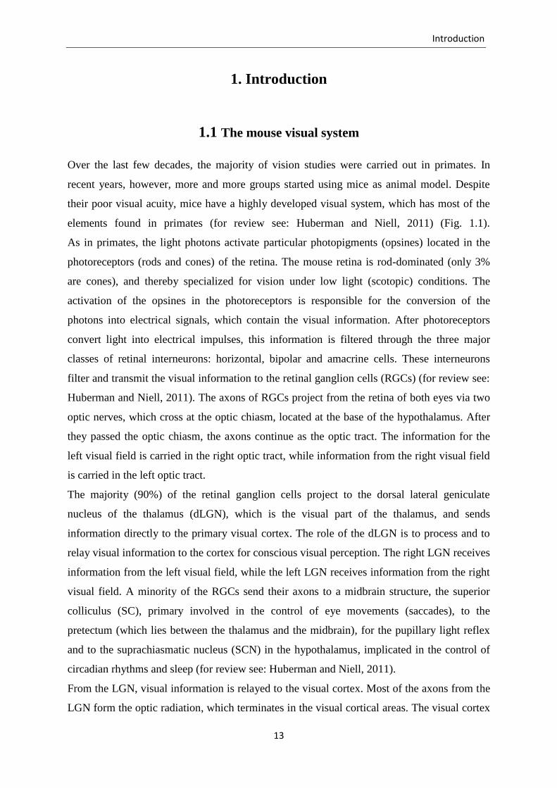


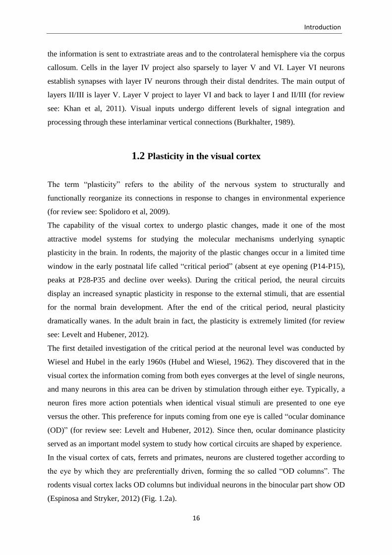


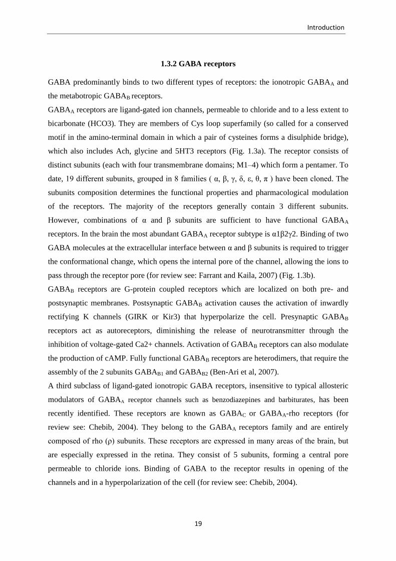


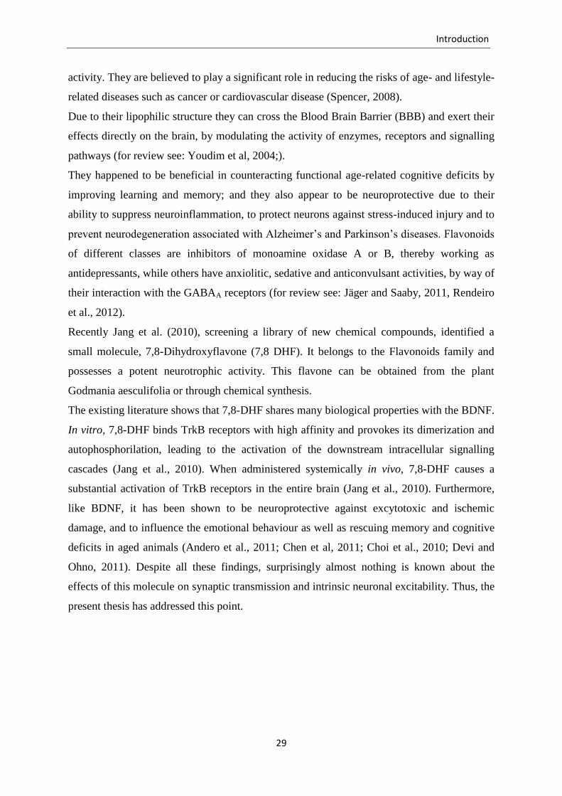
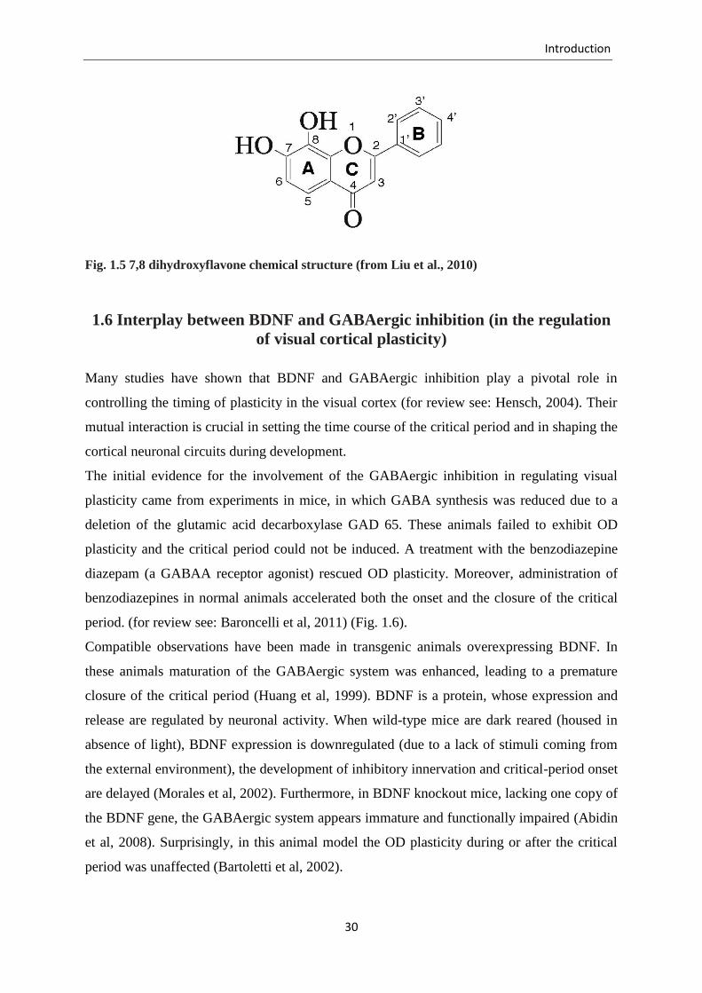

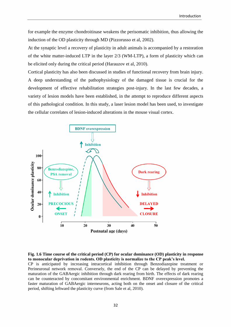


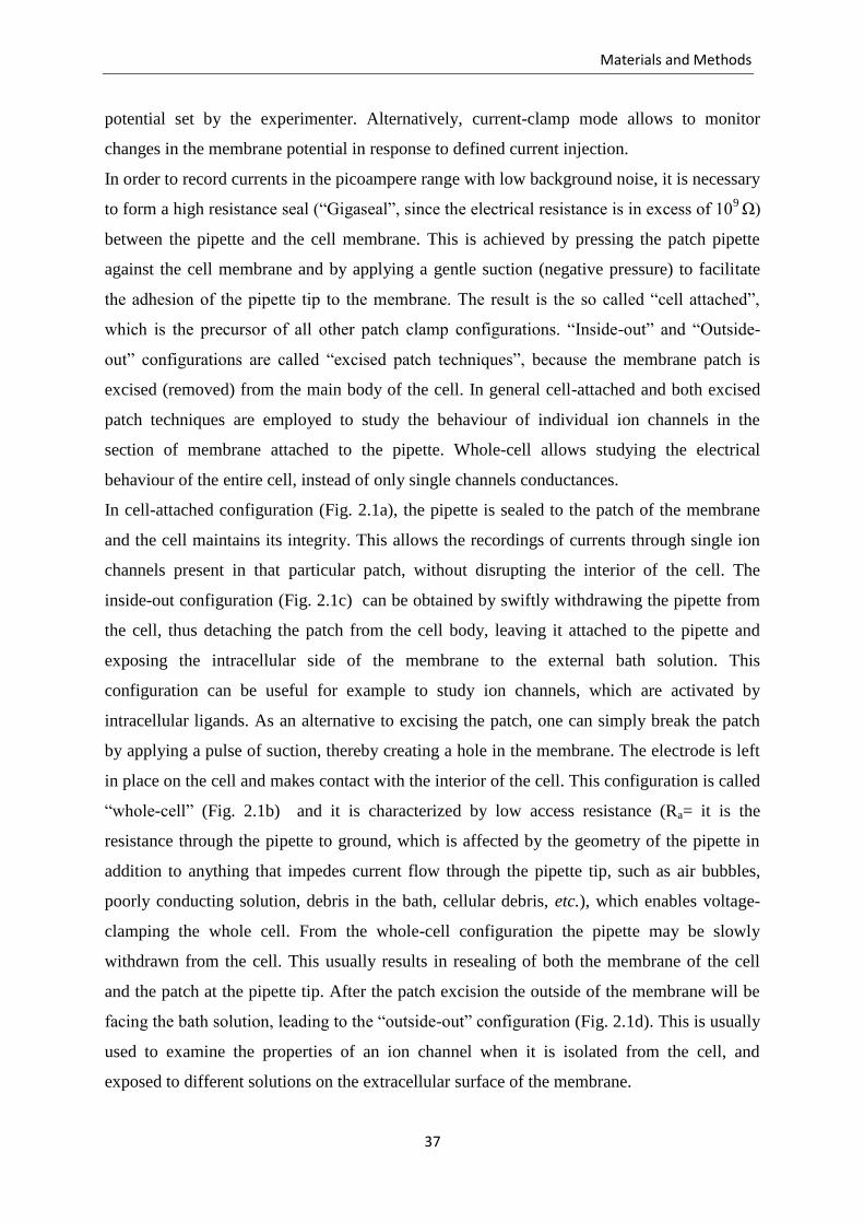


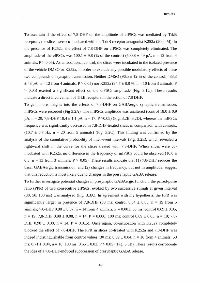

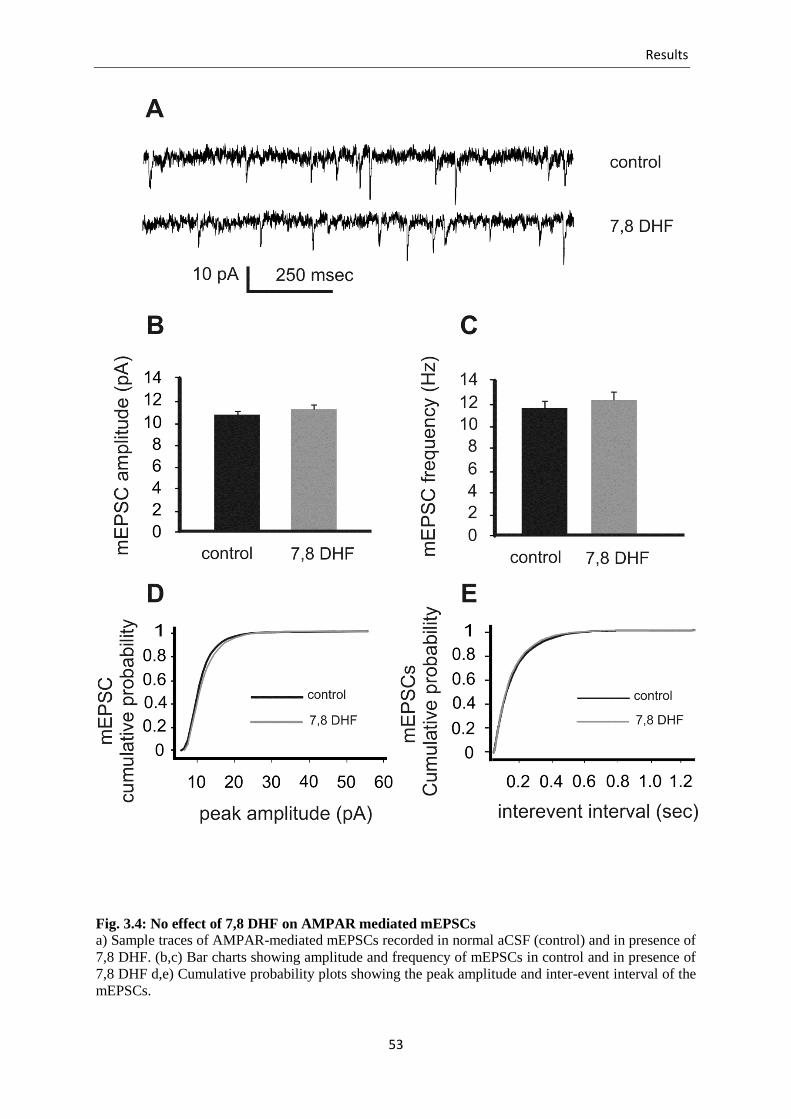


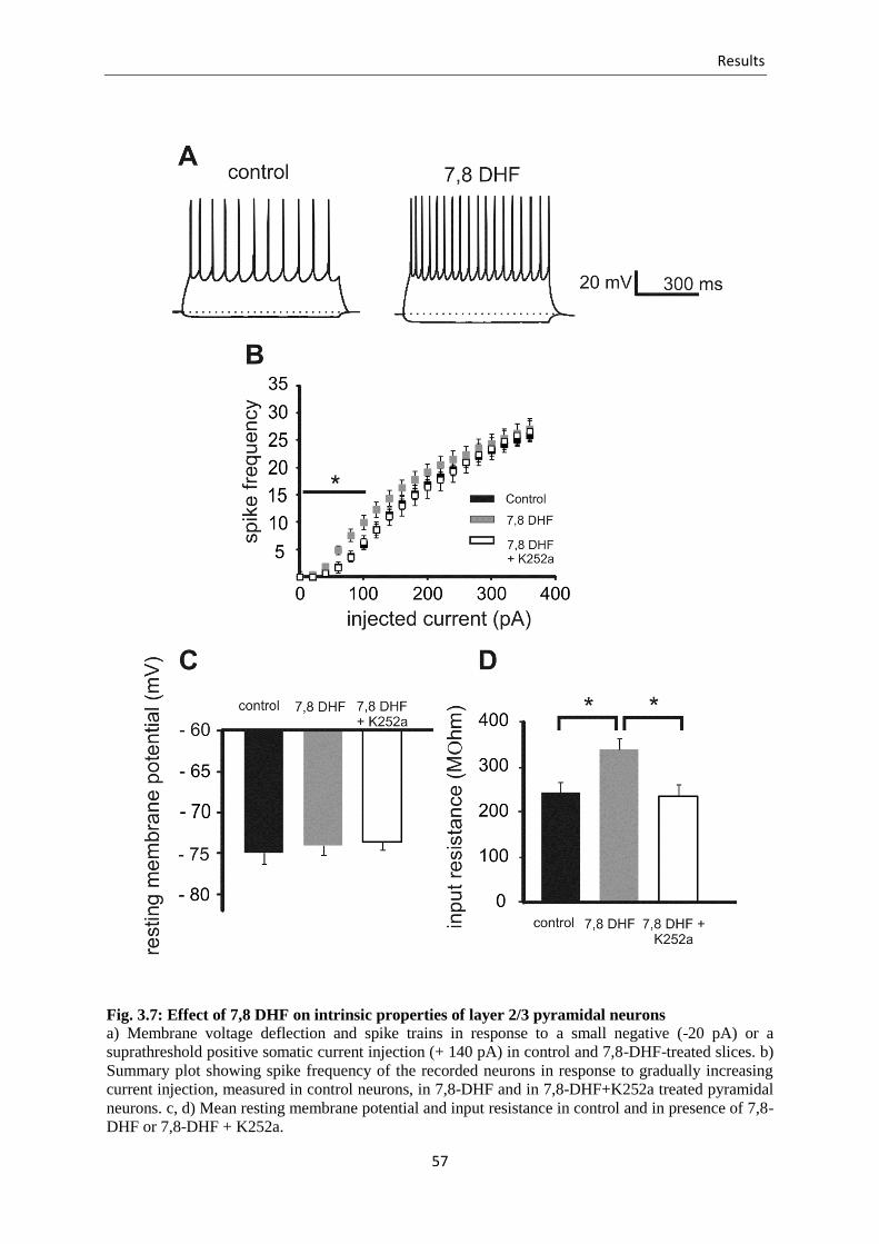

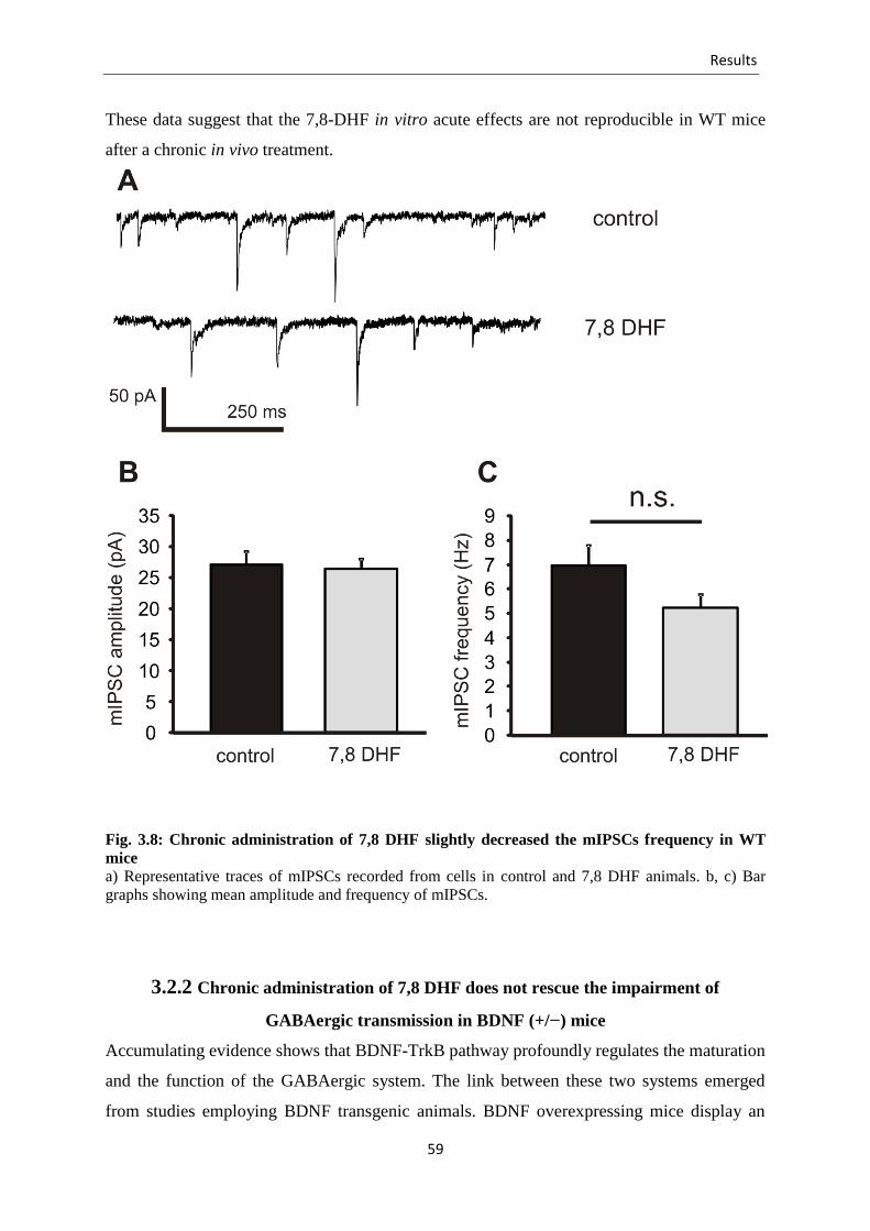

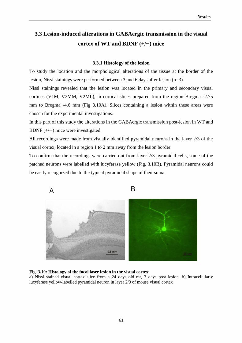

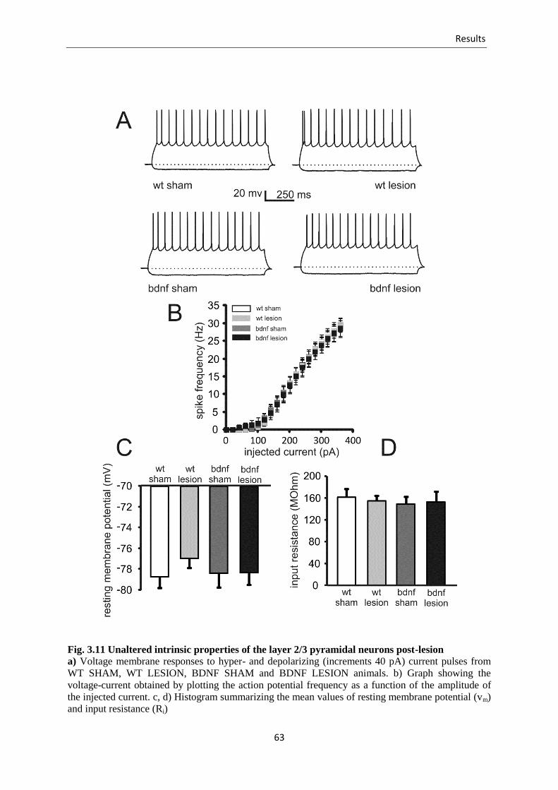
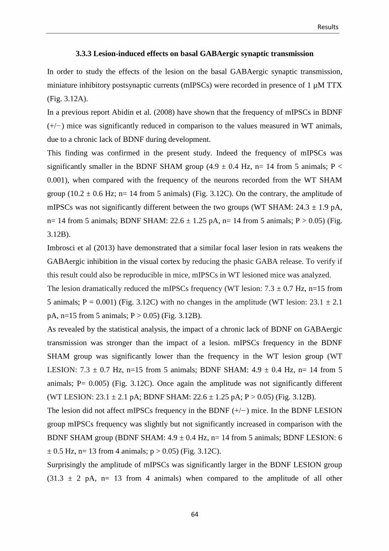


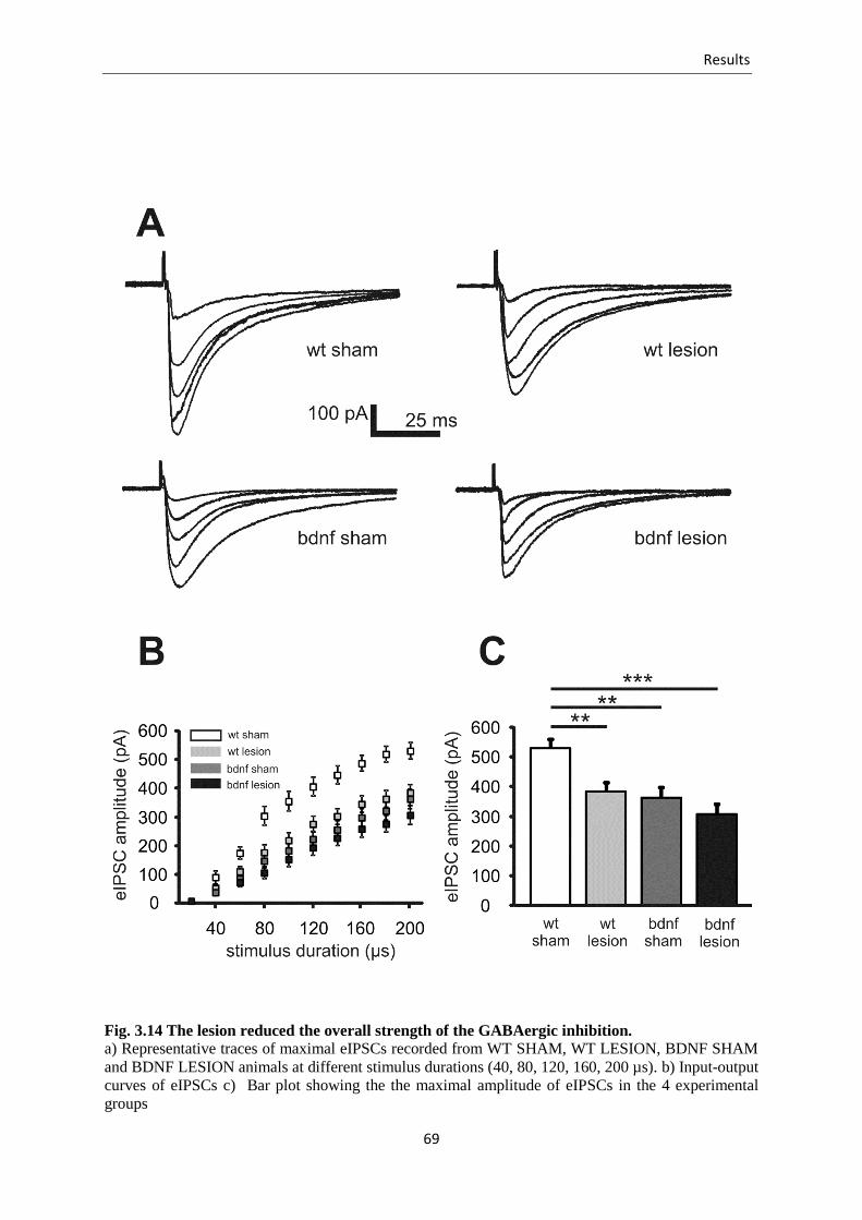


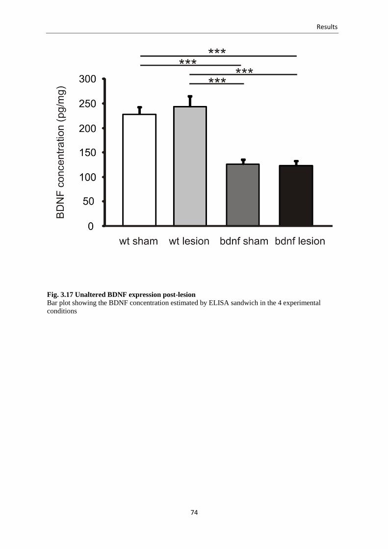


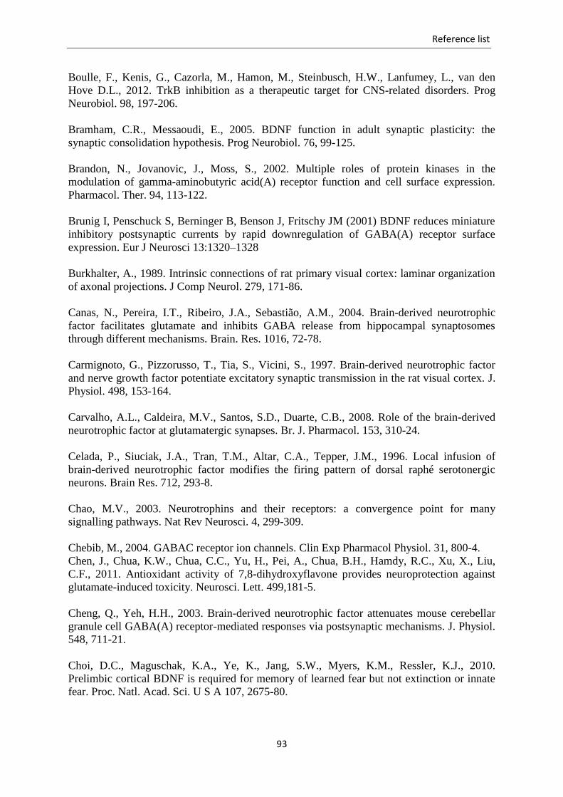


![Eine spektrale Methode f ur die Boltzmann-Gleichung · ly Accurate Approximation of the Collision Operator, (L. Pareschi and G. Russo; 2000)\, [4], die Grundlagen der molekularen](https://static.documents.pub/doc/80x56/5d4e6c1988c99303708ba86c/eine-spektrale-methode-f-ur-die-boltzmann-ly-accurate-approximation-of-the.jpg)


