44
Interstitial Lung Diseases Yang,Dong (杨冬) [email protected] Department of Pulmonary Medicine, Zhongshan Hospital, Fudan Universtiy

Interstitial Lung Diseases
Yang,Dong (杨冬)
Department of Pulmonary Medicine,
Zhongshan Hospital, Fudan Universtiy
Definition
a group of disease which predominantly affect the
pulmonary connective tissue and interstitium
(between the air sacs of the lungs) of the alveolar
walls and share a common response of the lung to
injury: alveolitis; inflammation; fibrosis of the
interalveolar septum
also known as―interstitial pulmonary fibrosis‖or
―diffuse parenchymal lung disease (DPLD)‖

Common clinical feature
1. Exertional dyspnea or nonproductive cough
2. Tachypnea and bibasilar end-inspiratory dry cracks
3. Bilateral diffuse infiltrates on chest radiographs
4. Presents as a RESTRICTIVE lung disease, DLco↓,
PA-aO2↑
5. Varying degrees of fibrosis and inflammation, with or
without evidence of granulomatous or secondary
vascular changes

What is the cause of ILD? >180 kinds
Chief known cause (1/3)
Inhale inorganic dusts
Inhale organic dusts
Radio-active injury
Micro-organic infection
Drugs
Lymphocytic carcinoma
Pulmonary edema

What is the cause of ILD?
Chief unknown cause
Primary lung diseases
ILD associated with system disorder (CTDs)
Alveolar filling disorder
ILD associated with pulmonary vasculitis
Inherited disorders

Classfication-1
1.According to process: Acute/Subacute/Chronic
2. According to cause: Clear /Not clear
3. According to pathology
Granulomatous disease / non-tumor and non-
inflammatory diseases / non-specific diseases /
occupational diseases / hyperplastic and
tumourous diseases / interstitial lung diseases
and honey lung

Classfication-2 (According to pathology )
Interstitial lung Diseases
1.usual interstitial pneumonia (UIP)
2.nonspecific interstitial pneumonia (NIP)
3. respiratory bronchiolitis (RB)
4. organizing pneumonia (BOOP)
5. diffuse alveolar damage
6. desquamative interstitial pneumonia (DIP)
7. lymphocytic interstitial pneumonia (LIP)

Pathogenesis The lung heals in a predictable, consistent
manner
Causes may differ but pathogenesis remains the
same
Cause = insult, injury, infection, toxin, etc
Cause is what differentiates the individual
diseases and their treatments
leads to alveolitis→initial event in all interstitial
lung diseases

Injury
Alveolitis?- the presence of immune cells within
the alveolar walls
Immune cells
Secrete cytokines recruiting more immune
cells and triggering complex chain of events
called ―inflammation‖
Cause direct tissue injury
Generate oxygen free radicals
Release proteases

Pathogenesis
Alveolar wall and tissue damage lead to leakage
of a fibrin-rich exudate into the alveolar spaces

Pathogenesis – Key point
Type I pneumocytes
Very susceptible to injury
Cannot regenerate
Type II pneumocytes
More resistant to injury
Regenerate and differentiate into Type I pneumocytes
Proliferate in injury laying down a new epithelial layer

Fibroblasts migrate across the damaged
alveolar wall into the alveolar space and into
the fibrin-rich exudate
Fibrin is organized, collagenized and covered
by alveolar epithelium
Pathogenesis

If the injury persists… Fibrosis continues, leading to obliteration of
the delicate interstitium
Normal lung architecture is distorted because
fibrosis scars down ―pulling‖ on bronchiolar
spaces causing dilatation and a cyst-like
architecture
Final stage is referred to as End Stage Lung
or Honey Comb Lung

How to make a diagnosis?

Diagnostic path
Diagnostic Tests Used for Identification
1. Blood Tests
2. Pulmonary Function Tests
3. Chest X-ray
4. CT Scan
5. Bronchoscopy : TBLB or BAL
6. Lung Biopsy : TBLB or OLB or TGLB

Pulmonary Function Tests Restrictive pattern
lung volume
compliance
diffusing capacity
Thus
FVC
FEV1
TLC
DLco
But, NORMAL FEV1/ FVC ratio
Chest imaging studies
X-ray
Diffuse reticular infiltrates, micronodular
CT scan
Fine to coarse reticular or reticulonodular
infiltrates, lung fibrosis and honeycomb
Peripheral ground- glass opacities
Bronchoalveolar lavage (BAL)
-to make a diagnosis ,differential diagnosis, treatment
-to exclude infections and malignancies (cancer)
-to identify inflammation in lung tissue

Lung biopsy
1. Bronchoscopy (TBLB)
20-40%(+), to diagnosis of sarcoidosis – 80%
2. Open-lung biopsy (OLB)
3. Thoracoscopy-guide lung biopsy (TGLB)
Idiopathic pulmonary fibrosis (IPF)
Alao named as cryptogenic fibrosing alveolitis,
chronic inflammation of the alveolar walls with
progressive fibrosis, of unknown etiology

IPF - Epdemiology
Age :40s- 50s
Male to female ratio 2/1 (1/1)
Prevalence: 5/100,000

Usual interstitial pneumonia (UIP) (IPF)
Histologic findings
1) dense fibrosis with remodeling of lung
architecture with honeycomb change
2) fibroblastic foci & fibrotic zones with
temporal heterogeneity
3) Smooth muscle hyperplasia in areas of
fibrosis
4) in septa and beneath the pleura.
5) patchy lung involvement

Symptoms and Signs
Symptoms
dyspnea on exertion, Progression over months,
nonproductive cough
Physical Examination
tachypnea
clear lungs or ―velcro‖ rales ( inspiratory crackles)
signs of pulmonary hypertension (Incr. P2, cor pulmonale)
digital clubbing and cyanosis

Radiography exam.
CXRs: lower
zones,
interstitial,
ground glass,
honey combing

Usual interstitial pneumonia (UIP) (IPF)
CT Features
1) reticular attenuation with lobular
septal thickening
2) architectural distortion
3) honeycomb pattern
4) ground-glass attenuation
5) predominantly basal &
peripheral in distribution
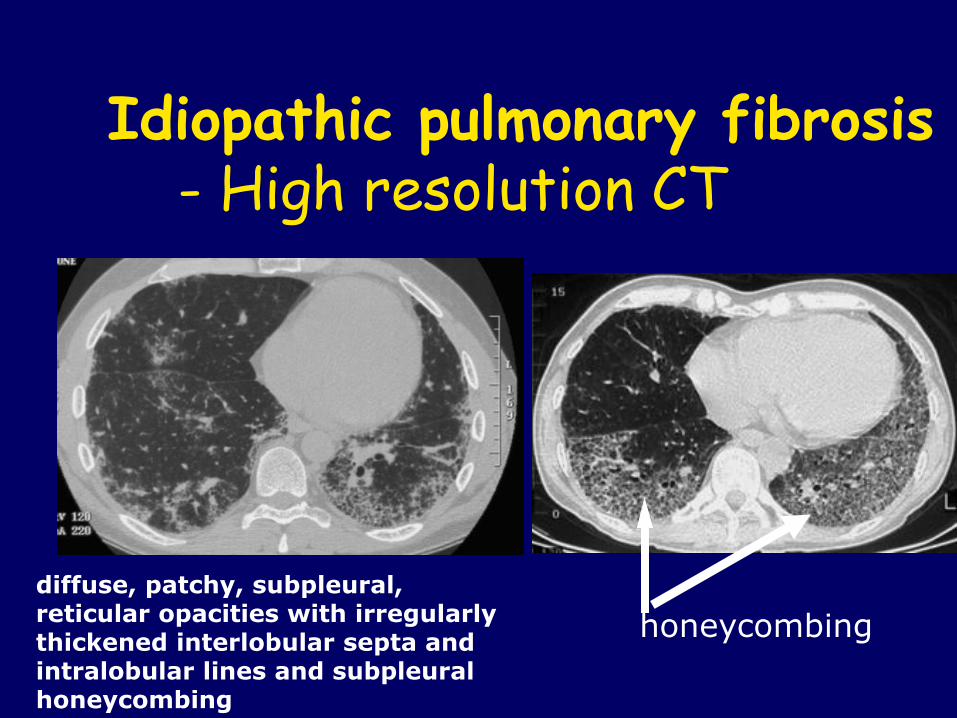
Idiopathic pulmonary fibrosis - High resolution CT
diffuse, patchy, subpleural, reticular opacities with irregularly thickened interlobular septa and intralobular lines and subpleural honeycombing
honeycombing

IPF -- Labs
PFTs: Restrictive
Hypoxemia:
first with exercise
later at rest
LDH, γ-Globulin↑; RF +
ANA can be (+)
lung biopsy: open or video-assisted thoracoscopic; not
indicated when x-rays show extensive
honeycombing

Diagnosis
Clinical features and lab findings
Exclude other kind of ILD
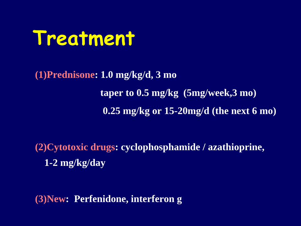
Treatment
(1)Prednisone: 1.0 mg/kg/d, 3 mo
taper to 0.5 mg/kg (5mg/week,3 mo)
0.25 mg/kg or 15-20mg/d (the next 6 mo)
(2)Cytotoxic drugs: cyclophosphamide / azathioprine,
1-2 mg/kg/day
(3)New: Perfenidone, interferon g
Treatment
(4) Supportive : O2 , treat infections, etc.
(5) Lung transplantation: end-stage
(6) Rehabilitation and education programs
Prognosis
Poor - mean survival is 4 years;
median survival: 4 - 6 yr after diagnosis
Worse if > 10% PMNs + Eos. on BAL
Better if lymphocytes on BAL
Histology: DIP

Sarcoidosis
Chronic multisystem disorder of unknown
cause characterized in affected organs by an
accumulation of T lymphocytes, noncaseating
epithelioid granulomas and derangements of
the normal tissue architecture

Etiology
Unknown
Exaggerated celluar immune response
(acquired, inherited or both)
Incidence and Prevalence
All ages, most between the ages of 20 and 40
Both sexes, female slightly more susceptible
than males

Symptoms and Signs
Acute or subacute (20%-40%)
fever, fatigue,malaise,anorexia,weight loss
cough, dyspnea, a vague retrosternal chest
discomfort, and /or polyarthritis
Symptoms and Signs
Lung
Primarily an interstitial lung disease
Distal atelectasis
Large-vessel pulmonary granulomatous
arteritis
Lymph node
Lymphadenopathy is very common
Intratharacic nodes are enlarged—75%-90%

Complication
Respiratory tract abnormalities---morbility and
mortality
Eye
Central nervous system---most serious

Larboratory Abnormalities
ACE
Graph
type I—bilateral hilar adenopathy acute subacute
type II--with diffuse parechymal changes chronic
type III-- diffuse parechymal changes chronic

Diagnosis
Clinical features and lab findings
Exclude other kind of noncaseating granulomas

Prognosis
Overall is good
Most acute disease with no significant sequelae
50% have some permanent organ dysfunction--
-most is mild,stable
15%-20%----active or recurs intermittently

Treatment
Glucocorticoids
Methotrexate
Other drugs
when to treat?

Treatment
Prednisone: 1.0 mg/kg/d, 4 mo -6mo
Slow taper over 2 to 3
Methotrexate: 5 to 15 mg/week in a single oral dose
when glucocorticoids are contraindicated or in
refractory cases
Lung transplant-------end-stage

Question? What are the Common clinical features ?
What can we find from pulmonary function test ?
How to make a diagnosis?


