Page 1
RESEARCH ARTICLE
Intravenous injection of extracellular vesicles
to treat chronic myocardial ischemia
Laura A. ScrimgeourID, Brittany A. Potz, Ahmad Aboul Gheit, Yuhong Liu, Guangbin Shi,
Melissa Pfeiffer, Bonnie J. Colantuono, Neel R. Sodha, M. Ruhul Abid, Frank W. Sellke*
Division of Cardiothoracic Surgery, Department of Surgery, Cardiovascular Research Center, Rhode Island
Hospital, Brown University Warren Alpert Medical School, Providence, RI, United States of America
* [email protected]
Abstract
Background
Mesenchymal stem cell-derived extracellular vesicles (EVs) appear to be a very exciting
treatment option for heart disease. Here, we used a swine model of chronic myocardial
ischemia to evaluate the efficacy of a less-invasive method of injection of EVs via a periph-
eral intravenous route.
Methods
Sixteen Yorkshire swine underwent placement of an ameroid constrictor on the left circum-
flex (LCx) artery at age 11 weeks to induce chronic myocardial ischemia. Two weeks later,
they were divided into two groups: control (CON; n = 8), and intravenous injection of EVs
(EVIV; n = 8). At 18 weeks of age, animals underwent final analysis and euthanasia. The
chronically ischemic myocardium (LCx territory) was harvested for analysis.
Results
Intravenous injection (IV) of EVs induced several pro-angiogenic markers such as MAPK,
JNK but not Akt. Whereas IV injections of EVs decreased VEGFR2 expression and inhibited
apoptotic signaling (caspase 3), they increased expression of VEGFR1 that is believed to
be anti-angiogenic. Injection of EVs did not result in an increase in vessel density and blood
flow when compared to the control group.
Conclusions
Although IV injection of EVs upregulated several pro-angiogenic signaling pathways, it failed
to induce changes in vascular density in the chronically ischemic myocardium. Thus, a lack
of increase in vascular density at the doses tested failed to elicit a functional response in
ischemic myocardium.
PLOS ONE
PLOS ONE | https://doi.org/10.1371/journal.pone.0238879 September 11, 2020 1 / 14
a1111111111
a1111111111
a1111111111
a1111111111
a1111111111
OPEN ACCESS
Citation: Scrimgeour LA, Potz BA, Aboul Gheit A,
Liu Y, Shi G, Pfeiffer M, et al. (2020) Intravenous
injection of extracellular vesicles to treat chronic
myocardial ischemia. PLoS ONE 15(9): e0238879.
https://doi.org/10.1371/journal.pone.0238879
Editor: John Calvert, Emory University, UNITED
STATES
Received: April 4, 2020
Accepted: August 25, 2020
Published: September 11, 2020
Copyright: © 2020 Scrimgeour et al. This is an
open access article distributed under the terms of
the Creative Commons Attribution License, which
permits unrestricted use, distribution, and
reproduction in any medium, provided the original
author and source are credited.
Data Availability Statement: All relevant data are
within the manuscript and its Supporting
Information files.
Funding: Funding for this research was provided
by the National Heart, Lung, and Blood Institute
(NHLBI) [R01HL46716 (F.W.S.), RO1HL128831-
01A1 (F.W.S.]; American Heart Association Grant-
in-Aid 14GRNT20460291 and NHLBI
1R01HL133624-01A1 (to M.R.A.); NIH/NIGMS
Training Grant 2T32 GM065085-12 (B.A.P.) and
2T32 GM065085-13 (L.A.S.). The funders had no
role in study design, data collection and analysis,
Page 2
Introduction
Despite numerous advances in prevention, diagnosis and treatment of cardiovascular disease,
ischemic heart disease continues to increase in prevalence and remains the leading cause of
mortality worldwide [1]. Furthermore, while catheter-based interventions are able to help an
increasing number of patients with compromised coronary arteries, there are many patients
whose disease burden is so diffuse that it is not amenable to percutaneous treatments. Addi-
tionally, even with surgical coronary artery bypass grafting, many patients retain suboptimal
cardiac function or are unable to undergo grafting procedures [2, 3]. Therefore, the explora-
tion of alternative and adjunctive therapies for ischemic heart disease is essential.
Stem cells offer a potential method for the treatment for ischemic cardiomyopathy. How-
ever, there are concerns related with stem cell therapies including immune-activation causing
the need for immunosuppression, cell death, lack of honing capabilities, and the need for re-
implantation of new cells to achieve a long-lasting effect on myocardial ischemia [4, 5]. To
avoid immunosuppression, the need for time-intensive and invasive intra-operative harvesting
for autologous stem cell transfers would be required [6]. These drawbacks make stem cell ther-
apies a less-palatable option for much of the large population in need of cardiac revasculariza-
tion. Furthermore, there is little data to suggest long-term in-vivo survival of transplanted
stem cells into myocardium [7].
Recently, secretion products of stem cells known as extracellular vesicles (EVs) have been
identified as carrying miRNAs, mRNAs, cytokines, growth factors and angiogenic proteins,
making them a promising target for ischemic treatments [8, 9]. These vesicles have been
shown to migrate to sites of injury and reprogram endogenous repair systems while limiting
local inflammation. EVs are defined by their size and mechanism of release: microvesicles are
released by budding off the plasma membrane and are large sized particles of>200nm; exo-
somes are endosomes that are released from intraluminal vesicles and are smaller in size rang-
ing from 50-200nm [10]. Given the complicated terminology, we use the term extracellular
vesicles to indicate our use of both microvesicles and exosomes. One of the exciting strengths
of using extracellular vesicles is that their small size and non-cellular treatment allow flow
through capillaries associated with cell-based therapies.
EVs have shown some promise of improved function in both small and large animal models
of myocardial infarction [11–14]. However, none of these studies specifically address chronic
myocardial ischemia, which likely has farther-reaching therapeutic benefits than only address-
ing myocardial infarction. Furthermore, to our knowledge, there are no studies evaluating
peripheral intravenous injection of EVs. For widespread applications, minimizing the inva-
siveness of procedures for translation to human studies would be optimal. Here, we analyze
the effect of extracellular vesicles administered via intravenous injection in a porcine model of
chronic myocardial ischemia.
Materials and methods
Sixteen intact male Yorkshire swine (Tufts University, Boston, MA) underwent placement of
an ameroid constrictor (Research Instruments SW, Escondido, CA) on the left circumflex
artery at age 11 weeks to induce chronic myocardial ischemia. They were then divided into
two groups: control (CON; n = 8) and intravenous injection of extracellular vesicles (EVIV;
n = 8). The EVIV group received intravenous injections of extracellular vesicles two weeks
after the placement of the ameroid constrictor (age 13 weeks). Animals underwent euthanasia
at 18 weeks of age, five weeks after treatment with intravenous extracellular vesicles or no
treatment, and ischemic myocardium was harvested for Western blot analysis. Blood was also
collected at the time of harvest and analyzed for cytokine levels.
PLOS ONE Extracellular vesicles & the myocardium
PLOS ONE | https://doi.org/10.1371/journal.pone.0238879 September 11, 2020 2 / 14
decision to publish, or preparation of the
manuscript.
Competing interests: The authors have declared
that no competing interests exist.
Page 3
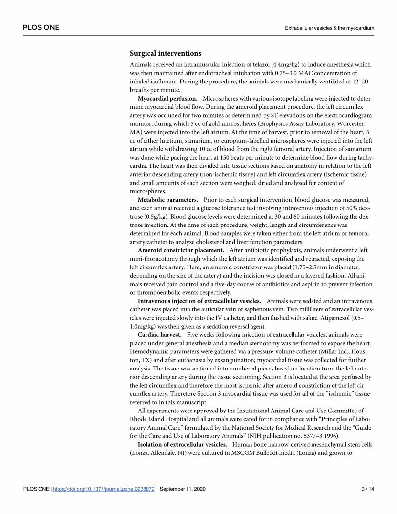
Surgical interventions
Animals received an intramuscular injection of telazol (4.4mg/kg) to induce anesthesia which
was then maintained after endotracheal intubation with 0.75–3.0 MAC concentration of
inhaled isoflurane. During the procedure, the animals were mechanically ventilated at 12–20
breaths per minute.
Myocardial perfusion. Microspheres with various isotope labeling were injected to deter-
mine myocardial blood flow. During the ameroid placement procedure, the left circumflex
artery was occluded for two minutes as determined by ST elevations on the electrocardiogram
monitor, during which 5 cc of gold microspheres (Biophysics Assay Laboratory, Worcester,
MA) were injected into the left atrium. At the time of harvest, prior to removal of the heart, 5
cc of either lutetium, samarium, or europium-labelled microspheres were injected into the left
atrium while withdrawing 10 cc of blood from the right femoral artery. Injection of samarium
was done while pacing the heart at 150 beats per minute to determine blood flow during tachy-
cardia. The heart was then divided into tissue sections based on anatomy in relation to the left
anterior descending artery (non-ischemic tissue) and left circumflex artery (ischemic tissue)
and small amounts of each section were weighed, dried and analyzed for content of
microspheres.
Metabolic parameters. Prior to each surgical intervention, blood glucose was measured,
and each animal received a glucose tolerance test involving intravenous injection of 50% dex-
trose (0.5g/kg). Blood glucose levels were determined at 30 and 60 minutes following the dex-
trose injection. At the time of each procedure, weight, length and circumference was
determined for each animal. Blood samples were taken either from the left atrium or femoral
artery catheter to analyze cholesterol and liver function parameters.
Ameroid constrictor placement. After antibiotic prophylaxis, animals underwent a left
mini-thoracotomy through which the left atrium was identified and retracted, exposing the
left circumflex artery. Here, an ameroid constrictor was placed (1.75–2.5mm in diameter,
depending on the size of the artery) and the incision was closed in a layered fashion. All ani-
mals received pain control and a five-day course of antibiotics and aspirin to prevent infection
or thromboembolic events respectively.
Intravenous injection of extracellular vesicles. Animals were sedated and an intravenous
catheter was placed into the auricular vein or saphenous vein. Two milliliters of extracellular ves-
icles were injected slowly into the IV catheter, and then flushed with saline. Atipamezol (0.5–
1.0mg/kg) was then given as a sedation reversal agent.
Cardiac harvest. Five weeks following injection of extracellular vesicles, animals were
placed under general anesthesia and a median sternotomy was performed to expose the heart.
Hemodynamic parameters were gathered via a pressure-volume catheter (Millar Inc., Hous-
ton, TX) and after euthanasia by exsanguination; myocardial tissue was collected for further
analysis. The tissue was sectioned into numbered pieces based on location from the left ante-
rior descending artery during the tissue sectioning. Section 3 is located at the area perfused by
the left circumflex and therefore the most ischemic after ameroid constriction of the left cir-
cumflex artery. Therefore Section 3 myocardial tissue was used for all of the “ischemic” tissue
referred to in this manuscript.
All experiments were approved by the Institutional Animal Care and Use Committee of
Rhode Island Hospital and all animals were cared for in compliance with “Principles of Labo-
ratory Animal Care” formulated by the National Society for Medical Research and the “Guide
for the Care and Use of Laboratory Animals” (NIH publication no. 5377–3 1996).
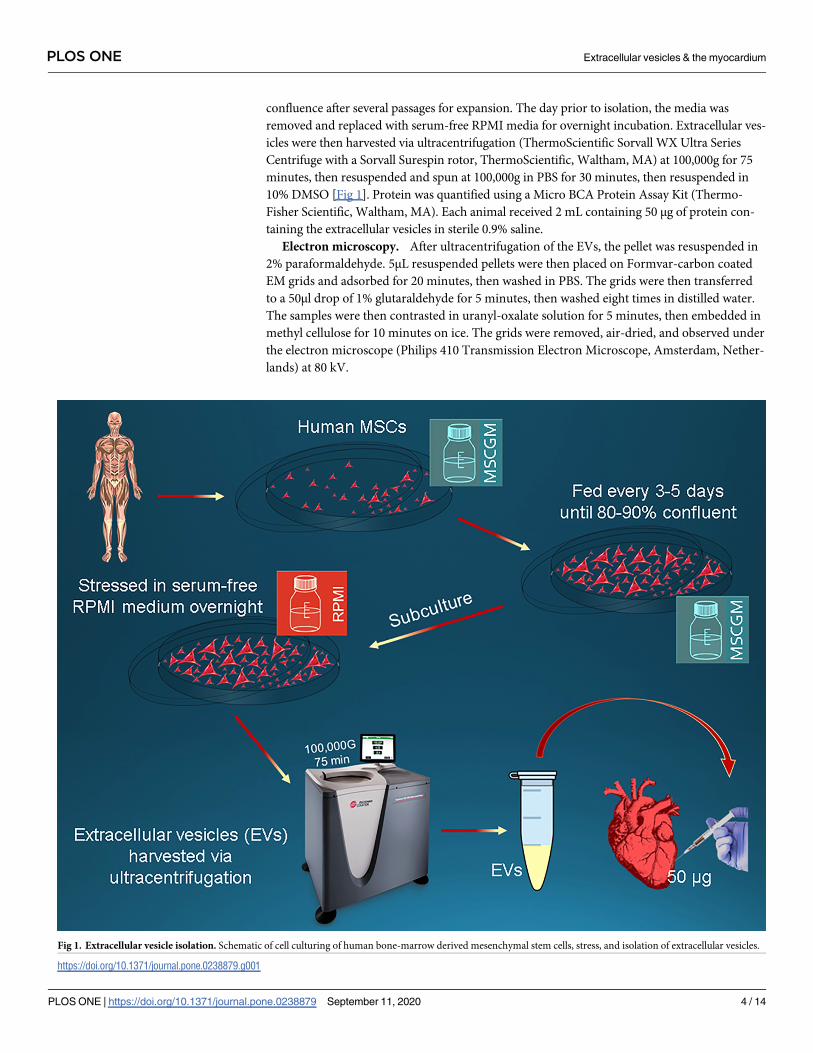
Isolation of extracellular vesicles. Human bone marrow-derived mesenchymal stem cells
(Lonza, Allendale, NJ) were cultured in MSCGM Bulletkit media (Lonza) and grown to
PLOS ONE Extracellular vesicles & the myocardium
PLOS ONE | https://doi.org/10.1371/journal.pone.0238879 September 11, 2020 3 / 14
Page 4
confluence after several passages for expansion. The day prior to isolation, the media was
removed and replaced with serum-free RPMI media for overnight incubation. Extracellular ves-
icles were then harvested via ultracentrifugation (ThermoScientific Sorvall WX Ultra Series
Centrifuge with a Sorvall Surespin rotor, ThermoScientific, Waltham, MA) at 100,000g for 75
minutes, then resuspended and spun at 100,000g in PBS for 30 minutes, then resuspended in
10% DMSO [Fig 1]. Protein was quantified using a Micro BCA Protein Assay Kit (Thermo-
Fisher Scientific, Waltham, MA). Each animal received 2 mL containing 50 μg of protein con-
taining the extracellular vesicles in sterile 0.9% saline.
Electron microscopy. After ultracentrifugation of the EVs, the pellet was resuspended in
2% paraformaldehyde. 5μL resuspended pellets were then placed on Formvar-carbon coated
EM grids and adsorbed for 20 minutes, then washed in PBS. The grids were then transferred
to a 50μl drop of 1% glutaraldehyde for 5 minutes, then washed eight times in distilled water.
The samples were then contrasted in uranyl-oxalate solution for 5 minutes, then embedded in
methyl cellulose for 10 minutes on ice. The grids were removed, air-dried, and observed under
the electron microscope (Philips 410 Transmission Electron Microscope, Amsterdam, Nether-
lands) at 80 kV.
Fig 1. Extracellular vesicle isolation. Schematic of cell culturing of human bone-marrow derived mesenchymal stem cells, stress, and isolation of extracellular vesicles.
https://doi.org/10.1371/journal.pone.0238879.g001
PLOS ONE Extracellular vesicles & the myocardium
PLOS ONE | https://doi.org/10.1371/journal.pone.0238879 September 11, 2020 4 / 14
Page 5
Western blot analysis. Homogenized tissue lysates were fractionated on 4–20% Tris-Gly-
cine gels (Novex™ Midi Protein Gels, Invitrogen, Carlsbad, CA) then transferred to polyvinyli-
dene difluoride membranes (Millipore, Bedford, MA). Protein concentration was determined
using a radio-immunoprecipitation assay (Pierce BCA Protein Assay Kit, ThermoFisher Scien-
tific, Waltham, MA). Primary antibodies [phosphoinositide 3-kinase (PI3K), Akt, phosphory-
lated Akt, extracellular signal-regulated kinase (ERK1/2), microtubule associated protein
kinase (MAPK), B cell lymphoma-2 (BCL-2), phosphorylated BCL-2, Bcl-2-associated death
promoter (BAD), phosphorylated BAD, caspase 3, caspase 9, cleaved caspase 9, c-Jun N-termi-
nal kinase (JNK), vascular endothelial growth factor receptor 1 (VEGF-R1), and vascular endo-
thelial growth factor receptor 2 (VEGF-R2), all from Cell Signaling, Danvers, MA] at a 1:1000
dilution were incubated overnight at 4˚C. Membranes were washed, and then a secondary
antibody at appropriate dilution was added and incubated for one hour at room temperature.
GAPDH was added to all membranes for loading control. Chemiluminescent images were
viewed and recorded using a digital camera (GBox, Syngene, Cambridge, England) and ana-
lyzed using Image-J software (National Institutes of Health, Bethesda, MD) to quantify band
densitometry. Data are reported as arbitrary light units representative of protein band density
normalized to GAPDH and fold-change values compared to controls.
Immunofluorescence. At the time of harvest, tissue from ischemic myocardium was fixed
in 10% formalin and 24-hours later, transferred to 70% ethanol. Slides were stained with clus-
ter of differentiation 31 (CD31, Abcam, Cambridge, UK) and α-smooth muscle actin (α-SMA,
Cell Signaling, Danvers, MA), as previously described [16]. After paraffinization, slides were
deparaffinized, rehydrated, then blocked with peroxide, rinsed, and blocked with an antibody
at 1:50 dilution overnight. A second antibody was incubated for an hour at room temperature
after rinsing. Images were captured at 20x objective with a Nikon Eclipse TE2000-U micro-
scope (Nikon Instruments, Melville, NY). These images were then analyzed using Image-J soft-
ware (National Institutes of Health, Bethesda, MD).
Statistical analyses
GraphPad Prism 5.0 Software (GraphPad Software Inc., San Diego, CA) was used to perform a
Mann-Whitney two-tailed t-test. Data are presented as mean ± SEM. A p value of<0.05 was
used for statistical significance.
Results
Metabolic parameters
Metabolic parameters including glucose levels, bilirubin, protein, albumin, CRP, insulin levels,
cholesterol levels (HDL, LDL, triglycerides) and fructosamine were measured via arterial
blood sampling and no significant difference was seen between the groups at the time of har-
vest. The alkaline phosphatase was higher in the CON than the EVIV group (p = 0.04).
Myocardial perfusion
Blood flow to the ischemic area of the myocardium (perfused by the chronically occluded left
circumflex) was not significantly different for those treated with intravenous injection of extra-
cellular vesicles (p>0.9). When blood flow was assessed during cardiac pacing to 150 beats per
minute, there was again no significant difference observed from the control group. One pig
from each group was excluded because the result was an outlier at more than 2 standard devia-
tions from the mean. [Fig 2]
PLOS ONE Extracellular vesicles & the myocardium
PLOS ONE | https://doi.org/10.1371/journal.pone.0238879 September 11, 2020 5 / 14
Page 6
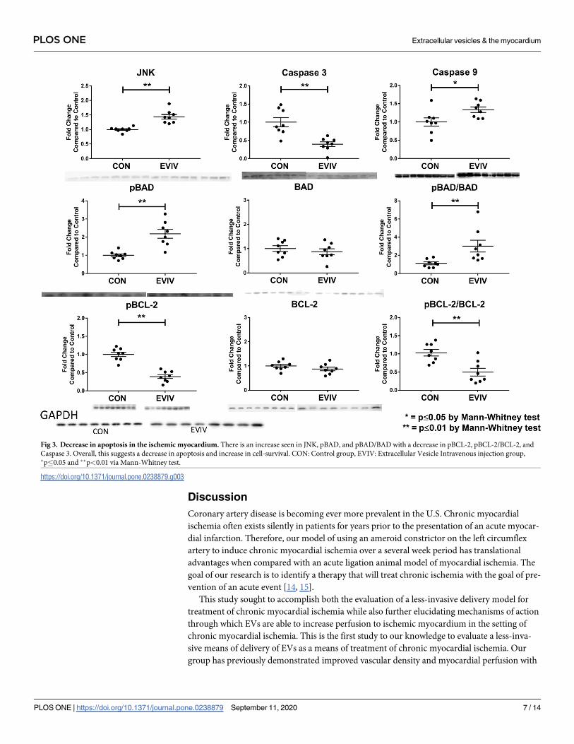
Apoptotic protein signaling
There were significantly upregulated levels of phosphorylated BAD compared to the control
(p = 0.002), despite no significant change in the overall amount of Akt (p>0.9). JNK was also
significantly upregulated (p = 0.008). While we do not see a significant change in BCL-2, phos-
phorylated BCL-2 was significantly decreased compared to the control (p = 0.004). Further-
more, there is a decrease in activation of the caspase cascade via decreased caspase 3
(p = 0.001). Interestingly, there is an increase in caspase 9 (p = 0.02). The overall significant
decrease in caspase 3, however, suggests downregulation of apoptosis. [Fig 3 and Table 1]
Angiogenic protein signaling
VEGF-R1 was significantly increased over the control group (p = 0.001), however VEGF-R2
was significantly downregulated (p = 0.002). Additionally, we observed a significant upregula-
tion of in MAP kinase (p = 0.001) and an increase in phosphorylated MAP kinase (p = 0.01).
[Fig 4 and Table 1].
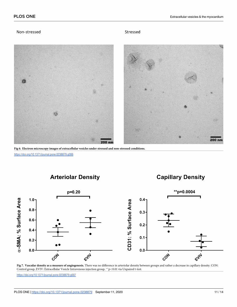
Size differences between stressed and non-stressed EVs
Mesenchymal stem cells which are subjected to stressed conditions for 24 hours produce sig-
nificantly larger EVs than cells which are not subjected to this stress. [Figs 5 and 6]
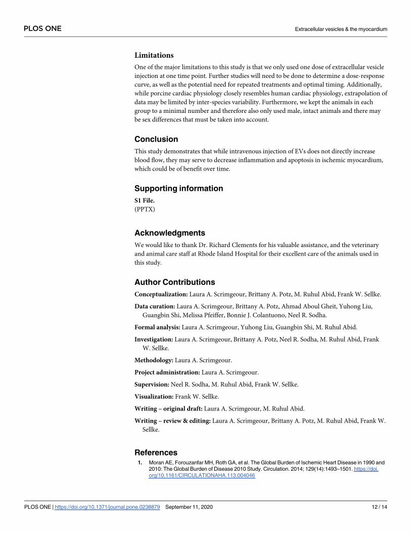
Myocardial arteriolar density
There was not an increase in capillary density counts as determined by CD31 staining in the
animals treated with EVs via IV when compared to the controls, rather, there was actually a
significant decrease in CD31 staining (EVIV: 0.07±0.02 vs CON: 0.24±0.02, p = 0.004). [Fig 7]
Furthermore, there was no significant difference in arteriolar density between the two groups
as determined by α-SMA staining, although there was a trend toward increase arteriolar den-
sity. This suggests a relative lack of angiogenesis in the ischemic myocardium (EVIV: 0.55±0.1
vs CON: 0.36±0.09, p = 0.2).
Fig 2. Intravenous injection of extracellular vesicles. No significant change was seen in blood flow (ml/min/g) either at rest or during pacing. CON: Control group,
EVIV: Extracellular Vesicle Intravenous injection group.
https://doi.org/10.1371/journal.pone.0238879.g002
PLOS ONE Extracellular vesicles & the myocardium
PLOS ONE | https://doi.org/10.1371/journal.pone.0238879 September 11, 2020 6 / 14
Page 7
Discussion
Coronary artery disease is becoming ever more prevalent in the U.S. Chronic myocardial
ischemia often exists silently in patients for years prior to the presentation of an acute myocar-
dial infarction. Therefore, our model of using an ameroid constrictor on the left circumflex
artery to induce chronic myocardial ischemia over a several week period has translational
advantages when compared with an acute ligation animal model of myocardial ischemia. The
goal of our research is to identify a therapy that will treat chronic ischemia with the goal of pre-
vention of an acute event [14, 15].
This study sought to accomplish both the evaluation of a less-invasive delivery model for
treatment of chronic myocardial ischemia while also further elucidating mechanisms of action
through which EVs are able to increase perfusion to ischemic myocardium in the setting of
chronic myocardial ischemia. This is the first study to our knowledge to evaluate a less-inva-
sive means of delivery of EVs as a means of treatment of chronic myocardial ischemia. Our
group has previously demonstrated improved vascular density and myocardial perfusion with
Fig 3. Decrease in apoptosis in the ischemic myocardium. There is an increase seen in JNK, pBAD, and pBAD/BAD with a decrease in pBCL-2, pBCL-2/BCL-2, and
Caspase 3. Overall, this suggests a decrease in apoptosis and increase in cell-survival. CON: Control group, EVIV: Extracellular Vesicle Intravenous injection group,�p�0.05 and ��p<0.01 via Mann-Whitney test.
https://doi.org/10.1371/journal.pone.0238879.g003
PLOS ONE Extracellular vesicles & the myocardium
PLOS ONE | https://doi.org/10.1371/journal.pone.0238879 September 11, 2020 7 / 14
Page 8
the intramyocardial injection of extracellular vesicles [16]; however, the invasive nature of an
additional thoracotomy limits the translational applications of this research to future human
studies. Other groups have evaluated intracoronary infusion via catheterization, however not
in a model of chronic ischemia [15, 17]. Therefore, we sought to explore a less-invasive, intra-
venous infusion approach of delivering treatment. Unfortunately, we did not find a significant
increase in the blood flow to the ischemic area while using a less-invasive, intravenous
approach. One plausible explanation for this would be that we used similar dosages of EVs for
IV injection as has been used in previous studies of intramyocardial injections; since IV pro-
vides a less direct application of therapy, we plan on repeating the experiment using an
increased dose of EVs in the IV group. Other explanations include an inactivation of EV’s
when injected into the blood, versus the myocardial as was done in other experiments where a
clear benefit was evident [16, 18]. It is interesting that EV’s actually decreased the density of
capillaries in the ischemic myocardium and there was a tread toward an increase in arteriolar
density. This suggests that the intravenous injection may have a differential effect on angio-
genic signaling compared to that observed when the EV’s are injected directly into the
myocardium.
Improved blood flow to areas of ischemia remains an overarching goal in the treatment of
myocardial ischemia. Treatments which improve angiogenesis are an obvious target for
achieving this goal. VEGF-R2 is a pro-angiogenic modulator while the actions of VEGF-R1 are
more dynamic. We observed a decrease in VEGF-R2 in the EVIV group, which may explain
Table 1. Markers of apoptosis and angiogenesis.
CON EVIV p-value
JNK 1 1.43 ± 0.08 0.0002�
Caspase 3 1 0.395 ± 0.065 0.0011�
Caspase 9 1 1.34 ± 0.07 0.0207�
Cleaved Caspase 9 1 0.91 ± 0.17 0.5054
Cleaved Caspase 9/Caspase 9 1.08 ± 0.16 0.71 ± 0.15 0.1049
pBAD 1 2.18 ± 0.24 0.0003�
BAD 1 0.87 ± 0.12 0.5737
pBAD/BAD 1.11 ± 0.15 3.01 ± 0.65 0.0019�
pBCL-2 1 0.39 ± 0.05 0.0002�
BCL-2 1 0.88 ± 0.07 0.2345
pBCL-2/BCL-2 1.03 ± 0.09 0.50 ± 0.11 0.0047�
MAPK p42 1 1.95 ± 0.18 0.0011�
VEGF-R1 1 3.35 ± 0.56 0.0006�
VEGF-R2 1 0.53 ± 0.08 0.0019�
MAPK 1 0.90 ± 0.04 0.5737
pMAPK 1 1.8 ± 0.12 0.0148�
TGF-β 1 1.25 ± 0.09 0.0148�
eNOS 1 0.087 ± 0.056 0.2786
pAkt 1 0.85 ± 0.09 0.1049
Akt 1 0.95 ± 0.10 0.5054
pAkt/Akt 1.03 ± 0.07 0.90 ± 0.09 0.2786
All markers are load controlled to GAPDH and normalized to CON and listed as mean ± SEM. P values determined via at two-tailed t-test (Mann-Whitney). CON:
Control group, EVIV: Extracellular Vesicle Intravenous injection group.
�p�0.05 and
��p<0.01 via Mann-Whitney test.
https://doi.org/10.1371/journal.pone.0238879.t001
PLOS ONE Extracellular vesicles & the myocardium
PLOS ONE | https://doi.org/10.1371/journal.pone.0238879 September 11, 2020 8 / 14
Page 9
why we do not see increased blood flow in that group compared to the control group. Further-
more, we did not observe an increase in capillary nor arteriolar density in tissue sections of
ischemic myocardium in the EVIV treated group, which further demonstrates the lack of
angiogenesis in this tissue. VEGF-R1 has been shown to affect vascular development differen-
tially at different stages. It is initially inhibitory to sprouting, but later becomes essential for
branching and connections with other vessels [19]. Interestingly, we see an increase in
VEGF-R1 but perhaps the levels are so high that they cause an overall inhibitory effect on
sprouting and the initiation of the angiogenic response.
Activation of VEGF-R2 has been shown to have downstream effects on the MAPK/ERK1/2
pathways and inhibition of this interaction is associated with inhibition of angiogenesis [20].
Upregulation of intracellular signaling cascades including MAP kinase are essential in carrying
out intracellular functions required for angiogenesis. It is very interesting to note the higher
levels of MAPK and pMAPK in the animals treated with IV injections of extracellular vesicles
The lack of change observed in blood flow in the IV-injected group in this study could suggest
Fig 4. Angiogenesis signaling in the ischemic myocardium. The EVIV group had a significant increase in MAPK, pMAPK, and VEGF-R1. CON: Control group,
EVIV: Extracellular Vesicle Intravenous injection group, �p�0.05 and ��p<0.01 via Mann-Whitney test.
https://doi.org/10.1371/journal.pone.0238879.g004
PLOS ONE Extracellular vesicles & the myocardium
PLOS ONE | https://doi.org/10.1371/journal.pone.0238879 September 11, 2020 9 / 14
Page 10
the need for a direct myocardial treatment as an initial treatment, but the opportunity for
repeated administrations of extracellular vesicles through peripheral intravenous administra-
tion at future time points to augment the beneficial effects. Collectively, this suggests that
while increased myocardial blood flow was not observed in the EV-injected group at the time
of harvest, this group had a greater intracellular angiogenic response via the MAP kinase path-
way and expression of VEGF receptors that may be indicative of a delay in response to the
peripheral circulation of extracellular vesicles in comparison to local injection at the area of
chronic ischemia.
Akt is known to inhibit the apoptosis via mitochondrial and calcium-induced cell death
pathways by inhibition of BAD and the caspase cascade [21, 22]. Similarly, the MAPK/ERK
pathway is involved in regulating intracellular signaling involved in apoptosis [23]. Here, we
note an increase in JNK, pBAD, and pBCL-2, which collectively work to downregulate apopto-
sis and promote cell survival. The significant downregulation of caspase 3 further supports evi-
dence demonstrating a decrease in apoptosis in the ischemic tissue after treatment. Taken
together, the findings suggest that extracellular vesicles promote cell survival and intrinsic
repair mechanisms in injured cardiac muscle tissue.
Fig 5. Stressed conditions cause a size increase in EVs.
https://doi.org/10.1371/journal.pone.0238879.g005
PLOS ONE Extracellular vesicles & the myocardium
PLOS ONE | https://doi.org/10.1371/journal.pone.0238879 September 11, 2020 10 / 14
Page 11
Fig 6. Electron microscopy images of extracellular vesicles under stressed and non-stressed conditions.
https://doi.org/10.1371/journal.pone.0238879.g006
Fig 7. Vascular density as a measure of angiogenesis. There was no difference in arteriolar density between groups and rather a decrease in capillary density. CON:
Control group, EVIV: Extracellular Vesicle Intravenous injection group, ��p<0.01 via Unpaired t-test.
https://doi.org/10.1371/journal.pone.0238879.g007
PLOS ONE Extracellular vesicles & the myocardium
PLOS ONE | https://doi.org/10.1371/journal.pone.0238879 September 11, 2020 11 / 14
Page 12
Limitations
One of the major limitations to this study is that we only used one dose of extracellular vesicle
injection at one time point. Further studies will need to be done to determine a dose-response
curve, as well as the potential need for repeated treatments and optimal timing. Additionally,
while porcine cardiac physiology closely resembles human cardiac physiology, extrapolation of
data may be limited by inter-species variability. Furthermore, we kept the animals in each
group to a minimal number and therefore also only used male, intact animals and there may
be sex differences that must be taken into account.
Conclusion
This study demonstrates that while intravenous injection of EVs does not directly increase
blood flow, they may serve to decrease inflammation and apoptosis in ischemic myocardium,
which could be of benefit over time.
Supporting information
S1 File.
(PPTX)
Acknowledgments
We would like to thank Dr. Richard Clements for his valuable assistance, and the veterinary
and animal care staff at Rhode Island Hospital for their excellent care of the animals used in
this study.
Author Contributions
Conceptualization: Laura A. Scrimgeour, Brittany A. Potz, M. Ruhul Abid, Frank W. Sellke.
Data curation: Laura A. Scrimgeour, Brittany A. Potz, Ahmad Aboul Gheit, Yuhong Liu,
Guangbin Shi, Melissa Pfeiffer, Bonnie J. Colantuono, Neel R. Sodha.
Formal analysis: Laura A. Scrimgeour, Yuhong Liu, Guangbin Shi, M. Ruhul Abid.
Investigation: Laura A. Scrimgeour, Brittany A. Potz, Neel R. Sodha, M. Ruhul Abid, Frank
W. Sellke.
Methodology: Laura A. Scrimgeour.
Project administration: Laura A. Scrimgeour.
Supervision: Neel R. Sodha, M. Ruhul Abid, Frank W. Sellke.
Visualization: Frank W. Sellke.
Writing – original draft: Laura A. Scrimgeour, M. Ruhul Abid.
Writing – review & editing: Laura A. Scrimgeour, Brittany A. Potz, M. Ruhul Abid, Frank W.
Sellke.
References1. Moran AE, Forouzanfar MH, Roth GA, et al. The Global Burden of Ischemic Heart Disease in 1990 and
2010: The Global Burden of Disease 2010 Study. Circulation. 2014; 129(14):1493–1501. https://doi.
org/10.1161/CIRCULATIONAHA.113.004046
PLOS ONE Extracellular vesicles & the myocardium
PLOS ONE | https://doi.org/10.1371/journal.pone.0238879 September 11, 2020 12 / 14
Page 13
2. Ram E, Goldenberg I, Kassif Y, et al. Real-life characteristics and outcomes of patients who undergo
percutaneous coronary intervention versus coronary artery bypass grafting for left main coronary artery
disease: data from the prospective Multi-vessel Coronary Artery Disease (MULTICAD) Israeli Regis-
try†. Eur J Cardio-Thoracic Surg. March 2018. https://doi.org/10.1093/ejcts/ezy115
3. Moon MR, Sundt TM, Pasque MK, Barner HB, Gay WA, Damiano RJ. Influence of internal mammary
artery grafting and completeness of revascularization on long-term outcome in octogenarians. Ann
Thorac Surg. 2001; 72(6):2003–2007. https://doi.org/10.1016/S0003-4975(01)03144-7
4. Kraitchman DL, Tatsumi M, Gilson WD, et al. Dynamic imaging of allogeneic mesenchymal stem cells
trafficking to myocardial infarction. Circulation. 2005; 112(10):1451–1461. https://doi.org/10.1161/
CIRCULATIONAHA.105.537480
5. Wu R, Hu X, Wang J. Concise Review: Optimized Strategies for Stem Cell-based Therapy in Myocardial
Repair: Clinical Translatability and Potential Limitation. Stem Cells. January 2018. https://doi.org/10.
1002/stem.2778
6. Natsumeda M, Florea V, Rieger AC, et al. A Combination of Allogeneic Stem Cells Promotes Cardiac
Regeneration. J Am Coll Cardiol. 2017; 70(20):2504–2515. https://doi.org/10.1016/j.jacc.2017.09.036
7. Bartunek J, Terzic A, Davison BA, et al. Cardiopoietic cell therapy for advanced ischemic heart failure:
results at 39 weeks of the prospective, randomized, double blind, sham-controlled CHART-1 clinical
trial. Eur Heart J. 2016; 38(9):ehw543. https://doi.org/10.1093/eurheartj/ehw543
8. EL Andaloussi S, Mager I, Breakefield XO, Wood MJA. Extracellular vesicles: biology and emerging
therapeutic opportunities. Nat Rev Drug Discov. 2013; 12(5):347–357. https://doi.org/10.1038/nrd3978
9. Bronckaers A, Hilkens P, Martens W, et al. Mesenchymal stem/stromal cells as a pharmacological and
therapeutic approach to accelerate angiogenesis. Pharmacol Ther. 2014; 143(2):181–196. https://doi.
org/10.1016/j.pharmthera.2014.02.013
10. Phinney DG, Pittenger MF. Concise Review: MSC-Derived Exosomes for Cell-Free Therapy. Stem
Cells. 2017; 35(4):851–858. https://doi.org/10.1002/stem.2575
11. Wang Y, Zhang L, Li Y, et al. Exosomes/microvesicles from induced pluripotent stem cells deliver car-
dioprotective miRNAs and prevent cardiomyocyte apoptosis in the ischemic myocardium. Int J Cardiol.
2015; 192:61–69. https://doi.org/10.1016/j.ijcard.2015.05.020
12. Timmers L, Lim SK, Arslan F, et al. Reduction of myocardial infarct size by human mesenchymal stem
cell conditioned medium. Stem Cell Res. 2008; 1:129–137. https://doi.org/10.1016/j.scr.2008.02.002
13. Arslan F, Lai RC, Smeets MB, et al. Mesenchymal stem cell-derived exosomes increase ATP levels,
decrease oxidative stress and activate PI3K/Akt pathway to enhance myocardial viability and prevent
adverse remodeling after myocardial ischemia/reperfusion injury. Stem Cell Res. 2013; 10:301–312.
https://doi.org/10.1016/j.scr.2013.01.002
14. Lai RC, Arslan F, Lee MM, et al. Exosome secreted by MSC reduces myocardial ischemia/reperfusion
injury. Stem Cell Res. 2010; 4:214–222. https://doi.org/10.1016/j.scr.2009.12.003
15. Bolli R, Tang X-L, Sanganalmath SK, et al. Intracoronary delivery of autologous cardiac stem cells
improves cardiac function in a porcine model of chronic ischemic cardiomyopathy. Circulation. 2013;
128(2):122–131. https://doi.org/10.1161/CIRCULATIONAHA.112.001075
16. Potz BA, Scrimgeour LA, Pavlov VI, Sodha NR, Abid MR, Sellke FW. Extracellular Vesicle Injection
Improves Myocardial Function and Increases Angiogenesis in a Swine Model of Chronic Ischemia. J
Am Heart Assoc. 2018 Jun 12; 7(12). pii: e008344. https://doi.org/10.1161/JAHA.117.008344
17. Malliaras K, Makkar RR, Smith RR, et al. Intracoronary cardiosphere-derived cells after myocardial
infarction: evidence of therapeutic regeneration in the final 1-year results of the CADUCEUS trial (CAr-
diosphere-Derived aUtologous stem CElls to reverse ventricUlar dySfunction). J Am Coll Cardiol. 2014;
63(2):110–122. https://doi.org/10.1016/j.jacc.2013.08.724
18. Scrimgeour LA, Potz BA, Aboul Gheit A, et al. Extracellular Vesicles Promote Arteriogenesis in Chroni-
cally Ischemic Myocardium in the Setting of Metabolic Syndrome. J Am Heart Assoc. 2019; 8(15):
e012617. https://doi.org/10.1161/JAHA.119.012617
19. Chappell JC, Cluceru JG, Nesmith JE, et al. Flt-1 (VEGFR-1) coordinates discrete stages of blood ves-
sel formation. Cardiovasc Res. 2016; 111(1):84–93. https://doi.org/10.1093/cvr/cvw091
20. Bazzazi H, Popel AS. Computational investigation of sphingosine kinase 1 (SphK1) and calcium depen-
dent ERK1/2 activation downstream of VEGFR2 in endothelial cells. PLoS Comput Biol. 2017; 13(2):
e1005332. https://doi.org/10.1371/journal.pcbi.1005332
21. Grisanti LA, Talarico JA, Carter RL, et al. β-Adrenergic receptor-mediated transactivation of epidermal
growth factor receptor decreases cardiomyocyte apoptosis through differential subcellular activation of
ERK1/2 and Akt. J Mol Cell Cardiol. 2014; 72:39–51. https://doi.org/10.1016/j.yjmcc.2014.02.009
PLOS ONE Extracellular vesicles & the myocardium
PLOS ONE | https://doi.org/10.1371/journal.pone.0238879 September 11, 2020 13 / 14
Page 14
22. Gao Y-X, He W-T, Pan L-F, et al. Downregulation of Akt2 attenuates ER stress-induced cytotoxicity
through JNK-Wnt pathway in cardiomyocytes. Bioorg Med Chem Lett. December 2017. https://doi.org/
10.1016/j.bmcl.2017.12.031
23. Rose BA, Yokota T, Chintalgattu V, et al. Cardiac myocyte p38α kinase regulates angiogenesis via
myocyte-endothelial cell cross-talk during stress-induced remodeling in the heart. J Biol Chem. 2017;
292(31):12787–12800. https://doi.org/10.1074/jbc.M117.784553
PLOS ONE Extracellular vesicles & the myocardium
PLOS ONE | https://doi.org/10.1371/journal.pone.0238879 September 11, 2020 14 / 14