82
Introduction to EKG’s The Basics Sheri Tokarczyk, MS, PA-C
Introduction to EKG’s The Basics
Sheri Tokarczyk, MS, PA-C
Introduction to EKG’s
• August 8th: Basics • August 15th : Review of Rate, Axis, Rhythm • August 22nd : Conduction abnormalities;
Chamber enlargement • August 29th: Ischemia, Injury, Infarct,
Practice EKG’s
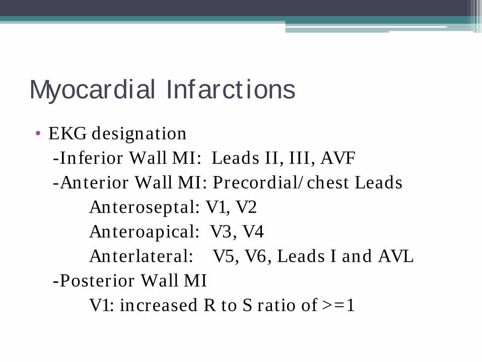
Myocardial Infarctions
• EKG designation -Inferior Wall MI: Leads II, III, AVF -Anterior Wall MI: Precordial/chest Leads Anteroseptal: V1, V2 Anteroapical: V3, V4 Anterlateral: V5, V6, Leads I and AVL -Posterior Wall MI V1: increased R to S ratio of >=1
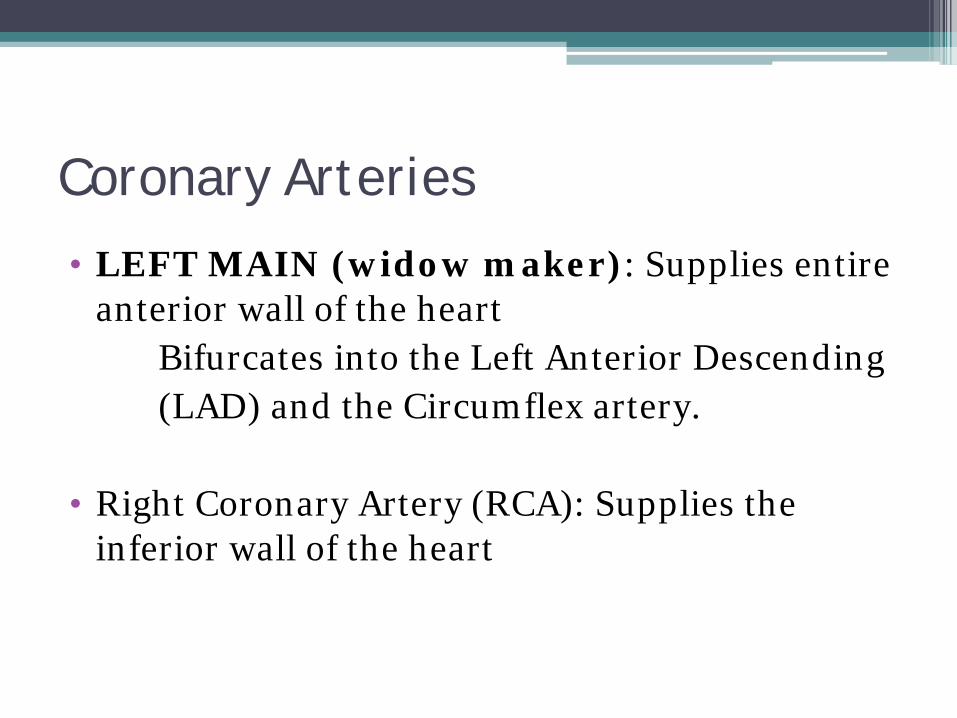
Coronary Arteries
• LEFT MAIN (widow maker): Supplies entire anterior wall of the heart
Bifurcates into the Left Anterior Descending (LAD) and the Circumflex artery.
• Right Coronary Artery (RCA): Supplies the
inferior wall of the heart
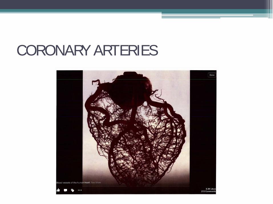
CORONARY ARTERIES
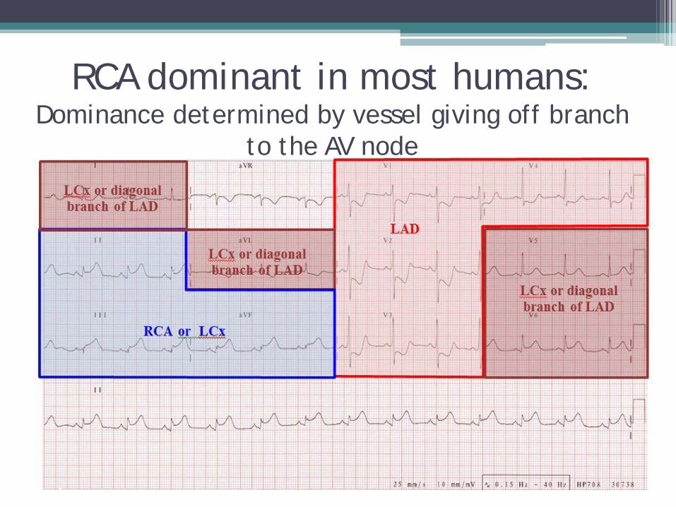
RCA dominant in most humans: Dominance determined by vessel giving off branch
to the AV node
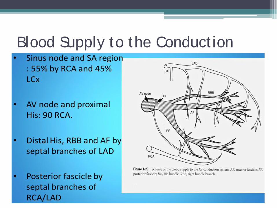
Blood Supply to the Conduction System
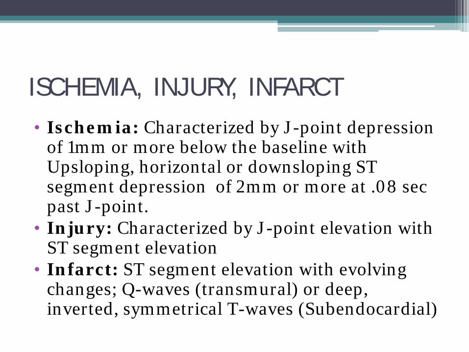
ISCHEMIA, INJURY, INFARCT • Ischemia: Characterized by J-point depression
of 1mm or more below the baseline with Upsloping, horizontal or downsloping ST segment depression of 2mm or more at .08 sec past J-point.
• Injury: Characterized by J-point elevation with ST segment elevation
• Infarct: ST segment elevation with evolving changes; Q-waves (transmural) or deep, inverted, symmetrical T-waves (Subendocardial)
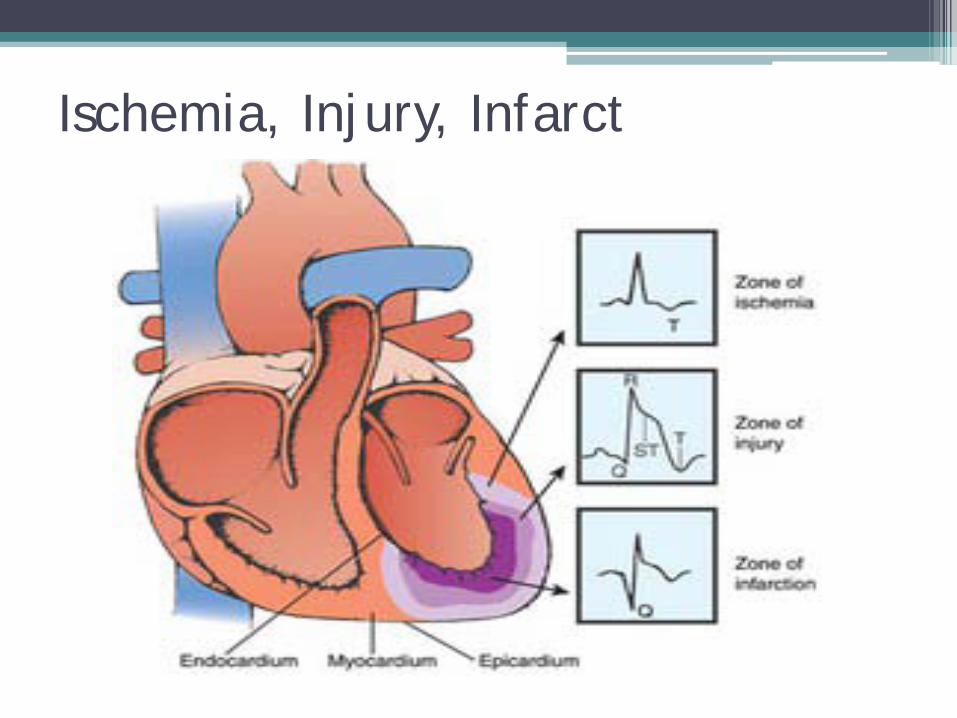
Ischemia, Injury, Infarct
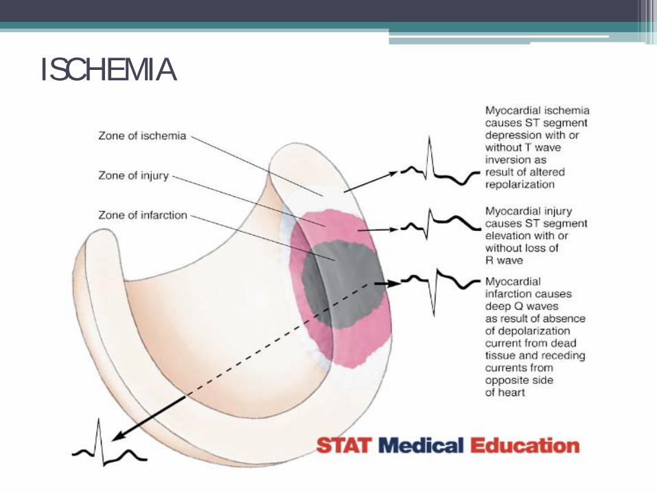
ISCHEMIA
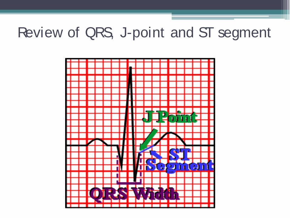
Review of QRS, J-point and ST segment
Determining significance of ST depression • Find the J-point
• Measure .08 sec (two small boxes) to the right
• If the ST segment is depressed at least 1 mm (1
small box) it is considered significant possible ischemic changes.
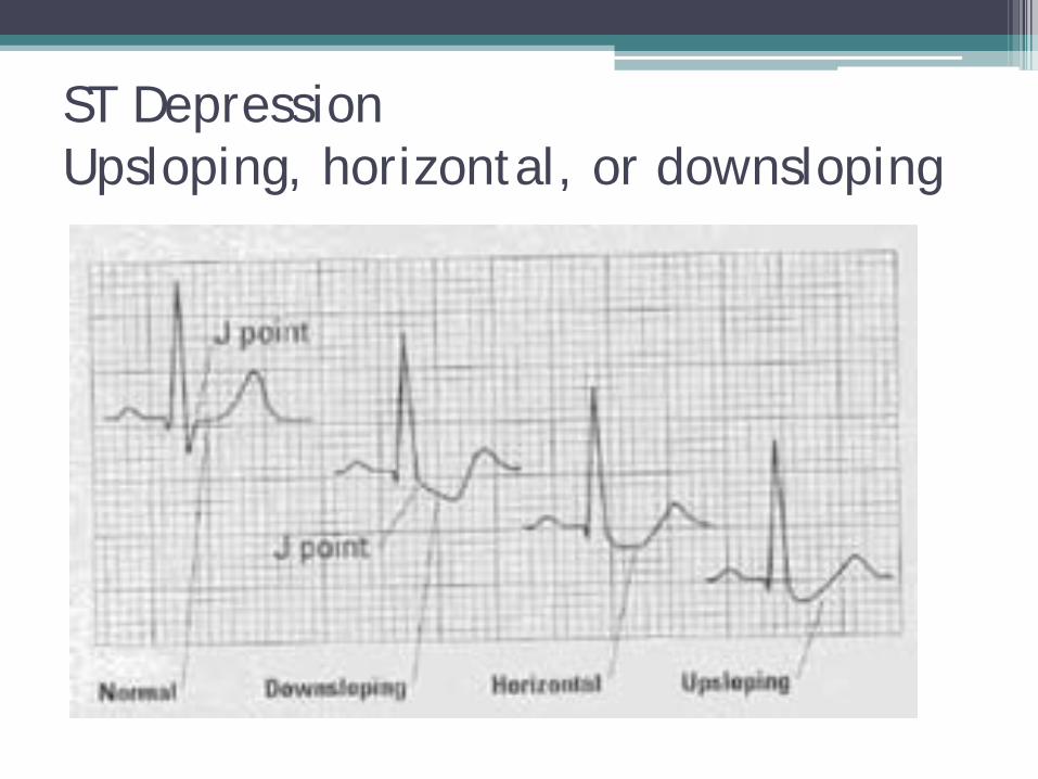
ST Depression Upsloping, horizontal, or downsloping
How do I know when to be concerned?
• Upsloping ST segment depression is of concern
• Horizontal ST segment depression is of more concern
• Downsloping ST segment depression is of
greatest concern
• NOTE: clinical correlation is ALWAYS required
Additional factors • ST depression is of greater concern when you see
it with minimal amounts of exertion (lower heart rates) when compared to peak exertion
• ST depression is of greater concern when it takes longer to return to baseline after exercise
• ST depression is of greatest concern when changes continue to occur after exertion is terminated
But clinical correlation is ALWAYS required
• Always monitor the patient Vitals Symptoms Just the way the patient looks: Pallor, sweating, ashen
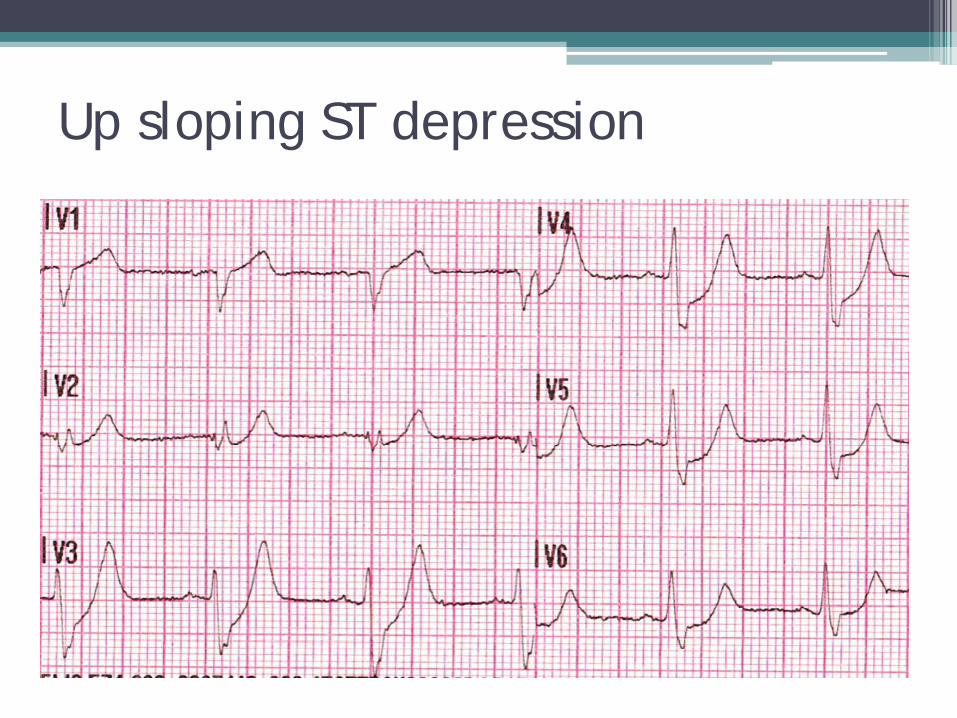
Up sloping ST depression
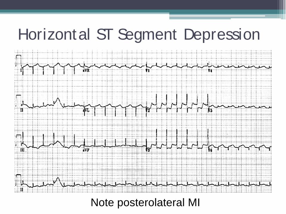
Horizontal ST Segment Depression
Note posterolateral MI
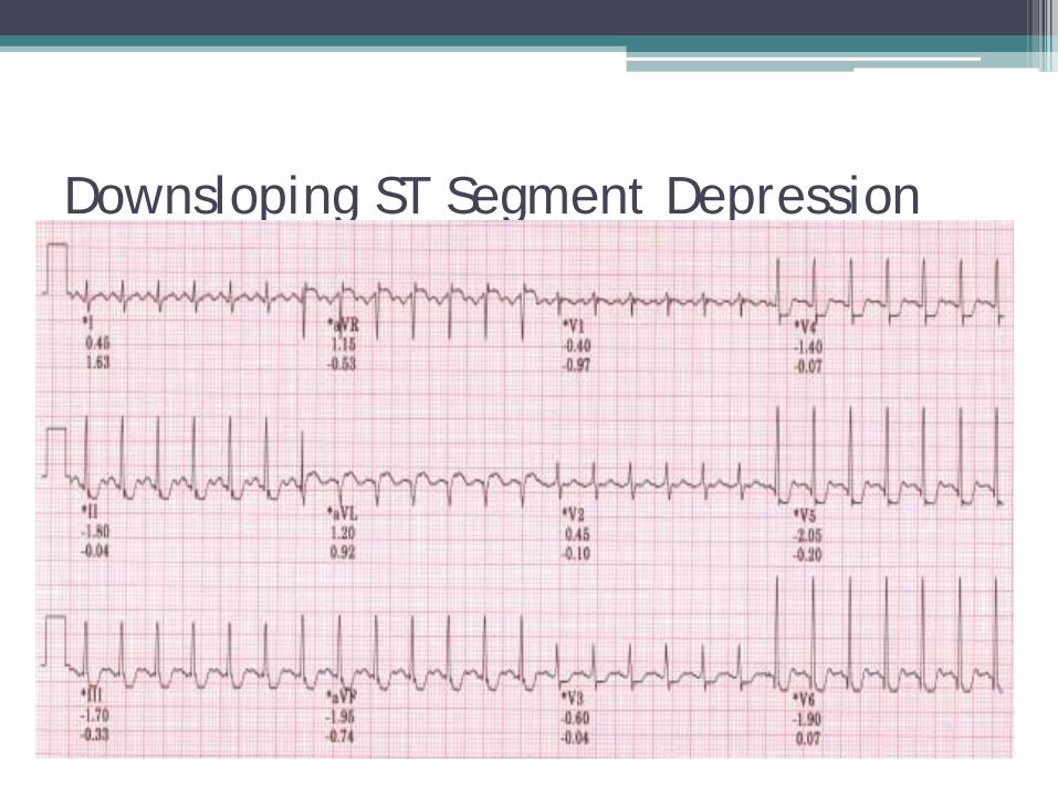
Downsloping ST Segment Depression
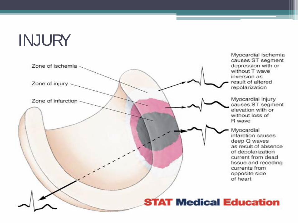
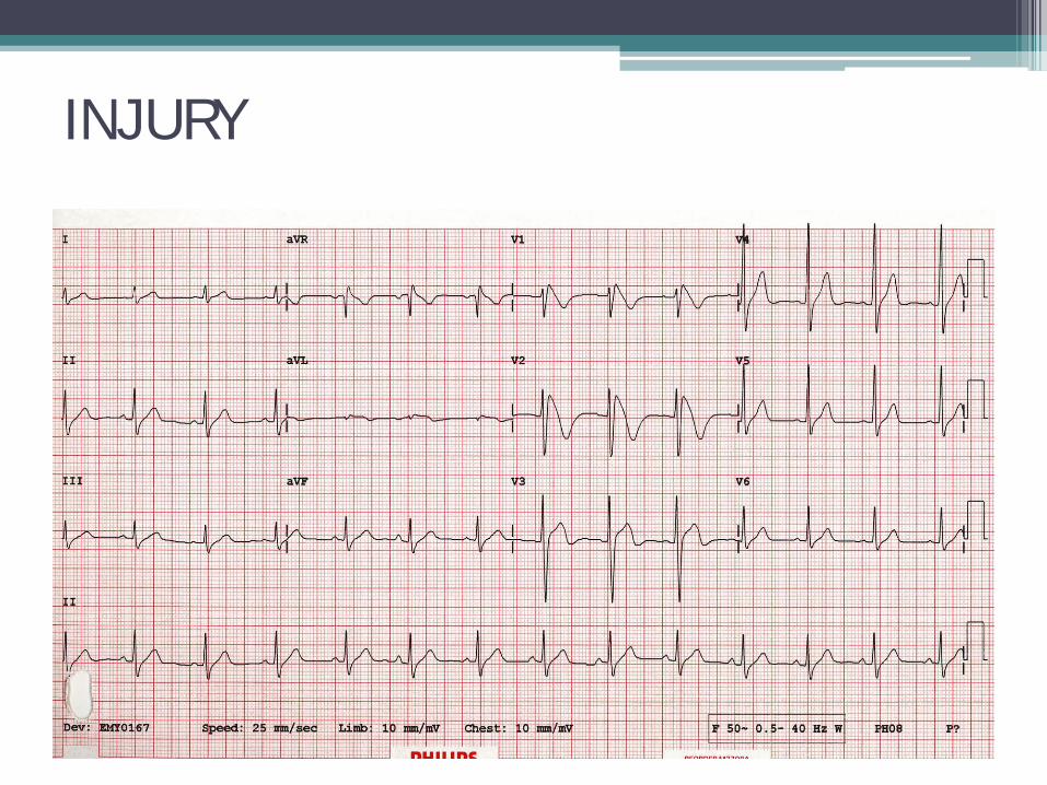
INJURY
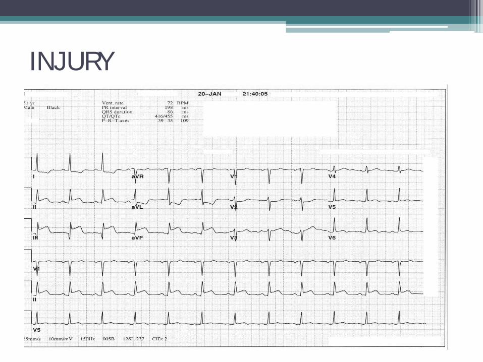
INJURY
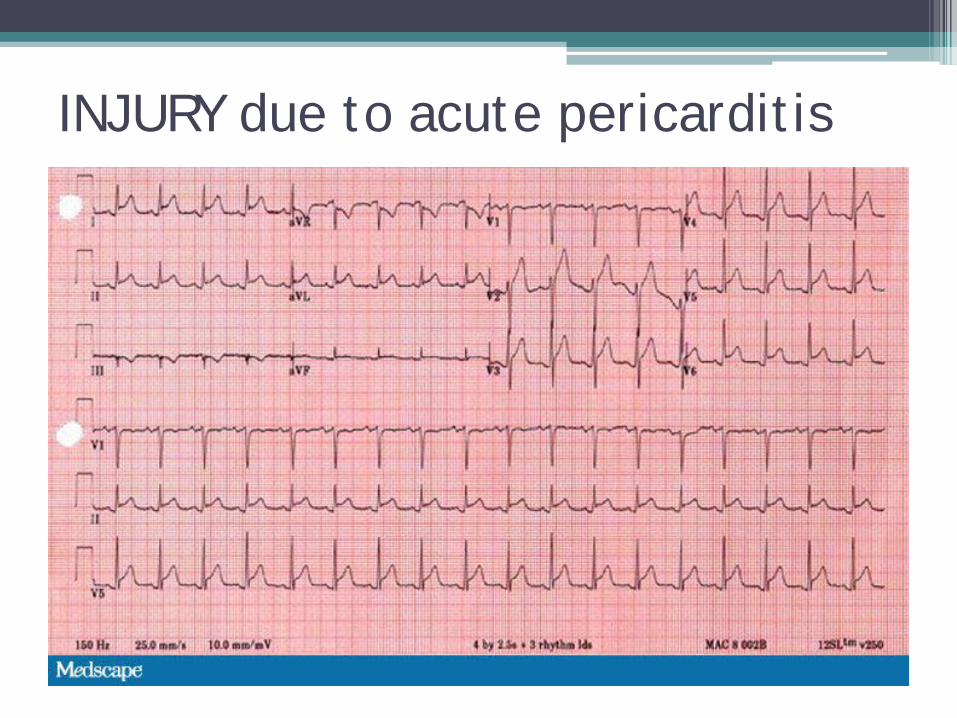
INJURY due to acute pericarditis
INJURY
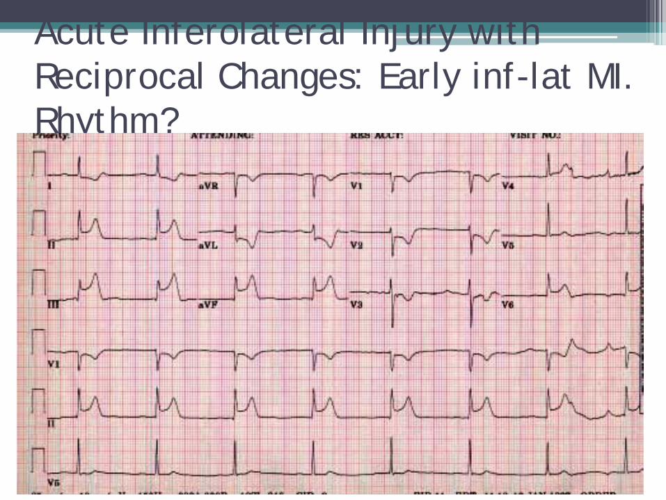
Acute Inferolateral Injury with Reciprocal Changes: Early inf-lat MI. Rhythm?
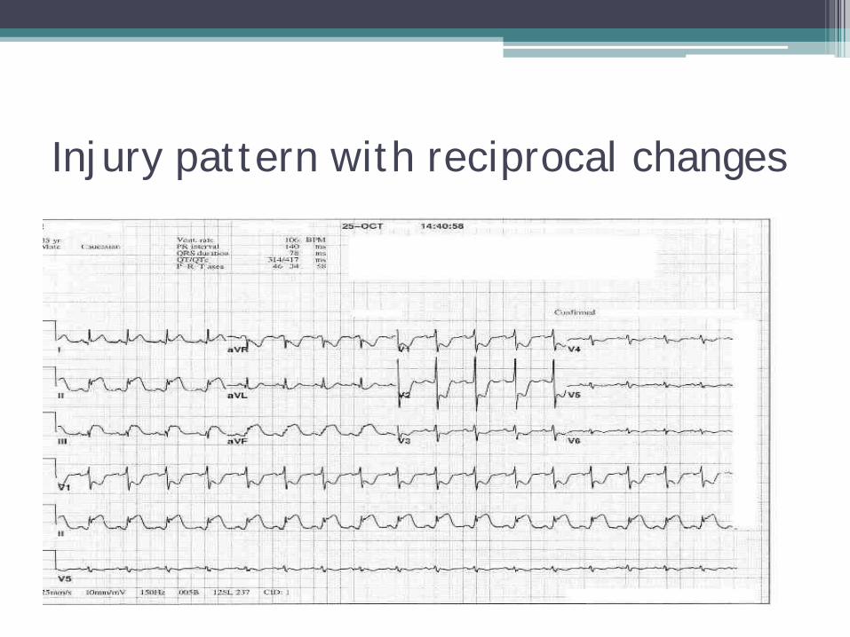
Injury pattern with reciprocal changes
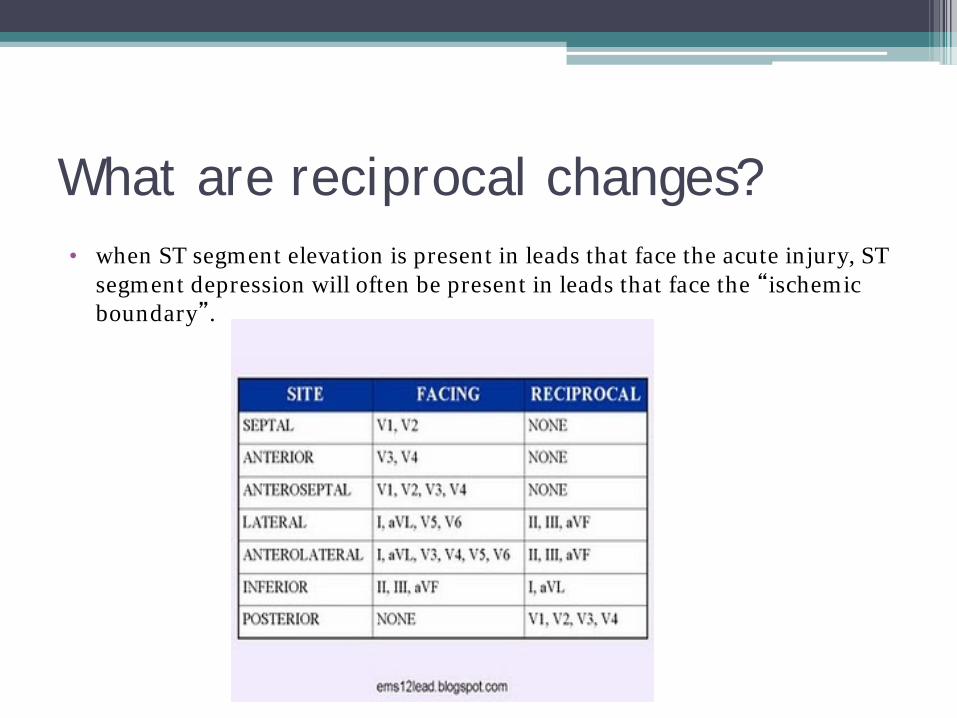
What are reciprocal changes? • when ST segment elevation is present in leads that face the acute injury, ST
segment depression will often be present in leads that face the “ischemic boundary”.

INFARCT
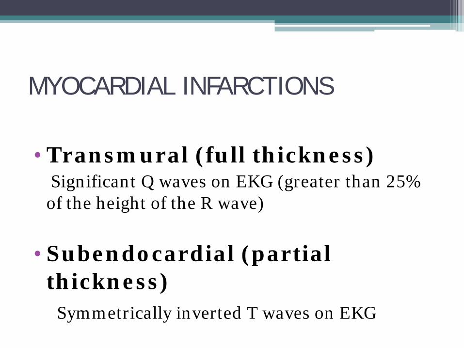
MYOCARDIAL INFARCTIONS
• Transmural (full thickness) Significant Q waves on EKG (greater than 25%
of the height of the R wave)
• Subendocardial (partial thickness)
Symmetrically inverted T waves on EKG
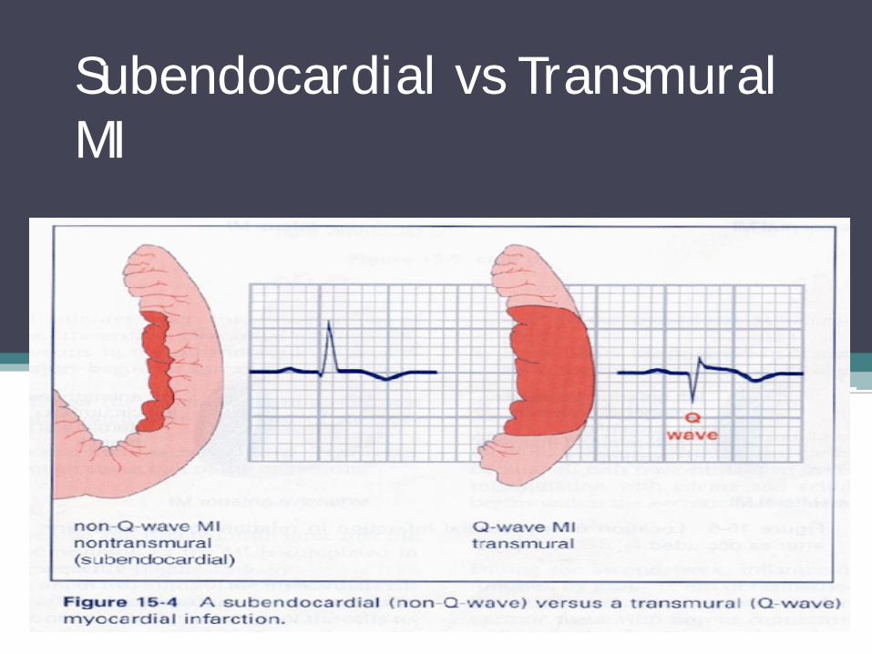
Subendocardial vs Transmural MI

Transmural Inferior Wall MI
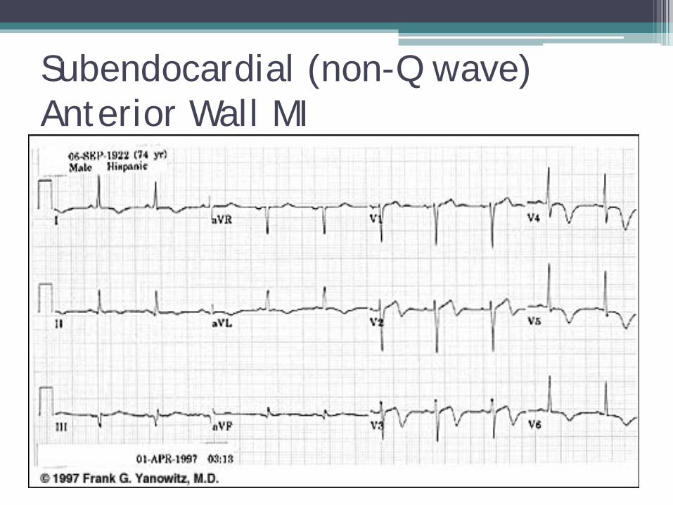
Subendocardial (non-Q wave) Anterior Wall MI
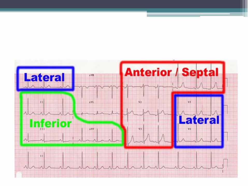
Myocardial Infarctions • Locations Inferior Wall Anterior Wall Lateral Wall Posterior Wall
INFERIOR WALL MI’s
• Usually due to blockage of the Right Coronary Artery (RCA)
• EKG: primary changes seen in Leads II, III and AVF with reciprocal changes in V5 and V6
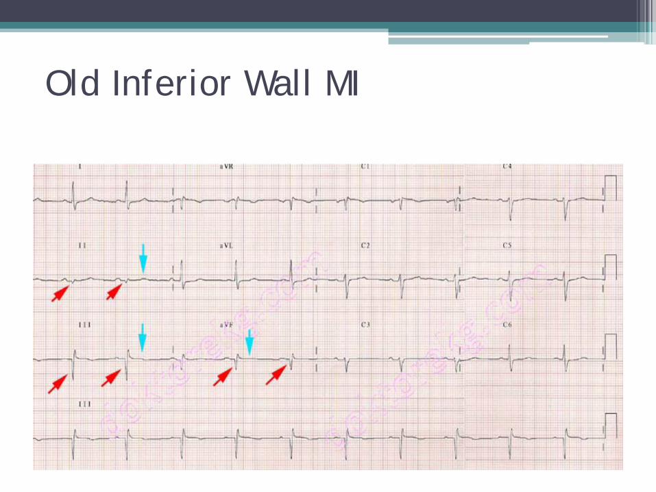
Old Inferior Wall MI
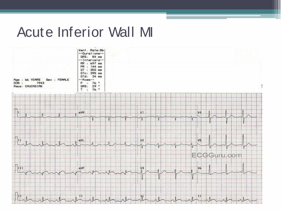
Acute Inferior Wall MI
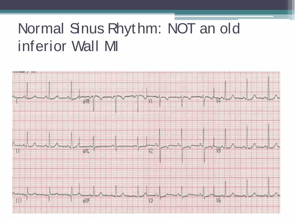
Normal Sinus Rhythm: NOT an old inferior Wall MI
Anterior Wall MI’s
• Can be due to blockage of Left Main Coronary Artery,
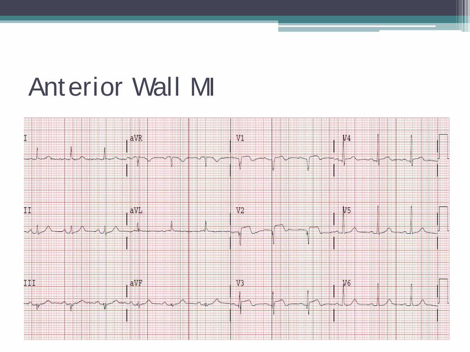
Anterior Wall MI
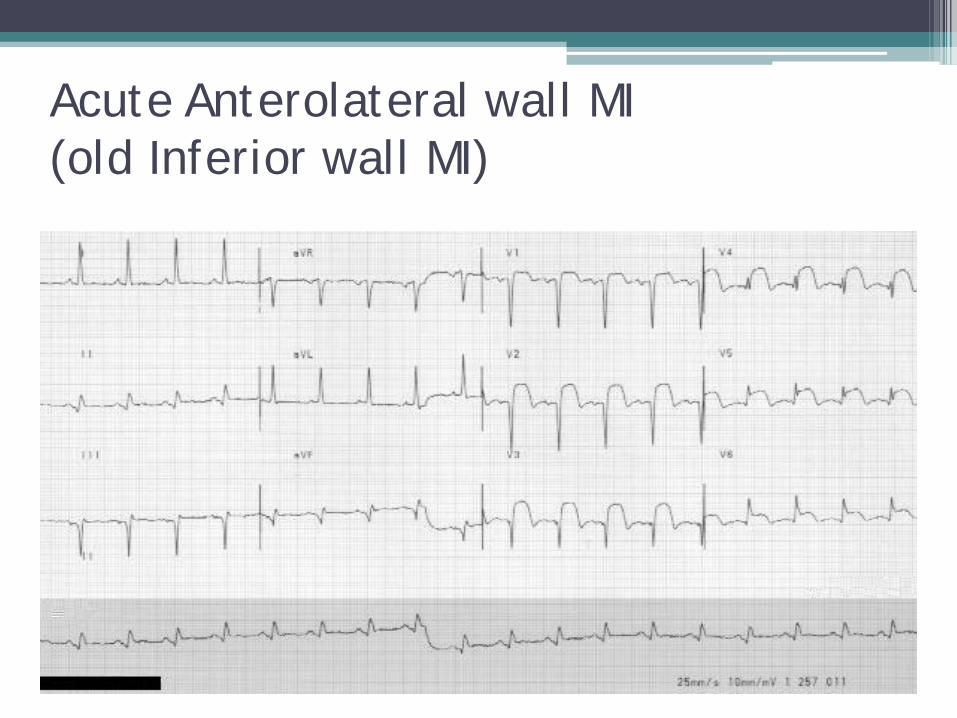
Acute Anterolateral wall MI (old Inferior wall MI)
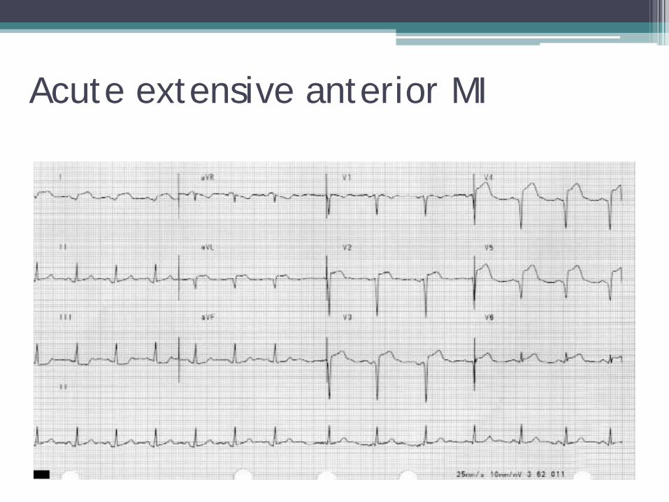
Acute extensive anterior MI
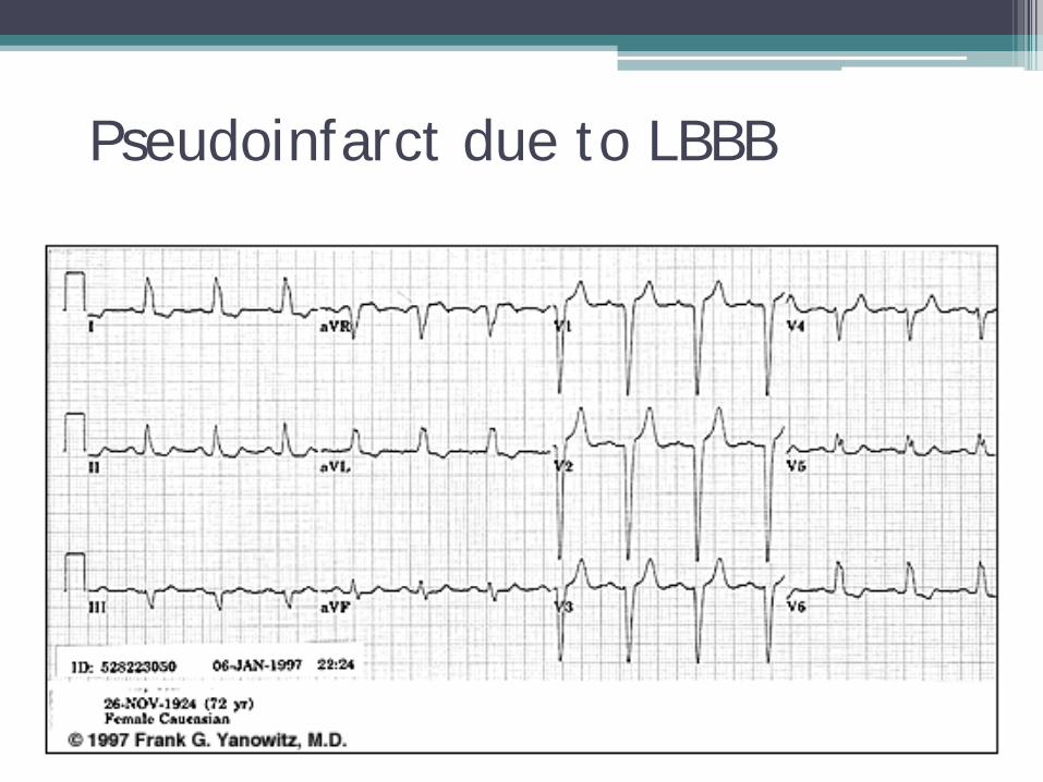
Pseudoinfarct due to LBBB

What about the posterior wall infarction? • There are no ECG leads overlaying the posterior
wall
• Use reciprocal changes in right chest leads. Do additional right chest leads if suspected
• Difficult to diagnose
• Posterior MI not uncommonly associated with inferior or lateral wall MIs
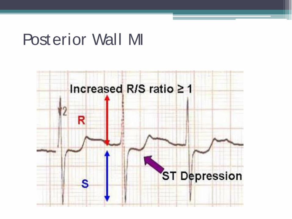
Posterior Wall MI
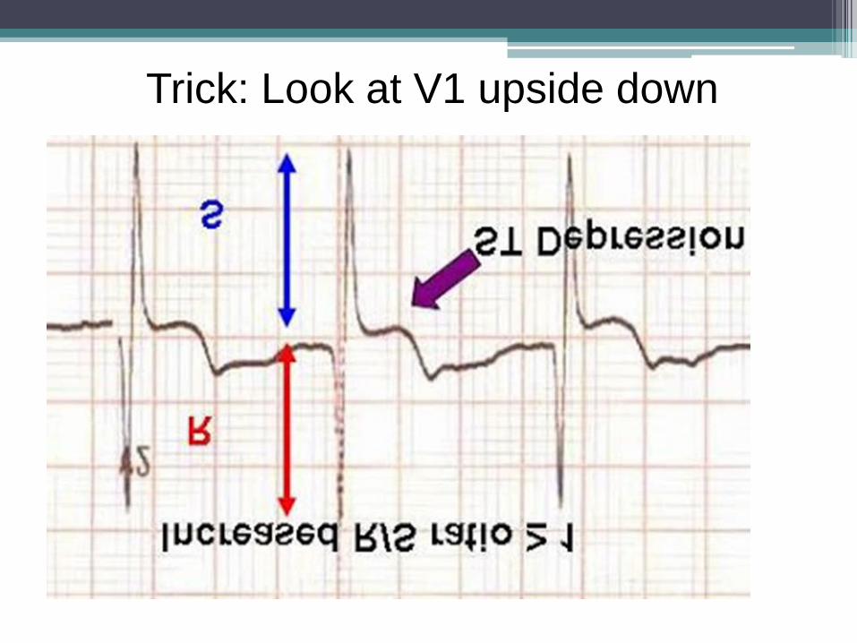
Trick: Look at V1 upside down
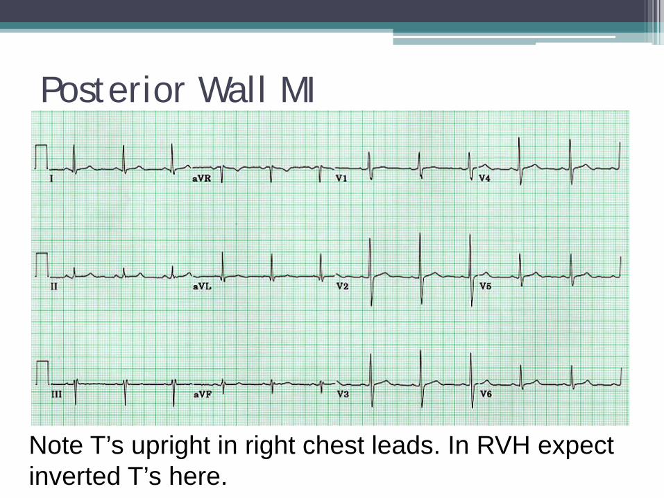
Posterior Wall MI
Note T’s upright in right chest leads. In RVH expect inverted T’s here.

Break Time
Real Patient Case
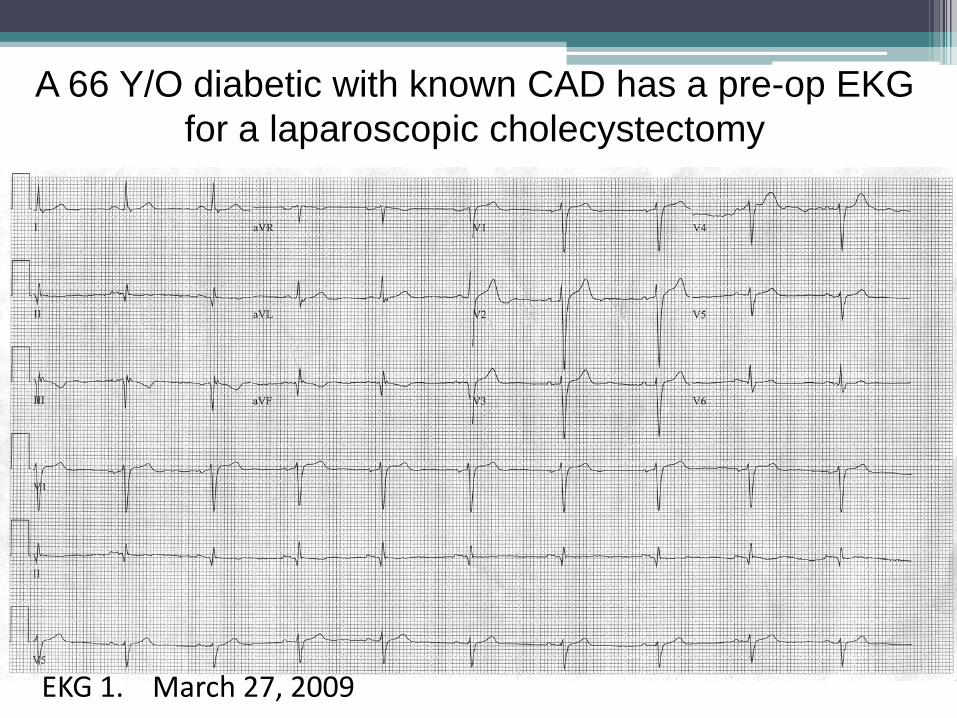
A 66 Y/O diabetic with known CAD has a pre-op EKG for a laparoscopic cholecystectomy
EKG 1. March 27, 2009
DH is a 66 Y/O diabetic with known CAD who was “cleared” for a laparoscopic cholecystectomy after an examination and EKG #1. His Plavix & ASA were stopped pre-op. Surgery was uncomplicated. In the recovery room anesthesia saw him for some PVC’s on the monitor. He was then allowed to return to his room. That evening he complained of pain in his left biceps. The nurse gave him Tylenol, which had been ordered for pain, and he did not require another dose.
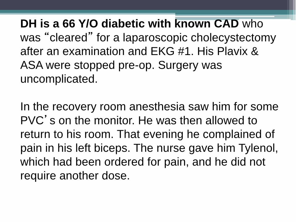
EKG ordered post op day 1 by surgical PA-C. Patient had C/O left biceps pain & had tachycardia.
EKG 2. April 27, 2009 07:27:19
Day 1 post op: On morning rounds a surgical PA noted tachycardia and the previous biceps pain. She obtained EKG 2 and called her supervisor who immediately had a cardiologist review the EKG by fax. He told her the changes showed an extensive acute MI, the kind often associated with complications such as papillary muscle rupture, a ruptured septum or heart, heart failure and/or shock. He urged immediate interventional consultation for treatment and an emergency cardiac catheterization.
Day 1 post op: A formal emergency cardiology consult followed, revealing stable hemodynamics but a new systolic murmur. Meanwhile the supervising PA checked the previous hospital notes. She noted that the patient’s Plavix & ASA had been stopped pre-op. Reviewed rhythm strips from the recovery room revealed the noted PVCs but also some ST elevation. Cardiac markers and an emergency cardiac catheterization were ordered.
Day 1 post op: The cardiac catheterization revealed total occlusions of both the LAD and RCA and a VSD consistent with a ruptured septum. Cardiac markers had come back positive. Post cath EKG #3 was obtained. He underwent emergency CABG and repair of the VSD. His post op EF was 35%. Several weeks later his VSD repair failed and repeat surgery was needed. He tolerated this well.
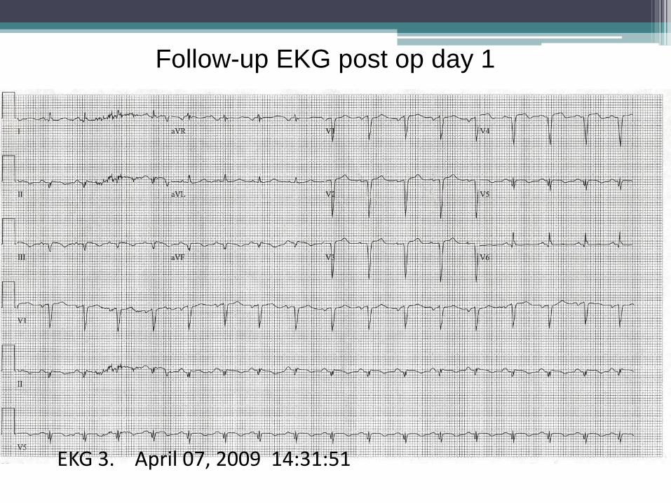
Follow-up EKG post op day 1
EKG 3. April 07, 2009 14:31:51

THOUGHTS????
Acute Pericarditis • EKG can easily be confused with an evolving MI • The ST segment and T wave changes in
pericarditis tend to be diffuse (although not always) and involve far more leads than the localized effect of infarction
• No reciprocal changes • Q wave formation does not occur • Elevated ST returns to baseline before the T
wave inverts
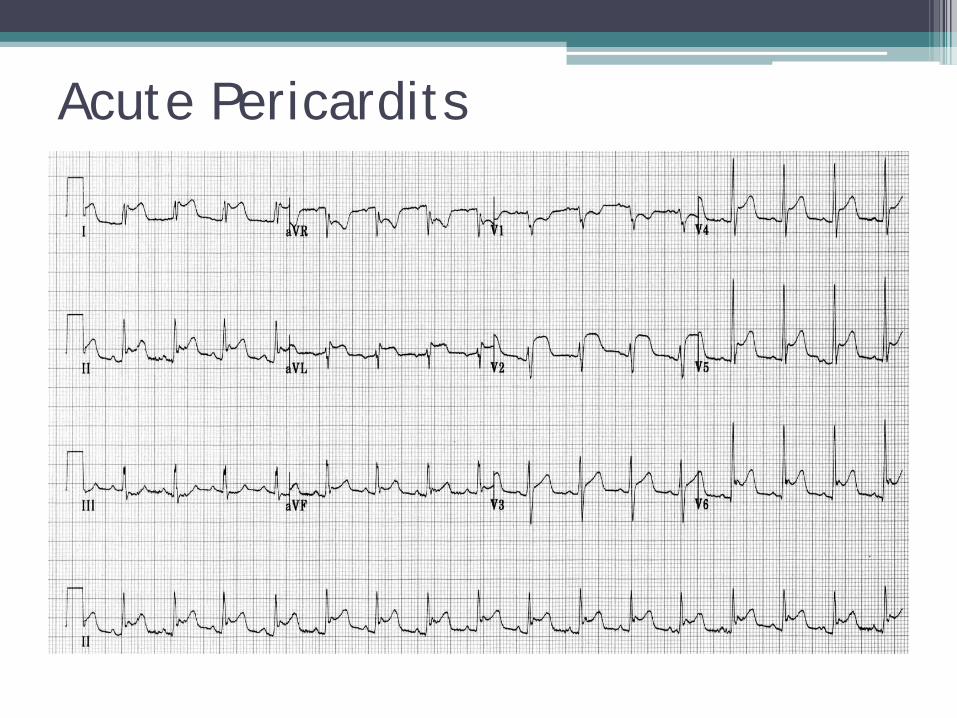
Acute Pericardits
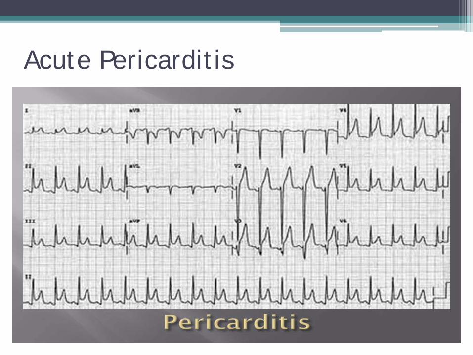
Acute Pericarditis
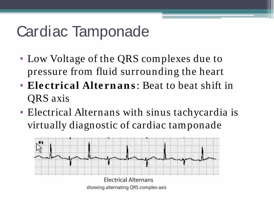
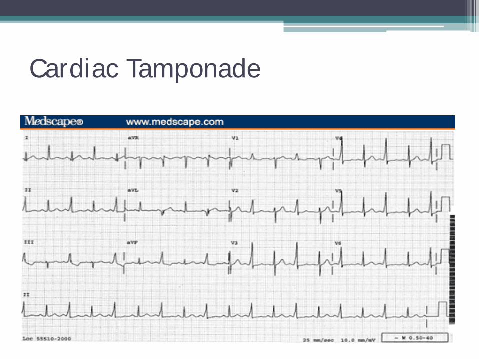
Cardiac Tamponade
• Low Voltage of the QRS complexes due to pressure from fluid surrounding the heart
• Electrical Alternans: Beat to beat shift in QRS axis
• Electrical Alternans with sinus tachycardia is virtually diagnostic of cardiac tamponade
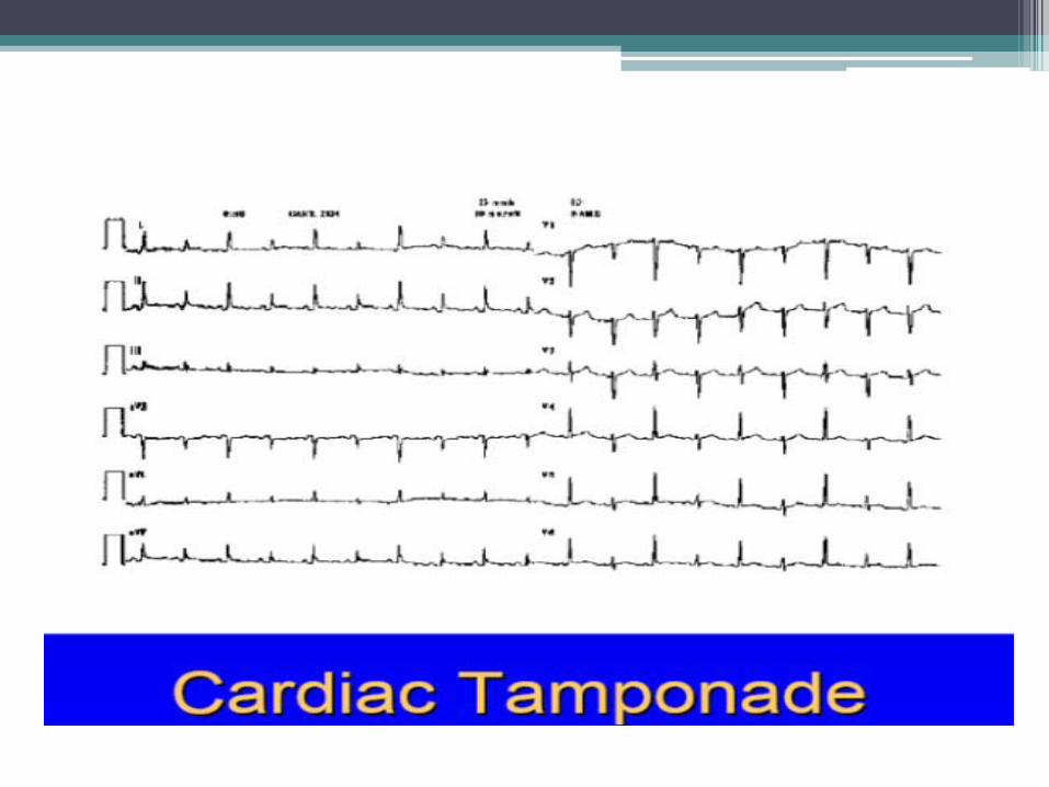
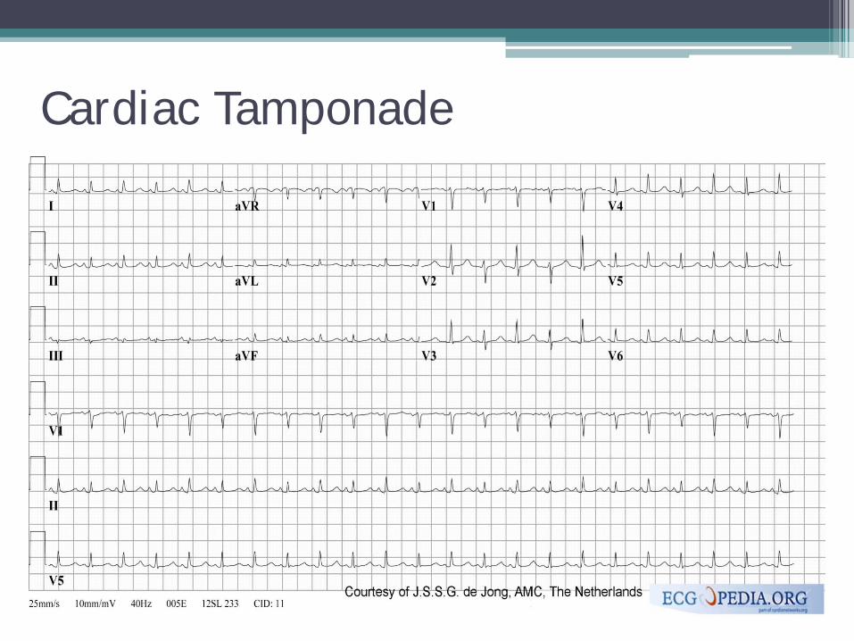
Cardiac Tamponade
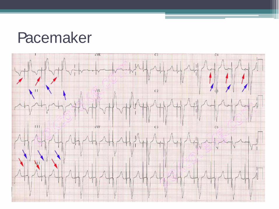
Pacemaker

LET’s PRACTICE
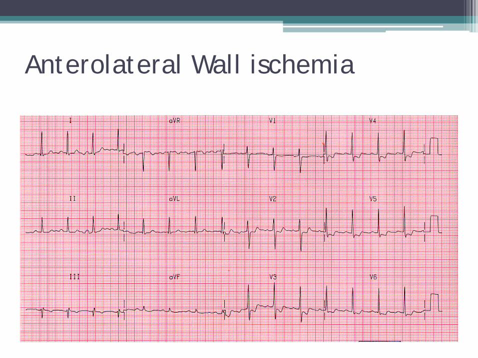
Anterolateral Wall ischemia

Normal Sinus Rhythm
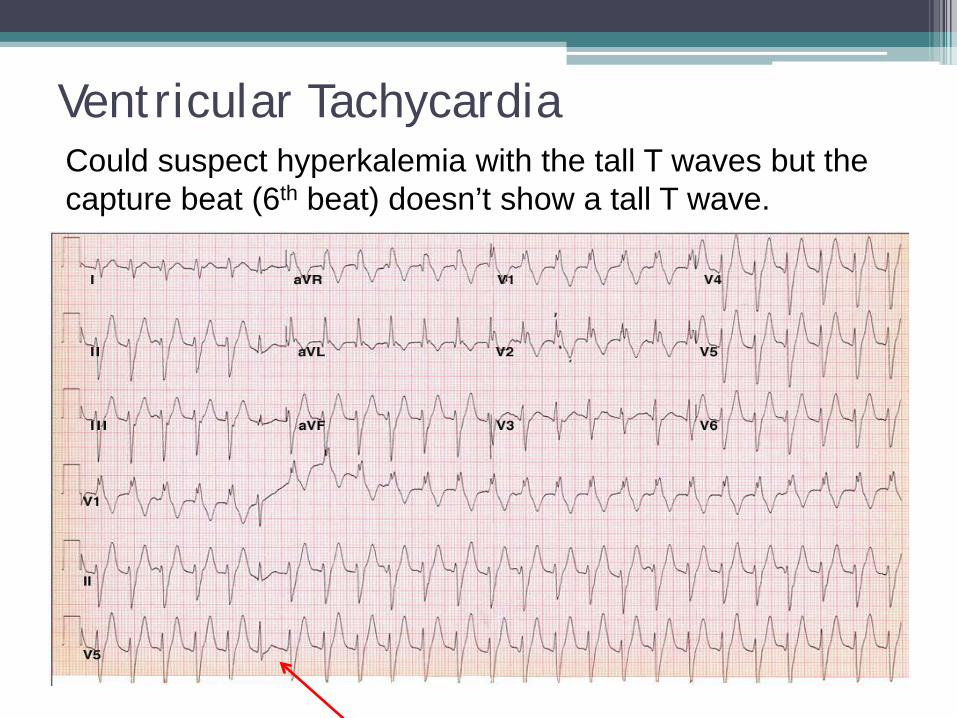
Ventricular Tachycardia
Could suspect hyperkalemia with the tall T waves but the capture beat (6th beat) doesn’t show a tall T wave.
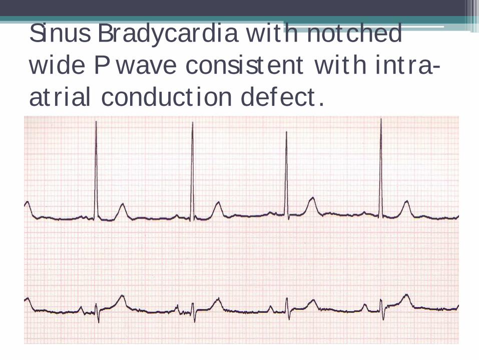
Sinus Bradycardia with notched wide P wave consistent with intra-atrial conduction defect.
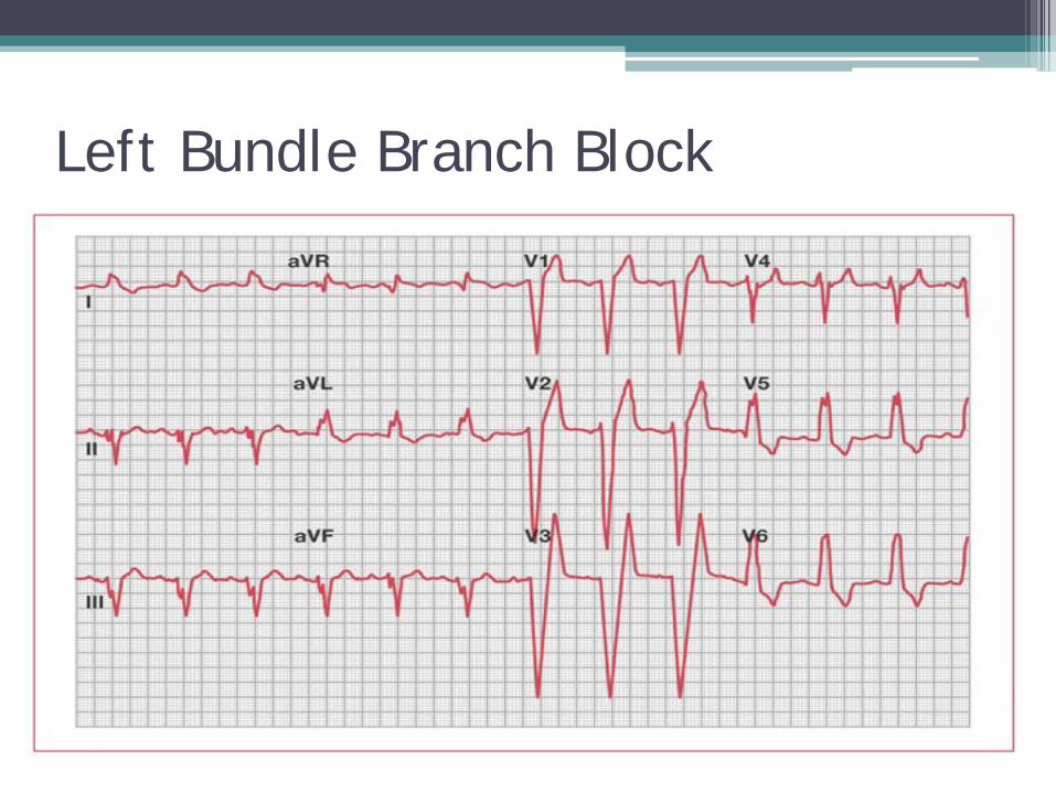
Left Bundle Branch Block
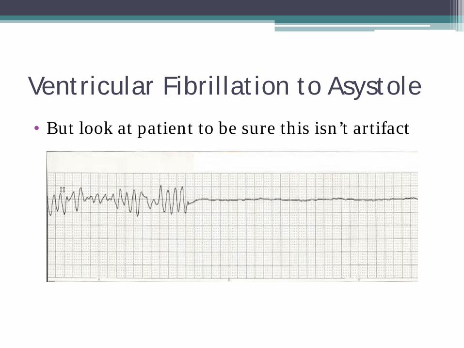
Ventricular Fibrillation to Asystole
• But look at patient to be sure this isn’t artifact
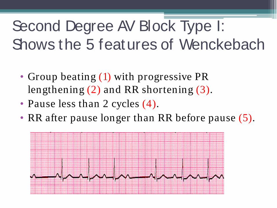
Second Degree AV Block Type I: Shows the 5 features of Wenckebach
• Group beating (1) with progressive PR lengthening (2) and RR shortening (3).
• Pause less than 2 cycles (4). • RR after pause longer than RR before pause (5).
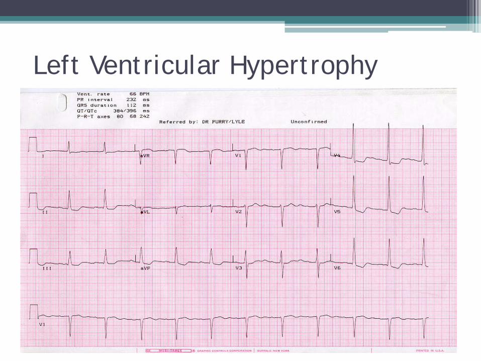
Left Ventricular Hypertrophy
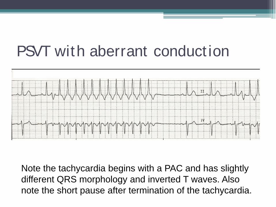
PSVT with aberrant conduction
Note the tachycardia begins with a PAC and has slightly different QRS morphology and inverted T waves. Also note the short pause after termination of the tachycardia.
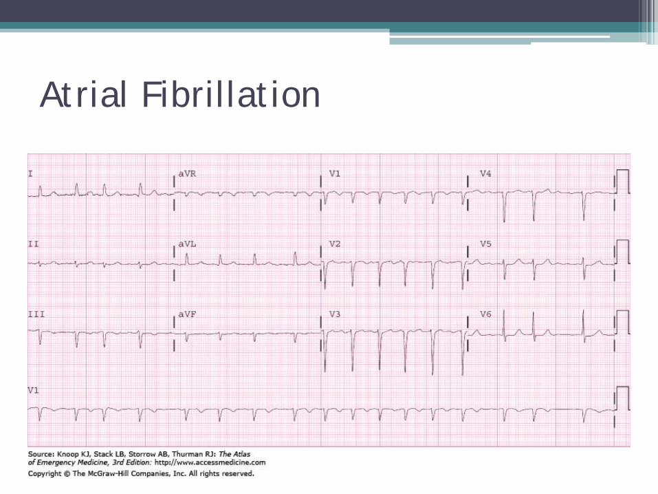
Atrial Fibrillation
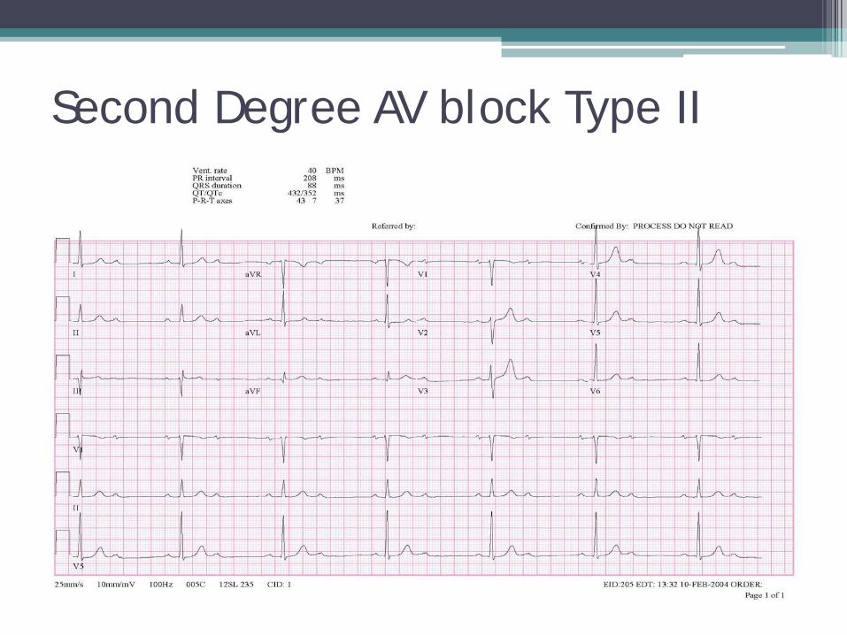
Second Degree AV block Type II

Atrial Flutter (variable conduction)
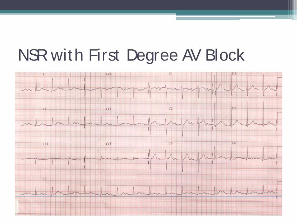
NSR with First Degree AV Block
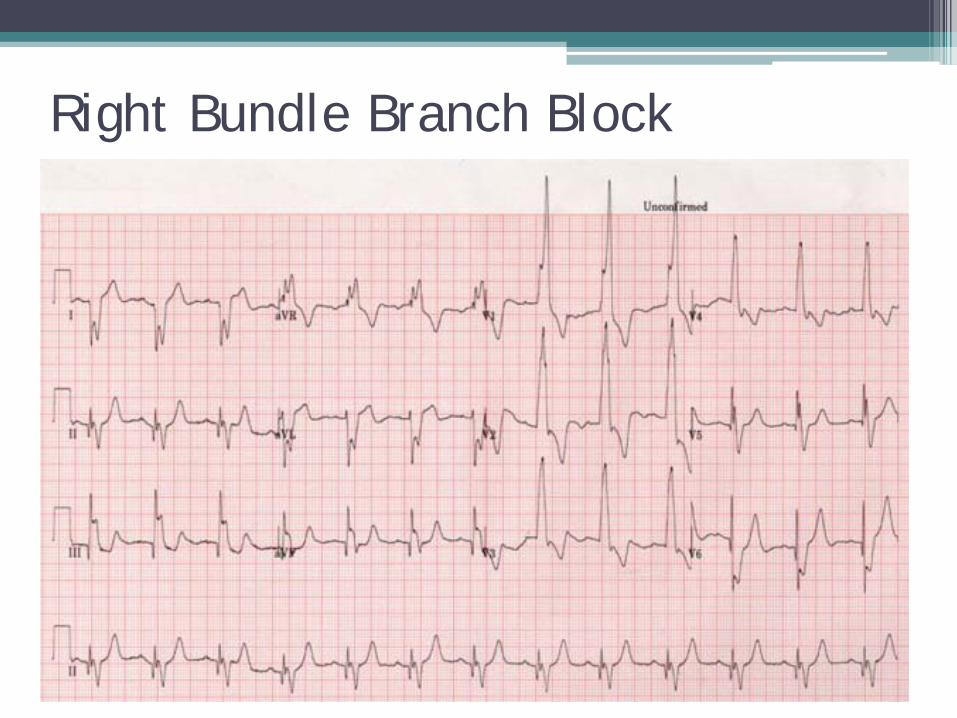
Right Bundle Branch Block
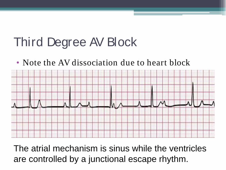
Third Degree AV Block
• Note the AV dissociation due to heart block
The atrial mechanism is sinus while the ventricles are controlled by a junctional escape rhythm.
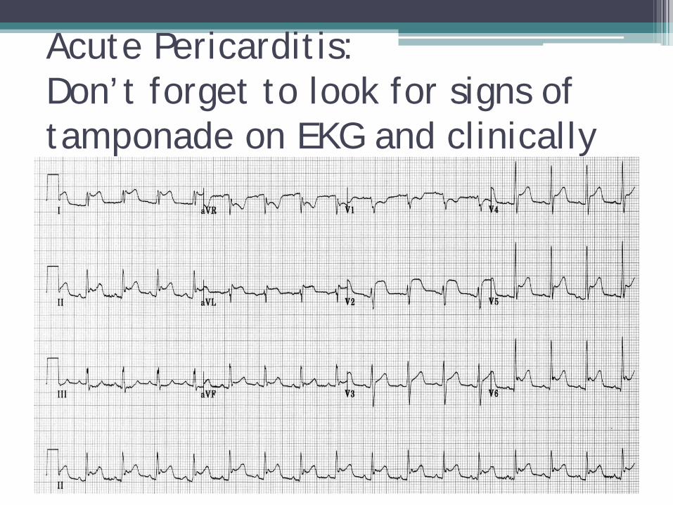
Acute Pericarditis: Don’t forget to look for signs of tamponade on EKG and clinically
Cardiac Tamponade


