137
X-Ray Scattering Studies of Thin Polymer Films Introduction to Neutron and X-Ray Scattering Sunil K. Sinha UCSD/LANL Acknowledgements: Prof. R.Pynn( Indiana U.) Prof. M.Tolan (U. Dortmund)

X-Ray Scattering Studies of Thin
Polymer Films Introduction to Neutron and X-Ray Scattering
Sunil K. Sinha
UCSD/LANL
Acknowledgements: Prof. R.Pynn( Indiana U.)
Prof. M.Tolan (U. Dortmund)
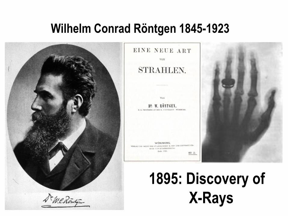
Wilhelm Conrad Röntgen 1845-1923
1895: Discovery of
X-Rays
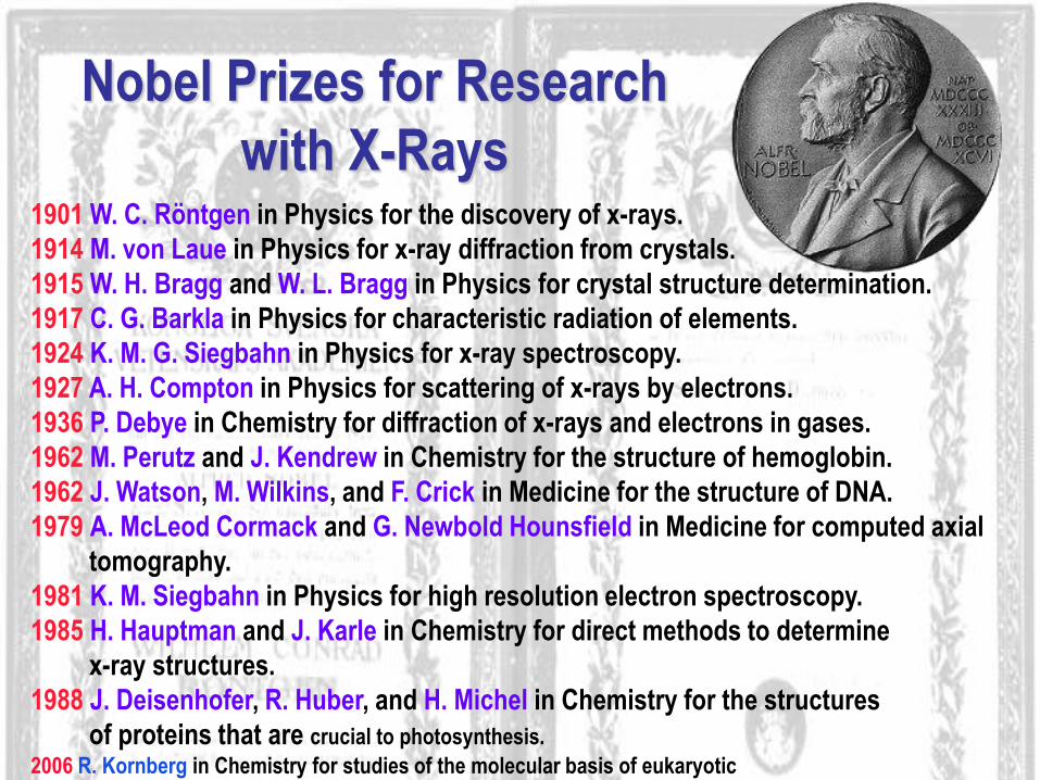
1901 W. C. Röntgen in Physics for the discovery of x-rays.
1914 M. von Laue in Physics for x-ray diffraction from crystals.
1915 W. H. Bragg and W. L. Bragg in Physics for crystal structure determination.
1917 C. G. Barkla in Physics for characteristic radiation of elements.
1924 K. M. G. Siegbahn in Physics for x-ray spectroscopy.
1927 A. H. Compton in Physics for scattering of x-rays by electrons.
1936 P. Debye in Chemistry for diffraction of x-rays and electrons in gases.
1962 M. Perutz and J. Kendrew in Chemistry for the structure of hemoglobin.
1962 J. Watson, M. Wilkins, and F. Crick in Medicine for the structure of DNA.
1979 A. McLeod Cormack and G. Newbold Hounsfield in Medicine for computed axial
tomography.
1981 K. M. Siegbahn in Physics for high resolution electron spectroscopy.
1985 H. Hauptman and J. Karle in Chemistry for direct methods to determine
x-ray structures.
1988 J. Deisenhofer, R. Huber, and H. Michel in Chemistry for the structures
of proteins that are crucial to photosynthesis.
2006 R. Kornberg in Chemistry for studies of the molecular basis of eukaryotic
transcription.
2009 V.Ramakrishnan, T.A.Steitz and A.E.Yonath for studies of the structure and
function of the ribosome.
Nobel Prizes for Research
with X-Rays

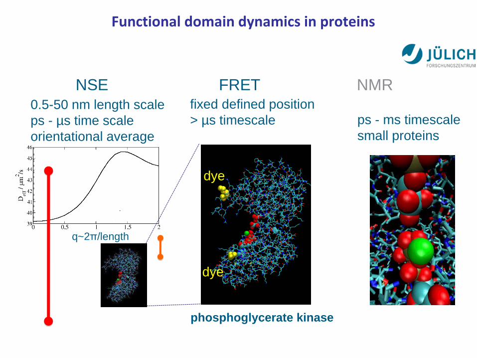
q~2π/length
NSE FRET NMR
0.5-50 nm length scale
ps - µs time scale
orientational average
fixed defined position
> µs timescale ps - ms timescale
small proteins
phosphoglycerate kinase
dye
dye
Functional domain dynamics in proteins
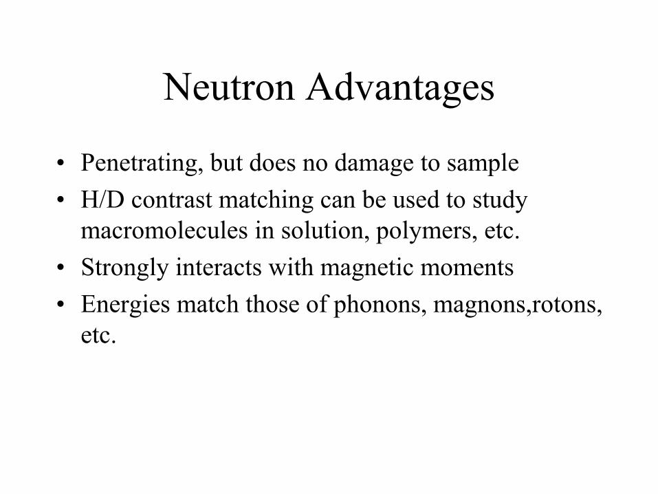
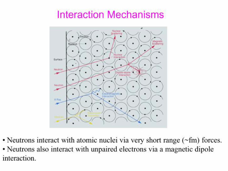
Neutron Advantages
• Penetrating, but does no damage to sample
• H/D contrast matching can be used to study
macromolecules in solution, polymers, etc.
• Strongly interacts with magnetic moments
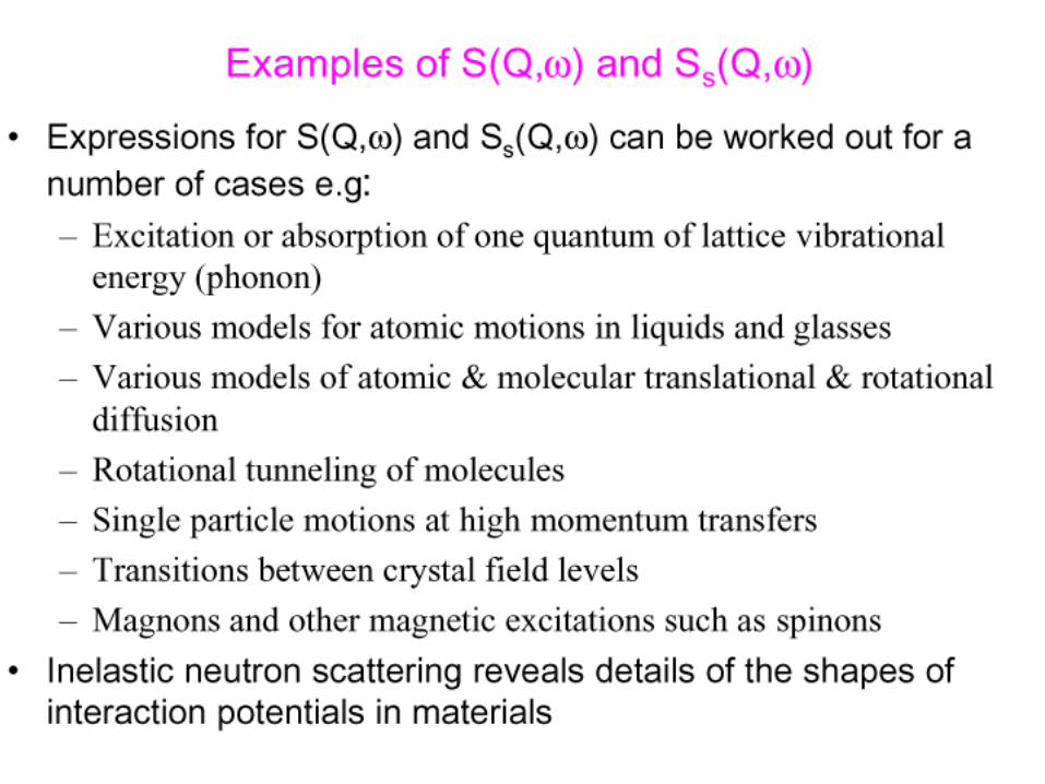
• Energies match those of phonons, magnons,rotons,
etc.
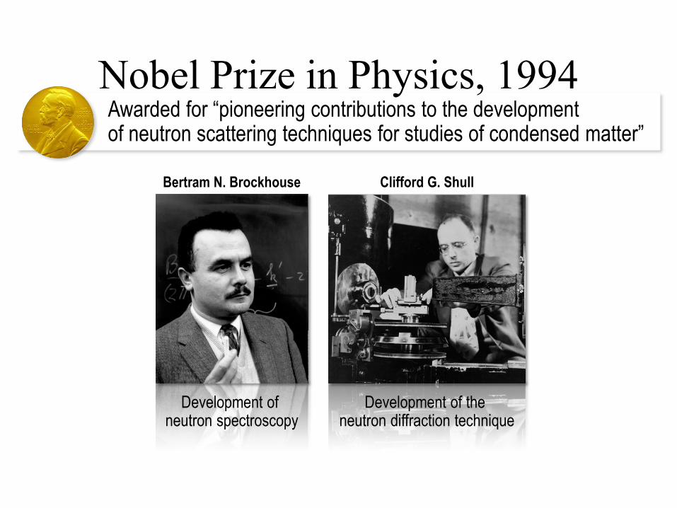
Awarded for “pioneering contributions to the development of neutron scattering techniques for studies of condensed matter”
Nobel Prize in Physics, 1994
Bertram N. Brockhouse Clifford G. Shull
Development of neutron spectroscopy
Development of the neutron diffraction technique


Antiferromagnetic Structure of MnO
(Shull and Wollan Phys. Rev. 83, 333 (1951)
First Study of an Antiferromagnetic Structure
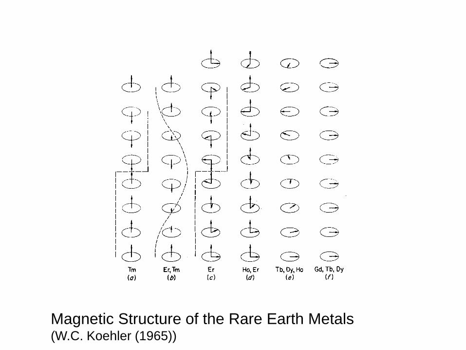
Magnetic Structure of the Rare Earth Metals (W.C. Koehler (1965))

Science with X-Rays
• Diffraction and crystal structures
• Structure Factors of liquids and glasses
• Structures of Thin Films
• ARPES
• EXAFS, XANES
• Studies of Magnetism with resonant XMS
• Inelastic X-ray scattering: phonons, electronic excitations
• X-ray Photon Correlation Spectroscopy
• Microscopy
• Imaging/Tomography
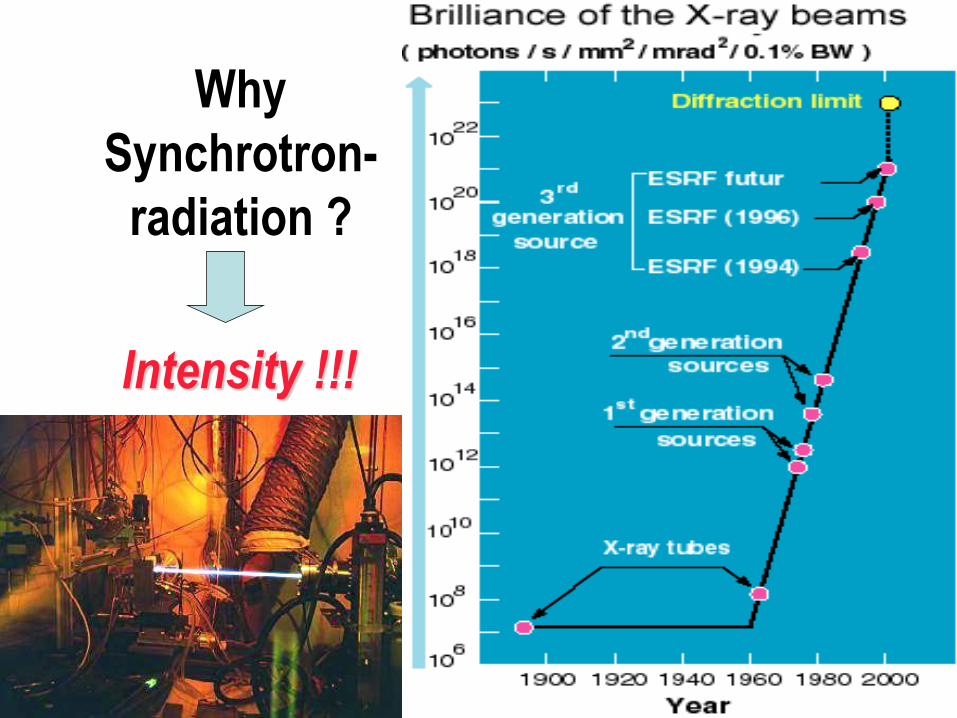
Why
Synchrotron-
radiation ?
Intensity !!!
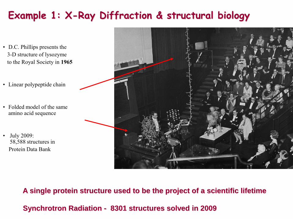
• D.C. Phillips presents the
3-D structure of lysozyme
to the Royal Society in 1965
• Linear polypeptide chain
• Folded model of the same amino acid sequence
• July 2009: 58,588 structures in
Protein Data Bank
A single protein structure used to be the project of a scientific lifetime
Synchrotron Radiation - 8301 structures solved in 2009
Example 1: X-Ray Diffraction & structural biology
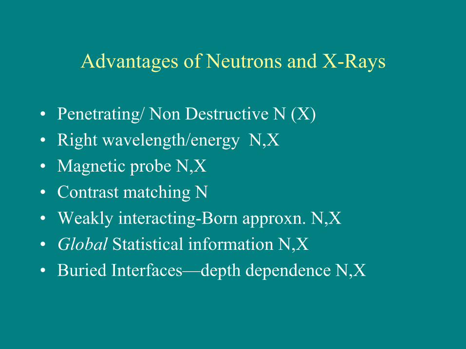
Advantages of Neutrons and X-Rays
• Penetrating/ Non Destructive N (X)
• Right wavelength/energy N,X
• Magnetic probe N,X
• Contrast matching N
• Weakly interacting-Born approxn. N,X
• Global Statistical information N,X
• Buried Interfaces—depth dependence N,X
Neutron and X-ray Scattering:
“small” science at big
facilities!

Historic accomplishments (Neutrons)
•Antiferromagnetic Structures
•Rare earth spirals and other spin structures
•Spin wave dispersion
•Our whole understanding of the details of exchange interactions in solids
•Magnetism and Superconductivity
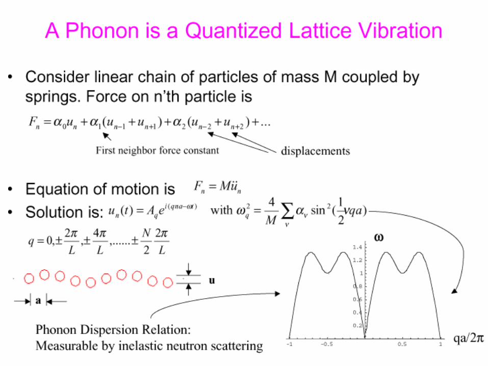
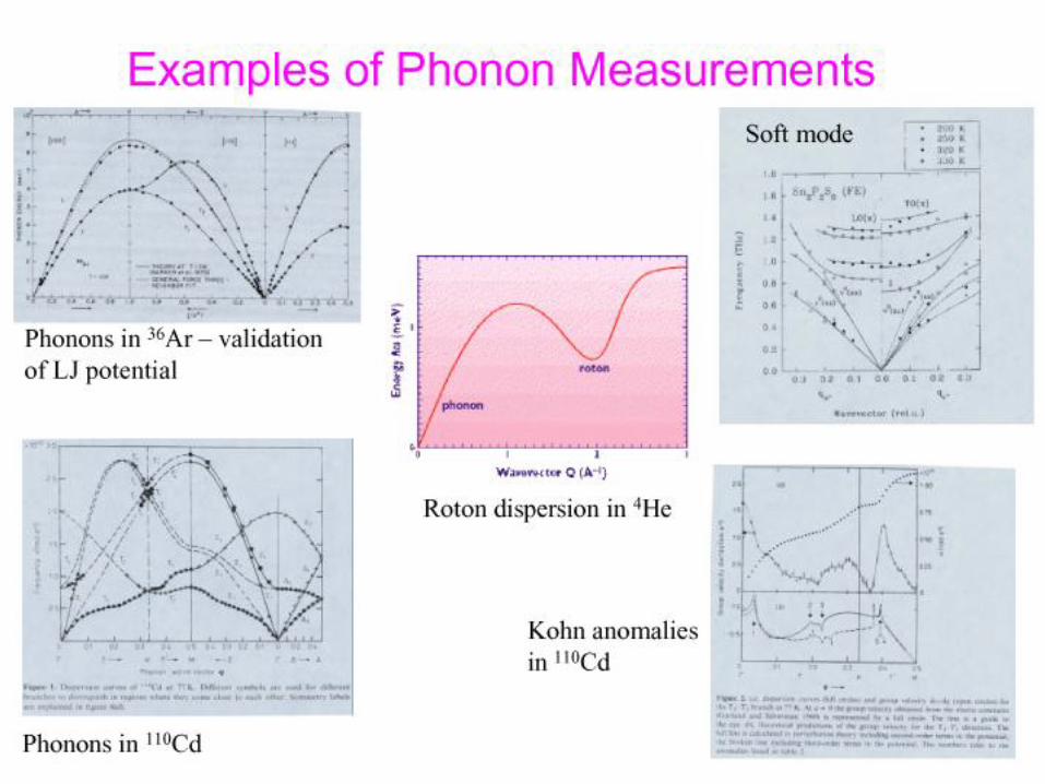
•Phonon dispersion curves in crystals; quantum crystals and anharmonicity
•Crystal fields
•Excitations in normal liquids
•Rotons in superfluid helium
•Condensate fraction in helium

Recent Applications
• Quantum Phase Transitions and Critical points
• Magnetic order and magnetic fluctuations in the high-Tc cuprates
• Gaps and low-lying excitations (including phonons) in High-Tc
• Magnetic Order and spin fluctuations in highly-correlated systems
• Manganites
• Magnetic nanodot/antidot arrays
• Exchange bias
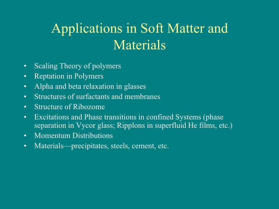
Applications in Soft Matter and
Materials
• Scaling Theory of polymers
• Reptation in Polymers
• Alpha and beta relaxation in glasses
• Structures of surfactants and membranes
• Structure of Ribozome
• Excitations and Phase transitions in confined Systems (phase separation in Vycor glass; Ripplons in superfluid He films, etc.)
• Momentum Distributions
• Materials—precipitates, steels, cement, etc.
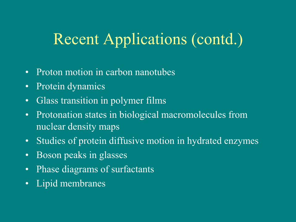
Recent Applications (contd.)
• Proton motion in carbon nanotubes
• Protein dynamics
• Glass transition in polymer films
• Protonation states in biological macromolecules from
nuclear density maps
• Studies of protein diffusive motion in hydrated enzymes
• Boson peaks in glasses
• Phase diagrams of surfactants
• Lipid membranes
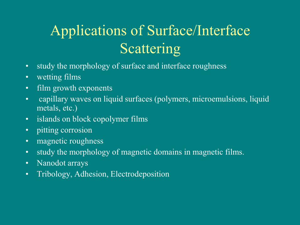
Applications of Surface/Interface
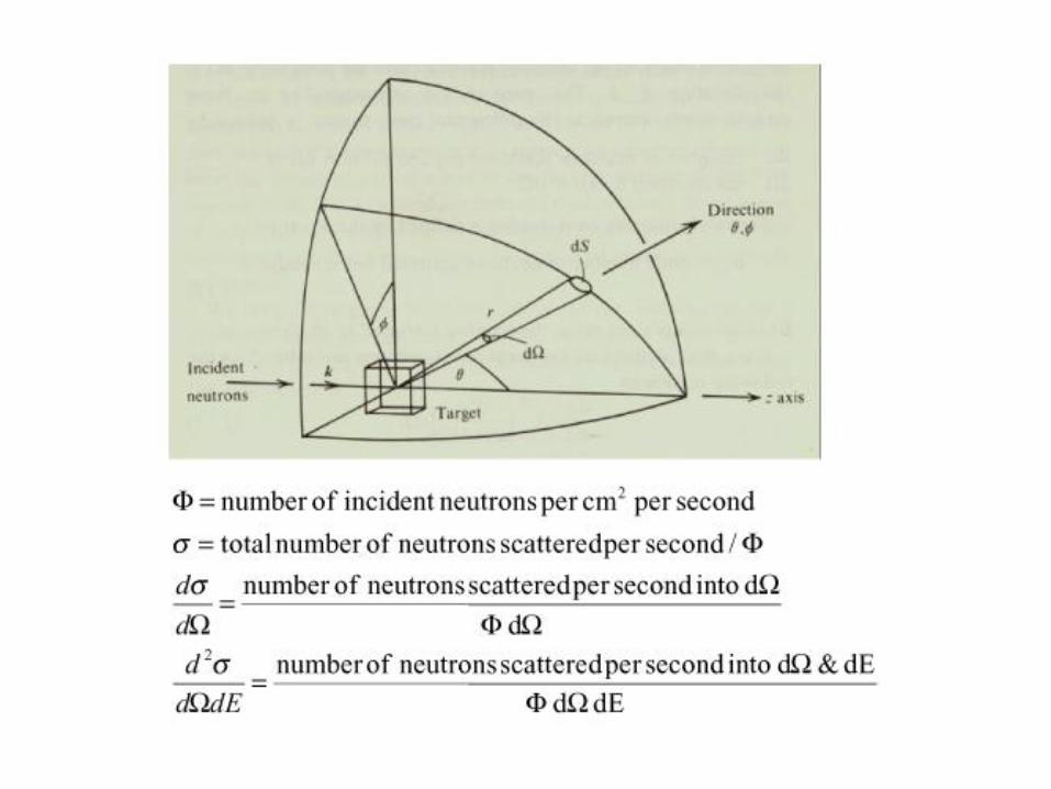
Scattering • study the morphology of surface and interface roughness
• wetting films
• film growth exponents
• capillary waves on liquid surfaces (polymers, microemulsions, liquid metals, etc.)
• islands on block copolymer films
• pitting corrosion
• magnetic roughness
• study the morphology of magnetic domains in magnetic films.
• Nanodot arrays
• Tribology, Adhesion, Electrodeposition
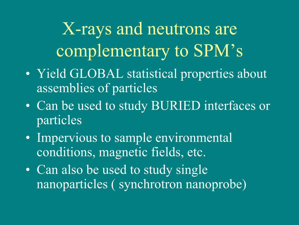
X-rays and neutrons are
complementary to SPM’s
• Yield GLOBAL statistical properties about assemblies of particles
• Can be used to study BURIED interfaces or particles
• Impervious to sample environmental conditions, magnetic fields, etc.
• Can also be used to study single nanoparticles ( synchrotron nanoprobe)
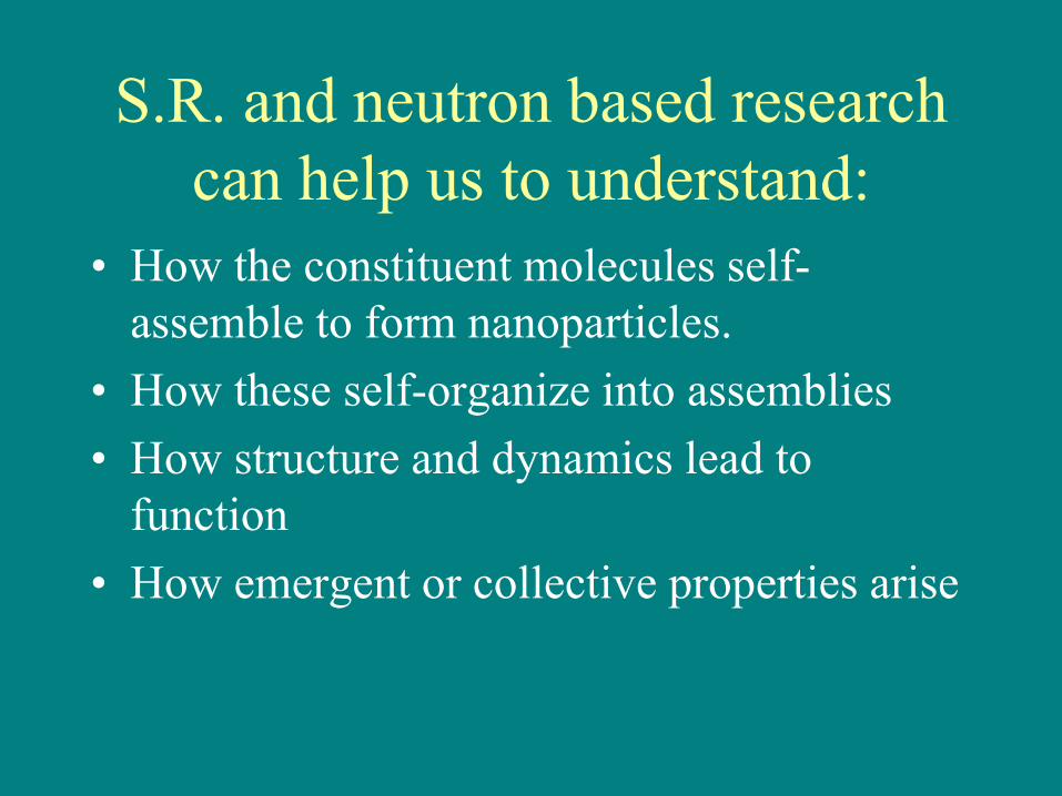
S.R. and neutron based research
can help us to understand:
• How the constituent molecules self-
assemble to form nanoparticles.
• How these self-organize into assemblies
• How structure and dynamics lead to
function
• How emergent or collective properties arise

Brightness & Fluxes for Neutron &
X-Ray Sources Brightness
dE/E
Divergence
Flux
Neutrons 2
Rotating
Anode 0.02
Bending
Magnet 0.1
Undulator
(APS) 10
1510 1110
2010 14105
1010
)( 121 sterms (%) )( 2mrad )( 21 ms
105.0
1.001.0
51.0 20105
24103310
2710QuickTime™ and a
TIFF (Uncompressed) decompressorare needed to see this picture.
LCLS Brilliance : Peak 8.5 . 1032
Ave 2.7 . 1022
Ph/s/mrad2/mm2/0.1%

Synchrotron-
and Neutron
Scattering
Places

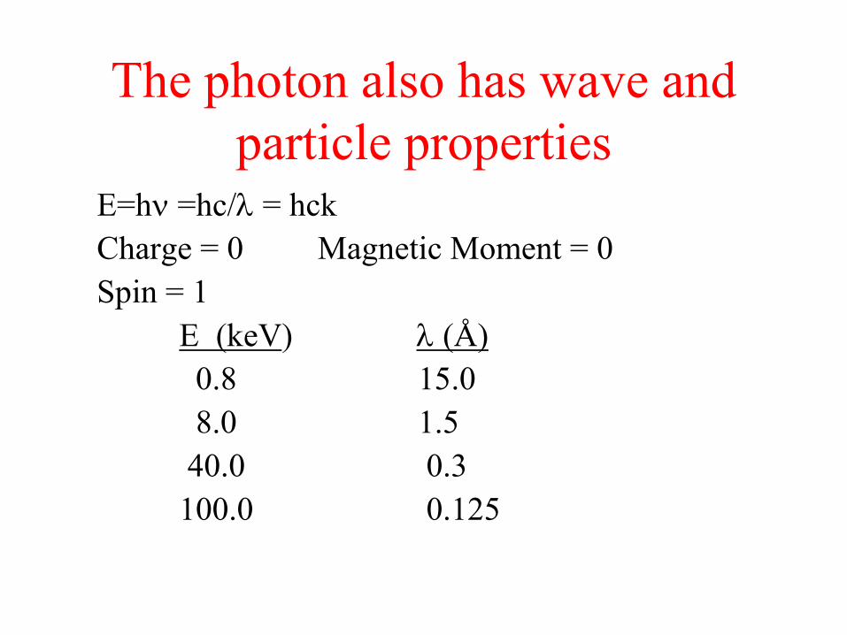
The photon also has wave and
particle properties E=h =hc/ = hck
Charge = 0 Magnetic Moment = 0
Spin = 1
E (keV) (Å)
0.8 15.0
8.0 1.5
40.0 0.3
100.0 0.125

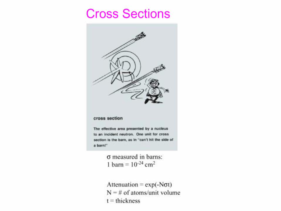
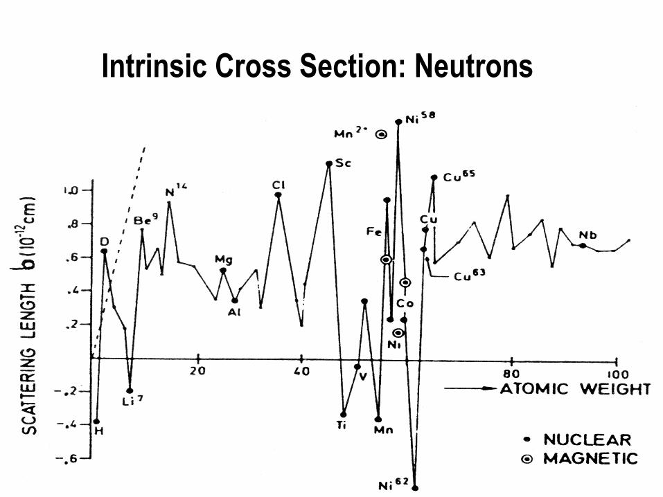
Intrinsic Cross Section: Neutrons

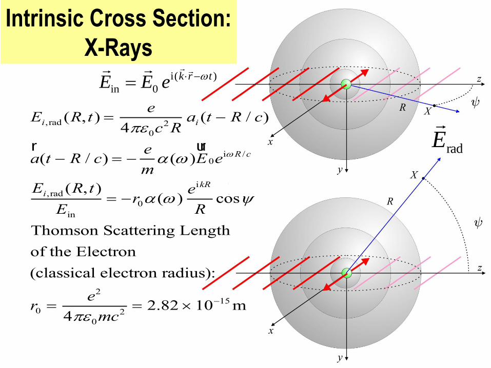
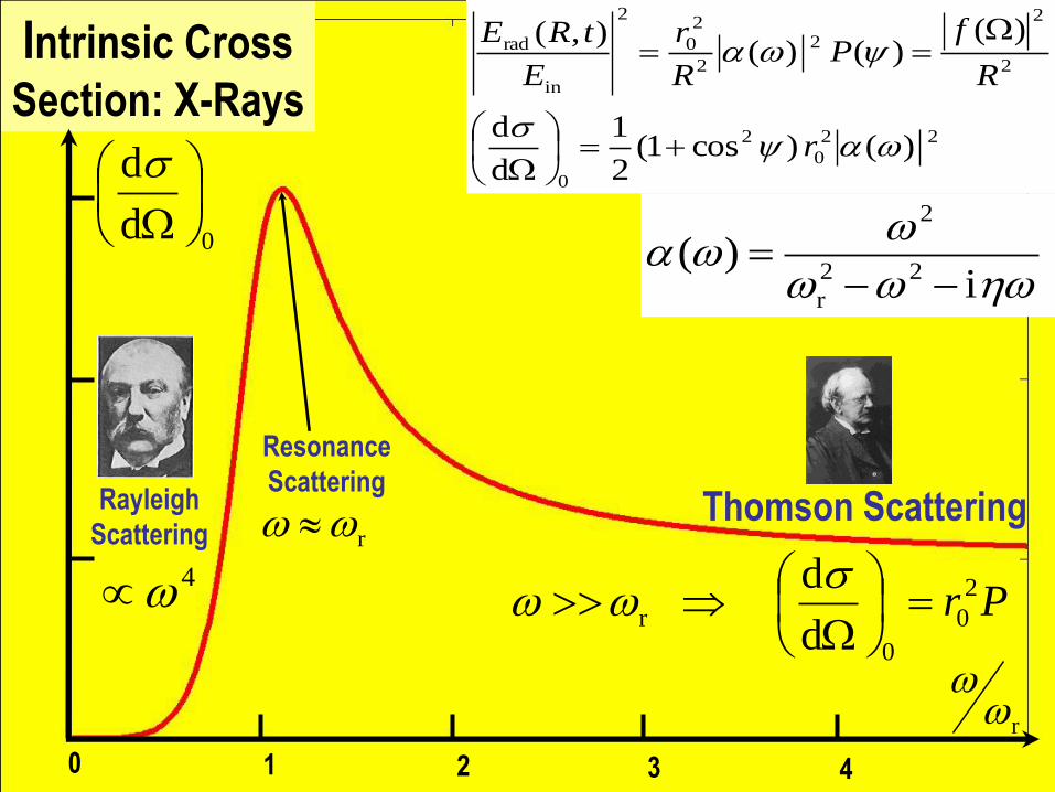
Intrinsic Cross Section:
X-Rays
Ei,rad (R, t) e
40c2Rai (t R / c)
ar(t R / c)
e
m( )E
ur0e
iR /c
Ei,rad (R, t)
Ein
r0( )eikR
Rcos
Thomson Scattering Length
of the Electron
(classical electron radius):
r0 e2
40mc2
2.82 1015 m
)(i
0in
trkeEE
radE
22
0
2
0
2
2
2
2
2
0
2
in
rad
)()cos1(2
1
d
d
)()()(
),(
r
R
fP
R
r
E
tRE
Intrinsic Cross
Section: X-Rays
0d
d
r
1 2 3 4 0
i)(
22
r
2
Resonance
Scattering
r Thomson Scattering
Pr 2
0
0
rd
d
4
Rayleigh
Scattering

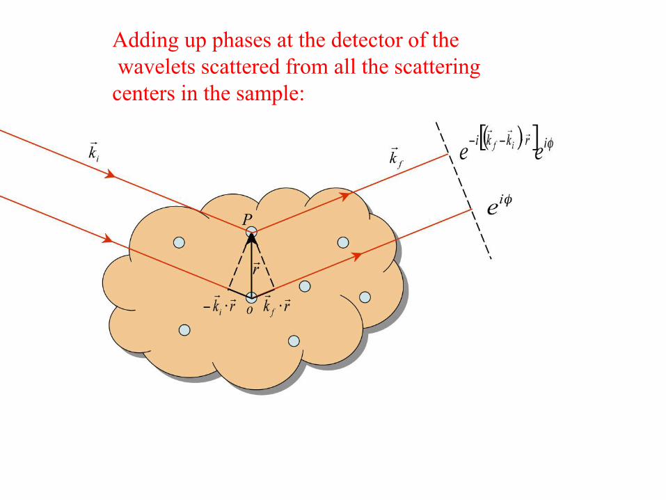
Adding up phases at the detector of the
wavelets scattered from all the scattering
centers in the sample:

q = kf - ki
Wave vector transfer is defined as
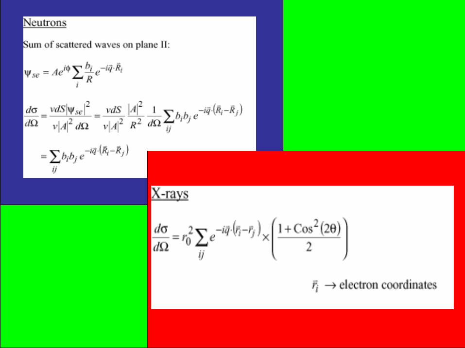

X-rays
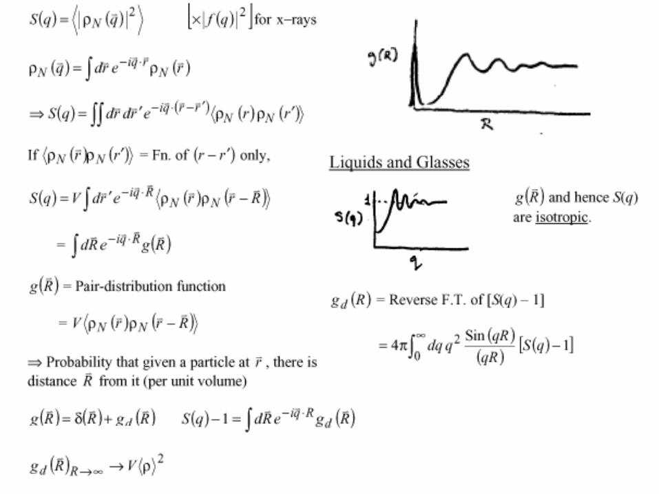
d = r021 + Cos2(2) S(q)
d 2
S(q) = ij exp[-iq.(ri-rj)]
{ri} == electron positions.

Now, i exp[-iq.Ri] = N(q) Fourier Transform of nuclear density
[ sometimes also referred to as F(q) ]
Proof:
N(r) = i ( r - Ri)
N(q) = ∫ N(r) exp[-iq.r] dr = ∫ i ( r - Ri) exp[-iq.r] dr
= i exp[-iq.Ri]
Similarly,
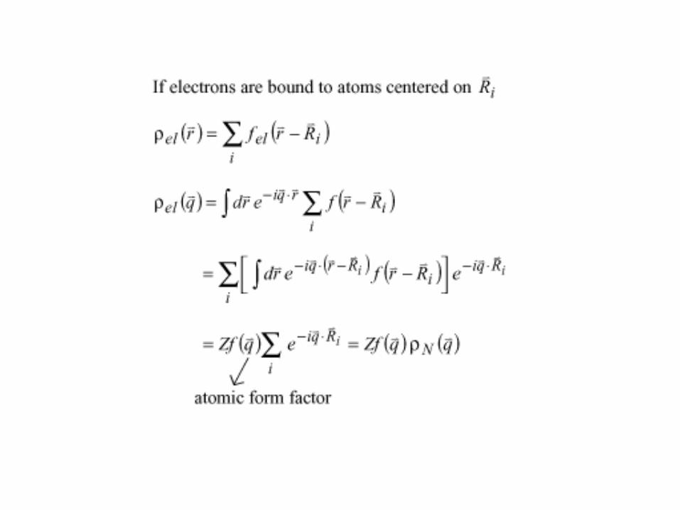
i exp[-iq.ri] = el(q) Fourier Transform of electron density
So, for neutrons, S(q) = N(q) N*(q)
And, for x-rays, S(q) = el(q) el*(q)
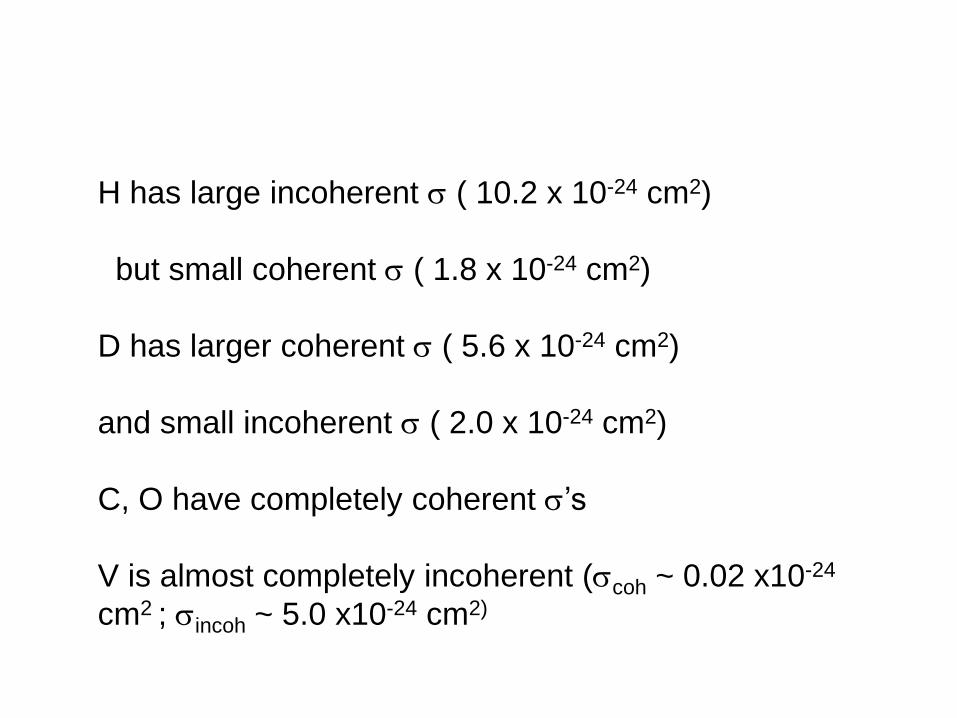
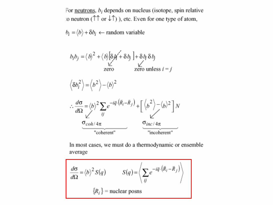
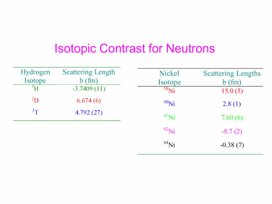
H has large incoherent ( 10.2 x 10⁻²⁴ cm2)
but small coherent ( 1.8 x 10⁻²⁴ cm2)
D has larger coherent ( 5.6 x 10⁻²⁴ cm2)
and small incoherent ( 2.0 x 10⁻²⁴ cm2)
C, O have completely coherent ’s
V is almost completely incoherent (coh ~ 0.02 x10⁻²⁴
cm2 ; incoh ~ 5.0 x10⁻²⁴ cm2)


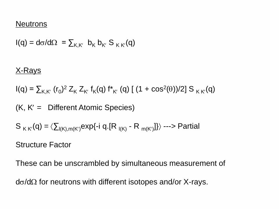
Neutrons
I(q) = d/d = ∑K,K bK bK S K K(q)
X-Rays
I(q) = ∑K,K (r0)2 ZK ZK fK(q) f*K (q) [ (1 + cos2())/2] S K K(q)
(K, K = Different Atomic Species)
S K K(q) = ∑l(K),m(K)exp{-i q.[R l(K) - R m(K)]} ---> Partial
Structure Factor
These can be unscrambled by simultaneous measurement of
d/d for neutrons with different isotopes and/or X-rays.

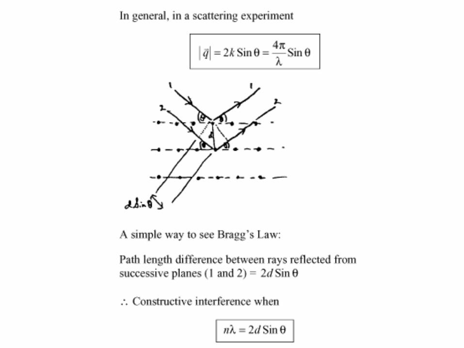
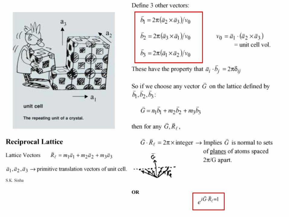
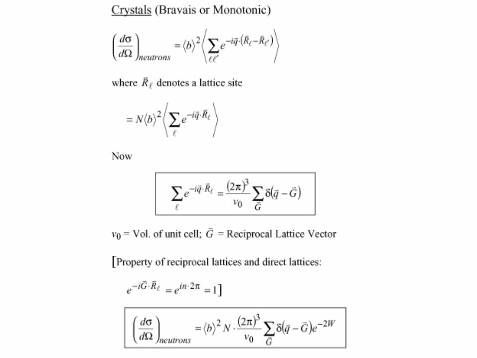
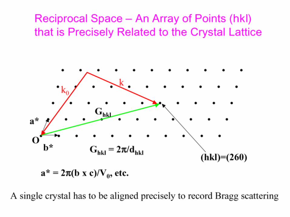
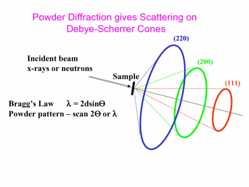
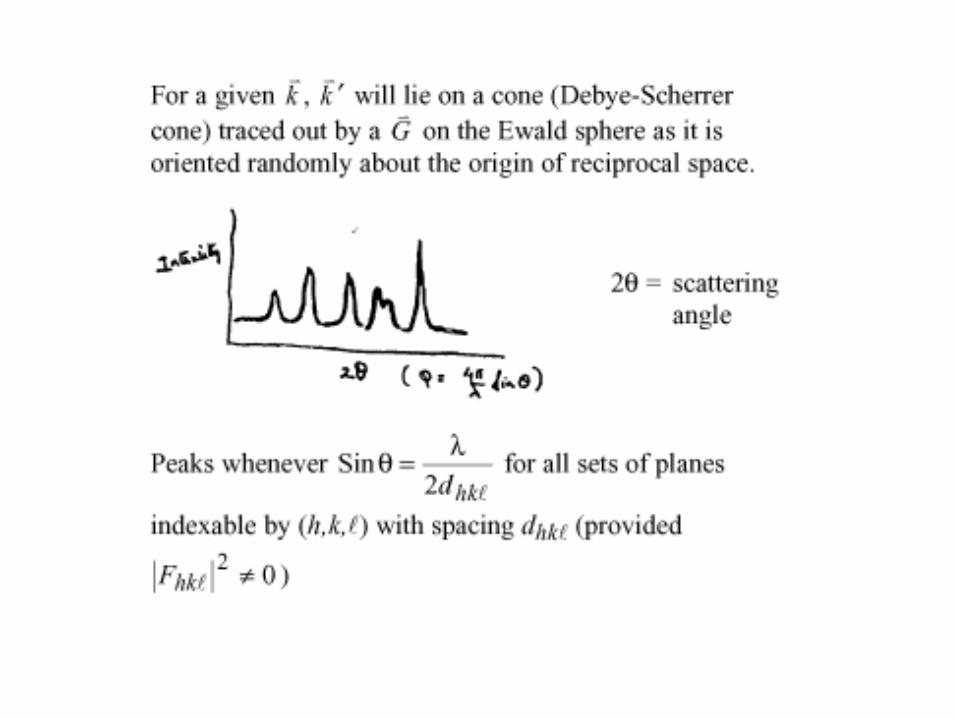
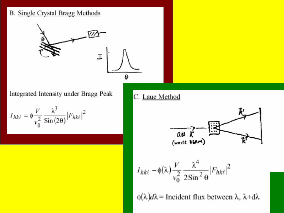
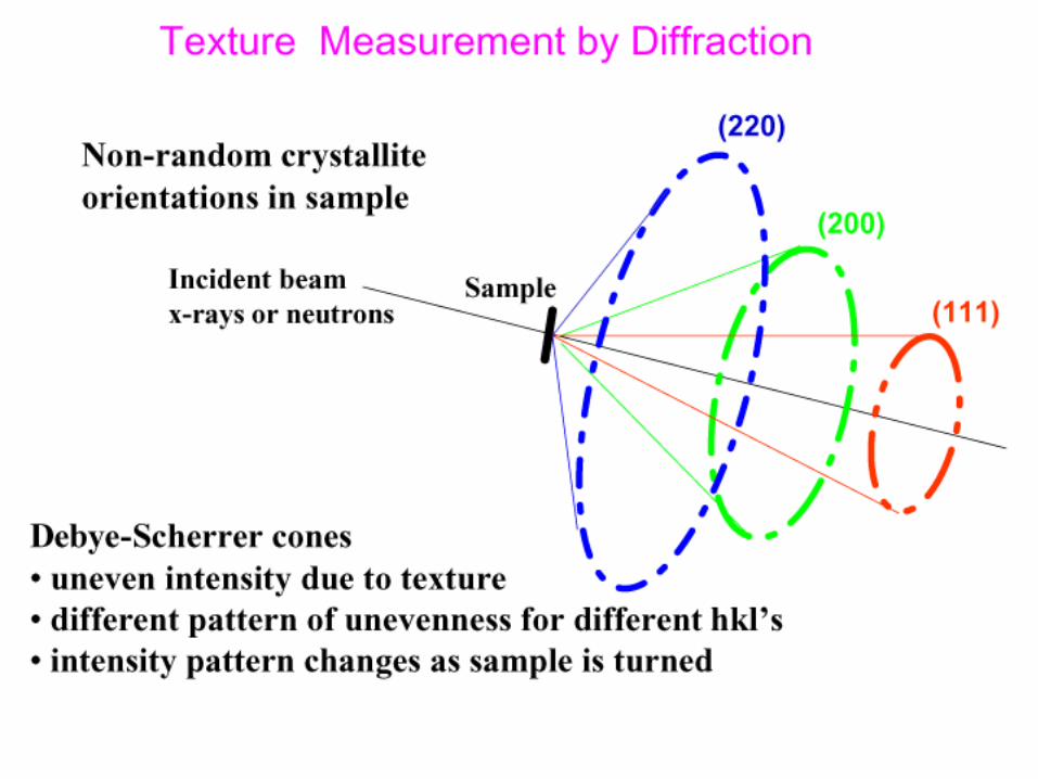
Diffraction from Crystals


qx
qy
qz


(S = 6) Ds = Surface fractal dimension.
If Ds =2, S(q) ~ 1/q4 (Porod’s Law for
smooth internal surfaces)
If 2 < Ds < 3, S(q) ~ 1/qn where 3< n <4
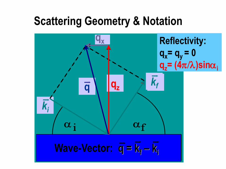
ki
kf q qz
qx
Scattering Geometry & Notation
Wave-Vector: q = kf – ki
Reflectivity:
qx= qy = 0
qz= (4)sini
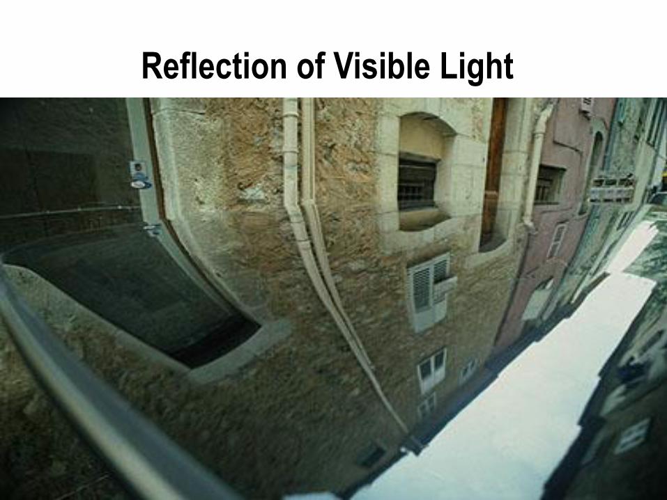
Reflection of Visible Light

Perfect & Imperfect „Mirrors“

Basic Equation: X-Rays
Helmholtz-Equation & Boundary Conditions
2 E( r ) + k2 n2( r ) E ( r ) = 0
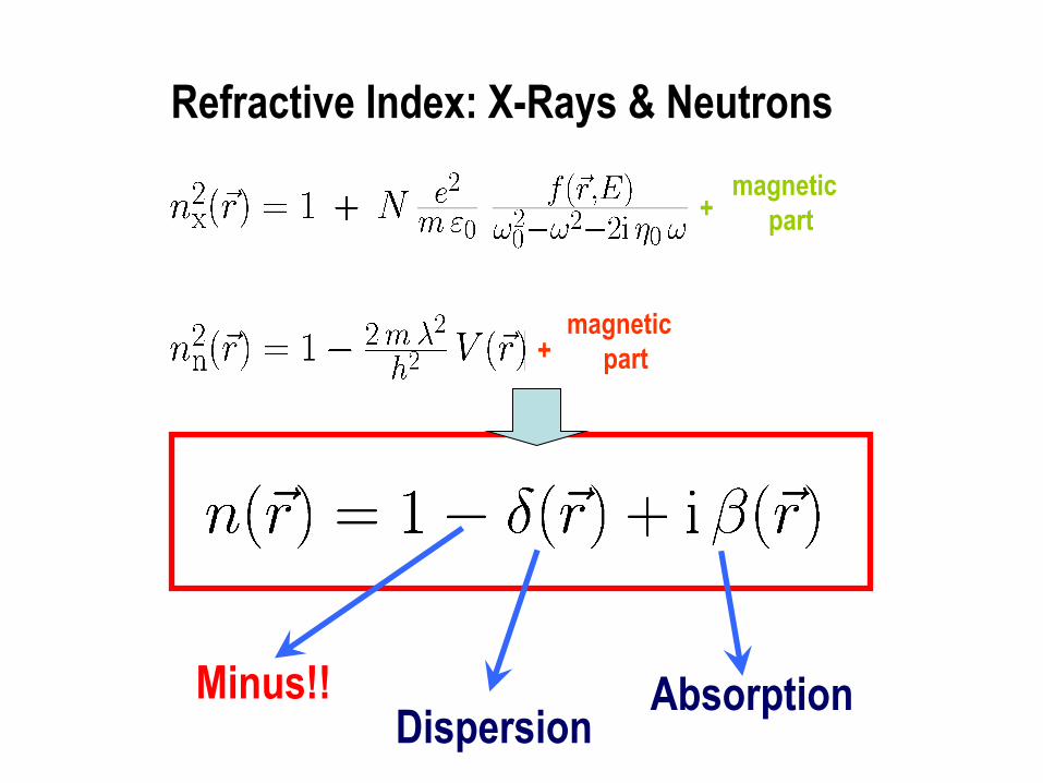
magnetic
part +
magnetic
part +
Dispersion Absorption Minus!!
Refractive Index: X-Rays & Neutrons

Derivation of n for neutrons:
Consider Schrodinger Eqn.
-(ћ2/2m)2 + (V -E) = 0 E = (ћ2/2m)k02
can be written:
2 +[1 - (2m/ћ2 k02)V] k0
2 = 0
V= (2ћ2/m)b N; k0 = 2/
so:
n2 = (1 - (2m/ћ2 k02)V) = 1 - (2b/) N
2nd term <<1, so n = 1 - (2b/2) N
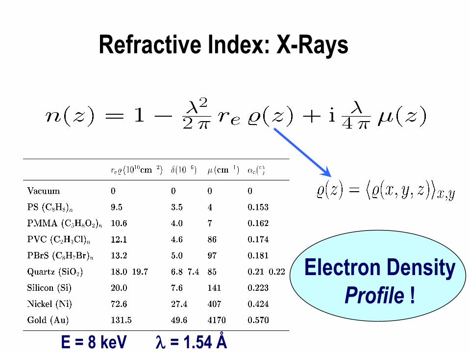
Electron Density
Profile !
Refractive Index: X-Rays
E = 8 keV = 1.54 Å

Reflected
Amplitude
Transmitted
Amplitude
Wave-
Vectors
Single Interface: Vacuum/Matter
Fresnel-
Formulae
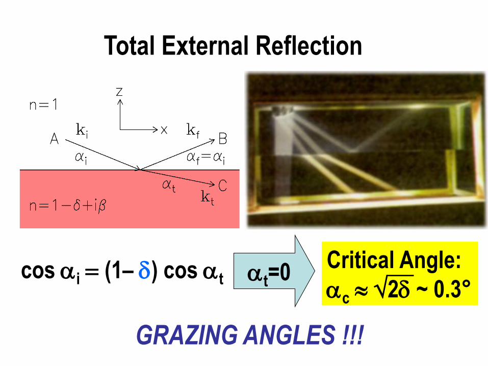
cos i (1– ) cos t t=0 Critical Angle:
c 2 ~ 0.3°
GRAZING ANGLES !!!
Total External Reflection
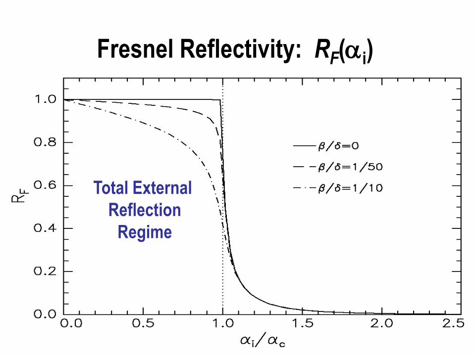
Total External
Reflection
Regime
Fresnel Reflectivity: RF(i)
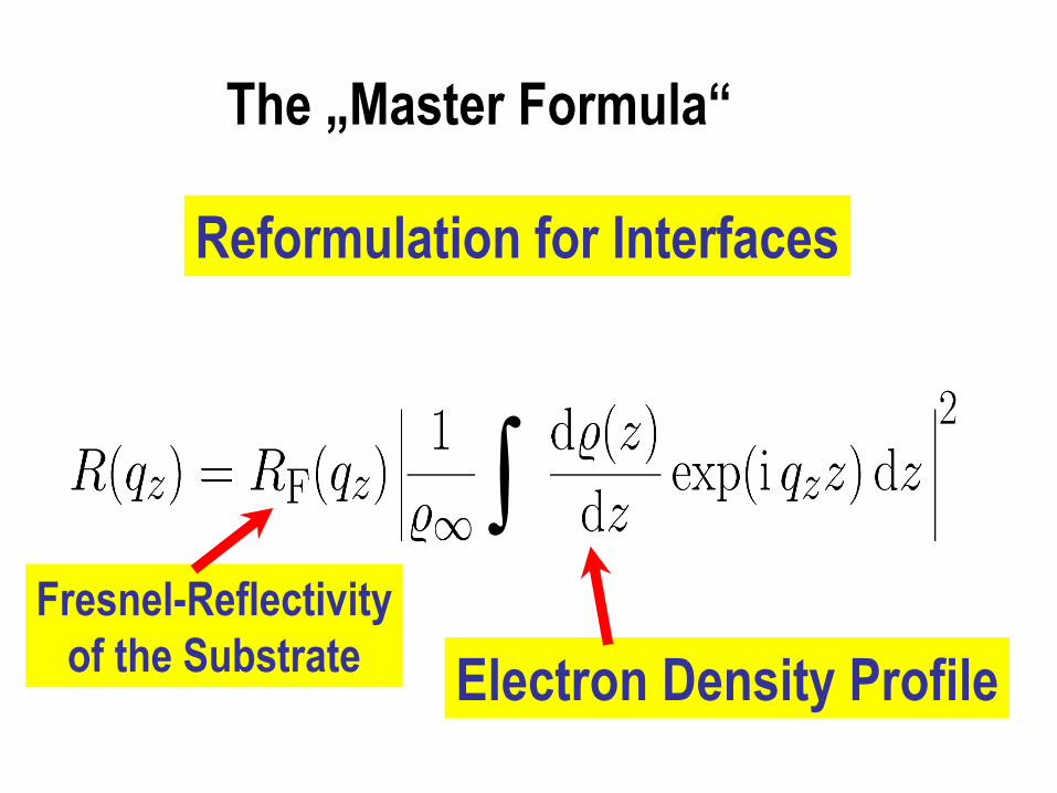
Reformulation for Interfaces
The „Master Formula“
Electron Density Profile
Fresnel-Reflectivity
of the Substrate
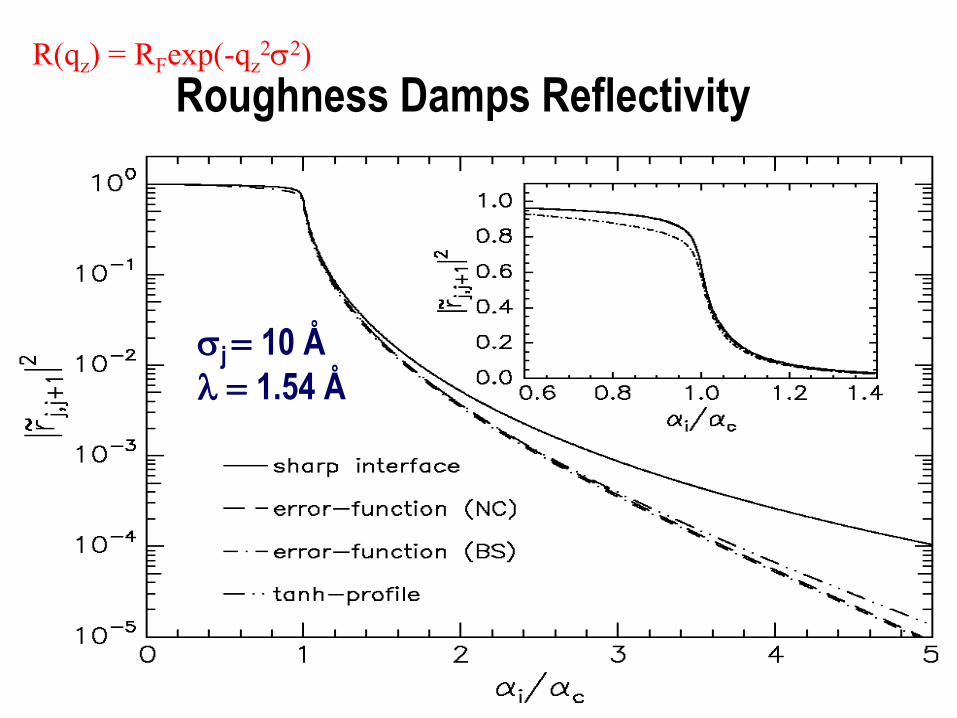
j 10 Å
1.54 Å
Roughness Damps Reflectivity R(qz) = RFexp(-qz
22)
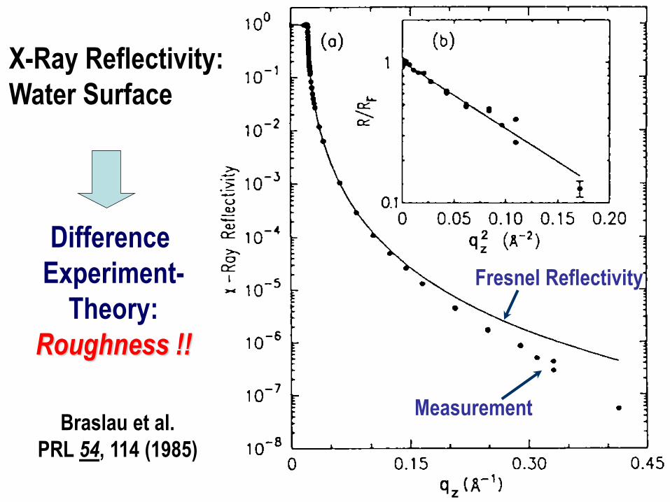
Braslau et al.
PRL 54, 114 (1985)
Fresnel Reflectivity
Measurement
X-Ray Reflectivity:
Water Surface
Difference
Experiment-
Theory:
Roughness !!
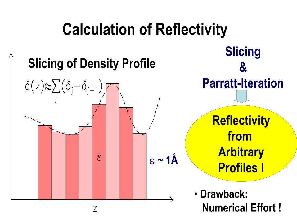
Slicing of Density Profile
~ 1Å
Slicing
&
Parratt-Iteration
Reflectivity
from
Arbitrary
Profiles !
• Drawback:
Numerical Effort !
Calculation of Reflectivity
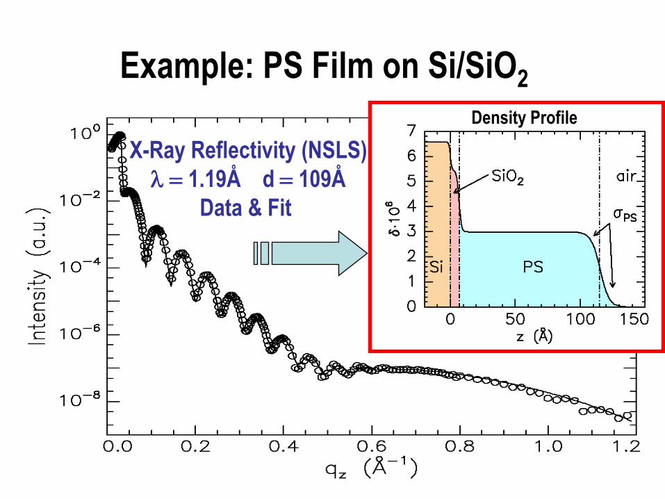
Example: PS Film on Si/SiO2
X-Ray Reflectivity (NSLS)
1.19Å d 109Å
Data & Fit
Density Profile
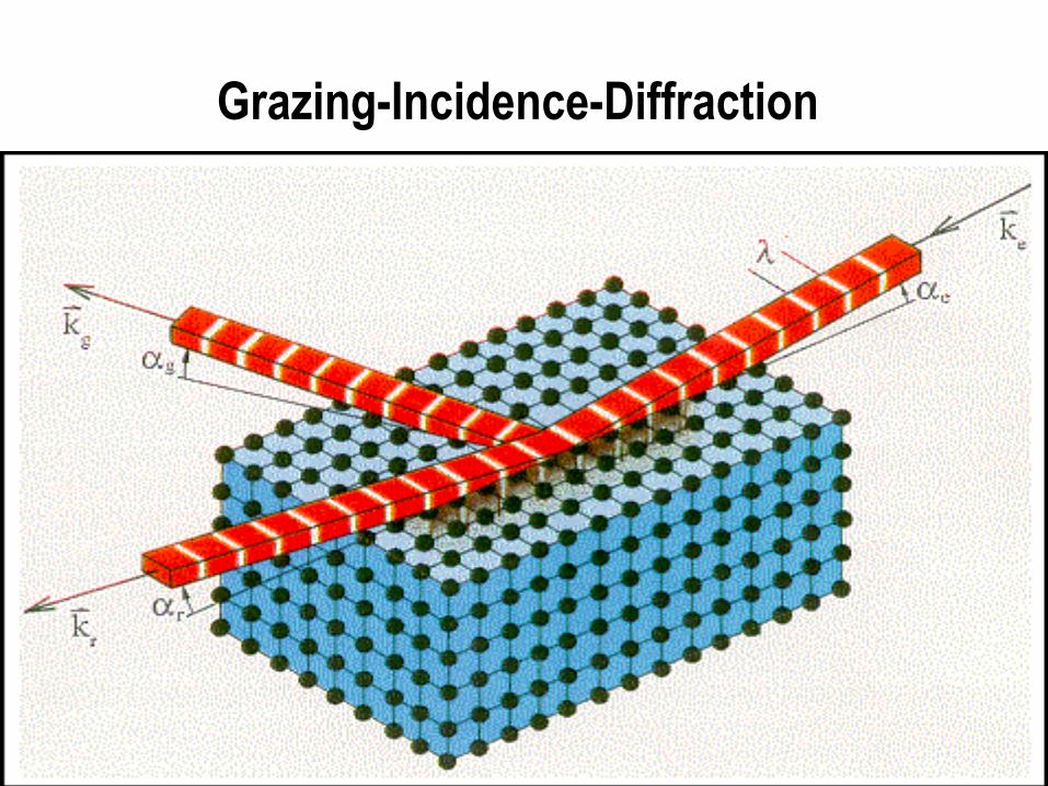
Grazing-Incidence-Diffraction
ki
kf Q Qz
Qx
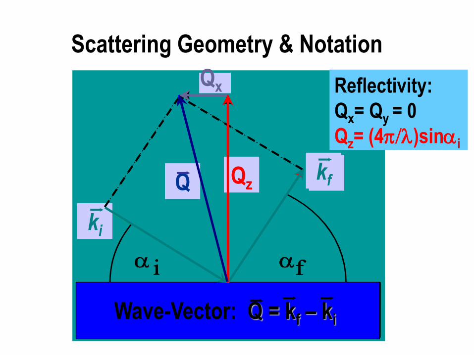
Scattering Geometry & Notation
Wave-Vector: Q = kf – ki
Reflectivity:
Qx= Qy = 0
Qz= (4)sini
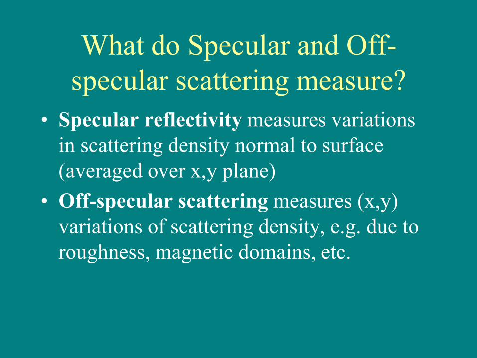
What do Specular and Off-
specular scattering measure?
• Specular reflectivity measures variations
in scattering density normal to surface
(averaged over x,y plane)
• Off-specular scattering measures (x,y)
variations of scattering density, e.g. due to
roughness, magnetic domains, etc.
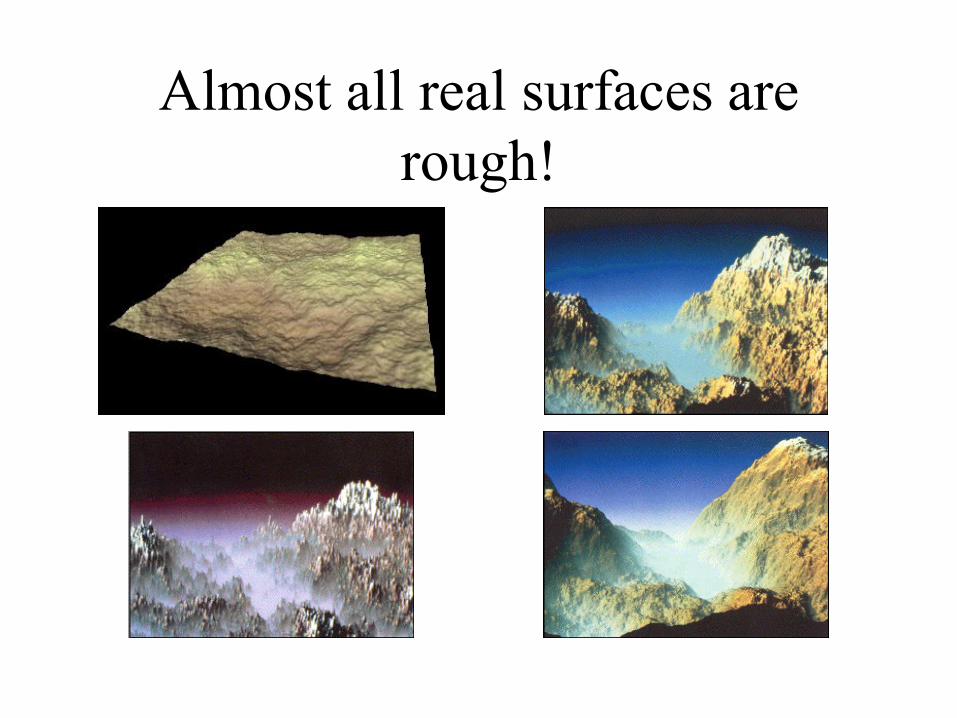
Almost all real surfaces are
rough!
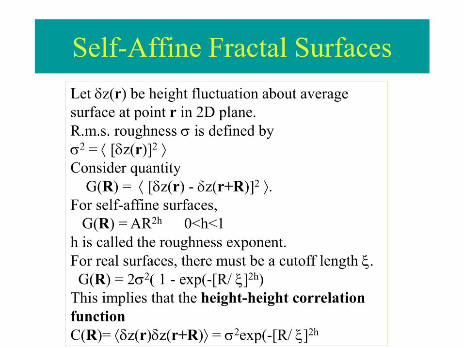
Self-Affine Fractal Surfaces
Let z(r) be height fluctuation about average
surface at point r in 2D plane.
R.m.s. roughness is defined by
2 = [z(r)]2
Consider quantity
G(R) = [z(r) - z(r+R)]2 .
For self-affine surfaces,
G(R) = AR2h 0<h<1
h is called the roughness exponent.
For real surfaces, there must be a cutoff length .
G(R) = 22( 1 - exp(-[R/ ]2h)
This implies that the height-height correlation
function
C(R)= z(r)z(r+R) = 2exp(-[R/ ]2h
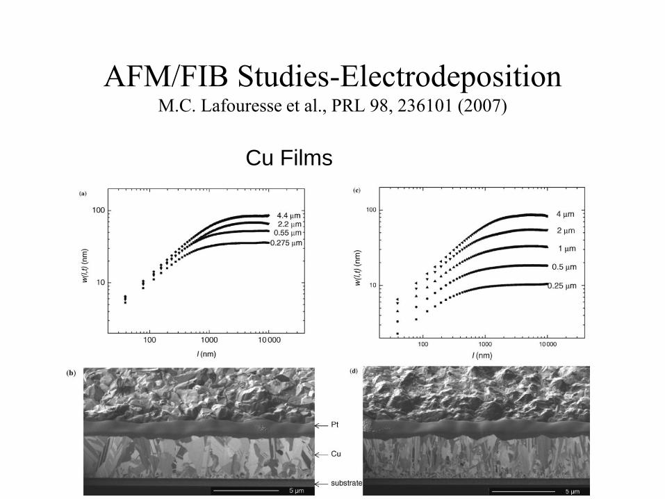
AFM/FIB Studies-Electrodeposition M.C. Lafouresse et al., PRL 98, 236101 (2007)
Cu Films

Scattering from a Self-Affine
Fractal Surface
S(qr) (Ar0
2 / qz2 )e
qz2 2
dXdYeqz
2C(R)
ei(qxXqyY )
SKS et al., Phys. Rev. B 38, 2297 (1988)
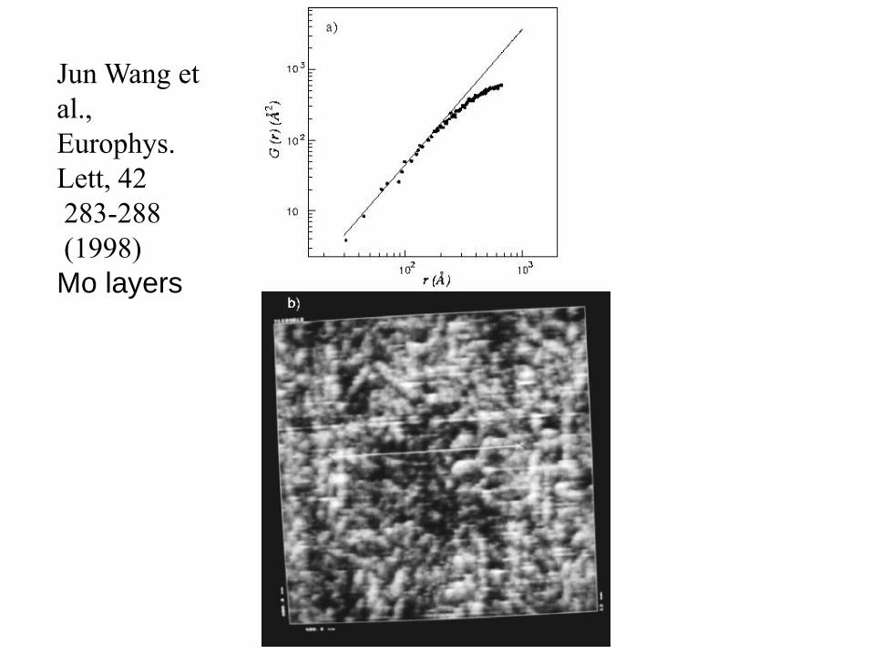
Jun Wang et
al.,
Europhys.
Lett, 42
283-288
(1998)
Mo layers
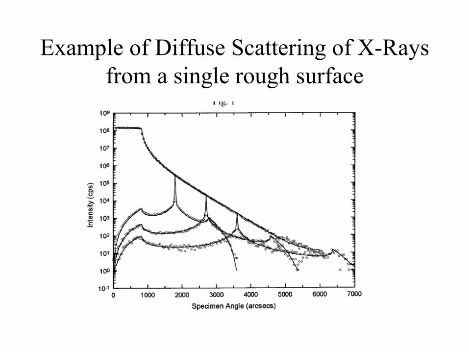
Example of Diffuse Scattering of X-Rays
from a single rough surface

Multilayers
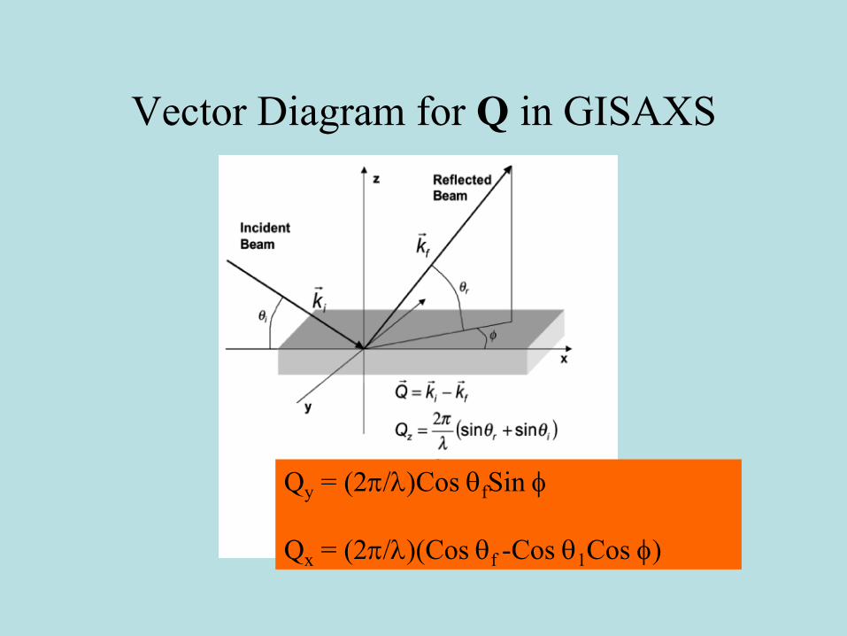
Vector Diagram for Q in GISAXS
Qy = (2/)Cos fSin
Qx = (2/)(Cos f -Cos 1Cos )

Measurement of GISAXS

X-Ray Reflectometers
Laboratory
Setup
HASYLAB: CEMO
Synchrotron
Setup
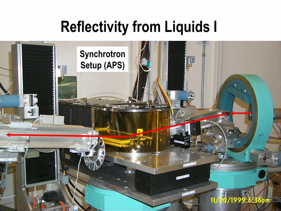
Synchrotron
Setup (APS)
Reflectivity from Liquids I


Magnetic Neutron Scattering

Core level resonances
2p3/2
2p1/2
EF
spin polarized3d bands
e0
ef
f = f1 + if2
Fe
Kortright et al., Phys. Rev. B 65, 12216 (2000)
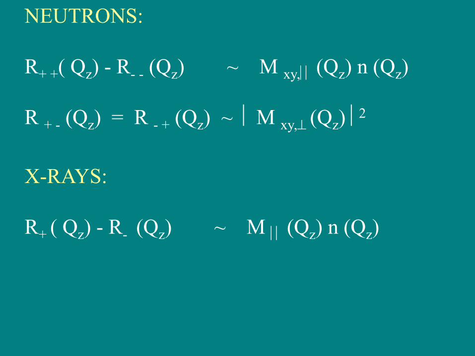
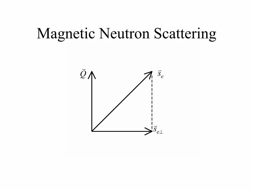
NEUTRONS:
R+ +( Qz) - R- - (Qz) ~ M xy, (Qz) n (Qz)
R + - (Qz) = R - + (Qz) ~ M xy, (Qz) 2
X-RAYS:
R+ ( Qz) - R- (Qz) ~ M (Qz) n (Qz)

l = 2 D
= (D )-1
t = R s
(hor., vert.)
Coherence Lengths
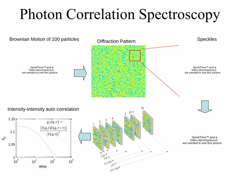
Photon Correlation Spectroscopy�
QuickTime™ and aVideo decompressor
are needed to see this picture.
Brownian Motion of 100 particles
QuickTime™ and aVideo decompressor
are needed to see this picture.
Speckles
Intensity-intensity auto correlation
100
101
102
103
1
1.05
1.1
1.15
delay
g2
2
2
),(
),(),(
),(
tqI
tqItqI
qg
Diffraction Pattern
QuickTime™ and aVideo decompressor
are needed to see this picture.

Photon Correlation Spectroscopy
sample detectorcoherent
beam
X-ray speckle pattern from a static silica aerogel

6/20/2011 Miao thesis 124
Finer sampling; larger array; smaller transform; “finite support” (area around specimen must be clear!)
“Oversampling”: Non-crystals: pattern continuous, can do finer sampling of intensity
6/20/2011 Miao thesis 125
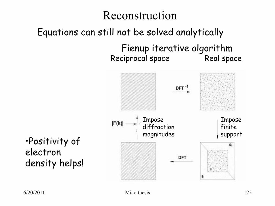
Reconstruction
Equations can still not be solved analytically
Fienup iterative algorithm Reciprocal space Real space
•Positivity of electron density helps!
Impose diffraction magnitudes
Impose finite support
6/20/2011 from Howells 126
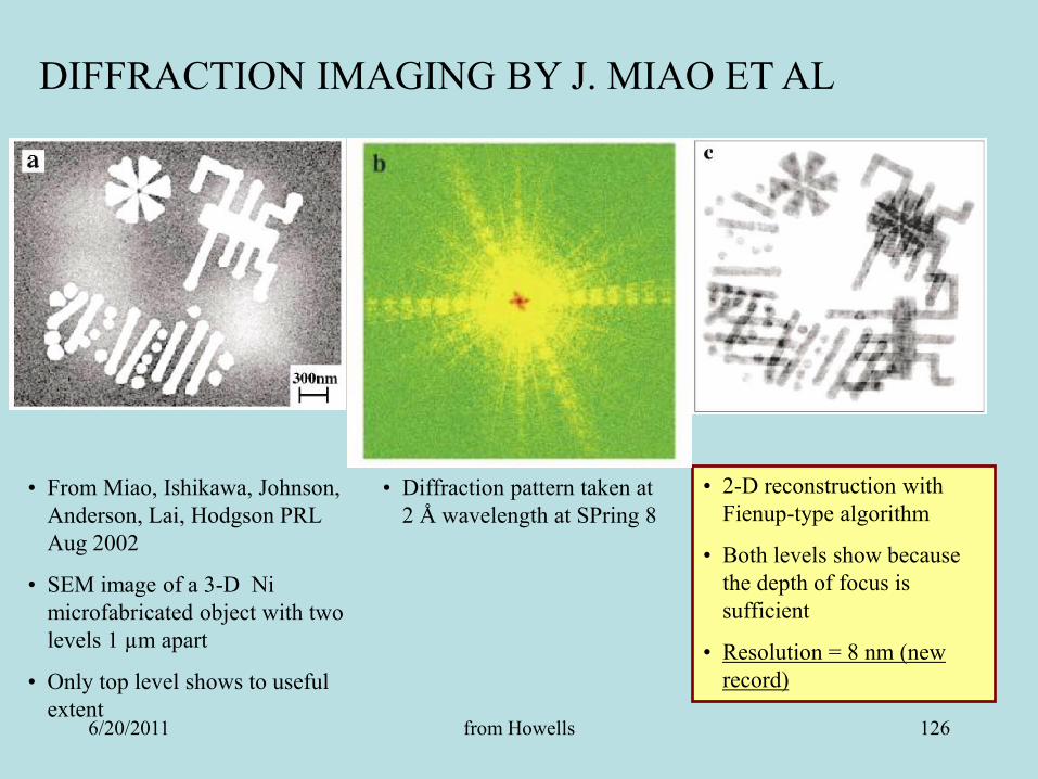
DIFFRACTION IMAGING BY J. MIAO ET AL
• From Miao, Ishikawa, Johnson,
Anderson, Lai, Hodgson PRL
Aug 2002
• SEM image of a 3-D Ni
microfabricated object with two
levels 1 µm apart
• Only top level shows to useful
extent
• Diffraction pattern taken at
2 Å wavelength at SPring 8
• 2-D reconstruction with
Fienup-type algorithm
• Both levels show because
the depth of focus is
sufficient
• Resolution = 8 nm (new
record)
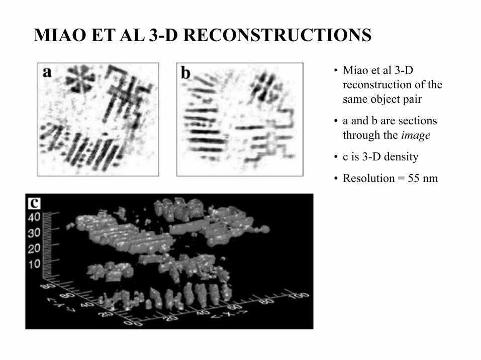
• Miao et al 3-D
reconstruction of the
same object pair
• a and b are sections
through the image
• c is 3-D density
• Resolution = 55 nm
MIAO ET AL 3-D RECONSTRUCTIONS

Imaging of individual nanoparticles at the APS
I.K. Robinson, et al., Science 298 2177 (2003)
170 nm silver cubes
Coherent diffraction pattern
from 170 nm Ag particle
inversion of
diffraction pattern
‘lensless imaging’
Ross Harder, University of Illinois, Champaign