Giorgia Basile 1 UNIVERSITÀ DEGLI STUDI DELLA TUSCIA DI VITERBO DIPARTIMENTO DISCIENZE ECOLOGICHE E BIOLOGICHE Corso di Dottorato di Ricerca in GENETICA E BIOLOGIA CELLULARE – XXVII Ciclo. "Investigating the role of Werner syndrome protein in the activation of the ATR-dependent checkpoint in response to mild replication stress" (s.s.d. BIO/11) Tesi di dottorato di: Dott.ssa Giorgia Basile Coordinatore del corso Tutore Prof. (Giorgio Prantera) Dott.ssa (Annapaola Franchitto) Data della discussione 08/05/2015
Transcript
Giorgia Basile
1
UNIVERSITÀ DEGLI STUDI DELLA TUSCIA DI VITERBO
DIPARTIMENTO DISCIENZE ECOLOGICHE E BIOLOGICHE
Corso di Dottorato di Ricerca in
GENETICA E BIOLOGIA CELLULARE – XXVII Ciclo.
"Investigating the role of Werner syndrome protein in the
activation of the ATR-dependent checkpoint
in response to mild replication stress"
(s.s.d. BIO/11)
Tesi di dottorato di:
Dott.ssa Giorgia Basile
Coordinatore del corso Tutore
Prof. (Giorgio Prantera) Dott.ssa (Annapaola Franchitto)
Werner syndrome (WS) is a human chromosomal instability and cancer-prone disease
caused by mutations in the WRN gene, encoding for the Werner syndrome protein (WRN)
that is a member of the RecQ helicases.
It has been previously proposed that WRN helicase activity is a key regulator of
common fragile site (CFS) stability, which are the preferential targets of genome instability in
precancerous lesions. Moreover, it is known that, under mild replication stress inducing by
low dose of Aphidicolin (Aph), WRN and ATR act in a common pathway preventing
accumulation of DNA breaks at CFS. Despite WS cells exhibit an ATR-like instability at
CFS, and that WRN has been found phosphorylated by ATR under robust replication stress
caused by treatments with hydroxyurea (HU), there is no evidence of a functional requirement
of WRN in the establishment of the replication checkpoint response to mild replication stress,
like that inducing CFS expression.
The aim of this study was to analyze the functional requirement of WRN in the ATR-
dependent checkpoint activation under mild replication stress using low doses of aphidicolin
treatments.
Our data establish that WRN plays a role in mediating CHK1 activation, a principal
target of the ATR kinase activity under replication stress. Moreover, our results demonstrate
that WRN and the ATR-mediated WRN phosphorylation are required to phosphorylate
CHK1, as they are important for chromatin loading of checkpoint mediators under untreated
as well as Aph-treatment conditions. In contrast, although WRN helicase activity is not
required for CHK1 phosphorylation, it results essential in supporting replication fork
recovery, possibly by the resolution of DNA secondary structures, thus promoting CFS
stability.
Analysis of replication fork dynamics shows that loss of WRN checkpoint mediator
function, as well as of WRN helicase activity, hamper replication fork progression, and lead
to new origin activation to allow recovery from replication slowing upon mild replication
stress. Furthermore, bypass of WRN checkpoint mediator function through over-expression of
a phospho-mimic form of CHK1 restores fork progression and chromosome stability to the
wild-type levels.
Loss of WRN also greatly hampers the phosphorylation of histone H2AX (γ-H2AX),
which is the earlier target of ATR kinase following replication stress. Indeed, although, upon
mild replication stress, in wild-type cells H2AX is activated by ATR in a time-dependent
Giorgia Basile
5
manner, its phosphorylation is reduced in WS cells, further confirming defects in the ATR
signalling.
Furthermore in this study, others cellular consequences of WRN loss have been
explored in response to mild replication stress. Evidences demonstrate that the absence of
WRN leads to an ATM pathway activation, which is harmful to the cells, as confirmed by
positive effects obtained on cellular survival and chromosomal damage by ATM inhibition in
the last part of the treatment with Aph. One way by which ATM inhibition could protect
genome stability in WS cells is the recovery of CHK1 defective activation. Noteworthy, in
cells expressing the unphosphorylable form of WRN, WSWRN6A, ATM inhibition is not able
to rescue CHK1 activation, possibly because the presence of the protein, although mutated,
could prevent the activation of such alternative pathway. Furthermore, WRN deficiency leads
the formation of 53BP1 foci in S phase, after prolonged mild replication stress. The increase
of 53BP1 foci in all cell cycle phases has been previously associated with CHK1 depletion,
and so it is consistent with defective CHK1 activation observed in WRN-deficient cells
following Aph exposure. These findings suggest that, in S phase, in absence of the ATR
checkpoint activation, 53BP1 recruitment in foci is instrumental in attracting other proteins
implicated in the response to mild replication stress damage.
Therefore, our results suggest a novel role of WRN as checkpoint mediator in response
to moderate replication stress and give strong mechanistic support to the notion that defective
fork repair/recovery undermines integrity of chromosomes at CFS. This study also unveils a
complicated network in which several proteins work tightly linked together. Loss of one
protein means altering this network and changing the interaction among proteins.
Moreover, our findings may contribute to shed light into the origin of chromosome
instability in WS and more in general to clarify how genome instability accumulates in pre-
neoplastic lesions, thus promoting cancer development.
Giorgia Basile
6
INTRODUCTION
Giorgia Basile
7
INTRODUCTION
DNA replication and genome stability
Genome instability is a common feature of cancer cells. Most of the chromosomal
abnormalities arising in tumors come from defective DNA replication (Myung and Kolodner,
2002). Thus, in eukaryotic cells DNA replication process is tightly monitored to ensure that
genome is replicated just once per cell cycle, and that DNA duplication is complete before
mitosis begins (Branzei and Foiani, 2010).
Given to the complexity of the replication process, it is not surprising that defects in
DNA replication or in its regulation may give rise to several human diseases. Therefore,
investigating the DNA replication process and the pathways that are involved in preventing
genome instability is fundamental to understand the mechanisms by which cancers and others
pathological disorders arise.
DNA replication represents a crucial moment in the life of a cell, as chromosomal
integrity can be seriously threatened by replication stress, that is the slowing and/or stalling of
replication fork progression (Zeman and Cimprich, 2014). In fact, replication stress interferes
with fork stability and can be caused by endogenous side-products of cellular metabolism,
exogenous agents capable to interfere with DNA replication, as well as intrinsic structural
features of specific genomic regions, such as the common fragile sites (CFS). Fork stalling is
a very frequent event occurring during S-phase. To guarantee genome integrity , replication
forks are endowed with an extraordinary potential to coordinate fork stalling with fork
resumption processes. When protection of stalled forks or their processing and replication
restart fail, mutations and aberrations accumulate in the genome. Mutations in genes that
protect the genome integrity during replication characterize a variety of human genetic
syndromes, which lead to cancer predisposition (Branzei and Foiani, 2005).Among these
human disease, there is Werner syndrome, that shows defects in resolving DNA replication
stress.
To minimize the risk of chromosomal rearrangement accumulation and deal with
problems encountered during S-phase, cells have evolved a sophisticated apparatus deputed to
the resolution of problems arising at replication forks: the replication checkpoint.
Giorgia Basile
8
The replication checkpoint response
The link between replication defects, human diseases and cancer underscores the
requirement of an efficient and accurate monitoring of genome integrity during DNA
replication, and the presence of multiple checkpoint activities in the S-phase may be
explained with the complexity of the DNA duplication process.
The replication checkpoint is a complex and coordinated network under the control of
the ATR kinase (Abraham, 2001; Zou and Elledge, 2003). These biochemical network
contains a class of protein, named mediators or adaptors, which promote functional
interactions between sensor and effector proteins. In the case of replication stress, replication
checkpoint activation leads to inhibition of origin firing, cell cycle arrest, stabilization, and
then restart of stalled forks, and prevention of the entry into mitosis until the DNA has been
completely replicated (Budzowska and Kanaar, 2009).
ATR was discovered in the human genome database as a gene with sequence
homology to ATM and SpRad3, hence the name ATR (Cimprich et al., 1996). The gene
encodes a protein of 303 kDa with a C-terminal kinase domain and regions of homology to
other PIKK family members. ATR deficiency in mice results in early embryonic death
(Brown and Baltimore, 2000), and mutations causing a partial loss of its activity have been
reported to be associated with the human autosomal recessive disorder, Seckel syndrome
(O’Driscoll et al., 2003). ATR is capable of specifically phosphorylating Serine or Threonine
residues in SQ/TQ sequences (Abraham, 2001). In human cells, ATR exists in a stable
complex with ATR-interacting protein (ATRIP), a potential regulatory partner (Cortez et al.,
2001; Sancar et al., 2004). ATR is essential for embryonic cell viability and, for this reason, it
probably has an important function during cell cycle progression.
Although ATR is activated in response to many different types of DNA damage,
including double strand breaks (DSBs), base adducts, crosslinks, it is thought to be mainly
responsible for the replication stress response.
The first step of the cellular response to stalled replication forks requires the
recognition of a such event. In eukaryotes, replication stress usually results in the formation of
stretches of single-stranded DNA (ssDNA) that plays crucial roles in its cellular recognition.
When forks are stalled, for example, by hydroxyurea or by Aphidicolin, uncoupling of
replicative helicase and DNA polymerases takes place, generating a ssDNA of sufficient
length (Byun et al., 2005; Sogo et al., 2002). In fact, often replicative helicases continue to
Giorgia Basile
9
unwind the parental DNA after the polymerase has stalled. RPA binds to these ssDNA, and
protects DNA from erosion. ATRIP brings the sensor/master kinase ATR to the site of the
fork stall (Zou, 2007). However, the ssDNA-RPA complex is not sufficient for checkpoint
activation (Masai et al., 2010). RPA-coated ssDNA recruits ATR–ATRIP and facilitates the
loading of 9–1–1 clamp to ds/ssDNA junctions by the Rad17 complex. Rad17 and Rad9 are
phosphorylated by ATR, and the phosphorylated Rad17 and Rad9 recruit Claspin and
TopBP1, respectively, allowing them to be efficiently phosphorylated by ATR (Zou, 2007).
The binding of TopBP1 with RAD9 localizes TOBP1-ATR-activating domain near ATR,
further stimulating the kinase activity of ATR (Kumagai et al., 2006). Once ATR is fully
activated at assembled stalled forks, signaling to coordinate cell cycle, repair and replication
can begin.
The list of ATR substrates is rapidly expanding thanks to the use of large-scale
proteomic profiling methodologies (Matsuoka et al., 2007; Mishmar et al., 1998; Mu et al.,
2007; Stokes et al., 2007). However, the best studied is the Ser/Thr kinase checkpoint kinase-
1 (CHK1). The phosphorylation of Claspin by ATR may promote its interaction with CHK1,
a serine/threonine-protein kinase. Claspin binds to phosphorylated RAD17 (a component of
the 9-1-1 clamp loader) and this interaction is important for sustaining CHK1 phosphorylation
(Kumagai and Dunphy, 2000; Wang et al., 2006). Claspin interacts with CHK1 in a damage-
dependent manner, and this interaction requires the phosphorylation of Claspin on at least two
sites (Ser864 and Ser895 in X. laevis) (Kumagai and Dunphy, 2003). In addition to Claspin, a
second replication-fork-associated complex that is composed of timeless, and timeless-
interacting protein (tipin) might also mediate the activation of CHK1 by ATR (Errico et al.,
2007; Leman et al., 2010; Unsal-Kaçmaz et al., 2007).
CHK1 activation requires phosphorylation by ATR on Ser317 and Ser345, which
seems to be a reliable indicator of CHK1 activation (Liu et al., 2000; Lopez-Girona et al.,
2001; Walworth and Bernards, 1996) and these post-translational modifications are used to
amplify the signal. Once phosphorylated, CHK1 is released from chromatin to phosphorylate
its substrates(Zhao and Piwnica-Worms, 2001) and signal DNA damage to the rest of the
nucleus. Reduced CHK1 activity has been associated with accumulation of ssDNA (Syljuåsen
et al., 2005), impaired replication fork progression (Maya-Mendoza et al., 2007), and
increased fork stalling (Maya-Mendoza et al., 2007; Petermann and Caldecott, 2006). Once
phosphorylated, CHK1 plays a critical role in suppressing late replication origin firing and
maintaining fork integrity (Lopes et al., 2001; Maya-Mendoza et al., 2007; Petermann and
Giorgia Basile
10
Caldecott, 2006).
CHK1 activation inhibits the entry into G2 or M phase by targeting CDC25
phosphatases (Boutros et al., 2006). Human cells have three CDC25 proteins that regulate
cell-cycle transitions by removing the inhibitory phosphorylation of cyclin-dependent kinases
(CDKs). CHK1 phosphorylation of the CDC25 proteins inhibits their activity and prevents
CDK activation (Furnari et al., 1999; Peng et al., 1997; Sanchez et al., 1997). This is a major
checkpoint mechanism that prevents entry into mitosis.
ATR signalling through CHK1 is also crucial for regulating replication. In eukaryotes,
DNA replication originates on multiple chromosomes from multiple origins that form
bidirectional replication forks. The ability to replicate the genome from multiple origins was
probably a crucial event in the evolution of eukaryotes, nevertheless, the presence of multiple
origins presents challenges to ensure that all parts of the genome are fully replicated in each
S-phase and no origin initiates for a second time in one cell cycle. For these reasons cells
developed a two-step mechanism that consists in “origin licensing” and “origin firing” that are
processes separated temporally and tightly coupled to distinct phases of the cell cycle. Before
S-phase, each origin is ‘licensed’ by the loading of the replicative helicases and a combination
of replication initiation proteins to prepare the chromatin for replication at future origins
(Masai et al., 2010). Origin firing involves the subsequent activation of the replicative
helicases(Masai et al., 2010).Replication origins fire according to a cell-type-specific
temporal program, which is established in the G1 phase of each cell cycle. In an unperturbed
S-phase only ∼10% of replication origins licensed are normally used in the firing process,
while the majority remaining dormant. In response to conditions causing the slowing or
stalling of DNA replication forks, the program of origin firing is altered in two contrasting
ways, depending on chromosomal context: first, inactive or ‘dormant’ replication origins in
the vicinity of the stalled replication fork become activated and, second, the checkpoint
induces a global shutdown of further origin firing throughout the genome. In this way, when
DNA replication fork progression is slowed or stalled, nearby dormant origins initiate (Ge et
al., 2007; McIntosh and Blow, 2012; Woodward et al., 2006) to ensure the completion of
DNA replication at stalled replication forks (Zeman and Cimprich, 2014). ATR signalling
globally slows down DNA replication at least in part by inhibiting origin firing , that is
important even in the absence of added exogenous replication stress agents (Maya-Mendoza
et al., 2007; Shechter et al.). ATR-dependent inhibition of origin firing is crucial in reducing
the rate of DNA synthesis under different DNA-damaging conditions (Alvino et al., 2007;
Giorgia Basile
11
Baynton et al., 2003a; Feijoo et al., 2001; Heffernan et al., 2002; Merrick, 2004; Mickle,
2007; Otterlei et al., 2006; Pichierri et al., 2003; Sakamoto et al., 2001; Shechter et al.;
Shirahige et al., 1998; Tercero and Diffley, 2001).
Figure 1 Replication stress leads to replication fork stalling and accumulation of RPA-coated
ssDNA regions, which recruit the ATR/ATRIP and the RAD17/RFC2-5 complexes. Loading of the 9-1-1
complex by RAD17/RFC2-5 and stimulation of the ATR kinase activity by the 9-1-1-associated protein
TOPBP1 result in the activation of the ATR signaling cascade and CHK1 phosphorylation.
Posttranslational modifications of the DDR factors depicted here are represented by different colored
shapes, as indicated by the legend. (Ciccia et al., 2010).
Giorgia Basile
12
Defective ATR-dependent signaling in the replication regulation might represent one
of the majority cause that leads to genome instability. In the human genome there are regions,
the common fragile sites, which had been found to be the preferential targets for genome
instability in the early stages of tumorigenesis. Interestingly, the ATR-dependent checkpoint
together with several proteins involved in response to replication fork stalling have been
implicated in maintaining common fragile site stability.
Common fragile sites
Common fragile sites (CFS) are loci that preferentially exhibit chromosome instability
visible as gaps and breaks on metaphase chromosomes following partial inhibition of DNA
synthesis (Durkin and Glover, 2007). Unlike rare fragile sites, CFS represent a component of
normal chromosome structure and are not the result of nucleotide repeat expansion mutations.
It was determinatedthat the great majority of CFS are also specifically and reproducibly
induced by low doses of aphidicolin (APH), an inhibitor of DNA polymerase α, δ, and
(Cheng and Kuchta, 1993; Durkin and Glover, 2007; Ikegami et al., 1978). At low
concentrations APH does not greatly affect mitotic index, and slows replication fork
movement causing mild replication stress. CFS are considered not only themselves as a
source of replication stress but also, DNA breakage at these sites is considered a symptom of
replication stress (Zeman and Cimprich, 2014) even at mild levels (Bartkova et al., 2005;
Gorgoulis et al., 2005). Until today, there is a consensus considering that moderate slowing of
replication fork movement delays completion of CFS replication more than the rest of the
genome, and that breaks occur at under-replicated sequences upon chromosome condensation
at mitotic onset. In addition to gaps and breaks, CFS display a number of characteristics of
DNA instability in cultured cells: following induction, they are ‘hotspots’ for increased sister
chromatid exchange (SCE)and show high frequency of translocations and deletions in somatic
cell hybrid systems.
In vivo, CFS correlate with chromosomal breakpoints in tumors (Hecht and Glover,
1984; Ma et al., 2012a), and were found to be involved in deletions of tumor suppressor genes
and genomic amplification of oncogenes (Hellman et al., 2002; Ozeri-Galai et al., 2012). CFS
are hotspots of genome instability since early stages of tumorigenesis, in fact chromosomal
instability at these loci precedes the instability in other genomic regions and it is thought to be
a driving force in cancer progression (Ma et al., 2012b).
Giorgia Basile
13
Rassool and colleagues (Rassool et al., 1991) demonstrated that fragile sites are
preferred sites of recombination or integration with pSV2neo-plasmid DNA transfected into
cells pre-treated with aphidicolin. Perhaps related to this characteristic are reports of the
coincidence of viral integration sites in tumors or tumor cell lines and fragile sites(de
Braekeleer et al., 1992).
Fragile sites have also been implicated in intrachromosomal gene amplification events
in cultured Chinese hamster ovary (CHO) cells and in cancer cells by leading to DNA strand
breaks that trigger breakage–fusion–bridge cycles (Coquelle et al., 1997). Despite their
inherent stability, CFS have been observed in several other mammalian species(Coquelle et
al., 1997; McAllister and Greenbaum, 1997; Ruiz-Herrera et al., 2004; Smeets and van de
Klundert, 1990; Soulie and De Grouchy, 1981; Stone et al., 1991, 1993; Yang and Long,
1993), thus suggesting a conserved function. Of those species, CFS are currently best
characterized in the laboratory mouse.
Sequence analyses of cloned fragile sites did not clarify why these sites are unstable.
The molecular basis of their fragility, indeed, are not fully understood yet. All fragile sites
cloned to date are relatively AT-rich (Arlt et al., 2002; Boldog et al., 1997; Ried et al., 2000;
Shiraishi et al., 2001), and have no expanded di- or trinucleotide repeats.
Figure 2Examples of common fragile sites. Human G-
banded metaphase chromosomes with breaks at fragile sites
FRA3B and FRA16D (arrows). (Glover et al. 2005).
Giorgia Basile
14
Mishmar and colleagues (Mishmar et al., 1998) designed the FlexStab program to
measure local variation in the twist angle between bases, and they found that the FRA7H
region contained more areas of high flexibility, termed ‘flexibility peaks’, than the non-fragile
regions. These flexible sequences are composed of interrupted runs of AT- dinucleotides
(Zlotorynski et al., 2003a), showing similarity to the AT-rich minisatellite repeats that
underlie the fragility of the rare fragile sites, FRA16B and FRA10B. Such sequences have the
potential to form secondary structures and, hence, may affect replication at fragile sites
(Zlotorynski et al., 2003a).
Despite their biological and medical relevance, the molecular basis of CFS fragility in
vivo has not been fully elucidated. At present, different models have been proposed to explain
how instability at CFS. Mounting evidence suggests that instability at CFS depends on
multiple factors, but all the proposed models imply that replication fork progression along
these loci is perturbed, and that protection of their integrity relies on an accurate response to
replication stress (Glover et al., 2005; Lukusa and Fryns, 2008; Mishmar et al., 1998). Hence,
it is reasonable that proteins involved in the stabilization and safe recovery of replication
forks could play a crucial role in preserving CFS integrity (Mishmar et al., 1998; Zlotorynski
et al., 2003a).
Multiple factors underlying CFS instability
CFS are large genomic regions, spanning hundreds to thousands kilobases, which
possess common features but show often different chromosome localizations in different cell
types or tissues (Debatisse et al., 2012). About 80 CFS have been identified so far, but not all
are expressed at the same frequency and may present different cell-type-specific sensitivity
(Letessier et al., 2011; Le Tallec et al., 2011).
While the molecular basis of CFS instability still remains elusive, several factors may
contribute to the fragility of these regions. Computational studies proposed that AT-rich
sequences of CFS may perturb DNA replication because of their ability to adopt complex
secondary structures and their tendency of fork stalling or replication elongation perturbation
(Mishmar et al., 1998; Zlotorynski et al., 2003b). An indirect evidence that such sequences
may perturb DNA replication because of their potential ability to adopt complex structures
has been provided by in vivo studies in a yeast system (Mishmar et al., 1998; Zlotorynski et
al., 2003b). In that study, an AT-rich region within the fragile site, FRA16D was predicted to
have high flexibility and to form cruciform DNA. At this site replication fork frequently stall,
Giorgia Basile
15
and increased chromosome breakage was registred, mimicking what might happen at human
CFS, also independently from replication stress. This is the first demonstration that links a
sequence element within a CFS with replication fork arrest and chromosome breakage.
However, the most convincing, even if yet indirect proof is provided by the observation that a
stable ectopic integration of FRA3B into non-fragile loci recapitulates the CFS-like
phenotype (Zhang and Freudenreich, 2007).
Interestingly, the hypothesis that that intrinsic features of a CFS sequence are
associated with breakage at fragile regions has been proven by electron microscopy analysis
in human cells transfected with FRA16B-containing constructs. That study revealed a
propensity of the FRA16B replication fork template to promote spontaneous fork reversal and
DNA polymerase pausing at specific sites within the FRA16B region, suggesting that the
secondary structure-forming ability of FRA16B contributes to its fragility by stalling DNA
replication (Zhang and Freudenreich, 2007). Overall, these observations confirm that
generation of stable secondary structures may be a general mechanism accounting for the
fragility of CFS during DNA replication.
Apart from an involvement of DNA secondary structures in determining CFS
instability, a role for replication origin density has been described, and the idea that CFS
expression is epigenetically defined has been proposed (Letessier et al., 2011). According to
that study, fragility of the human FRA3B fragile site in lymphoblastoid cells, but not in
fibroblasts, is due to a paucity of initiation events, which forces forks coming from flanking
regions to cover long distances to finish replication. Treatment of the cells with aphidicolin
leads to reduction of replication fork velocity. Consequently, replication along CFS risks
remaining partial, resulting in unreplicated regions which show a remarkable propensity to
breakage respect to the rest of the genome (Letessier et al., 2011).
Moreover, it has also been proposed that commitment to fragile site instability in
different cell types depends on the same paucity of origins, but different chromosomal regions
are committed (Le Tallec et al., 2011). Thus, the scarcity of origins within FRA3B in
combination with it being a late-replicating region could be responsible for the incomplete
replication of the site at G2/M, leading to its elevated susceptibility to breakage.
Giorgia Basile
16
A direct demonstration of fork stalling along an endogenous human fragile site has
been provided (Ozeri-Galai et al., 2012). Indeed, along the human FRA16C region, high
levels of fork stalling close to the AT-rich sequences are observed, clearly indicating that
replication is intrinsically perturbed. Moreover, although replication stress further enhances
fork stalling, most of the origins are already activated under unperturbed conditions, thus CFS
are not able to compensate for replication stress resulting in wide unreplicated regions more
sensitive to breakage (Ozeri-Galai et al., 2012). Consistent with that study, the analysis of the
replication dynamics of FRA6E, that contains long AT-rich sequences, also showed a slower
replication rate along the site, a shorter inter-origins distance, and a higher frequency of
replication fork arrest with respect to the rest of the genome (Palumbo et al., 2010). These
studies suggest that both paucity of replication origins and fork arrest can contribute to the
destabilization of the FRA16C and FRA6E regions.
More recently, collision between replication and transcription complexes has also been
considered a potential source of CFS instability, due to the ability of stable R-loops to impede
Figure 3 General scheme of the potential sources of replication stress at CFS. Multiple factors can
threaten DNA replication contributing to replication perturbation, and all the proposed causes implicate the
requirement of a replication recovery mechanism to avoid CFS instability. (Franchitto and Pichierri 2014).
Giorgia Basile
17
replication fork progression (Helmrich et al., 2011). Since not all the fragile sites co-localize
within very large genes, this mechanism can only explain the fragility of some CFS.
Collectively, all these findings clearly indicate that, although distinct replication
features may explain the instability of different fragile sites, replication fork pro-gression
along these loci is perturbed, and raise the possibility that maintenance of genome stability
depends on an accurate response to replication stress.
Replication Checkpoint Is Actively Involved in the Maintenance of CFS
Integrity
The ATR-dependent checkpoint together with several proteins involved in response to
replication fork stalling have been implicated in maintaining common fragile site stability. A
number of targets or modifiers of the ATR pathway have now been shown to influence fragile
site stability, including CHK1, the 9-1-1 complex, the Fanconi anemia (FA) pathway proteins,
Claspin, SMC1, BRCA1 and WRN protein (Dillon et al., 2010; Franchitto and Pichierri,
2011).The dependency of CFS stability on checkpoint activity supports the hypothesis that
their instability derives from stalled forks or incomplete replication.A first correlation
between the replication checkpoint and CFS has been provided by the discovery of the critical
role of the replication checkpoint kinase ATR in maintenance of fragile site integrity. Under
conditions of mild replication stress ATR protein preferentially binds (directly or through
complexes) to fragile site FRA3B as compared to non-fragile site regions (Wan et al.,
2010).Moreover, ATR disruption or hypomorphic mutation dramatically and specifically
results in CFS expression, even without addition of Aphidicolin (Casper et al., 2002, 2004). In
the same way, inactivation of Mec1, the yeast ATR homolog, elicits persistent fork stalling at
the replication slow zones, an example of fragile sites in yeast, leading to chromosome breaks
at these loci (Cha and Kleckner, 2002).Interestingly, the fact that ATR deficiency alone
results in CFS expressionsuggests, once again, that replication fork stalling may occur
spontaneously at these regions even during the normal replication. Altogether, these findings
demonstrate that ATR plays an important function in recognizingand responding to stalled or
incomplete replication at these sites. The model that explains how ATR prevents instability at
CFS proposes that in normal cells fragile sites are single-stranded (ssDNA), unreplicated
regions, derived from stalled or collapsed forks upon replication stress. When some of the
ssDNA regions escape checkpoint, CFS are expressed (Casper et al., 2002).
Giorgia Basile
18
Several other factors of the ATR-pathway and ATR substrates have been shown to
contribute to CFS stability (Dillon et al., 2010; Franchitto and Pichierri, 2011).Among them
CHK1, the apical kinase, deputed to the ATR- pathway activation, plays a crucial role in the
maintenance of CFS stability. Interestingly, upon replication perturbation at CFS, both CHK1
and CHK2 were activated, but only depletion of CHK1 induces CFS expression(Durkin et al.,
2006a). The elevated chromosome instability observed in the absence of CHK1 might be
explained by its proposed role in maintaining replication fork integrity upon replication stress,
and the high CFS expression may be due to loss of replication checkpoint function after fork
stalling.
Downregulation of two other upstream regulators of CHK1 activation in response to
replication stress significantly affects CFS stability: Claspin, an adaptor protein in the ATR
pathway; and HUS1, member of the 9.1.1 complex a of the RAD9/RAD1/HUS1 (9.1.1)
complex (Focarelli et al., 2009; Zhu and Weiss, 2007); SMC1 Component of the cohesion
complex, contributes to the replication checkpoint activation.
After exposure to low doses of aphidicolin, down regulation of Claspin or HUS1
significantly affects CFS stability (Zhu and Weiss, 2007; Focarelli et al., 2009).
Studies from the yeast model suggest that the Claspin homolog Mrc1 may be involved
in the stabilization of the replisome, by counteracting fork stalling at DNA secondary
structures (Katou et al., 2003). Loss of Mrc1 causes fork collapse, accumulation of ssDNA,
and then Rad9 activation to trigger checkpoint signaling and allow efficient restart of DNA
synthesis (Katou et al., 2003). In human cells Claspin is involved in the maintaining of stalled
fork stability and could contribute to dealing with the potential DNA secondary structures
formed at CFS, which would hinder replication fork progression leading to fork collapse. The
inhibition of the CLSPN gene leads to both genome instability and fragile site expression.
Following aphidicolin treatment, Claspin synthesis transiently increase due to its requirement
in checkpoint activation. However, Claspin synthesis decreased after a prolonged aphidicolin
treatment. It has been proposed that, CLSPN modulation, following an extreme replication
block, allows rare cells to escape checkpoint mechanisms and enter mitosis with a defect in
genome assembly (Focarelli et al., 2009)
Furthermore, since, in response to replication arrest, RAD9 regulates the S-phase
checkpoint activation by mediating CHK1 phosphorylation (Dang et al., 2005), promotes
phosphorylation of ATR- substrates, loss of HUS1, which leads to the disruption of the whole
Giorgia Basile
19
complex, might result in the loss of the checkpoint signal and then of the correct restart of
DNA synthesis.
Besides its role in sister chromatid cohesion, the structural maintenance of
chromosomes 1 (SMC1) is phosphorylated in an ATR-dependent manner under conditions of
replication stress, and has been implicated in the maintenance of CFS integrity (Musio et al.,
2005). Notably, SMC1 shows a preferential binding affinity for DNA secondary structures
and a strong preference for AT-rich sequences (Akhmedov et al., 1998). Thus, SMC1 might
contribute to the activation of the ATR-checkpoint upon replication perturbation at CFS,
probably allowing error-free recovery of DNA replication (Dang et al., 2005; Musio et al.,
2005).
Figure 4Proteins of the ATR-pathway involved in the maintenance of CFS stability. Adapted from:
Ozeri-Galai et al., 2012.
Giorgia Basile
20
Interestingly, maintenance of CFS stability requires the collaboration of ATR and
another of their targets, the Werner syndrome protein, WRN (Ammazzalorso et al., 2010;
Otterlei et al., 2006; Pichierri et al., 2003). Indeed, WRN, a member of the RecQ family of
DNA helicases, appears to be essential for fruitful rescue from replication fork arrest
(Baynton et al., 2003b; Pichierri et al., 2001; Sakamoto et al., 2001), and it is the first protein
involved in this process to be correlated with instability at CFS (Pirzio et al., 2008).
Therefore, if replication checkpoint functions are somewhat impaired, then the entire
pathway is probably inactivated and recovery of stalled forks compromised. As a
consequence, fork collapse and the inability to accurately replicate through fragile sites might
occur.
Although ATR is considered to be the major kinase mediating the response to
replication stress because of its ability to activatethe intra-S phase checkpoint, same
evidences support a role forprotein kinase ataxia-telangiectasia mutated (ATM) in the
activation of a response to replication stress(Mazouzi et al., 2014). This protein is best known
for its role as an apical activator of the DNA damage response in the face of DNA double-
strand breaks (DSBs). One aspect of ATM function under replication stress conditions could
be the activation of the homologous recombination repair pathway, which is important for
restart of collapsed replication forks and recovery of replication (Petermann and Helleday,
2010). ATM can also influence replication fork restart by directly regulating the DNA
helicases WRN and BLM, both required for resolution of replication intermediates
(Ammazzalorso et al., 2010; Davalos et al., 2004).
ATM plays another important role upon mild replication stress, like that caused by low
aphidicolin doses. In these conditions the frequency of chromosomal lesions that are
transmitted to daughter cells increases (Lukas et al., 2011). Unresolved replication
intermediates can occur during S/G2 phases of the cell cycle and can be converted into DNA
lesions in M phase, for example into DSBs. It has been shown that a protein that binds p53,
53BP1, forms nuclear bodies at such sites of unrepaired DNA lesions in the subsequentG1
phase, to shield these regions against erosion in an ATM-dependent manner (Lukas et al.,
2011).
Repair of lesions at CFS
Little is known about how lesions at fragile sites are repaired. Most studies of repair
responses have focused on DNA double-strand break (DSBs), whereas little is known about
Giorgia Basile
21
the repair of stalled replication forks or lesions resulting from replication stress that occur at
fragile sites. As CFS are late replicating region, induced with inhibitors of DNA replication,
the major hypothesis on the instability of these regions is that CFS sequences present
difficulties during replication process and that the breakage can results from an extreme
delayed or incomplete replication, leading to single-stranded gaps on newly replicated DNA
strands.
It has been demonstrated that DSBs are formed at CFS as a result of replication
perturbation and that the repair of these breaks by both homologous recombination (HR) and
non- homologous end- joining pathways NHEJ is essential for chromosomal stability at these
sites (Schwartz et al., 2005). Replication stress, in fact, leads to focus formation of RAD51
and phosphorylated DNA-PKcs, key components of the homologous recombination (HR) and
nonhomologous end-joining (NHEJ), DSB repair pathways, respectively. Down-regulation of
RAD51, DNA-PKcs, or Ligase IV, an additional component of the NHEJ repair pathway,
leads to a significant increase in fragile site expression under replication stress (Schwartz et
al., 2005).
HR plays the major role in responding to DSBs and stalled or collapsed replication
forks during S and G2, when the sister chromatid is present. Interestingly, Glover and
Stein(Glover and Stein, 1987) reported that, on average, 70% of all gaps and breaks at
FRA3B after aphidicolin treatment had an SCE at that site. The molecular basis for formation
of SCEs in mammalian cells is not well-understood, but it has been hypothesized that SCEs
are formed by the action of HR during replication repair. It has been shown, moreover, that
other proteins, such as the Fanconi anemia proteins (FA), which are involved in the HR-
dependent replication recovery, can be required for the regulation of CFS stability (Howlett et
al., 2005). These findings suggest that, even under conditions that slow DNA replication,
DSBs are formed at fragile sites and that stability at these genomic regions is dependent on
the DSB repair pathways.
Werner syndrome protein
WRN, the gene defective in WS, encodes a protein that is homologous to the E. coli
RECQ helicase, which plays an important role in the maintenance of genome stability.
In addition to WRN, four other RECQ-like proteins have been identified in humans,
including BLM (which is defective in Bloom syndrome), RECQL4 (whichis defective in
Rothmund–Thomson syndrome), RecQ1 andRecQ4. These five distinct RecQ helicases
possess all a hallmark RecQ helicase conserved domain. RecQ family also includes Sgs1 in
Giorgia Basile
22
Saccharomyces cerevisiae, Rqh1 in Schizosaccharomyces pombe,and homologs in
Caenorhabditis elegans, Xenopus laevis, and Drosophila melanogaster.Indirect
immunofluorescence using polyclonal anti-human WRN shows a predominant nucleolar
localization in human cells (Marciniak et al., 1998).
WRN gene encodes a large protein of 1432 amino acid (~162kDa). WRN possesses
amino terminal exonuclease domain conserved in proteins of the DnaQ family(Huang et al.,
1998), and a central helicase domain characteristic of the RecQ family(Gray et al., 1997). In
addition, DNA binding (RQC) and protein interaction (HRDC) domains exist distal to the
helicase domain.
WRN is a nuclear protein with both NLS and NoLS sequences situated at the C
terminus (residues 949-1092 ) (von Kobbe and Bohr, 2002). WRNappears to be mainly
located in the nucleoli, except during S phase or upon DNA damage, when it is redistributed
to sites of DNA replication or repair, and visible by immunofluorescence as nuclear foci
(Baynton et al., 2003a; Otterlei et al., 2006; Pichierri et al., 2003; Sakamoto et al.,
2001).Electron microscopy data indicates that WRN is found as a dimer in solution, yet as a
tetramer in complex with DNA (Compton et al., 2008). Still, while unwinding DNA, WRN
acts as a monomer (Choudhary et al., 2004). Together these results suggest that WRN’s
oligomeric state may be dependent on its catalytical activity and its interacting with DNA
(Rossi et al., 2010).
Loss of function mutations of the WRN gene give rise to a severe human disease: the
Werner syndrome (WS)(Oshima, 2000; Salk et al., 1985). Individuals with Werner syndrome
(WS) prematurely develop an aged appearance with many common features associated with
normal aging and cancer predisposition. Individuals with Werner syndrome develop normally
until the end of the first decade. The first sign of the disease is the lack of a growth spurt
during the early teen years (Belmaaza and Chartrand, 1994; Epstein et al., 1966; Goto, 1997;
Figure 5 Werner syndrome protein domain
Giorgia Basile
23
Tollefsbol and Cohen, 1984). Early findingsgenerally occurs in the fifth decade of life
beginning in early adulthood (usually observed in the 20s) include loss and graying of hair,
hoarseness, and scleroderma-like skin changes, followed by bilateral ocular cataracts, type 2
diabetes mellitus, hypogonadism, skin ulcers, and osteoporosis in the 30s. Myocardial
infarction and cancer are the most common causes of death; the mean age of death in
individuals with Werner syndrome is 54 years (Oshima et al., 2014).
Figure 6 Schematic representation of selected members of the RecQ family of DNA helicases.
Family members have been identified in bacteria (RecQ), fission yeast (Rqh1), budding yeast (Sgs1), flies
(DmBLM), amphibians (xBLM, FFA-1) and humans (WRN, RECQ4, BLM, RECQL, RECQ5), as
indicated on the left. Proteins are aligned by their conserved helicase domain, which is shown as a green
box. The conserved RQC and HRDC domains are shown as orange and purple boxes, respectively. The
exonuclease domain in the amino-terminal region of WRN and its orthologues is shown as a blue box.
Regions containing patches of acidic residues are shown as violet boxes. The nuclear localization signal
sequences identified at the extreme carboxyl terminus of certain family members is shown as a black bar.
The remaining pale yellow portions of each protein represent regions that are poorly conserved. At least
three splice variants of the human RECQ5 protein are expressed, only one of which is shown. The size of
each protein (in amino acids) is indicated on the right.
Giorgia Basile
24
Cellular phenotype caused by Werner syndrome protein loss
Cells from WS individuals have a short replicative lifespan in culture WS, in fact cells
undergo highly premature replicative senescence, failing to proliferate after only 9-11
population doublings, compared with the 50-60 doublings characteristic of wild type
fibroblasts (Hayflick, 1979). Transcriptomic studies have demonstrated that gene expression
profiling in Werner syndrome closely resembles those of normal aging, with >90% gene
expression changes associated with normal ageing seen in young WS cells (Kyng et al.,
2003).Moreover, WS cells exhibit genomic instability characterized by chromosomal
variegated translocation mosaicism (Salk et al., 1985), and more in general spontaneous
chromosomal abnormalities and large deletions in many genes(Fukuchi et al., 1989; Gowans
et al., 2005), which may represent an important determinant of the increased risk of cancer
and of the aging phenotype (van Brabant et al., 2000; Goto, 1997).WS cells show phenotypes
such as non-homologous chromosome exchanges and large chromosomal deletions, caused by
deficiency of DSBR (Singh et al., 2009). WRN can also catalyse branch migration of
Holliday junctions and melting of D-loops, which represent recombination intermediates.
Moreover, it has been established that WRN participates in a multi-protein complex including
ATR and the recombination proteins RAD51, RAD52, RAD54 and RAD54B, supporting a
role for WRN in the later steps of the HR process (Otterlei et al., 2006). WS cells are sensitive
to hydrogen peroxide (Von Kobbe et al., 2004), supporting, together with biochemical
evidence (Harrigan et al., 2006), the involvement of WRN in base excision repair BER, that is
one of the major DNA repair pathways next nucleotide excision repair (NER), double strand
break repair (DSBR), and mismatch repair (MMR).WS cells are very sensitive to a well
known DSB generating agent. Rapid accumulation of WRN at laser-induced DSBs has been
shown, and it remains at the DSB site for at least for 4 h (Singh et al., 2009). DSBs are
repaired by either non-homologous end joining (NHEJ) or homologous recombination (HR)
processes. WRN is known to physically and functionally interact with two key proteins
involved in NHEJ, Ku and DNA-PKcs(Chen et al., 2003; Cooper et al., 2000; Karmakar,
2002).
Notably, not only WS patients are more susceptible to cancer on WRN
loss. Epigenetic transcriptional silencing associated with CpG island-promoter
hypermethylation of the WRN gene promoter has been reported both in epithelial and in
mesenchymal cancers with value in prognosis in colorectal cancer.. Moreover specific WRN
Giorgia Basile
25
SNPs have been correlated with breast cancer incidence, suggesting that breast cancer can be
driven by the aging associated with variant WRN, even though such genetic changes do not
alter the helicase or exonuclease activities of the protein or modulate the levels
expressed(Ding et al., 2007).
WRN is therefore of interest not only to those attempting to understand the molecular
basis of human ageing, but also to cancer biologists. In fact, WRN knockdown is likely to
promote cancer cell death and hypersensitise cells to current chemotherapeutic agents. WRN
hypermethylation in colorectal tumors is a predictor of good clinical response to the
camptothecin analogue irinotecan, a topoisomerase inhibitor commonly used in the clinical
setting for the treatment of this tumor type (Agrelo et al., 2006). Therefore, small molecules
that specifically inhibit or modulates WRN have attracted great interest for their therapeutic
potential (Aggarwal et al., 2011).
Roles of Werner Syndrome Protein during DNA replication
The multiplicity of interactions make very difficult to determine the prominent
biological function of WRN protein. Moreover, the characteristics of WS syndrome are not
correlable with the loss of a specific activity of WRN protein, due to the pleiotropic nature of
the protein. Based on its in vitro substrate preferences, it is thought that in vivo WRN may
participate in several DNA metabolic pathways, such as replication, recombination and repair
,telomere maintenance, but also in transcription. Studies both in vitro and in vivo indicate that
the roles of WRN in a variety of DNA processes are mediated by post-translational
modifications, as well as several important protein-protein interactions (Rossi et al.,
2010).WRN is primarily a multifunctional nuclease widely involved in genome stability
maintenance. The nuclease activities of WRN are critical for these functions, but WRN plays
also nonenzymatic roles, for example in preserving nascent DNA strands from exonuclease
activity of MRE11 following replication stress(Su et al., 2014).
Firstly, cellular analyses reveal a role of WRN in DNA replication, because of the
observed delay in S-phase in WS cells. The delay has been attributed to either decreased rates
of DNA extension(Hanaoka et al., 1985)and replication fork propagation(Rodríguez-López et
al., 2002)(Kamath-Loeb et al., 2012) or to disruptions in replication initiation or origin
firing(Fujiwara et al., 1985; Hanaoka et al., 1983, 1985; Takeuchi et al., 1982). DNA combing
studies have demonstrated a problem with replication fork progression in WS cells, resulting
in marked asymmetry of bidirectional forks (Rodríguez-López et al., 2002). Such studies led
Giorgia Basile
26
to the proposal that replication forks stall at high frequency in cells lacking WRN protein, that
WRN could act in normal DNA replication to prevent collapse of replication forks or to
resolve DNA junctions at stalled replication forks, and that the loss of this capacity may be a
contributory factor in premature aging (Rodríguez-López et al., 2002).
Moreover, Pol δ synthesizes the lagging strand during replication of genomic DNA
and also functions in the synthesis steps of DNA repair and recombination. It has been shown
that WRN assists pol δ (possibly on the lagging strand during Okazaki fragment synthesis) by
removing 3’ mismatches, thus allowing the polymerase to extend primers (Kamath-Loeb et
al., 2012). This supports a direct role for WRN in Okazaki fragment synthesis and in DNA
editing. Indeed, WRN could play a role in editing DNA, either during DNA synthesis or in
processing free ends, in collaboration with and stimulated by the end-binding protein Ku
(Perry et al., 2006). Structural and biochemical similarities have been established between
WRN functional exonuclease domain (WRN-exo) and DnaQ-family replicative proofreading
exonucleases. Hence, WRN-exo is a human DnaQ family member and supports DnaQ-like
proofreading activities stimulated by Ku70/80 (Perry et al., 2006). With regard to WRN role
in Okazaki fragment processing, this has not been fully explored. RNA-primed Okazaki
fragments must be matured into a single covalent DNA strand, that requires PolB1, Fen1 and
Lig1 catalytic activities, coordinated by DNA sliding clamp, proliferating cell nuclear antigen
(PCNA) (Beattie and Bell, 2012). WRN binds to and stimulates the nuclease activity of Fen1,
which may contribute to efficiency of Okazaki fragment processing (Brosh et al., 2001).
Functional interaction is mediated by a 144 amino acid domain of WRN, that shares
homology with RecQ DNA helicases. As WRN binds to Fen1 immediately adjacent to its
Figure 7Werner syndrome patient
Giorgia Basile
27
PCNA binding site, it is likely that there is some interplay between the three proteins (Sharma
et al., 2005), that may be important in Okazaki fragment processing (Mason, 2013).
The most efficient mode of replication involves the removal of barriers to fork
progression before they lead to fork stalling. In vitro studies demonstrate that the WRN
helicase activity can unwind G4-tetraplex structures of the Fragile X syndrome repeat
sequence d(CGG)n and other DNA secondary structures such as hairpins or forked DNA,
more efficiently than double-stranded duplex DNA. WRN has been shown to be required by
DNA pol δ to unwind G4 DNA (Kamath-Loeb et al., 2001a), bubbles and D loops (Kamath-
Loeb et al., 2012) to allow pol δ-mediated synthesis over such template sequences without
leading to fork stalling.
In vitro and in vivo data demonstrate functional interaction between WRN and the
translesion DNA polymerases Pols, Polη, Polκ, and Polι (human cells have four TLS Pols,
REV1, Polη, Polκ, and Polι, that belong to the Y family, and a family B Pol, Polζ),
specialized Pols whose primary function is to insert nucleotides across DNA lesions that
block progression of replicative Pols(Kamath-Loeb et al., 2007). Some lesions such as those
caused by MMS or 4NQO present an insurmountable barrier to templating for the high
fidelity B family DNA polymerases, but error-prone replication through these small lesions is
often less costly for the cell than replication pausing and recruitment of repair complexes.
WRN has been found to promote the processivity of Y-family TLS pols on a wide range of
substrates including oxidized bases, abasic sites, and thymine dimmers. The functional
interaction between WRN and TLS Pols may promote replication fork progression, at the
expense of increased mutagenesis, and obviate the need to resolve stalled/collapsed forks by
processes involving chromosomal rearrangements.
Recovery from replication fork stalling
WRN has been extensively linked to replication fork recovery (Pichierri et al., 2011).
Several groups have shown that WRN cells are sensitive to treatment with replication
inhibitors and DNA damaging agents that cause replication fork stalling (Pichierri et al.,
2001; Sidorova et al., 2013a).The focus of many current investigations has largely been on the
response of WRN deficient cells to these replication disruptions, and the role of WRN in
recovery from replication-dependent DNA damage. Upon replication fork stalling, WRN acts
to limit fork collapse and/or to promote repair of DSBs.
Giorgia Basile
28
It is a target of ATR/ATM and interacts with several checkpoint factors, such as the
9.1.1 complex (Ammazzalorso et al., 2010; Pichierri et al., 2012), which are recruited at
stalled forks .Even though WRN has also been shown to carry out a function during
recombinational repair of DSBs (Prince et al., 2001), the primary function at perturbed forks
seems to be unrelated to recombination (Pichierri et al., 2001) and is probably more linked to
fork remodeling. WRN is involved in replication resumption after fork arrest induced by
DNA damage or nucleotide depletion by HU (Ammazzalorso et al., 2010; Sidorova et al.,
2013b), and supports replication at CFS. WRN could act in preventing the replisome
disassembly or in the removal of DNA secondary structures that impede fork progression by
its helicase activity, and its function could be regulated by the replication checkpoint. This is
in agreement with the proposed coordinated action of WRN and DNA polymerase delta in the
replication of DNA substrates containing G4-tetraplex structures (Kamath-Loeb et al., 2001b;
Shah et al., 2010a). Moreover WRN binds and/or functionally interacts with several proteins
at the replication fork, telomere ad proteins involved in fork recovery after stalling. For
instance, RPA physically interacts with WRN in vitro, stimulates its helicase activity, and,
following HU exposure co-localizes with WRN at replication fork stalling sites and assists
WRN in the resolution of replication arrest. Coimmunoprecipitation experiments suggest that
WRN and RPA association is enhanced in response to fork blockage inducing-treatments and
this interaction is instrumental for the WRN-mediated displacement of RPA from DNA that
contributes to fork recovery (Machwe et al., 2011).
During replication, topoisomerases relieve supercoiling in the DNA that occurs as a
result of strand separation. Incomplete topoisomerase release from DNA, such as occurs upon
treatment with topoisomerase inhibitors, leads to formation of covalent topoisomerase-DNA
complexes that can pose a barrier to replication and can result in the formation of strand
breaks(Leppard and Champoux, 2005). When exposed to topoisomerase I inhibitor topotecan
(TPT), cells with a knockdown of WRN have a greater arrest in S-phase and inhibition of
replication compared to control cells. This effect is specific for topoisomerase I inhibitors
since the effects are not seen when cells depleted of WRN are treated with the topoisomerase
II inhibitor etoposide (ETO).
In WRN knockdown versus wild-type cells, there is an increased propensity for
conversion of TPT-induced single strand breaks (SSBs) into double strand breaks (DSBs),
suggesting that WRN prevents SSBs at replication forks from being converted into DSBs
(Christmann et al., 2008). DSBs accumulate in WRN deficient cells in response to HU
Giorgia Basile
29
treatment, which induces replication fork stalling. These DSBs form as a result of collapsed
replication forks, as indicated by proliferating cell nuclear antigen (PCNA) release from
chromatin during S-phase. In the absence of WRN, stalled replication forks are processed via
a compensatory pathway, which can be dependent on MUS81 endonuclease, causing DSB
formation (Franchitto et al., 2008a). Together the results indicate that WRN functions in
protecting cells from DSB formation that can occur as a result of replication fork stalling and
collapse.
Figure 8Roles of WRN in S-phase at stalled replication forks. (A) WRN may participate
in repair of double-strand breaks (DSBs) following DNA damage-induced replication fork collapse.
Alternatively, WRN may function to regress the fork and allow for synthesis bypass of DNA
damage. (B) Secondary structures which block DNA polymerases may be resolved by WRN (see
text for details), (Rossi et al., 2010).
Giorgia Basile
30
Moreover, WRN is involved in the response to replication fork stalling induced by
agents that generate crosslinks within the DNA. Chromium is an environmental genotoxin
known to affect DNA replication through formation of interstrand crosslinks that inhibit
polymerase elongation of the DNA and also through creation of DSBs during S-phase that can
lead to replication fork collapse(Bridgewater et al., 1998). Cells depleted of WRN are
hypersensitive to chromium exposure, showing increased cell cycle arrest and cell death
compared to wild-type cells. In WRN deficient cells exposed to chromium, WRN colocalizes
with damage sites, as detected by phosphorylated histone H2AX (γ-H2AX) foci. These cells
display a longer recovery time for stalled replication forks and repair of DSBs than control
cells. Altogether the results indicate that WRN is involved in the recovery and/or repair of
chromium-induced replication stress and DNA damage during replication (Liu et al., 2009).
One potential avenue of WRN participation in the restart of damage-induced stalled
replication forks is through processing of regressed forks. When damage is encountered at the
fork, replication halts and the fork regresses into a chicken foot structure. In this structure, the
lagging strand serves as a template for leading strand synthesis. Subsequently, WRN can
mediate reverse branch migration of the chicken foot to bypass the damage, which can then be
repaired by alternate pathways (Sharma et al., 2004).
WRN and the replication checkpoint
In the last years, several studies on model organisms implicated RecQ helicases in the
S-phase checkpoint response. For instance, the budding yeast RecQ, Sgs1, has been found to
directly participate in the replication checkpoint response downstream to Mec1 (ATR) and
alongside Rad24 (RAD17) (Cobb et al., 2003), and even the bacterial RecQ might be
necessary, at least under certain conditions, for the induction of the SOS response(Hishida et
al., 2004).In vertebrates, BLM is phosphorylated by ATR and seems to cooperate with
checkpoint proteins, such as the MRE11 complex, BRCA1 and 53BP1, after DNA damage
induced at traveling forks or replication inhibition by HU (Davalos et al., 2004; Franchitto
and Pichierri, 2002). Thus, a connection between WRN and the replication checkpoint is
likely to occur and WRN may have a role in the recovery of stalled forks independently from
recombination. The first evidence supporting this cross-talk derives from observations, in
vitro and in vivo, that WRN can be phosphorylated by ATR after replication fork arrest
induced by HU or aphidicolin treatment (Pichierri et al., 2003). Additional data emerged from
in silico analysis of the potential ATR/ATM phosphorylation sites in the WRN sequence
(Kim et al., 1999; Traven and Heierhorst, 2005), and more recent findings demonstrate that in
Giorgia Basile
31
response to replication stress, WRN undergoes phosphorylation in an ATR/ATM-dependent
manner and co-localizes with ATR at nuclear foci (Ammazzalorso et al., 2010). Moreover,
WRN interacts or co-localizes with proteins involved either in the intra-S or replication
checkpoint, such as ATR or the MRE11 complex (Ammazzalorso et al., 2010; Cheng et al.,
2004; Franchitto and Pichierri, 2004). Of particular interest is that WRN helicase activity and
ATR-mediated checkpoint response collaborate in a common pathway to maintain CFS
stability.
Interestingly, upon replication arrest, WRN re-localization is completely abrogated in
cells depleted of the 9.1.1 complex (Pichierri et al., 2011), suggesting that the replication
checkpoint controls WRN function at stalled forks acting at multiple levels. Further
supporting the possibility that ATR-dependent phosphorylation may be required to fasten
WRN at stalled forks and that phosphorylation and ability to form nuclear foci are two
separable events. Indeed, while 9.1.1 complex down-regulation prevents both assembly of
WRN nuclear foci and phosphorylation, depletion of TopBP1 reduces WRN phosphorylation
without affecting its localization in nuclear foci(Pichierri et al., 2011). Altogether, it seems
likely that phosphorylation of WRN follows its recruitment at sites of stalled forks, probably
to “activate” fork processing. It is tempting to speculate that phosphorylation of WRN might
affect separately helicase or exonuclease activity.
Altogether, these findings reinforce the hypothesis that WRN plays an essential role in
the maintenance of genome stability by repairing damaged forks, whenever they stall, most
likely in collaboration with ATR-dependent checkpoint.
Werner syndrome helicase activity is essential in maintaining CFS stability
WRN is a key regulator of particular genome regions, called Common Fragile Sites
(CFS), that are naturally occurring replication fork stalling sites. WRN is required to limit the
formation of single stranded DNA regions and gaps during replication of common fragile
sites (CFS) (Ammazzalorso et al., 2010; Murfuni et al., 2012) and enhances processivity of
DNA pol δ on fragile site FRA16D over hairpins and microsatellite regions, requiring either
the helicase or DNA binding activities of WRN (Shah et al., 2010b).
Notably, the safeguard role of WRN at CFS requires the helicase activity of the
protein and its cooperation in an ATR-pathway (Pirzio et al., 2008). Since WRN deficiency
Giorgia Basile
32
recapitulates ATR defects in terms of CFS instability, it is likely that the ATR-mediated
stabilization of stalled forks may be basically carried out through phosphorylation and
regulation of WRN by ATR. WRN is mainly located in the nucleoli and relocalizes to nuclear
foci after DNA damage or replication fork arrest. It is recruited to sites of DNA synthesis,
possibly through association with the sliding clamp PCNA, and to sites of stalled/collapsed
forks probably by RPA in concert with the S phase checkpoint kinase ATR and its
downstream effectors and mediators Chk1, Rad53, Mec1 and Mrc1. In response to
replication perturbation induced at CFS, WRN deficient cells display an increase CFS
instability and breaks compared to wild-type even in the absence of treatment (Pirzio et al.,
2008). The expression in WS cells of missense mutant forms of WRN protein, that inactivate
the exonuclease (WRN-E84A) or helicase (WRN-K577M) activity (Chen et al., 2003; Gray et
al., 1997; Huang et al., 1998), led to a significant increase in chromosomal damage after
aphidicolin exposure compared to cells with a wild type WRN (WSWRN) (Pirzio et al., 2008).
However, FISH analyses performed on metaphases after 24 h of treatment indicated that the
induction of FRA3B, FRA7H, and FRA16D was enhanced in a statistically significant
manner only in WS and WRN-K577M cells (Pirzio et al., 2008). Given the specific
requirement of the WRN helicase activity for regulating CFS stability and the high propensity
of CFS to adopt DNA secondary structures during DNA replication (Mishmar et al., 1998;
Pirzio et al., 2008; Zlotorynski et al., 2003b), it is possible that the helicase activity of WRN
is necessary to the unwinding of these structures in order to facilitate replication fork
progression or support fork restart. Moreover, WRN could act in preventing the replisome
disassembly or in the removal of DNA secondary structures that impede fork progression at
these sites by its helicase activity, and its function could be regulated by the replication
checkpoint. This is in agreement with the proposed coordinated action of WRN and DNA
polymerase delta in the replication of DNA substrates containing G4-tetraplex structures
(Kamath-Loeb et al., 2012; Shah et al., 2010b). Since WRN deficiency recapitulates ATR
defects in terms of CFS instability, it is likely that the ATR-mediated stabilization of stalled
forks may be basically carried out through phosphorylation and regulation of WRN by ATR
(Casper et al., 2002).
Giorgia Basile
33
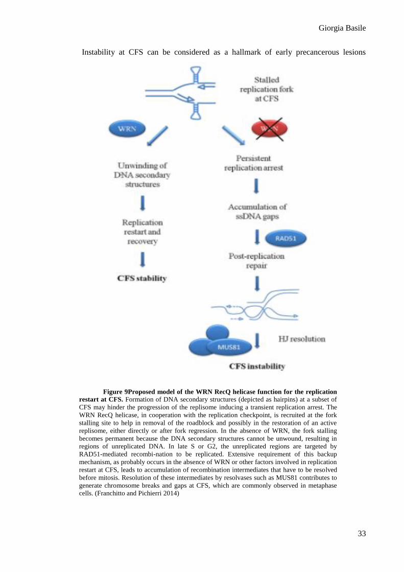
Instability at CFS can be considered as a hallmark of early precancerous lesions
Figure 9Proposed model of the WRN RecQ helicase function for the replication
restart at CFS. Formation of DNA secondary structures (depicted as hairpins) at a subset of
CFS may hinder the progression of the replisome inducing a transient replication arrest. The
WRN RecQ helicase, in cooperation with the replication checkpoint, is recruited at the fork
stalling site to help in removal of the roadblock and possibly in the restoration of an active
replisome, either directly or after fork regression. In the absence of WRN, the fork stalling
becomes permanent because the DNA secondary structures cannot be unwound, resulting in
regions of unreplicated DNA. In late S or G2, the unreplicated regions are targeted by
RAD51-mediated recombi-nation to be replicated. Extensive requirement of this backup
mechanism, as probably occurs in the absence of WRN or other factors involved in replication
restart at CFS, leads to accumulation of recombination intermediates that have to be resolved
before mitosis. Resolution of these intermediates by resolvases such as MUS81 contributes to
generate chromosome breaks and gaps at CFS, which are commonly observed in metaphase
cells. (Franchitto and Pichierri 2014)
Giorgia Basile
34
(Gorgoulis et al., 2005) and it is widely accepted that most gross chromosomal
rearrangements accumulating in solid tumors originate from fragile sites (Arlt et al., 2006).
WS is a cancer-prone and chromosome fragility syndrome characterized by gross
chromosomal rearrangements (Martin and Oshima, 2000; Oshima, 2000). Because instability
of CFS is readily detected in cells depleted of WRN even under normal division, it is possible
that chromosomal instability observed in WS cells could correlate with breaks accumulating
at these sites. However, a recent study suggests that most of the chromosomal abnormalities
arising in WS cells could be related to erosion of telomeric sequences (Crabbe et al., 2007).
These hypotheses are not necessarily incompatible: both the common fragile site and
telomere stabilities might require the helicase activity of WRN to clear the way for the
replisome, and chromosomal rearrangements observed in WS are most likely derived from a
common protective mechanism at telomeric and nontelomeric sequences. Consistently,
instability at CFS was also observed in Epstein-Barr virus–transformed lymphoblasts derived
from WS patients, which are telomerase proficient and thus protected from telomere erosion
(Pirzio et al., 2008).
Giorgia Basile
35
AIM OF THE WORK
Giorgia Basile
36
AIM
The checkpoints are surveillance mechanisms of genomic integrity, that is
fundamental to ensure genetic identity of cells in a multicellular organism. Failure of a
checkpoint often causes mutations and genomic arrangements, resulting in genetic instability,
which appears to be a leading cause in the development of many diseases, especially cancer.
Therefore, studies focusing on checkpoints are very important for understanding mechanisms
of genome maintenance, also because resulting data have direct impact on cancer biology.
The link between replication defects, human diseases and cancer, underscores the
requirement of an efficient and accurate monitoring of genome integrity during DNA
replication, which is provided by a complex and coordinated protein network, under the
control of the ATR kinase (Abraham, 2001; Zou and Elledge, 2003). In fact, DNA replication
represents a crucial moment in the life of a cell, as chromosomal integrity can be seriously
threatened by replication stress, that is the slowing and/or stalling of replication fork
progression (Zeman and Cimprich, 2014). Replication stress interferes with fork stability and
can be caused by endogenous side-products of cellular metabolism, exogenous agents capable
to interfere with DNA replication, as well as intrinsic structural features of specific genomic
regions, such as the common fragile sites (CFS).
CFS are difficult-to-replicate regions of the genome, especially prone to fork stalling
(Durkin and Glover, 2007). It was determined that the great majority of CFS are specifically
and reproducibly induced by low doses of Aphidicolin (Aph), an inhibitor of replicative DNA
polymerases,(Cheng and Kuchta, 1993; Durkin and Glover, 2007; Ikegami et al., 1978),
which do not greatly affect mitotic index, but slow replication fork progression.
Although they are considered as “hotspots” of genome instability, a recognized
causative factor in tumor development, very little is known about the molecular mechanisms
of CFS expression. As CFS are normally occurring replication fork stalling sites, they can be
considered as useful means to understand how replication fork stalling can be recovered in
vivo.
It has been previously proposed that WRN, mutated in the cancer-prone disease
Werner syndrome, is a key regulator of CFS stability, even under unperturbed conditions
through its helicaseactivity (Pirzio et al., 2008).These data support a role of WRN in
facilitating replication fork progression of regions affected by replication stress, and suggest
Giorgia Basile
37
that CFS may represent the physiological substrates of this protein (Pirzio et al.,
2008).Moreover, it is known that, under mild replication stress inducing by low doses of Aph,
WRN and ATR act in a common pathway preventing accumulation of DNA breaks at CFS.
Despite WS cells exhibit an ATR-like instability at CFS, and that WRN has been found
phosphorylated by ATR under replication stressupon HU‐induced replication stress (Pichierri
et al., 2003; Ammazzalorso et al.,2010), there is no evidence of a functional requirement of
WRN in the establishment of the replication checkpoint upon mild replication stress, such as
the one that causes breaks at CFS.
The aim of this study was to analyze the functional requirement of WRN in the ATR-
dependent checkpoint activation under mild replication stress, like that inducing CFS
expression.
Giorgia Basile
38
RESULTS PART 1
Nucleic Acids Res. 2014 Nov 10;42(20):12628-39. doi: 10.1093/nar/gku1022. Epub 2014 Oct 28.
Giorgia Basile
39
RESULTS (part 1)
WRN deficiency results in defective ATR-dependent checkpoint activation
under mild replication stress
In order to assess the role for WRN in ATR pathway activation in response to mild
replication stress, phosphorylation status of the main target of ATR, CHK1, was examined.
To compare isogenic cell lines, we first generated HEK293T cells stably expressing
scrambled (WRN-wt) or WRN-targeting shRNA (WRN-kd).WRN-kd cells showed about
80% depletion of WRN protein under the experimental conditions used in this study (Figure
1A).
A time course analysis was performed treating WRN-wt and WRN-kd cells with low
dose (0,4 μM) of Aphidicolin (Aph), and the phosphorylation of CHK1 was measured in cell
lysates by Western blot using an antibody that specifically recognizes phospho-CHK1 on
Ser345.Treatment with low dose of Aph induced a time-dependent phosphorylation of CHK1
in WRN-wt cells, already noticeable after 1 h and peaking at 24 h (Figure 1A), suggesting that
also a modest replication perturbation can trigger a quick checkpoint response.
In contrast, in WRN-kd cells CHK1 phosphorylation was not detectable, or very
weak, and it did not rich the wild-type levels even at the late time-points (Figure 1A).
In contrast, treating the cells for 4h with 1 mM HU, a specific inhibitor of DNA
synthesis which leads to a strong genome‐wide replication arrest, comparable CHK1
phosphorylation levels in both WRN-wt and WRN-kd cells was observed (Figure 10B, lanes
4 and 8).Although CHK1 phosphorylation is hampered in WRN-deficient cells, similar
amounts of Cyclin A were detected after treatments in both cell lines, suggesting that
defective CHK1 phosphorylation was not attributable to a smaller S-phase population in
WRN-kd cells (Figure 10B).
Giorgia Basile
40
Figure 10 WRN is required for CHK1 activation following mild replication stress. Western blot
detection of CHK1 phosphorylation: (A) in total extracts of WRN-wt or WRN-kd cells untreated (-) or treated
with Aph, as indicated; (B) in WRN-wt and WRN-kd cells treated with Aph or HU (s.e., short-exposure; l.e.,
long-exposure); ((E) in WS and WSWRN treated with 4 µM Aph for the indicated time points; D) in total
extracts of hTERT-immortalized primary fibroblasts, both WRN-proficient (GM01604) and WRN-deficient
(AG12975), untreated (-) or treated with 0.4 μM Aph at the indicated times. (A-B-C-D-E) In WRN-kd cells,
down-regulation of the WRN protein was verified using a specific anti-WRN antibody. The presence of
activated, i.e. phosphorylated, CHK1 was assessed using S345 phospho-specific antibody (pS345). Total amount
of CHK1 was determined with an anti-CHK1 antibody. Equal loading was confirmed probing with an anti-
Lamin B1 antibody. (B) Cyclin A was used to quantify S-phase cells.
Giorgia Basile
41
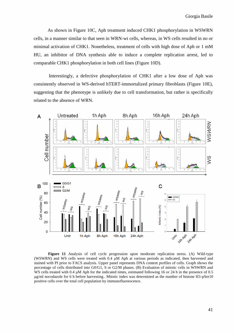
As shown in Figure 10C, Aph treatment induced CHK1 phosphorylation in WSWRN
cells, in a manner similar to that seen in WRN-wt cells, whereas, in WS cells resulted in no or
minimal activation of CHK1. Nonetheless, treatment of cells with high dose of Aph or 1 mM
HU, an inhibitor of DNA synthesis able to induce a complete replication arrest, led to
comparable CHK1 phosphorylation in both cell lines (Figure 10D).
Interestingly, a defective phosphorylation of CHK1 after a low dose of Aph was
consistently observed in WS-derived hTERT-immortalized primary fibroblasts (Figure 10E),
suggesting that the phenotype is unlikely due to cell transformation, but rather is specifically
related to the absence of WRN.
Figure 11 Analysis of cell cycle progression upon moderate replication stress. (A) Wild-type
(WSWRN) and WS cells were treated with 0.4 μM Aph at various periods as indicated, then harvested and
stained with PI prior to FACS analysis. Upper panel represents DNA content profiles of cells. Graph shows the
percentage of cells distributed into G0/G1, S or G2/M phases. (B) Evaluation of mitotic cells in WSWRN and
WS cells treated with 0.4 μM Aph for the indicated times, estimated following 16 or 24 h in the presence of 0.5
μg/ml nocodazole for 6 h before harvesting.. Mitotic index was determined as the number of histone H3-pSer10
positive cells over the total cell population by immunofluorescence.
Giorgia Basile
42
ATR-checkpoint response in WRN-deficient cells was also investigated by flow-
cytometric analysis. As expected, Aph slowed down cell cycle progression of WSWRN cells,
and delayed S-G2 phase transition (Figure 11A). In contrast, WS cells exhibited a higher
proportion of G2/M phase cells accumulated starting from 8 h of treatment (Figure 11A) with
a more pronounced accumulation of cells in the M phase, as evaluated by immunostaining for
the mitosis-specific marker phospho-histone H3 (Figure 11C).
Overall, these findings imply that WRN plays a critical function in response to mild
replication stress, and support a possible role for WRN as specific mediator of CHK1
activation.
WRN is phosphorylated in an ATR-dependent manner after Aph-treatment
Often checkpoint mediators are themselves targets of apical kinases, and WRN is
indeed phosphorylated by ATR after both HU and CPT treatments (Pirzio et al., 2008; Shah et
al., 2010).
To reinforce the existence of a functional relationship between WRN and the ATR-
dependent checkpoint after mild replication stress, we asked whether WRN was targeted by
ATR kinase under Aph treatment.
HEK293T cells were treated with low dose of Aph and cell lysates were subjected to
WRN IP. Using phospho-S/TQ antibodies, which specifically recognize phosphorylated
ATM/ATR substrates, phosphorylation of WRN was barely detectable in untreated conditions
but strongly increased after treatments (Figure 12B), suggesting that moderate stress is able to
induce WRN modification.
In an independent experiment, we transfected HEK293T cells with plasmids
expressing the Flag-tagged full-length wild-type WRN (wt) or a mutant Flag‐WRN6A, which
makes WRN unphosphorylable by ATR after replication stress (Pirzio et al., 2008).Following
Aph treatment, cell extracts were prepared and subjected to IP using anti-Flag tag antibodies.
The expression of wild-type WRN resulted in the expected phosphorylation at S/TQ sites,
whereas mutant WRN6A protein abrogated pS/TQ immunoreactivity (Figure 12C), suggesting
that, even after low dose of Aph, WRN is mainly phosphorylated at C-terminus residues.
Collectively, these results demonstrate that mild replication stress, like that inducing
breaks at CFS, may trigger ATR dependent pathway that requires the presence of WRN.
Giorgia Basile
43
WRN phosphorylation by ATR, but not WRN helicase activity, is required for
checkpoint activation upon mild replication stress
WRN is indeed phosphorylated by ATR after both HU and CPT
treatments(Ammazzalorso et al., 2010; Pirzio et al., 2008).To verify whether WRN
phosphorylation by ATR is a prerequisite for checkpoint activation after replication stress
induced by Aph, we studied the ability of WS cells stably expressing the mutant form of
WRN unphosphorylable by ATR (WSWRN6A) (Pirzio et al., 2008) to phosphorylateCHK1.
WSWRN, WS and WSWRN6A cells were used and CHK1 phosphorylation evaluated.
Figure 12ATR-dependent WRN phosphorylation upon Aph-treatment. (A) Western blot
detection of WRN phosphorylation in HEK293T cells treated or not with 0.4 µM Aph for the indicated time
points. Cell extracts were immunoprecipitated using anti-WRN antibody followed by immunoblotting with
an anti-S/TQ antibody. Total WRN was used to assess the amount of WRN immunoprecipitated. The ratio
of phosphorylated protein to total protein is reported below each lane. (B) Analysis of WRN status in
HEK293T cells transfected with plasmids expressing the Flag-tagged full-length wild-type WRN (wt) or a
full-length carrying Ala substitutions at all the six S/TQ sites (6A) and treated or not with Aph as indicated.
Cell extracts were prepared 48 h post-transfection and used to immunoprecipitate ectopic WRN with anti-
Flag tag antibody, followed by immunoblotting with an anti-S/TQ antibody. Total WRN was used to assess
the amount of wild-type or mutant form of WRN immunoprecipitated. All experiments are representative
images of at least two replicates.
Giorgia Basile
44
Figure 13ATR-dependent WRN phosphorylation, but not defective WRN helicase activity, fails
to activate CHK1 upon moderate replication stress. (A) WS cells, WSWRN or expressing an
unphosphorylable mutant form of WRN (WSWRN6A) were treated or not with Aph for the indicated times.
CHK1 phosphorylation was analysed by Western blot using phospho-specific antibodies (pS345). (B) WS
cells, WSWRN or expressing a mutant form of WRN affecting helicase (WRN-K577M) were treated or not
with Aph for the indicated times. (A-B) CHK1 phosphorylation was analysed by Western blot using phospho-
specific antibodies (pS345). Total amount of CHK1 was determined with an anti-CHK1 antibody. Lamin B1
was used as loading control. The ratio of phosphorylated protein to total protein and then normalized to the
untreated wild type is listed below each lane. (B) DNA content profiles of WRN-K577M cells treated or not
with Aph 0.4 μM for the indicated times and stained with PI prior to FACS analysis. Graph shows the
percentage of cells distributed into G0/G1, S or G2/M phases.
Giorgia Basile
45
As expected, WSWRN cells were proficient in activation of CHK1 after treatment (Figure
13A). In contrast, WSWRN6A cells showed defective CHK1 phosphorylation as WS cells.
Therefore, these data suggest that phosphorylation of WRN may play a role in the ATR-
checkpoint activation in response to moderate replication stress.
WRN helicase activity, which is crucial in maintaining CFS stability (Sakamoto et al.,
2001),could facilitate formation of the single-strand DNA (ssDNA) leading to checkpoint
activation (Franchitto et al., 2008). Thus, to verify whether suppression of helicase function
hinders CHK1 phosphorylation, WS fibroblasts and WS cells stably expressing the wild-type
WRN (WSWRN) or its helicase-dead form (WRN-K577M) were treated with Aph and cell
lysates subjected to Western blot (WB). The data confirmed that, in WSWRN cells, CHK1
was properly phosphorylated after Aph treatment, and comparable levels of CHK1
phosphorylation were observed in WRN-K577M cells, indicating that expression of the
helicase-dead WRN protein is sufficient to recover from the defective phenotype (to restore
checkpoint activity) (Figure 4B). Consistently, flow-cytometric experiments indicated that
loss of the WRN helicase activity had no effect on the delay of cell cycle progression induced
by Aph (Figure 13C).
We therefore can conclude that the WRN helicase activity is not implicated in the
activation of the ATR-dependent pathway in response to low-levels of replication stress,
whereas the ATR-dependent phosphorylation of WRN results essential for CHK1 activation.
Analysis of the ATR-signaling pathway in WRN-deficient and mutant cells
after low-levels of replication perturbation
Activation of the ATR-pathway depends on RPA binding to ssDNA (Franchitto et al.,
2008). To determine whether impaired RPA-coated ssDNA accumulation contributes to
defective checkpoint activation observed in the absence of WRN, the amount of chromatin-
bound RPA was measured in Aph-treated WSWRN and WS cells. Biochemical analysis of
fractionated cell extracts showed that RPA32, a component of the RPA heterotrimer, was only
slightly more chromatin-associated in WS cells, either exposed to Aph or left untreated,
respect to WSWRN cells (Figure 14A). Indeed, even though WS cells showed more
chromatin-bound RPA32, they also seemed to contain more RPA32 in total extracts (Figure
14A, bottom). Consistently with previous results, Western blot analysis showed that the level
of Ser33-phosphorylated RPA32, which represents a direct read-out of replication stress (Liu
et al., 2000), was greater in WRN-kd cells in comparison with WRN-wt cells (Figure 14C).
Giorgia Basile
46
Figure 14 Analysis of RPA32 accumulation and ssDNA formation in cells upon mild replication
perturbation. (A) WB chromatin recruitment ofRPA32 in WSWRN and WS cells. Total amount of RPA32 was
determined with an anti-RPA32 antibody. Lamin B1 was used as loading control. WB of whole cell extracts
(Input) (bottom) . (B) Immunostaining with RPA32 of cells untreated or treated with 0.4 μM Aph, representative
images. Graph shows the percentage of RPA32-positive cells (bottom). (C) Western blot detection of RPA32
phosphorylation in total extracts of WRN-wt and WRN-kd cells untreated (-) or treated with 0.4 µM Aph or
1mM HU for 4 h. Phosphorylated RPA32 was assessed using phospho-specific antibodies (pS33), total amount
of RPA32 was determined with an anti-RPA antibody. Equal loading was confirmed with anti-Tubulin antibody.
(D-E) Detection of BrdU incorporation under non-denaturing conditions in the parental DNA of WSWRN and
WS cells treated as indicated in the scheme. (F) Graph shows the percentage of BrdU-positive nuclei. (G) Dot
plot of the number of ssDNA foci per cell for a representative experiment.
Giorgia Basile
47
In agreement with this result, a time course analysis of non-extractable RPA32 foci by
immunofluorescence revealed that, in the absence of WRN, a higher number of RPA32-
positive nuclei accumulated under unperturbed conditions, while only a slight increase at 8 h
Aph treatment was detected, even if the RPA32 fluorescence appeared to be more intense than
in the control cells (Figure 14B).
Next, to support the evidence that loss of WRN does not affect the generation of
ssDNA after exposure to low dose of Aph, the extent of ssDNA formation was quantified
through native BrdU staining (Figure 14D-E-F-G). The analysis revealed that the percentage
of ssDNA-positive nuclei was roughly similar in both WSWRN and WS cells (Figure 14F),
but the number of foci per cell was higher in WS cells (Figure 14G).
Together, these results indicate that, in WS cells, CHK1 activation is not defective
because of the inability to form the RPA-coated ssDNA after moderate replication
perturbation.
It is known that the TopBP1 mediator protein is required for efficient CHK1
phosphorylation, and that it is recruited to stalled forks by the chromatin-bound RAD1-
RAD9-HUS1 (9.1.1) complex (Jackson and Pombo, 1998; Zou and Elledge, 2003). In
addition, WRN and the 9.1.1 complex physically interact (Delacroix et al., 2007). To test the
possibility that WRN could stabilize 9.1.1/TopBP1 association following mild replication
stress, we analyzed chromatin loading of TopBP1 and RAD9 by WB after cellular
fractionation in WSWRN and WS cells treated or not with Aph at the indicated times. As
shown in Figure15A, in WSWRN cells the amount of chromatin-bound TopBP1 started to
increase at 1 h after treatment, and remained high at 8 and 24 h, even if it decreased at 16 h in
agreement with other reports (Kumagai et al., 2006).
Similarly, RAD9 was loaded on the chromatin at all times of Aph exposure except at
16 h (Figure 15A). In contrast, in WS cells low-levels of RAD9 loaded to chromatin was
detectable under untreated and low dose Aph-treated conditions (Figure 15B). Interestingly,
the expression of the unphosphorylable form of WRN, but not, as expected, loss of WRN
helicase activity, compromised chromatin association of checkpoint mediators, and a clear
reduction of TopBP1 and RAD9 recruitment was found in WSWRN6A cells at 24 h of Aph
(Figure 15B). Therefore, defective CHK1 activation upon replication perturbation induced by
Aph, in WS and WSWRN6A cells, appears related to the levels of chromatin-bound
checkpoint mediators.
Giorgia Basile
48
Figure 15Analysis of chromatin loading of ATR-pathway sensors and mediators
in cells upon mild replication stress. (A) WSWRN and WS cells were treated with Aph for
various times or left untreated as indicated. Total amount of TopBP1 and RAD9 were
determined with an anti-TopBP1 or anti-RAD9 antibody respectively. Lamin B1 was used as
loading control. (B) WSWRN cells and WS cells expressing an ATR-unphosphorylable form
of WRN (WSWRN6A) or mutant form of WRN helicase (WRN-K577M) were treated or not
with Aph for the indicated times. Total amount of TopBP1 and RAD9 and equal loading of
total proteins were determined as in (B). Quantitative immunoblotting was used and the ratio
of TopBP1 or RAD9 protein to Lamin B1 then normalised to wild-type is reported below each
lane.
Giorgia Basile
49
Replication fork dynamics are altered in WRN-deficient and mutant cells
Having demonstrated that correct CHK1 activation relies on both the presence of
WRN and its phosphorylation by ATR, and since CHK1 has been implicated in maintaining
fork integrity and proper cell cycle progression under conditions of replicative stress
(Koundrioukoff et al., 2013; Pichierri et al., 2012), we examined how the absence of WRN or
expression of mutant forms could influence replication dynamics.
We first monitored replication in WSWRN, WS, WSWRN6A and WRN-K577M cells
under unperturbed conditions as indicated in the scheme (Figure 16A). Under these
conditions, WSWRN showed an average fork progression rate of 1.1 kb/min (Figure 16B). By
contrast, WRN-deficient cells or cells expressing the mutant forms of WRN displayed a
significant reduction of fork speed (about two-fold) as compared to control cells (Figure 16B).
A logical expectation from this result might be that replication stress exacerbated the effect,
however, all the cell lines reached identical values of fork velocity upon Aph treatment
(Figure 16C). This implies that fork progression is mainly affected by WRN deficiency or
expression of WRN mutated forms, and that Aph is able to further delay forks only in control
cells.
It has been previously proposed that cells lacking CHK1 activity show reduced rate of
replication fork progression and increased origin firing (Feijoo et al., 2001). Thus, we verified
whether the inability of Aph to reduce further fork speed in WRN-deficient and WRN-mutant
cells could be the end-result of new origins activated to compensate for replication problems.
We used a modified double labeling protocol that enables us to study fork recovery
from Aph (Figure 17A). Under such conditions, all the cell lines showed an analogous ability
to replicate after removal of Aph, evaluated as length of green tracks (Figure 8B and C).
Interestingly, using roscovitine, a CDK inhibitor that prevents origin firing, a reduced length
of green tracks in WS, WSWRN6A and WRN-K577M cells was detected, but no effect was
found in WSWRN cells (Figure 17B). Such a finding could indicate that DNA is partly
replicated from ongoing forks and partly from a downstream fork fired during roscovitine
treatment. Consistent with more new origin firing in WS, WSWRN6A and WRN-K577M cells,
roscovitine also decreased the number of isolated green tracks detected during the recovery
from Aph (Figure 17D). Therefore, we conclude that the increased replication elongation
observed in WRN-deficient or WRN-mutant cells is due to increased origin firing of
Giorgia Basile
50
neighboring dormant origins, and that this back-up mechanism obscures a more severe
reduction of fork progression occurring in these cells.
Figure 16Replication fork progression is impaired in WRN-deficient and mutant cells. (A)
Experimental scheme of dual labelling of DNA fibres. (B) Analysis of replication fork velocity in WSWRN
(wild-type), WRN-deficient (WS) and mutant cells (WSWRN6A and WRN-K577M) under unperturbed
conditions or (C) exposed to 0.4 µM Aph as indicated in (A). The length of the green tracks were measured.
Median values are represented as horizontal black lines. (D) Representative images of actual DNA fibres from
WSWRN, WRN-deficient and mutant cells. Scale bars, 10 µm. **** = Statistically significant P < 0.0001
(Student’s t-test).
Giorgia Basile
51
Figure 17Replication fork restart is affected by roscovitine in WRN-deficient and mutant cells.
(A) Experimental scheme of dual labelling of DNA fibres in presence or absence of roscovitine. (B) Analysis of
replication fork recovery in WSWRN (wild-type), WRN-deficient (WS) and mutant cells (WSWRN6A and
WRN-K577M) treated as indicated in (A). Aph (0.4 µM) and roscovitine (100 µM) were added for the indicated
time. Median values are represented as horizontal black lines. (C) Representative images of replication tracks
from WSWRN, WRN-deficient and mutant cells with or without roscovitine treatment. (D) Percentage of new
origin firing in cells treated as in (B). Data are reported as mean from three independent experiments. Error bars
represent standard error. **** = Statistically significant P < 0.0001 (Student’s t-test).
Giorgia Basile
52
Expression of phospho-mimic CHK1 mutant recovers cells from sensitivity to
Aph treatment and restores fork elongation
Since low-levels of replication stress induce CFS expression, we asked whether
defective checkpoint activation found in WS and WSWRN6A cells could be related to CFS
Figure 18Enhanced CFS induction in cells expressing an ATR-unphosphorylable form of WRN.
(A) Average overall chromosome gaps and breaks in WS cells (WS), WS cells expressing an ATR-
unphosphorylable form of WRN (WSWRN6A) and in WS cells in which wild-type WRN was reintroduced
(WSWRN). Cells were treated with two different doses of Aph for 24 h before harvesting. Data are presented as
means of three independent experiments. Error bars represent standard error. (B) Frequency of gaps and breaks
at CFS by FISH analysis. Cells were treated as described in (A). (A) Representative Giemsa-stained metaphases
of wild-type (WSWRN), WS and WSWRN6A fibroblasts untreated or treated with 0.2 µM Aph. Arrows indicate
chromosomal aberrations. FISH was performed using BACs probes corresponding to the FRA3B region.
Frequency of fragile site induction is presented as the percentage of chromosome 3 homolog with gaps and
breaks at FRA3B. Data are presented as means of three independent experiments. Error bars represent standard
error on the mean.
Giorgia Basile
53
instability. We first investigated the sensitivity of WSWRN6A to Aph. A dose-dependent
enhancement of chromosomal aberrations, similarly to that previously seen in WS cells
(Sakamoto et al., 2001), was observed in WSWRN6A,which showed approximately two-fold
more aberrations than control cells at the higher dose (Figure 18A). These results suggest that
WSWRN6A cells are sensitive to Aph treatment, and that loss of ATR-mediated WRN
phosphorylation is responsible for chromosome instability.
Next, we examined if increased chromosomal damage induced by Aph in WSWRN6A
took place at CFS. As shown in Figure 9C, instability at CFS FRA3B was induced in a dose-
dependent manner in WSWRN6A cells, with a number of gaps and breaks higher than in
control cells, and at levels comparable to those of WS cells. Interestingly, enhanced FRA3B
induction was detected even in the absence of Aph in both WSWRN6A and WS cells (Figure
18C). Similar results were observed testing other CFS (Figure 19).
These findings demonstrate that phosphorylation of WRN by ATR is essential not
only for correct CHK1 phosphorylation but also, and most notably, for CFS stability. Thus,
we wondered whether forced activation of CHK1 could overcome sensitivity to Aph in WS
and WSWRN6Acells, that is in cells where CHK1 is not phosphorylated.
Figure 19 Analysis of CFS expression in cells expressing an ATR-unphosphorylable form of
WRN. Frequency of fragile site FRA7H and FRA16D expression in WS cells (WS), WS cells expressing an
ATR-unphosphorylable form of WRN (WSWRN6A) and in WS cells in which wild-type WRN was
reintroduced (WSWRN). Cells were treated the indicated doses of Aph and harvested 24 h later. Frequency of
fragile site induction is presented as the percentage of chromosome 7 or 16 homologues with gaps and breaks at
FRA7H and FRA16D,R espectively. Data are presented as means of three independent experiments. Error bars
represent standard error.
Giorgia Basile
54
Cells were transfected with a construct expressing a mutant form of CHK1 in which
Ser317 and Ser345 were changed into Asp (CHK1317/345D), mimicking the phosphorylated
status of the protein (15). WB analysis confirmed that the Flag-tagged phospho-mimic CHK1
mutant (Flag-CHK1317/345D) was expressed at comparable levels in both cell lines (Figure
20A). After transfection, cells were treated with Aph and metaphase chromosomes were
collected and scored for total gaps and breaks. As expected, Aph treatment resulted in
enhanced chromosomal damage in WS and WSWRN6Acells (Figure 20B).
Interestingly, expression of the CHK1317/345Dprotein into both WS and WSWRN6A
cells resulted in the rescue of the chromosomal fragility induced by low dose of Aph and also
determined a slight reduction of the spontaneous number of aberrations per cell in WSWRN6A
cells (Figure 20B). These results indicate that DNA breakage observed in both WS and
WSWRN6A cells upon mild replication perturbation actually correlates with incorrect CHK1
activation.
Next, we asked whether the introduction of the phospho-mimic CHK1 mutant could
allow cells to recover from fork stalling, possible without activating new origins. WS and
WSWRN6A cells were transfected or not with the Flag-CHK1317/345Dplasmid, then treated with
Aph and roscovitine prior to analyze their replication recovery (Figure 20C).
Analysis of the length of replicating tracks on DNA fibers showed that the presence of
phospho-mimic CHK1 mutant enabled both cell lines to resume replication in a wild-type
fashion, overcoming the requirement of new origin firing, as demonstrated by the similar
track lengths in cells treated or not with roscovitine (Figure 20C), and the analysis of isolated
green tracks, showing that less dormant origins were activated (Figure 20D).
Altogether, these data strongly support the hypothesis that, in WS and WSWRN6A
cells, the inability to trigger CHK1 activation leads to replication fork demise and origin
firing in the attempt to complete replication.
Giorgia Basile
55
Figure 20Expression of phospho-mimic CHK1 rescues sensitivity to Aph and replication fork
slowing in WS and WSWRN6A cells. (A) WS and WSWRN6A cells were transfected with Flag-tagged CHK1
S317D/S345D mutant (CHK1317/345D). Lysates were collected 48 h thereafter and expression levels of Flag-
CHK1317/345D were determined by immunoblotting with anti-Flag antibody. WSWRN cells were used as negative
control. Total WRN was used to assess the amount of wild-type or mutant form of WRN. Lamin B1 was used as
loading control. (B) Effect of the expression of the Flag-CHK1317/345D plasmid on chromosomal damage. After
transfection, cells were treated or not with Aph for 24 h. The graph shows the average overall chromosome gaps
and breaks. Data are presented as means of three independent experiments. Error bars represent standard error on
the mean. (C) Evaluation of replication fork recovery in WS and WSWRN6A cells transfected with Flag-
CHK1317/345D. Cells were treated as indicated in the experimental scheme. Aph (0.4 µM) and roscovitine (100
µM) were added for the indicated time. Median values are represented as horizontal black lines. (D) Percentage
of new origin firing in cells treated as in (C). Data are reported as mean from three independent experiments.
Error bars represent standard error. **** = Statistically significant P < 0.0001 (Student’s t-test).
Giorgia Basile
56
RESULTS PART 2
Giorgia Basile
57
RESULTS (part 2)
H2AX is phosphorylated in an ATR-dependent manner in response to mild
replication stress induced by Aph
Figure 21-H2AX foci formation in wild-type cells depends on the ATR kinase after low doses of
Aph. (A) In the panel, representative images of each experimental point are shown. Error bars represent standard
errors. Scale bars: 10µm. (B) ATR depletion was verified by immunoblotting 48h after transfection using anti-
ATR antibody. Tubulin was used as loading control. (C) After depletion of ATR using RNAi and/or inhibition of
ATM activity by the KU55933 compound, cells were treated with 0,4µM Aph for 8h and immunostained with
anti-γ-H2AX (pSer139) antibody. Graph shows quantification of the number of nuclei with >5 nuclear γ-H2AX
foci (γ-H2AX-positive nuclei). Representative images are shown in the panel for each experimental condition.
The data are means from triplicate experiments.
To further analyze the cellular response to mild replication stress, we verified whether
other targets of the ATR kinase resulted affected by loss of WRN. Among the earlier ATR
targets is H2AX, which is phosphorylated in response to single-stranded DNA breaks as well
as during genome-wide-induced replication stress, such as replication fork arrest,and whose
Giorgia Basile
58
activation is easily detected as nuclear foci by specific antibody (anti-γ-H2AX pSer139) for
the phosphorylated form of H2AX (Ward and Chen, 2001).
We first verified whether H2AX could be a target of the ATR kinase also under
replication perturbation induced by low dose of Aph. To this aim, we performed
immunofluorescence experiments in wild-type cells in which ATM (ATMi), ATR (siATR) or
both (ATMi/siATR) were depleted.
Down-regulation of ATR was achieved by transfection with siRNA directed against
ATR, while ATM activity was inhibited by the use of KU55933 compound. Forty-eight hours
after interference, cells were pre-treated with the ATM activity inhibitor (KU55933) for 1h,
then Aph was added to a final concentration of 0,4μM for 8h.
The reduction of the ATR level was verified by Western blot (Figure 21B). Our results
showed that the percentage of γ -H2AX-positive cells was enhanced by Aph in wild-type cells
or cells in which ATM is chemically inhibited, but was absent, or very weak, in cells in which
ATR was depleted alone or in combination with ATM (Figure 21A and C).
These data suggest that H2AX phosphorylation induced by Aph-treatment depends
mainly on the ATR kinase.
Reduced activation of γ-H2AX in WS cells upon Aph-induced replication
perturbation
Next, having confirmed that H2AX is an ATR target upon Aph-induced replication
stress, we explored whether H2AX was phosphorylated in WS cells.
To this aim, we examined γ‐H2AX foci formation in WSWRN and WS cells,treated
with 0,4μM Aph for 8 or 16h, and then immunostained with anti-γ-H2AX antibody.
Our results show that, in wild-type cells, Aph increased the percentage of γ-H2AX-
positive nuclei in a time-dependent manner (Figure 2A and B). By contrast, in WS cells, we
detected high levels of γ-H2AX foci formation already under unperturbed conditions, but
reduced induction of γ-H2AX-positive nuclei after 8h Aph-exposure in comparison to wild-
type cells (Figure 22A and B). Moreover, in contrast to wild-type cells, WS cells showed a
small increase in the percentage of γ-H2AX foci at later time of treatment (Figure 22A and
B).
Giorgia Basile
59
Consistently with the results obtained by immunofluorescence, Western blot analysis
revealed a reduced accumulation of γ-H2AX in WS cells at earlier time of Aph-exposure, and
an increased level at later time of treatment (Figure C).
Altogether, these findings suggest that loss of WRN results in reduced activation of γ-
H2AX following Aph-induced replication perturbation.
Figure 22 Loss of WRN results in low levels of γ-H2AX foci formation after
Aph-treatment. A) and B) Immunofluorescence analysis of γ-H2AX. WSWRN and WS
cells were left untreated or treated with 0,4µM Aph for the indicated times, and then
immunostained with anti-γ-H2AX (pSer139) antibody. Graph shows quantification of the
number of nuclei with >5 nuclear γ-H2AX foci (γ-H2AX-positive nuclei). The data are
means from triplicate experiments. Error bars represent standard errors. Representative
images are shown in the panel for each experimental condition. (C) Western blot
detection of γ-H2AX in total extracts from WSWRN or WS cells left untreated or treated
with 0,4µM Aph for 8 or 24h using anti-γ-H2AX (pSer139) antibody. Equal loading was
confirmed probing with an anti-GAPDH antibody.
Giorgia Basile
60
Loss of WRN leads to the ATM pathway activation after mild replication stress
In this study, we demonstrate a defect in the ATR-signaling pathway in WRN-
deficient cells following mild replication stress (see Results part 1). Since it has been
previously reported that phosphorylation of H2AX in ATR-deficiency largely depends on the
ATM kinase (Chanoux et al., 2009), and that ATR phosphorylates checkpoint proteins in AT
cells (Tomimatsu et al., 2009), we asked whether an ATM-mediated pathway is activated
upon Aph-treatment in the absence of WRN.
To test this hypothesis, WSWRN and WS cells were left untreated or treated with Aph
under the same conditions used for the biochemical determination of CHK1 activation (see
Result part 1), with or without the concomitant addition of the selective inhibitor of ATM
kinase activity (KU55933). After fixation, cells were immunostained with anti-γ-H2AX
(pSer139) antibody. As shown in Figure 23, in untreated conditions, chemical inhibition of
ATM was able to reduce the percentage of -H2AX positive nuclei in both cell lines, but with
a greater effect in WS cells. However, after Aph-treatment, KU55933-exposure did not impair
-H2AX foci formation in wild-type cells, while in WS cells it decreased the percentage of -
Figure 23 Evaluation of ATM-pathway inhibition on the H2AX phosphorylation in WS cells.
Immunofluorescence analysis of γ-H2AX in WSWRN and WS cells left untreated or treated with 0,4µM Aph for
8h, with or without addition of 10µM chemical inhibitor of the ATM activity (KU55933). At the end of treatment,
cells were immunostained with anti-γ-H2AX (pSer139) antibody. Graph shows quantification of the number of
nuclei with >5 nuclear γ-H2AX foci (γ-H2AX-positive nuclei) (B).Representative images are shown in the panel for
each experimental condition (A).
Giorgia Basile
61
H2AX positive nuclei, even if at levels not comparable to those observed in untreated
conditions (Figure 23A and B).
These findings suggest that WS cells likely triggers an ATM-dependent pathway,
which remains active in response to mild replication stress, when the ATR-signaling pathway
results compromised due to loss of WRN mediator function.
Analysis of the consequences of ATM pathway activation in WS cells
It is well-documented that the checkpoint signaling functions of ATM and ATR are
not redundant (Abraham, 2001), and given that we observed that loss of WRN likely elicits an
ATM pathway, we investigated the functional consequences of its disruption for WS cells in
response to mild replication stress. We first evaluated the effect of inhibition of ATM kinase
activity on cell viability. To this purpose, WSWRN and WS cells were treated with Aph for
16h, with or without the addition of the chemical inhibitor KU55933, and subjected to the
fluorescence-based LIVE/DEAD assay. As shown in Figure 4A and B, loss of WRN led per
se to about two-fold increase in cell death in comparison to wild-type cells. After Aph-
exposure, a comparable reduction of cell viability was observed in both WSWRN and WS
cells (Figure 24B). Interestingly, inhibition of ATM kinase resulted in a reduction of cell
viability in both cell lines, but, surprisingly, at a smaller extent in WRN-deficient cells upon
mild replication stress (Figure 24B).
These findings indicate that inhibition of ATM pathway seems to have a little
effect on cell survival in WRN-deficient cells following mild replication stress.
To further investigate the consequences of ATM inhibition on WRN-deficient cells, we
measured the DNA damage in WRN-proficient or deficient cells by alkaline Comet assay. To
do that, we treated WSWRN and WS cells with Aph 0,4μM for 8 or 24h, with or without the
addition of the chemical inhibitor KU55933. We performed Comet assay and observed that
treatment with low Aph dose led to a time-dependent enhancement of the tail moment both in
wild-type and WS cells, with slightly higher values for WS cells (Figure 24C and D).
Interestingly, combined adition of Aph and KU55933 reduced DNA damage accumulation in
both cell lines at 8h of treatment, conversely ATM inhibition is more tolerated by the cells in
the absence of WRN at 24h when the inhibition is performed throughout the whole exposure
with low Aph dose (Figure 24D), and it is even advantageous when the inhibitor is added at
the end of the treatment (Figure 24E).
Giorgia Basile
62
Figure 24 Evaluation of cellular
consequences of ATM inhibition. (A and B)
Evaluation of cell death in wild-type (WSWRN)
and WS cells after 0,4µM Aph and/or 10µM
KU55933 treatment. Aph was added for 16h and
KU55933 1h prior Aph treatment. Cell viability was
evaluated by the fluorescence-based LIVE/DEAD
assay as described in “Materials and Methods”.
Data are expressed as percentage of dead cells and
are mean values from three independent
experiments. Error bars represent standard error. In
the panel representative images from selected
samples are shown. (C-DE) Analysis of DNA
damage accumulation evaluated by alkaline Comet
assay. WSWRN and WS cells were treated with
0,4µM Aph and/or 10µM KU55933 as indicated
and KU55933 1h prior Aph treatment; (E)
KU55933 was added for 3 hours at the end of the
treatment .Graph shows data presented as mean tail
moment. In the panel representative images from
selected samples are shown. Median values are
represented as horizontal black lines.
Giorgia Basile
63
Therefore, although ATM inhibition does not result in a huge difference in cell
viability examined with the short-term LIVE/DEAD assay between wild-type and WRN-
deficient cells, it diminishes considerably DNA damage accumulation induced by Aph at
earlier time of treatment in WRN-deficient cells.
Figure 25 Analysis of chromosomal damage induced by ATM inhibition in Aph-treated cells.
(A and B) Evaluation of chromosome fragmentation in WSWRN and WS cells treated or not with 0,4µM Aph
for 24h, and in which 10µM of ATM inhibitor (KU55933) was added to the medium 3h before harvesting
with colchicine. Metaphases were distributed according to the number of gaps and breaks per metaphase in
cells. Graph shows the percentage of metaphases with different number of chromosome breakage.
Representative Giemsa-stained metaphases of WS cells normal or differently damaged after treatment with
Aph and KU55933. Insets show an enlarged portion of metaphases for a better evaluation of chromosome
breakage.
Giorgia Basile
64
It has been previously demonstrated that under conditions of mild replication stress
induced by Aph, ATM plays a role in maintaining genome stability at CFS, which is evident
only in the absence of ATR (Ozeri-Galai et al., 2012). Thus, we analyzed the frequency of
chromosomal damage in WSWRN and WS cells treated with 0,4μM Aph in combination with
the ATM inhibitor and, 24h later, metaphases chromosome were collected and scored for total
gaps and breaks (Figure 25). Metaphases were divided into three groups, according to the
level of chromosome fragmentation: metaphases harboring from 5 to 10 gaps and breaks (5-
10), from 10 to 20 (10-20), or metaphases in which chromosomes were fragmented with more
than 20 aberrations (>20). Under unperturbed conditions, as expected, WS cells showed
higher chromosome fragmentation respect to the wild-type cells (Figure 25). Partial
replication inhibition conferred by low dose of Aph, induced chromosomal damage in both
cell lines, but, also in this case as expected, with levels of damage higher in WS cells (Figure
25). However, the addition of the ATM inhibitor at the end of the treatment with Aph led to
increased DNA breakage in wild-type cells, but reduced levels of chromosomal aberrations in
WS cells as compared to the Aph-treated samples (Figure 25).
Together, these results indicate that inhibition of ATM activity seems to reduce the
levels of chromosomal instability in WS cells respect to the wild-type cells after mild
replication stress.
Loss of WRN leads to accumulation of 53BP1 NBs in G1 phase and small foci
in S phase upon mild replication stress
It has been previously reported that mild replication stress, such as low-dose of Aph,
induces the formation of 53BP1 nuclear bodies (NBs), which are largely confined to G1 or
very early S phase (Lukas et al., 2011). These structures are produced by replication stress
and originate from lesions generated at the G2/M transition, when a fraction of “under-
replicated” genomic loci enters into mitosis and is converted to DNA/chromatin lesions
(Lukas et al., 2011). Moreover, 53BP1 NBs are thought to protect CFS from attack by
replication stress, and inhibition of ATM kinase affects their accumulation (Lukas et al.,
2011). Thus, we investigated the 53BP1 NBs formation in WSWRN and WS cells, with or
without the ATM kinase activity. First, cells were exposed to Aph 0,4μM for 24h, and then
fixed and co-immunostained with antibodies against 53BP1 (green fluorescence) and Cyclin
A (red fluorescence) to mark individual S phase cells. In wild-type cells,53BP1 NBs were
correctly formed in G1 (Cyclin A-negative cells) after 24h Aph (Figure 26A and B). In
WSWRN cells the average number of 53BP1 NBs increased from 2,3 to 8,25 after Aph, and
Giorgia Basile
65
such treatment caused a marked shift towards nuclei with higher numbers of 53BP1 NBs
restricted largely to G1 nuclei (Figure 26B). In WS cells, in untreated conditions, the average
number of 53BP1 NBs per nucleus is almost two-fold than in control cells, that is about 4,4,
and increased to 10,4 after exposure to Aph for 24h (Figure 26B).
Figure 26 WRN loss increases 53BP1 relocalisation in all cell cycle phases. (A) Representative
images showing WSWRN and WS cells co-immunostained with antibodies against 53BP1 (green fluorescence)
and Cyclin A (red fluorescence) are reported. (B) WSWRN and WS cells were treated or not with Aph 0,4μM for
24h and co-immunostained with antibodies against 53BP1 or Cyclin A. The indicated number of non-
overlapping images were acquired and analyzed for the presence of 53BP1 foci in G1 nuclei (Cyclin A-negative
cells). (C) Graph shows distribution per cell of the 53BP1 foci in Cyclin A-positive cells in WSWRN and WS
cells treated or not with 24h Aph and subjected to dual immunofluorescence with 53BP1 and Cyclin A. Median
values are represented as horizontal black lines. Error bars represent standard error.
Giorgia Basile
66
Notably, small diffuse 53BP1 foci were observed in Cyclin A-positive cells, especially
in WS cells, where the 53BP1-positive foci per cell increased from an average number of 3
under untreated conditions to 10,75 after Aph treatment (Figure 26C).
These results indicate that WRN deficiency per se promotes 53BP1 NBs accumulation
in Cyclin A-negative cells, and a further enhancement was observed after exposure to low
dose of Aph. Given that these structures correlate with ATR depletion and are thought to be
the end-result of chromosomal stress, occurring during the previous S phase in response to
mild replication stress, and taking into account that in WS cells the ATR pathway is
compromised, these 53BP1 NBs could represent, also in our case, a system used for
protection of DNA lesions formed at CFS.
Furthermore, loss of WRN leads to the formation of small diffuse 53BP1 foci during S
phase upon mild replication stress. Since the increase of 53BP1 foci in all cell cycle phases
was previously associated with CHK1 depletion (Lukas et al., 2011), we can speculate that the
phenotype observed in WS cells could be due to defective CHK1 activation after 8h and 24h
of Aph (see Results part 1).
Since it has been reported that ATM kinase is required for efficient accumulation of
53BP1 NBs (Lukas et al., 2011), we asked whether also 53BP1-positive foci formation in S
phase are dependent on ATM activity. To test this, WSWRN and WS cells were exposed to
Aph 0,4μM for 24h, with or without the addition of ATM inhibitor (KU55933), then fixed
and co-immunostained with antibodies against 53BP1 (green fluorescence) and Cyclin A (red
fluorescence). First, we confirmed that in our cell lines, ATM inhibition was able to reduce
accumulation of 53BP1 NBs (Figure 27A). Then, we examined the relocalisation of 53BP1
into small diffuse foci in S phase (Cyclin A-positive cells), and we found that relocalisation of
53BP1 foci into S phase did not require a functional ATM kinase (Figure 27B).
Next, to verify whether, in WS cells, 53BP1-positive foci in S phase accumulate at
CFS after treatment with the low dose of Aph, for 24h, we used a combined
immunofluorescence and FISH approach (IF-FISH) to co-localise 53BP1 with CFS. We
found that, in WS cells, FISH probes to FRA3B, the most expressed CFS, co-localising with
53BP1 foci in S phase induced by Aph at low frequency (7%) (Figure 27A). These findings
suggest that, being fragile sites DNA regions particularly sensitive to replicative stress,
53BP1-S-foci are probably correlated with replication stress response, but not specifically
Giorgia Basile
67
with common fragile sites processing, actually WRN-deficient cells have enhanced instability
at common fragile sites after treatment with Aph (Pirzio et al., 2008).
Altogether, these data allow us to hypothesize that, thus, being fragile sites DNA
regions particularly sensitive to replicative stress, 53BP1-S-foci are probably correlated with
replication stress response, but not specifically at common fragile sites, actually WRN-
deficient cells have enhanced instability at common fragile sites after treatment with Aph
(Pirzio et al., 2008).
Figure 27 53BP1 foci formation in Cyclin A-positive cells is not dependent on the ATM activity. (A) Combined FISH with chromosome-specific probes (green fluorescence) and immunofluorescence staining
for53BP1 (red fluorescence) in WS cells after treatment with 0,4µM Aph for 24 h. A mix of BACs (94D19,
149J4 and 48E21) mapping to FRA3B fragile site region was used as probe for FISH analysis.(B) Efficient
formation of 53BP1 NBs requires ATM activity. Exponentially growing WSWRN cells were treated or not with
10μM of ATM inhibitor (KU55933) for 24h during 0,4μM Aph treatment and co-immunostained with antibodies
to 53BP1 (green fluorescence) and Cyclin A (red fluorescence). In the panel representative images are reported.
(B) Graph shows distribution of small diffuse 53BP1 foci in Cyclin A-positive cells using WSWRN and WS
cells treated with 0,4μM Aph for 24h, with or without the addition of 10μM of KU55933. Median values are
represented as horizontal black lines.
Giorgia Basile
68
ATM inhibition in WRN-deficient cells recovers CHK1 defective activation in
response to mild replication stress
In this study, we reveal that loss of WRN elicits an ATM-dependent pathway in response to
mild replication stress, when the ATR-signaling pathway is compromised (see Results part 1).
Moreover, it has been previously demonstrated an interplay between ATM and ATR in the
regulation of CFS stability (Ozeri-Galai et al., 2008). In particular, it is thought that both ATR
and ATM phosphorylate CHK1 under partial replication conferred by Aph (Ozeri-Galai et
al., 2008). In the light of these findings, we examined the effect of ATM inhibition on CHK1
activation in WS cells.
Figure 28Evaluation of CHK2 and CHK1 activation upon mild replication stress.
(A) Western blot detection of CHK2 in total extracts ofWSWRN and WS cells untreated or
treated with 0,4µMAph for the indicated time. Where indicated, cells were treated with
10µMKU55933 added to media 1h prior Aph treatment. The presence of activated, i.e.
phosphorylated, CHK2 was assessed using Thr68 phospho-specific antibody (pCHK2). Total
amount of CHK2 was evaluated with an anti-CHK2 antibody. Equal loading in WB was
confirmed probing with an anti-GAPDH. (B) WRN-wt (wild-type) and WRN-kd (WRN-
deficient) cells were treated as in A. Equal loading in WB was confirmed probing with an anti-
Lamin B1. (C and D) Western blot detection of CHK1 in WSWRN and WS cells or WRN-wt and
WRN-kd cells were treated as in A. The presence of activated, i.e. phosphorylated, CHK2 was
assessed using Ser345 phospho-specific antibody (pS345CHK1). Total amount of CHK1 was
evaluated with an anti-CHK1 antibody. Equal loading in WB was confirmed probing with an
anti-GAPDH. The ratio of phosphorylated protein to total protein normalised to the untreated
wild-type is reported below each lane.
Giorgia Basile
69
First, we investigated whether, under our conditions of mild replication stress, CHK2,
a target of ATM kinase, was phosphorylated as a symptom of ATM pathway activation in WS
cells.
To carry out our analysis, WSWRN and WS cells were exposed to low dose of Aph
for 8h, and phosphorylation status of CHK2 was assessed in cell lysates by Western blot using
Thr68 phospho-specific antibody (Figure 28A). Moreover, some parallel samples were treated
with ATM inhibitor (KU55933) to verify the dependency of CHK2 phosphorylation on ATM
kinase upon Aph (Figure 28A).
In wild-type (WSWRN) cells, a very faint band corresponding to CHK2
phosphorylation was detectable after 8h of Aph, but it was inhibited by KU55933. In contrast,
in WS cells CHK2 showed elevated levels of phosphorylation even under untreated
conditions. The phospho-CHK2 proportion was sickly intensified by Aph treatment, but it
was highly reduced by combined treatment of Aph with ATM inhibitor (Figure 8A).
By using HEK293T cells stably expressing scrambled shRNA (WRN-wt), we
observed a CHK2 activation after 24h Aph in wild-type cells (Figure 28C). On the contrary,
WRN-deficient (WRN-kd) cells showed increased levels of CHK2 phosphorylation under
untreated conditions, and also already at 8h of Aph-exposure (Figure 28C).
For what concerns CHK1 activation, as expected, treatment with Aph for 8h induced
CHK1 phosphorylation in WSWRN, which remained unchanged upon ATM inhibition
(Figure 28B). Interestingly, in WS cells, although, as expected, CHK1 was not
phosphorylated after mild replication stress, however, the addition of KU55933 to the culture
medium during Aph treatment recovered from faulty checkpoint activation, leading to CHK1
phosphorylation at levels comparable, if not major, to those of wild-type cells (Figure 28B).
Similar results were obtained using HEK293T cells (Figure 28D).
Therefore, our data demonstrate that chemical inhibition of ATM kinase activity
confers to WRN-deficient cells the ability to recover CHK1 defective phosphorylation
following mild replication stress.
Giorgia Basile
70
Analysis of the consequences of ATM inhibition in WS cells stably expressing
the unphosphorylable mutant form of WRN
Figure 29 ATM inhibition consequences in WS cells stably expressing the mutant form of WRN
unphosphorylable by ATR (A) Aberrations per cell in WSWRN, WS and WS cells stably expressing the
mutant form of WRN unphosphorylable by ATR (WSWRN6A) (B)Distribution of metaphases according to the
number of gaps and constrictions per metaphase in cells following 24 h treatment with 0.4 M aphidicolin and
ATM inhibition with KU55933. (B) DNA damage accumulation evalueted by Alkaline Comet assay
WSWRN6A.
Giorgia Basile
71
To further analyse the consequences of ATM inhibition in WS cells we studied the
response to ATM inhibition in WS cells stably expressing the unphosphorylable mutant form
of WRN (WSWRN6A). DNA damage was measured in WSWRN6A cells, compared to WS
and WSWRN, by alkaline Comet assay. To do that, WSWRN, WS and WSWRN6A cells
were treated with Aph 0,4μM for 24h, with or without the addition of the chemical inhibitor
KU55933. Combined addition of Aph and KU55933 is more tolerated in WSWRN6A cells
respect to wild type cells, but the damage observer is higher respect to WS cells. ATM
inhibition performed throughout the whole exposure with low Aph doses in WSWRN6A leads
also to a chromosomal fragmentation in a similar manner than in WSWRN cells, but with
minor percentage of metaphases harboring more than 20 aberrations (>20) (Figure 29A-B).
Data show that ATM inhibition during mild replication stress increased DNA damage, also at
a chromosomal levels, in WSWRN6A at a middle level between WRN-proficient cells and
WRN-deficient cells (Figure 29C).
To go deeper inside the causes of this intermediate phenotype, 53BP1 foci in S-phase
and CHK1 phosphorylation were evaluated. First, WSWRN6A cells were exposed to Aph
0,4μM for 24h, and then fixed and co-immunostained with antibodies against 53BP1 (green
fluorescence) and Cyclin A (red fluorescence) to mark individual S phase cells. By using WS
cells stably expressing the unphosphorylable mutant form of WRN, 53BP1 foci in Cyclin A-
positive cells are observed, with a similar percentage of cyclin A positive cells with detectable
53BP1 focal structures respect to WS cells. These foci could be a sign of the activation of
alternative pathways at later time point, as observed in the absence of WRN, which could
partially explain the intermediate phenotype, between that of wild-type cells and that of WS
cells, shown by WSWRN6Acells in the comet and metaphase analysis when ATM is
inhibited. The analysis of CHK1 phosphorylation point out that after treatment with Aph for
8h in WSWRN6A cells was not phosphorylated after mild replication stress, and the addition
of KU55933 to the culture medium does not change CHK1 phosphorylation levels. WSWRN
and WS cell treated with Aph for 8 h were used respectly as positive and negative control. In
conclusion, data demonstrate that the activation of hypothetic alternative pathways in
WSWRN6A cells remain probably incomplete, because, at earlier time, the phosphorylation of
CHK1 upon mild replication stress does not change when ATM is inhibited (Figure 29E). The
inability of WSWRN6A cells to rescue CHK1 activation could be due to the presence of
WRN that, although mutated, could prevent the activation of an alternative pathways. Further
Giorgia Basile
72
experiments will be needed to explore this issue. Our results show that WRN is tightly
regulated by both ATR and ATM to ensure genome stability.
Giorgia Basile
73
DISCUSSION
Giorgia Basile
74
DISCUSSION
We previously determined that WRN in association with the ATRpathway prevents
CFS breakage, but with largely unknown mechanisms(Pirzio et al., 2008). Here, we provide
evidence for a crucial role of the WRN protein in mediating CHK1 phosphorylation
specifically following low level of replicative stress, such as that causing CFS expression.
Loss of CHK1 has been related to CFS expression, and CHK1 undergoes
phosphorylation upon mild replication perturbation(Durkin et al., 2006). Although CHK1 has
been widely found activated in response to low-doses of Aph (14,26,27), a recent work
suggested that CHK1-dependent protection of CFS might be unrelated to its phosphorylation-
dependent activation (Koundrioukoff et al., 2013). Consistently with earlier reports (Durkin et
al., 2006; Petermann and Helleday, 2010; Zachos et al., 2005), we detect CHK1
phosphorylation after Aph treatment, albeit at reduced levels if compared to strong replication
perturbation as that induced by HU. Since CHK1 phosphorylation was demonstrated from
independent studies using different cell lines, it is unlikely that the genetic background of
cells could influence this specific phenotype. However, the percentage of S-phase cells might
markedly affect the levels of CHK1 phosphorylation, especially in the presence of partial
inhibition of DNA synthesis, perhaps accounting for these discrepancies between the data.
Noteworthy, in the study where CHK1 phosphorylation has not been detected after low Aph
doses, chromatin loading of ATR and its mediators, RAD9 and TopBP1, has been
demonstrated (Koundrioukoff et al., 2013). Our findings confirm that mild replication stress
determines chromatin accumulation of RAD9 and TopBP1, similarly to robust replication
perturbation induced genome-wide.
Interestingly, RAD9 and TopBP1 recruitment to chromatin involves the presence of
WRN under unperturbed conditions as well as upon mild replication stress, suggesting that
the reported association of WRN with 9.1.1, whose loss increases CFS expression (Pichierri et
al., 2012), is important for CHK1 activation. Therefore, our results indicate that the presence
of the WRN protein may be required for CHK1 activation, suggesting that WRN acts as a
checkpoint mediator protein, specifically when replication is partially inhibited. A similar
function of a RecQ helicase has been demonstrated in yeast, where Sgs1 plays a non-catalytic
role in correct activation of Rad53 after replication stress (Liu et al., 2000), bridging RPA and
Rad53 together (Ozeri-Galai et al., 2008). Consistent with a possible role as mediator protein,
WRN is phosphorylated at S/TQ sites by ATR also after Aph-induced replication stress, and
this modification of WRN appears to be crucial for promoting efficient CHK1
phosphorylation upon mild replication perturbation, as well as for stabilisation of CFS.
Giorgia Basile
75
In WS cells expressing the ATR-unphosphorylable mutant form of WRN we detect a
decrease of RAD9 and TopBP1 levels respect to the other cell lines especially at the later
time. This is not surprisingly since the phospho-WRN mutant is a double mutant that cannot
be targeted by ATR and ATM (Pirzio et al., 2008), and we cannot exclude that loading on
chromatin of mediators at later times could rely on the ATM-dependent phosphorylation of
WRN. Altogether, it is plausible that, upon moderate replication perturbation, WRN may act
as a mediator of CHK1 activation, and this function can be regulated by the ATR-dependent
phosphorylation. Conversely, WRN helicase mutant does not affect CHK1 activation upon