Seton Hall University eRepository @ Seton Hall eses 2010 Investigation of Poly (C) Binding Protein 1-Interact Proteins by Screening cDNA Library Hamidah N. Sultan Seton Hall University Follow this and additional works at: hps://scholarship.shu.edu/theses Part of the Psychology Commons Recommended Citation Sultan, Hamidah N., "Investigation of Poly (C) Binding Protein 1-Interact Proteins by Screening cDNA Library" (2010). eses. 223. hps://scholarship.shu.edu/theses/223
Transcript
Seton Hall UniversityeRepository @ Seton Hall
Theses
2010
Investigation of Poly (C) Binding Protein 1-InteractProteins by Screening cDNA LibraryHamidah N. SultanSeton Hall University
Follow this and additional works at: https://scholarship.shu.edu/theses
Part of the Psychology Commons
Recommended CitationSultan, Hamidah N., "Investigation of Poly (C) Binding Protein 1-Interact Proteins by Screening cDNA Library" (2010). Theses. 223.https://scholarship.shu.edu/theses/223
Investigation of Poly (C) Binding Protein 1-Interacting Proteins by Screening a cDNA Library
Hamidah Nizar Abdul Sultan
Submitted in partial hlfillment of the requirements of the degree of Masters of Science from the Department of Biological Sciences of Seton Hall University -
May 2008
APPROVED BY:
y Jane L. o, PhD Mentor
Carolyn S. Bentivegna, PhD Committee Member
Heping Zhou, PhD Committee Member
Carolyn S. Bentivegna, PhD Chair, Department of Biology
ACKNOWLEDGEMENTS
This thesis not just a representation of research done in a laboratory; it is a depiction of the
support and guidance I have received from my mentor, professors, lab colleagues, friends, and
family.
First and foremost, I would like to express my deepest gratitude and appreciation to my mentor
Dr. Jane L. KO for her guidance, patience, support, and understanding. I thank her not only for
the opportunity to be a member of her lab, but also for the moral support she provided during the
most challenging times of my graduate career.
I would also like to thank the members of the thesis committee, Dr. Carolyn Bentivegna and Dr.
Heping Zhou, for their valuable time and contribution to this thesis.
I wish to thank all my professors and faculty at Seton Hall University for giving me the gift of a
higher education.
Thank you to the members of Dr. KO's lab: Ying-Chih Lin, Kelly Flock, Adnan Malik, Pranjal
Nahar, and Oliver Surriga for their continuous support and help.
Finally, I would like to thank my family and friends for all the encouragement and support they
have given me everyday.
TABLE OF CONTENTS
SECTION PAGE
Abstract 1
Introduction 2
Materials and Methods 12
Results 16
Discussion 34
Conclusion 37
References 38
LIST OF FIGURES AND TABLES
FIGURE / TABLE PAGE
Table 1
Figure 1
Figure 2
Figure 3
Figure 4
Figure 5
Figure 6
Figure 7
Figure 8
ABSTRACT
Poly (C) binding protein 1 (PCBPI) is a single strand DNA binding protein belonging to
the K Homology Domain superfamily. It is a multifunctional protein involved in various aspects ,
of gene regulation including mRNA stabilization, transcriptional and translational control.
Recent studies have further demonstrated that PCBPl participates in the regulation of neuronal
p-opioid receptor gene expression. In order to further understand the multifunctional roles of
PCBP1, PCBPI was used as the bait protein to identify its functional protein partners via protein-
protein interactions by screening a cDNA library using the bacteria two-hybrid system.
PCBP1 was cloned into the bait (pBT) vector, which resulted in a bait fusion protein: hcI-
PCBPI. The plasmid containing this fusion protein, pBT-PCBPI, was then used to screen over
one million clones of a human brain cDNA library on media containing three selective markers
(3-AT, chloramphenicol, and tetracycline), and then on a high stringent media containing four
selective markers (3-AT, chloramphenicol, tetracycline, and streptomycin). The potential PCBPl
interacting proteins from surviving colonies under high stringent selection were then verified for
a protein-protein interaction in vivo by extracting and re-co-transforming the target plasmids
(containing a cDNA insert from the human brain library) with the pBT-PCBPI plasmid into
bacterial competent cells. Further analysis of the extracted target plasmids using restriction
enzyme digestion and gel electrophoresis revealed a 750 base pair cDNA insert. This insert was
then cloned into the pGEX vector of the GST Gene Fusion System. The resulting plasmid,
pGEX-Clone, was used to over-express the GST-fusion protein in bacterial cells. The over-
expression of this protein was confirmed by analyzing bacterial lysates using SDS-PAGE. This
protein was then purified through the GST purification system, and now can be used for further
investigation of a direct physical interaction with PCBP1.
INTRODUCTION
Poly (C) binding protein 1 (PCBP1) is a single strand (ss) DNA-binding protein that has
multifunctional roles in mRNA stabilization (Reimann et al., 2002), transcriptional (Gines-
Rivera et al., 2006, KO and Loh, 2005, and Malik et. al, 2006) and translational (Kim et al., 2005
and Makeyev and Liebhaber et al., 2002) regulation. It belongs to the K Homology domain
superfamily, which is made up of two classes of proteins: the hnRNPs and the poly (C) binding
proteins (PCBPs), and is involved in many biological processes (Gines-Rivera et al., 2006,
Lynch et al., 2005, Malik et al., 2006, Makeyev and Liebhaber et al., 2002, and Mournen et al.,
2005)
Heterogeneous Nuclear Ribonucleoproteins (hnRNPs)
In eukaryotic cells, mRNA precursors are packaged with mRNA binding proteins to form
heterogeneous nuclear ribonucleoprotein complexes known as heterogeneous nuclear
ribonucleoproteins (hnRNPs). hnRNPs are involved in many molecular and cellular functions,
including chromatin remodeling, mRNA splicing, transcription, and translation (Bomsztyk et al.,
1997, Makeyev and Liebhaber et al., 2002, and Moumen et al., 2005). The hnRNPs also play a
role in the cross talk between kinases and factors that mediate nucleic acid directed processes by
acting as a docking platform (Inoue et al., 2007). More than 20 hnRNPs have been identified,
with hnRNP K being one of the most studied (Bromsztyk et al., 2004, Lee et al., 1996, and
Rahman-Roblick et al., 2007).
hnRNP K is found in both the nucleus and cytoplasm of many tissue types (Matunis et
al., 1992). It interacts with DNA, RNA, protein kinases, and proteins involved in chromatin
remodeling (Denisenko and Bomsztyk, 2002, Dreyfuss et al., 1993, Michelotti et al., 1996, Siomi
et al., 1994, and Tomonaga and Levens., 1996). hnRNP K has multiple domains including three
K Homology (KH) domains that mediate binding to DNNRNA, as well as a highly interactive K
protein region (KI) that recruits protein binding partners (Bomsztyk et al., 1997, Bomsztyk et al.,
2004, and Paziewska et al., 2004).
The K Homology Domain
KH domains are evolutionarily conserved and bind to short ribonucleotide sequences. ,
The KH domain has a pl-al-a2-P3-al configuration, in which three stranded antiparallel P-
sheets are supported by three a-helices (Paziewska et al., 2004). The KH domain was first
identified in hnRNP K as an mRNA binding motif. It exists in a variety of RNA-bindmg proteins
either singularly or repetitively, and can function independently or jointly in specific nucleotide
binding activities (Malik et al., 2006 and Paziewska et al., 2004). The KH domain family of
proteins is able to bind RNA homopolymers such as poly-cysteine (C) residues as well as single
stranded (ss) and double stranded (ds) DNA (Leffers et al., 1995, KO and Loh, 2005, and Malik
et al., 2006).
K Homology Domain Superfamily
The KH domain superfamily is made up of two classes of proteins: the hnRNPs and the
poly (C) binding proteins (PCBPs). The PCBPs are a family of four proteins PCBP 1-4 (Kim et
al., 2005 and Makeyev et al., 2002). Similar to hnRNP K, the PCBPs have three KH domains:
two KH domains found at the N-terminus and a third KH domain found near the C-terminus. A
third domain found between the second and thud KH domain carries the greatest sequence
variance. This variable domain contains a nuclear localization signal (NLS) that allows for
shuttling between the cytoplasm and nucleus (Berry et al., 2006). Unlike the PCBPs, the variable
domain in hnRNP K has a N-terminal bipartite nuclear localization signal (Chkheidze et al., 1999
and Makeyev and Liebhaber et al., 2002). Both the hnRNP and PCBP family of proteins have
multifunctional roles in gene regulation.
Transcriptional Control
The role hnRNP K plays in transcription is well characterized. For example, hnRNP K
enhances the expression of c-myc by binding to cysteine-rich sequences (the CT element) in the
c-myc promoter. hnRNP K also associates with the kappa-B enhancer motif, and enhances
expression of the EGR and BRCAl genes (Stains et al., 2005 and Lynch et al., 2005). In
addition, hnRNP K activates and represses RNA polymerase I1 transcription in a context-
dependent manner (Mournen et al., 2005).
PCBPs also play a role in the transcription of specific genes. Recent studies have shown
PCBPl to participate in the transcriptional activation of p-opioid receptor gene (MOR) by
binding to a ssDNA element located in the proximal promoter (Gines-Rivera et al., 2006 and
Malik et. al, 2006). Also, PCBPl was demonstrated to play a regulatory role in eZF4E
transcription of growth factor-stimulated cells by binding to a region in the promoter containing
the eIF4e basal element (4EBE) (Meng et al., 2007).
Posttranscriptional Control
PCBP 112 and hnRNP K have been found to mediate mRNA stabilization by binding to
pyrimidine-rich motifs (Chkheidze et. al, 1999) such as the cytidine-rich Differentiation Control
Element (DICE), a multifunctional cis-element (Reimann et al., 2002) found in the 3' -
untranslated region (UTR) of many eukqotic mRNAs. For example, PCBPl stabilizes Collagen
a-1 in hepatic stellate cells (HSC), which are the major cell type responsible for collagen
synthesis in cirrhosis of the liver. Collagen a-](I) mRNA increases during activation of HSCs by
a fibrogenic stimulus and stabilization occurs through a RNA complex containing PCBPl that
binds to the C-rich region in its 3' UTR (Stefanovic et al., 1997).
In addition, hnRNP K and PCBP112 stabilize reticulocyte-15-lipoxygenase (rl 5-LOX)
mRNA during the differentiation of erythrocytes and granulocytes. A model has been suggested
whereby PCBPl b i d s to hnRNP K, which binds to DICE of rl5-LOX- mRNA 3'UTR. The
hnRNP KIPCBPl-DICE complex blocks the 80s ribosome assembly, resulting in translational
silencing of rl5-Lox mRNA (Ostareck-Ledere and Ostareck, 2004). The same model of
translational silencing by PCBPl has been suggested for human papilomavirus type 16 L2 (L2)
mRNA (Makeyev and Liebhaber, 2002).
PCBPl and 2 have also been shown to stabilize Tyrosine Hydroxylase (TH) and
Erythropoietin (EPO) mRNA. A ribonucleoprotein complex binds to the hypoxia-induced
protein binding sequence (HIPBS), an RNA stability element in the 3'UTR of TH rnRNA. A
similar sequence is found in the 3' UTR of EPO mRNA. Both PCBPl and 2 bind to these
sequences, thereby stabilizing the mRNAs for translation (Czyzyk-Krzeska and Bendixen, 1999).
Other mRNAs that are stabilized by the PCBPs include a-globin, poliovirus, and renin
mRNA. The stability of a-globin mRNAs has been conferred by binding of an a-complex
composed of a single PCBP (either PCBPl or one of two isoforms of PCBP2) to the pyrimidine
rich cis-acting stability element in the 3'UTR (Chkheidze et. al, 1999). BCBP 1 and 2 bind and
stabilize the 5'-terminal cloverleaf of poliovirus mRNA that serves as a template for viral
negative-stranded RNA synthesis for RNA replication (Muny et al., 2001). Renin, also known as
angiotensinogenase, is an enzyme released by juxtanglomerular cells in the kidney in response to
low blow volume or decreased NaCl concentration. PCBPl stabilizes human renin mRNA by
binding at the 3' UTR (Morris et al., 2004).
Translational Control
In addition to transcriptional and posttranscriptional controls, PCBPs have been shown to
play a role in translational control of folate receptor mRNA and Hepatitus C Virus (HCV)
rnRNA. PCBPl interacts with an 18 base cis-element in the 5' UTR of folate receptor mRNA by
activating translation in vitro (Anthony et al., 2004). The 5' UTR region of HCV contains a
highly structured internal ribosome entry site (IRES) to which PCBPs bind. PCBP 1 and 2 have
been shown to interact with IRES in the 5' UTR HCV mRNA (Spangberg, 1999).
Poly Cysteine (C)-Binding Protein 1
As described above, both hnRNP K and the PCBPl are structurally similar and are
involved in transcription and translation of a variety of eukaryotic mRNAs. The exact roles these
proteins play are circumstance-dependent. In other words, each protein can have diverse effects
on different rnRNAs in the same cell or on the same mRNA in response varying environmental
signals (Thyagarajan and Szaro, 2004).
PCBPl has multifunctional roles in mRNA stabilization (Chkheidze et. al, 1999 and
Reimann et al., 2002), translational activation and silencing (Anthony et al., 2004 and
Spangberg, 1999), and transcriptional regulation (Makeyev and Liebhaber et al., 2002) PCBPl
is a ss DNA-binding protein that was found to participate in p-opioid receptor gene expression
by screening a mouse cDNA library using the yeast one-hybrid system (KO and Loh, 2005).
PCBPl recognizes (C)-rich ssDNA through its KH domain (Malik et al, 2006). It is found in
both the nucleus and cytoplasm of numerous tissues (Aasheim et al., 1994) including, but not
limited to, neuronal tissue (Beny et al., 2006).
Protein-Protein Interactions
The multiple-functionality of the hnRNP and PCBP proteins suggests that their roles may
be mediated by protein-protein interactions. Yeast-two-hybrid studies have shown hnRNP K and
the PCBPs to interact with different proteins. hnRNP K binds to eIF4E (Lynch et al., 2005) and
TATA-binding protein (Michelotti et al., 1996), as well as transcriptional repressors Eed, Zik-1,
Kid-1, and MZF-1 (Denisenko et al., 1996, Denisenko and Bromsztyk, 1997, and Bomsztyk et
al., 1997). hnRNP K dimerizes and oligomerzies with multiple proteins, including the Src family
of tyrosine kinases (Taylor and Shalloway, 1994 and Weng et al., 1994), the proto-oncogene Vav
(Bustelo et al., 1995 and Van Seuningen et al., 1995), and with protein kinase C (Schullery et al.,
1999). PCBP2 can form homodimers (Gamarnik and Andino, 1997, Kim et al., 2000) and can
interact with hnRNP L (Funke et al., 1996, and Kim et al., 2000), hnRNP K and I (Kim et al.,
2000), Y-box binding proteins, splicing factor 9G8, and filamin (Funke et al., 1996). In addition,
hnRNP K and PCBP2 interact with each other (Kim et al., 2000) and have common protein
partners including Y-box-binding protein, splicing factor 9G8, and hnRNP L (Makeyev and
Liebhaber, 2002 and Shnyreva et al., 2000).
Several proteins have been shown to interact with PCBPl in different tissue types, such
as hnRNP A2 (Kosturko et al., 20006), Pakl (Meng et al., 2007), and Lamin A/C (Zhong et al.,
2005). Table 1 lists the known protein-protein interactions with PCBPl.
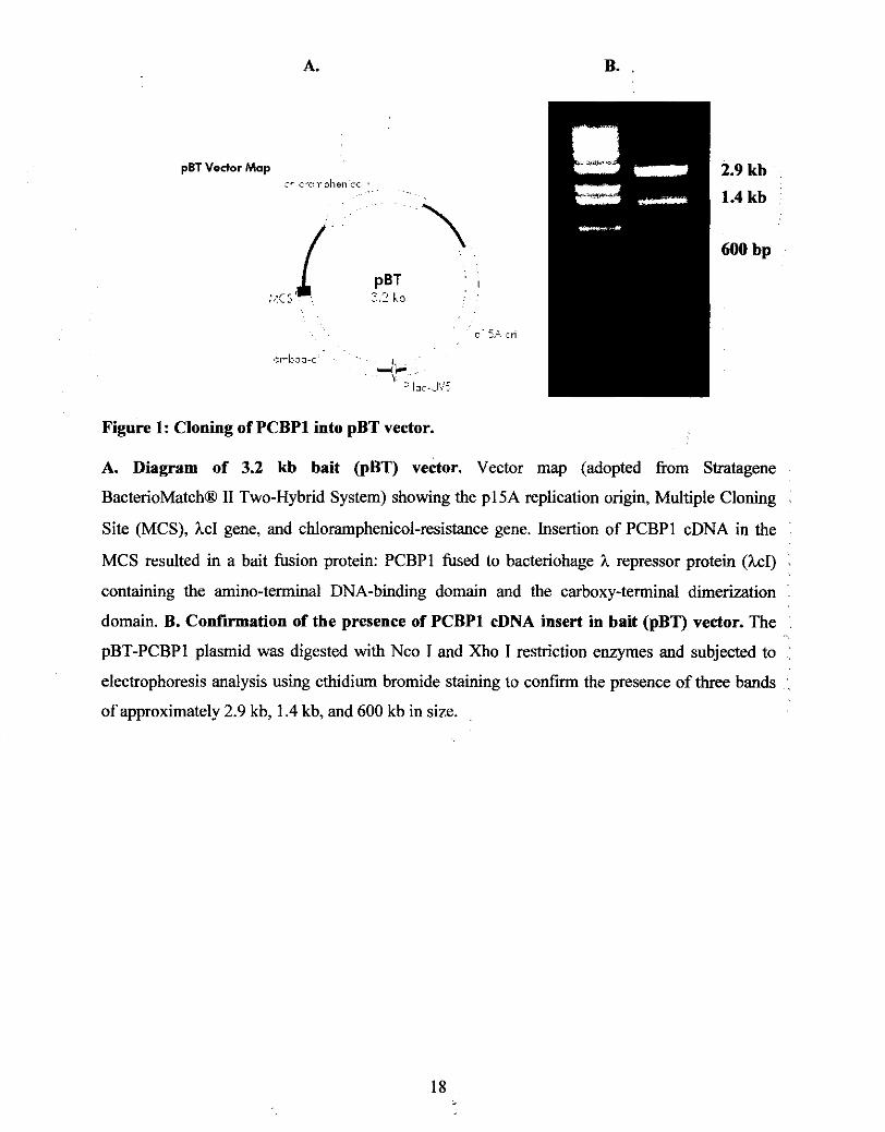
Site (MCS), hcI gene, and chloramphenicol-resistance gene. Insertion of PCBPl cDNA in the
MCS resulted in a bait fusion protein: PCBPl fused to bacteriohage h repressor protein (hcI) ~
containing the amino-terminal DNA-binding domain and the carboxy-terminal dimerization : domain. B. Confirmation of the presence of PCBPl cDNA insert in bait (pBT) vector. The : pBT-PCBPl plasmid was digested with Nco I and Xho I restriction enzymes and subjected to : electrophoresis analysis using ethidiurn bromide staining to confirm the presence of three bands :
of approximately 2.9 kb, 1.4 kb, and 600 kb in size.
Self-activation test of the hcI-PCBP1 fusion protein
To prevent a high background (i.e. a high number of false positives) during the library
screen, the bait fusion protein must be tested to determine if it can self-activate the reporter
genes: HIS3 and aadA. The 4.4 kb target @TRG) vector (Figure 2A) is used in the co-
transformation and contains the amino-terminal domain of RNA polymerase a subunit, which is
under the control of IPTG-inducible tandem promoter lpp/lac-UV5, a low-copy ColEl
replication origin, and a tetracycline-resistance gene.
The bait fusion proteins are tested for self-activation, as some bait proteins are known to
activate transcription of the reporter genes with out an interacting partner. Also, there is a chance
that the bait protein may be toxic to the host, and could therefore impact the ability of the host to
survive on selective media. The bait protein may also strongly or weakly activate the
transcription of the reporter genes. If the bait protein strongly activates the transcription of the
reporter genes, a high background may result. Weak interactions may result in the lack of
transcriptional activation, thereby affecting the results of the library screen.
In theory, the reporter genes should not be activated when the recombinant bait vector is
co-transformed with an empty target vector @TRG-Empty). This is because pTRG-Empty does
not contain a target protein that can interact with the bait fusion protein. Thus, bacteria are
unable to gmw on media containing selective markers for the reporter genes. However, if the
recombinant bait plasmid is capable of activating the reporter genes on its own, bacterial growth
is observed on media containing the appropriate selective markers (3-AT, chloramphenicol, and
tetracycline), and for that reason modifications are need to certain residues or domains.
The recombinant bait plasmid, pBT-PCBPl was tested for self-activation of the reporter
genes by co-transformation with the pTRG-Empty vector (does not contain a fusion protein) into
bacteria competent cells and plated on media containing three selective markers: 3-AT,
chrolamphenicol, and tetracycline. As shown in Figure 2B, colonies were not observed
confirming that pBT-PCBP1 is a suitable for screening a cDNA library.
Vedor Map
A= lcc c < J's
,- ' ---E*: , ,, v..*.p.,:,-c - r ~ ~ : ~ c l : r e I
Figure 2: Self-activation test of the recombinant bait plasmid pBT-PCBPI.
A. Diagram of the 4.4 kb target @TRG) vector. Vector map (adopted from Stratagene
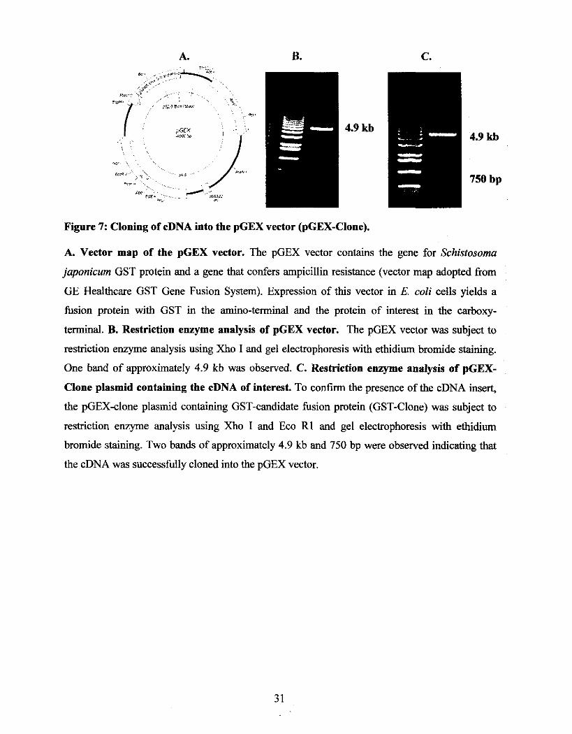
Figure 8: Silver stain analysis of bacterial lysate containing GST and GST-Clone.
Bacterial lysates from cells transformed with pGEX or pGEX-clone were analyzed for the GST
and GST-fusion protein: GST-Clone (29 kDa and 43 kDa, respectively) using SDS-PAGE
analysis with protein silver staining (Lanes 1-3). The GST and GST-clone proteins were purified
from bacterial lysates using Glutathione Sepharose 4B beads. The expected size bands at 29 kDa
for GST and 43 kDa for GST-Candidate Clone protein were observed canes 5-7).
DISCUSSION
In order to understand PCBPl's multiple functions, it is important to identify its
interacting proteins given that protein-protein interactions are essential to many protein functions
in biological processes. One method of identifying protein-protein interactions is the two-hybrid
system, which can be conducted in bacteria or yeast hosts. The bacteria two-hybrid system has
certain advantages such as the ability to analyze larger libraries as a result of higher
transformation efficiencies, faster growth rates (as observed in Escherichia coli), and a lower
false-positive rate of approximately 3 x lo-*. False-positives pose a difficult problem in yeast
two-hybrid screens due to the chance that the yeast host may harbor a eukaryotic homologue of
one of the interacting partners and thereby activate the transcription of the reporter genes. In
addition, the bait fusion proteins require a nuclear localization signal that may interfere with
target-binding activities (Chien et al., 1991 and Joung et al., 2000).
Therefore, in this study we used the bacteria two-hybrid system to screen for PCBPI-
interacting protein partners. The bait fusion protein, hcI-PCBPI, did not self-activate
transcription of the reporter genes and was used to screen a human brain cDNA library on media
containing three (chloramphenicol, tetracycline, 3-AT) and four (chloramphenicol, tetracycline,
3-AT, streptomycin) selective markers. The pTRG-cDNA plasmids from candidate clones that
survived high stringent selection were extracted and verified for an in vivo protein-protein
interaction by co-transformation with pBT-PCBPI. The pTRG-cDNA plasmids from verified
clones were analyzed, revealing a 750 base pair cDNA insert.
Various PCBP1-interacting proteins have been reported in different earlier yeast two-
hybrid studies (as listed in Table 1). There are several possibilities for these differences. For
example, different genes are expressed in diverse tissues and the genes being expressed in a
given host may also vary due to the host's age, race, sex, and environment. Consequently, the
variations observed may be a result of examination of the protein-protein interactions using
different tissue types. For example, the identification of Lamins A/C as a PCBP1-interacting
protein was detected in lymphoblasts (Zhong et al., 2005), while hnRNP A2 was observed to
bind to PCBPl in oligodendrocytes (Kosturko et al., 2006). Another possible reason may be due
in fact that some studies used a different protein as bait, such as c-MYC (Koch et al., 2007) and
Pak 1 (Meng et al., 2007). Also, the bait plasmid of the yeast-two hybrid kit varied, which may
have affected the binding properties of the bait fusion protein as a result of i n - h e sequencing
with the cDNA sequence of PCBPI. For example, AUFl was identified as a PCBPl interacting
protein using the pGBD plasmid (Kiledjian et al., 1997) and the pDONR223 vector of the
Gateway system was used to identify several other proteins such as hnRNP K (Lim et al., 2006).
Furthermore, variations may also be due to the cDNA library being used, which may not
represent all genes due to the construction of the cDNA library from different manufacturers. In
addition, the binding properties of target proteins can vary as a result of in-frame sequencing to
generate fusion proteins. For example, PCBPl was detected as a hnRNP A2 binding protein from
a ~ a t c h m a k e r ~ ~ Pre-transformed human cDNA library obtained from ClonetechIBD (Kosturko
et al., 2006), whereas a number of other PCBP1-interacting protein were identified from
screening a human brain cDNA library purchased from ProQuest, Invitrogen (Lim et al., 2006).
While several PCBP1-interacting proteins have been identified through the yeast two-hybrid
system, not all have been verified indicating there are more PCBP1-interacting proteins yet to be
discovered.
In this study, a candidate PCBP1 -interacting protein has been found and verified for an in
vivo protein-protein interaction with PCBPl through a re-co-transformation test of the pBT-
PCBPl and pTRG-Clone plasmids. This protein-protein interaction should be further analyzed in
additional in vivo and in vitro assays. The reason for this is that there may be factors in the host's
environment (i.e. bacteria) that may mediate or enhance the protein-protein interaction that may
not be present in other cellular environments. The examination of a direct interaction between
the candidate protein and PCBPl in viho, such as a GST pull down assay, is still necessary. For
this reason, the GST-fusion protein, GST-Clone, was established for in viho testing, which may
help to identify the interaction without the disturbance of certain environmental influences that
may occur in vivo. Further in vivo experiments will allow for confirmation of a protein-protein
interaction in the proteins' actual cellular environment where it is possible that the bait and target
proteins may be modified by factors in the cells that can either enhance or impair the interaction.
Also, it may be possible to identify proteins (i.e. in the form of a signaling cascade) that mediate
or interfere with the interaction, as well as identify different isoforms of the same proteins.
The identification of the candidate protein may help to understand the physiological roles
of PCBP1, as well as the role of the candidate protein in cellular processes. In particular, any
signaling cascades affecting the protein-protein interaction that may lead to identifying new
functions and targets for therapeutic usages.
CONCLUSION
In summary, the bacteria two-hybrid system was used to identify the protein-interacting
partners of PCBPl. PCBPl was first cloned into the bait vector @BT-PCBPI) and analyzed for
in-frame sequence with the hcI protein by DNA sequencing. Results of the co-transformation of
pBT-PCBPI with an empty target vector (pTRG-Empty) verified that the hcI-PCBPI fusion
protein did not self-activate the reporter genes (HIS3 and aadA) and was used to screen a human
brain cDNA library. In addition, the bacteria two-hybrid system was successfully setup using
pilot experiments with positive (pBT-LGF2 and pTRG-Gall IP) and negative control @BT-
Emtpy and pTRG-Empty) plasmids.
Approximately 1.65 x lo6 clones of a human brain cDNA library were screened using
media containing three selective markers: 3-AT, chloramphenicol, and tetracycline. In order to
verify positive clones, colonies that grew on this media were further screened using high
stringent media containing four selective markers: 3-AT, chlorarnphenicol, tetracycline, and
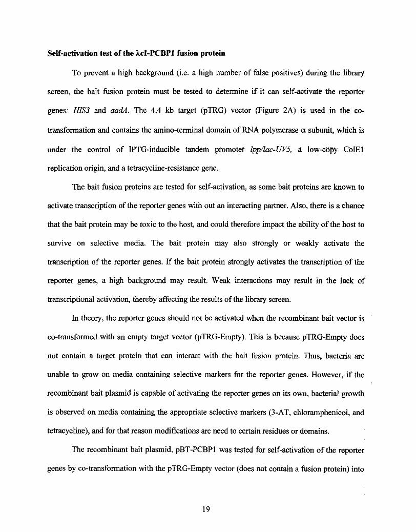
streptomycin. The candidate clone containing the cDNA insert @TRG-Clone) was isolated and
extracted. Restriction enzyme analysis demonstrated that the candidate clone contained a 750-
base pair cDNA insert. pTRG-Clone was then re-co transformed with pBT-PCBPl into bacteria
competent cells to validate a positive protein interaction in vivo.
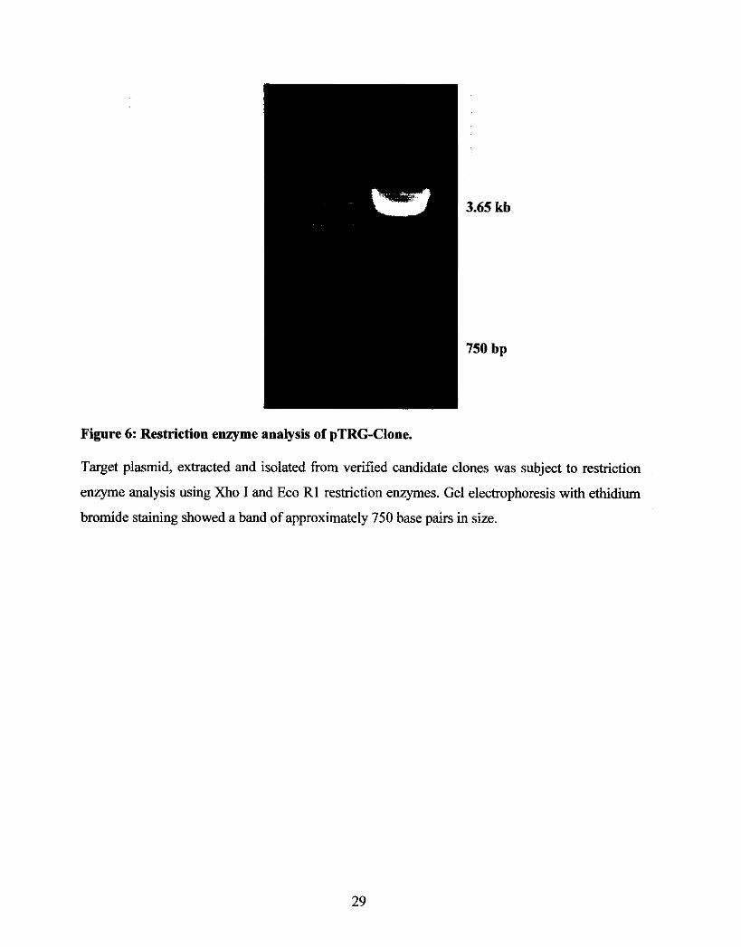
To further confirm the physical interaction of the candidate protein with PCBPI, the 750
base pair cDNA insert was cloned into the pGEX vector and over-expressed in bacterial cells.
Expression and purification of the GST-hsion proteins (GST and GST-Clone) were confirmed
by SDS-PAGE analysis with silver staining.
Future experiments are necessary to verify the candidate protein's physical interaction
with PCBP1 and to identify the functional roles of this interaction.
REFERENCES
Anthony, A.C., Tang, Y., Khan, R.A., Biju, M.P., Xiao, X., Sun, X., Hiremagalur, J.N., and Stabler, S.P. (2004). Translational upregulation of folate receptors is mediated by homocysteine via RNA-heterogeneous nuclear ribonucleoprotein El interactions. Journal of Clinical Investigations. 113(2): 285-301.
Aasheim, H.C., Loukianova, T., Deggerdal, A., and Smeland, E.B. (1994). Tissue specific expression and cDNA structure of a human transcript encoding a nucleic acid binding [oligo(dC)] protein related to the pre-mRNA binding protein K. Nucleic Acids Research. 22: 959-964.
Berry, A.M., Flock, K.E., Loh, H.H., and KO, J.L. (2006). Molecular basis of cellular localization of poly C binding protein 1 in neuronal cells. Biochemical and Biophysical Research Communications. 349(4): 1378-1 386.
Bromsztyk, K., Denisenko, O., and Ostrowski, J. (2004). hnRNP K: One protein multiple processes. BioEssays. 26(6): 629-638.
Bomsztyk, K., Van Seuningen, I., Suzuki, H., Denisenko, O., and Ostrowski, J. (1997). Diverse molecular interactions of the hnRNP K protein. Federation of European Biochemical Studies. 403: 113-115.
Bustelo, X.R., Suen, K.L., Michael, W.M., Dreyfuss, G., and Barbacid, M. (1995). Association of the vav proto-oncogene product with poly(rC)-specific RNA-binding proteins. Molecular and Cellular Biololy. 15: 1324-1332.
Chien, C., Bartel, P.L., Sternglanz, R., and Fields, S. (1991). The two-hybrid system: A method to identify and clone genes for proteins that interact with a protein of interest. Proceedings of the National Academy of Sciences. 88: 9578-9582.
Chkheidze, A.N., Lyakhov, D.L., Makeyev, A.V., Modes, J., Kong, J., and Liebhaber, S.A. (1999). Assembly of the a-Globin mRNA stability complex reflects binary interaction between the pyrimidine-rich 3' untranslated region determinant and Poly(C) Binding Protein aCP. Molecular and Cellular Biology. 19(7): 4572-4581.
Czyzyk-Krzeska, M.F. and Bendixen, A.C. (1999). Identification of the Poly(C) Binding Protein in the complex associated with the 3' untranslated region of erythropoietin messenger RNA. Blood, Journal of the American Sociely of Hematology. 93(6): 21 11-2120.
Denisenko, O.N. and Bomsztyk,K. (1997). The product of the murine homolog of the Drosophila extra sex combs gene displays transcriptional repressor activity. Molecular and Cellular ~ i o j o g y 17: 4707-4717.
Denisenko, 0. and Bomsztyk, K. (2002). Yeast hnRNP K-like genes are involved in regulation of the telomeric position effect and telomere length, Molecular and Cellular Biologv. 22: 286- 297.
Denisenko, O.N., O'Neill, B., Ostrowski, J., Van Seuningen, I., Bomsztyk, K. (1996). Zikl, a transcriptional repressor that interacts with the heterogeneous nuclear ribonucleoprotein particle K protein. Journal of Biological Chemistry. 271: 27701-27706.
Dreyfuss, G., Matunis, M.J., Pinol-Roma, S. and Burd, C.G. (1993). hnRNP proteins and the biogenesis of mRNA. Annual Review: Biochemistry. 62: 289-321.
Gamarnik, A.V. and Andino, R. (1997) Two functional complexes formed by KH domain containing proteins with the 59 noncoding region of poliovirus RNA. RNA. 3: 882-892.
Funke, B., Zuleger, B., Benavente, R., Schuster, T., Goller, M., Stevenin, J., and Horak, I. (1996). The mouse poly(C)-binding protein exists in multiple isoforms and interacts with several RNA-binding proteins. Nucleic Acids Research. 24: 3821-3828.
Inoe, A., Sawata, S.Y., Taira, K., and Wadhwa, R. (2007). Loss-of-function screening by randomized intracellular antibodies: Identification of hnRNP-K as a potential target for metastasis. Proceedings of the National Academy of Sciences. 104(2): 8983-8988.
Joung, J.K., Ramm, E.I., and Pabo, C.O. (2000). A bacteria two-hybrid selection system for studying protein-DNA and protein-protein interactions. Proceedings of the National Academy of Sciences. 97(13): 7382-7387.
Kiledjian, M., DeMaria, C.T., Brewer, G., and Novick, K., (1997). Identification of AUFl Heterogeneous Nuclear Ribonucleoprotein D as a component of the a-globin mRNA stability complex. Molecular and Cellular Biology. 17: 4870-4876.
Kim, J.H., Hahm, B., Kim, Y.K., Choi, M., and Jang, S.K. (2000). Protein-protein interaction among hnRNPs shuttling between nucleus and cytoplasm. Journal of Molecular Biology. 298: 395-405.
Kim, S.S., Pandey, K.K., Choi, H.S., Kim, S.Y., Law, P.Y., Wei, L.N., and Low, H.H. (2005). Poly(C) binding protein family is a transcription factor in mu-opioid receptor gene expression. Molecular Pharmacology. 68(3): 729-736.
KO, J.L. and Loh, H.H. (2005). Poly C Binding Protein, a single stranded DNA binding protein, regulates mouse p-opoid receptor gene expression. Journal of Neurochemistry. 93(3): 749-761.
Koch, H.B., Zhang, R., Verdoodt, B., Bailey, A., Zhang, C., Yates, J.R., Menssen, A., and Hermeking, H. (2007). Large-scale identification of c-MYC associated proteins using a combined TAP/MudPIT approach. Cell Cycle. 6(2): 205-217.
Kosturko, L.D., Maggipinto, M.J., Korza, G., Lee, J.W., Carson, J.H., and Barbarese, E. (2006). Heterogeneous nuclear ribonucleoprotein (hnRNP) El binds to hnRNP A2 and inhibits translation of A2 response element mRNAs. Molecular Biology of the Cell. 17(8): 3521-3533.
Lee, M., Mori, S., and Raychaudhuri, P. (1996). Trans-Activation by the hnRNP K protein involves an increase in RNA Synthesis from the reporter genes. Journal of Biological Chemistry. 271(7): 3420-3427.
Leffers, H., Dejgaard, K., and Celis, J.E. (1995). Characterization of two major cellular poly(rC)- binding human proteins, each containing three K-homologous( KH) domains. European Journal of Biochemisq. 230: 447-453.
Lim, J., Hao, T., Shaw, C., Patel, A.J., Szabo, G., Rual, J., Fisk, C.J., Li, N., Smolyar, A., Hill, D.E., Barabasi, A,, Vidal, M., and Zoghbi, H.Y. (2006). A protein-protein interaction network for human inherited ataxias and disorders of Purkinje Cell Degeneration. Cell. 125: 801-814.
Lynch, M., Chen, L., Ravitz, M.J., Mehtani, S., Korenblat, K., Pazin, M.J., and Schmidt, E.V. (2005). HnRNP K b i d s a core polypyrimidine element in eukaryotic translation initiation factor 4E (eIFE) promoter, and its regulation of eIF4E contributes to neoplastic transformation. Molecular and Cellular Biology. 25(15): 6436-6453.
Mailk, A.K., Flock, K.E., Godavarthi, C.L., Loh, H. H., and KO, J.L. (2006). Molecular basis underlying the poly C binding protein 1 as a regulator of the proximal promoter of mouse MOR (p-opioid) receptor gene. Journal of Brain Research. 11 12: 33-45.
Makayev, A.V. and Liebhaber, S.A. (2002). The poly(C)-binding proteins: a multiplicity of functions and a search for mechanisms. RNA. 8: 265-278.
Matunis, M.J., Michael, W.M., and Dreyfuss, G. (1992). Characterization and primary structure of the poly(C)-binding heterogeneous nuclear ribonucleoprotein complex K protein. Molecular ' and Cellular Biology. 12: 164- 171.
Meng, Q., Rayala, S.K., Gururaj, A.E., Talukder, A.H., O'Malley, B.W., and Kurnar, R. (2007). Signaling-dependent and coordinated regulation of transcription, splicing, and translation resides in a single coregulator, PCBPI. Proceedings of the National Academy of Sciences. 104(14): 5866-5871.
Michelotti, E.F., Michelotti, G.A., Aronsohn, A.I. and Levens, D. (1996). Heterogeneous nuclear ribonucleoprotein K is a transcription factor. Molecular and Cellular Biology. 16: 2350- 2360.
Moms, B.J., Adams, D.J., Beveridge, D.J., van der Weyden, L., Mangs, H., and Leedman, P.J. (2004), CAMP controls human rennin mRNA stability via specific RNA-binding proteins, Acta Physiologica Scandinavica. 181 : 369-373.
Moumen, A., Masterson, P., O'Conner, M.J., and Jackson, S.P. (2005). HnRNP K: An HDM2 target and transcriptional coactivator of p53 in response to DNA Damage. Cell. 123: 1065-1078.
Ostareck-Lederer, A,, and Ostareck, D.H. (2004). Control of mRNA translation and stability in haematopoietic cells: The function of hnRNPs K and El/E2. Biology ofthe Cell. 96: 407-41 1.
Paziewska, A,, Wywicz, L.S., Bujnicki, J.M., Bomsztyk, K., and Ostrowski, J. (2004). Cooperative binding of the hnRNP K three KH domains to mRNA targets. Federation of European Biochemical Studies. 57: 134-140.
Reimann, I., Huth, A,, Thiele, H., and Thiele, B. (2002). Suppression of 15-lipoxygenase Synthesis by hnRNP El is dependent on repetitive nature of LOX mRNA 3'UTR control element DICE. Journal of Molecular Biology. 315: 965-974.
Rivera-Gines, A,, Cook, R.J., Loh, H.H., and KO, J.L. (2006). Interplay of Sps and poly(C) binding protein 1 on the p-opioid receptor gene expression. Biochemical and Biophysical Research Communications. 345: 530-537.
Rahman-Roblick, R., Roblick, U.J., Hellman, U., Conrotto, P., Liu, T., and Becker, S. (2007). p53 targets identified by protein expression profiling. Proceedings ofthe National Academy of Sciences. 104(13): 5401-5406.
Schullery, D.S., Ostrowski, J., Denisenko, O.N., Stempka, L., Shnyreva, M., Suzuki, H., Gschwendt, M., and Bomsztyk, K. (1999) Regulated interaction of protein kinase Cd with the heterogeneous nuclear ribonucleoprotein K protein. Journal of Biological Chemisfry. 274: 15101-15109.
Shnyreva, M., Schullery, D.S., Suzuki, H., Higaki, Y., and Bomsztyk, K. (2000). Interaction of two multifunctional proteins; Heterogeneous nuclear ribonucleoprotein K and Y-box-binding protein. Journal of Biological Chemistry. 275: 15498-15503.
Siomi, H., Choi, M., Siomi, M. C., Nussbaum, R. L., and Dreyfuss, G. (1994). Essential role for KH domains in RNA binding: impaired RNA binding by a mutation in the KH domain of FMRl that causes fragile X syndrome. Cell. 77: 33-39
Spangberg, K. and Schwartz, S. (1999). Poly(C)-binding protein interacts with hepatitis C virus 5' untranslated region. Journal of General Virology. 80: 1371-1 376.
Stains, P.J., Lecanda, F., Towler, D.A., and Civitelli, R. (2005). Heterogeneous nuclear ribonucleoprotein K represses transcription from a cytosinelthymidine-rich element in the ostecalcin promoter. Biochemical Journal. 385: 613-623.
Stefanovic, B., Hellerbrand, C., Holcik, M., Briendl, M., Liebhaber, A.L., and Bremer, D.A. (1997). Posttranscriptional regulation of collagen al(1) mRNA in Hepatic Stellate cells. Molecular and Cellular Biology. 17(9): 5201-5209.
Sun, A., Balasubramaniyan, N., Liu, C., Shahid, M., and Suchy, F.J. (2004). Association of the 16 kDa subunit c of vacuolar proton pump with the ileal ~a+-dependent bile acid transporter: . Protein-protein interaction and intracellular trafficking. Journal of Biological Chernishy. 279(16): 16295-16300.
Takano, H., Hosono, K., Beppu, T., and Ueda, K. (2003). Involvement of oH and related sigma factors in glucose-dependent initiation of morphological and physiological development of Streptomyces griseus. Gene. 320: 127-135.
Thyagarajan, A. and Szaro, B.G. (2004). Phylogenetically conserved binding of specific K Homology domain proteins to the 3' untranslated region of the vertebrate middle neurofilament mRNA. The Journal of Biological Chemistry. 279: 49680-49688.
Tomonaga, T. and Levens, D. (1996). Activating transcription from single stranded DNA. Proceedings of the National Academy o f Sciences. 93: 5830-5835.
Van Seuningen, I., Ostrowski, J., Bustelo, X.R., Sleath, P.R., and Bomsztyk, K. (1995) The K protein domain that recruits the interleukin 1-responsive K protein kinase lies adjacent to a cluster of c-Src and Vav SH3-binding sites: Implications that K protein acts a s a docking platform. Journal of Biological Chemistry. 270: 26976-26985.
Verma, A. and Maurelli, A.T. (2003). Identification of two eukaryotic-like serine/threonine kinases encoded by Chlamydia trachomatis Serovar L2 and characterization of interacting partners of Pknl . Infection and Immunity. 71(10): 5772-5784.
Wang, Z., Day, N., Trifillis, P., and Kiledjian, M. (1999). An mRNA stability complex functions with poly(A)-binding protein to stabilize mRNA in vitro. Molecular and Cellular Biologv, 19: 4552-4560.
Weng, Z., Thomas, S.M., Rickles, R.J., Taylor, J.A., Brauer, A.W., Seidel-Dugan, C., Michael, W.M., Dreyfuss, G., and Brugge, J.S. (1994). Identification of Src, Fyn, and Lyn SH3-binding proteins: Implications for a function of SH3 domains. Molecular and Cellular Biologv. 14: 4509- 4521.
Zbong, N., Radu, G., Ju, W., and Brown, W.T. (2005). Novel progerin-interactive partner proteins hnRNP El, EGF, Me1 18, and UBC9 interact with lamin A/C. Biochemical and Biophysical Research Communications. 338: 855-861.