California State University, San Bernardino California State University, San Bernardino CSUSB ScholarWorks CSUSB ScholarWorks Theses Digitization Project John M. Pfau Library 1987 Investigation of the neutralizing activity for Treponema Pallidum Investigation of the neutralizing activity for Treponema Pallidum of neonatal rabbit basal serum taken at 2, 3, and 4 weeks of age of neonatal rabbit basal serum taken at 2, 3, and 4 weeks of age Helen Ceclie Mercier Follow this and additional works at: https://scholarworks.lib.csusb.edu/etd-project Part of the Immunology and Infectious Disease Commons Recommended Citation Recommended Citation Mercier, Helen Ceclie, "Investigation of the neutralizing activity for Treponema Pallidum of neonatal rabbit basal serum taken at 2, 3, and 4 weeks of age" (1987). Theses Digitization Project. 397. https://scholarworks.lib.csusb.edu/etd-project/397 This Thesis is brought to you for free and open access by the John M. Pfau Library at CSUSB ScholarWorks. It has been accepted for inclusion in Theses Digitization Project by an authorized administrator of CSUSB ScholarWorks. For more information, please contact [email protected].
Transcript
California State University, San Bernardino California State University, San Bernardino
CSUSB ScholarWorks CSUSB ScholarWorks
Theses Digitization Project John M. Pfau Library
1987
Investigation of the neutralizing activity for Treponema Pallidum Investigation of the neutralizing activity for Treponema Pallidum
of neonatal rabbit basal serum taken at 2, 3, and 4 weeks of age of neonatal rabbit basal serum taken at 2, 3, and 4 weeks of age
Helen Ceclie Mercier
Follow this and additional works at: https://scholarworks.lib.csusb.edu/etd-project
Part of the Immunology and Infectious Disease Commons
Recommended Citation Recommended Citation Mercier, Helen Ceclie, "Investigation of the neutralizing activity for Treponema Pallidum of neonatal rabbit basal serum taken at 2, 3, and 4 weeks of age" (1987). Theses Digitization Project. 397. https://scholarworks.lib.csusb.edu/etd-project/397
This Thesis is brought to you for free and open access by the John M. Pfau Library at CSUSB ScholarWorks. It has been accepted for inclusion in Theses Digitization Project by an authorized administrator of CSUSB ScholarWorks. For more information, please contact [email protected].
Test and control suspensions were diluted by the addition of
0.9 ml HI'NRS just prior to injection for a final suspension
of 1x10 T. pallidum/ml. Each site received 0.1 ml suspen
sion for a total inoculum of 1x10 T. pal1idum/site. Posi
tive (+) neutralization was indicated by the absence of
lesion development, negative (-) neutralization by the
appearance of typical lesions within the appropriate
incubation period established by the (-) Controls. Delayed
lesion development was indicative of partial (+7-)
neutralizatipn, Representative lesions of the mlcro-NZ assay
are pictured in Figures 14-24.
Inoculations
Five VDRL nonreactlye male NZ-W rabbits with "good"
backs (Fig. 8) (as opposed to "bad" backs. Fig. 9) were
obtained in advance of each experimental run (Bio Robotics,
10
Van Nuys, CA) and their backs were shaved just prior to the
time of inoculation. Each rafabit was inoculated in the
designated pattern illustrated in Figure 10. Each of the
test and control suspensions were drawn into sterile 1 ml
leurlok tuberculin syringes (Fig. 11), and administered for
a total of five replicate inoculation sites, one site on
each rabbit back (Fig. 12). All animal backs were clipped
and monitored daily for lesion development. Incubation
periods and lesion diameters (Fig. 13) and durations were
recorded daily. Aspirates of representative lesions were
examined for motile treponemes by darkfield microscopy. VDRL
serology tests were performed on all test animals upon
termination of each experimental run.
Statistical analysis
the incubation periods of neutralization lesions were
analyzed by the Student's t test. The differences in the
results were considered to be significant if p< 0.05,
11
Figs. 1-2. Extraction of Treponema pal 1idum from rabbittestes.--!. Testes infected with a minimum suspension of
72x10 T. pal 1idum/ml/testis removed for maceration.— 2.T. pal 1idum suspension following extraction from testes, centrifugation, and removal of gross cellular debris.
12
Figs. 3-6. Dilution procedures for the micro-NZ assay.6
— 3. Pipeting of I0)il aliquots of a 1x10 Treconena pal 1idum/ml suspension.--4. Serum samples were immediately placed on ice.--5. The addition of 90yl of test or control serum.— 6. Preparation of test and control suspensions for 16 hour incubation period.
13
Fig. 7. Anaerobic atmosphere jar, 34®C incubator, and syringe labels.
Figs. 8-9. Rabbit back characteristics.--8. Test rabbitwith a good back, i.e., smooth skin with no hair patterns after shaving. Black dots mark inoculation sites.--9. A rabbit with hair patterns that make lesion interpretation
15
diff icult
ds my> 1
fO mCOa:
OJ □c
z: CDo 3 1OL OU .
z; UJ •
3 u i nz oU J zi n -»r
inII u
t(TZ
a oz LU(T z
Vi IIm
u .PS
(NJ 3o c
• Ii n 1oz z
z3a.UJUlz3 a□c UJu 1 zi n CE 3UJ QCa X UJUJ z i nt 3(E 1-3 II I.H1.̂ my 1 1 mu I CE(E IZz t-1 1 3 31 O o:1 I z(E K QCUJ 4.H OI z zII II
in4 □cz z
> 1 »1 z3 o14m tCE (E1-4 CD3 3z O3 Z z
u 3UJ ai n 4-4 o
3 czH- 3 3* f (E M□3 aCD 1(E (E zIZ zUJ 3U i z OZ a QC3 CL 1Z UJ ZZ oc oH.I H u
II II
i n o□c uH l 3
inu3O> II-11341CD COZ z1-4 4-13 zo
3Z 33 o za u.o44 143 3 H-3 o zZ □c CDa ( 3z U• o z1-CJ 4-4
II VJ3.4o3
16
10.
Inoc
ulat
ion
patt
ern
for
test r
abbits.
-—
-—
--
— -—
-
—- - —
--
—
— -
-
-
---
Figs. 11-12. Inoculation procedure. — 11. Sterile, labeledluer lok syringes each containing either incubated test or
4control suspensions of 1x10 Treponema cal Iidum/ml. Only 17 syringes appear because zero hour viability control injections had been administered.--12. Inoculation of one test animal with 0.1 ml L. pallidum suspension at the VC site.
16
17
Fig. 13. Example of measurement of lesion diameter and inspection of lesion development.
18
RESULTS
Micro-neutralization assays performed with neonatal
basal sera from 2, 3/ and 4 week old rabbits failed to
demonstrate neutralizlnq activity for Trepohema pallldum.
Table 1 summarizes the results of this study. Among the
neonate groups 96-100% of Inoculated s1tes developed les1ons
from unheated serum preparations. There were no significant
differences among incubation periods (number of days from
the day of Inoculation to the first day of lesion develop
ment) of lesions using sera from the three neonatal age
gfroups, nor were there any significant differences among
sera from adult NRS control groups and neonatal groups. The
mean incubation period for lesion development among neonate
groups ranged from 16.4 +1.8 to 17.4 ±2.1 days and the mean
incubation period of the NRS was 18.6+4.4 days. However, in
two Instances <3 wk-HI and 4 wk-HI) the examination of data
On Individual sera showed either notably delayed or totally
absent lesion development which is not evident from the sum
marized data on Table 1.
Aliquots of both control and test sera were examined
for a heat-labile component(s) by heat-inactivating <56 C
for 30 minutes) sera prior to use in the micro-neutraliza
tlon assays. As expected, the Immune rabbit serum controls
neutralized T. pal1Idum (no lesions) when serum was not
heated and resulted in either no lesions (54% of sites) or
19
delayed lesions when heat-inactivated. Interestingly, in one
experimental run, one serum sample from each of the 3 and 4
week old heat-inactivated neonatal age groups demonstrated
partial neutralization by the absence of lesions at 3 of 5
sites and 2 of 5 sites respectively. Lesions that did devel
op from these serum samples were notably delayed.
The neonates whose basal sera were used for these
assays were infected with X- callidum following extrac
tion of their basal serum by Gamboa and Miller (1984). The
resistance among the neonates they inoculated was not uni
form. Some neonates developed atypical dermal lesions at
one or both inoculation sites while others remained free of
lesions. The atypical designation was defined as any lesion
that was small, indurated, nonulcerative and of short dura
tion as compared to adult controls inoculated similarly. As
shown in Table 2, no apparent correlation was demonstrable
between the development or absence of atypical lesions among
neonates and the neutralizing activity of their basal sera
in any of the age groups. Both heat-inactivated and unheated
serum samples from the seven which developed atypical le
sions <+ neonates), failed to neutralize the treponemes at
95% of the inoculated sites. Likewise, sera from 11 that had
not developed lesions <- neonates), failed to demonstrsite
neutralizing activity at 97% of the inoculated sites.
Figures 14-24 follow the progressive lesion development
20
of three representative animals from a total of fifteen used
to test for the neutralizing activity in neonatal rabbit
serum. These figures illustrate the first appearance of ery
thema (day 15 post-inoculation. Figs. 14, 15) and continue
through the healing stages (day 48 post-inoculation. Fig.
24).
Aspirates of representative lesions routinely drawn
just prior to ulceration (Fig. 17), and selected from both
test and control sites, demonstrated actively motile trepo
nemes by darkfield microscopy. Upon termination of the ex
periments, all test animals had converted to reactive VDRL
aerologies.
21
TflBLE 1- Neutra 1 izing act1Vity of neonatal basal sera from 2, 3, and 4 week . ; a
old rabbits.
Unheated Sera Heat-I nacti vated Sera
No. of No. of
No. Lesions/No. I ncubat ion Lesions/No. Incubation
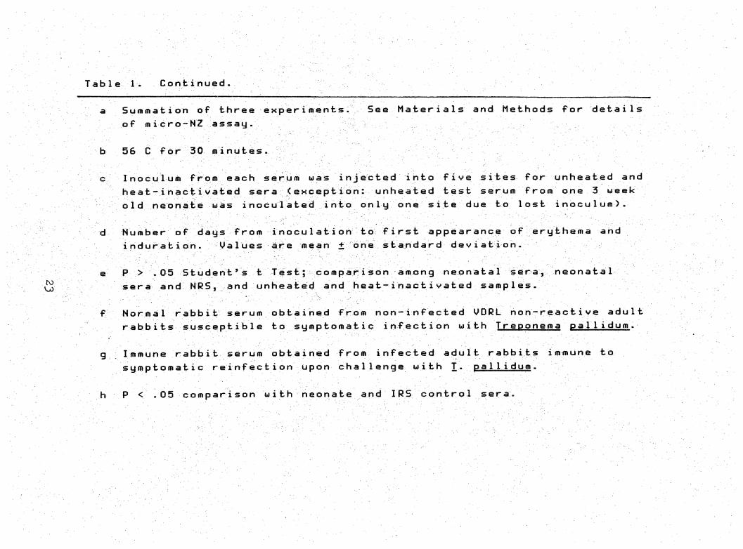
a Summation of three experiments. See Materia1s and Methods for details of micro-NZ assay-
b 56 C for 30 minutes-
Inoculum from each seru'" injected into five sites for unheated and heat-inactivated sera <exception: unheated test serum from one 3 ueek
old neonate was inoculated i nto only one site due to lost inoculum).
d Number of days from inoculation to first appearance of erythema and induration. Values are mean one standard dev i atIon.
e P > 05 Student's t Test; comparison among nepnata1 sera, neonata 1 sera and MRS, and unheated and heat-inactivated samp1es.
f Normal rabb it serum obta i ned from non-infected ODRL non>-reacti ve adu11
rabbits susceptib1e to symptomatic infection with Treponema pal 1idum.
g Immune rabbit serurn obtained from infected adult rabbits immune to symptomatic reinfection upon challenge with T. pa11idum.
h P < .05 comparison with neonate and IRS control sera.
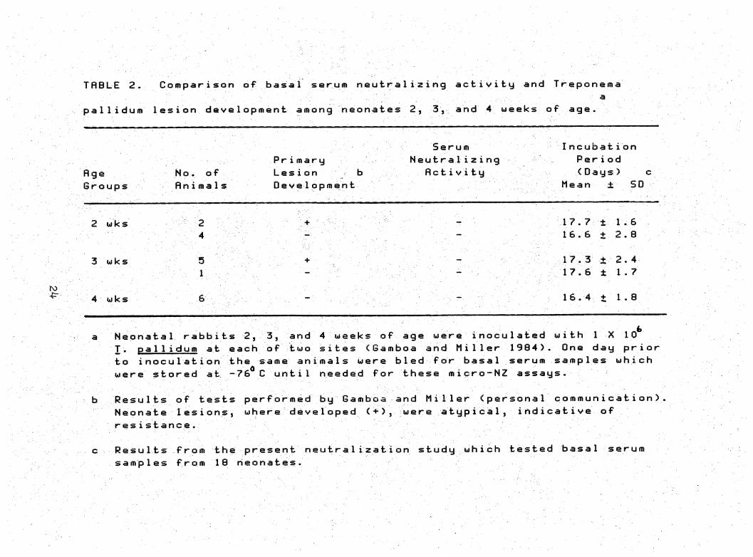
THBLE 2. Comparison of basal serurn neutralizing activity and Treponema a,
pallidum 1esion development among neonates 2, 3, and 4 weeks of age.
Serurn Incubati on
Pr i mary Neutra1izi ng Period
fl ge No. of Lesion b flcti V ity (Days) c
Groups Rn i m a 1s Development Mean ± SD
17.7+ 1.62 wks 2
16.6 + 2.04
17.3 + 2-43 wks 5
17.6 ± 1.71
ro
16.4 + 1.84 wks
Neonata1 rabbits 2, 3, and 4 weeks of age were i nocu1ated with I X 10 T. pal 1 idurn at each of two sites (Gamboa and Miller 1984). One day prior
to inoculation the same animals were bled for basal serum samples which were stored at -76^C until needed for these micro-NZ assays-
ResuIts of tests performed by Gamboa and Miller (persona1 commun icati on) Neohate Iesions, where developed <+), were atypica1, indicative of resistance.
Results from the present neutra1ization study which tested basal serum samples from 10 neonates.
Figs. 14-24. Representative examples of post-inoculation
lesion development on rabbit backs, demonstrating the
results of micro-NZ assays for Treponema pallidum
neutralizing activity by basal sera from 2, 3, and 4 week
old neonatal rabbits.
25
Figs. 14-15. Day 15. — 14. Rabbit no. 3. Erythema clearly demonstrable at most test sites, except at 3C-HI and 4B-HI. Development is absent at IRS and IRS-HI sites as predicted. — 15. Rabbit no. 4. Erythema development replicates that seen in rabbit no. 3.
26
16”17. Day 22.~“16. Rabbit no. 3. Test sites areconsistently erythematous and indurated, except 3C-HI and4E-HI. Controls NRS, NRS-HI, IRS, IRS-HI, VC and VC
0 16are as predicted.--17. Rabbit no. 4. Lesion developmentclosely replicates rabbit no. 3. Note site VC is near
16
27
ulceration.
3C-HI andFigs. 18-19. Day 32.--18. Rabbit no. 3.4E-HI sites are erythematous, indurated; other test sites and, NRS and NRS-HI are near ulceration; IRS and IRS-HI sites remain lesion free.--19. Rabbit no. 4. Test sites are ulcerated, except 3C-HI and 4E-HI; NRS and NRS-HI are near ulceration; and IRS and IRS-HI remain lesion free.
28
Figs. 20-21. Day 43.--20. Rabbit no. 3. All sites ulcerated, except positive NZ control sites, IRS and IRS-HI.--21. Rabbit no. 4. Most lesions are beginning to heal.
29
30
Figs.
22-24. D
ay 4
8.—
22.
Rabb
it n
o. 3
. Ul
cera
tion
s persist, n
o le
sion
s at p
osit
ive
NZ c
ontrol s
ites
.--23.
Rabb
it n
o. 4
. Le
sion
s ha
ve n
earl
y he
aled
.—24.
Rabb
it n
o. 1.
Exam
ple
of h
eale
d back.
DISCUSSION
Results of this study show an absence of detectable
neutralizing activity in the sera of 2, 3, and 4 week old
rabbits. A correlation between neutralizing activity of
neonatal basal sera and resistance to symptomatic infection
was suggested in a previous study by Gamboa and Miller
(1984). Their sugges.tion was based on 1) the presence of
neutralizing activity in basal sera from one week old
rabbits and their resistance to symptomatic infection fol
lowing ihtradermal inoculation with Irejaoiigjn^ EsXLi^iliiL
and 2) the absence of neutralizing activity in basal sera
and the waning resistance to T. oallidum infection of
five week old neonates. The resistance to symptomatic
infection following intradermal inoculation with T.
palliduih in the same animals from which our sera is de
rived throws suspicion on the influence of a neutralizing
factor(s) on their resistance. Apparently, serum neutraliz
ing activity 1) may contribute nothing to the resistance
demonstrated by neonatal animals, 2) may only partially
contribute, or 3) the sensitivity of the micro-neutraliza
tion assay is insufficient to detect the full in vivo poten
tial of serum as concentrations of its activity begin to
drop in neonates after one week of age.
The last option is of particular interest. As mentioned
in the results, there appears to be a hint of residual
31
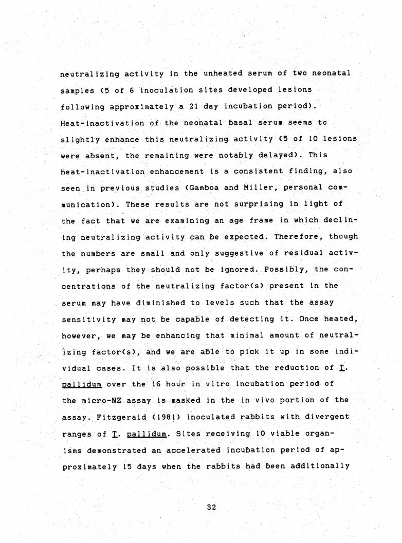
neutralizing activity in the unheated serum of two neonatal
samples <5 of 6 inoculation sites developed lesions
following approximately a 21 day incubation period).
Heat-inactivation of thshepnatal basal serum seems to
slightly enhance this neutralizing activity C5 of 10 lesions
Were absent, the remaihlng were notably delayed). This
heat-inactivation enhancement is a consistent finding, also
seen in previous studies (Gamboa and Miller, personal com
munication). These results are not surprising in light of
the fact that we are examining an age frame in which declin
ing neutralizing activity can be expected. Therefore, though
the numbers are small and only suggestive of residual activ
ity, perhaps they should not be Ignored. Possibly, the con
centrations of the neutralizing factorCs) present in the
serum may have diminished to levels such that the assay
Sensitivity may not be CapaEile of detecting it. Once heated,
however, we may be enhancing that minimal amount of neutral
izing factor(s), and we are able to pick it up in some indi
vidual cases, it is also possible that the reduction of T«
pallidum over the 16 hour in vitro incubation period of
the micro-NZ assay Is masked in the in vivo portion of the
assay. Fitzgerald (1981) Inoculated rabbits with divergent
ranges of T. pallidum. Sites receiving 10 viable organ
isms demonstrated an accelerated incubation period of ap
proximately 15 days when the rabbits had been additionally
32
Injected with 10 viable organisms at other sites^ (The
normal incubation period for 10 organisms averages approx
imately 24 days.). The delayed incufaatioh periods indicative
of partial neutralizing activity may have been masked by
sites on our animals receiving the full viable inoculum of
10 T. nal1idum; therefore, partial neutralization, which
could be expected as the factor declines in concentrations,
is not detected in our assay.
The delayed incubation periods and absence of lesions
at sites receiving HI~NRS is a consistent finding (Bishop
and Miller 1976, Blanco et al. 1984, Gamboa and Miller
1984). Blanco et al. (1984) have demonstrated that the IgG
nature of neutralizing activity in immune rabbit serum, and
its largely abrogated activity upon heating, is most likely
due to the elimination of complement. It has been suggested
that endogenous complement from the extraction of J..
pa11idum frcm rabbit testicles may account for the resid
ual neutralization seen in these suspensions (Bishop and
Miller 1976). On the other hand, it is possible that some
neutralizing activity in IRS is independent of complement.
It has been demonstrated that HI-MRS enhances phagocytosis
of X* pallidum bv proteose peptone-induced rabbit peri
toneal macrophages (Lukehart and Miller 1978). Therefore it
is feasible that opsonization may account for the residual
neutralization seen in these suspensions.
33
Interest was focused on the fetal disease as early as
19i3j Uhlenhuth and Mulzer (1913) set up studies on the in-^
heritance of syphi1is in rafabits, controlling the experimen
tal conditions to resemble human syphilis whenever possible.
Unfortunately, the ir methods were not sufficiently out!ined
to permit comparisons with later studies. Other research
groups fol1owed, Grigoriew (1929) described a single experi
mental case of congenital transmission of syphi1is from one
doe to her offspring. In yet another study, Bessemans and
Van CanneytC1932) concluded that, although many suggestions
of congenital infection resulted, they Could not prove con
clusively the existence of congenital syphilis in 34 rabbits
born from parents having ocular syphilitic lesions. Seiffert
(1934) briefly described eight experiments dealing with in
fection With X* sis a result of cohabitation or
cross-placental transmission but failed to describe the
route of infection of his experimental animals (mice and
rabbits). Kemp and Rosahn (1937) did not sufficiently des
cribe their experimental methods, making questionable their
conclusions that a placental barrier prevented the spread of
infection from doe to offspring, or the existence of a trep
onemicidal factor(s) in the fetus. In addition, and in rath
er forceful terms, Kemp and Fitzgerald (1938) concluded that
syphi1is is not transmitted from an infected doe to her off
spring. In 1957, Pautrizel et al. concluded that 1) the
34
rabbit fetus possesses a natural immunity to infection by
T. pallidum and 2) maternal antibodies play only a sec
ondary role in the prevention of transmission. Interesting
ly, nearly twenty years elapsed after the results of Kemp
and Fitzgerald (1938) were published before additional work
using the rabbit as a possible model for congenital syphilis
was again presented. Festenstein and Bokkenheuser (1961) and
Festenstein et al. (1967) attempted to tolerize neonatal
rabbits to X. pal1idum and found an increased suscep
tibilty as defined by the appearance of a runting syndrome.
In 1985 Fitzgerald listed four factors that select
against congenital syphilis in rabbits: 1) fetal damage
requires large numbers of T♦ pal1idum that accumulate in
a short period of time (Magnuson et al. 1948, Fitzgerald et
al. 1982j 2) female steroids, which are elevated during
(Frazier et al. 1935); 3) rabbit pregnancy results in multi
ple births, further diluting the numbers of organisms per
fetus (Fitzgerald 1985); and 4) possibly, the heat-stable
treponemicidal factor found in the serum of 4 to 6 day old
rabbits (Gamboa and Miller 1984) begins killing X.
pallidum before birth. Taking these factors into account
Fitzgerald (1985) was successful in demonstrating the
passage of X* pal1idum from infected does to fetal
rabbits, but only after multiple intravenous injections of
35
X. pal1idum over a period of four weeks for a total of
4x10 treponemes.
The large numbers of organisms necessary to demonstrate
overt symptoms of transmission may be a reflection on the
priesenee of additional resistance factors. As discussed by •
Gamboa and MiHer (1984), resistance of 5 to 8 day old neo
natal rabbits to dermal lesion development after intradermal
inoculation with X. pal1idum may be influenced by a num
ber of factors. Group housing (nesting) could create unfav
orable temperatures for the survival of X.. pallidum.
Experimental syphilis in rabbits requires that the animals
be kept in cool quarters <18-21 C) to allow for proper
lesion development following intradermal challenge with X*
pal1idum. In addition, inoculation sites must be kept
clipped. Therefore, higher temperatures due to huddling
of neonates in a nest may contribute to their resistance,
even though this influencing factor was Shown not to be
totally responsible for the absence of lesion deveiopment.
Nursing was also considered as a potential influencing
factor (Brambe11 1970a, b, Wilson and Miles 1975). Colostrum
and milk of several mammals are known to contain factors
which may influence resistance (Reiter and Oram 1967,
Goldman and Smith 1973, Head and Beer 1979). Although there
has been no evidence to substantiate a role for similar fac
tors in rabbits, and several investigators have concluded
36
that the systemic protective factors transmitted in utero to
the rafabit fetus are not supplemented fay nursing after birth
CBramfaell et al. 1951, Kraehenfauhl and Campiche 1969,
Bramfaell 1970c), conclusive evidence for or against this
theory remains lacking.
The association of a "natural antibody with innate re
sistance has been suggested as anpther factor^ p res
ponsible for neonatal serum neutralizing activity. Several
facts, however, have negated this as a possible explanation.
Natural antibody has classically been associated with the
IgM class of immunogiobulins CSolomon 1971) and,in the
rabbit, IgM is transmitted in utero CHemmings and Jones
1962). Therefore, were natural antibody a participant in
resistance, does' sera would also demonstrate neutralizing
activity and this has not been the case (Gamboa and Miller
1984). "
Gamboa and Miller (1984) also proposed that the absence
of a nutritional factor(s) necessary for optimum survival
and multiplication of the treponemes may influence resist
ance. The inability thus far to cultivate T. callidum in
pure culture makes the direct investigation of nutritional
requirements difficult.
While several hypotheses have been advanced to explain
the natural resistance of heonates to.syphil itic infection,
the definitive mechanism<s) has yet to be identified.
37
Studies on the isolation and identification of the neutral
izing factor(s) of basal sera from dnfe week old rabbits are
ongoing CGamboa, personal communicatioh), but in light of
this study/ may still be only half the story.
Congenital and neonatal human syphilis is not a ghost
of the past, but remains very much a disease of the present.
A total of 159 cases of early congenital syphilis have been
reported in the United States during 1982, an increase of 44
cases in four years (ASRMM 1983). These numbers "without
doubt underestimate the true magnitude of the problem,
because of misdiagnosis, and the occurrence of undocumented
cases manifested by spontaneous abortion or stillbirth"
(Hansfield and Lukehart 1984). Pregnant women and their
health care providers need to be aware of the significance
of the diagnosis of syphilis during pregnancy so that the
truly innocent victims may be spared readily preventable
suffering. :
38
LITERATURE CITED
ASRMM, 1983. Annual Summary, Reported Morbidity and Mortality in the United States. 1984 Publication CDC-85-8241, United States Public Health Service, Washington, D.C.
Bessemans, A. and Van Canneyt, 1932. Heredo-syphi1is Chez les lapereaux issus de parents attaints de manifestations oculaires specifiques. Soc. Beige de Biol. 110; 116-119.
Bishop, N.H. and Miller, J.N. 1976. Humoral immunity in experimental syphilis. II. The relationship of neutralizing factors in immune serum to acquired resistance. J. Immunol. 117: 197-207.
Blanco, D.R., Miller, J.N., and Hanff, P.A. 1984. Humoral immunity in experimental syphilis: the demonstration of IgG as a treponemicidal factor in immune rabbit serum, J. Immunol. 133: 2693-2697.
Brambell, F.W.R. 1970a. Transmission of immunity in the pig and horse. A. Neuberger and E.L. Tatum (eds.). In: Transmission of Passive Immunity from Mother to Young, Frontiers of Biology, vol. 18. North-Holland Publ. Co., Amsterdam, pp. 166-200. Cited in Gamboa and Miller 1984
Brambell, F.W.R. 1970b. Transmission of immunity in the ruminants. A. Neuberger and E.L. Tatum (eds.). In: Transmission of Passive Immunity from Mother to Young, Frontiers of Bioloav. vol. 18. North-Holland Publ.
Co., Amsterdam, pp. 201-233. Cited in Gamboa and Miller 1984.
Brambell, F.W.R. 1970c. Transmission of immunity in the rabbit. A. Neuberger and E.L. Tatum (eds.). In: Transmission of Passive Immunity from Mother to Young, Frontiers of Bioloav. vol. 18. North-Holland Publ.
Co., Amsterdam, pp. 42-79. Cited in Gamboa and Miller 1984.
Brambell, F.W.R., Hemmings, W.A., Henderson, M., Oakley, C.L., and Rowlands, W.T. 1951. The accumulation of antibodies in the stomach contents of foetal rabbits. Proc. R. Soc. London B 138: 195-204.
Brown, W.J. and Moore, M.B. 1963. Congenital syphilis in the United States. Clin. Pediatr. 2: 220-222.
Festenstein, H. and Bokkenheuser, U. 1961. Attempting induction of immunological tolerance in rabbits using living Treoonema oallidum. Br. J. Exp. Pathol. 42: 158-165.
Festenstein, H., Abrahams, C., and Bokkenheuser, U. 1967. Runting syndrome in neonatal rabbits infected with Treponema pal1idum. 01in. Exp. Immunol. 2: 311-320.
Fitzgerald, T.J. 1981. Accelerated lesion development in experimental syphilis. Infec. Immun. 34: 478-482.
Fitzgerald, T.J. 1985. Experimental congenital syphilis in rabbits. Canad. J. Microbiol. 31: 757-762.
Fitzgerald, T.J., Repesh, L.A., and Oakes, S.G. 1982. Morphological destruction of cultured cells by the attachment of Treponema pal1idum. Br. J. Vener. Dis. 58: 1-11.
Frazier, C.N., Mu, J.W., and Hu, O.K. 1935. Influence of estrogenic substance upon experimental syphilis of the adult male rabbit. Proc. Soc. Exper. Biol. and Med. 33: 65-69.
Gamboa, D. and Miller, J.N. 1984. Experimental neonatal syphilis. I. Evidence of resistance to symptomatic infection in neonatal rabbits following intradermal inoculation with Treponema oallidum (Nichols
strain). Pediatr. Res. 18: 967-971.
Gamboa, D., Miller, J.N., Lukehart, S.A., Bakerzander, S.A., and Sell, S. 1984. Experimental neonatal syphilis. II. Immunological responses of neonatal rabbits to intra dermal inoculation with Treoonema pallidum (Nichols strain). Pediatr. Res. 18: 972-979.
Goldman, A.3. and Smith, C.W. 1973. Host resistance factors in human milk. J. Pediatr. 82: 1082-1090.
Grigoriew, P. 1929. Angeborene durch ein in die vordere Augenkammer infiziertes Kaninchen iibertragene Syphilis. Dermatol. Wochenschr. 89: 1122. (English translation provided by Dr. Dexter Howard).
Grossman, J. 1977. Congenital syphilis. Teratology 16: 217224.
40
Mansfield, H.H. and Lukehart, S.A. 1984. Prevention of congenital syphi1is. JAMA 252: 1750-1751.
Harter, C.A. and Bernischke, K. 1976. Fetal syphilis in the first trimester. Am. J. Obstet. Gynecol. 124: 705-711.
Head, J.R. and Beer, A.L. 1979. In vivo and in vitro assess ment of the immunologic role of leukocytic cells in milk. In: Immunology of Breast Milk. P.L. Ogra and D. Dayton (eds.) Raven Press, New York, pp. 207-226.
Hemmings, W.A. and Jones, R.E. 1962. The occurrence of macroglofaulin antibodies in maternal and foetal sera of rabbits as determined by gradient centrifugation. Proc. R. Soc. London B.157: 27-32.
Kemp, J.E. and Rosahn, P.O. 1937. Experimental study of congenital syphilis, including a study of the infectiousness of blood, uterus, and placenta of pregnant rabbits with early syphi1is. Bull. John Hopkins Hosp. 60: 45-55.
Kemp, J.E. and Fitzgerald, E.M. 1938. Studies in experimen tal syphilis and the transference of immunity from immune syphilitic female rabbits to their offspring. J. Invest. Dermatol. 1: 353-365.
Kraehenbuhl, J.P. and Campiche, M.A. 1969. Early stages of intestinal absorption of specific antibodies in the newborn. J. Cell Biol. 42: 345-365.
Lukehart, S.A. and Miller, J.N. 1978. Demonstration of the in vitro phagocytosis of Treponema oallidum by rabbit peritoneal macrophages. J. Immunol. 121: 2014. Cited in Blanco et al. 1984.
Magnuson, H.J., Eagle, H. and Fleischman, R. 1948. The minimal infectious inoculum of Soirochaeta pal1ida (Nichols strain), and a consideration of its rate of multiplication in vivo. Am. J. Syph. Neurol. 32: i-18.
Pautrizel, R., Mayer, G., Rivasseau, A., and Szersnovicx, F. 1957. Immunite naturelle du foetus de lapin vis-a-vis de Treponema pal1idum. Immunol. Rev. 21: 382-392.
Peterson, J.C. 1973. Congenital syphilis: a review of its present status and significance in pediatrics. South. Med. J. 66: 257-263.
41
PHS. 1968. Syphilis: a synopsis. United States Department of Health, Education and Welfare, Public Health Service Publication No. 1660.
Reiter, B. and Oram, J.D. 1967. Bacterial inhibitors in milk and other biological fluids. Nature 216: 328-330.
Saxoni, F., Lapatsanis, P., and Pantelakis, S.N. 1967. Congenital syphilis: a description of 18 cases and re-examination of an old but ever present disease. Clin. Pediatr. 6: 687-691.
Seiffert, W. 1934. Experimentalle Untersuchungen uber die Infektion mit Spir. pallida durch Kohabitation und
durch die Plazenta. Zeitschr. Immun. Exp. Ther. 83: 386-389. (English translation provided by Dr. Dexter Howard).
Sokol, A.B. and Aroujo, T.R. 1973. Congenital syphilis, a new (old) diagnostic problem. J. Indiana State Med. Assoc. 66: 23-29.
Solomon, J.B. 1971. Foetal and neonatal immunology. In: A. Neuberger and E.L. Tatum (eds.) Frontiers of Biology, vol. 20. American Elsevier Publ. Co., New York, p. 50. Cited in Gamboa and Miller 1984.
Stokes, J.H., Beerman, H., and Ingrahara, N.R. 1944a. Familial and Prenatal Syphi1is. In Modern C1inical Syphilology, W.B. Saunders Co., Philadelphia, pp. 1068-1169.
Stokes, J.H., Beerman, H., and Ingraham, N.R. 1944b. The Current Developments-Penicillin. In Modern Clinical Syphilology. W.B. Saunders Co., Philadelphia, p.1247.
Tan, K.L. 1973. The re-emergence of early congenital syphilis. Acta. Paediat. Scand. 62: 601-607.
Teberg, A. and Hodgman, J.E. 1973. Congenital syphilis in newborn. Calif. Med. 118: 5-10.
Tietz, R.W. (ed.) 1982. Fundamentals of Clinical Chemistry W.B. Saunders Co., Philadelphia, pp. 758-784.
42
Uhlenhuth, P. and Mulzer, P. 1913. Beitrage zur experimentellen Pathologie und Therapie der Syphilis mit besonder Berucksichtigung der Impf-Syphi1is der Kaninchen. Arbeit. Kaiserf. Gesund. 44: 307. (English translation provided by Dr. Dexter Howard).
Wistreich, G.A. and Lechtman, M.D. 1984. Microbiology Macmillan Publishing Co., New York, pp. 778-780.
Wilson, G.S. and Miles, A. (eds.) 1975. Passive congenital immunity. In Tonlev and Wilson's Principles of Bacteriology. Viroloav and Immunity. The Williams and
Wilkins Co., Baltimore, pp. 1421-1425. Cited in Gamboa and Miller 1984.
Woody, N.C., Sistrunk, W.F. and Platou, R.V. 1963. Congenital syphilis: a laid ghost walks. J. Pediatr. 64: 63-67.