Progress on chemistry and application of chitin and its ... Volume XIII, 2008 107 1. Introduction Chitosanolytic enzymes have a wide-ranging applications such as generation of glu- cosamine, N-acetylglucosamine and size-specific chitooligosaccharides which have strong biological activity (antimicrobial, antiviral, antitumor, and antioxidant activities, immuno- logical effects) [1 - 2]. Chitosanases (EC 3.2.1.132) are endo-hydrolytic enzymes acting on internal glycosidic bonds within the biopolymer chains thereby releasing low molecular weight oligomers. Numerous bacteria and fungi secrete extracellular chitosanases. Some intracellular enzymes are found in plants and zygomycetes fungi like Mucor rouxii [3] or Absidia orchidis [4]. Beside the chitosanase, the unspecific chitosanolytic activity of lipase (EC 3.1.1.3., glycerol ester hydrolases) preparations from several microorganisms was fre- quently reported [5-11]. All lipase preparations, which have been tested in various labora- tories (wheat germ lipase [5-6, 9], recombinant lipase B from Candida antartica [6], lipase from Candida cylindracea [8], porcine pancreas lipase [9-10]) significantly depolymerized chitosan and its derivatives. The unspecific activity of lipase may result from the occurrence of chitosanase contaminating the preparation or the similarity of active sites of both these enzymes [7]. On the other hand, the activity of the recombinant lipase B from Candida antartica in the chitosan depolymerization [6] definitely rules out the action of unidentified impurities in enzyme preparations. Mucor circinelloides strain from Institute of Technical Biochemistry TUL, is a known pro- ducer of intracellular lipase, crude preparations of which have been used in chitosan hydro- lysis [12 - 14]. In the present study we have developed the method of purification of Mucor circinelloides intracellular proteins yielding the preparation displaying both lipolytic and chi- tosanolytic activities, which could be used for large-scale production of chitosan oligomers. ISOLATION AND PURIFICATION OF INTRACELLULAR CHITOSANOLYTIC ENZYMES OF Mucor circinelloides Katarzyna Struszczyk, Mirosława Szczęsna-Antczak, Matra Walczak, Tadeusz Antczak Institute of Technical Biochemistry Technical University of Łódź (TUL) Stefanowskiego 4/10, 90-924 Łódź, Poland E-mail: [email protected]
Transcript
Progress on chemistry and application of chitin and its ... Volume XIII, 2008 107
1. IntroductionChitosanolytic enzymes have a wide-ranging applications such as generation of glu-cosamine, N-acetylglucosamine and size-specific chitooligosaccharides which have strong biological activity (antimicrobial, antiviral, antitumor, and antioxidant activities, immuno-logical effects) [1 - 2]. Chitosanases (EC 3.2.1.132) are endo-hydrolytic enzymes acting on internal glycosidic bonds within the biopolymer chains thereby releasing low molecular weight oligomers. Numerous bacteria and fungi secrete extracellular chitosanases. Some intracellular enzymes are found in plants and zygomycetes fungi like Mucor rouxii [3] or Absidia orchidis [4]. Beside the chitosanase, the unspecific chitosanolytic activity of lipase (EC 3.1.1.3., glycerol ester hydrolases) preparations from several microorganisms was fre-quently reported [5-11]. All lipase preparations, which have been tested in various labora-tories (wheat germ lipase [5-6, 9], recombinant lipase B from Candida antartica [6], lipase from Candida cylindracea [8], porcine pancreas lipase [9-10]) significantly depolymerized chitosan and its derivatives. The unspecific activity of lipase may result from the occurrence of chitosanase contaminating the preparation or the similarity of active sites of both these enzymes [7]. On the other hand, the activity of the recombinant lipase B from Candida antartica in the chitosan depolymerization [6] definitely rules out the action of unidentified impurities in enzyme preparations.
Mucor circinelloides strain from Institute of Technical Biochemistry TUL, is a known pro-ducer of intracellular lipase, crude preparations of which have been used in chitosan hydro-lysis [12 - 14]. In the present study we have developed the method of purification of Mucor circinelloides intracellular proteins yielding the preparation displaying both lipolytic and chi-tosanolytic activities, which could be used for large-scale production of chitosan oligomers.
ISOLATION AND PURIFICATION OF INTRACELLULAR CHITOSANOLYTIC ENZYMES OF Mucor circinelloides
Katarzyna Struszczyk, Mirosława Szczęsna-Antczak,
Matra Walczak, Tadeusz Antczak
Institute of Technical BiochemistryTechnical University of Łódź (TUL)
Progress on chemistry and application of chitin and its ... Volume XIII, 2008108
K. Struszczyk, M. Szczęsna-Antczak, M. Walczak, T. Antczak
2. Materials and methods2.1. Microorganism and culture conditionsThe strain of Mucor circinelloides from the culture collection of the Institute of Technical Biochemistry of TUL was cultivated for 72 h at 30 °C with agitation at 180 r.p.m. The cul-ture medium was composed of corn steep liquor (3.7% w/v) and olive oil (2.7% v/v) - the medium was optimized for lipase biosynthesis [12]. The initial pH of the medium was 4.7. Mycelium of M. circinelloides was harvested by filtration, washed with water, defatted with acetone and air-dried at room temperature.
2.2. Chemicals and substratesCNBr-Sepharose 4B, Sephadex G-100, bacitracin, chitin, glucosamine, N-acetylglu-cosamine and sodium carboxymethyl cellulose were purchased from Sigma (USA). Chito-san preparations with various molecular weight (Mv) ranging from 121 kDa to 421 kDa and different deacetylation degree (DD) ranging from 66% to 98% were obtained from Vanson, Redmont (USA) and Chemopol Complex Pvt. Ltd. Tada (India). All other reagents were of analytical grade.
2.3. Enzymes extraction from Mucor mycelium2.3.1. Extraction by detergentsAir-dried and defatted M. circinelloides mycelium (1 g) was suspended in 0.1 M phosphate buffer, pH 7.2 (12 cm3) and supplemented with one of the following detergents (0.5% w/v): Triton X-100, Brij35, Tween 80 or sodium cholate. The mixture was stirred for 30 min at 4 °C and centrifuged at 13,000 × g for 20 min and the supernatant was used as a crude en-zymatic extract.
2.3.2. SonificationAir-dried and defatted M. circinelloides mycelium (1 g) was suspended in 0.1 M phosphate buffer, pH 7.2 (12 cm3). The mixture was sonicated by using two frequencies of ultrasounds (22 kHz and 30 kHz) for 6 min at 4 °C and centrifuged at 13 000 × g for 20 min. The super-natant was used as a crude enzymatic extract.
2.3.3. HomogenizationAir-dried and defatted M. circinelloides mycelium (1 g) was suspended in 0.1 M phosphate buffer, pH 7.2 (12 cm3) either supplemented with Triton X-100 (0.5% w/v) or not. The suspension was homogenized for 5 min and centrifuged at 13 000 × g for 20 min. The su-pernatant was used as a crude enzymatic extract.
2.3.4. Freezing and grindingAir-dried and defatted M. circinelloides mycelium (1 g) was suspended in 0.1 M phosphate buffer, pH 7.2 (12 cm3) either supplemented with Triton X-100 (0.5% w/v) or not, frozen at –20 °C and ground (2 times) with glass ballotines in a mortar (at 0 °C for 10 min). The homogenate was centrifuged at 13 000 × g for 20 min and the supernatant was used as a crude enzymatic extract.
Progress on chemistry and application of chitin and its ... Volume XIII, 2008 109
Isolation and purification of intracellular chitosanolytic enzymes of Mucor circinelloides
2.4. Enzyme purification2.4.1 Chromatography on bacitracin-CNBr-Sepharose 4BThe enzyme preparation obtained by mycelium extraction with 0.5% w/v Triton X-100 (method described in section 2.3.1.) was applied on bacitracin-CNBr–Sepharose 4B column (2 cm × 50 cm) previously equilibrated with 0.2 M phosphate buffer (pH 7.2). The column was washed with the same buffer. The adsorbed proteins were eluted with 0.2 M phosphate buffer (pH 7.2) supplemented with 0.15% w/v Brij 35. The elution was carried out at a flow rate of 14 cm3cm-2h-1 and 2.5 cm3 fractions were collected. Fractions containing chi-tosanolytic and lipolytic enzymes were pooled and concentrated by ultrafiltration (30 kDa membrane, Amicon).
2.4.2. Gel filtration on Sephadex G-100The concentrated fractions from affinity chromatography were applied on a Sephadex G-100 column (2 cm × 100 cm) equilibrated with 0.2 M phosphate buffer (pH 7.2). The elution was carried out at a flow rate of 8 cm3cm-2h-1 and 4.0 cm3 fractions were collected. Fractions containing active chitosanolytic and lipolytic enzymes were pooled and concen-trated by ultrafiltration (30 kDa membrane, Amicon). Albumin (66 kDa), peroxidase from horseradish (40 kDa) and lysozyme (14.7 kDa) were used as standards for the molecular mass determination.
2.5. Determination of enzymatic activity2.5.1. Chitosanolytic activityThe chitosanolytic activity was determined on the basis of a decrease in an average molecu-lar weight of chitosan (endo-chitosanolytic activity) and on the basis of a rise in reducing sugars content after the hydrolysis of this biopolymer (exo-chitosanolytic activity).
Reduction of an average molecular weight of chitosanReaction mixture contained: 1 cm3 of 2 % chitosan in 2 % acetic acid, 0.85 cm3 of 1 M CH3COONa and 0.15 cm3 of enzyme solution (pH 5.5). Chitosan digestion was carried out at 37 °C for 60 min and was terminated by 5 min incubation in a boiling water bath. To pre-pare the respective control, the mixture with the same composition as above was incubated for 5 min in the boiling water bath to inactivate the enzyme and for 60 min at 37 °C.
An average molecular weight of chitosan and its digestion products was determined by the viscometric method described in [15]. The viscosity measurements were conducted at 25 °C using an Ubbelohde’s viscometer (Shott, K ≈ 0.01 mm2s-1).
The hydrolytic activity of endo-chitosanolytic enzymes (Aendo-CH) was expressed in units defined as an amount of enzyme necessary to decrease an average molecular weight of chi-tosan by 1 kDa per min under the conditions described above [unit = 1 kDa min-1].
Saccharification of chitosan Reaction mixture contained: 1 cm3 of 2% chitosan in 2% acetic acid, 0.70 cm3 of 0.1 M phosphate buffer (pH 7.2) and 0.3 cm3 of enzyme solution (pH 5.5). Chitosan digestion was carried out at 37 °C for 24 h and was terminated by 5 min incubation in a boiling water bath. As a control the same mixture was first incubated for 5 min in the boiling water bath and
Progress on chemistry and application of chitin and its ... Volume XIII, 2008110
K. Struszczyk, M. Szczęsna-Antczak, M. Walczak, T. Antczak
next for 24 h at 37 °C.
The content of reducing amino-chitooligomers was determined by the Somogyi-Nelson method [16] using glucosamine or N-acetylglucosamine as the standards.
One unit (U) of hydrolytic activity of exo-chitosanolytic enzymes (Aexo-CH) was expressed as an amount of enzyme necessary to produce 1 µmol of reducing sugar per 1 min.
2.5.2. Lipase activityThe hydrolytic activity of lipase was determined using 20% olive oil emulsion stabilized with 2% polyvinyl alcohol (PVA). Reaction mixture contained 2.5 cm3 of oil emulsion, 1.5 cm3 of 0.1 M phosphate buffer (pH 7.2) and 0.5 cm3 of enzyme solution. The enzymatic reaction was carried out at 37 °C for 30 min with agitation at 120 r.p.m. and terminated by adding 10 cm3 of ethanol. The amount of acids released during olive oil hydrolysis was determined by titration with 0.05 M NaOH up to pH 10 (Schott-titrator TitroLine).
One unit [U] of lipolytic activity (AL) denoted the release of 1 µmol of fatty acid in 1 min under the conditions described above.
2.6. Protein assay and electrophoresisProtein was determined by the Lowry method [17] using bovine serum albumin as the stan-dard. During enzyme purification the protein concentration was estimated by measurements of absorbance at 280 nm.
The purity and molecular mass of the separated proteins were determined by sodium dodecyl sulfate-polyacrylamide gel electrophoresis (SDS-PAGE) using a 12% acrylamide gel [18].
2.7. Enzyme characterizationThe effect of pH on activity of chitosanolytic enzymes (exo- and endo-hydrolases) was es-timated on the basis of assays carried out at 37 °C and over pH range between 3.5 and 8.0. The temperature optimum of chitosanolytic enzymes was determined by measuring residual activity (section 2.5.1.) over a temperature range between 5 °C and 60 °C at pH 5.5.
The pH stability of the purified chitosanolytic enzymes was determined by measuring resid-ual activity (section 2.5.1.) after pre-incubating (for 60 min at 4 °C) in 0.1 M citrate buffer (pH 3.0 - 6.0) or in 0.1 M phosphate buffer (pH 6.0 - 8.0). Thermostability of chitosanolytic enzymes was evaluated by their incubation for 30 min at temperature varying from 5 to 100 °C followed by residual activity assays under standard conditions (section 2.5.1.).
For determination of substrate specificity of M. circinelloides chitosanolytic enzymes, vari-ous substrates e.g. chitosan (Mv ranging from 121 kDa to 421 kDa, DD ranging from 66% to 98%), colloidal chitin and sodium carboxymethyl cellulose were used.
Progress on chemistry and application of chitin and its ... Volume XIII, 2008 111
Isolation and purification of intracellular chitosanolytic enzymes of Mucor circinelloides
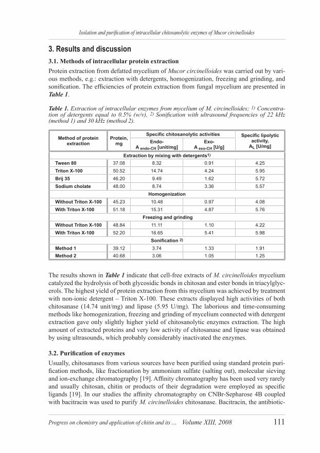
3. Results and discussion3.1. Methods of intracellular protein extractionProtein extraction from defatted mycelium of Mucor circinelloides was carried out by vari-ous methods, e.g.: extraction with detergents, homogenization, freezing and grinding, and sonification. The efficiencies of protein extraction from fungal mycelium are presented in Table 1.
Table 1. Extraction of intracellular enzymes from mycelium of M. circinelloides; 1) Concentra-tion of detergents equal to 0.5% (w/v), 2) Sonification with ultrasound frequencies of 22 kHz (method 1) and 30 kHz (method 2).
Method of protein extraction
Protein, mg
Specific chitosanolytic activities Specific lipolytic activity,
The results shown in Table 1 indicate that cell-free extracts of M. circinelloides mycelium catalyzed the hydrolysis of both glycosidic bonds in chitosan and ester bonds in triacylglyc-erols. The highest yield of protein extraction from this mycelium was achieved by treatment with non-ionic detergent – Triton X-100. These extracts displayed high activities of both chitosanase (14.74 unit/mg) and lipase (5.95 U/mg). The laborious and time-consuming methods like homogenization, freezing and grinding of mycelium connected with detergent extraction gave only slightly higher yield of chitosanolytic enzymes extraction. The high amount of extracted proteins and very low activity of chitosanase and lipase was obtained by using ultrasounds, which probably considerably inactivated the enzymes.
3.2. Purification of enzymesUsually, chitosanases from various sources have been purified using standard protein puri-fication methods, like fractionation by ammonium sulfate (salting out), molecular sieving and ion-exchange chromatography [19]. Affinity chromatography has been used very rarely and usually chitosan, chitin or products of their degradation were employed as specific ligands [19]. In our studies the affinity chromatography on CNBr-Sepharose 4B coupled with bacitracin was used to purify M. circinelloides chitosanase. Bacitracin, the antibiotic-
Progress on chemistry and application of chitin and its ... Volume XIII, 2008112
K. Struszczyk, M. Szczęsna-Antczak, M. Walczak, T. Antczak
cyclopeptide, is known as an efficient ligand of serine, aspartyl and metalloproteins and is used for separation of serine enzymes like e.g. lipases [20]. On the other hand, bacitracin can also interact with several different types of biomolecules, including DNA, RNA, lipo-proteins, receptors, and lipids from various sources [21]. It also binds transition metal ions, including Zn(+2), Mn(2+), Co(2+), Ni(2+), and Cu(2+) [22].
Crude enzymatic solution obtained by extraction of Mucor mycelium with Triton X-100 (0.5% w/v) was purified by affinity chromatography on CNBr-Sepharose 4B-bacitracin. As is shown in Figure 1, the proteins from the same peak (fractions from 123 to 136) displayed endo-, exo-chitosanolytic and lipase activities.
The fractions from affinity chromatography were concentrated and applied to gel chroma-tography on Sephadex G-100. Also this step of purification gave only one peak of proteins displaying endo-, exo-chitosanolytic and lipase activities (Figure 2). Their molecular mass was close to 42-43 kDa.
The results of purification are displayed in Tables 2 and 3. The purification procedure yield-ed a 23–fold purified chitosanase (endo-type enzyme) with 4.6% recovery of its activity and a 12–fold purified lipase with 2.4% recovery of activity. The ultimate specific activities of chitosanase and lipase were 231.5 unit/mg and 91.0 U/mg, respectively.
Figure 1. Affinity chromatography of chitosanase/lipase on CNBr-Sepharose 4B-bacitracin. Before protein separation, the column was equilibrated with 0.2 M phosphate buffer (pH 7.2). Unbound proteins from the protein extract (24 cm3) were washed with 280 cm3 of the latter buf-fer and the adsorbed proteins were eluted with 100 cm3 of the same buffer supplemented with Brij35 (0.15% w/v).
Progress on chemistry and application of chitin and its ... Volume XIII, 2008 113
Isolation and purification of intracellular chitosanolytic enzymes of Mucor circinelloides
The purified Mucor enzymes significantly lowered the chitosan Mv (average molecular weight) and their exo-chitosanolytic activity was minor. For comparison, all the prepara-tions of lipases described in literature [5 - 11] and applied for chitosan digestion also rapidly depolymerized this biopolymer and rapidly decreased viscosity of its solutions.
It is to note that the purified enzymes preparation migrated as only one protein band with the molecular mass of 42.5 kDa in the SDS-polyacrylamide gel electrophoresis (gel surface
Figure 2. Gel filtration of M. circinelloides intracellular proteins on Sephadex G-100. The col-umn was equilibrated with 0.2 M phosphate buffer (pH 7.2) and proteins from CNBr-Sepharose 4B-bacitracin (4.5 cm3) were applied. The column was eluted with the same buffer.
Table 2. Purification of M. circinelloides chitosanase.
Progress on chemistry and application of chitin and its ... Volume XIII, 2008114
K. Struszczyk, M. Szczęsna-Antczak, M. Walczak, T. Antczak
of 8.0 cm × 7.3 cm, Figure 3). But, when a distance of protein migration was longer (gel surface of 16.0 cm × 17.5 cm), the SDS-PAGE revealed two main protein bands with the molecular mass of 42 kDa and 43 kDa (Figure 4).
Most of the described in literature chitosanases and lipases from bacteria and fungi are char-acterized by the low molecular mass, usually ranging from 10 kDa to 50 kDa [19] and from 20 kDa to 80 kDa, respectively [23]. For example, the molecular masses of two chitosanases (A and B) from Mucor rouxii were 58 kDa and 76 kDa [24], from Penicillum islandicum - 30 kDa [19] and from Aspergillus niger - 29 kDa [25].
3.3. The effect of pH and temperature on M. circinelloides chitosanolytic enzymes The optimum pH for activity of M. circinelloides endo- and exo-chitosanolytic enzymes was 5.5-6.0 and these enzymes were stable in the pH range between 4.5 and 7.5. The highest chitosanolytic activities (endo- and exo-) were observed at temperature around 37 °C and these enzymes were relatively stable below 50 °C.
The optimum pH and temperature for activity of M. circinelloides chitosanolytic enzymes were similar to those of other fungal chitosanases. For example, enzymes from P. islandi-cum were optimally active at pH between 4.5 and 6.0 and 45 °C, while chitosanase from F. solani f. sp. phaseoil was optimally active at pH 5.6 and 40 °C. The other fungal chito-sanolytic enzymes were found to be stable up to 50 °C [19].
Bovine serum albumin 66 kDa
Peroxidase from horseradish
44 kDa
Chitosanolytic and lipolytic enzyme(s)
Figure 3. SDS-PAGE (8.0 cm × 7.3 cm) of M. circinelloides chitosanolytic enzymes. Molecular weight markers (lane 1), the purified M. circinelloides proteins (lane 2).
Figure 4. SDS-PAGE (16.0 cm × 17.5 cm) of M. circinelloides chitosanolytic enzymes. The purified M. circinellides proteins (lane 1), crude extract of proteins (lane 2).
1) 2)
1) 2)
Progress on chemistry and application of chitin and its ... Volume XIII, 2008 115
Isolation and purification of intracellular chitosanolytic enzymes of Mucor circinelloides
Different preparations of lipases that catalyzed chitosan hydrolysis, displayed the endo-chitosanolytic activity at temperature between 25 °C and 50 °C and pH between 3.6 and 7.0 [5 - 6, 8, 10].
3.4. Substrate specificity The purified enzymatic preparation was incubated with different substrates (1.0% w/v) such as chitosan (Mv ranging from 121 kDa to 421 kDa, DD ranging from 66% to 98%), colloidal chitin and sodium carboxymethyl cellulose at 37 °C and pH 5.5 for 1 h (endo-chitosanolytic activity) or 24 h (exo-chitosanolytic activity). Further steps were as described in 2.5.1.
Table 4. Substrate specificity of M. circinelloides enzymes.
The results shown in Table 4 indicate that the purified Mucor chitosanase prefers chitosan with the high degree of deacetylation (DD). The colloidal chitin and Na-CMC were not hydrolyzed by M. circinelloides enzymes.
4. SummaryApplication of the authors’ new, two-step (bacitracin affinity chromatography and gel filtra-tion on Sephadex G-100) method of purification of M. circinelloides intracellular enzymes gave the preparation with the relatively high activity of both endo-chitosanases and lipases. Our investigations indicate that this purified preparation contains two proteins with almost the same molecular masses and different activities. We have been trying to separate these proteins and purify them to homogeneity. We also intend to determine their 3 D structures and properties. However, the hitherto published results and our findings suggest that we may also identify the new oligomeric lipase, which efficiently depolymerizes chitozan.
5. AcknowledgmentThis work is supported by the Ministery of Science and Higher Education, project No. N507 181 32/2133.
6. ReferencesKim S.K. Rajapakse N., Carbohydrate Pol. 62, pp. 357-368, (2005).1. Prashanth K.V.H. Tharanthan R.N., Trends in Food Science & Technology 18, pp. 117-131, 2. (2007).
Progress on chemistry and application of chitin and its ... Volume XIII, 2008116
K. Struszczyk, M. Szczęsna-Antczak, M. Walczak, T. Antczak
Kołodziejska I., Malesa-Ciećwierz M., Górna E., Wojtasz-Pająk A. Chitin Enzymology, vol. II, 3. R.A.A. Muzzarelli (ed.), pp. 415-426, (1996).Jaworska M., Konieczna E., Kusaoke H., Progress on Chemistry and Application of Chitin and 4. Its Derivatives, vol.8, Struszczyk H. (ed.), pp. 43-49, (2002).Muzzarelli R.A.A., Xia W., Tomasetti M., Ilari P., Enzyme and Microbial Technology, vol.17, no.6, 5. pp. 541-545, (1995).Muzzarelli R.A.A. Chitin Handbook, Muzzarelli R.A.A., Peter M.G. (eds.), European Chitin 6. Society, pp. 153-163, (1997).Muzzarelli C., Francescageli O., Tosi G., Muzzarelli R.A.A., Carbohydrate Pol. 56, pp. 137-146, 7. (2004).Luckachan G.E., Pillai C.K.S. Carbohydrate Pol. 64, pp. 254-266, (2006).8. Muzzarelli R.A.A., Orlandini F., Pacetti D., Boselli E. Carbohydrate Pol. 66, pp. 363-371, 9. (2006).Pantaleone D., Yalpani M., Scollar M., Carbohydrate Res. 237, pp. 325-332, (1992).10. Roncal T., Oviedo A., Lopez de Armentia I., Fernandez L., Villaran M. Carbohydrate Res. 342, 11. pp. 2750-2756, (2007).Szczęsna-Antczak M., Antczak T., Piotrowicz-Wasiak M., Rzyska M12. ., Binkowska N., Bielecki S., Enzyme and Microbial Technology 39, pp. 1214-1222, (2006).Struszczyk K., Szczęsna-Antczak M., Antczak T. Rzyska M., Bielecki S. Struszczyk H., Progress 13. on Chemistry and Application of Chitin and Its Derivatives, vol.11, Jaworska M.M. (ed.), pp. 153-158, (2006).Struszczyk K., Szczęsna-Antczak M., Gajewska M., Antczak T., Progress on Chemistry and 14. Application of Chitin and Its Derivatives, vol 12, Jaworska M.M. (ed.), pp. 157-164, (2007).Roberts G.A.F. Chitin Chemistry, The Macmillan Press LTD., pp. 106-110, (1996).15. Wood T.M., Bhat K.M., Methods in Enzymology, Wood T.M., Kellogg S.T. (eds.) 163, pp. 87-16. 111, (1988).Lowry O.H., Rosenbrough N.J., Farr A.L., Randal R.J., Journal of Biological Chemistry 193, 17. pp. 265-275, (1951).Laemmli U.K., Nature, 227, p. 680, (1970).18. Somashekar D., Joseph R., Bioresource Technology 55, pp. 35-45, (1996).19. Stephanov V.M., Rudenskaya G.N. Journal of Applied Biochemistry 5, pp. 420-428, (1983);20. Ming L.J., Medical Research Review. 23, pp. 697-762, (2003).21. Ming L.J., Epperson J.D., Journal of Inorganic Biochemistry. 91, pp. 46-58, (2002).22. Kazlauskas R.J., Bornscheuer U.T., Biotechnology 2-nd ed. vol 823. a, Biotransformation I, Kelly D.R. (ed.), Wiley-VHC, Weinheim, pp. 37-191, (1998).Alfonso C., Martinez M.J., Reyes F. FEMS Microbiology Letters vol. 95, pp. 187-194, (1992).24. Chen X., Xia W., Tu X. Food Research International 38, pp. 315-322, (2005).25.


![Isolation, Partial Purification and Characterization of ... · Isolation and purification of lectins may be done through a variety of protein purification methods [24]-[33]. Methods](https://static.documents.pub/doc/80x56/5f54b2510bf11f58165072ba/isolation-partial-purification-and-characterization-of-isolation-and-purification.jpg)


