+ All Categories
Home > Documents > Isolation three for mitotic disjunction · to'\'Q5 tr Isolation and characterisation of three rows,...

Isolation three for mitotic disjunction · to'\'Q5 tr Isolation and characterisation of three rows,...

Date post: 11-Nov-2020
Category:
Upload: others
View: 1 times
Download: 0 times
Share this document with a friend
198
to'\'Q5 tr ¿ ,} Isolation and characterisation of three rows, a gene essential for mitotic chromosome disjunction in Drosophila melano gaster A thesis submitted for the degree of Doctor of Philosophy by Ulrik Peter John, B.Sc.(Hons), M.Sc. Departments of Biochemistry and Genetics University of Adelaide Adelaide, S.4.,5005 Australia 10 January 1995
Transcript

to'\'Q5 tr¿

,}

Isolation and characterisation of three rows,

a gene essential for mitotic chromosome disjunction

in Drosophila melano gaster

A thesis submitted for the degree of Doctor of Philosophy

by

Ulrik Peter John, B.Sc.(Hons), M.Sc.

Departments of Biochemistry and GeneticsUniversity of AdelaideAdelaide, S.4.,5005Australia 10 January 1995

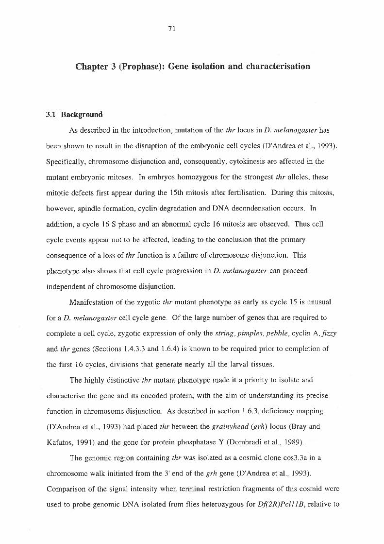
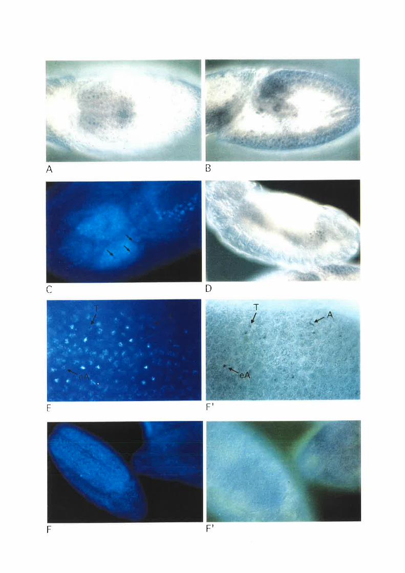
Confocal image of syncytial blastoderm embryo showing nuclei in metaphase (diagonal

from top right to bottom left) proceeding into anaphase. Chromosomes shown in green,

mierotubules (mitotic spindle) in red, and centrosomes in blue. Some nuclei have become

dissociated from pairs of centrosomes.

Table of contents

Abstract

Statement

Acknowledgments

Chapter 1 (G2)

Introduction..............

1.1 General principles of mitosis..

1.1.1 Microtubule organising centres

1.1.2 The assembly of the spindle......

1. 1.3 Centromere and kinetochore structure....

1.1.4 Chromosome motion in mitosis.

1.1.5 Force generation in mitosis................

1. 1.6 Sister chromatid disjunction

1. 1.7 Trouble shooting..........

1.2 How mitosis is regulated by the cell cycle control machinery........

1.3 Genes involved in chromosome segregation in other well studied

organlsms

1.3.1 Saccharomyces cerevisiae

I.3.2 Schizosaccharomyces pombe

t.41.3.3 Aspergillus nidulans

Mitosis in D ro s o phila melano I as te r ............,

1.4.1 Mitosis in embryonic development

l.4.I.l Syncytial divisions

I.4.1.2 Post-cellularisation. divisions...

I .4.2 Mitosis in postembryonic development..............

1.4.3 The nuclear and cytoskeletal organisation of mitosis...........

1.5 Genes involved in mitosis in D. melanogaster...

1.5.1 Maternal effect genes

1.5.2 Meiotic mutants

1.5.3 Zygotically regulated genes

1.5.3.1 The influence of the maternal contribution on the

time of onset of the zygotic phenotype

1.5.3.2 Late larval lethals

1.5.3.3 Embryonic lethals

1.5.4 Genes identified by sequence conservation/functional

complementation, and reverse genetics....

1.5.5 Genes identified by immunodetection of their encoded

proteins....

II2

3

5

7

8

11

12

t4

18

18

19

20

2l2I2t25

28

28

31

32

33

34

34

35

36

..37

..40

1.6 The three rows gene of D. melanogaster.

1.6. 1 Identification.....

1.6.2 Origin of aIIeIes................

1.6.3 Mapping

1.6.4 Mutant phenotype

1.7 This study

4l4I42

42

43

45

Chapter 2 (Gzl1Ñ4)

Materials and Methods...

2.1 Materials.

2.1.I Chemical reagents

2.1.2 Enzymes..........

Radio-labelled compounds

E. coli strains.......

Drosophila strains

2.1.6 Media and buffers..

2.I.7 Llbraries .................

2.I.8 Plasmids...

2.I.9 Oligonucleotides ...................

2.1.rc Molecular weight markers.....

2.2 Methods

2.2.I l" bacteriophage propagation...

2.1.3

2.r.4

2.1.5

47

4l41

48

48

48

49

5l53

54

54

55

55ËEJJ

56

56

56

51

57

57

51

58

58

58

58

59

59

59

59

59

60

60

2.2.2 )," bacteriophage library screening....

2.2.3 Isolation of I bacteriophage DNA2.2.4 Plasmid library screening

2.2.5 Radiolabelling of DNA fragments......

2.2.6 Hybridisation of radiolabelled probes to membrane

immobilised nucleic acids ............

2.2.7 Autoradiography ....

2.2.8 "Miniprep" isolation of plasmid DNA.........

2.2.9 Restriction analysis of DNA....

2.2.10 DNA fragment purification

2.2.1I Creation of recombinant plasmids.............

2.2.12 Transformation of recombinant molecules ..........

2.2.13 Nucleotide sequence analysis.....

a) Generation of nested deletions

b) Sequencing template preparation...................

c) Sequencing reactrons....

d) Electrophoresis

e) Sequence Analysis.

2.2.14 Maintainence of Drosophila stocks ..

2.2.t9

2.2.20

2.2.2r

2.2.22

2.2.23

2.2.24

Chapter 3 (Prophase)

Gene isolation and characterisation......

3.1 Background.....

3.2 Isolation of three rows coding sequences.

3.3 Genomic rescue of three rows mutants...

3.4 Structure of the three rows gene.

3.5 The three rows encoded product

2.2.15 Genetic transformation

2.2.16 Egg collects .........

2.2.17 Fixation of embryos for in si/rz hybridisation and

immunostaining ......

2.2.18 Expression studies....

a) RNA isolation

b) Northern analysrs.

c) RNA probe synthesis....

d) RNase protection analysis.....

e) Radiolabelling of oligonucleotide

f) Primer extension analysis.....

g) V/hole mount in situ hybridisation to mRNA...................

Electrophoresis of proteins.....

Bacterial expression of thr derived protein......

a) T7 system

b) Glutathione S-transferase fusion protein

Antibody production

IgG purification.........

Construction of affinity column

Affinity purification of antibodies......

2"2.25 Western analysis....

a) Sample preparation

b) Blotting

c) Immunodetection...

2.2.26 Immunostaining of embryo whole mounts

2.2.27 Image capture.....

2.2.28 Isolation of genomic DNA from adult Drosophila..

2.2.29 Southern blotting.....

2.2.30 Southern hybridisation under nonstringent conditions..............

2.2.31, Regulatory considerations............

2.3 Abbreviations.....

60

60

60

61

6T

6T

6t62

62

62

62

63

63

63

64

64

64

64

65

66

66

66

66

67

6l68

68

68

69

69

7l7T

t273

73

75

183.6 Discussion......

Chapter 4 (Prometaphase)

Analysis of expression ........

Chapter 5 (Metaphase)

Immunodetection

5.1 Background

5.2 Antibody production ......

5.3 Antibody purification .....

5.4 Western analysis

5.5 Immunolocalisation........

5.6 Discussion

Background ...............

Northern analysis

RNase protection analysis.....

Primer extension analysis.....

Whole mount in situ hybridisation to mRNA

4.6 Discussion

4.1

4.2

4.3

4.4

4.5

6.2

6.3

6.4

7.3

8l

.88

81

8l

89

89

91

Chapter 6 (Anaphase)

Isolation of a homologue from D. erecta..

6.1 Background ...

Genus bIot...........

Isolation of homologous sequences..........

Characterisation of D. erecta three rows

6.5 Discussion

Chapter 7 (Telophase)

Summary and prospects for future work ................ 104

7.1 Summary...

7.2 Future work

7.2.1 Further characterisation of mutant phenotype... r04

91

97

98

99

00

02

1

I

. r04

. t04

1 .2.2 Further analysis of expression .............

1 .2.3 Immunodetection................

7.2.5 Isolation of a diverged homologue........

1.2.6 Identification of interacting proterns

7.2.6.1 Genetic screens

7 .2.6.2 Immunological approaches ..........

7 .2.6.3 Exogenous reconstitution of interactions.............

Conclusion: three rows and its likely contribution to current issues

in mitosis.......

. to7

108

110

111

111

rt2113

tt4

References.. 115

I

Abstract

Zygotic expression of the three rows (thr) gene of Drosophila melanogaster is

required for normal cell proliferation during embryogenesis (D'Andrea et al., 1993).

Mitotic defects in thr mutant embryos begin during mitosis 15, and all subsequent

divisions are disrupted. Chromosome disjunction and consequently cytokinesis fail during

these defective mitoses, although the initial mitotic processes, and subsequent cell cycle

progression are not affected.

The thr gene has been identified, in a chromosome walk from the nearby grainyhead

gene, by correlation with a P element insertional polymorphism in the hybrid dysgenic

allele thrBH. Cloning of thr was confirmed by complementation of lethality in a homo-

zygous mutant background, with a genomic fragment from the region. The P element in-

sertion site has been defined by nucleotide sequencing and shown to interrupt a long ORF

corresponding with cDNA clones isolated from early embryonic libraries. thr encodes a

1,3'79 aa protein that shares no extended sequence similarity with known proteins.

rl¿r mRNA is present as abundant, maternally conferred transcript which degrades

at the time of cellularisation. At this and all subsequent times during development,

zygotic expression correlates with mitotic proliferation. These observations suggest that

the embryonic phenotype results from exhaustion of the maternal thr contribution and does

not reflect a developmentally restricted requirement for thr function. The delay in the

manifestation of the mutant phenotype until cycle 15 is believed to reflect persistance of

protein derived from maternal mRNA.

Immunostaining of embryos with three rows specific antibodies has revealed a cell

cycle dependent pattern of localisation, consistent with the defect in chromosome

disjunction observed in mutants. Three rows, undetectable in metaphase, is localised to

the chromosomes in anaphase, initially to the region of the presumptive kinetochore.

By the criteria of low stringency hybridisation to genomic Southern and library

filters, sequences homologous to thr can only be detected in Drosophila species thought to

have shared a common ancestor with D. melanogaster ûp to 20 mya. A homologue of thr

isolated from D. erecta encodes a protein with 88.37o sequence identity with three rows of

D. melanogaster over the common region.

ll

Statement

This work contains no material which has been accepted for the award of any other

degree of diploma in any university of other tertiary institution and, to the best of my

knowledge and belief, contains no material previously published or written by another

person, except where due reference has been made in the text.

I give consent for this copy of my thesis, when deposited in the University Library,

being available for loan and photocopying.

Ulrik John, l0ll/95

lll

Acknowledgments

I owe a debt of gratitude to my supervisor Rob Saint, whose enthusiasm and

commitment helped me see the advantages of working on the cell cycle in Drosophila, for

his honesty and integrity, and even for doing some crosses for me.

Sincere thanks to Richard D'Andrea who kindly allowed me to join the quest for

three rows, and whose departure was a great loss to the project.

Also to Paul Moretti for his patience dechorionating and in the fly lab. Rick Tearle,

with whom every interaction was insightful. Helena Richardson, truly the lab fairy

godmother, for so modestly giving the benefit of her vast experience and expertise.

Julianne Camerotto and Leanne Prior, princesses both, for reading the (at times)

unreadable. Louise O'Keefe for sharing her sequencing gels and enduring my taste in

music. Siv, Dan Kortschak, Stephen Gregory and Stanley Robert for not making me feel

too inadequate about my lack of computeracy. Gary Hime for help with microscopy and for

always being prepared to drop everything. All past and present members of the Saint lab

who have done things for me, made my time here enjoyable, or just put up with me" I

apologise to those for whom this catchall acknowledgement does not give sufficient credit.

Brian Miller and Joe Wrin for help with, the not always pleasant task of, generating

antibodies. Peter Kolesik for his expertise with the confocal microscope. There are many

people in technical and administrative support roles in both the Biochemistry and Genetics

departments who don't receive adequate recognition for their dedication and skill.

And to Maynard, Roy and HG for making work on the weekends bearable.

Especially to my parents Birte and Ian for making possible the fulltime pursuit of

my research, and for supporting me for the past six months.

Chapter 1 (G2): Introduction

This study concerns the characterisation of a gene, three rows, whose mutant

phenotype of failure of chromosome disjunction in anaphase, is indicative of an essential

but unknown function in mitosis.

1.1 General principles of mitosis

Mitosis is the process by which eukaryotic cells faithfully segregate their

duplicated genomes into two complete sets, usually at cell division. Mitosis occurs during

the stage of the eukaryotic cell cycle (Figure 1.1), referred to as M phase, and follows

replication of the chromosomal content in S phase. Interspersed between these two

phases are the "Gap" phases, Gl and G2 (Figure 1.1), during which commitment to, and

preparation for, the ensuing S and M phases occurs.

Mitosis can be viewed as the result of interactions between three major

multicomponent systems: i) the spindle, a microtubule (MT) based machine whose bipolar

organisation achieves the equipartition of ii), highly compacted chromosomes with

specialised structures upon them for engaging the spindle, and iii), a self governing

molecular oscillator which regulates the first two systems by controlling the level and

activity of their constituent proteins. The molecular oscillator will be described in section

1.2.

Despite obvious differences in the appearance of spindles and chromosomes in

various eukaryotes it is believed that the fundamental mechanisms of mitosis have been

conserved in evolution from yeast to humans. Our present understanding of mitosis has

come from the integration of data from genetically tractable organisms, such as the fission

and budding yeasts and Drosophila melanogaster, with cytological observations

predominantly from vertebrate cells.

Cytogeneticists studying mitosis have defined a series of sequential stages:

prophase, prometaphase, metaphase, anaphase and telophase (Figure 1.2). In prophase

Figure L.1 The eukaryotic cell cycle (from Murray and Hunt, 1993).

See text for details

Mitosis

lnterphase

Figure 1.2 Stages of mitosis (from Murray and Hunt, 1993).

See text for details.

Prometaphase

Prophase

Metaphase

Spindle pole

Chromosome

Condensing chromosomes

Nucleus

lnteçhase

Anaphase

I

I Cytokinesis

/\

\./

\

Microtubule

lnterphase

-/ Growth

)

chromosomes that have been replicated undergo condensation. The chromosomes are

captured by the mitotic spindle in prometqphase which positions them at the spindle

equator at metaphas¿. In anaphase the sister chromatids separate and are transported by

the spindle to opposite ends of the cell. Finally in telophas¿ the chromosomes decondense

and reestablish their interphase (a collective term for Gl, S and G2 phases) state.

1.1.1 Microtubule organising centres

The microtubule organising centre (MTOC) is the major nucleator of MTs in both

mitosis and interphase. Despite the structural dissimilarity between the MTOCs of fungi

(the spindle pole body) and animals (the centrosome) they do have protein components in

common. However it is yet to be demonstrated that they are homologous structures by

virtue of common ancestry.

The MTOC of yeast is the disc shaped Spindle Pole Body (SPB). Consistent with

the closed form of mitosis in yeast the SPB remains embedded in the nuclear envelope

throughout the yeast cell cycle where it simultaneously nucleates spindle MTs from its

intranuclear surface, and cytoplasmic MTs from the converse side. The SPB is a trilaminar

structure with its central layer contiguous with the nuclear membrane and MTs emanating

from the amorphous surface layers.

The centrosome of animal cells is a poorly defined cytoplasmic organelle consisting

of two orthogonally arranged centrioles surrounded by the amorphous pericentriolar

material (PCM) from which MTs emanate. Centrioles are related to, and in some

circumstances interchangable with, the basal body of flagellae. Each centriole is composed

of a short barrel of MTs in a "9+0" arrangement. It is believed that centrioles are unable

to arise de novo, their duplication being template driven by a preexisting centriole. While

various centrosomal components have been identified by immunological means (Kellogg

et al., 1989; Balczon and West, 1991) our knowledge of their arrangement and function is

scant.

Evidence of shared function in the MTOCs of fungi and animals has come from the

identification of an evolutionarily conserved minor tubulin, y-tubulin. y-tubulin was

originally identified in Aspergillus nidular?s as a suppressor of a p-tubulin mutation

J

(Oakley and Oakley, 1989). Localisation of y-tubulin to the SPB and demonstration that

mutations in y-tubulin in A. nidulans are severely compromised in MT formation led to the

hypothesis that T-tubulin may act as the site of MT nucleation (Oakley et al., 1990).

Furthermore, this function appears to be highly conserved, as y-tubulin has been detected

immunologically in the PCM in mammalian cells (Stearns et al., 1991), and cloned in

Schizosaccharomyces pombe, D. melanogaster, Xenopus laevis, mouse and humans (Horio

et al., l99I; Stearns et al., l99l;Zheng et al., 1991; Joshi et al., 1992). y-tubulin has also

been shown to be part of a complex with other centrosomal proteins in D. melanogaster

(Raff et al., 1993). Although y-tubulin's direct interaction with the minus ends of MTs is

as yet unproven, the demonstration that antibodies directed against it inhibit the

nucleation of new MTs but do not affect extant ones (Joshi et al., 1992), is highly

suggestive.

1.1.2 The assembly of the spindle

The assembly of a bipolar spindle is tied irrevocably to duplication and separation

of the MTOCs. Studies in fungi and mammals have revealed similar proteins involved in

both of these processes.

The replicate SPB is formed adjacent to the preexisting one, first appearing as a

"satellite" on the cytoplasmic side of the nuclear envelope early in Gl. Two daughter

SPBs arise following passage through "Start" (Hartwell et al., 1974), the transition point

late in Gl in which the cell becomes committed to undergo a complete cell cycle. The

daughter SPBs remain joined by a bridge until separated in S phase. The assembly of

SPBs is poorly understood, in part because few mutants have been found that affect the

process (Kilmartin, 1994). The product of one gene required for SPB duplication in

Saccharomyces cerevisiae, MPSI, is an essential protein kinase (M. Winey, pers. comm.)

Another gene, CDC31, encodes a low molecular weight calcium-binding protein (Baum

er al., 1986) of the EF-hand superfamily. The ubiquitous homologue of CDC3l , centrin,

has been isolated in protozoans (Huang et al., 1988), higher plants (Zhu et al., 1992) and

humans (Errabalou et al., 1994), and localised to the centrosome (Huang et al., 1988;

Errabalou et al., 1994).

4

In most animal cells centrosome duplication begins in Gl when the centrioles cease

their orthogonal arrangement and move slightly apart. Centriole replication is initiated

early in S phase, with the appearance of the procentriole perpendicular to the base of each

existing centriole, and is completed in G2 phase.

The MTs nucleated by MTOCs are polar structures with plus and minus ends

defined by their polymerisation properties and the asymmetry of their tubulin subunits.

The more stable minus end is proximal to the MTOC and the dynamic plus end is distal.

MTs are highly unstable structures existing in two states, either shrinking or growing.

Transitions from the shrinking to growing state are known as "rescue" and the converse

as "catastrophe". Because of the dynamic instabity of MTs the mitotic spindle is capable

of undergoing rapid changes in structure. Spindle assembly at the onset of mitosis is allied

to an increase in the catastrophe frequency relative to that of rescue (Belmont et al.,

1990), leading to the production of increased numbers of short, spindle MTs.

Many different types of proteins associate with MTs. Some MT associated

proteins (MAPs) modify MT dynamics while others are mechanochemical motors. Motor

proteins couple energy from nucleotide hydrolysis to rnovernent along the MT. Different

motor proteins may have different "adaptors" that enable them to engage and move

subcellular components, or to interact with other MTs and thus exert tension (Goldstein,

1991). Reflecting the polarity of their substrate, motor proteins usually move

unidirectionally being classified as either "plus end" or "minus end" directed.

Following their duplication MTOCs must be separated for a functional bipolar

spindle to formed. MTOC separation is a MT dependent process mediated by members of

a class of minus end directed molecular motors in both higher and lower eukaryotes. The

"bimC" family are phylogenetically distinct (Goodson et al., 1994) members of an

abundant, multifunctional class of Kinesin Related Proteins (KRPs) (Endow and Hatsumi,

1991). Mutants of bimC in A. nidulans (Enos and Morris, 1990), cutT in S. pombe (Hagan

and Yanagida, 1990), CinSp and Kiplp in S. cerevisiae (Hoyt et al., 1992), KLP6lF

(encoded by the urchin gene) in D. melanogaster (Heck et al., 1993) have MTOC

separation blocked. The same defect has been observed in in vitro spindle assembly

assays with antibodies against the X. laevis KRP Eg5 (Sawin et al., 1992a).

5

Immunolocalisation of cut7, Cin8p, Kiplp and Eg5 (Hagan and Yanagida, 1992; Hoyt et al.,

1992; Roof et al., 1992; Sawin et al., I992a) to MTs between the MTOCs is consistent

with a hypothesis that MTOC separation is conferred by sliding of antiparallel MTs, driven

by KRPs at the spindle midzone. Another KRP, human MKLP, with antiparallel MT

sliding activity in vitro is localised to the spindle midzone (Nislow et al., 1992) but is not

a member of the bimC family (Goodson et al., 1994). Evidence of an additional mechanism

for MTOC separation in anaphase in vertebrates is discussed in 1.1.5 below.

KRPs are also involved in maintaining the integrity of the spindle once it has been

assembled, possibly by the generation of opposing forces. Thus, deletion of CinSp and

Kiplp which causes collapse of the spindle can be partially suppressed by loss of function

in another KRP Kar3p (Saunders and Hoyt, 1992)" This is consistent with recent data that

Kar3p functions as a minus end directed motor (Endow et al., 1994). Similarly, in

A. nidulans defective bimC can be compensated for by deletion of klpA, a Kar3p related

KRP (O'Connell et al., 1993).

The contribution of motor proteins to force generation in chromosome movement is

discussed in 1.1.5 below.

1.1.3 Centromere and kinetochore structure

Kinetochores are plate shaped complexes of specialised proteins that bind to

specific centromeric DNA sequences, enabling chromosomes to engage the spindle. An

apparent lack of conservation of kinetochore proteins and centromeric sequences

throughout evolution (Bloom, 1993) have hindered the gaining of functional insights. To

date the most productive source of characterised kinetochore proteins has been humans,

whilst the only defined centromeric DNA sequence is that of S. cerevisiae.

The centromeric DNA of S. cerevisiae is only 125 nt in length and consists of three

distinct sequence elements CDEI, CDEII and CDEIII (Fitzgerald-Hayes et al., 1982).

All three elements are essential but the 25bp CDEIII is absolutely required for

centromeric function (Ng and Carbon, 1987). CDEIII is specifically bound, in a

phosphorylation dependent manner, by CBF3, a 240 kDa complex of three major proteins

CBF3A, B and C, and some minor proteins (Lechner and Carbon, 1991). The complex

6

harbours MT minus end directed motor activity (Hyman et al., 1992) for which the minor

component Kar3p is probably responsible (Endow et al., 1994; Middleton and Carbon,

1994). Genes encoding the CBF3A and C proteins have been cloned, both having been

independently isolated in genetic screens for mitotic defects (see Bloom,1993). Two of

the other minor components are CBF5p a putative MAP, and DNA topoisomerase II

(Jiang et al., 1993).

Investigations of human centromere and kinetochore structure have exploited

autoantibodies from patients with the syndrome CREST (Calcinosis, Raynaud's

phenomenon, Esophageal (sic) dismotility , Schlerodactly, and Telangiectasia) (Moroi

et al., 1980). These autoantibodies react with four distinct kinetochore proteins (CENPs)

(Earnshaw and Rothfield, 1985) and also two from the inner centromere (INCENPs)

(Earnshaw and Cooke, 1991). Another protein, CENP-E (Yen et al., 1991) has

subsequently been added to the group. The corresponding genes have been cloned and

their products characterised (Table 1.1).

Table L.L Mammalian kinetochore proteins (from Bloom, 1993),

Protein Mofifs Phenotvne

CENP-A

CENP-B

CENP-C

CENP-D

CENP-E

INCENPA

INCENPB

Histone-like

Acidic serine-rich region

Hydrophilic and highly basic

GTP-binding (RCCI homolog)

Kinesin-like microtubule based motor

Coiled-coil domain

Coiled-coil domain

G2-M arrest

Metaphase arrest

The characterisation of centromeric sequences in mammals has been greatly

hindered by their enormous size. For example the centromere of the smallest human

7

chromosome,2l, may occupy more than 5 Mb of its approximately 40 Mb total (Earnshaw

and Tomkiel, 1992). Short functional centromeric sequences may reside amongst long

stretches of repetitive DNA that characterise centromeric regions or else the repetitive

sequences themselves may confer aspects of centromere function. Evidence for the latter

hypothesis comes from the demonstration that the consensus binding site for CENP-B,

the CENP-B box, is a 17 nt sequence found in cr-satellite DNA (Muro et al., 1992). ü-

satellite DNA is present in abundance in mammalian centromeres, as higher order repeats

of a Il I nt monomer" However CENP-B binding is clearly not sufficient for centromere

function as it has been detected in inactive centromeres (Earnshaw et al., 1989).

The presence of other proteins at vertebrate kinetochores has been demonstated,

most notably the minus end directed motor dynein. Anti-dynein antibodies stain the

kinetochores of mitotic cells (Pfarr et al., 1990; Stuer et al., 1990) and isolated

chromosomes (Wordeman et al., 1991). KRPs have also been detected immunologically

(Sawin et al., 1992b) and the human KRP CENP-E localises to kinetochores during

prometaphase and metaphase (Yen et al., 1992). These results are consistent with in

vitro kinetochore motility assays which have identified two different motor activities of

opposite polarities (Hyman and Mitchison, 1991b).

1.1.4 Chromosome motion in mitosis

Because of the inadequacies of cytology in yeast (closed mitoses, low levels of

chromosome condensation, and minimalist spindles) descriptions of chromosome

behaviour in mitosis have come almost exclusively from vertebrate cells. In particular

newt lung epithelial cells have been highly favoured because of their large chromosomes,

flatness and optical clarity. These studies have revealed that chromosomes undergo

continuous oscillatory movements relative to the spindle poles throughout mitosis

(Skibbens et al., 1993), and have lent weight to the concept of the "smart kinetochore"

(Mitchison, 1989).

Chromosomes, when first attached to the spindle, are mono-orientated.

Kinetochores initially interact with the lateral surface of one of the MTs extending from, or

retracting to, the spindle poles. Immediately upon attachment the chromosome moves

8

rapidly (an order of magnitude faster than all subsequent movements (Rieder and Salmon,

1994)) along the side of the MT towards the spindle pole from which it is nucleated

(Rieder and Alexander, 1990). As it nears the pole it slows and the kinetochore

encounters more MTs, the ends of which it now interacts with. These MTs are known as

kinetochore MTs (kMTs). Oscillatory movements now commence which are characterised

by abrupt transitions in direction termed "directional instability" (Skibbens et al., 1993).

From the distortion of the centromeric region it has been inferred that force is generated by

alternate pushing or pulling movements at the kinetochore (Skibbens et al., 1993)"

In a process that may be aided by the oscillations, the other unoccupied kinetochore

eventually captures a MT (usually) from the opposite pole. The bi-orientated

chromosome now undergoes "congression" to take up a position on the spindle equator at

metaphase. Bi-orientated chromosomes also continue oscillatory movements in a manner

which suggests "cooperative switching" (Skibbens et al., 1993). If one kinetochore is

moving polewards, thenl5Vo of the time its sister is moving away from its pole. The

mechanism by which the behaviour of one kinetochore can influence that of the attached

one is unknown, but it is argued the "smart" kinetochores can sense tension at the sites of

kMT attachment and alter the frequency of phase transitions accordingly (Skibbens et al.,

l ee3).

In anaphase the sister chromatids disjoin and are moved to opposite ends of the

cell. Anaphase consists of two types of movement: anaphase A and anaphase B. In

anaphase A the sister chromatids move towards the spindle poles concommitant with

kMT shortening, whilst in anaphase B the spindle elongates and the poles move apart. In

animal cells anaphase A and B occur simultaneously. In S. pombe both spindle elongation

and kMT shortening have been observed concommitant with sister chromatid separation

(Ding et al., 1993).

1.1.5 Force generation in mitosis

The forces that move chromosomes in mitosis remain an area of contention. It has

been demonstated that under certain conditions either mechanochemical motor activity, or

9

MT dynamics at either the plus or minus ends can produce sufficient force to realistically

account for observations of chromosome movements during mitosis.

As discussed in section 1.1.3, immunological data and results from in vitro

experiments localise both plus and minus end directed motor activity to the kinetochore.

Certainly, the initial movement of a mono-orientated chromosome poleward along the

lateral surface of a MT is difficult to reconcile, other than with the action of a minus end

directed kinetochore motor. On the basis of its localisation to the kinetochore, and its

kinetic properties, cytoplasmic dynein has been proposed as the motor responsible for this

movement (Rieder and Alexander, 1990)" However in yeast, disruptions of the dynein

gene merely perturb spindle positioning (Li et al., 1993), and anti-dynein antibodies have

no effect on chromosome movement in mammalian cells (Vaisberg et al., 1993).

MT dynamics alone can also be sufficient to generate bidirectional chromosome

movement" MT polymerisation at the kinetochore can induce chromosome movement

away from the poles (Shelden and Wadsworth, 1992), and in the absence of ATP

kinetochores can remain bound to a MT as it depolymerises (Koshland et al., 1988)"

Furthermore, fluorescent marking of spindle MTs have revealed there is a continuous flux

of MTs polewards in metaphase and early anaphase caused by MT disassembly at the

pole (Mitchison and Salmon, 1992). Such flux could account for up to 37Vo of chromosome

to pole movement (Mitchison and Salmon, 1992).

The resolution of the relative contributions of kinetochore motors and MT dynamics

may come from the observation that kinetochores interact almost exclusively with the

ends of MTs. In such an arrangement kinetochore movement must be coordinated with

MT plus end polymerisation or depolymerisation. Thus MT motors may simply function to

maintain contact with the labile MT ends. MT polymerisation kinetics would then

determine the velocity of chromosome movement. Alternatively, the motor proteins

themselves could regulate MT dynamics.

Another less well characterised MT mediated force producing mechanism is the

astral ejection force or "polar wind". The spindle exerts a pressure, demonstrated by the

active transportation away from the pole of chromosome arms severed with a laser

microbeam (Rieder et al., 1986). Astral ejection forces may contribute to the preanaphase,

10

away from pole, movement of oscillating chromosomes (Cassimeris et al., 1994). The

nature of the force is unknown but it could simply result from the impact of growing MT

ends (Rieder et al., 1986).

Polewards movement of sister chromatids during anaphase A is believed to be

conferred by the same forces as in earlier stages, as chromosomal oscillations can still be

observed (Rieder and Salmon, 1994). Clearly however there is a shift in bias to pole

directed movement. This may simply be a product of abolishing the connection between

chromatids and as the magnitude of the astral ejection force is proportional to the

chromosomal area, the chromatid arms undergo sustained polewards movement (Rieder

and Salmon, 1994). The changes in MT dynamics that accompany disassembly of the

spindle in anaphase would hasten this process. Alternatively there may be regulatory

changes at the kinetochore which alter MT dynamics or motor activity.(Hyman and

Mitchison, l99la; Rieder and Salmon, 1994).

Anaphase B spindle elongation is an analogous process to MTOC separation

required for spindle assembly (Section 1.1.2). There are two hypotheses for how this may

occur. Either the force is produced by antiparallel pushing of lvITs in the spindle midzone,

as shown in yeast SPB separation, or from pulling forces between the astral MTs and

some peripheral anchor.

In lower eukaryotes there is evidence that the same forces are responsible for SPB

separation in prophase and in anaphase. Anti KRP antibodies localise to the midzone of

isolated diatom metaphase spindles and inhibit elongation (Hogan et al., 1992). Evidence

also comes from a cold sensitive mutant of S. cerevisiae, that only forms spindle but not

astral MTs, in which anaphase B is normal (Sullivan and Huffaker, 1992).

In vertebrates the latter mechanism clearly operates as centrosome migration

continues when half spindle MT arrays no longer overlap (Waters et al., 1993). However

both mechanisms probably contribute since spindle elongation occurs at a faster rate when

there is overlap, implying the antiparallel MTs act as a ratchet to govern the rate and

direction of centrosome separation (Waters et al., 1993). Minus end directed motors

tethered in the cytoplasm could provide the force for the proposed astral pulling, and

11

antibodies against dynein have been shown to inhibit centrosome separation (Vaisberg

et al., 1993).

One striking feature of mitotic force production is the many instances of seeming

redundancy of the mechanisms employed. A number of examples of this have been

discussed here. Thus: both MT dynamics or motor proteins may be adequate to move

chromosomes, more than one motor protein may contribute to the same process as for

CinSp and Kiplp in S. cerevisiae, anaphase B is driven by both pushing and pulling

mechanisms in vertebrates, and anaphase A and anaphase B both contribute to

chromosome segregation. While such redundancy may simply reflect inadequacies in our

understanding of these processes, it may also exist to enhance the accuracy of mitosis, a

process with strong selection for high fidelity (Goldstein, 1993; Thomas, 1993)

1.1.6 Sister chromatid disjunction

At anaphase the linkages which have held sister chromatids together throughout

mitosis are abolished almost simultaneously as a result of an unknown signal or reaction.

These linkages are not only proximal to the centromere but extend the entire length of the

chromosome (Rattner et al., 1988). Sister chromatid separation is a MT independent

process in some systems (reviewed in Rieder and Palazzo, 1992) but this is evidently not

the case in D. melanogaster (Gonzalez et al., 1991; Williams et al., 1992). However the

dependence in D. melanogaster, of chromatid disjunction on spindle integrity, probably

does not reflect a requirement for force production in disjunction but is a function of

checkpoint surveillance (Section 1.1.7).

On the basis of their localisation to the region of contact between sister chromatids

in metaphase, the human INCENPs have been proposed to function in chromatid

separation (Cooke et al., 1981). However there is no direct evidence for this. Indeed

INCENPS become redistributed to the spindle midzone significantly in advance of

anaphase (Earnshaw and Cooke, 1991). By contrast the presence of another class of

antigens, the CLiPs (Chromatid Linking Proteins) is correlated with chromatid association

(Rattner et al., 1988).

l2

One product whose activity is essential for sister chromatid separation is type II

DNA topoisomerase (topo II). Eukaryotic chromosomes typically complete DNA

replication with multiple intertwinings between the DNA strands of the two chromatids.

Disjunction in such a state would be extremely deleterious. Topo II can resolve

chromosome tangling by producing a double strand cut in the DNA of one chromatid,

passing the DNA of the other through the cut, then religating. Disruption of topo II

activity in yeasts, X. Iaevis and D. melanogaster leads to failure of chromatid separation

and also causes defects in chromosome condensation (DiNardo et al., 1984; Uemura et al.,

1987; Shamu and Murray, 1992;Buchenau et al.,1993).

1.1.7 Trouble shooting

To achieve the exceptionally high rates of fidelity (<tO-s mistakes per cell division

(Hartwell and Smith, 1985)) observed for chromosome segregation, eukaryotes have

developed dependency relationships that couple anaphase to the successful completion of

earlier events. Mechanisms exist to delay or block mitotic progression if a functional

bipolar spindle has not been formed, if chromosomes are malorientated, or if kinetochores

are defective.

Some, but clearly not all, of these arrests or delays are the product of "checkpoints"

(Hartwell and Weinert, 1989), where an extrinsic surveillance mechanism monitors the

successful completion of an earlier process. Checkpoints can be thought of as signal

transduction pathways that feed forward to downstream events empowering them to

proceed (Hartwell, 1991). Other arrests are simply a consequence of dependencies

analogous to a substrate-product mechanism (Hartwell and Weinert, 1989), in which

downstream events will not proceed in the absence of a pre-existing product or condition.

Prometaphase is prolonged to variable degrees (Kung et al., 1990) in cells that

have been treated with agents that either disrupt or stabilise MTs, even at levels that

produce no observable effects (Rieder andPalazzo, 1992; Jordan et al., 1993; Wendel et

al., 1993). Prolongation is also observed in the absence of spindle bipolarity (Hunt et al.,

1992).

13

Similarly, uncongressed chromosomes produced either by irradiation (Zirkle, 1970),

micromanipulation (Rieder and Alexander, 1989), treatment with kinase inhibitors

(Nicklas etal., 1993), or occurring naturally (Nicklas and Arana,1992) delay anaphase

onset.

Lesions in kinetochores also cause mitotic arrest. In S. cerevisiae a mutation in

the centromeric DNA of one chromosome, in the presence of 32 normal centromeres, can

retard mitosis (Spencer and Hieter, 1992), as does a mutation in the CTFI3 gene encoding

one of the CBF3 kinetochore complex proteins (Doheny et al., 1993)" Anaphase is also

delayed in cells injected with a mixture of antibodies, against several CENP kinetochore

proteins (Bernat et al., 1990), and against the kinetochore KRP CENP-E (Yen et al.,

199 1).

While many of these observed delays have been ascribed to the action of

checkpoints, very few have satisfied the criteria of being mediated by an extrinsic

mechanism that can be mutated or disrupted in order to bypass the delay. Assembly of a

functional spindle is one process that is clearly subject to checkpoint monitoring. Mutants

have been isolated in S. cerevisiae, the bub- and mad- strains, that fail to block or delay

mitotic progression in response to inhibitors of MT polymerisation (Hoyt et al., I99I; Li

and Murray,l99I).

It has been suggested that checkpoint monitoring of spindle assembly, and the

arrests due to chromosome malorientation and kinetochore defects may be part of the

same surveillance mechanism that specifically monitors the interaction of kinetochores

with MTs (Zirkle" 1970; Murray and Hunt, 1993). Evidence for a possible mechanism of

signalling kinetochore attachment to the spindle has come from the detection of a

phosphorylated epitope expressed on kinetochores in prometaphase but lost at metaphase

(Gorbsky and Ricketts, 1993). Most striking is the observation that a misaligned

chromosome, capable of delaying anaphase onset, strongly expresses the phosphoepitope

while the remaining chromosomes at the metaphase plate do not.

Interestingly no checkpoint mechanism for monitoring anaphase chromosome

segregation have yet been discerned. Evidence that this process is not subject to

monitoring comes from the occurrence of mutants which fail in disjunction yet undergo

l4

subsequent DNA replication to become polyploid (Sections 1.3 and 1.5.1). Some

dependency relationships however clearly operate on later events, as cytokinesis is often

defective in the absence of disjunction. For instance, cytokinesis is still blocked in bub

mutants in presence of MT inhibitors (Hoyt et al., 1991).

While the spindle assembly checkpoint is involved in troubleshooting at the

terminal stages of mitosis other checkpoints control entry into mitosis. Checkpoint

pathways have been genetically characterised in yeasts, that delay the onset of mitosis in

the event of DNA damage or failure to complete replication (reviewed in Murray, 1992).

Mutants have been isolated in S. cerevisiae and S. pombe that prevent arrest in response

to damaged DNA, or to unreplicated DNA, and some that are defective in both pathways.

Checkpoint mechanisms are not a universal phenomena. Their absence is a feature

of early embryogenesis in some organisms, including sea urchins (Sluder et al., 1994) and

X. Iaevis (Kimelman et al., 1987). This is believed to reflect a requirement for speed and

synchrony in early divisions (Hartwell and Weinert, 1989). In early D. melanogaster

embryogenesis whilst the spindle integrity checkpoint clearly operates, the checkpoints

controlling entry into mitosis do not. D. melanogaster, however, possesses an alternative

mechanism to eliminate defective nuclei (Section 1.4.1.1).

Checkpoint controls are believed to delay mitotic progression by modulating the

activity of the molecular oscillator which controls the cell cycle. The way this oscillator

regulates mitosis is described in the next section.

1.2 How mitosis is regulated by the cell cycle control machinery

Cyclin dependent kinases (CDKs) are the core of the molecular oscillator that

controls cell cycle progression in all eukaryotes. Homologues of the founder CDK, p34cdcz

of S. pombe, control entry into mitosis in all eukaryotes. CDKs are also responsible for

controlling other important transition points in the cell cycle such as the Gl to S phase

transition. In yeasts the Gl to S transition is also controlled by p34cdcz, but in

vertebrates and possibly D. melanogaster (Section 1.5.4) related but distinct CDKs appear

to be involved.

l5

p34cdcz is a highly stable protein whose enzymatic activity (phosphorylation of

serine and threonine residues) is regulated by two mechanisms: positively by physical

association with regulatory subunits known as cyclins; and either positively or negatively

depending on the phosphorylation state of two critical residues. Cyclins are unstable

proteins whose patterns of accumulation and loss are correlated with phases of the cell

cycle (Evans et al., 1983). Cyclins can be distinguished on the basis of their sequence

relationships and the transition points they are believed to influence. Cyclins A and B

participate in the regulation of mitosis.

The current biochemical model for the regulation of mitosis by p3{cdc2 (Figure 1.3)

is a synthesis of results from many different organisms. Many mechanistic details are still

to be determined, and the importance of particular regulatory steps in controlling cell cycle

progression shows variation from species to species, and even within species across the

life cycle (for example see Section 1.4.1.1).

Monomeric p34cdc2 is inactive and unphosphorylated (Figure 1.3). Association

with cyclins A and B, which accumulate to a threshold level in G2, is a prerequisite for

activation (Murray and Kirschner, 1989a)" Formation of the complex induces

phosphorylation at tyrosine 15 (nomenclature for human p34cdcz't by kinases, first

identified as the products of the weel (Russell and Nurse, 1987b) and mikl (Lundgren

et al., 1991) genes in S. pombe. Tyrosine 15 phosphorylation is inhibitory and dominant to

cdc2 activating kinase (CAK) phosphorylation at threonine 161 (Solomon et al., 1992)by

p40Mots (Fesquet et al., 1993; Poon et a1.,1993; Solomon et al., 1993)" itself a CDK

(Makela et al., 1994). Phosphorylation at these two residues produces inactive mitosis

promoting factor (preMPF) which lacks protein kinase activity (Figure 1.3). Both

phosphorylations are antagonised by the action of protein phosphatases, the former by

homologues of S. pombe Cdc25 (Russell and Nurse, 1986; Moreno et al., 1990).

As the pool of preMPF increases a small amount of active MPF accumulates,

dephosphorylated on tyrosine 15 (Figure 1.3). When the level of MPF reaches a

threshold level it initiates an explosive process of activation that drives cells rapidly and

irreversibly into mitosis. Weel is negatively regulated by yet another kinase Niml,

known from ^S.

pombe (Russell and Nurse, 1987a; Coleman et al., 1993). MPF activates

Figure 1.3 Biochemical model of the mitotic oscillator (from Murray and Hunt, 1993).

The varied forms of the Cdc?-cyclin B complex, during the cell cycle, are shown. Also

indicated are the enzymes that catalyse the modifications. The phosphorylation state of

two critical residues corresponding to tyrosine 15 (left) and threonine 161 (right) in

S. pombe are shown.

Cyclin B

Cdc2

Cyclin B

Cd,c2

Cdc2activating

kinase

<-Phosphatase

Cyclinsynthesis

Cdc25-------+<-

Weel

Phosphatase

Cyclinprotease

Chromosomecondensation

Nuclearenvelope

breakdown

lnact¡ve

+

YT YT

lnactive

YTlnactive

'!;

w""r f f "o"ru

+

YT

Y

Active MPFSpindle

assemblyCyclin B

Cdc2

Cyclin B

Cdc2

Cdc2 Cdc2

Cyclin B

t6

Cdc25 establishing positive feedback (Solomon et al., 1990; Hoffman et al., 1993) (Figure

1.3), and may also activate Niml (Murray, 1993). In attaining full activity MPF also

ensures its own demise by triggering the the cyclin degradation machinery which

contributes to the exit from mitosis (see below).

MPF is believed to elicit its function by phosphorylating target proteins that

mediate the events of mitosis, such as nuclear envelope breakdown, chromosome

condensation and spindle formation. Although many proteins are phosphorylated in

mitosis (Karsenti et al., 1987), and a plethora of proteins are substrates for MPF i¡¿ vitro,

very few proteins are known whose in vivo phosphorylation has consequences for mitotic

progression. Probably the best characterised MPF substrates are the nuclear lamins,

whose phosphorylation induces their depolymerisation (Hearld and McKeon, 1990),

necessary but not sufficient for nuclear envelope breakdown (Nigg, 1993). MPF can

modulate MT dynamics, and their nucleation by centrosomes in cell-free systems (Verde

et al., 1990; Buendia et al., 1992), implicating MPF kinase activity in spindle formation.

Furthermore, chromosome condensation is correlated with extensive phosphorylation of

chromatin associated proteins (Reeves, 1992)" However neither spindle formation nor

chromosome condensation has yet been shown be dependent on the in vivo

phosphorylation by MPF of a specific substrate.

The degradation of Cyclins A and B around the time of metaphase is necessary for

inactivation of MPF, and coincides with onset of anaphase. Cyclin A is degraded during

prometaphase, in advance of cyclin B, whose level drops precipitously at the metaphase-

anaphase transition (Evans et al., 1983; Lehner and O'Farrell, 1990b; Whitfield et al.,

1990). Cyclin degradation is believed to occur by ubiquitin mediated proteolysis, conferred

by a motif in the N-terminal region, the "destruction box" (Glotzer et al., 1991).

Demonstration that deletion of the cyclin B destruction box prevented inactivation of MPF

and produced mitotic arrest in X. laevis (Murray et al., 1989), and ,S. cerevisiae (Ghiara

etal., 1991) provided evidence for the hypothesis that cyclin destruction triggered

anaphase onset by inactivating MPF (Murray and Kirschner, 1989b; Glotzer et al., 1991).

By contrast, recent findings from two studies indicate that initiation of anaphase

requires neither cyclin degradation nor MPF inactivation. Addition of nondegradable, but

t7

otherwise active cyclin B (same form as in Murray et al., 1989) to mitotically cycling

X. Iaevis egg extracts prevents MPF inactivation but does not inhibit anaphase onset as

determined by sister chromatid separation (Holloway et al., 1993). However other events

of anaphase such as chromosome decondensation and spindle disassembly are blocked.

The validity of these findings rests on interpretation of the arrested state as being

anaphase. Earlier in vivo experiments (Murray et al., 1989) were reported to produce

metaphase arrest"

Experiments with S. cerevisiae also provide evidence that mitotic cyclin

degradation/lv1PF inactivation is not required for anaphase onset but functions in the final

exitfrommitosis(Suranaetal., 1993). Mutants of cdcl5, aproteinkinase(Schweizerand

Phillipsen, 1991), which arrest in telophase with disjoined chromosomes (Pringle and

Hartwell, 1981), can undergo anaphase with high levels of MPF (Suranaet al., 1993).

Furthermore, overexpression of a B-type mitotic cyclin causes arrest in telophase not in

metaphase.

If MPF inactivation is not a prerequisite for initiation of anaphase then what is the

nature of the signal? Accumulating evidence supports the idea that chromosome

disjunction is mediated by the same ubiquitin dependent proteolysis that degrades

cyclinB. InX. laevi,s egg extracts sister chromatid separation is inhibited in a dose

dependent manner by an N-terminal fragment of cyclin B that cannot activate MPF but is

proposed to elicit its effect by competing, as a substrate for ubiquitination, with a

hypothetical "anaphase trigger protein" (Holloway et al., 1993). Furthermore a mutant

form of the N-terminal peptide that is not recognised by the ubiquitin conjugating system

does not delay chromosome disjunction, while addition of the inhibitor, methylated

ubiquitin does. Evidence also comes from mutations in genes encoding subunits of the 265

ATP/ubiquitin dependent protease in S. pombe (Gordon et al., 1993) and S. cerevisiae

(Ghislain et al., 1993) that cause defective chromosome segregation. To date the

hypothetical anaphase trigger protein targeted for proteolysis has not been identified in

any organism.

Other lines of evidence point to a role for protein phosphatases in chromosome

disjunction. The action of phosphatase(s) is a logical means of initiating disjunction as it

18

could simply counter the activity of a preexisting MPF substrate. Inhibition of protein

phosphatases with okadaic acid inhibits disjunction in HeLa cells but permits cell cycle

progression (Ghosh and Paweletz, 1992). Rat embryo fibroblast cells arrest at metaphase

when injected with anti-protein phosphatase I (PPl) antibodies at the start of mitosis

(Fernandez et al., 1992). Mutants in PPl show defects in chromatid separation in

A. nidulans (Doonan and Morris, 1989), S. pombe (Ohkura et al., 1989; Kinoshita et al.,

1990), and D. melanogaster (Axton et al., 1990) (Section 1.5.3.2).

A transient rise in intracellular CaZ+ has also been invoked in the regulation of

anaphase onset. Unfertilised X. laevis eggs arrested in metaphase II of meiosis enter

anaphase when fertilisation triggers a cytoplasmic CaZ+ spike (Busa et al., 1985). Egg

extracts have been used to develop in vitro systems that mimic many features of mitosis

(Shamu and Murray, 1992; Holloway et al., 1993). Addition of Ca2+ to extracts with

assembled metaphase spindles leads to chromosome segregation (with qualifications, see

above), MPF inactivation, and passage into interphase. In sea urchin embryos sharp Ca2+

transients are correlated with cell cycle events including the metaphase-anaphase

transition (Ciapa et al., 1994). Ca2+ levels may modulate the activity of

calcium/calmodulin dependent protein kinases, substrates of which include a MAP whose

phosphorylation at the metaphase-anaphase transition induces MT depolymerisation

(Dinsmore and Sloboda, 1988).

The challenge remains to establish the relative contributions of, and the probable

interactions between, proteolytic degradation, phosphorylation states, and Ca2+ levels in

sister chromatid disjunction.

1.3 Genes involved in chromosome segregation in other well studied organisms

1.3.1 Saccharomyces cerevisiae

In a seminal achievement Hartwell and co-workers were the first to systematically

identify genes involved in cell division. Many cell division cycle (cdc) mutants were

isolated in a genetic screen for temperature sensitive mutants whose uniform morphology

t9

at the nonpermissive temperature indicated they were defective in cell cycle progression

(Hartwell et al., 1910). These and further cdc mutants subsequently isolated have

enabled the analysis of the S. cerevisiae cdc2 homologue, CDC28, as well as genes

required for budding, SPB duplication, DNA synthesis, spindle formation, and cytokinesis

(reviewed in Hartwell, 1991).

Three cdc mufants, cdcl6, cdc23 and cdc27 defective in mitosis appear to arrest in

metaphase (Sikorski et al., 1990; Sikorski et al., 1991). Their wild-type genes encode

proteins containing multiple tandem copies of an imperfect 34 amino acid "tetratricopeptide

repeat" (TPR) present in, but not confined to, genes involved in mitosis (Sikorski et al.,

1990). TPRs are proposed to function in intra- and intermolecular interactions with other

TPRs (Sikorski et al., l99l; Sikorski et al., 1993) and all three proteins have now been

shown to be part of a macromolecular complex (Lamb et al., 1994)" Furthermore the

products of CDCI6 and CDC23 have been localised to the nucleus (Sikorski et al., 1993).

Other genetic screening strategies have identified mutants defective in

chromosome disjunction, including two components of the CBF3 kinetochore complex

(Section 1.1.3). Mutants in espl arose from a scrsen for defects causing increased ploidy

(Baum et al., 1988). espl cells form irregular spindles and are grossly defective in

chromosome segregation such that following cytokinesis the bulk of the DNA and both

SPBs are found in one of the progeny cells, usually the daughter (McGrew et al., 1992).

The hyperploid progeny are capable of undergoing at least one further round of DNA

replication (McGrew et al., 1992) and spindle formation, accompanied by normal patterns

of CDK activity and cyclin accumulation (Surana et al., 1993). This reinforces the notion

that anaphase execution is independent of CDK destruction and cell cycle progression

(Section 1.2).

1.3.2 Schízosaccharomyces pombe

Mutant screens have identified a number of genes required for chromosome

segregation in S. pombe (Hirano et al., 1986). These include disl, dis2, dis3, sdsl

andsds2, involved in the activity of protein phosphatase 1 (reviewed in Kinoshita et al.,

1991) (Section 1.2). nuc2 muÍ.ants arrest in a metaphase-like state with condensed

20

chromosomes and a short spindle (Hirano et al., 1988). p67nuc2 is an insoluble nuclear

TPR protein (Hirano et al., 1990) that is the putative homologue of the .S. cerevisiae

CDC27 product (Sikorski et al., 1991).

cut mutants are also defective in chromosome segregation but unlike nuc2-

cytokinesis is not blocked (Hirano et al., 1986). Consequently the nuclei are often

guillotined producing anucleate and hyperploid cells. Like S. cerevisiae espl it appears

that in cutl and cut2 mttants DNA synthesis and SPB duplication can continue (Uzawa et

al., 1990). Furthermore, cutl encodes a protein with a potential Ca2+ binding motif in a

C-terminus with similarity to that of ESPI (Uzawa et al., 1990). Genetic interactions

suggest cutI, cut2, cut4, cutS and cutl0 may participate in the same, as yet

uncharacterised, process (Uzawa et al., 1990; Murray and Hunt, 1993). cut7 appears to be

quite distinct, encoding a "bimC" KRP involved in SPB separation/spindle elongation

(Hagan and Yanagida, 1990; Hagan and Yanagida, 1992) (section r.1.2). cutg which

encodes a TPR protein is the apparent homologue of S. cerevisiae CDCIó (Goebl and

Yanagida, 1991).

1.3,3 Aspergíllus nidulans

Conditional mutants identified in a genetic screen for defects in proliferation have

been assigned to a number of classes (Morris, 1975). nim mutations (never ln ruitosis),

conceptually similar to cdc mutants, occur in genes so far shown to include homologues of

S. pombe cdc25, cdcl3 (cyclin B) and cdc2 (O'Connell et a1.,1992; Osmani et al., 1994).

bim mutants (ålocked ln rnitosis) include the metaphase arrested bimA, a TPR

protein gene functionally homologous to ,S. cerevisiae CDC27 and ^S.

pombe nzc2 (Sikorski

et al., 1991). The bimA product has been localised to the SPB implicating SPB function in

chromatid disjunction (Mirabito and Morris, 1993). bimB is the putative homologue of

S. cerevisiae espl and,S. pombe cutl and similarly, when defective, leads to failure in

chromosome segregation but not DNA and SPB replication (May et al., 1992). bimC is

the founder member of the distinct group of KRPs involved in SPB separation (Enos and

Morris, 1990) (Section 1.1.2). The bimE product is not required for chromosome

segregation per se but appears to be a unique negative regulator of mitosis that ensures

2l

the completion of interphase before mitosis (Engle et al., 1990). Mutations in the protein

phosphatase I gene bimG engender defects in chromosome segregation (Doonan and

Morris, 1989) (Section 1.2).

1.4 Mitosis in Drosophila melanogaster

1.4.1. Mitosis in embryonic development

l.4.l.l Syncytial divisions

The eggs of D. melanogaster complete oogenesis arrested in metaphase I of

meiosis (Huettner, 1924). Egg maturation (completion of meiosis) is induced by ovulation

(Doane, 1960), and in mated females is concurrent with fertilisation by a single stored

sperm (Sonnenblick, 1950). The entire spermatozoan penetrates the egg whereupon all

structures apart from its chromatin and a flagellar centriole pair are eventually lost (Karr,

1991). As the sperm nucleus chromatin decondenses one of the four female haploid nuclei

approaches. The centrosome derived from the centriole pair divides and the two

centrosomes orient to form the spindle poles for the first mitotic division (Huettner, 1924).

The maternal and paternal chromosomes undertake the first mitosis on a common

spindle but in separate gonomeric groupings and do not achieve syngamy until telophase

(Huettner, 1924). Postponement of pronuclear fusion until the end of cycle I is probably a

product of the semi-closed form of mitosis observed for later divisions (Stafstrom and

Staehelin, 1984) (Section 1.4.3).

Until prometaphase of cycle I the remaining three haploid products of female

meiosis exhibit cell cycle coordination with the pronuclei then they terminally arrest

(Rabinowitz, l94I). Their chromosomes remain condensed, their nuclear envelopes

disperse, and eventually they fuse (Huettner, 1924). These polar bodies remain

quiescent, except in the case of a class of maternal effect mutations (Section 1.5.1).

Apparently the act of fertilisation in some way initiates mitotic cycling that licenses

the female pronucleus but not the polar bodies. Foe et al. (1993) have suggested that this

22

factor is the centrosome since the polar bodies may arrest at a metaphase like state in

cycle l, without a spindle, simply for want of centrosomes. However abnormal spindle

(asp) females can produce eggs devoid of DNA that do not sustain proliferation of the

male pronucleus following fertilisation (Gonzalez et al., 1990), so some factor associated

with the female pronucleus must also be involved.

The centrosome in D. melanogaster is organised by paternally supplied centrioles,

derived from the sperm tail basal body. It is important to distinguish between

centrosomes and centrioles when considering reports of centrosomes arising de novo as in

unfertilised embryos of giant nuclei on asp mothers (Freeman and Gìover, l98J; Gonzalez

et al., 1990), or in rare cases of parthenogenesis (Carson, 1967). Conceivably

centrosomal/spindle pole structures may form that are not organised by centrioles. Indeed

an acentriolar D. melanogaster cell line can still perform mitosis (Debec and Montmory,

1992).

Following completion of cycle 1 the ensuing 12 cycles consist of a series of rapid,

nearly synchronous nuclear divisions in a syncytium. These divisions consist of

alternating rounds of S and M phases with no discernible gap phases. Until cycle 7 the

nuclei occupy the interior of the egg. Then in cycles 8 and 9 the majority of the nuclei

migrate towards the egg surface in two discrete steps (Foe et al., 1993) in a MT

dependent manner (Raff and Glover, 1989). During each of these steps some nuclei fail to

migrate and regress to the interior of the embryo. The approximately 200 vitellophage

nuclei that result, cease dividing after cycle 10, lose their centrosomes, and become

polyploid.

The first nuclei to reach the egg surface arrive at the posterior pole in cycle 9. The

remainder reach the periphery of the egg in cycle 10 and become distributed in an evenly

spaced monolayer, the syncytial blastoderm configuration. At this time the nuclei at the

posterior pole cellularise to form the pole cells, the progenitors of the germ line.

Mitotic cycles 2 to 9 are extremely rapid, but from cycle 10 onwards there is a

progressive lengthening of cycle times that coincides with increased levels of zygotic

transcription. At 24o cycles 2 through 9 take on average 8.4-8.8 minutes (Rabinowitz,

l94l; Foe et al., 1993), whilst cycles l0 ro 13 rake 8.8, 10, 13 and 16-17.5 minures

23

respectively at25o (Foe, 1989; Foe et al., 1993). This occurs independent of transcription

(Edgar et al., 1986) and is not contingent on any genomic region (Merrill et al., 1988;

Wieschaus and Sweeton, 1988). By experimental manipulation of nuclear density (Edgar

et al., 1986) and ploidy (Zalokar et al., 19151' Edgar et al., 1986) increasing cycle length

has been shown to be correlated with increased nuclear:cytoplasmic ratio. However the

factor(s) which elicit this phenomenon are unknown.

The syncytial blastoderm cycles 10 through 13 are notable for undergoing mitosis

"metachronously" (Foe and Alberts, 1983). Typically, mitosis initiates in nuclei at both

embryonic poles and propagates in the manner of a wave towards the equator. It has been

estimated that this mitotic wave takes as little as 30 seconds to traverse from pole to

equator (Foe and Alberts, 1983). Whilst nuclei at the egg termini are the first to begin

(and conclude) mitosis, during any cycle each nucleus has about the same cycle length.

Foe et al (1993) have suggested that this mitotic wave is propagated by diffusion

of an autocatalytic mitotic activator throughout the common cytoplasm of the syncytium.

They believe that MPF is a candidate for such an activator. Certainly p34cdc2, which

shows little fluctuation in activity in early cycles, becomes subject to greater oscillations in

kinase activity at the time of the metachronous divisions (Edgar et al., 1994). However

the inhibitory phosphorylation at tyrosine 15, whose dephosphorylation by Cdc25 is

subject to positive feedback in other systems (Section 1.2), cannot be detected in

D. melanogaster at this stage (Edgar et al., 1994). Rather, cycles l0 to 13 have been

shown to be timed by the accumulation of cyclins (Edgar et al., 1994), a form of regulation

for which positive feedback has not been demonstrated and is difficult to envisage.

Despite the diminished degree of cell cycle regulation in syncytial blastoderm

embryos the spindle assembly checkpoint clearly operates. Treatment with microtubule

inhibitors arrests mitosis with chromosomes in a metaphase like configuration (Zalokar

and Erk, 1976; Foe and Alberts, 1983). Consistent with the global action of a checkpoint

all subsequent nuclear and cytoplasmic events are blocked (Foe et al., 1993). In both

syncytial embryos (Foe et al., 1993) and in larval neuroblasts (Whitfield et al., 1990) the

arrest is correlated with elevated, metaphase like levels of cyclin B (Section 1.5.4).

24

By contrast the checkpoint mechanisms that block entry into mitosis as a result of

DNA damage or underreplication appear not to operate in syncytial embryos. Embryos

that have been X-irradiated (Schneider-Minder, 1966) or have had DNA replication

blocked with aphidicolin (Raff and Glover, 1988; Raff and Glover, 1989) continue nuclear

and cytoplasmic events unhindered for several cycles. It appears that these checkpoints

may commence operating following cellularisation (Foe et al., 1993).

In the absence of these checkpoints syncytial blastoderm embryos employ a

different mechanism to ensure that the progeny of abnormal nuclear divisions do not

contribute to the differentiated cellular complement. Abnormal nuclei that form, when

DNA replication is blocked (Raff and Glover, 1988; Yasuda et al., 1991), in mutants with

colliding nuclei (Sullivan et al., 1990; Postner et al., 1992;Fogarty er al., 1994), when

topo II is inhibited (Buchenau et al., 1993), as a consequence of delay in metaphase

alignment (Sullivan et al., 1993) or in rare cases of defective division in wild-type embryos

(Minden et al., 1989), lose their attachment to the centrosome (see frontpiece) and sink

into the interior of the embryo. The mechanism of this detachment is unknown.

The adaptability of this system for mitotic fidelity is indicated by the detection of

compensatory divisions in surrounding unaffected nuclei (Yasuda et al., l99l; Buchenau

et al., 1993) possibly stimulated by the decreased nuclear:cytoplasmic ratio (see above).

This may explain the report that some daughterless-abo-Iike mutant embryos have regions

of increased nuclear density, and a significant proportion can develop to adulthood

(Sullivan et al., 1990).

The syncytial divisions halt after l3 cycles via an unknown mechanism, proposed to

bezygotically activated (O'Farrell etal., 1989). Maternal mRNA is sufficient for all l3

divisions as the RNA synthesis inhibitor cx,-amanitin blocks all subsequent development if

injected into embryos at or before cycle 13 (Edgar et al., 1986). Zygotic transcription first

detected during syncytial blastoderm divisions may produce a factor, a ribonuclease has

been suggested (O'Farrell et al., 1989), which inactivates a product required for mitosis.

string, the D. melanogasr¿r homologue of the S. pombe cdc25 gene, is a credible candidate

for such targeted inactivation because its maternal mRNA is abruptly degraded following

25

mitosis 13 (Edgar and O'Farrell, 1989), and because of its demonstrated role in regulating

mitosis in subsequent cycles (see below).

1.4.1.2 Post-cellularisation divisions

A profound transition occurs at the commencement of cycle 14, characterised by a

number of coordinated phenomena. Zygotic transcription, first detectable in cycle 10,

increases dramatically in interphase 14 and it is these newly synthesised transcripts

which regulate most of these processes. Cycle 14 is the first cycle with an extended

interphase, resulting from the introduction of a G2 phase. During the first half of

interphase l4 (Foe and Alberts, 1983) membranes form between the blastoderm nuclei

generating an epithelial monolayer of blastoderm cells.

The cellular blastoderm is shortlived, for gastrulation begins immediately following

cellularisation transforming the epithelial monolayer into the multilayered tissues of a

larva" Groups of cells invaginate and the embryo elongates in a process termed "germ

band extension". As a consequence of its confinement within the chorion, the embryo folds

over on itself propelling the pole cells dorsally, anteriorally and ultimately internalising

them.

Simultaneous with germ band extension mitosis is initiated in a complex and

invariant pattern of domains that have been shown to coincide with primordia of some

larval organs and tissues (Foe, 1989). The cycle l4 mitotic domains represent the earliest

manifestation of the commitment of cells to a specific developmental fate. In a seminal

achievement these have been documented in remarkable detail by Victoria Foe (Foe,

1989)"

Cells within cycle l4 mitotic domains enter mitosis in close synchrony with each

other but temporally distinct from cells in other mitotic domains. As all cells complete

cycle 14 DNA replication at the same time, the order in which the mitotic domains are

activated is dependent on the duration of the G2 phase. This order is reflected in the

designation of mitotic domains (Foe, 1989). Thus domains 1 and 25 enter mitosis 70 and

115 minutes, respectively, after completing mitosis 13. Exceptions to this pattern of cycle

26

14 divisions are nonproliferating cells, of the amnioserosa (domain A) which arrest at G2

of cycle 14, and possibly a region of the presumptive head (domain B) (Foe, 1989).

The initiation of mitosis in the cycle 14 domains appears to be regulated by

expression of string. During cycle 14 (and the ensuing 2 cycles) string is expressed in a

spatially and temporally dynamic pattern that precisely anticipates, by 15 to 35 minutes,

the pattern of mitotic domains (Edgar and O'Farrell, 1989). Moreover ectopic expression

of string using a heat-shock promoter is sufficient to drive allG2 cells rapidly into mitosis

(Edgar and O'Farrell, 1990). As string mutants are blocked in G2 of cycle 14, string

expression is both sufficient and necessary for mitotic initiation in cellular divisions (Edgar

and O'Farrell, 1990).

If string controls the complex spatiotemporal pattern of proliferation in cycles 14-16

how is its expression regulated? The string locus is known to contain extensive arrays of

cis-acting regulatory elements that confer different subsets of the cycle 14 mitotic domains

(8. Patterson and R. Saint, pers. comm.; Foe et al., 1993). It is proposed (Edgar and

O'Farrell, 1989) that the timing and location of string expression is realised by the

integration of information from combinations of patterning genes expressed during

embryogenesis, such as those involved in segment formation and identity. The

experimental confirmation of this hypothesis, currently underway, involves the dissection

of promoter elements residing in tens of kilobases of flanking untranslated sequence

(B. Patterson and R. Saint, pers. comm.; Foe et al., 1993).

Despite the abundance of its maternal mRNA and protein in syncytial embryos the

regulatory role of string, if any, in cycles I to l3 remains enigmatic. Cell cycle dependent

phosphorylation of string, believed to control its activity, is detectable from cycle 5 but no

corresponding changes in cdc2 tyrosine phosphorylation are apparent. Instead fluctuations

incdc2 activity in syncytial embryos are correlated with cyclin levels (Edgar et al., l9g4).

Following cycle 14 most cells undergo two further division cycles. Cycle 15 and l6

divisions, like those in cycle 14, occur in domains, most of which represent subdivisions of

cycle 14 domains (Foe et al., 1993), and are similarly preempted by string expression

(Edgar and O'Farrell, 1989).

27

Certain embryonic cell lineages continue proliferating beyond cycle 16. Neuroblasts

of the Central Nervous System (CNS) which arise in cycle 14 by delamination from the

ventral ectoderm are capable of up to 9 asymmetric divisions in embryogenesis

(Hartenstein et al., 1987). Peripheral Nervous System (PNS) cells, derived from

precursor cells which separate from the dorsal-lateral ectoderm after cycle l5

(Hartenstein and Campos-Ortega, 1985), undergo up to 3 rounds of proliferation (Bodmer

et al., 1989; Ghysen and O'Kane, 1989). In addition a subset of dorsal epidermal cells in

thoracic segments I and 2 undergo cycle 17 (Bate and Martinez Arias, l99l) to form part

of the tracheal system (Knoblich et al., 1994).

Proliferation subsequent to cycle l6 is characterised by a further significant

transition, the addition of a Gl phase to the cell cycle (Figure 1.4). For cycles I to 15

initiation of DNA replication is constitutively coupled to mitosis (Edgar et al., 1986; Edgar

and O'Farrell, 1990) (Figure 1.4). Even additional cellular mitoses, driven by ectopic

string expression, are followed by obligate S phases (Edgar and O'Farrell, 1990). By

contrast, normal cycle l6 mitoses are not followed by an immediate round of replication

(Edgar and O'Farrell, 1990), and most cells enter Gl (Edgar and O'Farrell, 1990; Smith

and Orr-Weaver, 1991), where many become terminally arrested.

While regulation of string expression controls progression from G2 from cycle 14

onwards it appears that, following the introduction of a G1 phase, entry into S phase is

regulated by expression of the Gl cyclin DmcycE (Richardson et al., 1993; Knoblich et al.,

1994). In non-neural cells DmcycE, whose expression is constitutive in during cycles 14

to 16 (Richardson et al., 1993), is down-regulated just prior to Gl arrest in cycle 17

(Knoblich et al., 1994). Furthermore ectopic expression demonstrates that DmcycE is

sufficient for entry into S phase 17 in Gl arrested cells (Knoblich et al." 1994).

By 7.5 hours after egg deposition (AED) cycle 16 concludes in non-neural cells and

the embryo undergoes germ band retraction to assume a larval morphology. Although no

longer capable of mitosis many cell types continue DNA replication post cycle 16 without

karyokinesis to become polytene (polyploid) (Smith and Orr-Weaver, 1991). These

endoreplicative cycles begin 8 hours AED, in progenitors of the larval salivary glands,

midgut, hindgut, fat body, and Malpighian tubules, in a defined spatiotemporal order (Smith

Figure 1.4 Summary of the types of cell cycle during development of D. melanogaster

(from Orr-'Weaver, 1994),

The first 13 cycles occur, without discernible gap phases, in a syncytium. A GZ phase is

added to the cell cycle for the subsequent 3 cellular divisions. Following cycle 16 a Gl

phase is observed in proliferation of neural tissues, whilst many other larval tissues entor

an endo cell cycle in which mitosis does not oscur, leading to polyteny.

Fedilization Cellularization / n'\M S

S S \G

//

2

+ M

M\ G2 S

3Postblastoderm

divisions G

28

and Orr-Weaver, 1991), analogous to the cycle 14 mitotic domains. "Endocycles"

represent a modified form of the normal cell cycle consisting solely of alternating S and G

phases (Figure 1.4), but the nature of their regulation is unknown. Only one regulatory

factor, DmcycÛ, has been shown to be required for endoreplication (Knoblich et al., 1994).

The pole cells, which were set aside early in embryogenesis, follow their own

proliferative agenda. They divided a maximum of two times following cellularisation in

cycle 10 (Technau and Campos-Ortega, 1986), are propelled internally by gastrulation and

remain quiescent until incorporated into the presumptive gonads. Late in embryogenesis,

at about 16 hours AED, they divide a further one or two times (Sonnenblick, 1950).

1.4.2 Mitosis in postembryonic development

Two principal types of cells remain diploid and mitotically active after hatching.

Persistent embryonic CNS neuroblasts of the thoracic segments, which were arrested in

Gl, commence proliferation by 36 hours after hatching, continue dividing throughout larval

life and metamorphose into adult neurons (Truman and Bate, l98S).

The tissues of the adult (the imago) consist almost entirely of the progeny of discs

or nests of cells that are sequestered from embryonic lineages. The imaginal cells remain

quiescent until proliferation is initiated in different discs at varying stages of larval

development, beginning during the first larval instar (Madhavan and Schneiderman, 1977)

and ceasing in pupation.

1.4.3 The nuclear and cytoskeletal organisation of mitosis

Studies of the subcellular structures of mitosis have exploited the configuration of

the syncytial blastoderm. In cycles 10 and 11 numerous, relatively large spindles

undergoing metachronous divisions lie in a thin layer.just below the surface of the embryo,

making them accessible to visualisation and manipulation. These studies have revealed

the cyclical patterns of MT and actin cytoskeletal dynamics, as well as the nuclear and

centrosomal cycles (Foe and Alberts, 1983; Stafstrom and Staehelin, 1984; Karr and

Alberts, 1986; Warn and Warn, 1986; Warn et al., 1987; Kellogg et al., 1988).

29

In particular, evidence is accumulating for the pivotal role of the centrosome in

coordinating the nuclear and cytoskeletal aspects of the cycle. Apart from their well

established role in organising the mitotic spindle, the MT arrays that centrosomes

nucleate are essential for nuclear migration, organising the cytoplasm and plasma

membrane, cellularisation and cytokinesis" Centrosomes are capable of performing most of

these functions independent of nuclei (Raff and Glover, 1988; Raff and Glover, 1989;

Yasuda et al., 1991). If the DNA synthesis inhibitor aphidicolin is injected into syncytial

embryos at cycle 7 or 8 nuclear migration is blocked but the centrosomes continue to

replicate and migrate to the embryo surface, where they organise the cortical cytoskeleton

and initiate pole cell formation (Raff and Glover, 1989). Furthermore, cyclical patterns of

chromosome condensation and decondensation, and reorganisation of the nuclear envelope

continue in aphidicolin treated embryos (Raff and Glover, 1988). It therefore appears that

the nuclear aspects of division cycles can operate independently of the cytoplasmic events

organised by the centrosome, synchrony being dictated by the cell cycle machinery.

The duration of each phase of mitosis in cycle 10 has been quantified. Foe and

Alberts (1983) estimated the phase lengths by extrapolating from the proportion of

embryos counted in each stage. (Kellogg et al., 1988) timed the phases by observing the

characteristic changes in MT organisation in embryos injected with fluorescent derivatives

of tubulin" Incorporating the earlier data (Foe and Alberts, 1983) Kellogg et al. (1988)

report lengths of 0.4, 1.6, 1.6, 1.0, and 0.4 minutes for prophase, prometaphase,

metaphase, anaphase and telophase of cycle 10, with the ensuing interphase of cycle 11

taking 5.5 minutes.

In a typical nuclear cycle in the syncytial blastoderm a pair of centrosomes adjacent

to each other on the cortical surface of the nucleus migrate to opposite sides of the nuclear

envelope in interphase (Warn and'Warn, 1986; Kellogg et al., 1988). Centrosomal

migration is believed to be mediated by antiparallel interaction (Foe et al., 1993) (Section

I.I.2) between MTs that emanate from each centrosome and traverse the surface of the

nuclear membrane (Warn and Warn, 1986; Kellogg et al., 1988). Concomitant with

centrosomal migration the nuclei swell and chromatin decondenses indicative of DNA

replication.

30

ln prophas¿ chromosome condensation is initiated at multiple sites of attachment to

the nuclear envelope, and propagates along the chromosome arms (Hiraoka et al., 1989).

MT dynamics undergo a major transition which converts the interphase organisation of

long MTs to a dense array of short labile MTs emanating from each centrosome (Warn

et al., 1987; Kellogg et al., 1988). Possibly as a consequence of the impact of elongating

MTs in this region, the section of the nuclear envelope nearest the centrosome begins to

break down (Stafstrom and Staehelin, 1984).

During prometaphase the semi-closed form of mitosis in D. melanogaster becomes

apparent. The condensed chromosomes detach from the nuclear envelope (Hiraoka et al.,

1990) which remains intact except in the vicinity of the spindle poles (Stafstrom and

Staehelin, 1984). A second morphologically similar envelope forms around the remnant

nuclear envelope, collectively these are termed the spindle envelope (Stafstrom and

Staehelin, 1984). In spite of the incomplete breakdown of the nuclear envelope, nuclear

lamins, which form a latticework which underpins the nuclear envelope and whose

dissociation is requisite for open mitoses, are disassembled (Fuchs et al., 1983) as are

nuclear pore complexes (Stafstrom and Staehelin, 1984). MTs emanating from the

centrosomes permeate the spindle envelope to form the spindle while some MTs extend

around the envelope's perimeter (Warn et al., 1987; Kellogg etal., 1988). Spindle MTs

interact with kinetochores, chromosome arms, and with MTs nucleated by the other

spindle pole. As a result the condensed chromosomes become aligned on the metaphase

plate. It is assumed that MT dynamics and/or molecular motors contribute to metaphase

congression (and anaphase disjunction) in the same manner as in other organisms

(Section 1.1.5 ) but little is known of their precise contributions in D. melanogaster.

Anaphase is marked by a reversal of the pattern of MT dynamics established in

prophase, so that MT stability increases. Indicative is the appearance of prominent astral

MTs (Warn etal., 1987; Kellogg etal., 1988). The sister chromatids separate and move

towards the spindle poles as the kinetochore MTs shorten (anaphase A). Simultaneously

the spindle elongates, increasing the distance between the spindle poles, and further

disjoining the chromatids (anaphase B). Conceivably the force generation for anaphase B

could be conferred by interactions between spindle overlap MTs, or by interactions of the

31

astral MTs with the asters of neighbouring spindles or some other potential anchor

(Section 1.1.5).

In telophase the daughter nuclei continue to move apart as their chromosomes

begin decondensing. The spindle envelope dissociates to form the monolaminate nuclear

envelope coalescing last around the telomeric ends of the chromosomes, distal to the

centrosomes (Fuchs et al., 1983). As the spindle overlap MTs are lost an array of MTs,

known as the "midbody" (Warn et al., 1987) or "interzonal MTs" (Kellogg et al., 1988),

persists late into telophase spanning the space between daughter nuclei. Midbody MTs

are not nucleated by the centrosome, but run from nucleus to nucleus (Warn and Warn,

1986), raising questions as to their role (see grapes in Section 1.5.1) and how they are

organised. As telophase proceeds the nuclei undergo a rotation of 90o so that they

reorientate from the plane of division tangential to the embryo surface till the centrosomes

are on the cortical side of the nucleus. Also in telophase the duplication and separation of

centrosomes, which commenced in metaphase with the separation of the centrioles in each

centrosome, is completed (Callaini and Riparbelli, 1990). The centrioles then duplicate in

the ensuing interphase and prophase (Callaini and Riparbelli, 1990)"

The addition of the process of cytokinesis (not considered in detail here), after

completion of karyokinesis, is one of the few ways in which cellular mitoses differ from the

general pattern of mitosis described above. The spindle envelope has been observed in

these divisions (cited in Stafstrom and Staehelin, 1984)" However centrosome migration

occurs later, in prophase, in cellular divisions (Callaini and Riparbelli, 1990).

1.5 Genes involved in mitosis in D. melanogaster

By exploiting its amenability to genetic analysis D. melanogaster has become the

best understood multicellular organism with respect to mitosis. A plethora of genes

affecting cell proliferation have been indentified in screens for mutants employing various

strategies, but few have received more than superficial examination. Many descriptions of

phenotype are based on single alleles, without analysis of hemizygous interactions with

deficiencies or other alleles to determine the precise nature of the defect. I will document

32

genes whose mutant phenotype has been characterised in detail, and concentrate on those

in which the gene has been cloned and analysis of its product has provided clues to its

precise function in mitosis. These genes are shown in Figure 1.5 in relation to their

proposed role in molecular and subcellular events of mitosis.

The D. melanogaster embryo is richly endowed by its mother with products required

for mitosis. The three classes of mutants defective in proliferation commonly

distinguished, maternal effect, late larval lethal and embryonic lethal, loosely reflect the

stage to which the maternal complement normally persists, and when zygotic transcription

usually commences. These categories are not mutually exclusive. Mutants that are late

larval lethal when null, often manifest as maternal effect with weaker alleles. In addition

to their lethality in the F2 generation some maternal effect mutants exhibit (nonlethal)

defects in mitosis in the CNS neuroblasts of the preceding generation.

An additional class of mutants discussed here are those which affect meiosis as

well as mitosis.

1.5.1 Maternal effect genes

The genes defined by the maternal effect class of mutants are generally those

whose products are required for the very early (syncytial) divisions. Homozygous mutant

individuals develop normally, using products derived from their (heterozygous) mothers.

They are unable, however, to supply their progeny with these products and are thus

sterile. Foe et al. (1993) list more than 60 mutations whose mitotic phenotype in

syncytial embryos is evident as a maternal effect.

Three interacting genes have been identified which act to repress DNA replication

in unfertilised eggs and to negatively regulate S-phase in early cleavage divisions.

Females mutant in giant nuclei(gnu) (Freeman et al., 1986; Freeman and Glover, I98l)

plutonium (plu) and pan gu (Shamanski and Orr-Weaver, 1991) produce unfertilised eggs

which have undergone extensive DNA synthesis, and fertilised eggs in which S-phase is

uncoupled from mitosis resulting in giant polyploid nuclei. The plu gene encodes a small

basic protein with two ankyrin repeats, a domain implicated in protein-protein interactions

33

(Axton et al., 1994). Another maternal effect mutation supernova (szn) (Underwood et al.,

1990) has an analogous phenotype.

Embryos produced by fs( I )Ya mothers are arrested in the first, gonomeric,

embryonic mitosis (Lin and Wolfner, 1991). fs(l)Ya, encodes a 708 residue highly basic

protein (Lin and'Wolfner, 1989) which localises to the nuclear lamina in a cell cycle

dependent manner (Lin and Wolfner, 1991; Lopez et al., 1994).

Phenotypic analysis of aurora (Glover, 1989) and daughterless-abo-like (dal)

(Sullivan et al., 1990) suggest they are involved in the separation of centrosomes. aurora

encodes a serine/threonine protein kinase (D. Glover, pers. comm.).

The phenotypic consequences on embryos of mutation in abnormal spindle (asp)are

complex, including defects in mitotic synchrony and spindle formation (Gonzalez et al.,

1990). asp also affects meiosis and post embryonic mitoses (Ripoll et al., 1985). asp

encodes a highly basic 205kD "pioneer" protein (D. Glover, pers. comm.)"

Mutation of grapes causes failure of midbody/interzonal microtubule formation in the

syncytial blastoderm, and consequently to collision of daughter nuclei (Fogarty et al.,

1994).

The lodestar defect is tangling and chromatin breakage in anaphase. Iodestar

encodes a9l4 aa NTP binding protein, possibly a helicase, that shuttles between the

nucleus and cytoplasm in mitosis and interphase respectively (Girdham and Glover, 1991).

quartet (qrt) exhlbits complete failure of chromosome disjunction without impediment of

cell cycle progression (Zahner and Cheney, 1990).

Precise function is difficult to assign to abnormal chromatin (abc)(Yessey et al.,

1991) and a distinct supernova(spno) (Webster et al., 1992), as both exhibit extreme

pleiotropic effects from the first syncytial divisions.

1.5.2 Meiotic mutants

While some mutants that affect gametogenesis are exclusively meiotic, evidence

for shared mechanisms in meiosis and mitosis comes from the incidence of mutants

defective in both processes (see asp in Section 1.5.1).

Figure L.5 Flow diagram of events in various molecular and subcellular pathways during the cell cycle of D. melanogaster.

The role of various essential genes in these processes, as implied by their mutant phenotype, is indicated. Cell cycle, and mitotic oscillator

shown in late embryogenesis modes, although some genes shown may only act in syncytial divisions. gnu, plu, pangu and sun are believed

to act as inhibitors of DNA replication . cyclin A and B are shown at the stage at which they are believed to first associate with p34cdc2.

Because of the pleiotropic effects of their mutation no attempt to attribute function to polo, abc, supernova (spno), dhd or asp is made. In

addition aar,andckl9phosphatasesprobablyhaveabroadrangeof substrates. nuf (nuclear-fallout),sced(scrambled),spongeandsqh

(spaghetti-squash) are not described in the text.

Actin

nuî,

d, 67C

ncd

A cyclin B

+

Actin Caps

lnterphase organisation

gnu, plu,pangu,

sgt

Ya

fod,qrt

iizvaar,

cR19-+ p34"ou

Y T

B

Centrlole separatlon

Splnclle formatlon

MT nucleatlon/lntegrlty

IM Elñ.t!?ritn

Bud formation

Topolog¡cal resolutionCompaction

p34"""2TY

Decondensation

Centrlole duplication

p34

YT

icat¡on

Double layeredsplndle envelope

Partialbreakdown

S PHASEG1CYTOKINESISTELOPHASE

O¡slunctlon

ANAPHASEMETAPHASE

Con-gress.

MTcapt.

PROMETA.

Condensatlon

Detachmentlrom N.E.

PROPHASE

Separat¡on/Mlgratlon

S¡ster chromatid cohesion

G2

lnter. organ.

-Precellularisation

-Postcellularisation

CELL CYCLE

UCLEAR ENVELOPE

CHROMOSOMES

DNA

MITOTIC OSCILLATOR

CYTO

MTs/SPINDLE

OSOMES

Format¡on

34

Mutants in orientation disruptor (ord) cause premature sister chromatid

disjunction (i.e. intra- not interchromosomal) in meiosis in the male and female germline

(}v4iyazaki and Orr-Weaver, 1992). Although sister chromatid cohesion is also defective

in mitosis in male gametogenesis ord is evidently not required for somatic mitosis.

By contrast, a small group of mutants defective in meiosis also derange embryonic

mitoses. Unlike most tubulin isoforms which are ubiquitously expressed and can be

functionally substituted for by other isoforms oTUB6TC is essential for nuclear division in

the oogenesis and embryogenesis (Matthews et al., 1993). gOVo of eggs laid by deadhead

(dhd) mothers are nonviable due to defective meiosis and the remainder show asynchrony

and polyploidy in embryonic mitosis (Salz et al., 1994). dhd encodes a 107 aa homologue

of thioredoxins, capable of protein disulphide bond cleavage, implicating yet another form of

posttranslational modification in mitotic regulation.

Two genes encoding KRP motors required for meiosis also function in mitosis.

claret nondisjunctional (co"d) mutants of the non-claret disjunctional (ncd) locus are

defective in chromosome segregation in female meiosis, and embryonic mitosis up to cycle

5, as a consequence of severe disruption of the spindle (Hatsumi and Endow, 1992). ncd

encodes a minus end directed KRP (Endow et al., 1990; McDonald et al., 1990) localised

to the mitotic spindle poles and proposed to function in their maintainence (Endow et al.,

1994). By contrast loss-of-function alleles of no distributive disjunction (nod) which

disrupt chromosome segregation in meiosis do not appear to affect mitosis (Zhang et al.,

1990). However the nod transcript is detectable throughout development suggesting a

function in mitosis (Zhang et al., 1990) that may be obscured by redundancy (Section

1.1.5).

1.5.3 Zygotically regulated genes

1.5.3.1 The influence of the maternal contribution on the time of onset of the zygotic

phenotype

Following the approaches pioneered by Hartwell and coworkers in S. cerevisiae the

genetic tractability of D. melanogaster has made it a focus for the identification of essential

35

cell cycle genes in multicellular organisms. However initial attempts to define conditional

or embryonic lethal mutants in genes involved in control of cell proliferation were largely

unsuccessful. The conceptual breakthrough came from consideration of D. melanogaster

ontogeny (Baker et al., 1982). Following the completion of embryogenesis growth

throughout larval life is accomplished by increases in cell size not number. Proliferation in

larval development occurs in neural and imaginal tissues that are the progenitors of adult

structures but are dispensible for larval viability (Shearn et al., l97l). If most products

required for embryonic cell division are provided as maternal mRNA and/or protein then a

zygotic requirement for gene expression would not be manifest until the formative period of

the next life cycle stage requiring significant cellular proliferation, the adult. Accordingly,

progeny homozygous mutant for an essential cell division gene would die at the

larval/pupal transition from defects in imaginal tissue proliferation.

A prediction of the hypothesis of Baker, Gatti and coworkers is the occurrence, in

collections of "late larval lethals", of mutants in genes for essential proliferative functions.

Indeed cytological examination of CNS neuroblasts in 59 late larval lethals revealed 30

were defective in various aspects of mitotic chromsome behaviour (Gatti and Baker,

1989). Some of the better characterised of these mutants are amongst those described

below"

1.5.3.2 Late larval lethals

The phenotypic consequences of mutations in the merry-go-round (mgr) and

KLP6lF (Heck et al., 1993) (Section 1'l.2) genes are consistent with defects in the

separation of centrosomes. mgr mutants exhibit rings of condensed chromosomes around

monopolar spindles in larval neuroblasts and also perturb meiosis (Sunkel and Glover,

1 988).

Both rough deal (rod) (Karess and Glover, 1989) and l(l )2w10 (Williams et al.,

1992) are defective in chromosome segregation. l(l)zwl0 encodes alLlaa "pioneer"

protein, 2w10, with a cell cycle dependent pattern of localisation. In prometaphase zwI0

migrates from the cytoplasm to the spindle/nucleus, and associates in metaphase and

anaphase with structures coincident with kMTs and kinetochores respectively (V/illiams

36

et al., 1992). Unlike those of wild-type, chromosomes in I(I)2w10 mutant cells can

undergo "precocious sister chromatid separation" (PSCS) when mitotically arrested by

either colchicine or taxol (Williams and Goldbery,1994). Furthermore PSCS occurs in the

presence of high levels of cyclin B. l( I )zw I0 is proposed to be involved in the spindle

integrity feedback pathway (Williams and Goldberg, 1994). The pattern of localisation of

zw10 is significantly altered in two other mutants defective in chromosome segregation,

rod and abnormal anaphase resolution (this section), suggesting interaction with the

products of these genes.

Lesions in both protein phosphatase 1 and 2A have been identified in mutants with

defects in chromosome segregation. In abnormal anaphase resolution (aar) mutants sister

chromatids separate but are defective in polewards movement (Gomes et al., 1993;

Mayer-Jaekel et al., 1993). aar encodes the regulatory subunit of protein phosphatase 2A

(Mayer-Jaekel et al., 1993), which is also defective in the more severely compromised,

independently isolated allelic mutant twins (Uemura et al., 1993). Mutants of cklg

(PPl 878), encoding a protein phosphatase 1 isoenzyme, are defective in a number of

events late in mitosis including chromosome disjunction (Axton et al., 1990).

polo encodes a member of a conserved family of (5. cerevisiae) CDCS-ltke serine-

threonine protein kinases implicated in the regulation of late mitotic (and meiotic) events

(Golsteyn et al., 1994). polo mutants exhibit pleiotropic effects in mitosis and meiosis

(Sunkel and Glover, 1988; Llamazares et al., 1991). Polo kinase activity is cyclical,

peaking in late anaphase/telophase (Fenton and Glover, 1993).

1.5.3.3 Embryonic lethals

The third class of mutants commonly distinguished are those in genes whose

zygotic activation is required in embryonic divisions following cycle 13. This is either

because the maternal complement becomes depleted at the time of cellularisation, or

possibly as a consequence of a novel requirement in cellular mitoses. As these divisions

are the first to be zygotically regulated it is predicted that this restricted class of genes

will include critical regulators of cell cycle progression.

37

Embryonic lethal string mutants, originally isolated on the basis of a cuticle

phenotype (Jürgens et al., 1984), exhibit arrest inG2 phase of cycle l4 (Edgar and

O'Farrell, 1989). string is the functional homologue of the S. pombe cdc25 phosphatase

(Jimenez et al., 1990) proposed to control entry into mitosis in cycles 14-16 by removing

the inhibitory phosphate at tyrosine l5 of p34cdc2 (Edgar and O'Farrell, 1990; Edgar et al.,

1994) (Section 1.4.1.2).

Mutants in pebble are defective in cytokinesis beginning in cycle l4 of

embryogenesis. The syncytial divisions, cellularisation, and cycle 14-16 mitoses all

proceed (Hime and Saint, 1992), but no contractile rings are apparent (Lehner, 1992),

leading to the production of large multinucleate cells.

Embryos mutant for pimples have about half the normal complement of cells.

Mitosis is defective, commencing in cycle 15, but DNA replication continues producing

polyploid nuclei (Smith et al., 1993). In these respects the mutant phenotype of pimples is

indistinguishable from that of three rows (Section 1.6.4), the subject of this study.

Lesions in the fizzy gene cause defects in the formation of a variety of late

proliferating tissues. Cells in mitosis of cycle 15 in the ventrally derived epidermis, CNS

and PNS arrest at metaphase with lethal consequences (Dawson et al., 1993).

1.5.4 Genes identifled by sequence conservation/functional complementation, and

reverse genetics

Based on the assumption that all eukaryotes share the same central mechanism of

cell cycle control, many essential regulatory genes have been isolated in D. melanogaster

in the absence of a mutant phenotype. Conservation of gene sequence has enabled the

cloning of genes, by screening libraries with heterologous probes, or with PCR based

approaches. Functional conservation has also been exploited to obtain homologues of

yeast genes by complementation of loss of function mutants. In some instances, following

gene characterisation and mapping, "reverse genetic" approaches have been applied to

deduce the consequences of mutation for the organism.

The homologue of S. pombe cdc2 (Dm cdc2) has been independently cloned by

rescue of the yeast mutant (JimenezeIal., 1990), and by PCR using primers from

38

evolutionary conserved regions (Lehner and O'Farrell, 1990a). Both the Dm cdc2 mRNA

(Jimenez etal., l99O; Lehner and O'Farrell, 1990a) and protein (Foe et al., 1993; Edgar

et al., 1994) are present throughout embryogenesis and neither show cell cycle dependent

patterns of expression. Loss of function mutants exhibit a classical late larval lethal

phenotype and reduction of maternal contribution has revealed a requirement for Dm cdc2

at the GzlM transition (Stern et al., 1993). Dm cdc2 is not required for S phase (Smirh

et al., 1993; Stern et al., 1993). This function may be conferred by a variant form, Dm cdc2c

(Lehner and O'Farrell, 1990a), shown by reciprocal immunoprecipitation to associate with

the Gl specific cyclin E (Knoblich et al., 1994).

A variant homologue of cdc25,twine, has been isolated by its ability to complement

the S. pombe mutant (Jimenez et al., 1990). twine has a specific function in meiosis. It is

expressed in both male and female gonads, and mutants are defective in gametogenesis

(Alphey et al., 1992; Courtot et al., 1992). twine is also present during, and necessary for,

syncytial embryonic mitoses (Alphey et al., 1992).

Genes for cyclins A and B have been cloned using degenerate primers derived from

regions of evolutionarily conserved amino acid sequence (Lehner and O'Farrell, 1989;

whitfield et al., 1989; Lehner and o'Farrell, 1990b; whitfield et al., 1990). Abundant

maternally supplied transcripts of both are present throughout the syncytial divisions.

Zygotic transcripts replace the maternal complement as it is lost, for cyclin A in cycles 14

and 15, and for cyclin B abruptly from all but the pole cells during cellularisation (Whitfield

et al., 1989). Later in development zygotic transcription is correlated with proliferation in

the CNS and PNS (Lehner and O'Farrell, 1990b).

In addition to its concentration in the syncytial pole plasm and persistance in pole

cells, the cyclin B message also has a perinuclear distribution during syncytial divisions

(Whitfield et al., 1989; Lehner and O'Farrell, 1990b). This distrihution is microtubule

dependent (Raff et al., 1990), and directed by sequences in the transcript's 3' untranslated

region (Dalby and Glover, 1992). Discrete 3' untranslated sequence elements are also

responsible for the posterior pole accumulation of maternal cyclin B mRNAs and their

translational repression in the pole cells until the resumption of mitosis (Dalby and

Glover, 1993).

39

The marked differences in the temporal and spatial patterns of Cyclin A and B

proteins are evidence of distinct roles in the regulation of mitotic progression. In syncytial

divisions Cyclin A undergoes rapid turnover that is not cell cycle dependent. Levels of

Cyclin B, which are initially stable, undergo progressively greater fluctuations in the

syncytial blastoderm cycles as more protein is destroyed in each mitosis (Edgar et al.,

1994). Following cellularisation Cyclin A and B each exhibit distinct, highly dynamic, cell

cycle dependent patterns of accumulation and degradation (Lehner and O'Farrell, 1990b;

Whitfield et al., 1990). Cyclin A levels peak in prophase, decline in prometaphase, and are

lost by metaphase. By contrast Cyclin B is degraded in anaphase, consistent with a role

in anaphase initiation (but see Section 1.2).

Cyclins A and B can also be distinguished by their subcellular locations. Cyclin A,

which is cytoplasmic in interphase, associates with the chromosomes in prophase, and

when not degraded in syncytial divisions, remains associated with them until telophase

(Maldonado-Codina and Glover,1992). CyclinB is associated with the spindle and

particularly the centrosomes and astral microtubules (Debec and Montmory,1992;

Maldonado-Codina and Glover " 1992), and with microtubules in vitro (Kellogg et al.,

1991). Cyclin A degradation proceeds in cells mitotically arrested by treatment with

microtubule depolymerising agents but cyclin B levels remain high (Whitfield et al., 1990;

Edgar et al., 1994), and centrosomally associated (Debec and Montmory, 1992). The

association of cyclin B with the spindle and the dependence of cyclin B degradation on the

presence of an intact spindle has been proposed as a basis for the mechanism of

checkpoint monitoring of spindle integrity (Foe et al., 1993).

The phenotypic consequences of loss of function in cyclins A and B indicate that

their levels do not temporally regulate the cellular divisions and that they act

synergistically in the same processes. Cyclin A mutants arrest in S phase of cycle 16,

having progressed normally through cycles 14 and 15 on presumed trace amounts of

protein derived from residual maternal mRNA (Lehner and O'Farrell, 1990b). Cyclin B

mutants exhibit slight delays in the timing of cycles 10 to 13 (Edgar et al., 1994) but are

not blocked in cellular divisions (Knoblich and Lehner, 1993). Cell cycle progression

continues despite depletion of the maternal cyclin B contribution, although there are

40

temporal delays and spindle abnormalities. The nondescript phenotype of cyclin B mutants

may be explained by the isolation of a variant B type cyclin in D. melanogaster

(C.F. Lehner, pers. comm.). Evidence for the overlapping and synergistic functions of

cyclins A and B comes from the earlier arrest, inG2 of cycle 15, observed in double

mutants (Knoblich and Lehner, 1993).

Heterologous screening and complementation approaches have also lead to the

identification of two putative Gl cyclins. DmcycE the D. melanogasr¿r homologue of the

human Gl specific cyclin E gene (Section 1.4.1.2) was isolated by library screening with a

heterologous probe (Richardson et al., 1993). Overexpression of DmcycE is capable of

rescuing S. cerevisia¿ deficient in all three Gl cyclins. An additional putative

D. melanogaster Gl cyclin gene DmcycC (CLNDw) has been isolated independently by

two groups based on its ability to rescue S. cerevisia¿ deficient in Gl cyclin function

(Lahue et al., 1991; Leopold and O'Farrell, 1991).

PCR based approaches have also been used to clone six novel KRP encoding genes

(Stewart et al., 1991) and provide evidence for the existence of as many as 35 KRP genes

in the D. melanogaster genome (Endow and Hatsumi, 1991). The map positions of some

of these loci correspond to the KRP genes nod, ncd and KLP6IF with characterised roles

in mitosis (Sections I 5.2 and 1.5.3.2). Others are close to the position of loci known to

affect chromosome distribution (Endow,1993), making them credible candidate genes.

1.5.5 Genes identified by immunodetection of their encoded proteins

Yet another stategy for identifying products necessary for mitosis is to raise

antibodies to subcellular fractions enriched in mitotic components. Two initiatives have

exploited contrasting approaches.

A library of monoclonal antibodies has been generated against proteins from

nuclear fractions of D. melanogaster Kc cells and early embryos (Frasch et al., 1936).

These antibodies have revealed the varied subcellular locations of their corresponding

antigens and their patterns of redistribution during the nuclear cycle. The gene for one of

these antigens, 8x63, which preferentially associates with centrosomes has been isolated

by immunoscreening an expression library (Whitfield et al., l9B8).

4l

Taxol-stabilised MTs have been used as an affinity matrix to isolate a profile of

MT associated proteins (MAPs) from embryos, and antibodies have been generated

against 24 of them (Kellogg etal., 1989). These proteins localise in various temporal

patterns to mitotic spindles, kinetochores, and many to centrosomes. Coding sequences

for one of the centrosomal proteins, D-MAP 190 have been isolated, and been shown to

be identical to the Bx63 antigen (Kellogg and Alberts, 1992), demonstrating the

convergence of these two approaches. D-MAP 190 is part of a centrosomal complex

containing another MAP, D-MAP 60 (Kellogg and Alberts, 1992), and y-tubulin (Raff

et al., 1993) (Section 1.1.1). A further 205kD MAP, localised to spindle and cytoplasmic

MTs, has been isolated independently (Irminger-Finger et al., 1990).

It is apparent from this survey of genes involved in mitosis that, despite

D. melanogaster being the best understood multicellular organism in this regard, the

function of very few gene products is understood at the molecular or even subcellular level,

The information is fragmentary, even when considering those products studied in detail,

and almost nothing is known about the interactions between proteins in dependency

pathways and multicomponent complexes. However the sheer number of genes identified,

together with the experimental tractability of D. melanogaster, represent a valuable

resource that promise a major contribution to an understanding of the mechanisms and

regulation of mitosis in all eukaryotes. The challenge now is to elucidate the precise

function of further essential genes. One of these genes, three rows, is considered below.

1.6 The three rows gene of D. melanogaster

1.6.1 Identification

three rows (thr) was identified in a screen for ethyl methane sulphate (EMS)

induced (zygotic) embryonic lethal mutations on the second chromosome (Nüsslein-

Volhard et al., 1984). Embryonic lethal-bearing lines \t/ere scored on the basis of their

larval cuticle phenotype. "three rows" was so called because of the number of denticle

bands on the cuticle of each segment, reduced from the usual wild+ype complement of six

42

The authors made particular reference to the characteristic and unique morphology of the

larval cuticle in alleles of thr. With the benefit of hindsight this distinctive phenotype is

suggestive of a defect in proliferation leading to a reduction in cell number .

1.6.2 Origin of alleles

Of the nine alleles isolated in the original screen (Nüsslein-Volhard et al., 1984)

three remain, thrlB, thrlL, and the temperature sensi¡iu" ¡¡¡-IIV (Tearle and

Nüsslein-Volhard, 1981). Two additional EMS alleles have been generated, ¡¡¡-313 a¡¡¿

¡þ¡'321 (R. Tearle, University of Adelaide, Australia).

Of particular value to this study has been the P element allele thrBH generated by

P-M hybrid dysgenesis (P. Gergen, SUNY, Stonybrook, NY)"

1.6.3 Mapping

The thr mutation was originally mapped genetically to the position 2-86, and

cytologically, based on Df(2R)PC4's failure to complement, to 554-F (Nüsslein-Volhard

et al., 1984). This places thr on the right (long) arm of chromosome 2. Subsequent genetic

and cytological mapping located thr more precisely to the interval 54F-554 between the

proximal breakpoints of Df(2R)Pcll lB and Df(2R)PcIW5 (D'Andrea er al., 1993)

(Figure 1.6). This placedthrbetween the grainyhead (grå) locus (Bray and Kafatos,

1991) and the gene for protein phosphatase Y (Dombradi et al., 1989).

The localisation of thr between these two previously cloned genes was the basis of

the strategy to clone the thr coding sequences reported in this study. The region

containing thr was isolated in a cosmid derived chromosomal walk initiated from the grå

gene (D'Andrea et al., 1993). Although the mapping of cosmid clones and positioning of

the thr gene within them was the work of R.J. D'Andrea, this was done concomitantly with

the isolation and characterisation of coding sequences reported in this thesis. Accordingly

it is described, with appropriate attribution, in Chapter 3.

43

1.6.4 Mutant phenotype

Other than the clue provided by the mutant cuticle in the original screen, the

earliest description of the //rr phenotype (Tearle and Nüsslein-Volhard, 1987) was that

affected embryos had fewer and larger cells, resulting from failure of mitosis after cycle 14.

The thr mutant phenotype has been characterised in detail (D'Andrea et al., 1993)

and is summarised here.

Cyclins A and B have distinct temporal patterns of accumulation and degradation

during mitosis that define discrete stages of mitosis (Lehner and O'Farrell, 1990b;

Whitfield et al., 1990) (Section 1.5.4). In particular cyclin A is degraded in metaphase

before cyclin B. This property has been exploited to characterise the thr phenotype by

double immunostaining embryos for cyclin A and B (D'Andrea et al., 1993).

An example of an immunofluorescent anti-cyclin A antibody labelling of a wild-type

embryo is shown in Figure 1.74. This embryo was fixed at the stage during which the

cells of the dorsolateral epidermis are in the process of mitosis 15. The region of the

dorsolateral epidermis outlined with the white box in Figure 1.7A is shown at higher

magnification after double labelling with anti-cyclin B antibodies (Figure 1.7C) and a DNA

stain (Figure 1.7E). A great number of mitotic figures, including anaphase and telophase

figures (see arrowheads and arrows, respectively, in Figure 1.78) can readily be observed

in areas where anti-cyclin B labelling is absent or weak.

An embryo that is homozygous for the thr mutant allele thrlB is shown in

Figure 1.78,D,F. Anti-cyclinA antibody labelling (Figure 1.7B) reveal the embryo is at

approximately the same developmental stage as the wild-type embryo described above

(Figure 1.74,C,E). A close comparison, however, indicates that the mutant embryo is

slightly more advanced in development than the wild-type embryo. The areas in the

dorsolateral epidermis that are no longer labelled with anti-cyclin antibodies

(Figure 1.7C,D) are somewhat larger in the mutant embryo than in the wild-type embryo.

More cells are expected, therefore, to have completed mitosis 15. However, the DNA

labelling demonstrates that the completion of mitosis l5 is abnormal in mutant thr

embryos. In contrast to wild-type, anaphase figures are not found in mutant embryos

(Figure 1.7F). Anaphase cells in wild-type embryos can be identified after double

Figure 1.6 Deficiency mapping of three rows to 544-55F (courtesy of R. J. D'Andrea).

Deficiency breakpoints and the position of selected embryonic lethal complementation

groups are shown, with respect to intervals in the 54-55 region. thr lies between grh and

PPY loci as defined by the proximal breakpoints of the deficiencies Df(2R)PcIl lB and

Df(2R)PcMz5 (D'Andrea et al., 1993).

54 55

E F A B

PcI

c D

Df (2R)PC4

Df r2R)XM82

Df (2R)Pcl7B

Df(2R)Pcl11B

Df(2R)PclW5

rh

th'r

gsfau

HD

PV

Figure 1.7 Mitotic defect inthree rops mutant embryos (from D'Andrea et al., 1993).

Embryos were collected and aged to the stage during which the cells of the dorsolateral

epidermis progress through mitosis 15. Progression through mitosis l5 was analysed

after double labelling with anti-cyclin A antibodies (A,B), anti-cyclin B antibodies (C,D)

and with the DNA stain Hoechst 33258 (E,F). The region outlined in (A) and (B) is

shown at higher magnification in (C,E) and (D,F), respectively. In wildtype embryos

(A,C,E), anaphase figures (arrowheads) are readily detected in regions where anti-cyclin

B labelling is very weak (see star in C,E). In thr mutant embryos (B,D,F), anaphase

figures are never observed. Anaphase figures are also absent from cells with the weak

anti-cyclin B labelling characteristic of anaphase (see star in D,F). During telophase,

instead of the pairs of daughter nuclei present in wildtype (see for example arrows in E),

a single nucleus is observed in thr mutants (see for example arrow in F).

,&

* *-;

as

::::a

:,.

.#

i;o

¿1

9.

s'{ft'-u4

,$

*;

nl':-{"

ó

ot'

Ëìl

44

labelling with anti-cyclin antibodies. They are not labelled with anti-cyclin A antibodies,

but are still very weakly labelled with anti-cyclin B antibodies (see star in

Figure 1.7 C,E). However, in mutant embryos, regions which display the characteristic

absence of anti-cyclin A and weak anti-cyclin B labelling of anaphase cells contain no

anaphase figures (see star in Figure 1.7 D,F). In addition, normal telophase figures are

also absent in mutant embryos. Instead of the characteristic pairs of daughter nuclei with

decondensing chromatin, which are observed in wild-type embryos (see arrows in

Figure l.7E), chromatin decondensation is observed only in single nuclei in mutants (see

arrow in Figure 1.7F) and not in pairs.

The results of these double labelling experiments indicate that chromosome

separation is defective in thr mutant embryos (D'Andrea et al., 1993).

Labelling of wild-type and thr mutant embryos with anti-tubulin antibodies has

confirmed that it is chromosome disjunction that is defective in thr mutant embryos

(D'Andrea et al., 1993). While anaphase and telophase figuresare readily observed in

wild-type embryos thr mutant embryos are devoid of normal anaphase and telophase

figures. As a consequence of the failure of chromosome disjunction cytokinesis is also

defective in mutant embryos.

The analysis of subsequent development in thr mutant embryos indicates thr

function is specifically required for chromosome disjunction and not for cell cycle

progression (D'Andrea et al., 1993). Following the failure of chromosome disjunction in

cycle 15 other processes, such as chromosome decondensation in cycle 15, and S phase

and M phase in cycle 16 are not affected and continue normally.

Detailed examination suggests the mitotic spindle in thr mutant embryos is fully

functional at least until metaphase (D'Andrea et al., 1993). Spindle formation and

chromosome alignment in metaphase of cycle 15 appear normal in thr mutant embryos

However, only rudimentary signs of spindle elongation are observed during anaphase, in

association with failure of chromosome disjunction. Spindle formation also occurs in

mitosis 16 but some of these spindles are tripolar or tetrapolar suggesting that, in common

with the cell cycle, the centriole cycle is unimpeded in thr mutant embryos.

45

The phenotypic analysis of different thr alleles also provides evidence of maternal

contribution. There is a difference in the time of onset of phenotypic manifestations in

embryos transheterozygous for the amorphic allele thrlB and the partially functional

temperature sensitive allele ¡þvllv, depending on which allele is provided maternally. As

the only differences between these two types of progeny are the maternal versus paternal

genotype, the difference in phenotype is accounted for by a maternal effect of the thr gene

(D'Andrea et al., 1993).

1.7 This study

Phenotypic characterisation of the thr mutant had revealed a number of distinctive

features, particularly the early embryonic lethality and the failure of chromosome

disjunction without impediment of cell cycle progression. At the commencement of this

study I wished to account for these observations, and elucidate the function of thr, at the

molecular level. This thesis reports the analysis of the thr gene and its product. The

results of these investigations are divided into four sections.

The first section ("Prophase") reports on the isolation and characterisation of the

thr gene. Molecular cloning of thr has been unequivocably demonstrated and the sequence

of the entire coding region determined. The product encoded is unlike any known to date

but includes motifs consistent with function in mitosis. Three rows therefore defines a

previously unknown activity essential for mitotic chromosome disjunction.

The second section ("Prometaphase") describes the temporal and spatial patterns

of thr transcription. These were determined by Northern, RNase protection and primer

extension analysis, and by in situ hybridisation. They have revealed the pattern of thr

expression in relation to the cell cycle and with major phases of embryonic and post

embryonic proliferation, and provide an account for the cycle 15 mutant defect.

The application of three rows specific antibodies is described in the third section

("Metaphase"). These have detected a temporally and spatially dynamic pattern of in situ

antigen distribution. Cell cycle dependent localisation of three rows to a specific

46

component of the mitotic apparatus implicates three rows function in an aspect of

chromosome separation or movement.

In the fourth section ("Anaphase") I recount efforts to characterise a diverged thr

homologue that will reveal domains, conserved over the course of evolution, due to

functional constraints on their amino acid sequence. To this end the degree of evolutionary

conservation of homologous sequences has been determined and a diverged rår homologue

has been isolated. Analysis of the homologue suggests that thr, a gene essential in

D. melanogaster for the universal process of mitosis, is not highly conserved.

In the concluding discussion ("Telophase") the contribution of this work to our

understanding of mitosis in eukaryotes is considered and experiments are suggested that

will further elucidate the function of thr.

2.1 Materials

2.1.1 Chemical reagents

Acrylamide:

bisacrylamide

Actinomycin D:

Agarose

Ampicillin

ATP

chloramphenicol

Coomassie blue R-250

BCIG:

BCIP

digoxigenin-1 1-dUTP

dNTPs

NBT

47

Bio-Rad

Sigma

Boehringer Mannheim

Chapter 2 (GZIM): Materials and Methods

Sepharose CL-68 Pharmacia

nitrocellulose (8485) Schleicher and Schuell

All other reagents were of analytical grade, or the highest grade obtainable.

48

2.1.2 Enzymes

Restriction endonucleases

Alkaline calf intestinal phosphatase

AMV reverse transcriptase

Proteinase K

T4 DNA ligase:

Klenow

T4 Polynucleotide Kinase

DNase

RNase

2.1.3 Radio-labelled compounds

o-32P-dATP (3oooci/mmole)

cr-3sS-dATP ( 1 sooci/mmole)

cr-32P-uTP (3oooci/mmole)

T-32P-RTP (4oooci/mmole)

Boehringer Mannheim, New England Biolabs,

Pharmacia

Boehringer Mannhetm

Bresatec

Sigma

Bresatec

2.1.4 E. coli strains

LE392: F ,hsdR574, (rK-, mK*), supB44, supF58,IacYl, gaIKZ, gaï122, metBl,

trpR55 (Murray et al., 1977)

DH5cx,: F-, Q80, lacZLMlS, rec{I, end{l, gyrL96, thi-|, hsdRl7, (16-, mK*), supE44,

relA.l, deoR,L(lacZY A- ar gF) U 1 69 (Hanahan, 1 983)

49

BL2 1 (DE3): hsdS, gal (Ì,"cIts85J, indl,,Sam7, niní, IacUV5-T7 genel )

(Studier and Moffatt, 1986)

BL2l(DE3) plysS (Studier et al., 1990)

2.1.5 Drosophila strains

Unless otherwise indicated, strains are as described (Lindsley and Zimm, 1992)

and obtained from the Indiana Stock Centre, Bloomington, IA.

a) D. melanogaster

Wild-type strain

Canton-S

thr alleles

thrlL, thrlB and ¡þ¡IIV (Nüsslein-Volhard et al., 1984) (were provided by

C. Nüsslein-Volhard, MPI, Tübingen, Germany)

thrBH (P. Gergen, SUNY, Stonybrook, NY)

¡þy313, ¡¡r321 (R. Tearle, University of Adelaide, Australia)

All were crossed to the isochromosomal stock cn bw sp (R. Tearle)

50

Balancer chromosomes:

cvo

CyO wg PllacZl (N. Patel, Carnegie Institute of V/ashington, Baltimore,

MD)

SM6a

Deficiency chromosomes:

Df(2L)PC4 (G. Jürgens, MPI, Tübingen, Germany)

Df(2L)Pcl7B (I. Duncan, Washington University, St Louis, MO)

Strains employed in transformation:

wn IS

wII18. cn thrlB bwlSM1a (R. Tearle)

b) Drosophilø species

D. simulans (National Drosophila Species Resource Centre, Bowling Green, OH)

D. yakuba

D. erecta

D. eugracilis

D. ananassae

D. virilis

D. robusta

D. hydei

D. funebris

51

2.1.6 Media and buffers

a) Media

All buffers and media were prepared with distilled and deionised water and

sterilised by autoclaving, except heat labile reagents, which were filter sterilised.

All bacterial strains were propagated in L-broth or on L-agar plates, except L8392

which was grown in T-broth or on T-broth plates.

L-broth: IVo (wlv) amine A

0.5Vo least extract

17o NaCl, pH7.0

T-broth 0.57o NaCl

l7o tryptone

Plates: L or T-broth with I.5Vo bacto-agar

'Where required for selection ampicillin was added to a final concentration of 100 pglml,

and chloramphenicol to 30 pglml.

ÃlI Drosophila strains were grown on Drosophila culture media

IjVo treacle

20Vo yeast

l%o agar

IlVo Polenta

2.5Vo tegosept

l.5Vo propionic acid

b) Buffers

Commonly used buffers were:

Blocking mix: 5Vo Blotto

I xPBS

O.l%o Tween2O

O.02Vo sodium azide

Protein gel running buffer : l.5%o tris base

7.ZVo glycine

0.57o SDS

Protein gel load buffer:

52

62.5 mM Tris-HCl pH 6.8

IÙVo glycerol

2Vo SDS

5 Vo 2-þ -merc aptoethanol

0.00125Vo bromophenol blue

PBS: 7.5 mM Na2HPO4

2.5 mM NaH2POa

145 mM NaCl

PSB: 10 mM Tris-HCl pH7.4

10 mM NaCl

100 mM MgCl2

TBE: 50 mM Tris-borate pH 8.3

1 mM EDTA

53

TBS: 20 mM Tris-HCl pH 7.5

0.5 M NaCl

TE: 10 mM Tris-HCl pH7.4

0.1 mM EDTA

TAE: 40 mM Tris-acetate pH 8.2

1 mM EDTA

10 x agarose gel load buffer 507o sucrose

50 mM EDTA

O.l%obromophenol blue

Sequencing gel load buffer: 98 7o deionised formamide

l0 mM EDTA pH 8.0

0.0257o xylene cyanol

0.025 Vo bromophenol blue

SSC: 150 mM NaCl

15 mM Na citrate

2.1.7 Libraries

pNB40cDV 0-4, 4-8 h embryonic cDNA libraries (Brown and Kafatos, 1988)

î, gtl1 0-18 h embryonic cDNA library (Clontech)

},EMBL3 D. erecta genomic library (Hickey et al., 1991)

54

2.1.8 Plasmids

a) Cloning vectors

pCaSpeR4 (V. Pirotta, University of Geneva, Switzerland)

pBluescript (Stratagene)

pGEX-2T (Smith and Johnson, 1988)

pRK171 (4.H. Rosenberg, Brookhaven, NY)

b) Recombinant plasmids

pn25.7wc(L2-3) (G. Rubin, UC Berkeley, CA)

2.1.9 Oligonucleotides

a) Sequencing Primers

Mt3 -20:

Reverse:

T3:

T7:

SP6:

5'-d(GTAAAACGACGGCCAGT)-3'

5'-d(AACAGCTATGACCATG)- 3'

5' -d(ATTAACCCTCACTAAAGGGA)- 3'

5'-d(TAATACGACTCACTATAGGG)-3'

5'-d(GAATTTAGGTGACACTATAG)- 3'

b) Oligonucleotide for primer extension

5' -d(GCTTCTGCAGCAGCACATGG)- 3'

(complementary from 415 to 494,Figure3.4)

29t3:

55

2.1.10 Molecular weight markers

a) DNA

î" DNA digested with BsTEII and SalI produces fragments of sizes (in kb): 14.14,7.24,

4.82, 4.32,3.68, 3.13, 2.14,2.32, 1.93, r.37 , 1.26,0.70,0.45,0.22 and 0.1 1

b) RNA

0.24-9.5 kb RNA Ladder (GIBCO BRL)

c) Protein

Prestained high molecular weight markers (GIBCO BRL)

2.2 Methods

2.2.I )," bacteriophage propagation

L8392 plating cells were prepared by resuspending a mid-log phase culture grown

in T-broth, 10 mM MgCl2, in 0.5 volume 10 mM MgCl2. Appropriate dilutions of À phage

were added to 100 lllLE392 plating cells, mixed with 3 ml molten0.JTo agar at42oC,

poured onto 85 mm T-broth plates and incubated at 3'7oC for 8-16 h. For 140 mm plates

volumes were scaled up three fold.

Agar plugs containing plaques of interest were removed with nalrow or wide end of

Pasteur pipette and phage eluted in 1 ml PSB at rt for 4 h. Serial dilutions for plating were

made in PSB. Phage stocks were stored at 4oC with addition of 50 pl CHCI3.

56

2.2.2 )," bacteriophage library screening

Phage DNA on 140 mm T-broth plates at a density of 5 x 10⁴ pfu/plate was

transferred to duplicate sets of "Plaquescreen" (New England Nuclear) filters by

overlaying. Orientation marks were made and filters where processed according to

manufacturer's instructions. Filters were hybridised in plastic petri dish using procedures

in2.2.6.

2.2.3 Isolation of À bacteriophage DNA

Plugs of single plaques were transferred into 0.5 ml of stationery phase L8392

culture + 16 pl MgSOa and incubated at 37oC for 30 min. This was transferred to 10 ml

T-broth and incubated at3loC with shaking until lysis (usually 4 h) whereupon 0.5 ml

CHCI3 was added for a further 10 min. DNase and RNase to 10 pglml were added for

30 min at3'loC and debris pelleted by spinning for 5 min at 15009. 0.5 g PEG 6000 and

0.44 g NaCl were gently dissolved in the supernatant and left at4oC for 16 h.

Phage were pelleted at 10,000 g,4oC, for 10 min and resuspended in 500 pl PSB,

20 mM EDTA, 50 pglml proteinase K,0.57o SDS at 65oC for t h. l, DNA was extracted

with phenol/CHcl3 twice and ethanol precipitated at rt. High molecular weight DNA was

"spooled" off and resuspended in 100 pl TE. Typical yield was 10 pg.

2.2.4 Plasmid library screening

DH5cr cells transformed (Section2.2.12) with plasmid library DNA were plated at

a density of 5 x 10⁴ colonies/plate onto nitrocellulose filters overlayed on 140 mm L-broth

plates, and grown at 30oC for 16 h. Replica filters were made and processed as described

(Brown and Kafatos, 1988). Filters were hybridised in plastic petri dishes using

procedures in section 2.2.6.

Master filters were stored on L-broth plates containing ampicillin supplemented

with IÙVo glycerol, at -ZOoC.

57

2.2.5 Radiolabelling of DNA fragments

DNA fragments were labelled by Klenow catalysed o-32P-dATP incorporation in

random oligonucleotide primed synthesis products (Feinberg and Vogelstein, 1983) using

a "Megaprime" kit (Amersham). Unincorporated nucleotides were removed by size

exclusion chromatography on Sepharose CL-68 mini column (Section 2.2.13).

2.2.6 Hybridisation of radiolabelled probes to membrane immobilised nucleic acids

Filters were pre-hybridised with hybridisation mix (5OVo formamide, 5 x SSC,

0.5Voblotto, 100 pglml sonicated and denatured salmon sperm DNA) at 42oC for at least

2h. If nylon based filters (Plaquescreen) were used hybridisation mix was supplemented

with 17o SDS. Heat denatured and snap cooled radiolabelled probe was added to the

membranes with fresh hybridisation mix and incubated at 42o for 4-16 h.

Membranes were washed typically (at high stringency) with two 10 min washes

each of 2 x SSC, 0.17o SDS at rt, then 0.1 x SSC, 0.17o SDS at 65oC"

2.2.7 A¡utoradiography

Membranes or dried gels were exposed for variable periods to X-Omat AR X-ray

film (Kodak) in an autoradiography cassette (Ilford) at rt, or at -80oC in the presence of a

calcium tungstate intensifying screen.

2.2.8 "Miniprep" isolation of plasmid DNA

A single colony was used to innoculate 10 ml of L-broth plus ampicillin and

incubated for 5-16 h at37oC with shaking. DNA was isolated by the "boiled lysis"

method (Murphy and Kavanagh, 1988) to the stage of isopropanol precipitation, where the

pellet (typical yield 50 ¡rg) was resuspended in 50 pl of TE. Proportional scaling down of

volumes was used in minipreps of smaller cultures.

58

2.2.9 Restriction analysis of DNA

DNA was digested with restriction endonucleases under conditions recommended

by the suppliers. 1/10th volume of agarose gel load buffer was added and samples were

run on a l.2Vo agarose horizontal minigel (Hoefer HE 33) in TAE buffer at 5-10 V/cm.

DNA was visualised by staining the gel with 10 pglml ethidium bromide and viewing

under UV light.

2,2.10 DNA fragment purification

DNA was isolated from agarose gel slices by centrifugation at 12,0009 through

25 ¡tl of acid washed glass beads in a 0.5m1 microfuge tube and collected in a 1.5 ml

microfuge tube. Contaminating micromolecules were removed on a Sepharose CL-68 mini

column (Section 2.2.I3).

2.2.11 Creation of recombinant plasmids

Plasmid vector DNA was prepared by digestion with the appropriate restriction

endonuclease in the presence of I U CIP to remove 5'terminal phosphates. Linear vector

molecules were then purified on Sepharose CL-68 mini-columns (Section 2.2.13).

Ligations of 100 ng total DNA were performed with insert:vector of 3:1 in 10 pl 0.05

M Tris-HCl pH 7.5, 0.01 M N4gC110.01 M DTT, I mM ATP and I U T4 DNA ligase at

14oC for 4-16h.

2,2.12 Transformation of recombinant molecules

A 50 ml mid-log phase culture of DH5a (or BL21(DE3)) was harvested,

resuspended in 20 ml 50 mM CaCl2 and left on ice lor 20 min. The cells were harvested

and carefully resuspended in 2 ml 50 mM CaCl2. 100 ¡rl cell suspension was typically

mixed with 1 pl ligation mix and left on ice for 30 min before heat shock at3loC for 3 min.

The mixture was incubated at 37oC for 30 min following addition of 0.5 ml L-broth, plated

on L-broth plates with ampicillin (and with chloramphenicol for BL21(DE3)piysS), and

grown at 37oC for 16 h. For "blue/white" selection of pBluescript recombinant clones 10 pl

each of 20Vo IPTG and lOVo BCIG was added to plating mixture.

59

2.2.13 Nucleotide sequence analysis

a) Generation of nested deletions

Recombinant pBluescript plasmid DNA was restriction digested at combinations of

unique sites in their polylinker sequences to generate linear molecules with 5' overhangs

adjacent to the insert and 3' (protecting) overhangs at the opposite ends. Additional

Sepharose CL-68 purifications (Section 2.2.13) were done prior to, and following

restriction digestion. Time courses of Exonuclease III digestion were performed using a

Double Stranded Nested Deletion Kit (Pharmacia). Digestion was assayed by agarose

gel electrophoresis and suitable timepoints where chosen for ligation and transformation

(Sections 2.2.II and 12). Colonies were "miniprepped" (Section 2.2.8) for insert size

analysis and sequencing.

b) Sequencing template preparation

9 ¡rg of plasmid DNA was RNase treated, alkali denatured and purified on a

Sepharose CL-68 mini column as described (Murphy and Kavanagh, 1988). 3 pg of this

was annealed with 10 ng of primer at3loC for t h.

c) Sequencing reactions

DNA was sequenced by the dideoxy method (Sanger et al., 1977), using

G-35S-dATP and a Sequenase Sequencing Kit (United States Biochemical).

d) Electrophoresis

Products of sequencing reactions were resolved on 0.4 mm 6Vo acrylamide

(acrylamide:bis-acrylamide, 20:l),8 M urea, TBE gels. Gels were fixed in IOVo acettc

acid,ZOVo methanol, transferred to 3MM paper (Whatmann), dried on a vacuum gel drier at

80oC for 30 min, and autoradiographed for 16 h.

60

e) Sequence Analysis

Sequence analysis and comparisons were carried out using computer programs of

Staden (1980) and the Genetics Computer Group (University of Wisconsin, WI).

Searches of GenBank and EMBL databases used the FASTA (Pearson and Lipman,

1988) and BLAST (Altschul et al., 1990) programs.

2.2.14 Maintainence of Drosophila stocks

Stocks were routinely cultured at 25oC in plastic vials or half-pint glass bottles

containing Drosophila culture medium. Stocks needed for collections of large numbers of

eggs were maintained in a population cage.

2.2.L5 Genetic transformation

The 11.Okb Notl fragment shown in Figure 3.1 was subcloned into the w+

transformation vector pCaSpeR4. DNA was purified using QIAGEN-tip 100 columns

(QIAGEN Inc.). Transformation construct was co-microinjected with pn25.7wc(L2-3)

(as a source of transposase activity) at concentrations of approximately I ¡tglml into the

posterior end of manually dechorionated 45-90 min AED wI118 embryos.

Adults that developed from the injected embryos were crosssl ¡s v¿l I I8'

cn thrlBbw/SMía and transformed lines recovered as indicated by w+ eye colour. In all,

three transformed lines were crossed to another thr mttant line to generate individuals

carrying the transformed fragment in a ç¡1 ¡þy313 bwlcn thrlB bw background.

2.2.16 Egg collects

Eggs were collected and aged on apple juice agar plates at 25oC before removal of

the chorion with 4Vo hypochlorite. Embryos were snap frozen and stored at -80oC for RNA

extraction, or fixed as below.

2.2.17 Fixation of embryos for in sifz hybridisation and immunostaining

Embryos were fixed and devitellinised as described (Karr and Alberts, 1986) and

stored at -20oC in methanol, 5Vo EGTA pH 7.5 (Kellogg et al., 1988).

6T

2.2.18 Expression studies

a) RNA isolation

RNA was extracted from staged embryos by homogenisation in

6 M guanidine-HCl,0.1 M CH3COONa, and pelleting at 37,000 rpm in a SW41 rotor for

16 h through a 4.8 M CsCl pad (MacDonald et al., 1987). Pellets were resuspended and

ethanol precipitated twice before being resuspended in H2O and yield quantified by reading

absorbance at 260 nm.

b) Northern analysis

20 ttg of total RNA from each life cycle stage was fractionated on low

formaldehyde, l.2Vo agarose gels (Ausubel et al., 1987). RNA was immobilised by

blotting onto Nytran-N (Schleicher and Schuell) with HETS buffer (CinnaÆiotecx).

Evenness of loading and transfer efficiency was assayed by staining membrane with

methylene blue (Herin and Schmidt, 1988). Filters were hybridised with radiolabelled

probe, washed and autoradiographed (Section 2.2.6.1)"

c) RNA probe synthesis

An antisense RNA probe was synthesised by T7 RNA polymerase incorporation of

cr-32P-UTP from the template of a SaII digested 421 bp genomic HindIII fragment

(nucleotides -326 to 96, Figure 3.4) subcloned into pBluescript, using a Message Maker

in vitro Transcription Kit (Bresatec), according to instructions.

Reaction was resolved by electrophoresis on a 14 x 14 x 0.5 cm 6Vo acrylamide

(acrylamide:bis-acrylamide, 201),8 M urea, TBE gel. The primary product of the labelling

reaction was detected by autoradiography, the corresponding region of the gel excised and

the RNA probe eluted into 500 pl 500 mM CH3COONH4, I mM EDTA, 0.17o SDS,

10 U RNase Inhibitor for 16 h at 37oC.

62

d) RNase protection analysis

A Ribonuclease Protection Assay Kit (Ambion) was used according to

instructions. 5 pg of total RNA from each stage (with yeast RNA as a control) was

co-ethanol precipitated with 50,000 cpm of RNA probe. The mixture was resuspended,

allowed to hybridise, RNase treated, phenol/CHCl3 extracted and ethanol precipitated.

Reaction products were resolved on a sequencing gel (Section2.2.13) with sequencing

reactions as approximate size standards.

e) Radiolabelling of oligonucleotide

Oligonucleotide 2913 was labelled in a 1Opl reaction containing 500 ng

oligonucleotide, 6 û y-32p-OtP, 50 mM Tris-HCl pP'7.6,10 mM MgCl2,lmM DTT,

0.1 mM spermidine, 0.1 mM EDTA and 3 U T4 Polynucleotide Kinase for I h at 37oC.

Products of labelling reaction were resolved on a 14 x 14 x 0.5 cm2OVo acrylamide

(acrylamide:bis-acrylamide, 20:l) TBE gel. The primary product of labelling reaction was

detected by autoradiography, the corresponding region of the gel excised, and the

oligonucleotide eluted into 500 pl TE for t h at 65oC.

l) Primer extension analysis

I pl labelled oligonucleotide was co-ethanol precipitated with 20 pg of total RNA

from each stage and resuspended in 10pl 10 mM Tris-HCl pH 8.3, 200 mM NaCl. The

mixture was denatured at 80oC for 3 min and slowly cooled to 42oC. The mixture was

made to 10 mM Tris-HCl pH 8.3,60 mM NaCl, 10 mM MgCl2,500 pM dNTPs, 5Opg/ml

ActinomycinD,24 U AMV reverse transcriptase in 35pl total volume, and incubated at

42oC for I h. 0.5 Fg of RNase A was added for 15 min at 37oC, the mixture extracted with

phenol:CHcl3 (1:1), and with CHCI3, ând ethanol precipitated. Products were resolved by

electrophoresis (Section 2.2.13) with sequencing reactions as size markers.

g) Whole mount in situ hybridisation to mRNA

200 ng ofAluUHhaUHaeIIURsøI digested pUJAS cDNA clone was labelled by

random priming with 10 times the normal concentration of oligonucleotides (C. Oh and

63

B. Edgar; unpublished modifications of Feinberg and Vogelstein, 1983) and incorporation

of digoxigenin-1 1-dUTP. Whole-mount in rirø hybridisations were performed with staged

Canton-S and Df(2L)PC4 embryos according to modifications (Lehner and O'Farrell,

1990b) of the protocol (Tautz and Pfeifle, 1989). Embryos were mounted for microscopy in

807o glycerol.

2.2.19 Electrophoresis of proteins

Proteins were resolved on discontinuous SDS-polyacrylamide gels (Laemmli,

l97O) in a Mini-PROTEAN II cell (Bio-Rad) according to instructions. Samples were

diluted at least l:4 with protein gel load buffer and heated at l00oC for 3-5 min before

resolution on J .57o acrylamide (acrylamide:bis-acrylamide, 3l .5:l) gels at 200 V for

45 min.

For analytical purposes typical 10 pl sample volumes per track were run on

0.75 mm thick gels and visualised by staining in 0.17o Coomassie blue, 4OVo methanol,

lOVo acetic acid for 30 min and destaining in 4OVo methanol, l07o acetic acid. For

preparative purposes 500 pl sample volumes were run across 1.5 mm thick gels and

visualised by staining in O.lVo Coomassie blue/H2O for 5 min and destaining in H2O.

2.2.20 Bacterial expression of thr derived protein

a) T7 system

The 2.0 kb NdeUBsrYI fragment of UJAS (nucleotides 1914-3923 in Figure 3.4)

was cloned (Section 2.2.11) into NdeUBamHI digested pRK17l, and used to transform

BL21(DE3) plysS (Section 2.2.12).

To induce expression a single colony was inoculated directly into 500 ml L-broth

with ampicillin and chloramphenicol and grown with shaking at 37oC till mid-log phase

(usually 4 h). IPTG to 0.84 mM was added and incubation continued for a further 2 h.

Cells were harvested, resuspended in 5 ml PBT and sonicated on ice 10 times for 30 sec

every min. The lysate was spun at 12,0009 in a microfuge and both pellet and supernatant

retained.

64

b) Glutathione S-transferase fusion protein

The 3.5 kb BgilUNorI fragment of UJAS (from nucleotide I 149 in Figure 3.4),

including 14 nt of pNB4OcDV, was cloned (Section 2.2.1I) into BamHUSmaI digested

pGEX-2T (Smith and Johnson, 1988). UJAS was first cut with NotI, the 5' overhang filled

in with 0.5 mM dNTPs, 1 U of Klenow at3loC for 30 min, then cut with BgIII, and the

fragment isolated. The clone was transformed into BL21(DE3) (Section 2.2.12).

Expression of the fusion protein was induced as described (Smith and Johnson,

1988) and as for T1 protein (above).

2.2.21 Antibody production

Tl and GST fusion protein preps were run on two preparative gels each, to the limit

of the gels resolving power caused by overloading. Following staining the gel slices were

excised, an equal volume (usually 1.5 ml) of PBS was added and the gel slices were

homogenised with a tissue homogeniser. The homogenate was mixed with an equal

volume of span 85:paraffin oil (1:5) and emulsified by passing between syringes through

20G hypodermic needles.

Three New Zealand white rabbits were pre-bled, inoculated at 4 subcutaneous

points with 2 ml total (containing about I mg of purified protein) of the emulsion, and

boosted 3 times at monthly intervals with similar quantities. 10-14 days after the final

boost rabbits were terminally bled.

2.2.22 IgG purifTcation

The IgG fraction from rabbit preimmune sera was purified using an Affi-Gel Protein

A MAPS II kit (Bio-Rad) according to instructions except that the bound IgGs were

eluted and neutralised as for high affinity elution in 2.2.24.

2.2.23 Construction of affinity column

T7 protein preps were run on four preparative gels. The lateral margins of each gel

were excised and stained to locate the T7 expressed protein, and the corresponding

regions were isolated. Protein was electroeluted from gel slices into 1:10 protein gel

65

running buffer in dialysis tubing. Dialysis tubing was placed in a horizontal gel tank of

protein gel running buffer and run at 12 mA and 3 'W for 16 h. Buffer replacement with

50 mM HEPES, 25 mM KCl, 0.IVo Tween 20 pH 7.6 was done by dialysis with 4 changes

of 1 I over 36 h.

Yield was quantified by measuring absorbance at 280 nm relative to BSA solutions

of known concentration.

The purified protein (about 6 *g) was coupled to 1 ml of Affigel 10 agarose

(Bio-Rad) according to the manufacturer's instructions. Efficiency of coupling was

assayed on Coomassie stained protein gels. A column was poured using a 5 ml plastic

syringe with a glass microfibre filter (Whatman), cut to size, at the base. Prior to initial

use the column was subjected to treatment by all the solutions used tn 2.2.24, to strip any

unbound antigen. The column was stored at -20oC in PBS containing 0.02Vo sodium azide,

5OVo glycerol.

2.2.24 Affinity purification of antibodies

Purification was performed at rt by gravity flow as described (Kellogg and Alberts,

1992)" 5 ml of serum was centrifuged at 100,000 g to remove any particulate matter and

applied repeatedly to the column over 2 h. The column was washed with 50 ml TBS to

remove nonspecifically bound protein. Low affinity antibodies were first eluted by the

application of 1.4 M MgCl2, lÙVo glycerol, 50mM HEPES. High affinity antibodies were

eluted with O.5Vo acetic acid, 0.15 M NaCl by sequential application of 0.5 rnl aliquots,

collection in microfuge tubes, followed by immediate neutralisation with

5-50 pl 1 M Na2PO 4, and addition of 5 ¡tl ZVo sodium azide. The column was finally

stripped with 100 mM triethylamine pH 11.5 and neutralised with TBS. Column fractions

were assayed for the presence of antibody activity by western analysis on bacterially

expressed protein (Section 2.2.25). Peak fractions were pooled and concentrated by

dialysis into PBS containing 0.O2Vo sodium az\de,5OVo glycerol.

66

2.2.25 Western analysis

a) Sample preparation

l-5 ug of bacterially expressed protein was run on 0.75 mm preparative gels. For

analysis of Drosophila embryonic protein approximately 20 methanol fixed (Kellogg et al.,

1988) embryos in 10 pl of protein gel load buffer were loaded per gel track.

b) Blotting

Following electrophoresis gels were equilibrated for I h in two changes of 100 ml

transfer buffer (48 mM Tris base, 39 mM Glycine, O.O375Vo SDS, 207o Methanol) and

electrophoretically transferred to nitrocellulose at 25 V,3 mVcm2 of gel, for I h using a

Trans-Blot SD Transfer Cell (Bio-Rad) according to instructions. After transfer,

orientation marks were made on the nitrocellulose and it was allowed to air dry and, where

appropriate, cut into strips.

c) Immunodetection

All steps were performed at rt with mixing by oscillation. Blots of bacterially

expressed protein were rinsed twice in PBS, incubated in blocking mix for I h, then in

1:200-500 dilution of antibody in 100-200 pl blocking mixlcm2 for 2 h. Nitrocellulose was

washed four times with PBS for 5 min and incubated with 1:500 dilution of biotinylated

anti-rabbit Ig antibody (Amersham) in blocking mix for 2 h. Following washing the

membrane was incubated with a 1:5000 dilution of streptavidin-AP conjugate (Boehringer

Mannheim) for 30 min, and washed again. The blot was equilibrated in two rinses of

100 mM Tris-HCl pH 9.5, I mM MgCl2 and chromogenic reaction performed in the dark

with 50 þl/cm2 of 0.3 mg/ml BCIP, 0.3 mg/ml NBT, 100 mM Tris-HCl pH 9.5, I mM MgCl2

Reaction was stopped by rinsingin2O mM EDTA.

Immunodetection of embryonic protein blots was performed identically except

washes were with PBT for 20 min each, tertiary compound was from Peroxidase standard

ABC Kit (Vectastain) and detection was with ECL Gene Detection reagents

(Amersham), each according to instructions.

67

2.2.26 Immunostaining of embryo whole mounts

Fixed embryos were progressively rehydrated through to PBT, left in PBT for t h,

and in blocking mix for 2 h. Embryos were incubated in 10 x their volume of 1:50 dilution of

affinity purified antibody in blocking mix at 4oC for 16 h, with rocking to maintain

suspension. All subsequent steps were performed at rt. 'Washes after each incubation

consisted of four quick changes of PBT followed by two washes of t h with rocking. The

secondary antibody was first preabsorbed against an identical batch of untreated embryos

at the final concentration (1:100) before incubation for 4 h.

Following incubation with Texas Red conjugated sheep anti-rabbit IgG (Jackson),

as a secondary antibody, embryos were counterstained for DNA with chromomycin A3

(Sigma) as described (Foe, 1989).

With biotinylated sheep anti-rabbit Ig (Amersham), streptavidin-HRP (Vectastain

ABC KiÐ was applied as the tertiary compound, and HRP activity was detected by

incubation with 0.5 mg/ml DAB, O.O45VoHzOzando.064Vo NiCl2" DNA was

counterstained with 10 pglml Hoechst 33258.

Embryos were mounted for microscopy in 8O7o glycerol.

2.2.27 Image capture

Fluorescent and brightfield microscopy was performed on a Zeiss Axiophot

microscope equipped for Normarski and epifluorescence. Objectives used were

Plan-Neofluar 20xl0.5,4}xl}.l5 and l00x/1.3 oil immersion. Photographs were taken with

a Zeiss Microphot system and recorded on Ektachrome 160T film (Kodak). Slides were

scanned with a Kodak RFS 2035 Film Scanner.

Confocal microscopy was performed with a Bio-Rad MRC 1000 Confocal Imaging

System in conjunction with a Nikon Optiphot microscope. Objectives used were

Plan-Apochromat 40x10.95, and 60x/1.4 and 100x/1.4 oil immersion.

The Adobe Photoshop program was used for image preparation. Colour prints were

obtained using a Kodak XLT 1720 Digital Continuous Tone Printer.

68

2.2.28 Isolation of genomic DNA from adult Drosophila

20 adult flies were collected in a microfuge tube and placed on ice. They were

macerated with glass pestle in 100 pl of ice cold 0.1 M Tris-HCl pH 9, 0 0.1 M EDTA

followed by addition of 100 ¡tl ol2Vo SDS and incubation at 65oC for 30 min. 42 pl of

5 M CH3COOK was added, the mixture left on ice for 30 min and cell debris pelleted at

12,0009 in a microfuge. The spin was repeated on the supernatant after another 10 min on

ice. DNA was pelleted by addition of 120 ¡tl isopropanol and spinning at 12,0009 for

10 min in a microfuge. DNA (typical yield20 pg) was resuspended in 20 pl H2O.

Routinely, 10 pg was restriction digested and run in each track of a

15 x 13.5 x 0.5 cm horizontal l.ZVo agarose gel for Southern analysis.

2.2.29 Southern blotting

After visualisation with ethidium bromide, agarose gels were soaked in

0.25 M HCl, then in 0.5 M NaOH, 1.5 M NaCl until bromophenol blue in the gel turned

yellow, then blue respectively. Following neutralisation in 1.0 M Tris-HCl pH 7 .4,

1.5 M NaCl for 30 mins the gel was placed in a sandwich that consisted of (from bottom):

5 sheets of 3MM paper drawing on a reservoir of 20 x SSC, the gel, a sheet of

nitrocellulose (prewetted in HzO), 3 sheets of 3MM paper, 5 cm of paper towels, and a

glass plate with a 0.5 kg weight upon it. Sandwich was left for 16 hours and then

dismantled, with orientation marks made on the nitrocellulose. The membrane was

allowed to air dry then baked in vacuo at 80oC for 2 h. Radiolabelled probe was hybridised

with nitrocellulose as in 2.2.6.

2.2.30 Southern hybridisation under nonstringent conditions

"Genus blot" was hybridised as tn 2.2.6 cxccpt hybridisation was carried out at

37oC with a mix containing 29 Vo formamide as suggested (O'Neil and Belote, 1992).

Filters were washed in 6 x SSC, O.l Vo SDS at rt.

69

2.2.31 Regulatory considerations

All manipulations involving recombinant DNA were carried out in accordance with

the regulations and approval of the Genetic Manipulation Advisory Commitee and the

University Council of the University of Adelaide.

All manipulations involving animals were carried out in accordance with the

regulations and approval of the Animal Ethics Committee and the University Council of the

University of Adelaide.

2.3 Abbreviations

Abbreviations are as described in "Instructions to authors" "

Biochem. J. (1978)

169, l-27.

In addition:

aa

AP

APS

BCIG

BCIP

bisacrylamide

bp

BSA

blotto

CIP

Da

DTT

HEPES

HRP

IPTG

kb

Klenow

amino acid residues

alkaline phosphatase

ammonium persulphate

5-bromo-4-chloro-3-indolyl- p-D-galactopyranoside

5-bromo-4-chloro-3 -indolyl-phosphate

N,N'-methylene-bisacrylamide

base pairs

bovine serum albumin

skim milk powder

alkaline calf intestinal phosphatase

Dalton

dithiothreitol

N-2-Hydroxyethyl piperazine-N-2-ethane sulphonic acid

horse radish peroxidase

isopropyl- p -D-thiogalactopyranoside

kilobase

(fragment) of E. coli DNA polymerase I

70

Mb

my

mya

NBT

nt

ORF

PEG

ptu

rpm

rt

SDS

TEMED

U

megabase

million years

million years ago

4-nitro blue tetrazolium chloride

nucleotide

open reading frame

polyethylene glycol

plaque forming unit

révolutions per minute

room temperature

lauryl sulphate sodium salt (sodium dodecyl sulphate)

N,N,N',N'-tetramethylethylenedi amine

unit

7t

Chapter 3 (Prophase): Gene isolation and characterisation

3.L Background

As described in the introduction, mutation of the thr locls in D. melanogaster has

been shown to result in the disruption of the embryonic cell cycles (D'Andrea et al., 1993)"

Specifically, chromosome disjunction and, consequently, cytokinesis are affected in the

mutant embryonic mitoses. In embryos homozygous for the strongest thr alleles, these

mitotic defects first appear during the 15th mitosis after fertilisation. During this mitosis,

however, spindle formation, cyclin degradation and DNA decondensation occurs. In

addition, a cycle 16 S phase and an abnormal cycle 16 mitosis are observed. Thus cell

cycle events appear not to be affected, leading to the conclusion that the primary

consequence of a loss of thr lunction is a failure of chromosome disjunction. This

phenotype also shows that cell cycle progression in D" melanogaster can proceed

independent of chromosome disjunction.

Manifestation of the zygotic thrmutant phenotype as early as cycle 15 is unusual

for a D. melanogaster cell cycle gene. Of the large number of genes that are required to

complete a cell cycle, zygotic expression of only the string, pimples, pebble, cyclin A,fizzy

and thr genes (Sections 1.4.3.3 and 1.6.4) is known to be required prior to completion of

the first 16 cycles, divisions that generate nearly all the larval tissues.

The highly distinctive thr mutant phenotype made it a priority to isolate and

characterise the gene and its encoded protein, with the aim of understanding its precise

function in chromosome disjunction. As described in section 1.6.3, deficiency mapping

(D'Andrea et al., 1993) had placed thr between the grainyhead (grh) locus (Bray and

Kafatos, l99I) and the gene for protein phosphatase Y (Dombradi et al." 1989).

The genomic region containing thr was isolated as a cosmid clone cos3.3a in a

chromosome walk initiated from the 3' end of the grh gene (D'Andrea et al., 1993).

Comparison of the signal intensity when terminal restriction fragments of this cosmid were

used to probe genomic DNA isolated from flies heterozygous for Df(2R)Pcll lB, relative to

72

flies carrying an isochromosomal 2nd chromosome, demonstrated that the proximal

breakpoint of Df(2R)Pcll IB lay within this cosmid (D'Andrea et al., 1993) (Figure 3.1).

In addition, Southern analysis with the terminal 6.5kb SalI fragment of this cosmid

revealed the presence of a complex polymorphism in the thrBH allele (D'Andrea et al.,

1993), a mutant derived from a hybrid dysgenic cross (Lindsley and zimm, 1992)

(P. Gergen, pers. comm.). The restriction mapping data of the thrBH mutant chromosome

is consistent with two insertion events, one of an apparently intact P element, and a

second of unknown type (Figure 3.1).

To characterise the nature of these polymorphisms in more detail, EcoRl fragments

from thrBHlcn bw sp genomic DNA were cloned into a Àgt10 vector (D'Andrea et al.,

1993). AnXbal-Sallfragment spanning the polymorphic fragments was used to probe for

recombinants containing these fragments. A 0.6kb internal SalI fragment was identified as

a fragment covering the site of the P element insertion. Although the precise nature of the

second lesion in the adjacent EcoRl fragment was not determined, Southern hybridisation

results were consistent with a non P element insertion generating the novel EcoRI

fragment of 3.7kb. Genetic analysis revealed that the thrBH allele was not revertable in

the presence of P transposase. It was not determined whether this non-reversion was

due to a defect in the P element insertion or due to an effect of the second insertion on

expression of the thr gene.

3.2 Isolation of three rows coding sequences

As a result of the detection of separate insertional polymorphisms in two flanking

genomic SalI fragments of thrBH relative to Canton-S, these two fragments were used as

probes to isolate cDNA clones from early embryonic libraries (Brown and Kafatos, 1988).

The cDNA clones isolated came from three clistinct transcription units (Figure 3.1).

Restriction mapping and Southern analysis allowed the transcribed regions to be

positioned accurately on the molecular map of this region (Figure 3.1).

One transcription unit, defined by cDNA clone UJA8, was shown to span the

P element insertion site in thrBH, while another, defined by UJB6, coincided with the

second non P element insertion event (Figure 3.1). It was not possible at this stage to

73

determine which of these polymorphisms was the primary thr mutational event and hence

which of the transcription units represented the best candidate for the thr gene.

Two complementary approaches were simultaneously adopted in an attempt to

distinguish which transcription unit represented the thr gene. Firstly the longest cDNA

clones of each transcription unit were subject to sequence analysis. Comparison of the

encoded product with extant proteins of known function could either reveal similarities in

derived sequence with proteins having demonstrated or conceivable roles in chromosome

disjunction, or allow the elimination of one transcription unit based on similarity to

proteins having no credible role in mitosis, such as products involved in metabolism.

The respective 4.6 kb and 3.8 kb inserts of cDNA clones UJB6 and UJAS , were

sequenced by assembly of data derived from nested deletions. The sequence of UJB6 was

not greatly informative. It did not contain a single extended ORF, and neither its

nucleotide sequence nor the products of the short regions of potential coding capacity

exhibited significant similarity with any known sequences (data not shown). The

sequence of the UJAS cDNA clone is presented in section 3.4.

The other strategy to unambiguously identify the transcription unit corresponding to

thr was by genomic complementation.

3.3 Genomic rescue of three rows mutants

An 1lkb Nofl genomic DNA fragment, which covered the transcript defined by

cDNA clone UJA8, but excluded parts of the neighbouring transcripts (Figure 3.1), was

introduced into the D. melanogaster germline by P element mediated transformation.

Three independent transformants, crossed into a homozygons thr mutant background,

rescued the thr mutant phenotype, confirming that this transcription unit corresponds to

the thr gene (Figure3.2)

3.4 Structure of thre three rolls gene

Ten independent r/zr cDNA clones were isolated from a 4-8 h embryonic cDNA

library. Curiously cDNA clones were not found in a 0-4 h library, despite the presence of

maternal thr transcripts (see Chapter 4). In spite of repeated attempts at screening

Figure 3.1 Restriction map of the grainyhead-three raws region.

The region deleted in chromosomal deficieny Df(2R)Pcl I IB is marked by dashes. Two insertion polymorphisms in the thrBH allele relative

to Canton-S from which the cosmid library was derived (J. Tamkun, pers. comm.), one of which was shown to be P element derived, are

shown. The position and orientation of the grainyhead and thr genes are indicated, as well as two cDNA clones UJB6 and UJ4c from

transcription units flanking thr. The longest rhr cDNA clones UJAS and UJE6 are represented. The region spanned by 3 cosmid clones is

shown, as well as that of an 1lkb fragment, derived from cosmid 3.3a, which was found to be capable of rescuing the thr mutanT phenotype.

PROXIMAL

cos3.3A

DISTAL

non P insertion

R Pcll 1 b

P insertion

E

<t-

ftJl4Þ::lf UJ86 IcDNAs: UJAS

UJE6--

Nof 11.0

cos3D

cos3C

thr

E

Figure 3.2 Strategy employed to demonstrate genomic rescue.

Transformant lines carrying the 11.0 kb Noil fragment on either the X or 3rd chromosome

(crosses for the 3rd chromosome shown here) were crossed to thr mutant lines to

generate individuals carrying the transformed fragment in a çn ¡l'¡y313 bwlcn thrlB bw

background. Survival of such individuals indicated that the thr lethality was rescued by

the presence of the 11.0 kb NorI fragment. The genotype of viable and phenotypically

distinguishable classes of progeny are shown along with their numbers in a typical

experiment.

lpCaSpeR4Notll .0 + pn25.'lwc(L2-3 ) l+ v¡1778;+ x

wl.7J.8;+; lpw+)

v/7718 ; +

XW cn thrrBbwSM6a

+; cn thr313 bwc1û

61 77J.8 .cn thrrBbw ; Ípw+lX+

cn thr313 bwcn thrÏB bw

(w since cn bw)

Will only survive ifIPrv*] can complement

Numbers: 1l-

i lPw+ 1 cn thr373 bw+ cyo

+

cvo

(v,f \

26

Cy

54

74

pNB4OcDV cDNA libraries for full length cDNA clones, no clone that contained the entire

protein coding region was isolated (Figure 3.3). Consequently, the 5' sequence of thr was

initially obtained from genomic fragments derived from cosmid clones (Figures 3.3 and

3.4). In the latter stages of this study two clones, CTI42 and CT15l, were obtained from

a newly available, random primed 0-18 h embryonic cDNA library, that extended further 5'

than the clones obtained from the pNB40 library (Figure 3.3). The assembled cDNA and

genomic sequences (Figure 3.4) revealed a large ORF, encoding a predicted 1,379 aa

polypeptide. The sequence in the vicinity of the putative initiation codon is in good

agreement with the consensus for D. melanogaster, including the highly favoured adenine

residue at -3 (Cavener and Ray, 1991). An in frame termination codon lies immediately

upstream of the presumptive initiation codon (Figure 3.4).

Comparison of the genomic and cDNA sequences in the regions of overlap revealed

the existence of two small introns towards the 5' end of the ORF (Figures 3.3 and 3.4).

These introns contain consensus splice sequences (Shapiro and Senepathy, 1987) at the

junctions with the ORF (Figure 3.4). Although restriction mapping of cDNA and genomic

DNA fragments failed to detect any other introns, the existence of other similarly sized

introns cannot be excluded.

Comparison of the cDNA clone UJAS and genomic DNA derived sequences also

revealed six nucleotide substitution polymorphisms in the immediate vicinity of the second

intron. Three result in changes in the amino acid sequence of the encoded product, two of

the changes being conservative (Figure 3.4). These polymorphisms may be the product of

error prone replication by reverse transcriptase in construction of the cDNA library.

However, the sequence of an additional 7 independent cDNA clones with different

5'termini, derived from the same cDNA library, revealed only a single nucleotide

difference (nucleotide 2893, Figure 3.4) in a2.4kb region of overlap with UJA8. These

data, and the silent or conservative nature of 5 of the 6 changes, make it more likely that

the polymorphisms observed between the cDNA and genomic DNA sequences reflect true

sequence differences between the different strains used to generate the cDNA and

genomic libraries.

75

A 0.6kb Sall fragment identified previously as defining the site of the P element

insertion (D'Andrea etal., 1993) (Section 3.1) was subcloned for sequence analysis.

Comparison of this sequence with that of UJAS revealed that the rl¿r ORF was interrupted,

in the mutant allele, by the right end of a P element (O'Hare and Rubin, 1983) immediately

5' to nt 3557 (Figures 3.4 and 3.5). The eight nucleotides adjacent to the insertion site

show five matches with the consensus derived for P element integration sites (O'Hare and

Rubin, 1983XFigure 3.5). The chimaeric fragment containing the junction between the

5' side of the rår ORF and the left P element end was not isolated. However the

consequences for the thrBH encoded product if the P element sequence matched that

reported (O'Hare and Rubin, 1983) would be premature termination of translation within

3 aa (or 1, or 5 aa for the other two reading frames), and a product 83Vo of theoretical

wild-type length.

Sequence analysis also revealed the existence of alternate polyadenylation sites in

rår transcripts. Of ten distinct cDNA clones obtained, eight (including UJE6, Figures 3.3

and 3.4) possessed a 111 nt 3' UTR, whilst UJAS (Figures 3.3 and 3.4) and one other

independent clone had a 356 nt 3' UTR. Polyadenylation signals that matched with

statistically favoured consensus sequences (Wickens, 1990) were found 5l and 2l nt,

respectively, upstream of the poly A tails (Figure3.4)"

3.5 The three rolrs encoded product

The only large open reading frame derived from the assembled sequence encodes a

basic (basic:acidic 2lI:145, pI=8.62), leucine and serine rich (Table 3.1) protein of

predicted M.W. l5l,46l Da. A hydropathy profile (Kyte and Doolittle, 1982) suggested

that thr encodes a protein, with no potential transmembrane domains (results not shown).

Figure 3.3 The gene structure of three rows.

Regions from which genomic sequence was obtained are shown above the gene structure, and the extent of cDNA clones shown below it

Rest¡iction sites: Fl HindIII, P PstI,E EcoRI, SSaiI. APsrI site absent in the genomic sequence is indicated

Genomic sequence:

HI

sI

PI

EI

PEttHPI

HI

PEtt

PEtt

PHHI

PI thr HH

(n

cDNAs: UJASP

I

PI

PI

PI

PHtl

HI

PI

H

H

HI

HI

sI

sI

PI

PI

EI

E

(A)n

UJE6

CT142

cr151 ..... I

(A)n

E_J_t.

P

P

-{

P element

three rows

1kb

crL42 >-112 ttbccagctttagcatggacggtcacactggatcccaaaatcEggcgcaaaagcaacaaaaaaggaagcgtcgcagttaaactagttaaa

-22 tgtgccaattcccttEgaaaaaÀTGTCIACTGÀTATÀC,CCÀCCCÀGCTGÀ\C{,GCÀGCCGCTcccÀI€TGG.AÀÀÀÀcTccccÀAÀÀcc,cTM S T D I À TQ L KG S R S DV E K V R K TV 23

6 9 C'GÀAC'CCÀÀÀTTCCGGGÀGTTCTETGGCGAIGGÀCTACCTCTCÀGATÀCGÀÀGTGÀÀTGTÀI"IC'CC'CCÀTATTTC'CCTCGCCITÀÀÀCCÀEÀK F R E L S G DGL P LRYEVNVLRH I C LÀL KD 53

Hindrrr159 TÀÂTCTGCACCÀGAÀCTCGGÀrcTTTÀCT€CGACÀTCÀTGGCCÀTÀ.A.1CC1ECCÀCGCGTÀ,G'I:TCCAGCGÀCÆAGÀÀGCCAÀççSIAÏG

NLH QN S DL YC D I MG I ML PRVVP S E E K P S L W 83

249 C€AAGCGCÀCTTCTCCÀGCCTC'cC€TÀCÀTACÀTCÀ1C{'cTI€Ì"rrc.A,rcÀcgÈaggagtcgctataagaatcttcgaatctttctaaccEÀHLSSLRYIHHGLFHOlOO

339 aattctcccgttcacagccÀrccÀTTcÀÀGccrcleÀGÀ.A,c€TcTÀcÀÀccTcÀTcccAcÄÀcÀAcccTccccrcrccÀÀcÀq,cÀÀrcAcR S I E À C Q K L Y N L I R Q Q P C R L Q E E S D],25

< crL424 2 9 ÀCTACAÀÀ¡,TATÀrcTGGÀCATÀCÀTTTGACCCÀCTÍCÂÀTGGÍITTCCÀTGTçCTC{1T,CÀGÀÀGCÀÀÀÀÀCTCCCCTTGGÀÀC{TÀ.CÀÀ

Y K I Y L D I H L T H F N G F H V L L Q K Q K L P L E À T S155

5 1 9 GTCÃÀT:I€TGTTÀTCCTT"IGGÄÀTCÀTTGCCÀGÀTCTÀTTCC{AC{CÀTGACGC.AÀÀGCCAÀÀTÀÀGrcTÀTCCC,CAACGCTTCTÀGTTC

O L C Y À L E S L G D L F A À M T Q R Q I S L C A T L L V Q185Ili¡dIIl

609 ÀÀCTÀÀACGÀGÀGT"TTÀTTTCGCÀÀjAJ\GÀAGTÀGÀTCÀTrcTTCÀÀGTCCCTA¿,GçITCCTCCCGTCGGAGÀGrcTTGCCAÀÀÀTGTTCÀL N E S L F G K R S R S F F K S L S F L P S E S L À K M F N215

789 À.4ÀTCGACÀTçT1CÀGTCCÀCÀGTCCÀÀTCATA,CÀGÀT€rcÀTTGCÀGCTGCTCCGCÀTGÀGCÀÀGGÀ]\CTÀTTTCGCCÀGGÀÀ1CÀÀÀCCI D M F S P Q S N Q A M S L Q L L R M S K E L F R Q E S N L275

UJE6 >879 IGIGCTATC'CTCTÀCÀGC1çÀTGTÀCTÀCTÀTÀTCÀÀGTT,AAICTTTGTTCGGGÀ.ACCCÀCÀGGCGÀCTTTÀÀGCC,CÀCCTÀCÀTTGACT

C Y A L Q L M Y Y Y I K L I F V R E P T G D F K R T Y I D L3O5< Genomic

969 TGrcTÀGCÀÀGTt{rcÀGCAC1*rcTrcGÀGCACÀÀÀGTÀGCCTCGCÀTC{CÀÀÀGÀÀCÀGTGGCTAGCGGÀAT TCTCCIGGCCÀTTCÀÀTS S K F Q H F F E H K V A S H À K E Q W L À D F L V À I Q L335

ÀLLMLLÀSSTSSNu.lÀ8 >

L À N L F P E C L S L T L À L V Q245

365

39s

1059 TC'CTÀCAGGTGCTCÀTCCATCÀAÀGTÀÀCAGTÀÀGTTGCÀGÀGTCCTrITCÄGÀTÀTTI"IEGCÀGCÀGTTTGACC€ÀGAGAC.CÀGTCCCGLQVL I HQSNSKLQSPFQIFWQQFDGES S PE

Genomic >11.49 ÀGÀTCTACACAGCC'CÀCTTTCÀGTTCCTICÀGACGTGTC,CTAC'CTTGGCGGTTÀÀTATTACGÀGGÀGrcCTI'IC{,C,C1TCÀGTTGCIICC

r YTÀH FQLLQTCÀSLÀVNI TRS PLGC S C S H

1239 ÀCGAC'C'CÀTGCAÀGÀGCGTGCGÀCC'C'CÀCTCCATÀTIEGCGTATCGÀTTGTGCGCATTAGATC,CGTÀTÀTTÀÀTTçG.AÀ¡,CCC.C.CGGCGGE À C K S V R R H C I L À Y G L C À L D À Y I N W K P À À E425

ÀA1329 ÀGCÀGÀGAGCÀÀÀTGIGgÈgggtattttCaEctaaEtttgtggaattttaatgctacatttcaatactattgcagÀcCCCrcÀCÀÀcCCC

QRÀNVSPHKP435KT

ÀÀc1419 TTGCTC€GAGTAGrcÀÀÀTACrcÀÀTçGATGTGC'CTAÀGÀCCÀTCÀÀGTGÍITTGC€TCCCÀCCÀGTGTGGAGÀTIÀTTÀÀGCTÀGTCCGC

L L GVVK Y S MDVÀK T MKC L G P 1 S VE I I K L V R 465< Genomic

1509 CAC'CTGÀCÀTÀCGTC{'CTGÀTCAGGrcÀCC1çICCGGAGCAÀÀTGTCCGTGCTGCTCCCÀCTT"TTCCÀGCCÀCTC.CAGÀÀGCTT.CGÀCCC

QL TYVÀDQVTC P E QI{SVL L P LL E PL Q K LR P 495.8coRI

1 5 9 9 TTGGTÍGCCGACCAGGÀTATGAC'CÀGTTTACTCCGÀCCCCrcTTTÀÀGGCCAC€TCCCÀTTGCC.CCGÀTTCCÀÀTÀ,TÀ,C.CATGTCGAÀTTLVAD QDMS S L L RRL F KAS S HC G D SN I ÀC R I 525

1689 qÄÀGCTÀGTTÀTTIGGCCTCGÀTTACGÀÀTCCCGCÀCGÀTTÀÀGArcÀCÀC€TCTCI"TTGTACTATCÀCÀÀrc1CGGÀÀÀÀÀÀC.GC.CÀCC

QÀ S YLÀ S I TNPÀRLR S OVC L Y YHNL G K KG T 555

1]79 GÀGÀ1CÀÀÀAGGTGTGTCTACGAGTC'C'CÀCGÀGrcCACGCCACTACCTTICCCTCTCACTCCC€ÀCCAGÀ,¡\GÀÀÀCÀGC1çTÀTGATÀCCE I KRCVYEWH E S T PL PF PL T P DQ KK Q L YD T 585

1869 GÀ1Trc1-TTGCCTTÀCTÀ,CACTÀTI"T€AGGÀGTCCTrcTÀCCffTCÀTATCGÀÀTCACTTATrcGTTCCCGÀÀCGÀGTGÀCTÀTCÀrcTGDF FÀLL HYIJR S P S TÀHME S L I RC RT S DYH L 615

1 9 5 9 GTACTCTTC'CCCÀGÀCÀÀÀTGCGÀÀÀGG.Ã.TGÀCrcGÀTTTCGÀÀGAÀÀTC.CÀTÀGÀGGT"rcÀTGÀTAÀ.GCTÀÀGGCÀÀCÀÀCGTTCC,CTCVL LARQ MRKD D S I S KKC I EVHD KL RQ Q RS L 645

2 04 9 ÀGrcGAÀTC'GÀTAÀCTTCTCCCTGGGCCACGCÀÀGTGT€C,GACTÀCTÀCTGGÀCC{ÀCIGGAGGCTCÀÀ.AÀÀÀCCÀÀÀGTTrcCACCÀÀGS RMDNL C L G H A S VGL L L DÀL E A Q KT K V S T K 6'15

2 1 3 9 GAGATÀÀCGGÀÀÀÀTÀTGTTCGÀGGAC'CTC'CTÀCTCAGCÀÀGÀÀTTIÀTCffÀGATGÀÀCATÀCÀ.AÀGGGÀGCÀGCGÀTTGGrcÀÀCTÀTE I T E N M F E E L L L S K N L W Q M N I Q R E Q R L V N L7O5

2 2 2 9 GCTAGTGÀÀGCCÀTCTCC'GCCT"rcÀGCÂÀCTTCTrcGÄ,TCGÀGCÀGATCÀÀGÀC{CATTCÀGCGCÀÀATG.AÀÀCGTCTATTGÀTTGC.GAGA S EÀ I S À F S N I' I' D RÀ D O E P L S À N E T S I D W E 735

2 3 19 C'CÀTTGÀTTGÀCGÀTGCCATCC'CTACICCCÀATGCÀCTTTCAÀGTÀTCGC.GTÀrcAGTCÀGAÀGÀC.GATGÀTGCCTCGCTGTTCCTTCTGÀ L I D DA T À TANÀ L S S MG Y Q S E E D DÀ W L L L L'165

2 4 O 9 AGGÀTGCCTCC{TICCTC'GÀ.AGÀTCGTTTTÀCCTÀTCTC.CGTGCCCTÀÀÀTCÀTTTTCTGTCACÀGÀÀTGÀC.GTTÀGTTCTCGTTTAÀÀTRMG RLL E DRF TYLRÀLNH F L S QNEV S S RLN 795

2499 CTGÀÀÀCTCC'CCGÀGGÀÀGTGGÀÀGTÀGCÀGÀC'GÀÀTTC,CTCGÀTGATTTGIçC.CCTCÀÀTTCÀÀÀÀÀTGGCÀÀÀT"TCT'TCÀÀGCGTCAGL K L G E E V E VA E E L L D D L [tI P Q L K N G K F F K R Q 825

2 5 8 9 CAÀÀCTÀCGGTÀÀTCCrcTGITTTTCrcÀCCTCCfCÀGTTÂCTÀTCCCAGÀÀTGGÀÀTCCTÀTÀGICÀTGCCCAGTTGCTTCTÀTTGCÀTQ TTVML C FC H LÀ S YYÀRMEC Y S HÀQ L L L L H 855

2 6 7 9 GTGGÀÀCÀACTTCGCGÀAGAGTTTCCTGÀGÀGÀCÀÀGGÀÀÀÀÀGTGÀTAT1çTÀT'IçCTTÀCÀCTC.CÀAÀCGIGTGCGCT"ITCGÀÀT1I{4VE QL RE E F P E RQ GKS D I VL L TL Q TVR F R I G 885

2769 TATCAGCÀÀÀGGÀÀGCCÀACGÀATICCAGGCTC'CCGÀCrcCTCTGCGTCÀÀTTC€ÀCÀTTCT"rcTCCÀCÀÀTGIC€GÀÀGTTTI"|GCÀÀTY Q QRK P TNC RL P T PLRQLD I LL DNVR S FC N 915

T (UJÀ9)2859 CTATCCAGT:TTÀGÀTGGCGC'CTCÀTIçCÀGCTGCCTCTTTCGÀCTCTTGTCÀGGGÀÀÀGCÀCCGÀGICGICTCCGÀÀCÀGÀTTÀÀGCGÀÀ

L S S LDGG S LQL P L S TLVRE S T E S S ÀNRL S E 945L

2949 ÀGÀCIGÍCCI"rcTCCAÀCÀTTGCÀCTACÀTITTGGTÀCICCÀÀTCTGGCTTGGCTTTÀÀGAGCCATAGÀGGTÀTrcCTTC,CÀTC€TTÀTGGRL S F S N I ÀL H L VL Q S G L ÀL RA I E V F L AWLW 9'15

3 O 3 9 ÀCCÀÀTTTGCÀÀÀTC'GÀÀÀGT"TrcGÀCÀÀGGCGCAATCGAÀGTTGÀGACTCÀTCGAGCÀTTGÍ"T1ÀGGTATÀÀÀÀCÀGCTCÀÀTCCÀÀCGT NL Q M E S F D KÀ Q S KL RL I E H C L G I K Q L N P T ]-OO5

3129 AGTÀCGCCC€ÀÀÀ.AC'GÀÀGCGÀTTÀÀCCÀTGTAGCÀÀTTAGTGATCIçGCTAGTÀÀTÀTC.CÀTCTCCTCCÀÀTTGGTGGAC.CCGÀ1CÀGGS RP E K EÀ I KDVA I S DLÀSNMHL L Q L VE P I R 1035

3219 ÀÀGCAGCÀGCTATTÀÀÀTÀTGGCCIIGCCGÀÀTCTGCICAÄÀÀ,TGCGACCÀCÀTÀC€CCÀÀÀTCCCCÀÀCTCGÀCTIGGÄ,TCCCTÀCÀTAK Q Q L LNMÀ S PNL LKMRPH S PNP Q L DL D RY I 1065

EcoRf3 3 0 9 ACÀCTCGATGTGC'CC'CCÀGCGÀÀrcI-ICGÀGAGÀÀTIcCCÀGCIECÀÀTtiCTIGTÀCTI"IGTÀÀCGGGCIGTCTACATC,CÀCGICTCCGC

T L D VÀ PÀN L R E N S Q L Q C L Y FV T G C L HA R L R 1095

3 3 9 9 TTrcTCCÀGÀGÀÀÀCÀGCGÀÀC¡ÀÎTGGÀC,GÀGTrcTÀTGGÀÀGÀGCGCATÀÀCTGGÀTGCÀGGÀÀÀÀÀCCTCCGÀTGÀGTÄC€GCITTGF L Q RN S E Q L E E F YG RAHNWM Q E K P P M S S À L LL25

< p elment3 4 8 9 TÀTCCCATGTTGCÀTGCCCÀGCÀGCTGTÀ1CÀICTCAÀCTÀrcTTCGCTI"ICCGCGÀÀÀGCÀTCTÀGÀGGCTATATCÀÀ.CGC,CTCÀÀ.ITG

Y P M L H À Q Q L YH LNY L RFÀR KH V E A I S T ÀQ L 1155SalI < BH74 Gqomic

3579 GGCCTTÀÀÀÀTC€GÀTCÀCC€C'CGGIçGAGÀTTÀÀTrrcGÀGTÀCÀÀTTTTITGCCTCÀGCTÀÀÀGÀCGGCTCAG1.TÀGÀÀCTCÀÀÀCCCG L K MR S RAVD T N F E YNF LÀ Q L K TA Q L E L K P 1185

3 669 GIGGGCCAGGATAÀC,CCÀCAC.GTCAÀÀÀICCTTCGÀCGTGCTTTCGTÀTTCÀÀTCÀCTCÀCCCGÀÀGÀTÀÀGÀ.AACGAACCGCÀÀCÀGGÀVG Q D K P Q V K I L R RÀL V FN H S P E DK K R T ÀT G L2T5

3 ? 5 9 ICGGTTTCAGCÀGTCÀÀGÀÀTACGGCGTCT.AÀÂGTTÀÀÀCÀGTCC,CCCA.AÀÀÀGGCACCTCGATTCÀGÀÀTTIÀCGÀGGÀGCTC.GÀÀCTÀS V S AVKN T À S K VKQ S AK KÀ P R F R I Y E E L E L 1245

3 8 4 9 CGACCACCÀÀGTGCTÀCCAGTTGCAGTÀGCÀGCC€TC€CÀCC CCGCCÎ"rcGGÀTCÀCGTC€ÀrcTÀÀÀTGCCTGCR P P S À T S C S S S G G S G T E N T P P S D H V D L NÀ C].2'15

3 939 CÀÀGCGÀrcGÀGÀTÀ.AC€GÀCGÀTGÀCGATrcÀCCTÎIEGTGTCCÀCÀÀÀGÀÀGÀCÀCÀÀCCÀÀÀÀÀGTAGGGÀGÀÀGGCAÀÀÀCCCÀÀG

QÀ I E I S D D DD S P LV S T KKT Q P K S R E KÀ K P K 1-305HindIII

4 O 2 9 GCCÀCÀTCCÀÀÀGCTTGTÀÀÀGTCCTÀÀCÀTTGGÀTÀÀTÀGCTTGGÀÀÀTÀGTÀGÀÀÀCGCCGÀCÀÀTAÀCTÀCÀÀGTÀCÀCGGÀGCÀCCAT S KÀC KVLTL DNS L E IVE T P T I T T S T RS T 1335

4 1 1 9 ÀGÀGCCÀGGCIT.CC.CCÀÀCCAGTÀGAGÀCÀCCÀ.AÀGÀCÀGCGÀCTCT"TTCÀTCCÀAGCGÀACGÀGGCGCCÀGGT"IIPTGGÀÀGCACAC.C.CTRAR L R Q P V E T P K TÀ T L S S K RT R R O V L E AQ A 1355

4209 CCCGÀÀÀCGGÀGTCCATTÀGCÀCACGCACACGGCÀTCGGCACTGÀt.tagaEagEagtctccttagttcgtatttattcthatcgaaaaEgPETESISTRTRHRH*T379

uJE6 (À)n >4299 tcEEqgtatttaaÀtataEaEaEaEatEgtgEgEaagtgttacaaaagagcttttgacataaattcatcgEttttEaÈ.ct.tgtggcaggq

4389 acagtagEgacgttctacagatgtccttgctccaaaaaccgtgct.gaacccgctgEccaattgctggcgt.gggtaaaatacacctqrag,cc

44?9 tgat.tÈgcag,ccEccttcaaaÈtgacccacgtagcgaEtEcccgtEgcEatggaEaacaEaÈtaacttattatchataat.ctÈtatgcatUJÀ8 (A)n >

4569 ttttaaagttgcgaaaaaaÈaÀaÈåaaatgaaacaaat 4606

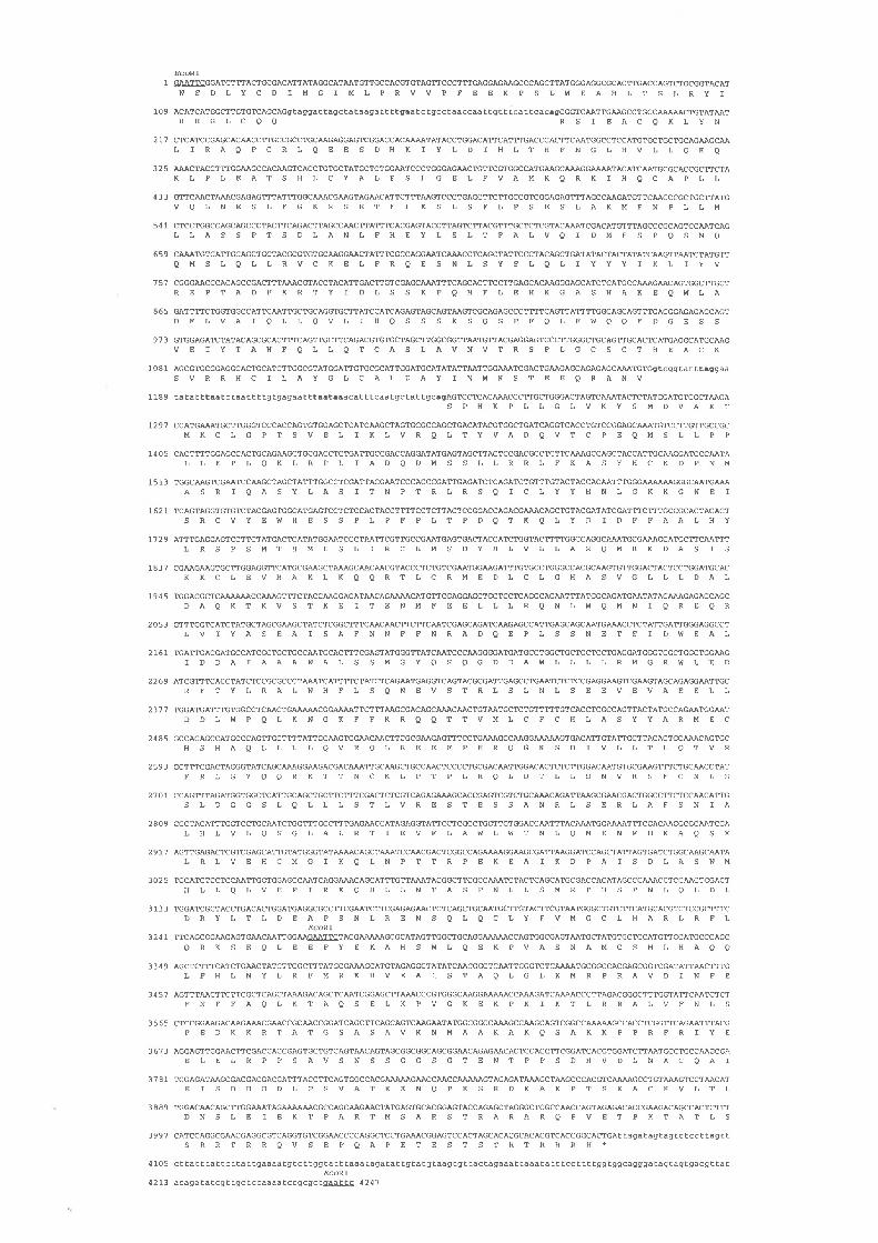
Figure 3.4 Nucleotide sequence and deriyed amino acid sequence of three rows.

The nucleotide sequence of Íhe three rows gene contains a long open reading frame encoding atheoretical protein of l3'79 amino acids. Coding sequence is shown in upper case, and 5', intron and 3'

noncoding sequence is shown in lower case. An in frame termination codon, 30 nt upstream of the

putative intiation codon, is shown in bold. HindIIl, EcoNI and S¿lI restriction sites are underlined.

Intron splice donator and acceptor sites are shown in bold. The derived amino acid sequence is

represented in one letter code below the nucleotide sequence. The 5' termini of the cDNA clones,

CTl42, UJAS and UJE6, are shown with arrowheads (the position of the arrowhead marks the firstnucleotide). The extent of nucleotide sequence derived from subcloned fragments of cosmid 3.3a, and

from CT142 is delineated by arrowheads. Six nucleotide substitution polymorphisms in the genomic

sequence, relative to the cDNA sequence, are shown above the cDNA sequence. The resulting amino

acid substitutions are shown below the cDNA amino acid sequence. A further nucleotide substitution

polymorphism, and consequent amino acid sequence difference, detected in cDNA clone UJA9 isindicated. Nucleotide sequence derived from the dysgenic allele thrBH extends from the arrowhead

marked "BH14 Genomic" to the position indicated as "P element" where the cDNA sequence is

intemrpted by a consensus P element right end (O'Hare and Rubin, 1983) (Figure 3.5). The nucleotide

at which poly A tails commence in cDNA clones UJA8 and UJE6 is arrowed in the 3' untranslated

sequence. Putative polyadenylation signals are shown in bold. A region of amino acid sequence withsimilarity to part of the product of the nuc2 gene of S. pombe (Figure 3.6) is underlined.

Figure 3.5 Alignment of sequence at the junction of P element and the 3' end of tllre thr ORF in thrBil, with corresponding regions in

UJAS and pn25.1.

* From O'Hare and Rubin (1983)

Consensus 8 nt sequenceadjacent to P element*: GGCCAGACil| t

U.JA8 : 3 5 2 5 AÄ.CTATCTTCGCTTTGCGCGzuU\GCATGTAGAGGCTATATCAACGGCTCA.A.TTGGGCCTTAJUU\TGCGATCACGCGCGGTCGAC 3 6 O 8

lllllllllllllllllllllllllllllllllllllllllllllllllllthTBH: CCGACGGGACCACCTTATGTTATTTCATCATGGGCTATATCA.A,CGGCTCAATTGGGCCTTÆVU\TGCGATCACGCGCGGTCG¡E

llllllllllllllllllllllllllllllll 'ea'zr

çm25 .1-* : 287 6 CCGACGGGACCACCTTATGTTATTTCATCATG 2907

t6

Table 3.1 Comparison of percentage amino acid content of three rows with that of

the average in proteins.*

^ -i-^ ^^:n fL--^ -^,,,- t;ff^-^-^^

AG

LS

V

KT

E

D

P

R

IN

aF

Y

C

H

Mw

7.t

2.1

14.8

9.5

4.5

5.1

4.9

6.2

4.3

4.4

6.5

4.1

3.8

6.4

3.6

2.8

2.5

3.0

2.2

og

8.6

8.4

1.4

1.0

6.6

6.6

6.1

6.0

5.5

5.2

4.9

4.5

4.3

3.9

3.6

3.4

2.9

2.0

r.7

1.3

- 1.5

-5.7

+7.4

+2.5

-2.1

-0.9

-1.1

+0.2

-1.2

-0.8

+1.6

-0.4

-0.5

+2,5

0

-0.6

-0.4

+1.0

+0.5

-o4*

according to Dayhoff (1978)

Inspection of the derived thr product revealed consensus motifs implicated in a

number of protein activities or modifications. Some of these are shown in Table 3.2. Given

the basic nature of the encoded product, particularly at the C-terminal end, the occurrence

of 3 potential nuclear targeting sequences is not surprising. In addition to those shown in

Table 3.2,three rows also contains many potential phosphorylation sites for cAMP/cGMP

dependent protein kinases, protein kinase C, calcium/calmodulin dependent protein kinase,

and casein kinases I and II (Kennelly and Krebs, l99l). The biological significance, of

these phosphorylation sites, and the leucine zipper motifs (Table 3.2) are somewhat

77

dubious. There are clearly other determinants to protein kinase activity than the amino

acid residues surrounding a phosphorylation site (Kennelly and Krebs, 1991). Consensus

leucine zippers are known to occur in proteins that are neither transcription factors nor

undergo dimerisation (GCG Prosite program notes), and their fortuitous occurence in a

protein as leucine rich as three rows (Table 3.1) is not unexpected. The putative nuclear

targeting sequence with the the best match to the consensus (Dingwall and Laskey,

1991), at residues lI97-I2Il, is coincident with motifs (Kennelly and Krebs, 1991) for

phosphorylation by cAMP/cGMP dependent protein kinases, protein kinase C, and casein

kinase II (Table 3.1).

Table 3.2 Putative sequence motifs in three rovÍs.

Activitv Consen stt s Position

Nucleartargetingsequence

(R/K)z-X r s- 1 2-(R/K)375

CyclinDependentProteinKinasePhosphorylationsite2

Tyrosine R/K-X2-3-DlE-X3-2-YKinasePhosphorylationsire3

Leucine L-X6-L-X6-L-X6-LZipper4

RKHVEAISTAQLGLKMRSRRRALVFNHSPEDKKR*KKTQPKSREKAKPK

SPHKTPLR

RLQEESDYRLLEDRFTY

S/T-P-X-R/K

tt44-tt62II91 -I2IIt292-1305

431-434898-901

tt9-t26769-777

3O-51 LSGDGLPLRYEVNVLRHICLAL]64-785 LLRMGRLLEDRFTYLRALNHFLI 08 3 - I 1 O4LYFVTGCLHARLRFLQRNSEQL

*Motifs matching the consensusl for Casein Kinase II, cAMP/cGMP dependent protein kinase and Protein

Kinase C are located at residues 1205-1208,1209-1212 and 12O9-1212 repectively.

References: I lDingwatl and Laskey, 1991)2 (Kennelly and Krebs, l99l)3 lcoop"r et al., 1984)4 (Landschulz et al., 1988)

78

The thr encoded product also contains 4 regions (3 at the C-terminal end) which give high

scores with the algorithm for identifying PEST sequences (Rogers et al., 1986)

(Table 3.3). PEST sequences are regions rich in proline, glutamic acid, aspartic acid,

serine and threonine, flanked by basic amino acids, that are present in many unstable

proteins.

Table 3.3 PEST sequences in three ro\ils.

Position Sequence T-FIND scorex

565-579

t246-1269

1312-1333

t358-t374

HESTPLPFPLTPDQK

RPPS ATS CS S SGGS GTENTPPSDH

KVLTLDNSLEIVETPTITTS TR

RQVLEAQAPETESISTR

+9.4

+14.9

+2.7

+2.5

*Calculated as described (Rogers et al., 1986). Scores can range from +45 to -45

Comparisons with sequence databases using the program FASTA (Pearson and

Lipman, 1988) revealed no extended sequence similarity to any known protein. By

contrast, the program BLAST (Altschul et al., 1990), which optimises matches between

short motifs, indicated that a sequence at residues 705-729 of the encoded product

exhibited 44Vo identity (smallest Poisson probability 0.015) with residues 220-244 of the

product of the nuc2 gene of S. pombe (Figure 3.6). nuc2 encodes a 665 aa TPR protein

with an essential role in mitosis (Hirano etal., l9S8) (Section 1.3.2). The possible

significance of this similarity is discussed below.

3.6 Discussion

The thr gene has been identified in a chromosomal walk from the neighbouring grh

gene, by association with a P element insertion event in the dysgenic allele thrBH.

Isolation has been confirmed by P element mediated germ line transformation of an 1l kb

79

genomic fragment, capable of complementing embryonic lethality when present in a

homozygous thr mutant background. Sequence analysis of cDNA and genomic clones

have revealed a large ORF encoding a predicted I,319 aa polypeptide. During the course

of this stldy thr was cloned independently using a different approach (Philp et al., 1993)"

In contrast to the findings of the present study Philp etal. (1993) were able to revert the

thrBH dysgenic allele" The sequence reported encodes a lI72 aa product that can be

resolved with the 1379 aa reported here by allowance for the 5' most intron and a

frameshift near the 3' end of the ORF. The frameshift results from a single extra

nucleotide in the sequence presented here (a C at4133, Figure 3.4), found in a region of

sequencing gel "compression". The sequence in this region has been confirmed by "dITP"

sequencing on both strands (data not shown), and furthermore is consistent with that of a

thr homologue (see Chapter 6). The results of Philp et al. (1993) also reveal the existence

of yet another polyadenylation site 82 nt downstream of the termination codon reported

here.

Analysis of the derived product of the thr gene does not give any clear indication of

its precise function in chromosome disjunction. Three rows contains motifs that suggest it

may be nuclear localised and could be unstable. The coincidence of phosphorylation sites

with a nuclear targeting sequence is provocative, in the light of findings that implicate

protein phosphorylation in modulating the efficiency of targeting (Moll et al., l99l; Rihs

et al., 1991).

Searches of the sequence data bases show the derived protein has no extended

sequence similarity with any known protein. Three rows is thus unlikely to be directly

responsible for chromosome movement, having no consensus NTP binding sites, and

exhibiting no homology to any known motor protein.. One potential clue to the function of

the protein is a very small region, with apparently significant similarity to a sequence

outside the TPR domains, in the píTnucz protein of S. pombe (Figure 3.6). nuc2 is required

for chromosome disjunction although, unlike thr, mutants appear to undergo a true cell

cycle arrest in a metaphase like stage (Hirano et al., 1988). However it is difficult to

conceive that this region of similarity is a product of functional conservation during

evolution, as the motif is not found in the nuc2 homologues in S. cerevisiae or A. nidulans

Figure 3.6 A region of amino acid sequence similarity between the three rolls encoded product and the S. pombe nuc2 product.

Identical residues are boxed and conservative substitutions using the following groupings: A, L, V, I, M; K, R; D, E; S, T; N, Q; y, F, are

circled. The derived nuc2 sequence is from Hirano et al. (1988).

Y,S. penle nuc2 220 SNSS @r wISAF

ÏSAFD. neTanqastæ thr 705 ASEA @m p

ASE PGS 244

QEP SAN 729

D

D

(,

A

FDR

FDR

L

L

E

E

80

(Section 1.3), nor indeed in those recently isolated from humans and D. melanogaster

(Tugendreich et al., 1993; Lamb et al., 1994).

It would therefore appear that three rows is a "pioneer" protein which defines an as

yet uncharacterised activity required for chromosome disjunction. The function of three

rows is further addressed in subsequent chapters.

8l

Chapter 4 (Prometaphase): Analysis of expression

4.1 Background

The thr mutant phenotype is especially noteworthy for its embryonic lethality. As

described in section 1.5.3 the maternal contribution of most components required for

proliferation appears to be sufficient for all the embryonic cycles in D. melanogaster.

Defects in embryonic proliferation with consequent embryonic lethality result from

mutations in genes encoding essential cell cycle functions if the maternal contribution is

insufficient to provide for the entire set of embryonic cell cycles. This is true in particular

for the products of genes encoding regulatory functions such as string and cyclins A and E

(Sections 1.5.3.3 and 1.5.4) since cell cycle progression is dependent on their periodic

synthesis and degradation.

The aim of this work was to characterise the expression of thr. In addition to

determining the number and length of transcripts, it was intended to establish the temporal

pattern of thr transcription in relation to both the cell cycle, and development. These data

would be used to: determine if levels of thr message exhibited cell cycle dependence as

would be expected if it had a regulatory function in mitosis; interpret the cycle 15 defect

observed in homozygous mutant embryos; and ascertain the role of thrin proliferation at

other (non-embryonic) stages of development.

4.2 Northern analysis

Hybridisation of a rår cDNA probe to a developmental Northern blot of total RNA

revealed high levels of thr transcripts in embryos up to 4 h post fertilisation and reduced

levels up to 16 h of embryonic development (Figure4.l). Northern analysis was unable to

detect transcripts late in embryogenesis, during larval development (Figure4.l), or in

pupae or adults (data not shown), but transcripts were clearly present in D. melanogaster

Schneider line 2 cultured cells (Figure 4.1).

Figure 4.1 Detection by Northern analysis of three rows transcripts during

development of D. melanogaster.

A. Northern blot of total RNA isolated from timed collects of embryos and larvae, and

Schneider line 2 tissue culture cells, hybridised with the UJA8 cDNA as probe. Two

hybridising bands corresponding to 4.4 and4.7 kb transcripts were apparent on shorter

autoradiographic exposures, but are obscured in the long exposure shown here to

illustrate the levels of thr mRNA during embryonic and larval development. B. Membrane

stained with methylene blue to illustrate relative levels of RNA loaded. C. Aligned

Ethidium bromide stained track of BRL RNA size markers.

A larvae \ø

ofîrù no .f."^1.$t rê.o ""'"4.7 kb4.4 t

<o9.49 kb7.46

4.40

2.37

1.35

o.24

cB

82

In some exposures it was possible to resolve two species of transcript of

approximate size 4.4 and 4.7 kb (Figure 4.1 and data not shown).

4.3 RNase protection analysis

RNase protection experiments using an antisense RNA probe derived from the

HindIII fragment at nucleotides 240 to 666 (Figure 3.4) confirmed the findings of the

Northern analysis but the increased sensitivity also enabled the detection of thr

transcripts in third instar larvae, adult males and females (Figure 4.2). These results

reinforce the correlation between thr expression and cell proliferation in all tissues.

Transcripts present in adult females presumably include maternal rår ftanscripts

generated during oogenesis (Section 4.5).

Although precise size determinations are impossible without RNA size standards

the mobility of the protected species, relative to a DNA sequence ladder (data not

shown), is consistent with the 310 nt product that would be expected to derive from

sequences 3' to the first of the introns (Figure 3.4). The presence of probe sized product in

some gel tracks (Figure 4.2) is indicative of incomplete RNase digestion.

4.4 Primer extension analysis

A primer complementary to the thr ORF at nucleotides 415 to 494 (Figure 3.4) was

employed to elucidate the position of transcription initiation. This revealed the presence of

products with 5'termini atpositions (relative to numbering in Figure3.4) of: 380+l ,96+2

(if genuine both predicted to terminate within the ORF), -58+5 (predominantly in early

embryos) and -190+30 (Figure 4.3). This result was reproducible and observed when the

extension reaction was performed in the presence of Actinomycin D, to reduce secondary

structure formation, and hence inhibit the production of anomolous extension products due

to polymerase pausing. The position of the priming site chosen, with respect to the 5' end

of the gene, was based on incomplete sequence information obtained early in this study.

In retrospect it would have been desirable to have utilised an oligonucleotide that primed

nearer the initiation codon, thus enhancing the yield of full length extension products.

83

The molecular basis for the variant lengths of cDNA extension products has not

been determined. They may derive from transcripts that arise from initiation at different

sites, or from the processing of a single primary transcript, such as by intron excision"

Two of the major products detected, if specific, are predicted on the basis of sequence data

(Figure 3.4) to terminate within coding sequence. However it is possible that these

products are not thr specific, as their abundance in 8 to 24 h embryos is inconsistent with

levels of thr transcript detected by other methods of expression analysis employed here.

Conversely, they may be authentic and could, for example, arise from the excision of

additional introns, not detected in the limited range of cDNA clones decribed in Chapter 3.

The second longest primer extension product is estimated to terminate as little as 10

nucleotides upstream of the 5' end of the cDNA clone CTl42 (Figure 3.4). In an

independent study (Philp et al., 1993) a thr transcript has been identified, by RACE PCR,

starting at -722+5 (their numbering) which appears to correspond to the longest primer

extension product identified here. Inspection of upstream sequences (this study and Philp

et al., 1993) has failed to detect "TATA" motifs, approximately 30 nt upstream of any of

the identified 5' termini.

The two longest 5'UTR lengths detected, in combination with the alternate 3'

lengths identified in the cDNA clones (Figure 3.4) can notionally give rise to transcripts

between 4.3 and 4.7 kb in length (excluding the poly(A) tail), which is consistent with

those sizes estimated from the Northern (Figure4.l).

4.5 Whole mount in situ hybridisation to mRNA

To characterise the spatial and temporal distribution of rizr mRNA during embryonic

development, digoxigenin labelled cDNA, derived from clone UJA8, was hybridised in situ

to fixed embryos (Figure 4.4). Throughout the syncytial divisions, prior to cellularisation,

embryos exhibited high levels of thr transcripts (Figure 4.4A). In these embryos, the thr

transcripts appeared to be concentrated around the nuclei. A similar distribution has been

observed for transcripts of cyclin B (Lehner and O'Farrell, 1990b) and shown to be

dependent on microtubule integrity (Raff et al., 1990) and sequences in the 3' UTR (Dalby

and Glover, 1992) (Section 1.5.4).

Figure 4.2 Detection by RNase protection analysis of îhree rorys transcripts during

development of D. melanogaster.

Product protected from digestion by RNase when a 421 nt RNA probe is hybridised to

total RNA isolated from life cycle stages and Schneider line 2 tissue culture cells, with

yeast RNA as a control. The results of two independent experiments are shown (A and

B), with a longer autoradiographic exposure (B) also revealing message in third instar

larvae and adult males. The position of the undegraded RNA probe and the protected

species is indicated.

sîq""'"*¡:"$d"î"";Ç;i:"Ëprobe >

protected >

probe ¡

protected >

A

B

Figure 4.3 Primer extension analysis of three rows transcripts during development of

D. melanogaster.

Resolution of oligonucleotide primed cDNA extension products from total RNA isolated

from: 1 . O-zh, 2.2-4h, 3. 4-8h, 4.8-12h, 5. 12-16, 6. l6-20h, 7.20-24h embryos,

8. lst, 9.2nd, 10. 3rd instar larvae, 11. early, 12.late pupae, 13. adult females,

14. males, 15. Schneider line 2 cells, 16. yeast. The results of two independent

experiments are shown (A and B). The mobilities of the extension products relative to

the sequence ladder and Figure 3.4 are indicated.

1. s. 8.9.10.16. 1. 2. 3. 4. 5. 6. 7. 8. 9.1011.13.15-. 12- 14. 16

-1 90+30

-58+5 --

(e612)

þ80r1)-

BA

Figure 4.4 Detection of three rows transcripts during development of

D. melanogaster by in situ hybridisation of digoxigenin labelled probes to wild type

and three rows defÏcient embryo whole mounts.

V/hole mount embryos were hybridised with the UJA8 cDNA labelled by incorporation of

digoxigenin-11-dUTP. Hybridisation was detected using an alkaline phosphatase

coupled secondary antibody detection system. All embryos are orientated with anterior to

the left and ventral side down unless otherwise indicated.

A. Syncytial stage embryo exhibiting perinuclear distribution of transcript.

B. An embryo at cellular blastoderm with relatively uniform message distribution.

C. Germ band extended embryo with uniform staining in all but the amnioserosa.

D. Germ band extended embryo with reduced staining in all but cells of the CNS.

E. Embryo in early stage of germ band retraction showing expression in cells of the CNS

and PNS.

F. Ventral view of an almost complete germ band retracted embryo showing transcripts

in segmentally reiterated cells of the CNS and PNS.

G. Fully germ band retracted embryo with staining in ventral nerve cord and brain lobe

(cNS).

H. Dorsal view of fully germ band retracted embryo with transcripts confined to cells in

the proliferating margins of the brain lobes.

I, J, K: Progeny of DfQR)PC4/CyO parents. Df(2R)PC4 removes thr in its entirety. One

quarter of the embryos at cellularisation and later stages were lackinE thr transcripts.

I. An embryo during cellularisation showing lack of transcripts in all cells except pole

(presumptive germ) cells.

J. Cellular blastoderm at higher magnification showing punctate staining in pole cells.

K. Early germ band extended embryo with transcripts restricted to the pole cells.

Dc

FE

H

I JK

84

Following cellularisation, levels of thr transcripts were seen to remain high and

evenly distributed, except for a higher concentration in the region of the pole cells

(Figure 4.48). During germ band extension, transcript levels were high except in the

amnioserosa (Figure 4.4C), but declined during cycles l5 and 16 (Figure4.4D) to be

undetectable in most tissues prior to germ band retraction. Significantly, there was no

indication of any stage-specific regulation of thr ûanscúpt levels during cycle 15. At the

germ band extended stage and the early stages of retraction, thr expression was clearly

observed in cells of the CNS and PNS (Figure4.4E, F). Following germ band retraction,

thr transcripts were confined to a subset of cells within the ventral nerve cord and in the

proliferative centres of the brain lobes, (Figure 4.4G, H). These are the only proliferating

tissues at this stage of development. Expression was not detected in cells undergoing

endo-replication cycles, cycles of DNA replication that proceed in the absence of mitosis

to generate polytene chromosomes (Smith and Orr-Weaver, 1991).

Levels of thr message appear to be relatively stable during embryogenesis and do

not fluctuate in an obvious cell cycle dependent manner. In the progressive snapshots of

development represented in collections of fixed embryos, the pattern of cells in which r/¿r

message was detected did not undergo marked changes. This is in contrast to the

temporally and spatially dynamic patterns of expression observed for established cell

cycle regulators such as string (Edgar and O'Farrell, 1989) and cyclin E (Richardson et al.,

1993).

To follow the fate of the maternal contribution during embryonic development,

embryos derived from parents heterozygous for the deficiency Df(2L)PC4, which removes

thr (Figure 1.5), were examined by in situ hybridisation. During the syncytial divisions,

all embryos exhibited uniformly high levels of transcript, as observed in the progeny of

wild-type parents. I)uring cellularisation and early germ bancl extension, a majority of the

embryos again resembled wild-type. At these stages and later in development, however,

approximately one quarter of the progeny exhibited dramatically lower levels of thr

transcripts in all cells, except the pole cells (Figure 4.4I-K). These embryos presumably

correspond to the homozygous deficiency embryos. Double staining with the DNA stain

Hoechst 33258 showed that, as expected, the unstained fully germ band extended

85

embryos exhibited the mitotic defect characteristic of homozygous thr embryos (data not

shown).

These findings indicate that maternal thr mRNA is rapidly degraded at the time of

cellularisation and that thr transcripts observed late in cycle 14 and in subsequent cycles

are the product of zygotic transcription. The transition from maternal transcripts to zygotic

transcripts during cycle 14 is not, however, marked by a discernible stage in which ¡hr

transcripts are absent"

At higher magnification, the distribution of thr mRNA in pole cells was observed to

be punctate and perinuclear (Figure 4.4J and results not shown). Such a subcellular

distribution appears identical to that observed in pole cells for cyclin B mRNA (Lehner and

O'Farrell, 1990b), which resides in cytoplasmic granules. The irregular, granular

distribution of thr mRNA within germ line cells is also observed in somatic tissues (see

staining in cells of PNS, Figure 4.48), and contrasts with the uniform cytoplasmic staining

observed for many other types of transcripts (for example see Richardson et al., 1993).

4.6 Discussion

The evidence presented here argues strongly that the early requirement for zygotic

expression of thr, as revealed by the mutant phenotype, results from the instability of the

maternal mRNA during cycle 14. The maternal thr mRNA appears to be destabilised upon

cessation of the syncytial divisions. An apparently constant pool of maternal r/rr mRNA

was found in all syncytial embryos. In embryos homozygous for a thr deficiency, this pool

was observed to rapidly decrease following cessation of the syncytial divisions, so that at

the time of cellularisation tår mRNA levels were reduced to undetectable levels in all cells

except pole (germ line) cells. In normal embryos, however, the levels of thr mRNA were

maintained at a much higher level during these stages, showing that zygotic transcription

was supplementing the rapidly degrading maternal mRNA.

The disappearance of maternal thr transcripts at this time is consistent with the

temporal onset of the mutant phenotype. Although it is possible that trace amounts of

mRNA may persist after the precipitous decline in levels at the commencement of cycle

14, it is more likely that limited function in mutants is conferred by the persistence of

86

protein. In embryos homozygous for amorphic alleles, sufficient thr product is presumably

translated from the maternal lftr mRNA prior to its degradation for a normal l4th mitosis

to occur, and perhaps for a rudimentary mitosis 15. Such a persistence of the maternally

derived thr protein is also suggested by the observation that the maternal thr genotype

affects the time at which the mutant phenotype becomes evident (D'Andrea et al., 1993).

The dependence of the zygotic phenotype on the maternal genotype, the absence of

any stage-specific regulation of thr transcripts during cycles 14 and 15, and the correlation

between expression and proliferation in embryonic and postembryonic development, all

argue against the scenario that the thr embryonic phenotype results from a

developmentally restricted requirement for thr function. Rather, these results are

consistent with the idea that thr function is provided maternally and that in homozygous

mutant embryos the maternal contribution becomes insufficient prior to mitosis 15. At this

stage the mutant phenotype manifests itself because of a failure to produce functional

zygotie rår products.

The apparent stability of the maternal rår mRNA during the syncytial divisions,

compared with the instability during cellularisation, suggests that upon completion of the

syncytial divisions, RNA degradative pathways, specific for certain transcripts, are

activated. The change in stability is dramatically evident for maternal string transcripts

(Edgar and O'Farrell, 1989) and for maternal D. melanogaster cyclin E transcripts

(Richardson et al., 1993). The basis for the decreased stability of maternal transcripts,

following the syncytial divisions, is yet to be determined for any of these genes.

If maternally conferred message is being actively degraded concomitant with

zygotically initiated transcription, then some mechanism should exist to differentiate

between transcripts of maternal and zygotic origin. Perhaps recognition sequences or

structures are present in the mRNA 3' and 5'UTRs where polymorphisms have been

detected. The maternal form may correspond to the -58+5 transcript, identified by primer

extension analysis, present in abundance only in early embryos (Figure 4.3).

Experiments designed to further elucidate the function of the thr product in mitosis

are described in the next chapter.

87

Chapter 5 (Metaphase): Immunodetection

5.1 Background

Evidence presented in Chapter 3 established that three rows encodes a "pioneer"

protein essential for mitotic chromosome disjunction. However its precise role in this

process is unknown. With few clues revealed by the sequence encoded by the ORF, or

the pattern of gene expression, other stategies were required to elucidate its precise

function. A powerful technique in this regard is the utilisation of antibodies specifically

directed to the encoded product.

The aim of this section of work was firstly to obtain substantial quantities of three

rows derived protein by bacterial expression. This protein would be employed to generate

specific antibodies by immunisation and the three rows directed antibodies purified from

the immune sera. Application of antibodies to whole mounts of embryos should reveal the

subcellular location of the antigen. In particular, localisation of three rows to a particular

element of the mitotic apparatus would enable informed speculation as to function, whether

it be signalling, structural or mechanical.

5.2 Antibody production

Two bacterial expression systems were exploited to raise antibodies specific to

three rows (Figure5.1). The C-terminal 1014 aaof the derived product (Figure5.1) were

expressed as a 142 kDa glutathione-S-transferase (GST) fusion protein (Figure 5.21^)

using the pGEX vectors (Smith and Johnson, 1988). A nonchimaeric JJ kDa protein

(Figure 5.2A) consisting of 670 aa of the thr putative product (Figure 5.1) was expressed

with the T7 transcription/translation system (Studier and Moffatt, 1986).

Although the pGEX system permits the purification of the fusion protein based on

the affinity of GST for glutathione agarose, this could not be applied for the

GST-three rows fusion. Following induction of expression and bacterial lysis the fusion

protein was recovered almost exclusively in the pellet. The small amount of soluble

Figure 5.1 Bacterial expression constructs employed to produce three rows derived proteins, in relation to t}¡e three roys ORF.

BgAl

1 149

Ndel

1914

pGEX2 BgllllNofl 3.5

pRK171 NdellBsfYl 2.0

Bsffl

3923 4253

three rows

Figure 5.2 Three rows derived bacterial expression products and detection of

immune response in rabbits inoculated with them.

A. Coomassie stained SDS-PAGE of three rows derived bacterial expression products.

Mobility of protein molecular weight markers is arrowed. B" Detection, by western blot

analysis, of immune response in rabbits inoculated with recombinant protein

Nitrocellulose strips consist of two separate gels. Rabbits: 1-3 inoculated with pGEX2

BgIIUNotI3.5, 4-6 with pRKlTl NdeUBstYI2.O. Position of bacterial expression

products is arrowed. All rabbits produce immune sera that reacts with both bacterial

expression products.

203 kDa >

1 05>

70.8>

43.6>

{pGEX2 BgllV Notl3.S

pRKl71 Ndell BstYl2.O

<pRKl71 Ndell BstYl 2.0

<pGEX2 BgllllÂlof I 3.5

1.2.3.4.5.6

B

88

protein was capable of binding to glutathione agarose, but could not be eluted.

Consequently, for antibody production, the recombinant protein was resolved on an

acrylamide gel and a homogenised gel slice containing the protein was used for

immunisation. The same technique was employed for the T7 expressed protein.

The rabbits immunised with the bacterially expressed, three rows derived proteins

exhibited an immune response as judged by the reaction of their sera to the recombinant

proteins on western blots (Figure 5.28). However the failure of the crude immune sera to

reproducibly detect a D. melanogaster product on westerns or in embryo whole mounts

made it necessary to purify the three rows specific antibodies.

5.3 Antibody purifTcation

Three rows specific antibodies generated in rabbits inoculated with recombinant

protein were purified on the basis of their affinity for the T7 expressed protein. This was

also feasible for immune sera against the GST fusion protein because of the considerable

stretch of sequence shared between the two bacterially expressed proteins (Figure 5.1).

Such an approach also had the desirable outcome of excluding antibodies directed against

the GST portion of the fusion protein. The T7 expressed protein was enriched by

resolution on SDS-PAGE, electroeluted from an excised gel slice (Figure 5.34) and

covalently coupled to Affigel 10 to form the matrix of an affinity column.

Antibodies with both low and high affinity for the T7 expressed protein were

purified with the affinity column as described (Kellogg and Alberts,1992). Crude immune

serum was passed over the column and, following extensive washing, antibodies that

recognise the T7 protein with low affinity were eluted with 50mM HEPES pH 7.6,

1.4 M MgCl2, 10Vo glycerol. High affinity antibodies retained on the column were eluted

with O.5Vo acetic acid, 0. l5 M NaCl, and neutralised. Peak fractions were detected by their

reactivity to recombinant protein on western blots (Figure 5.38), pooled and concentrated.

These high affinity antibodies are particularly suitable for western blotting and

immunostaining (Kellogg and Alberts, 1992).

89

5.4 Western analysis

V/hilst the antibodies purified with the affinity column from various immune sera

clearly reacted to both bacterially expressed three rows derived proteins (Figure 5.38), it

was imperative to demonstrate that they recognised the in vivo product. This was

accomplished by the detection in homozygous thrBH embryos of a truncated product

relative to that in wild-type (Figure 5.4). The molecular weight estimated from the

mobility relative to protein standards was 150 kDa for the wild-type protein and 125 kDa

for the mutant, which is in reasonably good agreement with 157 and approximately

l32kDa respectively predicted from the nucleotide sequence data (Chapter 3).

Homozygous mutant embryos were selected based on their exhibition of the mitotic defect,

which of necessity, was late in embryogenesis. The relatively low signal intensity relative

to background is consistent with the low levels of protein detected in immunostainings of

late embryo whole mounts (Section 5.5). Despite variation of conditions ECL detection

gave unsatisfactorily high levels of background" However much of this appears to be a

nonspecific product of the western blot, as it is also detected in the protein size marker gel

track (Figure 5.4)"

5.5 Immunolocalisation

Application of immunoaffinity purified antibodies to formaldehyde fixed embryos,

and detection with HRP in combination with Hoechst 33258 staining of DNA, revealed a

cell cycle dependent pattern of antigen localisation (Figures 5.5, 5.6). This was

particularly marked in syncytial embryos where the metachronous nature of the cortical

divisions made it possible to observe, in a single embryo, the transition from one mitotic

stage to another. Occasionally embryos arose where the mitotic wave propagated from

only one pole and, as a consequence, it was sometimes possible to observe the progress

of almost an entire mitotic cycle across the long axis of the embryo (Figure 5.5 A, B).

The antigen is not detected in interphase (Figure 5.5 A, C, F and results not

shown). However as chromosome condensation becomes apparent in prophase, the

nucleus becomes progressively and uniformly stained except for some concentration

coincident with chromatin (see arrows in inset, Figure 5.5C). As the nuclear envelope

tr'igure 5.3 Purification of pRK171 NdeUBstYI 2.0 bacterially expressed protein, and

utilisation for purification of three rows specific antibodies by immunoaffinity

chromatography.

A. Coomassie stained SDS-PAGE of pRKlTl NdeIlBstYI 2.0 bacterial protein prior to,

and following purification by electroelution. B. Detection, by westem blot analysis, of

three rows specific antibodies, from rabbit immune serum 3, eluted from a pRK171

NdeIlBstXI 2.0 bacterial protein column. Nitrocellulose strips consist of two separate

gels.

Unpurified Gel

Low affinity eluate:1. 2. 3.4. 5. 6. 7. I

High affinity eluate:1. 2. S. 4. 6. 8. 10.12.14.

{ pRK171 Ndell BstYl2.O

{ pGEX2 BgllV lVof 13.5

B

Figure 5.4 Detection of three rows in wild type and thrBH embryos by western blot

analysis.

l. Protein molecular weight markers

2. ¡l.1yBH/BH.

J. ¡þ¡.BH/+.

4. thr+/+.

1 2. 3. 4

203 kDa Þ

1 05>70.8>

I lengthtruncated

Figure 5.5 Immunostaining of syncytial blastoderm embryo whole mounts with anti-

three rows antibodies.

Antigen was detected with HRP and the DNA counterstained with Hoechst 33258.

A. Whole embryo exhibiting, from left to right, nuclei in prophase through to interphase.

B. Whole embryo, metaphase to telophase.

C. Interphase-metaphase, inset: early prophase nucleus showing staining associated

with condensing chromatin (arrows).

D. Prophase-anaphase.

E. Metaphase-anaphase.

F. Telophase-interphase.

G. Whole embryo showing staining in pole cells (bottom right).

G'. Internal focal view of same embryo as in "G." showing vitellophage nuclei staining.

BA

DC

Ë

G

F

G'

Figure 5.6 Immunostaining of cellularised embryo whole mounts with anti-three

rows antibodies.

Antigen was detected with HRP and in some cases the DNA visualised by

counterstaining with Hoechst 33258. All are dorsal views of germ band extended

embryos unless otherwise stated.

A. Staining of presumptive subepidermal neuroblasts in the neurogenic region.

B. Lateral view showing germ band staining and persistent staining of irregularly

dispersed presumptive vitellophage nuclei.

C. Anterior end with fluorescent visualisation of DNA, showing staining in presumptive

neuroblasts (arrows) at the margins of the brain lobes.

D. Lateral view of germ band retracted embryo showing general lack of staining, except

for pole cells associated with the posterior midgut.

E. High magnification fluorescent visualisation of epidermal cells showing staining in

cells in early anaphase (eA), anaphase (A), and telophase (T).

E'. Visible light view of same embryo as in "E".

F. Fluorescent visualisation of DNA in homozygous Dfl,L)Pcl7B (left) and

phenotypically wild type (right) embryos.

F'. Higher magnification visible light views of same embryos as in "F" showing absence

of staining in the deficiency embryo.

t

Ë

BA

a-^|'I

DC

T

EE

F F'

90

undergoes partial breakdown and chromosomes congress in prometaphase the nuclear

staining is lost, until it is essentially undetectable at metaphase (Figure 5.5 B, C, D, E).

Coincident with the first manifestation of chromosome movement in anaphase, the antigen

again becomes detectable on the chromosomes, first appearing at the regions where

polewards movement commences (Figure 5.5 D, E). These are presumably regions

around the kinetochore. As anaphase proceeds, the staining localises to the entire

chromosome and becomes progressively stronger (Figure 5.5 D, E), appearing to reach

maximum intensity as the nuclear envelope reforms around the chromosomes in telophase

(Figure 5.5F). The staining then declines as the nuclei take on the enlarged appearance

characteristic of S phase (Figure 5.5F).

Intense staining of yolk nuclei is also observed in many syncytial embryos

(Figure 5.5G'). Staining of vitellophage nuclei persists in development and is sometimes

detected in gastrulating embryos (Figure 5.68).

Consistent with the elevated levels of mRNA detected in them (Section 4.5), pole

cells are often strongly immunostained, relative to the neighbouring syncytium/cells

(Figure 5.5G). This staining is persistent, and can be detected late in embryogenesis

when the pole cells are internalised and associated with the posterior midgut

(Figure 5.6D).

The pattern of antigen localisation in embryos post cycle 13, although more difficult

to observe because of lower signal intensity and smaller nuclei, appears consistent with

that in syncytial embryos. Early in germ band extension, the nuclei of cells stain in a

manner that reflects, at least in part, the pattern of mitoses (Figure 5.6 A, ts). ln

particular, a characteristic arrangement of three subepidermal rows of large cells on either

side of the ventral midline (Figure 5.64) is consistent with the pattern of CNS neuroblast

distribution at this stage. Because of the internalisation of these cells it is difficult to

correlate this staining with a particular mitotic stage.

In epidermal cells the antigen exhibits a cell cycle dependent pattern of

detectability, similar to the precellular divisions, being most evident in anaphase and

telophase when associated with the chromosomes (Figure 5.6 E, E'). However the

prophase accumulation, observed in syncytial embryos, is not marked during cellular

91

divisions. Staining declines in level as embryogenesis proceeds until undetectable, in all

tissues of germ band retracted embryos, except isolated cells in the proliferating margins

of the brain lobes (Figure 5.6D).

The specificity of immunostaining was demonstrated by the the loss of antigen in

homozygow Df(2L)Pcl7B embryos exhibiting the thr phenotvpe late in embryogenesis

(Figure 5.6 F, F').

Immunofluorescent staining of embryos confirmed the cell cycle dependent pattern

of antigen localisation observed for HRP detection (Figure 5.7). Confocal and

epifluorescent imaging of syncytial embryos revealed staining of nuclei in prophase,

absence in metaphase, and mobilisation to chromosomes in anaphase and

telophase(Figure 5.7 A, B, B', C). DNA associated staining of chromosomes during

anaphase and telophase was also observed in cellularised embryos (Figure 5.7 E, E').

Interpretation of the immunolocalisation pattern was hampered by the variable

degree to which centrosomes are also stained, particularly in methanol fixed embryos, in

what appears to be a nonspecific fashion (see frontpiece). In homozygous Df(2L)Pcl7B

embryos, in which nuclear/chromosomal staining is abolished in cycles 15 and 16,

centrosomal staining can be detected fluorescently (Figure 5.J D, D' and results not

shown). The centrosome is a structure particularly prone to artefactual immunostaining

(J. Raff, pers. comm.). This apparently artefactual staining makes it impossible to rule out

a bona fide association of three rows with the centrosome. As a negative control,

embryos were incubated with the IgG fraction isolated from preimmune sera. Even when

preimmune IgGs were applied at 500 times the concentration of the affinity purified

antibodies, no staining was detected (data not shown).

5.6 Discussion

The considerable effort expended to obtain specific antibodies capable of in situ

detection is evidence that three rows may not be a particularly immunogenic protein.

Satisfactory preparations of affinity purified polyclonal antibodies were not obtained from

the sera of subcutaneously immunised mice and rats, despite clear ellicitation of an

immune response. Mice were also inoculated intraperitoneally with approximately I mg of

Figure 5.7 Immunofluorescent staining of embryo whole mounts with anti-three

rows antibodies.

Embryos have been double stained with a Texas-Red conjugated secondary antibody to

recognise three rows (red), and chromomycin A3 for DNA (green), and visualised by

epifluorescent (8, B', D, D', E, and E') or confocal (A and C) microscopy. All are wild

type syncytial embryos unless otherwise stated.

A. Double imaging of DNA and three rows in embryo in stages, from left to right, of

prophase through to anaphase.

B. Visualisation of DNA in nuclei in anaphase-telophase.

B'. Same embryo as in "B" showing visualisation of three rows.

C" Double imaging of DNA and three rows in nuclei in prophase-metaphase.

D" Visualisation of DNA in cells of homozygous germ band extended Df(2L)Pcl7B

embryo.

D'. Same embryo as in "D" showing persistent nonspecific staining by anti-three rows

antibodies of presumptive centrosomes.

E. Visualisation of DNA in epidermal cells of germ band extended embryo.

E'. Same embryo as in "D" showing DNA associated three rows staining in anaphase

and telophase (bottom right).

A

B'B

D'DC

E E

92

recombinant protein, along with Freund's complete adjuvant, for the purpose of obtaining

monoclonal antibodies. Of 35 hybridoma cell lines isolated from mice immunised with the

T7 expressed protein (J. Wrin, pers. comm.), none had more than a weak affinity for

recombinant protein on westerns (data not shown). It is possible that the isolation of

efficacious antibodies from rabbits is a product of the large volumes of sera obtained,

implying that specific high affinity antibodies may represent a very small proportion of total

circulating levels. It is also possible that because of masking by other proteins or

posttranslational modifications in vivo, or because of major differences in conformation

between the the bacterially expressed/SDS-PAGE resolved, and the in vivo expressed

forms, that antibodies to very few native epitopes were present in the immune sera raised.

The dynamic pattern of three rows localisation, whilst unique, is similar to some

other antigens associated with the mitotic spindle. Several antigens in D. melanogaster

also localise to the chromosomes in anaphase. The zwl0 protein, which is excluded from

the nucleus in telophase and interphase, accumulates within the spindle envelope in

prometaphase, appears spindle associated in metaphase, and rapidly localises to a region

at or near the kinetochores in anaphase (Williams et al., 1992). Prior to cycle 10 in

syncytial embryos proliferating cell nuclear antigen (PCNA) stains chromosomes in

anaphase, but not metaphase (Yamaguchi et al., l99l). In subsequent cycles PCNA

becomes associated with chromosomes/nuclei at progressively later stages of the cell

cycle, which is consistent with its function in DNA replication.

There exists in D. melanogaster a protein with a cell cycle dependent pattern of

localisation reminiscent to that of three rows. An antigen recognised by "Aj 1", one of a

collection of monoclonal antibodies directed against nuclear proteins (Frasch et al., 1986)

(Section 1.5.5), occupies the nucleus of syncytial embryos in prophase then disperses to

surround the spindle in metaphase whilst being excluded from the chromosomes. In

anaphase it specifically associates with the chromosomes, remains present in telophase,

and appears also to persist throughout interphase. Following cellularisation the pattern of

Aj I staining in mitosis is similar to that in syncytial embryos except that in interphase the

antigen localises not to the nucleus, but the nucleolus. In spite of the similarities to three

rows, Ajl clearly recognises a distinct protein of 47 kDa on westerns (Frasch et al.,

93

1986). There are other differences which may be significant, such as the continuing

detection of the antigen around the spindle in metaphase, and its persistence during

interphase, particularly its nucleolar localisation following cellularisation. In the latter

case, it may be pertinent to note that the organelles which stain with Aj 1 (the nucleolus),

and (nonspecifically) with anti-three rows antibodies (the centrosome) are both singular,

punctate and nuclear membrane associated during interphase.

Are the abrupt transitions in the detection of three rows in syncytial embryos the

product of cycles of protein synthesis and degradation, or the result of redistribution of a

relatively constant pool of protein? The velocity of the "mitotic wave" during the

metachronous syncytial divisions makes it unlikely fluctuations in antigen detection are

caused by proteolysis and de novo translation. It has been estimated that it takes fewer

than 30 seconds for the wave to propagate from the pole to the equator, although intervals

of up to 2.5 min have been observed (Foe and Alberts, 1983). As staining can be

observed in nuclei that are only one intervening nucleus away from nuclei with no staining

(Figure 5.5 C, D, E), spanning less than lOVo of the length of an embryo, it would appear

that the translocations of protein in both prometaphase and anaphase may take less than

5 seconds and probably no more than 30 seconds. Such intervals are difficult to reconcile

with protein degradation and synthesis. Periods of 10-12 s have been estimated for the

D. melanogaster zwt0 protein, which has a similarly rapid pattern of localisation to the

chromosomes in anaphase (Williams et al., 1992)"

If protein is being transported from the nucleus in prometaphase, why is it not

detected in the cytoplasm in metaphase? It is conceivable that protein dispersed into the

cytoplasm may be below the threshold of immunodetection, although the apparent levels in

prophase and anaphase make this unlikely. Alternatively, the antigen may be

immunologically masked in metaphase, as a consequence of association with other

proteins in a complex, due to posttranslational modification, or for some other reason.

Although the pattern of immunolocalisation is not particularly redolent of a

microtubule associated protein (MAP), experiments were performed to ascertain if three

rows had an affinity for microtubules. Western analysis of proteins isolated by

cosedimentation with taxol stabilised microtubules (Kellogg et al., 1989) demonstrated a

94

clear enrichment for DMAP 190 (antibody supplied by J. Raff and D. Kellogg), but failed to

detect three rows (data not shown).

The distinctive pattern of three rows localisation revealed by immunostaining is

consistent with some of the previously observed (Section 3.5) characteristics of the

derived product. The apparent association with chromosomes is consistent with the basic

nature of the protein. It is possible three rows interacts directly with DNA as do other

basic proteins like histones, although it is also feasible the association occurs via

interactions with other proteins. Although probably not degraded in prometaphase, the

identified PEST sequences may target the protein for degradation in interphase. The

accumulation of protein in the nucleus during prophase correlates with the presence of

nuclear targeting sequences. Furthermore, the cell cycle dependent fluctuations in nuclear

localisation may be a product of phosphorylation changes at some of the protein kinase

recognition sites that are coincident with, and possibly modulate the activity of, the best

candidate nuclear targeting sequence (Table 3.2).

The pattern of three rows immunolocalisation is also consistent with the three rows

mutant phenotype. Essentially, chromosome disjunction may be defective in mutants

because the protein fails to migrate to the chromosomes at the initiation of anaphase, or

because the protein migrates but is nonfunctional. Translocation of the protein could be a

consequence of MPF activation or other associated changes at the metaphase-anaphase

transition which would explain why chromosome disjunction is defective in mutants whilst

cell cycle progression is unimpeded.

With the lack of sequence similarity of the derived product offering no hint, the

pattern of immunolocalisation is presently the best indicator of three rows function. Even

though the mutant phenotype implies three rows is only essential for anaphase

chromosome movement any suggestions for function must be able to account for its

detection, in syncytial embryos, in prophase as well as anaphase. One explanation is that

three rows does finction in prophase but that, either, phenotypic manifestation in mutants

has not been elucidated by our superficial examination, or the consequences of the

prophase defect do not become apparent until chromosomes have congressed. Another

possibility is that three rows is not required early in mitosis, but that its accumulation in

95

the nucleus in prophase is a means of sequestering sufficient quantities of protein in the

vicinity of the presumptive spindle, particularly during the extremely rapid syncytial

divisions, to mediate its essential function late in mitosis. Accordingly, the protein

detected in prophase may not be functional, requiring some anaphase specific form of post-

translational modification to become activated or localised.

If three rows is required both early and late in mitosis there are at least two

possible functions. One is suggested by the antigen's association with chromosomes in

prophase, and anaphase, stages marked by states of intermediate chromosome

condensation. Chromosome condensation, however, appears to be unaffected in mutants

(D'Andrea et al., 1993) and there are no precedents for defective chromosome

condensation inhibiting disjunction. Detection of the antigen in both prophase and

anaphase is also evidence for a general requirement in chromosome movement" However

the mutant phenotype argues against a role in metaphase alignment, and immunostaining

appears to show that the antigen is dispersed in prometaphase before chromosomes have

fully congressed (Figures 5.5 C, D, 5.7C).

If three rows function is confined to anaphase then it could function either in a

signalling, or structural capacity to engender chromosome movement. Three rows could be

a kind of "licensing factor" whose rapid translocation to the chromosomes at the initiation

of anaphase in some way signals them to move polewards" Alternatively, it could have a

more direct role in chromosome movement by altering the relatively static interaction

between the kinetochore and the spindle MTs that exists at metaphase. Otherwise it may

be responsible for dissolution of the bonds that join sister chromatids until the

commencement of disjunction. All of these roles may only require redistribution of protein

to the kinetochore/centromere region. Accordingly, the mass decoration of the

chromosomes in syncytial anaphases could be a product of the high levels of protein at this

developmental stage, or to ensure the equitable distribution of the protein to the daughter

nuclei.

The role of three rows in mitosis later in development, implied by the data on gene

expression (Section 4.3), remains to be immunologically investigated. Results from a

preliminary immunostaining of third instar larval brain lobes reveal localisation to giant

96

neuroblast cells (data not shown) which are known to be proliferating at this stage. It will

be of interest to extend this investigation to look for the presence of three rows in imaginal

and gametogenic (both mitotic and meiotic) proliferation.

Further insights into three rows function by the characterisation of a diverged

homologue are described in the next chapter.

97

Chapter 6 (Anaphase): Isolation of a homologue from D. erecta

6.1 Background

In circumstances where a gene has been shown to encode a "pioneer" protein of

undetermined function, the isolation and characterisation of the homologous gene from an

appropriately diverged species has the potential to yield functional insights. The rationale

is that domains that have conserved their sequence during the course of evolution do so

because of functional constraints on that sequence. Accordingly, in a reductionist

approach, these conserved regions would be targeted for detailed investigation of function.

For example, the cyclins exhibit considerable evolutionary divergence in their amino acid

sequence such that from yeast to man only a single conserved region can be detected"

This domain, the "cyclin box" is absolutely required, being responsible for the cyclin's

interaction with, and activation of, the CDK catalytic subunit.

In a manner that approximates their degree of sequence conservation, homologues

of D. melanogaster genes are usually isolated from one of three different "sister" groups.

At the extreme end of the conservation scale are genes whose homologues have been

isolated from yeast or man. The histones are an example of genes which have been

conserved (Wells and McBride, 1989) across the estimated l-2 billion years of eukaryote

evolution. The homologues of intermediately conserved D. melanogaster genes are often

detected in other arthropods, like crustaceans, or in representative species of other insect

orders such as Tribolium, Manduca or Schistocerca. Genes that have been conserved

during this approximate 500 my period of divergence include the pair-rule gene

fushi-tarazu (for example see Dawes et al., 1994). Looking closer afield, the

characterisation of homologues of less evolutionarily conserved genes have exploited the

60-80 million year (Beverley and'Wilson, 1984; Spicer, 1988) radiation of the genus

Drosophila. At least 11 homologues of D. melanogaster genes having been isolated from

D. virilis (reviewed in O'Neil and Belote, 1992), the species of choice for this work.

98

It might reasonably be predicted that a product like three rows, which has been

shown by mutation to perform an essential role in a universal process such as mitosis,

would be highly conserved. The aim of this work was first to define, by Southern

hybridisation, the extent of conservation of the three rows coding sequences. This

information would be applied to isolate a homologue from a species at the limit of

detectability with the heterologous probe. Comparison of the derived sequence of the

homologous product with that of D. melanogaster should identify evolutionarily conserved

domains, that could be further analysed.

6.2 Genus blot

As a starting point for the isolation of a diverged thr homologue, the extent of

conservation of hybridising sequences in the genus Drosophila was gauged using a

genomic Southern "genus blot". On the basis of schemes for the phylogeny of Drosophila

(Beverley and Wilson, 1984; Lachaise et al., 1988) (Figure 6.1), nine species were

chosen, representing a range of suggested divergence times tiom the D. melanogaster line.

Along with D. melanogaster, their genomic DNA was isolated, restricted with HindIII,

resolved by electrophoresis and membrane immobilised. The genus blot was probed with

the UJA8 cDNA clone under nonstringent hybridisation and washing conditions as

suggested (O'Neil and Belote, 1992).

Remarkably, it was not possible to detect hybridising sequences in species thought

to have shared an ancestor with D. melanogaster more than 20 mya (Figure 6. I ). Under

the least stringent of conditions sequences complementary lo thr could only be detected

reproducibly in the genomes of species up to and including D. eugracillus. A major

radiation is believed to have occurred about 60 mya leading to the divergence of the

subgenus Drosophila, that includes D. virilis, from the subgenus represented by

D. melanogaster, the Sophophora (Beverley and Wilson, 1984; Spicer, 1988). Species from

the subgenus Drosophila contain no thr related sequences that can be detected by low

stringency genomic Southern hybridisation (Figure 6.1).

The presence and integrity of the genomic DNA on this genus blot has been

confirmed. Stripping and reprobing of the membrane allowed the detection in the subgenus

99

Drosophilø, of sequences complementary to the D. melanogaster genes Polycomblike and

deadringer under relatively stringent conditions (washing with 0.1 x SSC at 42oC,

S. Robert and D. Kortschak, pers. comm.).

6.3 Isolation of homologous sequences

In spite of the results of the genus blot, efforts were made to isolate, by library

screening, sequences homologous to thr from D. virilis. This was based on the greater

sensitivity expected for hybridisation to a library filter of nonconfluent, high titre plaques.

Attempts were made to screen a number of platings of three independently constructed

D. virilis genomic or cDNA libraries under suggested nonstringent conditions (O'Neil and

Belote, 1992). A number of weakly hybridising clones were isolated and the hybridising

sequences therein subcloned. Extensive sequencing and analysis of the nucleotide

sequences and those of the notional amino acids encoded revealed no significant

similarities to thr (data not shown).

As a consequence of these results it was decided to isolate a thr homologue from a

species more closely related to D. melanogaster, one in which thr related sequences could

be reproducibly detected on the genus blot (Figure 6.1). A number of aliquots were

obtained of a genomic library (Hickey et al., 1991) from D. erecta, a species believed to

have diverged from the D. melanogasterline about 17 mya (Figure 6.1). One aliquot of

this library (obtained from A. Lohe and D. Hartl, Washington University, St Louis, MO)

yielded 7 hybridising clones out of approximately 5 x 10⁵ plaques screened with cDNA

UJA8. Restriction analysis of the clones demonstrated they were related, and of two

types (data not shown), suggesting a low level of complexity in the library aliquot,

probably a result of the aliquot having been amplified at least twice. Southern

hybridisation indicated that sequences related to UJAS were confined, in the most

extensive of the two insert types, to two EcoRI fragments of 1.0 and3.2 kb (data not

shown). These were subcloned for sequence determination (Section 6.4).

Although D. melanogaster and D. erecta are each equally diverged from D. virilis

(Figure 6.1) it is possible that the D. erecta thr may be a better probe for isolating thr of

Figure 6.1 Genus blot and suggested phylogeny of Drosophíla.

A. Ethidium bromide stained gel. B. Southern blot. Blot was hybridised with UJAS cDNA

and washed under nonstringent conditions. A suggested phylogeny for the genus

Drosophila and timescale (see text for references) is aligned with corresponding gel

tracks on the Southern blot. Hybridising sequences can only be detected reproducibly in

genomes of species up to and including D. eugracilus. Gel tracks:

l. D. melanogaster

2. D. simulans

3. D. yakuba

4. D. erecta

5. D. eugracilis

6. D. ananassae

7. D. virilis

8. D. robusta

9. D. hydei

10. D. funebris.

1.2. 3.4. 5. 6. 7. 8. 9.10.

A

1. 2.

B

10

30

?o

40

Millionyearsago

50

60Subgenus Sophophora Subgenus Drosophila

r00

D. virilis. However an attempt to screen a D. virilis library with D. erecta /år sequences

as a probe was similarly unsuccessful (data not shown).

6.4 Characterisation of D. erecta three rows

Sequence of most of the three rows genomic region from D. erecta has been

obtained, from 170 nt 3' of the initiation codon identified in D. melanogaster to over 160 nt

3' of the termination codon (Figure 6.2)" This precludes comparison of the 57 N-terminal

amino acid residues of D. melanogaster three rows with the corresponding sequences in

D. erecta.

The D. erecta /zr ORF appears to be interupted by two small introns, at identical

position to those in D. melanogaster. No direct evidence has yet been obtained that these

regions are introns in D. erecta. However the occurrence of related sequences in these

positions (Figure 6.3 A, B), the conservation of presumptive splice donator and acceptor

sites (Figure 6.2), and the presence of in frame stop codons within the putative introns

(Figure 6.2) all support the proposition that these regions are noncoding in D. erecta.

The ORF terminates at the same TGA as in D. melanogaster and strikingly similar

sequences extend for a further 58 nt downstream (Figure 6.3C). The nucleotide sequence

in the ORF is 88.3Vo identical to that shared with D. melanogaster (Table 6.1) although the

3' most 1 kb of common coding sequence has a level of divergence (85.6Vo) comparable to

that of intron and 3' noncoding sequences (Table 6.1).

101

Table 6.1 Percentage identityx between genes of D. melanogaster and their

homologues in D" erecta.

three rows transþrmerl o-amylase2 5tt-j3 ¡¿¡4 ref(2)P5

UTR: - 5'plus 3'

-5'

- Intron

-3',

ORF:

Protein sequence

81.1,86.0+

84.8

88.3$

88.3$

85.5

86.0, 67.8+

93.1

87.1

77.3

85.0

n"a.

86.45

95.75

15.6#

69.3

68.5#

84.8

84.6

61.0

9s.7

96.r

n.a.

90.6

86.3

* Percentage identity of pairwise compared residues, i.e. relative insertions and deletions not scored as

mismatches.n.a. not applicable.# Includes long stretches of nontranscribed sequence.* three rows and transformer have two introns.i D. erecte sequence not complete at S'lN-terminal end.1,3,5 En"o¿ed proteins contain repeated sequences of low complexity

I 1O'Neil and Belote, 1992)2 1Hi"k"y et al., 1991)3 (Martin et al., 1988) scores deletions as mismatches.4 lMartin et al., 1988) scores deletions as mismatch¿s, derived from data of (Bodmer and Ashburner,

1984) on D. orena, a close relative of D. erecta.5 lDru et al., 1993)

The (incomplete) derived product of the thr gene of D. erecta is a I3I9 aa protein

which, consistent with the nucleotide data, is 89.7Vo identical to that of D. melanogaster

over the N-terminal 989 amino acids of shared sequence, and 84.2Vo over the C-terminal

333 (Figure 6.4). The two polypeptide sequences are colinear over the common region

except for the relative deletion, in D. erecta, of three amino acids near the C-terminal end

(Figure 6.4). Amino acid substitutions relative to three rows of D. melanogaster are

mostly singular and appear randomly dispersed throughout the product except for the

slightly elevated levels at the C-terminus. There are no strikingly conserved domains

embedded in regions of diverged sequence. Only one of the consensus amino acid

Figure 6.2 Nucleotide and derived amino acid sequence of three rows from D. erecta.

The nucleotide sequence of the D. erecta gene has a long open reading frame encoding an

incomplete theoretical protein of 1319 amino acids. Coding sequence is shown in upper

case, and putative intron and 3' noncoding sequence are shown in lower case. EcoRI sites

are underlined. Consensus splice donator and acceptor sites, and in frame termination

codons in the putative introns are shown in bold. The derived amino acid sequence is

shown in one letter code below the nucleotide sequence.

EcoRf1 GÀÀTfCGGÀTCTTTÀCTGCECÀTTÀTÀGGCÀTMTGMGCCÀCGTGTÀGTTCCCTMGÀGGÀGÀÀGCCCAGCTTÀTGGGAGGCGCÀCTTGÀCCÀGTCTGCGGTÀCÀT

NSDLYCDIMGIMLPRVVPFEEKPSLWEÀHLTSLRY I

109 ÀCATCÀTGGCTTGTGTCÀGCÀGgtaggattagctataagatLcÈg¡atctgcctaaccaattgrtttcattcaCIgCGGTCMTTGÀÀGCCTGCCMCTGTÀTMTHHGLCQQRSIEÀCOKLYN

2 17 CTCÀTCCGÀGCÀCMCCTTGCCGCCTGCMGÀGGÀGTCCGÀCCACÀÀÀÀTÀTÀCCTGGÀCÀTTCÀTTTGÀCCCACTTCMTGGCCTCCATGTGCTGClGCÀGMCCML IRÀQPCRLQEESDHKIYLDTHLTHFNGLHVLLOKO

3 2 5 MCTÀCCTTTGGÀÀGCCÀCÀÀGTCÀCCTGlGCTATGCTCTGGÀÀTCCCTGGGÀGMCTGTTCGTGGCCÀTGMCCÀÀÀGGAÀÀÀTÀCÀTCMTGCGCÀCCCCTTCTÀK L P L B ÀT S H L C Y À L E S L G E L P VÀ M K O R K I H O C À P L L

¡T ] 3 GTTCMCTMCGÀGÀGTMÀTTTCGCNCGMGTÀGMCÀTTCTTTÀÀGTCCCTGÀGCTTCTTGCCGTCGGÀGAGTTTAGCCMGÀTGTTCMCCCGCTGCTTÀTGVQLNESLFGKRSRTFFKSLSFLPS ESLÀKMFNPLLM

54 1 CTCCTGGCCÀGCÀGCCCTÀCTTCÀGÀCTTÀGCCÂÀCTTÀI"TTCÀCGÀGTACCTTÀGTCTTACGTTTGCTCTCGTÀCMTCGÀCÀTGTTTÀGCCCGCÀGTCCMTCÀGL L À S S P T S D LÀN L F H E Y L S LT F À L VQ I D M F S P O S N O

659 CÀ.AÀTGTCÀTTGCÀ6CTGCTÀCGCGTGTGCMGGMCTÀTTTCGCCAGGÀÀTCÀÀÀCCTCAGCTÀTTCCCTÀCÀGCTGÀTÀTACTÀCTATATCMGTTÀÀTCTÀTGTTO M S L O L L RV C K E L F R Q E S N L S Y S L Q L I Y Y Y I K L I Y V

7 5 7 CGGGMCCCÀCÀGCCGÀCMMCGTÀCCTÀCÀTTGÀCMGTCGÀGCMMTCÀGCÀCTTCCTTGÀGCACMGGGÀGCÀTCTCÀTGCCÀMGMCÀGTGGCTTGCTR E P T À D F K R T Y I D L S S K F QH F L E H K G À S H À K E OW L A

8 6 5 GÀTTTTCTGGTGGCCÀTTCÀÀTTGCTGCÀGGTGCTTÀTCCATCÀGÀGTÀGCÀGTÀÀGTCGCÀGÀGCCCTTTTCÀGTTÀTTTTGGCÀGCAGTTTGÀCGGAGÀGÀGCÀGTD E L VÀ I Q L L Q VL I H O S S S K S Q S P F O L F W Q Q F D G E S S

9 7 3 GTGGÀGÀTCTÀTACÀGCGCÀCTMCÀGTTGCTTCÀGÀCGTGTGCTÀCCTTGGCGGMMTGTTÀCGÀGGÀGTCCCMGGGCTGCÀGTTGCÀCTCATGÀGGCÀTGCÀÀGVEIYTÀHFQLLQTCÀS LÀVNVTRS PLGCSCT H EÀCK

1081 AGCGTGCGGÀGGCÀCTGCÀTCTTGGCGTÀTGGÀTTGTGCGCÀMGGÀTGCÀTÀTÀTTAÀTTGGÀÀÀTCGACTGMGÀGCÀGAGÀGCAÀÀTGTGgÈcggTatLtaggaaSVRRHC ILAYGLCÀLDÀYINWKSTEEORÀNV

1189 tatattÈÀÀtctaattttglgagaattÈ¡¿È¡ÀacatttcaatgrcÈactgÍC¡gÀGTCCTCÀCAÀÀCCCTTGCTGGGÀCTÀGTCMTÀCTCTÀTGGÀTGTGGCTMGÀSPHKPLLGLVKYSMDVÀKT

12 9? CCÀTGÀÀÀTGCMGGGTCCCACCÀGTGTGGÀGCTCATCÀÀGCTÀGTGCGCCÀGCTGÀCÀTÀCGTGGCTGATCÀGGTCACCTGTCCGGÀGCMTGTCCTTGTTGCCGCM KC L G P T S V E L I K L V R O L T Y VA D O VT C P E Q M S L L P P

1 4 O 5 CACTTTTGGÀGCCÀCTGCÀGMGCTGCGÀCCTCTGÀTTGCCGÀCCÀGGÀTÀTGÀGTÀGCTTÀCTCCGÀCGCCTCTTCMGCCÀGCTACCATTGCMCGÀTCCCÀÀTÀL L E P L O K L R P L I À D Q D U S S L L R R L F KÀ S Y H C K D P N M

1 5 1 3 TGGCÀÀGTCGMTCCMGCTÀGCTÀTTTGGCCTCGÀTTÀCGÀÀTCCCÀCCCGÀTTGÀGÀTCTCÀGÀTCTGTTTGTÀCTÀCCÀCÀÀTTTGGGNGGGCMTGMÀSR IQASYLÀS ITNPTRLRSOICLYYHNLGKKGNEI

1 6 2 1 TCÀGTÀGGTGTGTCTÀCGÀGlGGCÀTGÀGTCCTCTCCÀCTÀCCTTTTCCTCTTÀCTCCGGÀCCÀGÀCGÀÀÀCÀGCTGTÀCGÀTÀTCGÀTTTCTTTGCCGCÀCTÀCÀCTS R C V Y EW H E S S P L P F P L T P D Q T K O L Y D I D P F A À L H Y

17 2 9 ATTÎGÀGGAGTCCTTCTATGÀCTCÀTÀTGGMTCGCTMTTCGTTGCCGMTGÀGTGÀCTACCÀTCTGGTÀCTTTTGGCCÀGGCMTGCGMGGÀTGCTTCÀÀTTTLRS PSMTHMESL IRCRMSDYHLVLLÀRQMRKDÀS I S

1 8 3 ? CGÀÀGÀÀGTGCTTGGÀGGMCÀTGCGMGCTÀÀÀGCMCÀÀCGTÀCCCTCTGTCGÀÀTGGAÀGÀlTTGTGCCTGGGCCÀCGCAÀGTGTTGGÀCTÀCTCCTGGÀTGCÀCKKCLBVHÀKLKQQRTLCRMEDLCLCHÀSVGLLLDAL

1945 TGGÀCGCTCNCCMGTTTCTACCMGGAGÀTÀÀCAGÀÀÀÀCÀTGTTCGÀGGÀGCTGCTCCTCÀGGCÀGMTTTÀTGGCÀGATGÀÀTÀTÀCMGÀGÀGCÀGCD À O K T KV S T K E T T E NM F E E L L L R O N L W Q MN I Q R E O R

2053 GTTTGGTCÀTCTÀTGCTÀGCGMGCTÀTCTCGGCTTTCÀÀCÀÀCTTCTTCÀÀTCGAGCÀGÀTCÀÀGAGCCÀTTGÀGCÀGCÀÀTGÀÀÀCCTCTÀrIGÀTTGGGÀGGCCTLVIYÀSEÀ ISÀFNNFFNRÀDQEPLSSNETS I DWEÀL

2 1 6 1 TGÀTÎGÀCGÀTGCCÀTCGCTGCTCCCMTGCÀCTTTCGÀGTÀTGGGMÀTCMTCCCMGGGGÀTGÀTGCCTGCCTGCTGCTCCTGÀGGÀTGGGTCGCTGGCTGGÀÀGI DDA I ÀÀÀNÀLS SMGYOSQGDDÀWLLLLRMG RWL ED

22 69 ÀTCGTTTCÀCCTÀTCTCCGCGCCCTÀÀÀTCÀTTTTCTÀTCTCÀGMTGÀGGTCÀGTÀCGCGÀTTGÀGCCTGMTCTCTCCGÀGGMGTCGÀÀGTÀGCÀGAGGMTTGCRFlYLRALNHFLSQNEVSTRLSLNLS EEVEVÀEELL

2 3 7 ? TGGÀTGAMTGTGGCCTCMCTGÀÂMCGGAÀÀÀTTCTTTÀÀGCGÀCAGCÀÀÀCAÀCTGTÀÀTGCTCTGTTTTTCTCÀCCTCGCCÀGTTÀCTÀTGCCÀGMTGGMTD D L W P O L K NG K F F K R O O T T VM L C F C ¡¡ L À S Y Y À R M E C

2 4 B5 GCCACÀCCCÀTGCCCÀGTTGCTTÎTÀTTGCÀÀGTGGÀÀCÀÀCTTCCCGMGÀGTTTCCTGÀÀÀGGCMGGMGTGÀCÀMGTÀMGCTTÀCÀCTGCÀÀÀCAGTGCH S H A O L L L L QV E O L R E E F P E R O G K S D I V L L T L O T V R

2593 GCTTTCGÀCTÀGGGTÀTCAC,CNGGMGÀCGÀCAÀÀTTGCÀÀGCTGCCÀÀCTCCCClGCGÀCAÀTTGGÀCÀCTCTCT'TGGÀCÀÀTGTGCGÀÀGTTTCTGCMCCTÀTFRLGYQQRKTTNCKLPT PLRQLDTLLDNVRS FCNLS

2?01 CCÀGTTTÀGÀTGGTGGCTCÀTTGCAGCTGClTCMTCGÀC?CTCGTCÀGÀGMGCÀCCGÀGTCGTCTGCMCÀGÀTTÀÀGCGÀÀCGÀCTGGCCTTCTCCÀÀCÀTTGSLDGGSLQLLLSTLVRBSTESSÀNRLS ERLÀFSN IA

2809 CGCTÀCÀTTTGGTCCTGCMTCTCGTTTGGCMTGAGMCCÀTÀGÀGGTÀTTCCTCGCCTGGTTGTGGÀCCMTMÀCNTGGNTTTCGÀCMCGCGCAÀTCGÀLHLVLOSG LÀLRT I EVFLÀWLWTNLQMENF DKAOSK

2 9 1 7 ÀGTTGÀGÀCTCGTCGÀGCÀÎTGTÀTGGGTÀTÀÀÀÀCÀGCTMTCCMCGÀCTCGGCCÀGÀÀÂÀGGMGCGÀTTÀÀGGÀTCCÀGCTATTÀGTGÀTCTGGCMGCMTALRLVEHCMG I KQLNPTTRPEKEÀI KDPA ISDLÀSNM

3025 TGCÀTCTCCTCCMTTGGTCGÀGCCMTCÀGGNCÀGCÀTTTGTTMTÀCGGCTTCGCCMTCTACTCÀGCÀTGCGÀCCÀCÀTÀGCCCMCCTCCMCTGGACTHLLOLVEP IRKQHLLNTÀS PNLLSüRPHS PNLOLDL

3 1 3 ] TGGÀTCGCTÀCCTGÀCÀCTGGÀTGÀGGCGCCTTCGMTCTTCGÀGAGMCTCTCÀGCTGCMTGCTTGTÀCTTCGTMTGGCCTGTCTTCÀTGCÀCGTCTCCGCTTTCDRYLTLDBÀPSNLRENSQLQCLY FVMGCLHÀRLRFL

ECORI3241 TTCÀGCGGMGÀGTGMCMMGGMGÀÀTTqTÀCGMGCGCATÀGTTGGCTGCAGGMCCÀGTGGCGÀGTMTGCTÀTGTGCTCCÀTGMGCÀTGCCCÀGC

Q R K S E Q L E E F Y E KÀ H S W L Q E K P V À S N A M C S M L H À Q O

3 3 4 9 AGCTCTMCÀTCTGMCTÀTCTTCGCTTTÀTGCGMGCÀTGTÀGÀGGCTÀTÀTCMCGGCTCMTTGGGTCTCNTGCGGCCÀCGAGCGGTCGÀTÀTTMCTMGLFHLNYLRFMRKHVEÀI STÂOLGLKMRPRÀVDINFE

3457 ÀGTTTÀÀCTTCTTCGCTCÀGCTMGÀCÀGCTCMTCGGAGCTTÀÂÀCCCGTGGGCMGGMCCMGÀTCNCCCTTÀGÀCGGGCTTTGGTATTCÀATCTCTF N F F A Q L K T AO S E L K P V G K E K P K I K T L R R À L V P N L S

3 5 6 5 CTCCGGMGÀCMGNCGÀÀCCGCÀÀCCGGÀTCÀGCTTCÀGCÀGTCMGMTÀTGGCGGCCMGCCMGCÀGTCGGCCMGCCACCTCGGMCÀGMTTTÀCGPEDKKRTÀTGSÀSÀVKNMÀÀKÀKQSÀKKPPRPR IY E

3673 ÀGGAGTTGGMCTTCGÀCCÀCCGÀGTGCTGTCÀGTMCÀGTÀGCGGCGGCÀGCGGMCÀGÀGMCACTCCÀCCTTCGGATCÀCGTGGÀTCTTMTGCCTGCCMGCGÀBLELRPPS ÀVSNSSGGSGTENTPPSDHVDLNÀCOÀI

3 ? 8I TCGAGÀTMGCGÀCGÀCGÀCGÀTTTÀCCTTCÀGTGGCCACGÀÀÀÀÀGAÀCCÀÀCCÀMGTÀGÀGÀTÀÀÂGCTMGCCCÀCGTCMGCCTGTNGTCCTMCÀTE ISDDDDL PSVATKRNOPKSRDKÀK PTS KÀCKVLTL

] 8 8 9 TGGÀCMCÀGCTTGGAÂÀTÀGÀÀÀÀÀÀCGCCÀGCAÀGÀACTATGÀGTGCÀCGGÀGTÀCCAGÀGCTAGGGCTCGCCMCCÀGTÀGAGÀCÀCCGMGÀCÀGCCÀCTCTMDNSLE IEKTPÀRTMSÀRSTRÀRÀROPVETPKTÀTLS

3997 CÀTCCÀGGCGMCGÀGGCGTCÀGGTGTCGGMCCCCÀGGCTCCTGNCGGÀGTCCÀCTAGCÀCÀCGCÀCÀCGTCÀCCGGCÀCTGÀLragaragragtcCccLtagCLS R R T R R O V S E P Q À P E T B S T S T RT R H R H *

4105 cttatLÈatcctLaLtgaaaaÈgfEctLqgÈaLtÈaaaLagataLEgtaLgLaagLgLtacLagaaattaõataLttccttLLggtggcagggatagcagLgãcgLtàEECORI

42 13 acagaLatcattgctccaaaa tccgcsctqÂÀIlc 4 24 ?

Figure 6.3 Comparison of noncoding nucleotide sequences from D. melanogaster and.

D. erectø three roÍrs gene regions.

A. The first intron. Splice donator and acceptor sites are shown in bold.

B. The second intron.

C. 3'UTR. Shown in bold is the common termination codon and the EcoRI site in

D. erecta.

D.m" 3 O 1- GTAGGAGTCGCTATAÄGAATCTTCGAATCTTTCTAACCAATTCTCCCGTT||t il|t | ll|| It It ItD"e 1-3 1- GTAGGATTAGCTATAAGA. . TTTTGAATCTGCCTAÄCCAATTGTTTCATT

351- CACÀG 355

|llt1-79 CACÀG 183

D-m 1-346 GTGGGTAT. . . TTTTATC TAATTTTGTGGAATTTTAÀTGCTA

I | llt I ||t

A

B

C

ll I |t t

D. e " L1-1 4 GTCGGTATTTAGGAÀTATATTTAATCTAATTTTGTGAGAATTTAAT. AAA

13 85 CATTTCAATACTATTGCAG 1-403

illlllll llillil1.223 CATTTCAATGCTATTGCAG 1-241

D.m

D-e

42 5 L TC.ATTAGATAGTAGTCTCCTTAGTTCGTATTTATTCTTATCGA/UU\TGTC

||l||||lllillllill llllllllllllr lllllllll4 O B O TGÀTTAGATAGTAGTC TCC TTAGTTC TTATTTATTC TTATTG NUU\TGTC

4 3 O 1- TTGGTATTTNU\TATATATATATATTGTGTGTAAGTGTTAC NUU\GAGC T

||lllllll4I3O TTGGTATTTAA

It llIlll llllll||lt I

ATAGATATTGTATGTAAGTGTTAC TA

43 5 1, TTTGACATA,U\TTCATTGTTTTTTATCTTGTGGCAGGGACAGTAGTGACG

ll ll It t ||||||||||t41-61 . . . GAAATTAJU\TATTTCCTTTTGGTGGCAGGGATAGTAGTGACG

44OL TTCTACAGATGTCCTTGCTCCNVUV\CCGTGCTGAACCC 443 9

I tilll ll llllIllllr rr ilil r

4209 TTATACAGATATCGTTGCTCCNUU\TCCGCGCTC'AATTC 424]-EcoRI

Figure 6.4 Comparison of the derived amino acid sequences of three rows from

D. melanogaster and D. erectø.

Amino acid substitutions in D. erecta relative to D. melanogaster are shown beneath the

full D. melanogast¿r sequence. Relative deletions in the D. erecta sequence are indicated

with "-". Because of the limits of the D. erecta genomic clone no comparison is possible

with the N-terminal 57 amino acids of D. melanogdster three rows.

D.mD.e

1 MSTDIATQLKGSRSDVEKVRKWEAKFRELSGDGLPLRYEVNVLRHICLALKDNLHQNSD(")

6 1 LYcDIMGIMLPRWPSEEKPSLT¡¡EAHLSSLRYTHHGLFHQRSIEACQKLYNLIRQQPCRI,FTCQA

1 2 ]- QEESDYKIYLDIHLTHFNGFIIVLLQKQKLPLEATSQLCYALESLGDLFAAMTQRQI SLCA

H L H E V K KHQ

1 8 ]. TLLVQLNESLFGKRSRSFFKSLSFLPSESLAKMFNALLMLLAS STSSNI,ANLFPECLSLT

P T P PTD HY

2 4 1- LALVQ I DMF S PQSNQQMS LQLLRMSKELFRQE SNLCYALQL}TTTY I K], I FVRE PTGDFKR

F VC SS I Y A

3 O 1 TY I DL S S KFQHFFEHKVASHAKEQWLADFLVAÌ QLLQVL I HQ SNS KLQ S P FQ I FV\TQQFDG

LGSSL

3 6 t_ E S S pE r YTAHFQLLQTCAS r,AVNrrRS PLGC SC SHEÀC KSVRRHC r LAYGLCALDAYTNI/Ù

VVT

4 2 1 KPAAEQRANVS PHKPLLGWKYS¡4DVAKTMKCLG PT SVE I IKLVRQLTWADQWC PEQM

STELL

4 8 ]- SVLLPLLEPLQKLRPLVADQDMSSLLRRLFKÄ,SSHCGDSNIACRIQASYLASITNPARLRLP I Y KPMS T

5 4 ]- S QVCLYYI{NLGKKGTE T KRCVYEWHE ST PL P F PLT PDQKKQLYDTDFFALLFryLRS P S TA

INSSTI}ff

HMESLIRCRTSDYHLWLARQ¡4RKDDS I SKKC I EVHDKLRQQRSLSRMDNLCLGHASVGL

M A L A K TC ED

L LDAIEAQKTKVSTKE T TENMFEELLL S KNLVìIQMNI QREQRLVNYAS EAI SAF SNFFDRÀ

DRQINN

DQE PLSANETS I D!ÍEAL I DDAIATANAL S SMGYQSEEDDAWLLLLRMGRLLEDRFTYLRASAQGW

LNHFL S QNEVS SRLNLKLGEEVEVAEELLDDLVìIPQLKNGKF FKRQQTTVMLCFCHI,ASYYT SNS

ARMECYSFAQLLLLHVEQLREEF PERQGKSDTWLTLQTVRFR IGYQQRKPTNCRL PT PL

HQLTK

RQLDI LLDNVRSFCNLS SLDGGSLQLPLSTLVRESTES SANRL SERLSFSNIALHLVLQSTLA

GLALRÀI EVFLAWLWTNLQMESFDKAQSKLRLT EHCLGIKQLNPTSRPEKEAI KDVAI SD

TNVMTP

LASNMHLLQLVEP IRKQQLLNMAS PNLLKMRPH S PNPQLDI,DRY I TI,DVAPANLRENS QLHTSLLES

ecr,yFvTcci*"*"r,0Ñ, "O""""YGRÆI\wMO

¡xp pl¿s ser,vpu¡,ne,OOl,YHl,vv;M K EK SL ANMCS F

RFARKIIVEAI S TÀQLGLK¡4RS RAVDINF EYNFLAQLKTAQLELKPVGQDKPQVKI LRRÀLM P F F S KE KÏT

VFNHS PEDKKRTATGSVSAVKNTÀSKVKQSAKKAPRFRIYEELELRPPSATSC S S SGGSG

L A MAA P V_N

TENTPP SDHVDLNACQAI E I SDDDDS PLVSTKKTQPKSREKAKPKÀTSKACKVLTLDNSLLSA N D

E TVET PT I T' "'*S'*O*LRQ

PVET PKTETI S S TNTRRQVLEAQAP ETE ; I STRTRHru{ ; 1 3 7 9

EK ARMA A R SP T

60r-

66r

't2L

'7 B1_

84L

901_

96L

t021,

10 81_

1_1_41-

1-201,

]-26L

L32\

t02

sequence motifs identified in D. melanogaster, the tyrosine kinase phosphorylation site at

119-126 (Table 3.1) is significantly altered in three rows of D. erecta. Just one of the four

potential PEST sequences identified in D. melanogaster (at 1212-1233, Table 3.2) is

disrupted by relative substitutions in D. erecta. One of the residues identical in the short

region of similarity between three rows of D. melanogaster and p67nuc2 of .S. pombe, the

aspartic acid at 718 (Figure 3.6) is substituted with an asparagine in D. erecta

(Figure 6.4).

Comparisons of the protein sequence databases with three rows of D. erecta once

again reveal no regions of significant similarity to any known protein.

6.5 Discussion

The three rows Eene of D. melanogaster, whose product performs an essential role

in the evolutionarily conserved process of mitotic chromosome disjunction, may not be

conserved even within the genus. Complementary sequences to thr cannot be detected in

the genome of D. virilis by hybridisation under nonstringent conditions, both to genomic

Southern blots, and genomic and cDNA libraries. D. virilis is estimated to have diverged

from the D. melanogaster line about 60 mya at a major branch point that gave rise to the

subgenera Drosophila and Sophophora (Beverley and'Wilson, 1984; Spicer, 1988)

(Figure 6.1).

The homologue of thr has been cloned from a Sophophoran species , D. erecta,

proposed to have shared a common ancestor with D. melanogaster 17 mya (Lachaise

et al., 1988). I'he nucleotide and derived amino acid sequence of D. erecta thr areboth

88.3Vo identical to D. melanogaster. Relative to the other D. erecta homologues of

D. melanogaster genes reported (Table 6.1) thr appears to have been poorly conserved in

the course of evolution. Of the three genes, transþrmer, Sgs-3 and Ref(2)P (Table 6.1),

whose products exhibit more divergence, all are distinguished by regions of repetitive, low

complexity, amino acid sequence, seemingly capable of tolerating polymorphism. Relative

to the proteins without sequence degeneracy, alcohol dehydrogenase and c{,-amylase,

three rows is clearly the least constrained (Table 6.1).

103

Even within D. melanogaster, //rr sequence is demonstrably prone to variation.

Nucleotide polymorphisms, some of them leading to amino acid substitutions, have been

detected in short regions of common sequence derived from the Canton-S and iso-2 strains

(Section 3.4). Still further polymorphisms have been revealed by restriction analysis of

cDNA and genomic clones (J. Camerotto, pers. comm.) and in the other report of thr

sequence (Philp et al., 1993)"

The homologue of the tra gene has been isolated from D. virilis by low stringency

hybridisation (O'Neil and Belote,1992). By the criterion of the relative sequence

divergence of their homologues in D. erecta (Table 6.1), the tra gene is less evolutionarily

conserved than thr. So why is it not possible to detect the thr homologue in D. virilis in

the same manner as tra? One possible explanation pertains to the amino acid content of

three rows and the consequent degeneracy of the codon dictionary. 3O.8Vo of the amino

acids in three rows can be encoded by 6 different codons (R, L and S), compared to 19.3Vo

in the average content of proteins (Dayhoff, 1,978) (Table 3.1). This gives considerable

potential for nucleotide sequence divergence, by genetic drift, without resultant amino acid

variation.

Alternatively, the failure to isolate a homologue in D. virilis by hybridisation may

reflect divergence of the protein sequence. Evidence that the protein is not highly

conserved comes from the failure, in preliminary experiments, to immunodetect the

homologue of three rows in whole mounts of D.virilis (data not shown). The ability to

detect homologous sequences in D. virilis could be further hampered if the rate of

sequence divergence in three rows has varied during the evolution of the genus.

Despite the evidence from hybridisation approaches it remains likely that thr

homologues will be found in D. virilis, and further afield, and that their characterisation will

be informative. Strategies for the isolation of more diverged homologues will be discussed

in the next chapter.

t04

Chapter 7 (Telophase): Summary and prospects for future work

T.L Summary

three rows, a gene previously only defined by its distinctive mutant phenotype, has

been isolated and its coding sequence and product characterised. The encoded product is

unlike any protein known to date and therefore defines a previously unknown activity

essential for mitotic chromosome disjunction.

The temporal and spatial pattern of thr expression has been elucidated. The

expression pattern provides an explanation for why the mutant phenotype becomes

apparent in cycle 15, a complete cycle later than the initiation of zygotic transcription of thr

in wild-type embryos. Maternally derived mRNA appears to be actively degraded at the

time of cellularisation, and three rows function in cycle 14 in homozygous mutants is

believed to be conferred by persistant protein derived from the maternal complement.

Immunostaining of embryos with three rows specific antibodies has revealed a

dynamic, cell cycle dependent pattern of localisation, consistent with the defect in

chromosome disjunction observed in mutants. Three rows, undetectable in metaphase, is

localised to the chromosomes in anaphase, initially to the region of the presumptive

kinetochore, where it may function in chromatid separation or movement.

The isolation and characterisation of homologous sequences from D. erecta

indicates that, despite being essential in D. melanogaster for the universal process of

chromosome disjunction, thr is not highly conserved in the course of evolution.

7.2 Future work

7.2.1 Further characterisation of mutant phenotype

Although /år mutants are defective in chromosome disjunction in cycle 15,

subsequent nuclear events such as chromosome decondensation and DNA replication in

105

cycle 16 have been observed to proceed (D'Andrea et al., 1993). It remains to be

determined, however, whether these downstream events occur on a normal schedule. In

the phenotypically identical pimples mutant, an elevated frequency of mitotic Figures has

been observed (Smith et al., 1993), consistent with a prolongation of mitotic organisation.

Similarly, homozygous thr mutant embryos have a high mitotic index, particularly in cycle

16 (data not shown), suggesting a delay in downstream events.

How might mutations in thr hinder cell cycle progression? It has been established

that degradation of cyclins A and B occurs normally in cycle 15 in thr mutants (D'Andrea

et al., 1993)" Because of the causal relationship between cyclin degradation and MPF

inactivation (Murray et al., 1989) it is assumed that failure of MPF inactivation is not

responsible for the inferred cell cycle delay. However the consequenses of the thr mutant

defect on other regulatory events is unknown. In particular, the activity of specific

phosphatases and kinases has been implicated in regulating late events of mitosis.

Requirements for both PPl (Axton et al., 1990) and PP2A (Mayer-Jaekel et al., 1993)

activity in chromosome segregation in D. melanogaster have been revealed by the

phenotype of mutants. The product of polo, a member of a conserved family of protein

kinases (Clay etal., 1993), is necessary for late mitotic events (Llamazares etal., 1991)

and its activity appears to peak in anaphase/telophase (Fenton and Glover, 1993).

Determination of the patterns of accumulation or activity of protein phosphatases, or of

polo kinase, in thr mutant backgrounds, may reveal the regulatory events which result in

the apparent cell cycle delays post cycle 15.

Chromosome disjunction can be conceived of as two discrete events: the separation

of sister chromatids and, subsequently, their polewards movement. It remains to be

determined which of these two aspects of chromosome disjunction are defective in thr

mutants. Some spindles have been observed in thr mutants in which chromosome

separation has commenced but is apparently never completed (D'Andrea et al., 1993).

These observations imply that it is sustained polewards movement that is defective in

mutants, not sister chromatid separation (SCS).

Chromosome movement in anaphase is clearly a MT dependent process, whilst

SCS may simply result from the dissolution of bonds between chromatids. If so it should

106

be possible to establish specifically whether SCS proceeds, in thr mutants, by treatment

with MT depolymerising drugs, such as colchicine, followed by hypotonic shock.

Unfortunately SCS, which has been observed in other systems following treatment with

MT depolymerising drugs (Rieder andPalazzo, 1992), has been reported not to occur in

D. melanogaster (Gonzalez et al., 1991). This may be a product of the demonstrated

dependency, in D. melanogaster, of cyclin B degradation on an intact spindle (Whitfield

et al., 1990). Accordingly, dissolution of the spindle in thr mutants will inhibit cyclin B

degradation and thus block anaphase initiation. Without entry into anaphase it may not be

possible to test explicitly for SCS by spindle disruption.

Despite the failure to progress into anaphase it may still be informative to lreat thr

mutant cells with colchicine, to test for the phenomenon of precocious sister chromatid

separation (PSCS) (Smith et al., 1985). In I(l)zwI0 mutant neuroblasts following

treatment with both MT depolymerising, and stabilising, drugs 3O-6OVo of cells exhibit

PSCS compared to 2-JVo in treated wild-type brains.(Williams et al., 1992; Williams and

Goldberg, 1994). Cyclin B levels remain high in both wild-type and mutant cells treated

with colchicine suggesting PSCS is not a consequence of defects in a madlbub type of

checkpoint mechanism (Section 1rl.1). Rather it has been suggested that defects in zwl0

abolish the dependency of sister chromatid separation on spindle integrity by a novel

mechanism that is independent of MPF activity (williams and Goldbery, 1994).

Another aspect of thr that remains to be investigated phenotypically is its implied

role in nonembryonic proliferation. thr transcripts have been detected in all postembryonic

life cycle stages marked by significant levels of mitotic proliferation (Section 4.4¡. The

requirement for thr in imaginal proliferation can be tested by mosaic analysis, using the

FLP/FRT system (Golic and Lindquist, 1989) to generate adults with clones of cells in

which the thr gene is deleted. The generation of indivicluals with sections of the aclult

cuticular structures missing or hypotrophied would be evidence of an essential role for thr

in postembryonic proliferation.

t07

7.2.2 Further analysis of expression

Polymorphisms in the length of both the 3' and 5' UTRs have been detected in the

analysis of transcription products. It may be informative to determine how these variants

arise and what specific function, if any, they perform.

The polymorphisms in 3' UTR length revealed in sequence analysis of cDNA clones

appear to arise from alternate polyadenylation signals. The isolation of both forms from a

4-8 h embryonic cDNA library may indicate correspondence to maternal and zygotic forms

of transcript. This could be verified by utilising of 3' sequences specific to the longer cDNA

form as a probe to a population of fixed embryos that includes syncytial and cellularised

individuals" The maternal form would be predicted to be abundant in syncytial embryos,

but undetectable in cellularised embryos in all but the pole cells, whilst the pattern of

detection of the zygotic form would be essentially complementary.

The length polymorphisms detected in the transcript 5' UTR by primer extension

analysis also merit further investigation. cDNA clones of these variant forms could be

derived by RACE PCR and the mechanism of their generation, by processing or by

initiation of transcription at alternate sites, determined by sequence comparison with 5'

genomic regions. Evidence for developmental stage specificity has come from primer

extension analysis, with the detection of a 58+5 nt UTR transcript only in early embryos

(Section 4.4). Developmental stage specificity of these 5' UTR variants could be resolved,

as for the 3' forms, by the application of specific probes for in situ hybridisation"

In situ hybridisation to thr mRNA in early embryo whole mounts has revealed

distinctive spatial and temporal patterns of localisation (Section 4.5), the mechanisms of

which remain to be determined. Three noteworthy, possibly inter-related, patterns of thr

mRNA detection were discerned: perinuclear localisation in syncytial embryos, the inferred

specific degradation of maternally derived message at the time of cellularisation, and the

persistence in pole cells.

The perinuclear distribution and pole cell accumulations are similar to those

observed for transcripts of cyclin B (V/hitfield et al., 1989; Lehner and O'Farrell, 1990b;

Raff et al., 1990). The perinuclear distribution of cyclin B mRNA is known to involve MTs

(Raff et al., 1990) and recognition sequences in the 3' UTR (Dalby and Glover, 1992)

108

although the precise mechanism is unknown. The same, or possibly different, 3' UTR

sequence elements are responsible for the posterior pole accumulation of cyclin B mRNA

and its persistence in pole cells late in development (Dalby and Glover, 1993).

Comparison of the 3' UTR sequences in thr and cyclin B mRNAs failed to detect sequence

similarities (data not shown). However, if cyclin B and r/rr message do share a common

mechanisms of localisation these could conceivably be conferred by shared secondary

structures, not necessarily matching nucleotide sequences.

The precipitous decline in the level of maternal message at the time of

cellularisation is a characteristic thr shares with a number of other genes including dorsal

(Steward et al., 1988), srg (Edgar and O'Farrell, 1989), twine (Alphey et al., 1992) and

cycE (Richardson et al., 1993). The mechanism of this apparently active process of

degradation is unknown and is worthy of further investigation.

7.2.3 lmmunodetection

The immunostaining of whole mount embryos has revealed a distinctive pattern of

localisation that is consistent with the mutant phenotype and offers some functional

insights. The antibody can be further exploited to elucidate the function of thr in a number

of ways.

The question of three rows role in meiotic, as well as mitotic, proliferation can be

resolved by immunostaining of adult ovaries and testes. A similar pattern of localisation

in meiotic divisions in oocytes and spermatogenic cysts would suggest a function in all

forms of chromosome disjunction. In particular given the apparent similarities to

chromosome disjunction in mitosis, the participation of three rows in meiosis II would not

be unexpected. Immunostaining of ovaries may also reveal if three rows is part of the

maternal complement in nurse cells, and if it functions in gametogenesis associated mitotic

proliferation, for example, in follicle cells.

Conversely, one might expect the absence of three rows in tissues undergoing

polyteny. Institution of the endo cell cycle involves modifications in the patterns of

expression of certain genes, particularly the switching off of genes that catalyse entry into

r09

M phase (Smith and Orr-V/eaver, 1991). Three rows, with its function apparently

restricted to mitosis, would be one of the predicted targets of this down regulation.

Although clearly essential for chromosome disjunction, the detection of three rows

throughout mitosis could imply a role in earlier stages of mitosis. One method of defining

the temporal requirement for three rows is to observe the effect of injecting specific

antibodies into syncytial embryos, or tissue culture cells. A requirement for function

strictly in anaphase would be predicted to phenocopy the mutant, whilst an earlier

requirement may perturb or block events in prophase or prometaphase.

Immunostaining has revealed the rapid mobilisation of three rows to the

presumptive kinetochore region of the chromosomes, at the commencement of disjunction.

In order to confirm that three rows does localise to the kinetochore, double immunostaining

experiments could be performed with antibodies against established kinetochore proteins,

such as zw10 (Williams et al., 1992) or 55-39 (Kellogg et al., 1989).

The abrupt appearance of three rows on the chromosomes at the commencement of

disjunction is correlated temporally with the inactivation of MPF at the metaphase-

anaphase transition. To test if three rows localisation is dependent on MPF inactivation,

wild-type embryos or larval brains could be immunostained following treatment with

colchicine. Colchicine treated D. melanogaster cells arrest prior to anaphase with high

levels of MPF, due to the dependency of cyclin B degradation on MT integrity (Whitfield

et al., 1990) (Section 1.2.1). If dependent on MPF inactivation, three rows localisation to

chromosomes should not be observed in colchicine treated cells. Similar experiments have

demonstrated that zw10 translocation is not dependent on MPF inactivation, as it

continues to accumulate at the kinetochore in mitotically arrested cells (Williams and

Goldberg, 1994).

If, as the present study has indicated, three rows is not greatly immunogenic it may

be possible to exploit the antigenicity of other protein epitopes to enhance

immunodetection. "Epitope tags" consist of short polypeptides, to which highly specific

(usually monoclonal) antibodies are directed (Geli et al., 1988). Chimaeric proteins

comprising the epitope tag fused at either the N-terminal or C-terminal end to the protein

of interest, can be engineered by recombinant techniques. In D. melanogaster in vivo

110

expression can be induced following genomic insertional transformation and the pattern of

localisation of the chimaeric protein detected with the epitope tag directed antibody. To

confirm that the function and localisation of the chimaeric protein reflects that of the native

form, the ability to complement the mutant phenotype would first need to be demonstrated.

7.2,5 Isolation of a diverged homologue

The isolation of a thr homologue from D. erecta whose product exhibits 88.3To

identity to D. melanogaster three rows is indicative of a rapidly evolving product. Such a

level of divergence is not sufficient to uncover conserved, functional domains, although it

does suggest that the isolation and characterisation of a thr homologue from a more

distantly related member of the genus will be informative. Unfortunately thr is not

detectable by hybridisation under nonstringent conditions in the species of choice for this

form of analysis, D. virilis, estimated to have diverged from the D. melanogaster line 60

mya. Accordingly other methods will have to be employed to isolate a more diverged thr

homologue.

One strategy is to exploit the evolutionary conservation of a nearby gene. If a gene

in the vicinity of thr can be identified, that is conserved in D. virilis, it can be used as a

probe to initiate a chromosomal walk in D. virilis to eventually isolate homologous

sequences to thr. Such a strategy assumes that the genomic structure in the vicinity of thr

has been conserved during the estimated 60 my of evolution. For this reason it is

preferable that the starting point of the walk be close, to minimise the chances of

chromosomal rearrangements having occurred in the intervening genomic region during the

period since divergence. On the basis of hybridisation to the "genus blot", sequences

complementary to the cDNAs, UJB6 and UJ4c, representing the transcription units

immediately adjoining thr (Figure 3.1), appear not to be any more conserved than thr (data

not shown). Fortunately, the more distal gene grh, the starting point for the chromosomal

walk that resulted in the isolation of thr (Figure 3.1), has been conserved during the

course of Drosophil¿ evolution and the homologous sequences isolated from D. virilis

(S. Bray, pers. comm.). These sequences will be used in future work to span the region in

D. virilis that corresponds to the approximately 18 kb grh-thr interval in D. melanogaster.

111

An alternative strategy for isolation of a thr homologue in D. virilis would be by

PCR amplification with degenerate primers. A number of primers could be designed that

correspond to regions of amino acid sequence, conserved between D. melanogaster and

D. erecta, with low levels of degeneracy in coding sequence. These could be used in

varying combinations in an attempt to derive a PCR product from genomic or library DNA

sequences of D. virilis. The PCR products would be sequenced and the efficacy of this

approach confirmed by detection of derived amino acid sequence homologous to three rows

of D. melanogaster. If this strategy does not yield an homologous sequence from D. virilis

a more incremental approach through the genus may have to be adopted, starting with a

less diverged species such as D. ananassaa (Figure 6.1).

7.2.6 ldentification of interacting proteins

Most proteins in the cell do not function in isolation but as integral parts of

multisubunit complexes, or as components of signalling and dependency pathways.

Determination of the identity of proteins that interact with a newly characterised gene

product may reveal the biochemical basis for its function. This is particularly so in the case

of a "pioneer" protein like three rows for which immunolocalisation has not given a clear

indication of its role. A number of methods exist for identifying interacting proteins. Each

technique has its strengths, as well as potential to identify spurious interactions. A

systematic analysis should exploit a number of independent strategies to unambiguously

indentify interacting proteins. Most of these methods can be utilised in screens to identify

previously unknown interactors, or in a candidate approach to test for interaction with

proteins with similar patterns of immunolocalisation or those encoded by genes with

phenotypic similarities. In the candidate approach genes that would be worthy of

investigation would include pimples, mutants of which have a phenotype indistinguishable

from that of thr (Smith et al., 1993), as well as l(I)2w10, qrt, aar, rough deal and lodestar.

7.2.6.1 Genetic screens

The application of mutational screens, is a well established and reasonably reliable

means of identifying interacting gene products. Mutants in secondary loci, can be isolated

t12

and characterised that are capable of either enhancing or suppressing the severity of a

mutant phenotype. Because of the timing and lethality of its phenotype r/¿r is not an ideal

mutant for this type of screen. In particular, identification of enhancers of phenotype, and

recovery of the strain is effectively impossible. Potentially more fruitful is a screen for

second site suppressors that takes advantage of the partial function in the ¡þ¡'lIV allele.

The success of such an approach is still problematic, because an F1 screen demands that a

heterozygous second site suppressor can compensate sufficiently for loss of r/¿r function,

to ensure survival to adulthood. Because of the potential for the detection of nonspecific

effects, further characterisation of the secondary locus would be required to establish the

veracity of the interaction.

7.2.6.2 Immunological approaches

Specific antibodies can be exploited in a number of ways to ellucidate protein

interactions.

Antibodies can be used to examine the interaction of three rows with candidate

interactors, in particular the pim product. Application of the anti-three rows antibody to

pim homozygotes will reveal if three rows localisation, particularly at anaphase, is

dependent on pim function. Mutants of other candidate genes could also be examined.

Such an approach has demonstrated that mutation of aar or rough deal,butnotlodestar,

can affect the localisation of zw10 (IVilliams and Goldberg, 1994).

Using a reciprocal approach antibodies against proteins that are candidate

interactors with three rows could be used as probes to thr mutant embryos. In particular

the monoclonal antibody Aj1 (Frasch et al., 1986) has a similar pattern of

immunolocalisation to that of three rows. In the absence of a corresponding gene or

mutant for Aj1, this is presently the only method of establishing an interaction.

Antibodies can also be employed to demonstrate interaction between proteins in

cellular extracts by immunoprecipitation and immunoaffinity chromatography. Proteins that

form stable complexes in vivo can be coimmunoprecipitated with antibodies against one of

the components of the complex. Interacting proteins can then be visualised by

electrophoresis, and their identity determined by purification and protein microsequencing,

113

or by western analysis with antibodies against candidate interacting proteins. Candidates

for which antibodies exist, are the aforementioned Ajl antigen and 2w10. Genuine

interactions can be confirmed with the reciprocal approach; detecting three rows in

complexes immunoprecipitated with antibodies against other proteins in the complex.

Proteins that associate with three rows in multicomponent complexes can also be

purified by immunoaffinity chromatography. Low affinity antibodies against the

centrosomal protein DMAP 190 have successfully been used to identify 10 major

interacting proteins in cellular extracts, including one protein shown by immunostaining to

be centrosomally localised (Kellogg and Alberts, 1992)

7.2.6.3 Exogenous reconstitution of interactions

Other methods of identifying interacting proteins rely on the detection of

interactions between components that are exogenously expressed and reconstituted"

One such technique is the screening of an expression library using, as a probe, the

protein of interest (Defeo-Jones et al., 1991). The protein will bind to membrane

immobilised protein, produced by individual clones of an expression library for which it has

an affinity, and can be detected immunologically or radioactively. Although the interaction

occurs entirely in vitro, and may have considerable potential to reveal artefactual

associations, this method has been used successfully to isolate proteins that interact with

retinoblastoma (Defeo-Jones et al., 1991).

Another method for identifying interacting proteins, that is finding much favour, is

the "two hybrid" system (Fields and Song, 1989). Essentially, this system relies on the

reconstitution of a transcriptional activator to drive the expression of a selectable marker

in yeast cells, cotransformed with expression constructs of the gene of interest, and an

appropriate cDNA library. The two hybrid system be used to "fish" for previously

unknown interactors in a cDNA library screen, or to test for interaction with the products

of candidate genes. Furthermore, once an interaction has been detected, the system can

be employed to define precisely the regions of the proteins required for the interaction.

114

7.3 Conclusion: three rows and its likely contribution to current issues in mitosis

Despite the apparently rigorous model for the operation of the mitotic oscillator and

the detailed cytological descriptions of events in mitosis we are still remarkably ignorant

about the molecular mechanisms involved in the formation of a bipolar spindle, the

structural transformations that chromosomes undergo during mitosis, and their

movements. In particular, major foci of attention in contemporary research on mitosis are

the questions of the relative significance of molecular motors and microtubule dynamics to

force production, the signalling event(s) that trigger chromosome disjunction in anaphase,

and the mechanism of the dissolution of the bonds between sister chromatids at the

commencement of disjunction"

Because of its essential role in chromosome disjunction; its product, unlike any

other known to date; and its distinctive pattern of subcellular localisation, three rows

seems well placed to contribute to the resolution to these dilemmas. Elucidation of the

biochemical function of three rows,by application of some or all of the experimental

strategies outlined in Section 1.2, wtll identify a previously unsuspected activity required

for chromosome disjunction and contribute to an understanding of the mechanism of

mitosis in all eukaryotes.

115

References

Alphey, L., Jimenez, J., White-Cooper, H., Dawson, I., Nurse, P. and Glover, D. M.(1992). tvvine, a cdc25 Homolog That Functions in the Male and Female Germline ofDrosophila. CeII 69, 971 -988.

Altschul, S. F., Gish, W., Miller,'W'., Myers, E. V/. and Lipman, D. J. (1990). Basic LocalAlignment Search Tool. ,I. MoI. Biol.2l5,403-410.

Ausubel, S. F., Brent, R., Kingston, R. E., Moore, D., Smith, J. 4., Seidman, J. G. andStruhl, K. (1987). Current Protocols on Molecular Biology. Wiley, New York.

Axton, J. M., Dombradi, V., Cohen, P. T. W. and Glover, D. M. (1990). One of the ProteinPhosphatase 1 Isoenzymes in Drosophila Is Essential for Mitosis. CelI63,33-46.

Axton, J. M., Shamanski, F.L., Young, L. M., Henderson, D. S., Boyd, J. B. andOrr-Weaver, T. L. (1994). The inhibitor of DNA replication encoded by the Drosophilagene plutonium is a small, ankyrin repeat protein. EMBO J. 13, 462-410.

Baker, B. S., Smith, D. A. and Gatti, M. (1982). Region-specific effects on chromosomeintegrity of mutations at essential loci in Drosophila melanogaster.Proc. NatI Acad. Sci. USA79, I2O5-I209.

Balczon, R. and West, K. (1991). The Identification of Mammalian Centrosomal AntigensUsing Human Autoimmune Anticentrosomal Antisera. CelI Motil. Cytoskel.20, l2l-135.

Bate, M. and Martinez Arias, A. (1991). The embryonic origin of imaginal discs inD ro s ophila. D ev elopment ll2, 7 55 -7 6I.

Baum, P., Furlong, C. and Byers, B. (1986). Yeast Gene Required for Spindle Pole BodyDuplication: Homology of its Product with Ca2+-Binding Proteins.Proc. NatI Acad. Sci. USA 83, 5512-5516.

Baum, P., Yip, C., Goetsch, L. and Byers, B. (1988). A Yeast Gene Essential forRegulation of Spindle Pole Duplication. MoI. CeIl. 8io1.8,5386-5391.

Belmont, L. D., Hyman, A. 4., Sawin, K. E. and Mitchison, T. J. (1990). Real-TimeVisualization of Cell Cycle-Dependent Changes in Microtubule Dynamics in CytoplasmicExtracts. CeII 62, 579-589.

Bernat, R. L., G. G., 8., Rothfield, N. F. and Earnshaw, W. C. (1990). Injection ofAnticentromere Antibodies in Interphase Disrupts Events Required for chromosomeMovement in Mitosis. J Cell BioI. Lll., 1519-1533

Beverley, S. M. and Wilson, A. C. (1984). Molecular Evolution in Drosophila andtheHigher Diptera II. A Time Scale for Fly Evolution. "/. MoI. Evol.2I, I-I3.Bloom, K. (1993). The Centromere Frontier: Kinetochore Components, Microtubule-BasedMotility, and the CEN-Value Paradox. CeII73, 621-624.

Bodmer, M. and Ashburner, M. (1984). Conservation and change in the DNA sequencescoding for alcohol dehydrogenase in sibling species of Drosophila. Nature 309,425-430.

Bodmer, R., Carretto, R. and Jan, Y. N. (1989). Neurogenesis of the Peripheral NervousSystem in Drosophila Embryos: DNA Replication Patterns and Cell Lineages. Neuron 3,2t-32.

Bray, S. J. and Kafatos, F. C. (1991). Developmental function of Elf-1: an essentialtranscription factor during embryogenesis in Drosophila. Genes Dev. 5, 1612-1683.

116

Brown, N. H. and Kafatos, F. C. (1988). Functional cDNA Libraries from DrosophilaEmbryos. J. Mol. BioI.203, 425-437.

Buchenau, P., Saumweber, H. and Arndt-Jovin, D. J. (1993). Consequences oftopoisomerase II inhibition in early embryogenesis of Drosophila revealed by in vivoconfocal laser scanning microscopy. ,f. CeII S,ci.104, 1175-1185.

Buendia, 8., Draetta, G. and Karsenti, E. (1992). Regulation of the Microtubule NucleatingActivity of Centrosomes in Xenopus Egg Extracts: Role of Cyclin A-Associated ProteinKinase. J. Cell BioI.116, 143I-1442.

Busa, W. 8., Ferguson, J. E., Joseph, S. K., Williamson, J. R. and Nucitelli, R. (1985).Activation of the frog (Xenopus laevis) eggs by inositol triphosphate. I. Characterization ofCa2+ release from intracellular stores. J. CelI Biol.100, 677-682.

Callaini, G. and Riparbelli, M. G. (1990). Centriole and centrosome cycle in the earlyDrosophila embryo. l. CeII Sci. 97, 539-543.

Carson, H. L. (1967). Selection for parthenogenesis in Drosophila mercatorum. Genetics55, r5l-171.

Cassimeris, L., Rieder, C. L. and Salmon, E. D. (1994). Microtubule assembly andkinetochore directional instability in vertebrate monopolar spindles: implications for themechanism of chromosome congression. J. CeII Sci. 107,285-297 .

Cavener, D. R. and Ray, S. C. (1991). Eukaryotic start and stop translation sites.Nucl. Acids Res. L9, 3185-3192.

Ciapa, 8., Pesando, D., Wilding, M. and Whitaker, M. (1994). Cell-cycle calciumtransients driven by cyclic changes in inositol triphosphate levels. Nature 368, 875-878.

Clay, F. J., McEwan, S. J., Bertoncello, I., Wilks, A. F. and Dunn, A. R. (1993).Identification and cloning of a protein kinase-encoding gene, Plk,related to the polo geneof Drosophila. Proc. Natl Acad. Sci. USA 90, 4882-4886.

Coleman, T. R., Tang, Z. and Dunphy, W. G. (1993). Negative regulation of the Weelprotein kinase by direct action of the Niml/Cdrl mitotic inducer. Cell73, l-20.

Cooke, C. 4., Heck, M. M. S. and Earnshaw, W. C. (1987). The INCENP antigens:movement from the inner centromere to the midbody during mitosis. J. CelI Biol.I05,2053-2067.

Cooper, J. 4., Esch, F. S., Taylor, S. S. and Hunter, T. (1984). Phosphorylation Sites inEnolase and Lactate Dehydrogenase Utilized by Tyrosine Protein Kinase in Vivo and inVitro. J. Biol. Chem. 259, 7835-1841.

Courtot, C., Fankhauser, C., Simanis, V. and Lehner, C.F. (1992). The Drosophilacdc25homolog twine is required for meiosis. Development lL6, 405-416.

D'Andrea, R. J., Stratmann, R., Lehner, C.F., John, U. P. and Saint, R. (1993). Thethree rows Gene of Drosophila melanogaster Encodes a Novel Protein that is Required forChromosome Disjunction During Mitosis. Mol. BioI. Cell4, I16l-1174.

Dalby, B. and Glover, D. M. (1992).3' non-translated sequences in Drosophila cychnBtranscripts direct posterior pole accumulation late in oogenesis and peri-nuclearassociation in syncytial embryos. Development ll5, 989-997.

Dalby, B. and Glover, D. M. (1993). Discrete sequence elements control posterior poleaccumulation and translational repression of maternal cyclin B mRNA in Drosophila.EMBO J. 12, 1219-1227.

Dawes, R., Dawson, I., Falciani, F., Tear, G. and Akam, M. (1994). Dax, a locust Hoxgene relatedtofushi-tarazu but showing no pair-rule expression. DevelopmentI2O,1561- 1572.

TT7

Dawson, I. 4., Roth, S., Akam, M. and Artavanis-Tsakonas, S. (1993). Mutations of the

fizzy locls cause metaphase arrest in Drosophila melanogaster embryos. Developmentrl7, 359-316.

Debec, A. and Montmory, C. (1992). Cyclin B is associated with the centrosomes inDrosophila mitotic cells. Biol. CelI.75, l2L-126.

Defeo-Jones,D., Huang, P. S., Jones, R. E., Haskell, K. M., Vuocolo, G. 4., Hanobik,M. G., Huber, H. E. and Oliff, A. (1991). Cloning of cDNAs for cellular proteins that bindto the retinoblastoma gene product. Nature 352,251-254.

DiNardo, S., Voelkel, K. and Sternglanz, R. (1984). DNA topoisomerase II mutant ofSaccharomyces cerevisiae: topoisomerase required for segregation of daughter moleculesat the termination of DNA replication. Proc. NatI Acad. Sci. USA81.,2616-2620.

Ding, R., McDonald, K. L. and Mclntosh, J. R. (1993). Three-Dimensional Reconstructionand Analysis of Mitotic Spindles from the Yeast, Schizosaccharomyces pombe. J. CeIl Biol.120, r4t-t51.Dingwall, C. and Laskey, R. A. (1991). Nuclear targeting sequences- a consensus?Trends Biochem. Sci. 16, 418-481.

Dinsmore, J. H. and Sloboda, R. D. (1988). Calcium and Calmodulin-DependentPhosphorylation of a 62 kd Protein Induces Microtubule Depolymerisation in Sea UrchinMitotic Apparatuses. Cell 53, 7 69-1 80.

Doane, W. V/. (1960). Completion of meiosis in uninseminated eggs of Drosophilamelano gaster. Science I32, 677 -67 8.

Doheny, K. F., Sorger, P. K., Hyman, A. A., Tugendreich, S., Spencer, F. and Hieter, P.(1993). Identification of Essential Components of the S. cerevisiae Kinetochore. Cell73,761-774.

Dombradi, V., Axton, J. M., Glover, D. M. and Cohen, P. T. (1989). Molecular cloning andchromosomal localization of a novel Drosophila protein phosphatase. FEBS Lett. 247,39r-395.

Doonan, J. H. and Morris, N. R. (1989). The bimG gene of Aspergillus nidulans, requiredfor completion of anaphase, encodes a homolog of mammalian phosphoproteinphosphatase I. Cell57, 987-996.

Dru, P., Bras, F., Dezelee, S., Gay, P., Petitjean, A.-M., Pierre-Deneubourg, A.,Teninges, D. and Contanine, D. (1993). Unusual variability of the Drosophila melanogasterref(2)P Protein Which Controls the Multiplication of Sigma Rhabdovirus. Genetics 133,943-954.

Earnshaw, \V. C. and Cooke, C. A. (1991). Analysis of the distribution of the INCENPsthroughout mitosis reveals the existence of a pathway of structural changes in thechromosomes during metaphase and early events in cleavage furrow formation. J. CelI Sci.98,443-46t.

Earnshaw, W. C. and Tomkiel, J.E. (1992). Centromere and kinetochore structure"Curr. Opin. Cell BioI. 4,86-93.

Earnshaw, W. C., Ratrie III, H. and Stetten, G. (1989). Visualisation of centromericproteins CENP-B and CENP-C on a stable dicentric chromosome in cytological spreads.Chromosoma 98, l-12.

Earnshaw, V/. C. and Rothfield, N. (1985). Identification of a family of human centromereproteins using autoimmune sera from patients with schleroderma. Chromosoma9l,3t3-32t.

Edgar, B. 4., Kiehle, C. P. and Schubiger, G. (1986). Cell Cycle Control by theNucleo-Cytoplasmic Ratio in early Drosophila Development. CeIl 44, 365-372.

118

Edgar, B. A. and O'Farrell, P. H. (1989). Genetic Control of Cell Division Patterns in theDrosophila Embryo. Cell 57, 117-187 .

Edgar, B. A. and O'Farrell, P. H. (1990). The Three Postblastoderm Cell Cycles ofDrosophila Embryogenesis Are Regulated in G2 by string. CelI 62, 469-480.

Edgar, B.4., Sprenger, F., Duronio, R.J., Leopold, P. and O'Farrell (1994). Distinctmolecular mechanisms regulate cell cycle timing at successive stages of Drosophilaembryogenesis. Genes Dev. 8, 440-452.

Endow, S. A. (1993). Chromosome distribution, molecular motors and the claret protein.Trends Genet.9, 52-55.

Endow, S. A., Chandra, R., Komma, D.J., Yamamoto, A. H. and Salmon, E. D. (1994).Mutants of the Drosophila ncd microtubule motor protein cause centrosomal and spindlepole defects in mitosis. J. CeII Sci. I07,859-867.

Endow, S. A. and Hatsumi, M. (1991). A multimember kinesin gene family in Drosophila.Proc. Natl Acad. Sci. UÍA88,4424-4421.

Endow, S. 4., Henikoff, S. and Soler-Niedziela,L. (1990). Mediation of meiotic and earlymitotic chromosome segregation in Drosophila by a protein related to kinesin. Nature 345,8 1-83.

Endow, S.4., Kang, S. J., Satterwhite, L.L., Rose, M. D., Skeen, V. P. and Salmon, E. D"(1994). Yeast Kar3 is a minus-end microtubule motor protein that destabilizesmicrotubules preferentially at the minus ends. EMBO J.13,2108-2113.

Engle, D. 8., Osmani, S. 4., Osmani, A. H., Rosborough, S., Xiang, X. and Morris, N. R.(1990). A Negative Regulator of Mitosis rn Aspergillus ls a Putative Membrane-spanningProtein. J. BioI. Chem.265, 16132-16131.

Enos, A. and Morris, N. R. (1990). Mutation of a Gene that Encodes a Kinesin-LikeProtein Blocks Nuclear Division in A. nidulans. Cell60, 1019-1027.

Errabalou, R., Saunders, M. A. and Salisbury, J. L. (1994). Cloning of a cDNA encodinghuman centrin, an EF-hand protein of centrosomes and mitotic spindle poles. J. CeII Sci.107,9-16.

Evans, T., Rosenthal, E. T., Youngbloom, J., Distel, D. and Hunt, T. (1983). Cyclin: Aprotein specified by maternal mRNA in sea urchin eggs that is destroyed at each cleavagedivision. Cell 33, 389-396.

Feinberg, A. P. and Vogelstein, B. (1983). A technique for radiolabelling DNA restrictionendonuclease fragments to high specific activity. AnaI. Biochem.132,6-13.

Fenton, B. and Glover, D. M. (1993). A conserved mitotic kinase active at late anaphase-telophase in syncytial Drosophila embryos. Nature 363, 637-640.

Fernandez,4., Braughtigan, D. L. and Lamb, N. J. C. (1992). Protein Phosphatase Type 1

in Mammalian Cell Mitosis: ChromosomalLocalization and Involvement in Mitotic Exit.J. Cell Biol. 116, 142I-1430.

Fesquet, D., Labbe, J.-C., Derancourt, J., Capony, J.-P., Galas, S., Girard, F., Lorca, T.,Shuttleworth, J., Doree, M. and Cavadore, J.-C.(1993). The MOl5 gene encodes thecatalytic subunit of a protein kinase that activates cdc2 and other cyclin-dependentkinases (CDKs) through phosphorylation of Thrl6l and its homologues. EMBO J. 12,3Ilt-3t2t.Fields, S. and Song, O. (1989). A novel genetic system to detect protein-proteininteraction s. Nature 340, 245-246.

Fitzgerald-Hayes, M., Clarke, L. and Carbon, J. (1982). Nucleotide SequenceComparisons and Functional Analysis of Yeast Centromere DNAs. Ce\|29,235-244.

119

Foe, V. (1989). Mitotic domains reveal early commitment of cells in Drosophlla embryos.Development 107, l-22.

Foe, V. E. and Alberts, B. M. (1983). Studies of nuclear and cytoplasmic behaviour duringthe five mitotic cycles that precede gastrulation in Drosophila embryogenesis. "/. CeIl Sci.61,31-lo.

Foe, V. E., Odell, G. M. and Edgar, B. A. (1993). Mitosis and morphogenesis in theDrosophila embryo: point and counterpoint. In The Development of Drosophilamelanogaster. (ed. M. Bate and A. Martinez Arias). Cold Spring Harbour LaboratoryPress, Plainview, New York, pp149-300.

Fogarty, P., Kalpin, R. F. and Sullivan, W. (1994). The Drosophila maternal-effectmutation grapes causes a metaphase arrest at nuclear cycle 13. Development 120,2l3I-2142"

Frasch, M., Glover, D. M. and Saumweber, H. (1986). Nuclear antigens follow differentpathways into daughter nuclei during mitosis in early Drosophila embryos. J. CeII Sci. 82,r55-112"

Freeman, M. and Glover, D. M. (1987). The gnu mutation of Drosophila causesinappropriate DNA synthesis in unfertilized and fertilized eggs. Genes Dev.l, 924-930.

Freeman, M., Nüsslein-Volhard, C. and Glover, D. M. (1986). The Dissociation ofNuclear and Centrosomal Division in gnu, a Mutation Causing Giant Nuclei in Drosophila.CeL|46,457-468.

Fuchs, J. P., Giloh, H., Kuo, C. H. and Saumweber, H. (1983). Nuclear structure:Determination of the fate of the nuclear envelope in Drosophila during mitosis usingmonoclonal antibodies. J. CelI. Sci. 64, 331-349.

Gatti, M. and Baker, B. S. (1989). Genes controlling essential cell-cycle functions inDrosophila melanogaster. Genes Dev. 3, 438-453.

Geli, V., Baty, D. and Lazdunsky, C. (1988). Use of foreign epitope as a "tag" for thelocalization of minor proteins within a cell: the case of the immunity protein to colicin A.Proc. Natl Acad. Sci. USA 85, 689-693.

Ghiara, J. 8., Richardson, H. E., Sugimoto, K., Henze, M., Lew, D. J., Wittenberg, C. andReed, S. I. (1991). A Cyclin B Homolog in S. cerevisiae: Chronic Activation of the Cdc28Protein Kinase by Cyclin Prevents Exit from Mitosis. Cell65, 163-114.

Ghislain, M., Udvardy, A. and Mann, C. (1993). S. cerevisiae 265 protease mutants arrestcell division in G2lmetaphase. Nature 366,358-362.

Ghosh, S. and Paweletz, N. (1992). Okadaic Acid Inhibits Sister Chromatid Separation inMammalian Cells. Exp. CeII Res.200,215-217.

Ghysen, A. and O'Kane, C. (1989). Neural enhancer-like elements as specific cell markersin Drosophila. Development 105, 35-52.

Girdham, C. H. and Glover, D. M. (1991). Chromosome tangling and breakage atanaphase result from mutations in lodestar,aDrosophila gene encoding a putativenucleoside triphosphate-binding protein. Genes Dev. 5, 1186-1199.

Glotzer, M., Murray, A.W. and Kirschner, M. W. (1991). Cyclin is degraded by theubiquitin pathway. Nature 349, L32-138.

Glover, D. M. (1989). Mitosis in Drosophila. J. CelI Sci.92, 137-146.

Goebl, M. and Yanagida, M. (1991). The TPR snap helix: a novel protein repeat motif frommitosis to transcription. Trends Biochem. Sci. 16, 173-177.

Goldstein, L. S. B. (1991). The kinesin superfamily: tails of functional redundancy.Trends CeIl Biol. 1,,93-98.

120

Goldstein, L. S. B. (1993). Functional Redundancy in Mitotic Force Generation.J. CelI Biol.l20,l-3.Golic, K. G. and Lindquist, s. (1989). The FLP Recombinase of Yeast CaralyzesSite-Specific Recombination in the Drosophila Genome. Cell 59, 499-509.

Golsteyn, R. M., Schultz, S. J., Bartek, J., Ziemiecki, 4., Ried, T. and Nigg, E. A. ( 1994).Cell cycle analysis and chromosomal localization of human Plkl, a putative homologue ofthe mitotic kinases Drosophila polo and Saccharomyces cerevisiae Cdc5. J. CeIl Sci. 107,1 509- t5r1 .

Gomes, R., Karess, R. E., Ohkura, H., Glover, D. M. and Sunkel, C. E. (1993). Abnormalanaphase resolution (aar): a locus required for progression through mitosis in Drosophila.J. CeIl Sci. 104,583-593.

Gonzalez, C., Jimenez, J. C., Ripoll, P. and Sunkel, C. E. (1991). The Spindle is Requiredfor the Process of Sister Chromatid Separation in Drosophila Neuroblasts. Exp. Cell Res.192, lo-15.

Gonzalez, C., Saunders, R. D. C., Casal, J., Molina, I., Carmena, M., Ripoll, P. and Glover,D. M. (1990). Mutations at the asp locus of Drosophilalead to multiple free centrosome insyncytial embryos, but restrict centrosome duplication in larval neuroblasts. J. CeH Sci. 96,605-616.

Goodson, H. V., Kang, S. J. and Endow, S. A. (1994). Molecular phylogeny of the kinesinfamily of microtubule motor proteins. J. Cell\ci.107,1875-1884.

Gorbsky, G. J. and Ricketts, W. A. (1993). Differential Expression of a Phosphoepitope atthe Kinetochores of Moving Chromosomes. J. Cell Biol.122, l3ll-1321.Gordon, C., McGurk, G., Dillon, P., Rosen, C. and Hastie, N. D. (1993). Defective mitosisdue to a mutation in the gene for a fission yeast 265 protease subunit. Nature 366,355-351.

Hagan, I. and Yanagida, M. (1990). Novel Potential Mitotic Motor Protein Encoded by theFission Yeast cutT+ Gene. Nature 347, 563-566.

Hagan, I. and Yanagida, M. (1992). Kinesin-Related cutT Protein Associates with Mitoticand Meiotic Spindles in Fission Yeast. Nature 356,74-76.

Hanahan, D. (1983). Studies on transformation of Escherichia coli with plasmids.J. MoI. Biol. L66,557-580.

Hartenstein, V. and Campos-Ortega, J.A.(1985). Fate mapping in wildtype Drosophilamelanogaster.I. The pattern of embryonic cell divisions. Roux's Arch. Dev. Biol. 194,18 1- 195.

Hartenstein, V., Rudloff, E. and Campos-Ortega, J. A. (1987). The pattern of proliferationof the neuroblasts in the wild-type embryo of Drosophila melanogaster.Roux's Arch. Dev. Biol.196, 473-485.

Hartwell, L. H. (1991). Twenty-Five Years of Cell Cycle Genetics. Genetics 129,915-980.

Hartwell, L. H., Culotti, J., Pringle, J. R. and Reid, B. J. (1974). Genetic control of celldivision cycle in yeast. Science 183, 46-51.

Hartwell, L. H., Culotti, J. and Ried, B. (1970). Genetic control of the cell division cycle inyeast. I. Detection of mutants. Proc. Natl Acad. Sci. USA 66,352-359.

Hartwell, L. H. and Smith, D. (1985). Altered fidelity of mitotic chromosome transmissionin cell cycle mutants of S. cerevisiae. Genetics 110, 381-395.

Hartwell, L. H. andWeinert, T.A.(1989). Checkpoints: Controls That Ensure the Order ofCell Cycle Events. Science 246,629-634.

t2l

Hatsumi, M. and Endow, S. A. (1992). Mutants of the microtubule motor protein, nonclaretdisjunctional, affect spindle structure and chromosome movement in meiosis and mitosis.J. CeIl Sci. 101, 541-559.

Hearld, R. and McKeon, F. (1990). Mutations of phosphorylation sites in lamin A thatprevent nuclear lamina disassembly in mitosis. CeII6L,579-589.

Heck, M. M. S., Periera, 4., Pesavento, P., Yannoni, Y., Spradling, A. C. and Goldstein,L. S. B. (1993). The Kinesin-like Protein KLP6IF Is Essential for Mitosis in Drosophila.J. CelI BioI. 123, 665-679.

Herin, D. L. and Schmidt, G. W. (1988). Rapid, reversible staining of northern blots priorto hybridisation. Biotechniques 6, 196.

Hickey, D. 4., Bally-Cuif, L., Abukashawa, S., Payant, V. and Benkel, B. F. (1991).Concerted evolution of duplicated protein-coding genes in Drosophila"Proc. NatI Acad. Scí. 88, 161 l-1615.

Hime, G. and Saint, R. (1992). Zygotic expression of the pebble locus is required forcytokinesis during the postblastoderm mitoses of Drosophila. Development Ll4, 165-171.

Hirano, T., Funahashi, S., fJemura, T. and Yanagida, M. (1986). Isolation andcharacterization of Schizosaccharomyces cerevisiae cut mvtants that block nuclear divisionbut not cytokinesis. EMBO J. 5, 2973-2919.

Hirano, T., Hiraoka, Y. and Yanagida, M. (1988). A Temperature-sensitive Mutation ofthe Schizosaccharomyces pombe Gene nuc2+ That Encodes a Nuclear Scaffold-like ProteinBlocks Spindle Elongation in Mitotic Anaphase. J. CeIl Biol. 106, 1171-1183.

Hirano, T., Kinoshita, N., Morikawa, K. and Yanagida, M. (1990). Snap Helix with Knoband Hole: Essential Repeats in S. pombe Nuclear Protein nuc2+. CeIl60, 319-328.

Hiraoka, Y., Agard, D. A. and Sedat, J. W. (1990). Temporal and Spatial Coordination ofChromosome Movement, Spindle Formation, and Nuclear Envelope Breakdown DuringPrometaphase in Drosophila melanogaster Embryos. J. Cell BioI. lll, 2815-2828.

Hiraoka, Y., Minden, J. S., Swedlow, J. R., Sedat, J. W. and Agard, D. A. (1989). Focalpoints for chromosome condensation and decondensation revealed by three-dimensional invivo time-lapse microscopy. Nature 342, 293-296.

Hoffman, I., Clarke, P. R., Marcotte, M. J., Karsenti, E. and Draetta, G. (1993).Phosphorylation and activation of human cdc25-Cby cdc}-cyclin B and its involvement inthe self amplification of MPF at mitosis. EMBO J.12,53-63.

Hogan, C. J., Stephens, L., Shimizu, T. and Cande, W. Z. (1992). Physiological Evidencefor Involvement of Kinesin-Related Protein during Anaphase Spindle Elongation in DiatomCentral Spindles. J. CeII BioI.lI9, l21l-1286.

Holloway, S. L., Glotzer, M., King, R. W. and Murray, A. W. (1993). Anaphase IsInitiated by Proteolysis Rather Than by the Inactivation of Maturation-Promoting Factor.Cell73, 1393-1402.

Horio, T. S., Uzawa, S., Jung, M. K., Oakley, B. R., Tanaka, K. and Yanagida, M. (1991).The Fission Yeast y-Tubulin is Essential for Mitosis and is Localized at MicrotubuleOrganizing Centres. J. Cell Íci.99,693-100.

Hoyt, M. 4., He, L., Loo, K. K. and Saunders, W. S. (1992). TwoSaccharomyces cerevisi¿¿ Kinesin-Related Gene-Products Required for Mitotic SpindleAssembly. J. Cell BioI. Ll8, 109-120.

Hoyt, M. 4., Totis, L. and Roberts, B.T.(1991). S. cerevisiae Genes Required for CellCycle Arrest in Response to Loss of Microtubule Function. Cell66, 501-517.

122

Huang, 8., Mengersen, A. and Lee, V. (1988). Molecular Cloning of a cDNA for Caltractin,a Basal Body-associated Ca2+-Binding Protein: Homology in its Protein Sequence withCalmodulin and Yeast CDC3l Gene Product. "I. CeII BioI. 107, 133-140.

Huettner, ^.F.

(1924). Maturation and fertilization of Drosophila melanogaster.J. Morphol. 39, 249-265.

Hunt, T., Luca, F. C. and Ruderman, J.V. (1992). The requirements for protein synthesisand degradation, and the control of destruction of cyclins A and B in the meiotic and mitoticcell cycles of the clam embryo. J. CelI Biol.116,707-724.

Hyman, A. 4., Middleton, K., Centola, M., Mitchison, T. J. and Carbon, J. (1992).Microtubule-motor activity of a yeast centromere-binding protein complex. Nature 359,533-536.

Hyman, A. A. and Mitchison, T. J. (1991a). Regulation of the Direction of Chromosomemovement. Cold Spring Harbor Sy*p. Quant. BioI. 56,745-150.

Hyman, A. H. and Mitchison, T. J. (1991b). Two different microtubule-based motoractivities with opposite polarities in kinetochores. Nature 351,206-211.

Irminger-Finger, I., Laymon, R. A. and Goldstein, L. S.B.(1990). Analysis of the PrimarySequence and Microtubule-binding Region of the Drosophila 205K MAP. J. Cell BioI.lll,2563-2572.

Jiang, W., Middleton, K., Yoon, H.-J., Fourquet, C. and Carbon, J. (1993). An EssentialYeast Protein, CBF5p, Binds In Vitro to Centromeres and Microtubules. Mol. CeIl. Biol.13,4884-4893.

Jimenez, J., Alphey, L., Nurse, P. and Glover, D. M. (1990). Complementation of fissionyeast- cdc2Ls and cdc25ts mutants identifies two cell cycle genes from Drosophila: a cdc2homologue and string. EMBO J. 9, 3565-3511.

Jordan, M. 4., Toso, R. J., Thrower, D. and Wilson,L. (1993). Mechanism of mitotic blockand inhibition of cell proliferation by taxol at low concentrations. Proc. Natl Acad. Sci. USA90,9552-9556.

Joshi, H. C., Palacious, M. J., McNamara, L. and Cleveland, D. W. (1992).1-tubulin is acentrosomal protein required for cell cycle dependent microtubule nucleation. Nature 356,80-8 3.

Jürgens, G.,'Wieschaus, E., Nüsslein-Volhard, C. and Kluding, H. (1984). Mutationsaffecting the pattern of the larval cuticle in Drosophila melanogaster.Il. Zygotic loci on thethird chromosome. Willhelm Roux's Arch. Dev. BioI.l93,283-295.

Karess, R. E. and Glover, D. M. (1989). rough deal: A Gene Required for Proper MitoticSegregation in Drosophila. J. CelI Biol. 109,2951-2961.

Karr, T. L. (1991). Intracellular sperm/egg interactions in Drosophila: A three dimensionalanalysis of a paternal product in the developing egg. Mech. Dev.34, 101-l19.

Karr, T. L. and Alberts, B. M. (1986). Organization of the Cytoskeleton in EarlyDrosophila Embryos. J. Cell Biol.l02, 1494-1509.

Karsenti, E., Bravo, R. and Kirschner, M. (1987). Phosphorylation changes associatedwith the early cell cycle in Xenopus eggs. Dev. BioI.119, 442-453.

Kellogg, D. R. and Alberts, B. M. (1992). Purification of a Multiprotein ComplexContaining Centrosomal Proteins from the Drosophila Embryo by Chromatography WithLow-Affinity Polyclonal Antibodies. MoI. Biol. CeII3, l-11.

Kellogg, D. R., Field, C. M. and Alberts, B. M. (1989). Identification ofMicrotubule-associated Proteins in The Centrosome, Spindle, and Kinetochore of TheEarly Drosophila Embryo. J. Cell Biol.109,2977-2991.

t23

Kellogg, D. R., Mitchison, T. J. and Alberts, B. M. (1988). Behaviour of microtubules andactin filaments in living Drosophila embryos. Development 103, 675-686.

Kellogg, D. R., Sullivan, V/., Theurkauf,'W., Oegema, K., Raff, J. W. and Alberts, B. M.(1991). Studies on the Centrosome and Cytoplasmic Organisation in the Early DrosophílaEmbryo. CoId Spring Harbor Symp. Quant. Biol. 56,649-662.

Kennelly, P. J. and Krebs, E. G. (1991). Consensus Sequences as Substrate SpecificityDeterminants for Protein Kinases and Protein Phosphatases. J. BioL Chem.266,1 5555- I 5558.

Kilmartin, J.Y. (1994). Genetic and biochemical approaches to spindle function andchromosome segregation in eukaryotic microorganisms. Curr" Opin. CeII Biol.6,50-54"

Kimelman, D., Kirschner, M. and Scherson, T. (1987). The Events of the MidblastulaTransition in Xenopus Are Regulated by Changes in the Cell Cycle. Ce\|48,399-411.

Kinoshita, N., Ohkura, H. and Yanagida, M. (1990). Distinct, Essential Roles of Type Iand 2A Protein Phosphatases in the Control of the Fission Yeast Cell Division Cycle. Cell63,405-415.

Kinoshita, N., Yamano, H., Le Bouffant-Sladeczek, F., Kurooka, H., Ohkura, H., Stone,E. M., Takeuchi, M., Toda, T., Yoshida, T. and Yanagida, M. (1991). Sister-chromatidSeparation and Protein Dephosphorylation in Mitosis.Cold Spring Harbor Sy*p. Quant" Biol. 56,62I-628"

Knoblich, J. and Lehner, C. F. (1993). Synergistic action of Drosophila cyclins A and Bduring the Gr-M transition" EMBO J. L2,65-74.

Knoblich, J.4., Sauer, K., Jones, L., Richardson, H., Saint, R. andLehner, C.F.(1994).Cyclin E Controls S Phase Progression and Its Down-Regulation during DrosophilaEmbryogenesis Is required for the Arrest of Cell Proliferation. CelI77, 101-120.

Koshland, D. E., Mitchison, T.J. and Kirschner, M. W. (1988). Polewards chromosomemovement driven by microtubule depolymerisation in vitro. Nature 331, 499-504"

Kung, A. L., Sherwood, S. W. and Schimke, R. T. (1990). Cell line-specific differences inthe control of cell cycle progression in the absence of mitosis. Proc. Natl Acad. Sci. 87,9553-9551"

Kyte, J. and Doolittle, R. F. (1982). A simple method for displaying the hydropathiccharacter of a protein. J. MoI. BioI.1..57, lO5-132.

Lachaise, D., Cariou, M.L., David, J. R., Lemeunier, F., Tsacas, L. and Ashburner, M.(1988). Historical biogeography of the Drosophila melanogaster species subgroup.Evol. BioI. 22, 159-225.

Laemmli, U. K. (1970). Cleavage of structural proteins during the assembly of the head ofbacteriophage T4. Nature 227, 680-685.

Lahue, E. E., Smith, A. V. and Orr-'Weaver, T. L. (1991). A novel cyclin gene fromDrosophila complements CLN function in yeast. Genes Dev. 5,2166-2175.

Lamb, J. R., Michaud, W. 4., Sikorski, R. S. and Hieter, P. A. (1994). Cdc16p, Cdc23p,Cdc27p form a complex essential for mitosis . EMBO J. 13, 4321-4328.

Landschulz,'W. H., Johnson, P. F. and McKnight, S. L. (1988). The leucine zipper: a

hypothetical structure common to a new class of DNA binding proteins. Science 240,1759-1764.

Lechner, J. and Carbon, J. (1991). A24O kd Multisubunit Protein Complex, CBF3, is aMajor Component of the Budding Yeast Centromere. CeIl64,711-125.

Lehner, C. F. ( 1992). The pebble gene is required for cytokinesis in Drosophila. J. Cell Sci.

103, 1021-1030.

124

Lehner, C. F. and O'Farrell, P. H. (1989). Expression and Function of Drosophila Cyclin Aduring Embryonic Cell Cycle Progression . Ce\|56,951-968.

Lehner, C. F. and O'Farrell, P. H. (1990a). Drosophila cdc2 homologs: a functionalhomolog is coexpressed with a cognate variant. EMBO J.9,3573-3581.

Lehner, C. F. and O'Farrell, P. H. (1990b). The Roles of Drosophila Cyclins A and B inMitotic Control. CeIl 61, 535-541 .

Leopold, P. and O'Farrell, P.H.. (1991). An Evolutionary Conserved Cyclin Homolog fromDrosophila Rescues Yeast Deficient in Gl Cyclins. CelI66, 1201-1216.

Li, R. and Murray, A. V/. (1991). Feedback Control of Mitosis in Budding Yeast. Cell66,5 19-53 1.

Li, Y. Y., Yeh, E., Hays, T. and Bloom, K. (1993). Disruption of Mitotic SpindleOrientation in a Yeast Dynein Mutant. Proc. Natl Acad. Sci. USA 90, 10096-10100.

Lin, H. and'Wolfner, M. F. (1989). Cloning and analysis of fs(l) Ya, amaternal effect generequired for the initiation of Drosophila embryogenesis. MoI. Gen. Genet.2l5,257-265.

Lin, H. and Wolfner, M. F. (1991). The Drosophila Maternal-Effect Genefs(l) I¿ Encodesa Cell Cycle-Dependent Nuclear Envelope Component Required for Embryonic Mitosis.CeII64,49-62.

Lindsley, D. L. and Zimm, G. G. (1992). The Genome of Drosophila melanog,aster.Academic Press, San Diego.

Llamazares, S., Moriera, 4., Tavares, 4., Girdhafl, C., Spruce, B. 4., Gonzalez, C.,Karess, R. E., Glover, D. M. and Sunkel, C. E. (1991). polo encodes a protein kinasehomolog required for mitosis in Drosophila. Genes Dev. 5,2153-2165.

Lopez, J. M., Song, K., Hirshfield, A. 8., Lin, H. and Wolfner, M. F. (1994). TheDrosophila fs(l)Ya Protein, Which Is Needed for the First Mitotic Division, Is in theNuclear Lamina and in the Envelopes of Cleavage Nuclei, Pronuclei, and NonmitoticNuclei. Dev. Biol. 163, 202-21I.

Lundgren, K., Walworth, N., Booher, R., Dembski, M., Kirschner, M. and Beach, D.(1991). mikl and weel cooperate in the inhibitory tyrosine phosphorlation of cdc2. CeII64,1111-tt22.

MacDonald, R.J., Swift, G. H., Przybyla, A. E. and Chirgwin, J. M. (1987). Isolation ofRNA Using Guanidinium Salts. Methods Enzymol. l52, 219-227.

Madhavan, M. and Schneiderman, H. ^.

(1977). Histological analysis of the dynamics ofgrowth of imaginal discs and histoblast nests during the larval development of Drosophilamelanogaster. Roux's Arch. Dev. Biol. 183, 269-305.

Makela, T. P., Tassan, J.-P., Nigg, E. 4., Frutiger, S., Hughes, G. J. and Weinberg, R. A.(1994). A cyclin associated with the CDK-activating kinase MO15. Nature 37I, 254-257.

Maldonado-Codina, G. and Glover, D. M. (1992). Cyclins A and B Associate withChromatin and the Polar Regions of Spindles, Respectively, and Do Not UndergoComplete Degradation at Anaphase in Syncytial Drosophila Embryos. J. Cell BioI.116,967 -97 6.

Martin, C. H., Mayeda, C. A. and Meyerowitz,E. M. (1988). Evolution and Expression ofthe ^Sgs-3 Glue Gene of DrosophiLa. J. MoL BioL.20l,213-281.

Matthews, K. 4., Rees, D. and Kaufman, T. C. (1993). A functionally speciahzedcx,-tubulin is required for oocyte meiosis and cleavage mitoses in Drosophila. Developmenttl7 , 917 -991.

t25

May, G. S., McGoldrick, C. 4., Holt, C. L. and Denison, S. H. (1992). The bimB3 mutationof Aspergillus nidularzs uncouples DNA replication from the completion of mitosis.J. Biol. Chem.267 " 15731-15743.

Mayer-Jaekel, R. E., Ohkura, H., Gomes, R., Sunkel, C. E., Baumgartner, S., Hemmings,B. A. and Glover, D. M. (1993). The 55kd Regulatory Subunit of Drosophila ProteinPhosphatase 2A Is Required for Anaphase. CeII72,62l-633.

McDonald, H. 8., Stewart, R. J. and Goldstein, L. S. B. (1990). The Kinesin-llke ncdProtein of Drosophila Is a Minus End-Directed Microtubule Motor. CeII63, 1159-1165.

McGrew, J. T., Goetsch, L., Byers, B. and Baum, P. (1992). Requirement for ESPI in theNuclear Division of Saccharomyces cerevisiae" MoI. Biol. Cell3,1443-1454.

Merrill, P. T., Sweeton, D. and Wieschaus, E. (1988). Requirements for autosomal gene

activity during precellular stages of Drosophila melanogaster. Development 104, 495-509.

Middleton, K. and Carbon, J. (1994). KARS-encoded kinesin is a minus-end-directedmotor that functions with centromere binding proteins (CBF3) on an in vitro yeastkinetochore. Proc. Natl Acad. Sci. USA91-,7212-1216"

Minden, J" S., Agard, D. 4., Sedat, J. W. and Alberts, B. M. (1989). Direct Cell LineageAnalysis in Drosophila melanogaster by Time-Lapse, Three-Dimensional OpticalMicroscopy of Living Embryos. J. Cell Biol.l09,505-516.

Mirabito, P. M. and Morris, N. R. (1993). BIMA, a TPR-containing Protein Required forMitosis, Localizes to the Spindle Pole Body in Aspergillus nidulans. J. Cell Biol. 120,959-968.

Mitchison, T. J. (1989). Chromosome Alignment at Mitotic Metaphase: Balanced Forcesor Smart Kinetochores. In Kinesin, Dynein, and Microtubule Dynamics. (ed. F.D. Warnerand J.R. Mclntosh). Alan R. Liss, New York, pp42l-430.

Mitchison, T. J. and Salmon, E.D. (1992). Poleward Kinetochore Fiber Movement OccursDuring Both Metaphase and Anaphase-A in Newt Lung Cell Mitosis. J. CelI BioI.ll9,569-582.

Miyazaki, W. Y. and Orr-Weaver, T. L. (1992). Sister-Chromatid Misbehaviour inDrosophila ord Mutants. Genetics 342, lO47-1061.

Moll, T., Tebb, G., Surana, U., Robitsch, H. and Nasmyth, K. (1991). The Role ofPhosphorylation and the CDC28 Protein Kinase in Cell Cycle- Regulated Nuclear Importof the S. cerevisiae Transcription Factor SWI5. Ce\|66,743-758.

Moreno, S., Nurse, P. and Russell, P. (1990). Regulation of mitosis by cyclic accumulationof p8Qcdc25 mitotic inducer in fission yeast. Nature 344,549-552.

Moroi, Y., Peebles, C., Fritzler, M. J., Steigerwald, J. and Tan, E. M. (1980). Autoantibodyto centromere (kinetochore) in schleroderrna sera. Proc. Natl Acad. Sci. USA77,t621-1631.

Morris, N. R. (1975). Mitotic mutants of Aspergillus nidulans. Genet. Res.26,231-254.

Muro, Y., Masumoto, H., Yoda, K., Nozaki, N., Ohashi, M. and Okazaki, T. (1992).Centromere Protein B Assembles Human Centromeric Alpha-Satellite DNA at the 17-bpSequence, CENP-B Box. "/. Cell Biol.116, 585-596.

Murphy, G. and Kavanagh, T. (1988). Speeding-up the sequencing of double-strandedDNA. Nucleic Acids Res.16,5198.

Murray, A. and Hunt, T. (1993). The cell cycle: an introduction. W.H. Freeman and Co.,New York.

Murray, A. W. (1992). Creative blocks: cell-cycle checkpoints and feedback controls.Nature 359, 599-604.

126

Murray, A. W. (1993). Turning on mitosis. Curr. BioI.3,29l-293.

Murray, A. W. and Kirschner, M. W. (1989a). Cyclin synthesis drives the early embryoniccell cycle. Nature 339,275-280.

Murray, A. W. and Kirschner, M. W. (1989b). Dominoes and Clocks: The Union of TwoViews of the Cell Cycle. Science 246, 614-621.

Murray, A. W., Solomon, M. J. and Kirschner, M. W. (1989). The role of cyclin synthesisand degradation in the control of maturation promoting factor activity. Nature 339,280-286.

Murray, N. E., Brammar, W. J. and Murray, K. (1971). Lambdoid phages that simplify therecovery of in vitro recombinants. MoI. Gen. Genet 150, 53-58.

Ng, R. and Carbon, J. (1987). Mutational and in vitro protein-binding studies oncentromeric DNA from Saccharomyces cerevisiae. Mol. CeII. Biol.7, 4522-4534.

Nicklas, R. B. and Arana, P. (1992). Evolution and the meaning of metaphase. J. CelI Sci.102,681-690.

Nicklas, R. 8., Krawitz, L. E. and Ward, S. C. (1993). Odd chromosome movement andinaccurate chromosome distribution in mitosis and meiosis after treatment with proteinkinase inhibitors. J. CeII Sci.104,961-913.

Nigg, E. A. (1993). Targets of cyclin-dependent protein kinases. Curr. Opin. Cell Biol.5,t87 -193.

Nislow, C., Lombillo, V. 4., Kuriyama, R. and Mclntosh, J. R. (1992). A plus-end-directedmotor enzyme that moves antiparallel microtubules in vitro localizes to the interzone ofmitotic spindles. Nature 359, 543-547.

Nüsslein-Volhard, C., Wieschaus, E. and Kluding, H. (1984). Mutations affecting thepattern of the larval cuticle in Drosophila melanogaster. I. Zygotic loci on the secondchromosome. Roux's Arch. Dev. BioI. 193, 261 -282.

O'Connell, M. J., Meluh, P. 8., Rose, M. D. and Morris, N. R. (1993). Suppression of thebimC4 Mitotic Spindle Defect by Deletion of klpA, a Gene Encoding a KAR3-relatedKinesin-like Protein in Aspergillus nidulans. J. CeII BioI.l2O, 153-162.

O'Connell, M. J., Osmani, A. H., Morris, N. R. and Osmani, S.A. (1992). An extra copy ofn¡*BcvclinB elevates pre-MPF levels and partially suppresses mutation of nimTcdc25 inAspergillus nidulans. EMBO J. ll,2139-2149.

O'Farrell, P. H., Edgar, B. 4., Lakich, D. and Lehner, C. F. (1989). Directing Cell DivisionDuring Developmenf. Science 246, 635-640.

O'Hare, K. and Rubin, G. M. (1983). Structures of P Transposable Elements and TheirSites of Insertion and Excision in the Drosophila Genome. Cell34,25-35.

O'Neil, M. T. and Belote, J. M. (1992).Interspecific Comparison of the transþrmer Gene ofDrosophila Reveals an Unusually High Degree of Evolutionary Divergence. Genetics l3l,tt3-128.

Oakley, B. R., Oakley, C. E., Yoon, Y. and Jung, M. C. (1990). y-tubulin is a component ofthe spindle pole body that is essential for microtubule function in Aspergillus nidulans. Cell61,1289-1301.

Oakley, C. E. and Oakley, B. R. (1989). Identification of y-Tubulin, a New Member of theTubulin Superfamily by the mipA Gene of Aspergillus nidulans. Nature 338, 662-664.

Ohkura, H., Kinoshita, N., Miyatani, S., Toda, T. and Yanagida, M. (1989). The fissionyeast dis2+ gene required for chromosome disjoining encodes one of two putative type 1

protein phosphatases. Cell 57, 997-1007.

127

Orr-Weaver, T. L. (1994). Developmental modification of the cell cycle. Trends Genet.l0,32r-327.

Osmani, A. H., van Peij, N., Mischke, M., O'Connell, M. J. and Osmani, S. A. (1994). Asingle p34cdc2 protein kinase (encoded by nimXcdc2¡ is required at G, and G, in Aspergillus

nidulans. J. CeII Sci.I07, 1519-1528.

Pearson, V/. R. and Lipman, D. J. (1988). Improved tools for biological sequencecomparison. Proc. NatI Acad. Sci. USA 85,2444-2448.

Pfarr, C. M., Coue, M., Grissom, P. M., Hays, T. S., Porter, M. E. and Mclntosh, J. R.(1990). Cytoplasmic dynein is localised to kinetochores during mitosis. Nature345,263-265.

Philp, A. V., Axton, J. M., Saunders, R. D. C. and Glover, D. M. (1993). Mutations in theDrosophila melanogaster gene three rows permit aspects of mitosis to continue in theabsence of chromatid segregation. ,/. CeII S,ci.106, 87-98.

Poon, R. Y. C., Yamashita, K., Adamczewski, J. P., Hunt, T. and Shuttleworth, J. (1993).

The cdc2-related protein p40Mol5 is the catalytic subunit of a protein kinase that can

acrivate p33cdk2 and p34cdc2. nuno J.12,3123-3132.

Postner, M. 4., Miller, K. G. and Wieschaus, E. F. (1992). Maternal Effect Mutations ofthe sponge Locus Affect Actin Cytoskeletal Rearrangements in Drosophila melanogasterEmbryos. J. CelI BioI" ll9, 1205-1218.

Pringle, J. R. andHartwell, L. H. (1981). The Saccharomyces cerevisiaecell cycle. lnTheMolecular Biology of the yeast Saccharomyces: Life cycle and inheritance. (ed.J. N. Strathern, E. W. Jones and J. R. Broach). Cold Spring Harbor Press, Cold SpringHarbor, New York, pp97-142.

Rabinowitz, M. (1941). Studies on the cytology and early embryology of the egg inDrosophila melanogaster. J. Morphol. 69, l-49.

Raff, J. W. and Glover, D. M. (1988). Nuclear and Cytoplasmic Mitotic Cycles Continue inDrosophila Embryos in which DNA Synthesis Is Inhibited with Aphidicolin. J. CeII. BioL

107,2009-2019.

Raff, J. V/. and Glover, D. M. (1989). Centrosomes, and Not Nuclei, Initiate Pole CellFormation in Drosophila Embryos. CelI57, 6ll-619.

Raff, J. W., Kellogg, D. R. and Alberts, B. M. (1993). Drosophila y-Tubulin Is Part of aComplex Containing Two Previously Identified Centrosomal MAPS. J. CeIl BioL l2l,823-835.

Raff, J. W., Whitfield, W. G. F. and Glover, D. M. (1990). Two distinct mechanismslocalise cyclin B transcripts in syncytial Drosophila embryos. Development lL0,t249-t26t.

Rattner, J. B., Kingwell, B. G. and Fritzler, M. J. (1988). Detection of distinct structuraldomains within the primary constriction using autoantibodies. Chromosoma96,360-367.

Reeves, R. (1992). Chromatin Changes During the Cell Cycle. Curr. Opin. Cell BioI.4,413-423.

Richardson, H. E., O'Keefe, L. V., Reed, S. I. and Saint, R. (1993). A DrosophilaGr-specific cyclin E homolog exhibits different modes of expression during embryogenesis.

Development Ll9, 67 3-690.

Rieder, C. L. and Alexander, S. P. (1989). The attachment of chromosomes to the mitoticspindle and the production of aneuploidy in newt lung cells. In Mechanisms of ChromosomeDistribution and Aneuploidy. (ed. M.A. Resnick and B.K. Vig). Alan R. Liss, New York,pp185-194.

128

Rieder, C. L. and Alexander, S. P. (1990). Kinetochores Are Transported Poleward along aSingle Astral Microtubule during Chromosome Attachment to the Spindle in Newt LungCells. J. Cell Biol. I10,81-95.

Rieder, C.L., Davison, E. 4., Jensen, L. C. W., Cassimeras, L. and Salmon, E.D. (1986).Oscillatory Movements of Monoorientated Chromosomes and their Position Relative tothe Spindle Pole, Result from the Ejection Properties of the Aster and Half-spindle.J. CeII BioI. 103, 58 1-591.

Rieder, C. L. and Palazzo, R. E. (1992). Colcemid and the mitotic cycle. J. CeII Sci.l02,387 -392.

Rieder, C. L. and Salmon, E. D. (1994). Motile Kinetochores and Polar Ejection ForcesDictate Chromosome Position on the Vertebrate Mitotic Spindle" J. CeII BioI.124,223-233.

Rihs, H.-P., Jans, D.4., Fan, H. and Peters, R. (1991). The rate of nuclear cytoplasmicprotein transport is determined by the casein kinase II site flanking the nuclear localizationsequence of the SV40 T-antigen. EMBO "f. 10, 633-639.

Ripoll, P., Pimpinello, S., Valdivia, M. M. and Avila, J. (1985). A Cell Division Mutant ofDrosophila with a Functionally Abnormal Spindle. CeII 41, 901-912.

Rogers, S., Wells, R. and Rechsteiner, M. (1986). Amino Acid Sequences Common toRapidly Degraded Proteins: The PEST Flypothesis. Science 234, 364-368.

Roof, D. M., Meluh, P. B. and Rose, M. D. (1992). Kinesin related Proteins Required forAssembly of the Mitotic Spindle. J. Cell Biol.ll8,95-108.

Russell, P. and Nurse, P. (1986). cdc25+ functions as an inducer in mitotic control offission yeast. CeIl45, 145-153.

Russell, P. and Nurse, P. (1987a). The mitotic inducer niml+ functions in a regulatorynetwork of protein kinase homologs controlling the initiation of mitosis. CeII49, 569-516.

Russell, P. and Nurse, P. (1987b). Negative regulation of mitosis by weel +, a geneencoding a protein kinase homologue. CelI49, 559-567.

Salz, H. K., Flickinger, T. W., Mittendorf, E., Pellicena-Palle, A., Petschek, J. P. andAlbrecht, E. B. (1994). The Drosophila Maternal Effect Locus deadhead Encodes aThioredoxin Homolog Required for Female Meiosis and Early Embryonic Development.Genetics 136, 1075- 1086.

Sanger, F., Nicklen, S. and Coulsen, A.R.(1917). DNA sequencing withchain-terminating inhibitors. Proc. Natl Acad. Sci. USA 74, 5463-5467.

Saunders, V/. S. and Hoyt, M. A. (1992). Kinesin-related Proteins required for StructuralIntegrity of the Mitotic Spindle. CeII70, 451-458.

Sawin, K. E., LeGuellec, K., Phillippe, M. and Mitchison, T. J. (1992a). Mitotic spindleorganization by a plus-end-directed microtubule motor. Nature 359, 540-543.

Sawin, K. E., Mitchison, T. J. and Worfeman, L. G. (1992b). Evidence for kinesin-relatedproteins in the mitotic apparatus using peptide antibodies. J Cell Sci. 101,303-313.

Schneider-Minder, A. (1966). Cytologische untersuchung der embryonalentwicklung vonDrosophila melanogaster nach rontgenbestrahlung in fruhen entwicklungsstadien.Int. J. Rad. Biol.ll, I-20.

Schweizer, B. and Phillipsen, P. (1991). CDCl5, an essential gene in Saccharomycescerevisiae, encodes a protein kinase domain. Yeast 7, 265-213.

Shamanski, F. L. and Orr-'Weaver, T. L. (1991). The Drosophlla plutonium and pan guGenes Regulate Entry into S Phase at Fertilization. Cell66, 1289-1300.

t29

Shamu, C. E. and Murray, A. W. (1992). Sister chromatid separation in frog egg extractsrequires DNA topoisomerase II activity during anaphase. J. Cell Biol. ll7,92l-934.Shapiro, M. B. and Senepathy, P. (1987). RNA splice junctions of different classes ofeukaryotes: sequence statistics and functional implications in gene expression.Nucleic Acids R¿s. 15, 1155-lll4.Shearn, 4., Rice, T., Garen, A. and Gehring, W. (1971). Genetic control of imaginal discdevelopment in Drosophila. Proc. Natl Acad. Sci. USA 68,2594-2598.

Shelden, E. and Wadsworth, P. (1992). Microinjection of Biotin-Tubulin into AnaphaseCells Induces Transient Elongation of Kinetochore Microtubules and Reversal ofChromosome-to-pole Motion. J. CelI BioI.116, 1409-1420.

Sikorski, R. S., Boguski, M. S., Goebl, M. and Hieter, P. (1990). A Repeating Amino AcidMotif in CDC23 Defines a Family of Proteins and a New Relationship among GenesRequired for Mitosis and RNA Synthesis. CeII60, 307-317.

Sikorski, R. S., Michard, W. A. and Hieter, P. (1993). p62cdc23 of Saccharomycescerevisiae: A Nuclear Tetratricopeptide Repeat Protein with Two Mutable Domains.MoI. CeII. BioI.13, 1212-1221.

Sikorski, R. S., Michaud, W. 4., Wootton, J. C., Boguski, M. S., Connelly, C. and Hieter, P.(1991). TPR Proteins as Essential Components of the Yeast Cell Cycle.CoId Spring Harbor Sy*p. Quant. Biol. 56,663-613.

Skibbens, R., Skeen, V. P. and Salmon, E. D. (1993). Directional instability of kinetochoremobility during chromosome congression and segregation in mitotic newt lung cells:a push-pull mechanism. J. CeII BioI.l22,859-875.

Sluder, G., Miller, F. J., Thompson, E. A. and Wolf, D. E. (1994). Feedback Control of theMetaphase-Anaphase Transition in Sea Urchin Zygotes: Role of MalorientatedChromosomes. ,L CeII BioI.126, 189-198.

Smith, A. V., King, J. A. and Orr-Vy'eaver, T. L. (1993). Identification of Genomic RegionsRequired for DNA Replication During Drosophila Embryogenesis. Genetics 135, 817-829.

Smith, A. V. and Orr-Weaver, T. L. (1991). The regulation of the cell cycle duringDrosophilø embryogenesis: the transition to polyteny. Development 112,991-lOO8.

Smith, D. 4., Baker, B. S. and Gatti, M. (1985). Mutations in genes encoding essentialmitotic functions in D ro sophila melano gaster. Genetic s ll0, 647 -67 0.

Smith, D. B. and Johnson, K. S. (1988). Single-step purification of polypeptides expressedin Escherichia coli as fusions with glutathione S-transferase. Gene 67, 3l-40.

Solomon, M. J., Glotzer, M., Lee, T. H., Phillippe, M. and Kirschner, M. V/. (1990). CyclinActivarion of p34'd'2. Celt 63, loI3-1024.

Solomon, M. J., Harper, J. W. and Shuttleworth, J. (1993). CAK, the p34cdc2 activatingkinase, contains a protein identical or closely related to p46Mo15 . nUgO J. L2, 3133-3142.

Solomon, M. J., Lee, T. and Kirschner, M. W. (1992). Role of Phosphorylation in p34"d"2Activation: Identification of an Activating Kinase. MoI. Biol. CeII3, 13-21.

Sonnenblick, B. P. (1950). The early embryology of Drosophila melanogaster.In Biology ofDrosophila. (ed. M. Demerec). John Wiley, New York, pp62-163.

Spencer, F. and Hieter, P. (1992). Centromere DNA Mutations Induce a Mitotic Delay inSaccharomyces cerevisiae. Proc. Natl Acad. Sci. USA 89, 8908-8912.

Spicer, G. S. (1988). Molecular evolution among some Drosophila species groups as

indicated by two-dimensional electrophoresis. -/. Mol. Evol.27, 250-260.

130

Staden, R. (1980). Automation of the computer handling of gel reading data produced bythe shotgun method of DNA sequencing. Nucleic Acids Res. 8, 3613-3694.

Stafstrom, J. P. and Staehelin, L. A. (1984). Dynamics of the nuclear envelope and ofnuclear pore complexes during mitosis in the Drosophila embryo. Eur. J. CelI Biol.34,t79-189.

Stearns, T., Evans, L. and Kirschner, M. (1991). y-Tubulin is a Highly ConservedComponent of the Centrosome. CeII65, 825-836.

Stern,8., Ried, G., Clegg, N. J., Grigliatti, T. A. and Lehner, C. F. (1993). Genetic analysisof the Drosophila cdc2 homolog. Development ll7,219-232.

Steward, R., Zusman, S.8., Huang, L. H. and Schedl, P. (1988). The dorsal Protein IsDistributed in a Gradient in Early Drosophila Embryos. Cell55, 487-495.

Stewart, R. J., Pesavento, P. 4., Woerpel, D. N. and Goldstein, L. S. B. (1991).Identification and partial characterization of six new members of the kinesin superfamily inDrosophila. Proc. NatI Acad. Sci. U9A88,8470-8474.

Studier, F. V/. and Moffatt, B. A. (1986). Use of Bacteriophage T7 RNA Polymerase toDirect Selective High-level Expression of Cloned Genes. J. Mol. Biol.I89, I l3-130.

Studier, F. \V., Rosenberg, A. H., Dunn, J. J. and Dubendorff, J. W. (1990). Use of T7 RNAPolymerase to Direct Expression of Cloned Genes. Methods Enzymol.185, 60-89.

Stuer, E. R., Wordman, L., Schoer, T. A. and Sheetz, M. P. (1990). Localization ofcytoplasmic dynein to mitotic spindles and kinetochores. Nature 345,266-268.

Sullivan, D. S. and Huffaker, T. C. (1992). Astral Microtubules are not Required forAnaphase B in Saccharomyces cerevisiae. J. Cell Biol. 119,379-388.

Sullivan, W., Daily, D. R., Fogarty, P., Yook, K. J. and Pimpinelli, S. (1993). Delays inAnaphase Initiation Occur in Individual Nuclei of the Syncytial DrosophiLa Embryo.MoL Biol. CeII 4, 885-896.

Sullivan, W., Minden, J. S. and Alberts, B. M. (1990). daughterless-abo-like, a Drosophilamaternal-effect mutation that exhibits abnormal centrosome separation during the lateblastoderm divisions. Development ll0, 3II-323.

Sunkel, C. E. and Glover, D. M. (1988). polo, a mitotic mutant of Drosophila displayingabnormal spindle poles. J. CeII Sci. 89, 25-38.

Surana, U., Amon, 4., Dowzer, C., McGrew, J., Byers, B. and Nasmyth, K. (1993).Destruction of the CDC2SICLB mitotic kinase is not required for the metaphase toanaphase transition in budding yeast. EMBO J.12, 1969-1978.

Tautz, D. and Pfeifle, C. (1989). A non-radioactive in situ hybridisation methods for thelocalisation of specific RNAs in the Drosophila embryo reveals translational control of thesegmentation gene hunchback. Chromosoma 948, 8 1-85.

Tearle, R. and Nüsslein-Volhard, C. (1987). Tübingen mutants and stocklist.Dros. Inf Service 66,209-269.

Technau, G. M. and Campos-Ortega, J. A. (1986). Lineage analysis of translatedindividual cells in embryos of Drosophila melanogaster.III. Commitment and proliferativecapabilities of pole cells and midgut progenitors. Roux's Arch. Dev. Biol. L95, 445-454.

Thomas, J. H. (1993). Thinking about genetic redundancy. Trends Genet.9,395-399.

Truman, J. W. and Bate, M. (1988). Spatial and Temporal Patterns of Neurogenesis in theCentral Nervous System of Drosophila melanogaster. Devl Biol.125, 145-157.

131

Tugendreich, S., Boguski, M. S., Seldin, M. S. and Hieter, P. (1993). Linking yeastgenetics to mammalian genomes: Identification and mapping of the human homologue ofCDCL7 via the expressed sequence tag (EST) data base. Proc. NatI Acad Sci. 90,1003r-10035.

LJemura, T., Ohkura, H., Adachi, Y., Morino, K., Shiozaki, K. and Yanagida, M. (1987).DNA topoisomerase II is required for condensation and separation of mitoticchromosomes in S. pombe. Cell50,9Il-925"

{.Jemura, T., Shiomi, K., Togashi, S. and Takeichi, M. (1993). Mutation of n'vins encoding a

regulator of protein phosophatase 2A leads to pattern duplication in Drosophila imaginaldiscs. Genes Dev.7, 429-440.

Underwood, E. M., Briot, A. S., Doll, K. Z.,Ludwiczak, R. L., Otteson, D.C., Tower, J.,Vessey, K. B. and Yu, K. (1990). Genetics of 51D-524, a Region Containing SeveralMaternal-Effect Genes and Two Maternal-Specific Transcripts in Drosophila. Genetics126,639-650.

Uzawa, S. U., Samejima, I., Hirano, T., Tanaka, K. and Yanagida, M. (1990). The FissionYeast cutl+ Gene Regulates Spindle Pole Body Duplication and Has Homology to theBudding Yeast ESP I Gene. CeII 62, 913-925.

Vaisberg, E. 4., Koonce, M. P. and Mclntosh, J. R. (1993). Cytoplasmic Dynein Plays a

Role in Mammalian Mitotic Spindle Formation. J. Cell BioI. 123,849-858.

Verde, F., Labbe, J. C., Doree, M. and Karsenti, E. (1990). Regulation of microtubuledynamics by cdc2 protein kinase in cell-free extracts of Xenopus eggs. Nature 343,233-238.

Vessey, K. 8., Ludwiczak, R.L., Briot, A. S. and Underwood, E. M. (1991). abnormalchromatin (abc), a maternal-effect locus in Drosophila melanogaster. J. CeIl Sci.98,233-243.'Warn, R. M., Flegg, L" and Warn, A. (1987). An Investigation of Microtubule Organizationand Functions in Living Drosophila Embryos by Injection of a Fluorescently LabelledAntibody against Tyrosinated ct-Tubulin. J. CeII BioI.l05, l12l-1130.'Warn, R. M. and Warn, A. (1986). Microtubule Arrays Present during the Syncytial andCellular Blastoderm Stages of the Early Drosophila Embryo. Exp. CeII R¿s. 163, 2Ol-210.

Waters, J. C., Cole, R. W. and Rieder, C. L. (1993). The Force-Producing Mechanism forCentrosome Separation During Spindle Formation in Vertebrates Is Intrinsic to EachAster. J" CeII BioI.l22,361-372.

Webster, M., Moretti, P. and Brink, N. G. (1992). Supernova (spno), a new maternalmutant producing variable sized cleavage nuclei rn Drosophila. Genet. Res., Camb.60,t3t-131.

Wells, D. and McBride, C. (1989). A comprehensive compilation and alignment of histonesand histone genes. Nucl. Acids Res. 17, 3ll-346.

Wendel, K. L., Wilson, L. and Jordan, M. A. (1993). Mitotic block in HeLa cells byvinblastine: ultrastructural changes in kinetochore-microtubule attachment and incentrosomes. J. CeIl Biol. 104,261-274.

Whitfield, W. G. F., Gonzalez,C., Maldonado-Codina, G. and Glover, D. M. (1990). TheA- and B-type cyclins of Drosophila are accumulated and destroyed in temporally distinctevents that define separable phases of the G2-M transition. EMBO J.9, 2563-2572.

Whitfield, W. G. F., Gonzalez,C., Sanchez-Herrero, E. and Glover, D. M. (1989).Transcripts of one of two Drosophila cyclin genes become localized in pole cells duringembryogen esis. Natur¿ 338, 337 -340.

132

Whitfield, W. G. F., Millar, S. E., Saumweber, H., Frasch, M. and Glover, D. M. (1988).Cloning of a gene encoding an antigen associated with the centrosome in Drosophila.J, CelI Sci. 89, 467-480.

Wickens, M. (1990). How the messenger got its tail: addition of poly(A) in the nucleus.Trends Biochem. Sci. 15, 277-281.

Wieschaus, E. and Sweeton, D. (1988). Requirements for X-linked zygotic gene activityduring cellularization of early Drosophila embryos. Development 104,483-493.

Williams, B. C. and Goldberg, M. L. (1994). Determinants of Drosophila zw l0 proteinlocalization and function. J. Cell Sci. 107, 185-198.

Williams, B. C., Karr, T. L., Montgomery, J. M. and Goldberg, M. L. (1992). TheDrosophila I(I)zwl0 Gene Product, Required for Accurate Mitotic ChromosomeSegregation, Is Redistributed at Anaphase Onset. J. CeIl BioI.ll8,759-173.

Wordeman, L., Steuer, E., Sheetz, M. and Mitchison, T. (1991). Chemical SubdomainsWithin the Kinetochore Domain of Isolated CHO Mitotic Chromosomes. J. CelI Biol.ll4,285-294.

Yamaguchi, M., Date, T. and Matsukage, A. (1991). Distribution of PCNA in Drosophilaembryo during nuclear division cycles. J. Cell Sci. 100,729-733.

Yasuda, G. K., Baker, J. and Schubiger, G. (1991). Independent roles of centrosomes andDNA in organizing the Drosophila cytoskeleton. Development LlI,319-391.Yen, T. J., Comptom, D. 4., Wise, D., Zinkowski, R. P., Brinkley, B. R., Earnshaw, W. C.and Cleveland, D. W. (1991). CENP-E, a novel human centromere-associated proteinrequired for progression from metaphase to anaphase. EMBO J. 10, 1245-1254.

Yen, T. J.,Li, G., Scharr, B. T., Szilak, I. and Cleveland, D. W. (1992). CENP-E is aputative kinetochore motor that accumulates just before mitosis. Nature 359, 536-539.

Zahner, J. E. and Cheney, C. M. (1990). quartet: ADrosophilaDevelopmental MutationAffecting Chromosome Separation in Mitosis. Devl Gen.ll,27-4O.

Zalokar, M., Audit, C. and Erk, I. (1975). Developmental Defects of Female-SterileMutants of Drosophila melanogaster. Dev. Biol. 47, 419-432.

Zalokar, M. and Erk, I. (1976). Division and migration of nuclei during earlyembryogenesis of Drosophila melanogaster. J. Microbiol. CeIl. 25, 91 -106.

Zhang, P., Knowles, B. 4., Goldstein, L. S. B. and Hawley, R. S. (1990). A Kinesin-likeProtein Required for Distributive Chromosome Segregation in Drosophila. Cell62,1053- 1062.

Zheng, Y., Jung, M. K. and Oakley, B. R. (1991). y-Tubulin Is Present in Drosophilamelanogaster and Homo sapiens and Is Associated with the Centrosome. Cell65,gtt-823.

Zhu, J.-K., Bressan, R. A. and Hasegawa, P. M. (1992). Ãn Atriplex nummulari¿ cDNAwith sequence relatedness to the algal caltractin gene. Plant Physiol.99, 1734-1735.

Zirkle, R. E. (1970). Ultraviolet-Microbeam Irradiation of Newt-Cell Cytoplasm: SpindleDestruction, False Anaphase, and Delay of True Anaphase. Radiat. Res. 41, 516-531.


Recommended