Wu et al. BMC Cancer 2010, 10:269http://www.biomedcentral.com/1471-2407/10/269
Open AccessR E S E A R C H A R T I C L E
Research articleIsothiocyanates induce oxidative stress and suppress the metastasis potential of human non-small cell lung cancer cellsXiang Wu†, Yu Zhu†, Huiqin Yan, Boning Liu, Ying Li, Qinghua Zhou* and Ke Xu*
AbstractBackground: Isothiocyanates are natural compounds found in consumable cruciferous vegetables. They have been shown to inhibit chemical carcinogenesis by a wide variety of chemical carcinogens in animal models. Recent studies have also shown that isothiocyanates have antitumor activity, inhibiting the growth of several types of cultured human cancer cells. Our previous study showed that PEITC inhibited human leukemia cells growth by inducing apoptosis. However, the effect of isothiocyanates on lung cancer cell metastasis has not been studied. In the present study, we investigated the inhibitory effects of BITC and PEITC on metastatic potential of highly metastatic human lung cancer L9981 cells.
Methods: Cell migration and invasion were measured by wound healing assay and transwell chemotaxis assay. Expression of metastasis-related genes was assessed by quantitative RT-PCR and Western blotting. The mechanisms of action were evaluated by flow cytometry, reporter assay and Western blotting.
Results: Our data showed that both BITC and PEITC inhibited L9981 cell growth in a dose-dependent manner, the IC50 values were 5.0 and 9.7 μM, respectively. Cell migrations were reduced to 8.1% and 16.5% of control, respectively; and cell invasions were reduced to 2.7% and 7.3% of control, respectively. Metastasis-related genes MMP-2, Twist and β-catenin were also modulated. BITC and PEITC inhibited cell survival signaling molecules Akt and NFκB activation. Moreover, BITC and PEITC increased ROS generation and caused GSH depletion. Pretreatment with NAC blocked BITC and PEITC induced ROS elevation and NFκB inhibition.
Conclusion: Our results indicated that BITC and PEITC suppress lung cancer cell metastasis potential by modulation of metastasis-related gene expression, inhibition of Akt/NFκB pathway. Induction of oxidative stress may play an important role.
BackgroundLung cancer is the most common cancer worldwide, withapproximately 1.3 million cases recorded annually [1].Furthermore, lung cancer is the leading cause of cancer-related deaths and is responsible for 1.18 million deathsannually [2]. The 5-year relative survival rate is approxi-mately 15% [3]. Most patients present with locallyadvanced (37%) or metastatic (38%) disease at the time ofdiagnosis [4]. Surgery, chemotherapy and radiation have
been generally unsatisfactory, especially in the treatmentof advanced disease. As 90% of lung cancer patients die ofmetastasis [5], metastasis is of great importance to theclinical management.
Metastasis is an extraordinarily complex process, sev-eral discrete steps are discernable in the biological cas-cade of metastasis: loss of cellular adhesion, increasedmotility and invasiveness, entry and survival in the circu-lation, exit into new tissue, and eventual colonization of adistant site [6]. A wide variety of factors contributing tothe spread of tumor cells includes cytokines, hormones,growth factors, cell adhesion molecules, and extracellularmatrix proteins [7]. To date, the mechanism of metastasisis unclear, new strategies based on better understanding
* Correspondence: [email protected], [email protected] Key Laboratory of Lung Cancer Metastasis and Tumor Microenviroment, Tianjin Lung Cancer Institute, Tianjin Medical University General Hospital, Tianjin 300052, China† Contributed equallyFull list of author information is available at the end of the article
Wu et al. BMC Cancer 2010, 10:269http://www.biomedcentral.com/1471-2407/10/269
Page 2 of 11
of the mechanism are clearly needed to improve thetreatment efficacy of this fatal disease.
Numerous studies support the fact that phytochemicalsfound in certain food substances protect against cancer.Cruciferous vegetables have been widely accepted aspotential diet components that may decrease the risk ofcancer [8]. Isothiocyanates are abundant in cruciferousvegetables such as broccoli, watercress, Brussels sprouts,cabbage, Japanese radish and cauliflower, they play a sig-nificant role in cancer chemopreventive activity of thesevegetables. Some isothiocyanates derived from crucifer-ous vegetables, such as phenethyl isothiocyanate(PEITC), benzyl isothiocyanat (BITC), sulforaphane(SFN) are highly effective in preventing or reducing therisk of cancer induced by carcinogens in animal models[9]. The mechanisms of cancer chemopreventive activityof isothiocyanates are the inhibition of phase I enzymescytochrome P-450s involved in the activation of carcino-gen and/or induction of phase II detoxifying enzymes,such as glutathione S-transferases, quinone reductase,and UDP-glucuronosyltransferases [9]. The induction ofphase II enzymes is mediated by Nrf2-dependent path-way [10]. Recent studies have also shown that isothiocya-nates have antitumor activity, inhibiting the growth ofseveral types of cultured human cancer cells. Isothiocya-nates induce cancer cell apoptosis [11], cell cycle arrest[12], generation of reactive oxygen species (ROS) [12,13],regulate the activation of transcription factors STAT3,NFκB and Nrf2 [10,14,15], inhibit MAPK and PKC activi-ties [12,16], down-regulate estrogen receptor [17] etc.However, the mechanism is not fully understood.
In this study, we focused on two isothiocyanates: BITCand PEITC (Figure 1), investigated their inhibitory activi-ties on lung cancer cell metastasis potential. We haveestablished a pair of highly metastatic human large celllung cancer cell line L9981 and low metastatic cell lineNL9980, and examined the effect of BITC and PEITC oncell proliferation, invasion, migration, and expression ofmetastasis-related genes.
MethodsMaterialsPEITC, BITC, NAC were purchased from Sigma Chemi-cal Co. (St. Louis, MO). Rabbit monoclonal antibodiesagainst Twist, MMP-2, polyclonal antibodies against Akt,
p-Akt were purchased from Cell Signaling (Beverly, MA),mouse monoclonal antibody against β-actin were pur-chased from SIGMA, secondary antibodies coupled toHRP were purchased from ZSGB-BIO (Beijing, China).Trizol was purchased from Invitrogen (Carlsbad, CA),reverse transcription kit and real-time PCR kit were pur-chased from TaKaRa Biotechnology Co. (Dalian, China).pNFκB-luc was purchased from Clontech (MountainView, CA), pRL-SV40 was purchased from Promega(Madison, WI).
Cell linesHighly metastatic cell line L9981 and low metastatic cellline NL9980 were established from a human lung largecell carcinoma cell line (WCQH29801) [18]. Cells weregrown and maintained in RPMI-1640 medium supple-mented with 10% fetal bovine serum, 2 mmol/L glu-tamine (GIBCO BRL, Grand Island, NY) at 37°C, 5% CO2.Penicillin and streptomycin were not added into culturemedium to avoid the cross effects with isothiocyanates.
Cell proliferation assayCells were seeded at an initial density of 2 × 10⁵ cell/mLand incubated with 1 - 40 μM PEITC or BITC for 48 h at37°C. Stock solutions of the compounds (100 mM) wereprepared in DMSO and diluted into the growth mediumsuch that the final concentration of DMSO did notexceed 0.05% (v/v), a concentration that did not inducetoxicity in L9981 or NL9980 cells. The cell viability weredetermined by Vi-CELL Cell Viability Analyzer (BECK-MAN COULTER, Brea, CA), following the manufacture'sinstruction. The median inhibitory concentration IC50values were calculated using GraphPad Prism 5.0 soft-ware (La Jolla, CA).
Would healing assayCell migration was examined using a wound healingassay. Cells were cultured in six-well plates to 100% con-fluence. A plastic pipette tip was used to generate a cleanwound area across the center of the well. Cell debris wasremoved by washing with PBS, and cells were allowed tomigrate in the medium. The wound was assessed by amicroscope (Nikon, Tokyo, Japan) at ×40 magnification atindicated time points. Cells in each field of view werecounted by photographing through the microscope, andthe average number of cells present in each scrape witheach treatment was determined. At least five wound areaswere investigated on each plate to quantify the migration.
Cell invasion assayThe tumor cell invasion activity was assessed by CellInvasion Kit (CHEMICON INTERNATIONAL Inc., Bill-erica, MA). It was performed in an Invasion Chamber, a24-well tissue culture plate with cell culture inserts. The
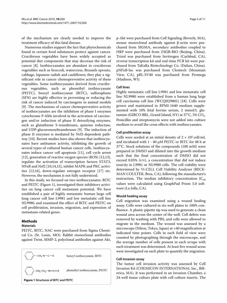
Figure 1 Structures of BITC and PEITC.
phenethyl isothiocyanate, PEITC
benzyl isothiocyanate, BITC
Wu et al. BMC Cancer 2010, 10:269http://www.biomedcentral.com/1471-2407/10/269
Page 3 of 11
inserts contain an 8 μm pore size polycarbonate mem-brane, over which a thin layer of ECMatrixTM is dried.L9981 cells were suspended to a final concentration of 2 ×10⁵ cell/mL in serum free medium with 0.1% BSA. Cellsuspensions (300 μL) were added to the upper compart-ment, medium collected from NIH3T3 cell culture wasadded with 0.1% BSA, then added to the lower compart-ment, and incubated for 24 h at 37°C in 5% CO2 atmo-sphere. Invasive cells on lower surface of the membranewere stained following the manufacturer's instruction,and counted by photographing the membrane throughthe microscope (×200 magnification).
Western blotting analysisWestern blottings were performed as previouslydescribed [19]. Briefly, L9981 cells were incubated withPEITC or BITC for 24 h, washed with PBS, and the cellpellets were lysed in lysis buffer (20 mM Tris (pH7.5), 150mM NaCl, 1% Triton X-100, sodium pyrophosphate, β-glycerophosphate, EDTA, Na3VO4, leupeptin) (BeyotimeInstitute of Biotech, Jiangsu, China) for 30 min on ice.Lysates were centrifuged (15,000 g, 20 min, 4°C). Five-fold concentrated SDS sample buffer (Beyotime Instituteof Biotech, Jiangsu, China) was added to cell lysates,boiled for 5 min, and electrophoresed on a 12% SDS-polyacrylamide gel. Protein molecular weight standards(Bio-Rad, Richmond, CA) were run concurrently. Pro-teins were transferred electrophoretically to nitrocellu-lose membranes. Membranes were blocked for 1 hour atroom temperature with 5% milk protein, 0.1% Tween 20in PBS (PBS-Tween), then were probed with rabbit anti-Twist, MMP-2, Akt, p-Akt antibodies at 1:1000 dilutionin PBS-Tween with 5% BSA overnight at 4°C. After wash-ing, membranes were probed with HRP-conjugated goatanti-rabbit antibody at 1:5000 dilution in PBS-Tweenwith 3% milk protein for 1 hour. After washing, blots weredeveloped with the Phototope HRP Western Blot Detec-tion system (Cell Singaling).
Reverse transcriptionTotal RNA was extracted from cells using Trizol (Invitro-gen, Carlsbad, CA). Reverse transcription was performedas preciously described [19] using TaKaRa kit followingmanufacturer's instruction, in the DNAEngine PeltierThermal Cycler (BIO-RAD, Richmond, CA). Briefly,RNA and random primers were denatured for 10 min at70°C; then M-MLV reverse transcriptase, deoxynucle-otide triphosphates, RNase inhibitor and reverse tran-scription buffer were added and incubated for 10 min at30°C, 60 min at 42°C and 15 min at 70°C.
Real-time PCRPrimers were synthesized by SBS Genetech (Beijing,China). SYBR Green was used to quantify mRNA levels.
All the real-time PCR reagents were purchased fromTaKaRa Biotechnology Co. (Dalian, China). PCR reac-tions were performed as preciously described [19].Briefly, PCR reactions were performed at the followingconditions: 10 seconds at 95°C, then 40 cycles at 95°C for5 seconds and 65°C for 34 seconds in the ABI Prism 7500Sequence Detector System (ABI, Foster City, CA). Theprimers for MMP-2 were 5"-CTTCCAAGTCTGGAGC-GATGT-3" (foward) and 5"-TACCGTCAAAGGGG-TATCCAT-3" (reverse), which amplified a product of 119bp. The primers for β-catenin were 5"-GCTGGGACCT-TGCATAACCTT-3" (foward) and 5"-ATTTTCACCAG-GGCAGGAATG-3" (reverse), which amplified a productof 86 bp. The primers for Twist were 5"-GCCAATCAGC-CACTGAAAGG-3" (foward) and 5"-TGTTCTTATAGT-TCCTCTGATTGTTACCA-3" (reverse), which amplifieda product of 83 bp. Glyceraldehyde-3-phoshate dehydro-genase (GAPDH) was used for normalization. TheGAPDH primers were 5"-CCACCCATGGCAAATTCC-3" (foward) and 5"-GATGGGATTTCCATTGATGACA-3" (reverse), which amplified a product of 71 bp.
Measurement of reactive oxygen species (ROS)DCFH-DA fluorescent probes were used to measured theintracellular generation of hydroperoxide (H2O2) andsuperoxide anions (O2
·-), respectively, using ReactiveOxygen Species Assay Kit (Beyotime Institute of Biotech,Jiangsu, China), following the manufacture's instruction[20]. Briefly, L9981 cells were incubated with or withoutBITC or PEITC for 4 h, then reacted with 10 μM ofDCFH-DA for 30 min at 37°C. The ROS levels weredetected by flow cytometry. The fluorescence was mea-sured at excitation 488 nm and emission 525 nm.
Measurement of glutathione (GSH)DTNB were used to measure the intracellular GSH byTotal Glutathione Assay Kit (Beyotime Institute of Bio-tech, Jiangsu, China), following the manufacture'sinstruction [21]. Briefly, L9981 cells were incubated withor without BITC or PEITC for 3-24 h, cell lysates wereprepared, and reacted with assay solution for 5 min at25°C. The absorbance at A412 was measured on a SpectraMax M5 microplate reader (Molecular Devices, Sunny-vale, CA). The GSH concentrations were determined bycomparison with standards.
DNA transfectionTransfection of L9981 cells was carried out using lipo-fectamine 2000 (Invitrogen, Carlsbad, CA), following themanufacture's instruction. Briefly, L9981 cells were platedin a 24-well plate at 1 × 10⁵ cell/well. Cells were co-trans-fected with 400 ng of pNFκB-luc, and 4 ng of pRL-SV40as an internal control. Cells were rested for 8 h aftertransfection, then were incubated with or without BITC
Wu et al. BMC Cancer 2010, 10:269http://www.biomedcentral.com/1471-2407/10/269
Page 4 of 11
or PEITC for 18 h. Luciferase assay were performed usingthe Dual-luciferase Reporter Assay System (Promega) fol-lowing the manufacture's instruction, on BERTHOLDTriStar LB 941 (BERTHOLD TECHNOLOGIES, BadWildbad, Germany)
Statistical analysisThe data were presented as mean ± standard deviation(S.D.). IC50 is the median growth inhibitory concentrationvalue, calculated using GraphPad Prism 5.0 software (LaJolla, CA). Variance analysis between groups was per-formed by one-way ANOVA and significance of differ-ence between control and treatment groups was analyzedusing Dunnett multiple comparison test. The differenceswith p < 0.05 were considered statistically significant.
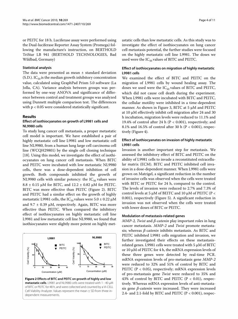
ResultsEffect of isothiocyanates on growth of L9981 cells and NL9980 cellsTo study lung cancer cell metastasis, a proper metastaticcell model is important. We have established a pair ofhighly metastatic cell line L9981 and low metastatic cellline NL9980, from a human lung large cell carcinoma cellline (WCQH29801) by the single cell cloning technique[18]. Using this model, we investigate the effect of isothi-ocyanates on lung cancer cell metastasis. When BITCand PEITC were incubated with low metastatic NL9980cells, there was a dose-dependent inhibition of cellgrowth. Both compounds inhibited the growth ofNL9980 cells with similar potency: the IC50 values were8.8 ± 0.15 μM for BITC, and 12.2 ± 0.82 μM for PEITC.BITC was more effective than PEITC (Figure 2). BITCand PEITC had a similar effect on the growth of highlymetastatic L9981 cells, the IC50 values were 5.0 ± 0.22 μMand 9.7 ± 0.39 μM, respectively. Again, BITC was moreeffective than PEITC. When compared the inhibitoryeffect of isothiocyanates on highly metastatic cell lineL9981 and low metastatic cell line NL9980, we found thatisothiocyanates were slightly more potent on highly met-
astatic cells than low metastatic cells. As this study was toinvestigate the effect of isothiocyanates on lung cancercell metastasis potential, the further studies were focusedon the highly metastatic cell line L9981. The doses weused were the IC50 values of BITC and PEITC.
Effect of isothiocyanates on migration of highly metastatic L9981 cellsWe examined the effect of BITC and PEITC on themigration of L9981 cells by wound healing assay. Thedoses we used were the IC50 values of BITC and PEITC,which did not cause cell death during the experiment.When L9981 cells were incubated with BITC and PEITC,the cellular motility were inhibited in a time-dependentmanner. As shown in Figure 3, BITC at 5 μM and PEITCat 10 μM effectively inhibit cell migration after 24 and 30h incubation, migration levels were reduced to 11.1% and19.4% of control after 24 h (P < 0.001), respectively; and8.1% and 16.5% of control after 30 h (P < 0.001), respec-tively (Figure 4).
Effect of isothiocyanates on invasion of highly metastatic L9981 cellsInvasion is another important step for metastasis. Weassessed the inhibitory effect of BITC and PEITC on theability of L9981 cells to invade a reconstituted extracellu-lar matrix (ECM). BITC and PEITC inhibited cell inva-sion in a dose-dependent manner. When L9981 cells weregrown on Matrigel, a significant reduction in the numberof invasive cells was observed when the cells were treatedwith BITC or PEITC for 24 h, compared to the control.The levels of invasion were reduced to 2.7% and 7.3% ofcontrol levels at 5 μM of BEITC and 10 μM of PEITC (P <0.001), respectively (Figure 5). A significant reduction ininvasion was not observed when the cells were treatedwith lower doses of BITC or PEITC.
Modulation of metastasis-related genesMMP-2, Twist and β-catenin play important roles in lungcancer metastasis. MMP-2 and Twist promote metasta-sis; whereas β-catenin inhibits metastasis. As BITC andPEITC inhibited L9981 cells migration and invasion, wefurther investigated their effects on these metastasis-related genes. L9981 cells were treated with 5 μM of BITCor 10 μM of PEITC for 4 h, the mRNA expression levels ofthese three genes were detected by real-time PCR.mRNA expression levels of pro-metastasis gene MMP-2were reduced to 32% and 51% of control by BITC andPEITC (P < 0.05), respectively; mRNA expression levelsof pro-metastasis gene Twist were reduced to 35% and43% of control by BITC and PEITC (P < 0.01), respec-tively. Whereas mRNA expression levels of anti-metasta-sis gene β-catenin were increased. They were increased2.4- and 2.1-fold by BITC and PEITC (P < 0.001), respec-
Figure 2 Effects of BITC and PEITC on growth of highly and low metastatic cells. L9981 and NL9980 cells were treated with 1 - 40 μM of BITC or PEITC for 48 h, and were collected and counted by a Vi-CELL Cell Viability Analyzer. Values represent the mean ± SD from three in-dependent measurements.
0 10 20 30 400
50
100
0 10 20 30 400
50
100
L9981 NL9980
Concentration (μM) Concentration (μM)
Cel
l Via
bilit
y (%
)
Cel
l Via
bilit
y (%
)
BITCPEITC
BITCPEITC
Wu et al. BMC Cancer 2010, 10:269http://www.biomedcentral.com/1471-2407/10/269
Page 5 of 11
tively (Figure 6A). We further detected the proteinexpression of these genes. Western blotting data demon-strated that both MMP-2 and Twist expression werereduced by BITC and PEITC, in a dose-dependent man-ner (Figure 6B). These results were consistent withmigration and invasion assay results.
Effect of isothiocyanates on ROS generationWe investigated whether the generation of intracellularROS is part of the mechanism by which isothiocyanatessuppress the metastasis potential of lung cancer L9981cells. The generation of ROS by isothiocyanates wasassessed by using fluorescent probes DCFH-DA by flowcytometry. Treatment with 5 μM of BITC or 10 μMPEITC showed similar effects, resulted in an increase inROS levels, compared with control (Figure 7). Howeverthese were only short term treatments. After a prolonged
time, when the Nrf2 targeted genes are expressed, theamount of ROS could decrease. We further investigatedthe effect of antioxidant NAC on ROS generation. NAC(1 mM) was added to the medium 1 h prior to isothiocya-nate treatment, and remained in the medium throughoutthe experiments. Pretreatment with NAC completelyblocked the increased ROS generation induced by bothBITC and PEITC.
Effect of isothiocyanates on intracellular glutathione levelsGlutathione is an intracellular antioxidant, helps protectcells from ROS such as free radicals and peroxides.Whether isothiocyanates exacerbated oxidative stress bycausing depletion of intracellular glutathione was investi-gated. Our data showed both BITC and PEITC decreasedtotal GSH concentration in a dose-dependent manner,but the total GSH concentration in control incubations
Figure 3 Effect of BITC and PEITC on L9981 cells migration. Wound healing assays were performed to assess cell migration. Cells were treated or untreated with 5 μM of BITC or 10 μM of PEITC for 24 and 30 h. Representative photographs of treated and untreated cells are presented (×40 mag-nification).
Control BITC (5 μM)
0 h
30 h
24 h
PEITC (10 μM)
Wu et al. BMC Cancer 2010, 10:269http://www.biomedcentral.com/1471-2407/10/269
Page 6 of 11
did not change significantly. When L9981 cells were incu-bated with 5 or 10 μM of BITC, there was a decrease intotal GSH concentration in the initial 3 h of incubation,and continues to decrease till 6 h, but by 12 h had recov-ered to high level. Thereafter, a further marked decreaseoccurred until 24 h (Figure 8). When L9981 cells wereincubated with 5 or 10 μM of PEITC, total GSH concen-tration decreased in the 3 to 6 h period. Similar to BITCtreatment, they were recovered to high level at 9 or 12 h,respectively. Then declined again and remained at lowlevels thereafter.
Effect of isothiocyanates on Akt activationAkt is an important cell signaling molecule. It blocksapoptosis, and promotes cell survival. Akt has been impli-cated as a major factor in many types of cancer. To evalu-ate whether Akt is a target of isothiocyanate on inhibitionof lung cancer cell metastasis, we detected the Akt activa-tion by western blotting (Figure 9). Both BITC and PEITCdecreased Akt phosphorylation, in a dose-dependent
Figure 4 Effect of BITC and PEITC on L9981 cells migration. Wound healing assays were performed to assess cell migration. Cells were treated or untreated with 5 μM of BITC or 10 μM of PEITC for 24 and 30 h. Number of cells migrated at 24 and 30 h time point are pre-sented. Values represent the mean ± SD of three independent experi-ments (*** P < 0.001).
24 h 30 h
Cel
l Num
ber
ControlBITCPEITC
*********
***
Figure 5 Effect of BITC and PEITC on L9981 cells invasion. Invasion Chamber Assays were performed to assess the effect on cell invasion. Cells were treated or untreated with 5 μM of BITC or 10 μM of PEITC for 24 h. (A) Representative photographs of treated and untreated cells are presented (×200 magnification). (B) Number of cells invaded at 24 h time point. Values represent the mean ± SD of three independent experiments (*** P < 0.001).
Control BITC PEITC
Control
Cel
l Num
ber
*** ***
BITC (5 μM) PEITC (10 μM)
(A)
(B)
Wu et al. BMC Cancer 2010, 10:269http://www.biomedcentral.com/1471-2407/10/269
Page 7 of 11
manner. At high concentrations, PEITC (20 μM) andBITC (10 μM) nearly completely blocked Akt phosphory-lation; meanwhile, total Akt level remained unchanged.
Effect of isothiocyanates on NFκB transcriptional activationThe nuclear factor kappa B (NF-κB) is believed to play animportant role in tumor cell growth, proliferation, angio-genesis, invasion, apoptosis and survival. In this study, weinvestigated the effects of BITC and PEITC on NF-κBtranscriptional activation, by luciferase reporter assay. Asshown in Figure 10, both BITC and PEITC inhibited thetranscriptional activation of NF-κB in a dose-dependentmanner. After treatments for 18 h, compared with con-trol, 10 and 20 μM of PEITC significantly inhibited thetranscriptional activity of NF-κB to 64.5 and 30.5% ofcontrol (P < 0.05), respectively. Similar to PEITC, 5 and10 μM of BITC significantly inhibited the transcriptionalactivity of NF-κB to 30.8 and 6.8% of control (P < 0.001),respectively. We further investigated the protective effectof antioxidant NAC. Pretreatment with antioxidant NAC(1 mM) for 1 h significantly attenuated the inhibitoryeffect of BITC and PEITC on NF-κB transcriptional acti-vation (P < 0.05).
DiscussionThe antiproliferative, antitumour activity of dietary iso-thiocyanates has been of recent research interest [9].These compounds have inhibitory effects on several typesof cancer cell growth, such as leukemia [22,23], prostatecancer [14], breast cancer [11,17], lung cancer [24], cervi-cal cancer [16], colorectal cancer [15] etc. Our previousstudies have demonstrated that allyl isothiocyanate(AITC), PEITC, PETC-Cys and related compounds
Figure 6 Effect of BITC and PEITC on metastasis-related gene ex-pression. (A) L9981 cells were treated with 5 μM of BITC or 10 μM of PEITC for 4 h. MMP-2, Twist and β-catenin mRNA expressions were de-tected by real-time PCR. (B) L9981 cells were treated with 5 - 20 μM of BITC or PEITC for 24 h. MMP-2 and Twist protein expression were de-tected by Western blotting analyses (* P < 0.05, ** P < 0.01, *** P < 0.001). Similar results were obtained in three independent experi-ments.
MMP-2TwistB-catenin
MMP-2
Twist
Actin
Control BITC PEITC
Rel
ativ
e E
xpre
ssio
n Le
vel
10μM PEITC - + - - -20 μM PEITC - - + - -
5 μM BITC - - - + -10 μM BITC - - - - +
******
**** ***
(A)
(B)
Figure 7 Effect of BITC and PEITC on ROS generation. L9981 cells were treated with 5 μM of BITC or 10 μM of PEITC for 4 h, then reacted with DCFH-DA. ROS levels were detected by flow cytometry. For NAC protection, cells were pretreated with NAC (1 mM) for 1 h.
DCF-DA Fluorescence DCF-DA Fluorescence
Cel
l Cou
nt
Cel
l Cou
nt
Control
BITC
BITC+NAC
Control
PEITC
PEITC+NAC
BITC PEITC
Wu et al. BMC Cancer 2010, 10:269http://www.biomedcentral.com/1471-2407/10/269
Page 8 of 11
induce apoptosis in HL60 cells [22,25]. The activation ofcaspases and JNK are part of the mechanism [23].
Metastasis is the most common cause of death in can-cer patients. Therefore, the research and development ofnovel anti-metastatic drugs is one of the most activefields in cancer research. Recent studies revealed that iso-thiocyanates have anti-angiogenic and anti-metastaticeffects. Isothiocyanates inhibited tumor-specific angio-genesis by down-regulating nitric oxide, TNF-alpha andproinflammatory cytokine production, and by inactiva-tion of Akt [26-28]. Isothiocyanates also suppressed themetastasis potential of human hepatoma cells [29], coloncancer cells [30] and breast cancer cells [31]. This effect ismediated by decreasing the expression of MMPs, pro-inflammatory cytokines, growth factors such as platelet-
derived growth factor (PDGF) and vascular endothelialgrowth factor (VEGF), transcription factor twist; andincreasing the expression of tissue inhibitors of matrixmetalloproteinase (TIMPs). However, there is no reporton the effect of isothiocyanates on lung cancer metasta-sis. In the present study, we investigated the effect ofBITC and PEITC on lung cancer cell metastasis potential,by using a highly metastatic human large cell lung cancercell line as an in vitro model.
Figure 8 Effect of BITC and PEITC on intracellular GSH levels. L9981 cells were treated with 5 or 10 μM of BITC or PEITC for 3 - 24 h, intracellular total GSH levels were detected at indicated time points by microplate reader. Values represent the mean ± SD of three independent experiments.
Incubation Time (h) Incubation Time (h)
GS
H (
uM)
BITC PEITC
GS
H (
uM)
0.0
50.0
100.0
150.0
200.0
250.0
0 3 6 9 12 24
Control5 uM10 uM
0.0
50.0
100.0
150.0
200.0
250.0
0 3 6 9 12 24
Control5 uM10 uM
Figure 9 Effect of BITC and PEITC on Akt activation. L9981 cells were treated with 5 - 20 μM of BITC or PEITC for 24 h. Cell lysates were prepared, p-Akt and total Akt were detected by Western blotting anal-yses. Similar results were obtained in three independent experiments.
10μM PEITC - + - - -20 μM PEITC - - + - -
5 μM BITC - - - + -10 μM BITC - - - - +
p-Akt
Akt
Figure 10 Effect of BITC and PEITC on NFκB transcriptional activa-tion. L9981 cells were transfected with pNFκB-luc, and treated with 5 - 20 μM of BITC or PEITC for 18 h. NFκB activation was detected by lu-ciferase reporter assay. For NAC protection, cells were pretreated with NAC (1 mM) for 1 h. Values represent the mean ± SD of three indepen-dent experiments (* P < 0.05, ** P < 0.01, *** P < 0.001).
0.0
200.0
400.0
600.0
800.0
1000.0
1200.0
1400.0
-NAC+NAC
Rel
ativ
e Lu
c A
ctiv
ity
10μM PEITC - + - - -
20 μM PEITC - - + - -
5 μM BITC - - - + -
10 μM BITC - - - - +
******
***
*****
***
**
Wu et al. BMC Cancer 2010, 10:269http://www.biomedcentral.com/1471-2407/10/269
Page 9 of 11
We performed wound healing and transwell chamberassays to examine the effect of BITC and PEITC on lungcancer cell metastasis potential, at the concentrationswhich did not cause cell death during the assays. Ourresults clearly demonstrate for the first time, that bothBITC and PEITC effectively inhibit lung cancer cellmigration and invasion in vitro. We then detected theeffect of isothiocyanates on metastasis-related geneexpression. MMPs are a family of zinc-binding endopep-tidases that collectively degrade most of the componentsof extracellular matrix (ECM), and they are necessary forcancer invasion and metastasis [32]. In particular, MMP-2 degrades components of the basement membrane andis strongly implicated in the invasion and metastasis ofmalignant tumors [32]. Our data showed that BITC andPEITC reduced MMP-2 expression at both mRNA andprotein level. Transcription factor Twist is a key regulatorof tumor metastasis and an inducer of epithelial-mesen-chymal transition. It plays an essential role in metastasis.Twist over-expression correlates with hepatocellular car-cinoma metastasis. Suppression of Twist expression inhighly metastatic mammary carcinoma cells specificallyinhibits its metastatic ability [33,34]. In our study, BITCand PEITC down-regulated Twist expression at bothmRNA and protein levels. Another metastasis correlatedgene we examined is β-catenin. β-catenin is an epithelialmarker, it is necessary for the creation and maintenanceof epithelial cell layers. It is down-regulated during lungcancer cell invasion and metastasis [35]. We found thatwhen BITC and PEITC suppressed cell metastasis poten-tial, β-catenin expression was increased. Taken together,these data indicated BITC and PEITC suppressed lungcancer cell metastasis potential by modulating metasta-sis-related gene expression.
To further explore the underlying mechanism, weinvestigated the effect of BITC and PEITC on cell survivalpathways. Akt/NFκB is a major anti-apoptotic/pro-sur-vival pathway that is frequently hyperactivated in mostcancers [36,37]. Akt phosphorylation promotes cellgrowth and survival by inactivating downstream pro-apoptosis substrates such as Bad, caspases, and activatingcell survival substrates such as NFκB. Both clinical analy-sis and in vivo studies showed that Akt plays an importantrole in cancer cell metastasis [38,39]. NFκB is a transcrip-tion factor that is activated by various intra- and extra-cellular stimuli such as cytokines, oxidant-free radicals,ultraviolet irradiation, and bacterial or viral products. Itcontrols the expression of numerous genes involved inimmune and inflammatory responses, cell proliferation,oncogenesis, angiogenesis and apoptosis. Inhibition ofNFκB activation effectively suppressed tumor cell inva-sion [40]. More interestingly, recent studies suggestedthat the activation of Akt/NFκB pathway contribute tothe migration of lung cancer cell [41,42]. In this study, weexamined the effect of BITC and PEITC on Akt/NFκB
pathway. BITC and PEITC inhibited both Akt phospho-rylation and NFκB transcriptional activation, in a dose-dependent manner. This suggested that Akt/NFκB path-way is a potential target of BITC and PEITC.
The altered cellular redox status and increased genera-tion of ROS have long been observed in cancer cells,especially the cells in advanced stage tumor, whichexhibit multiple genetic alterations and high oxidativestress. This drives us to investigate the effect of isothiocy-anates on ROS generation. ROS is generated intracellu-larly as byproducts of normal aerobic metabolism or assecond messengers in various signal transduction path-ways or in response to environmental stress. ROS isessential for biological functions. They regulate many sig-nal transduction pathways by directly reacting with andmodifying the structure of proteins, transcription factorsand genes to modulate their functions. ROS is involved insignalling cell growth and differentiation, regulating theactivity of enzymes, mediating inflammation by stimulat-ing cytokine production, and eliminating pathogens andforeign particles [43]. Cancer cells frequently exhibit highoxidative stress. The generation of ROS is part of themechanism by which most chemotherapeutic agents orionizing radiation kill tumor cells [44,45]. Recent studiesdemonstrate that ROS also plays an important role in cellinvasion. It regulates cell invasion via MMPs expression,MAPK pathways and NFκB activation [46-48]. In thisstudy, we investigated the role of ROS in isothiocyanate-induced inhibition of lung cancer cell metastasis. Ourfinding provide evidence of the generation of ROS byBITC and PEITC in lung cancer highly metastatic cells,this is consistent with studies in other type of cancer, suchas leukaemia [49], breast cancer [13] and pancreatic can-cer [12]. The hypothesis of the increased generation ofROS in response to BITC and PEITC was further sup-ported by the finding that pretreatment with NAC, a gen-eral antioxidant, blocked the ROS accumulation. NACpretreatment also blocked the suppression of NFκB acti-vation, this is in agreement with the finding that ROS-NFκB pathway mediates TGF-beta1-induced cell inva-sion [48]. It has been described that isothiocynates causerelease of Nrf2 from sequestration by Keap1, and its sub-sequent translocation into the nucleus. Nuclear Nrf2 acti-vates ARE-elements and induces expression of stress-responsive genes [10]. Although for short term treatmentthe ROS level increases, we expect that after a long termtreatment, the ROS level will decrease due to induction ofNrf2 dependent detoxification and antioxidative genes.We suggested that ROS generation may play a role in theinhibitory activity of isothiocyanates on lung cancermetastasis.
GSH is an antioxidant, helps protect cells from ROSsuch as free radicals and peroxides, it also maintainsexogenous antioxidants such as vitamins C and E in theirreduced (active) forms. PEITC is known to conjugate
Wu et al. BMC Cancer 2010, 10:269http://www.biomedcentral.com/1471-2407/10/269
Page 10 of 11
with GSH, leading to its exportation and depletion of cel-lular GSH. Depletion of cellular GSH leads to ROS accu-mulation. This is thought to be a major mechanism ofPEITC-induced ROS stress in cancer cells. Our previousstudy showed that GSH depletion is involved in leukae-mia cell apoptosis induced by PEITC and its cysteine con-jugate [50], a current study showed that GSHconcentration was decreased by BITC and PEITC. Therewere recoveries at 9 and 12 h time points, however, thismay be due to the stimulated GSH synthesis when cellsdetected the GSH level was low. After 24 h, GSH concen-tration decreased again, this is probably because the dys-function of cellular GSH synthesis system. Some tumorcells have higher GSH level, this is due to the over-expres-sion of Gamma-glutamyl transpeptidase and an interor-gan flow of GSH. The higher GSH level promotesmetastatic growth [51]. This suggested that GSH mayplay a role in tumor cell metastasis. Therefore GSH canbe a target for metastasis treatment. In supporting thishypothesis, Mena et al sensitized B16 melanoma to com-bination therapy and eliminates metastatic disease byGSH depletion [52]. Our finding that BITC and PEITCdecreased GSH level while suppressed tumor cell metas-tasis potential, also support this hypothesis.
Our results demonstrate that BITC and PEITC inhib-ited lung cancer highly metastatic L9981cell proliferation,migration and invasion. Metastasis-related genes weremodulated and Akt/NFκB pathway was inhibited. Oxida-tive stress could be part of the mechanism by which iso-thiocyanates suppressed lung cancer cell metastasispotential.
ConclusionIsothiocyanates are well-known chemopreventive agents.Understanding the mechanism of action of these com-pounds may provide valuable information for their possi-ble application in cancer prevention and therapy. Thereare already a number of studies that evaluate the effects ofisothiocyanates in human subjects [53-55], this couldpotentially facilitate clinical development of isothiocya-nates for cancer therapy.
Competing interestsThe authors declare that they have no competing interests.
Authors' contributionsKX, XW, and QHZ designed the study and wrote the manuscript. XW also per-formed the signaling pathway experiments. YZ performed cell proliferationexperiment, ROS and GSH assays, signaling pathway and transfection experi-ments. HQY performed the migration, invasion and gene expression experi-
ments. BNL and YL performed cell proliferation experiment. All authors readand approved the manuscript.
AcknowledgementsThis work was supported by the National Natural Science Foundation of China (30873035), the Start Up Fund of the Ministry of Education of China, the Key Project of Tianjin Municipal Education Commission (ZD200714), and the Key Project of Tianjin Municipal Science and Technology Commission (10JCZDJC20800)
Author DetailsTianjin Key Laboratory of Lung Cancer Metastasis and Tumor Microenviroment, Tianjin Lung Cancer Institute, Tianjin Medical University General Hospital, Tianjin 300052, China
References1. Stewart BWKP: WHO. World Cancer Report. IARC Press, Lyon, France;
2003. 2. Parkin DMBF, Ferlay J, Pisani P: Global cancer statistics, 2002. CA Cancer J
Clin 2005, 55(2):74-108.3. Ries LEM, Kosary C, et al.: Cancer Statistics Review, 1975-2002. National
Cancer Institute; 2005. 4. Jemal Aea: Cancer statistics, 2005. CA. Cancer J Clin 2005, 55:10-30.5. Gupta GP, Massague J: Cancer metastasis: building a framework. Cell
2006, 127(4):679-695.6. Chambers AF, Groom AC, MacDonald IC: Dissemination and growth of
cancer cells in metastatic sites. Nat Rev Cancer 2002, 2(8):563-572.7. Vitolo D, Ciocci L, Deriu G, Spinelli S, Cortese S, Masuelli L, Morrone S, Filice
MJ, Coloni GF, Natali PG, et al.: Laminin alpha2 chain-positive vessels and epidermal growth factor in lung neuroendocrine carcinoma: a model of a novel cooperative role of laminin-2 and epidermal growth factor in vessel neoplastic invasion and metastasis. Am J Pathol 2006, 168(3):991-1003.
8. Lam TK, Gallicchio L, Lindsley K, Shiels M, Hammond E, Tao XG, Chen L, Robinson KA, Caulfield LE, Herman JG, et al.: Cruciferous vegetable consumption and lung cancer risk: a systematic review. Cancer Epidemiol Biomarkers Prev 2009, 18(1):184-195.
9. Wu X, Zhou QH, Xu K: Are isothiocyanates potential anti-cancer drugs? Acta Pharmacol Sin 2009, 30(5):501-512.
10. Cheung KL, Kong AN: Molecular targets of dietary phenethyl isothiocyanate and sulforaphane for cancer chemoprevention. AAPS J 12(1):87-97.
11. Lee JW, Cho MK: Phenethyl isothiocyanate induced apoptosis via down regulation of Bcl-2/XIAP and triggering of the mitochondrial pathway in MCF-7 cells. Arch Pharm Res 2008, 31(12):1604-1612.
12. Sahu RP, Zhang R, Batra S, Shi Y, Srivastava SK: Benzyl isothiocyanate-mediated generation of reactive oxygen species causes cell cycle arrest and induces apoptosis via activation of MAPK in human pancreatic cancer cells. Carcinogenesis 2009, 30(10):1744-1753.
13. Xiao D, Powolny AA, Singh SV: Benzyl isothiocyanate targets mitochondrial respiratory chain to trigger reactive oxygen species-dependent apoptosis in human breast cancer cells. J Biol Chem 2008, 283(44):30151-30163.
14. Gong A, He M, Krishna Vanaja D, Yin P, Karnes RJ, Young CY: Phenethyl isothiocyanate inhibits STAT3 activation in prostate cancer cells. Mol Nutr Food Res 2009, 53(7):878-886.
15. Prawan A, Saw CL, Khor TO, Keum YS, Yu S, Hu L, Kong AN: Anti-NF-kappaB and anti-inflammatory activities of synthetic isothiocyanates: effect of chemical structures and cellular signaling. Chem Biol Interact 2009, 179(2-3):202-211.
16. Mukherjee S, Dey S, Bhattacharya RK, Roy M: Isothiocyanates sensitize the effect of chemotherapeutic drugs via modulation of protein kinase C and telomerase in cervical cancer cells. Mol Cell Biochem 2009, 330(1-2):9-22.
17. Kang L, Wang ZY: Breast Cancer Cell Growth Inhibition by Phenethyl Isothiocyanate is Associated with Downregulation of Estrogen Receptor-alpha36. J Cell Mol Med 2009.
Wu et al. BMC Cancer 2010, 10:269http://www.biomedcentral.com/1471-2407/10/269
Page 11 of 11
18. Qinghua WNGY ZHOU, Guowei CHE, Wen ZHU, Xiaohe CHEN, Xiaof eng CHEN, Zhilin SUN: Establishment and their biological characteristics of clonal cell subpopulations (NL9980 and L9981) from a human lung large cell carcinoma cell line (WCQH29801). Chinese Journal of Lung Cancer 2003, 6(6):464-468.
19. Xu K, Guidez F, Glasow A, Chung D, Petrie K, Stegmaier K, Wang KK, Zhang J, Jing Y, Zelent A, et al.: Benzodithiophenes potentiate differentiation of acute promyelocytic leukemia cells by lowering the threshold for ligand-mediated corepressor/coactivator exchange with retinoic acid receptor alpha and enhancing changes in all-trans-retinoic acid-regulated gene expression. Cancer Res 2005, 65(17):7856-7865.
20. Guan L, Han B, Li Z, Hua F, Huang F, Wei W, Yang Y, Xu C: Sodium selenite induces apoptosis by ROS-mediated endoplasmic reticulum stress and mitochondrial dysfunction in human acute promyelocytic leukemia NB4 cells. Apoptosis 2009, 14(2):218-225.
21. Zhang F, Wang X, Wang W, Li N, Li J: Glutamine reduces TNF-alpha by enhancing glutathione synthesis in lipopolysaccharide-stimulated alveolar epithelial cells of rats. Inflammation 2008, 31(5):344-350.
22. Xu K, Thornalley PJ: Studies on the mechanism of the inhibition of human leukaemia cell growth by dietary isothiocyanates and their cysteine adducts in vitro. Biochem Pharmacol 2000, 60(2):221-231.
23. Xu K, Thornalley PJ: Signal transduction activated by the cancer chemopreventive isothiocyanates: cleavage of BID protein, tyrosine phosphorylation and activation of JNK. Br J Cancer 2001, 84(5):670-673.
24. Mi L, Gan N, Cheema A, Dakshanamurthy S, Wang X, Yang DC, Chung FL: Cancer preventive isothiocyanates induce selective degradation of cellular alpha- and beta-tubulins by proteasomes. J Biol Chem 2009, 284(25):17039-17051.
25. Xu K, Thornalley PJ: Antitumour activity of sphingoid base adducts of phenethyl isothiocyanate. Bioorg Med Chem Lett 2000, 10(1):53-54.
26. Xiao D, Singh SV: Phenethyl isothiocyanate inhibits angiogenesis in vitro and ex vivo. Cancer Res 2007, 67(5):2239-2246.
27. Thejass P, Kuttan G: Inhibition of endothelial cell differentiation and proinflammatory cytokine production during angiogenesis by allyl isothiocyanate and phenyl isothiocyanate. Integr Cancer Ther 2007, 6(4):389-399.
28. Thejass P, Kuttan G: Allyl isothiocyanate (AITC) and phenyl isothiocyanate (PITC) inhibit tumour-specific angiogenesis by downregulating nitric oxide (NO) and tumour necrosis factor-alpha (TNF-alpha) production. Nitric Oxide 2007, 16(2):247-257.
29. Hwang ES, Lee HJ: Benzyl isothiocyanate inhibits metalloproteinase-2/-9 expression by suppressing the mitogen-activated protein kinase in SK-Hep1 human hepatoma cells. Food Chem Toxicol 2008, 46(7):2358-2364.
30. Lai KC, Huang AC, Hsu SC, Kuo CL, Yang JS, Wu SH, Chung JG: Benzyl isothiocyanate (BITC) inhibits migration and invasion of human colon cancer HT29 cells by inhibiting matrix metalloproteinase-2/-9 and urokinase plasminogen (uPA) through PKC and MAPK signaling pathway. J Agric Food Chem 58(5):2935-2942.
31. Hunakova L, Sedlakova O, Cholujova D, Gronesova P, Duraj J, Sedlak J: Modulation of markers associated with aggressive phenotype in MDA-MB-231 breast carcinoma cells by sulforaphane. Neoplasma 2009, 56(6):548-556.
32. Zucker S, Vacirca J: Role of matrix metalloproteinases (MMPs) in colorectal cancer. Cancer Metastasis Rev 2004, 23(1-2):101-117.
33. Lee TK, Poon RT, Yuen AP, Ling MT, Kwok WK, Wang XH, Wong YC, Guan XY, Man K, Chau KL, et al.: Twist overexpression correlates with hepatocellular carcinoma metastasis through induction of epithelial-mesenchymal transition. Clin Cancer Res 2006, 12(18):5369-5376.
34. Yang J, Mani SA, Donaher JL, Ramaswamy S, Itzykson RA, Come C, Savagner P, Gitelman I, Richardson A, Weinberg RA: Twist, a master regulator of morphogenesis, plays an essential role in tumor metastasis. Cell 2004, 117(7):927-939.
35. Davis R, Rizwani W, Banerjee S, Kovacs M, Haura E, Coppola D, Chellappan S: Nicotine promotes tumor growth and metastasis in mouse models of lung cancer. PLoS One 2009, 4(10):e7524.
36. Karin M, Cao Y, Greten FR, Li ZW: NF-kappaB in cancer: from innocent bystander to major culprit. Nat Rev Cancer 2002, 2(4):301-310.
37. Altomare DA, Testa JR: Perturbations of the AKT signaling pathway in human cancer. Oncogene 2005, 24(50):7455-7464.
38. Davies MA, Stemke-Hale K, Lin E, Tellez C, Deng W, Gopal YN, Woodman SE, Calderone TC, Ju Z, Lazar AJ, et al.: Integrated Molecular and Clinical
Analysis of AKT Activation in Metastatic Melanoma. Clin Cancer Res 2009, 15(24):7538-7546.
39. Matsuoka H, Tsubaki M, Yamazoe Y, Ogaki M, Satou T, Itoh T, Kusunoki T, Nishida S: Tamoxifen inhibits tumor cell invasion and metastasis in mouse melanoma through suppression of PKC/MEK/ERK and PKC/PI3K/Akt pathways. Exp Cell Res 2009, 315(12):2022-2032.
40. Kim A, Kim MJ, Yang Y, Kim JW, Yeom YI, Lim JS: Suppression of NF-kappaB activity by NDRG2 expression attenuates the invasive potential of highly malignant tumor cells. Carcinogenesis 2009, 30(6):927-936.
41. Fong YC, Liu SC, Huang CY, Li TM, Hsu SF, Kao ST, Tsai FJ, Chen WC, Chen CY, Tang CH: Osteopontin increases lung cancer cells migration via activation of the alphavbeta3 integrin/FAK/Akt and NF-kappaB-dependent pathway. Lung Cancer 2009, 64(3):263-270.
42. Huang CY, Fong YC, Lee CY, Chen MY, Tsai HC, Hsu HC, Tang CH: CCL5 increases lung cancer migration via PI3K, Akt and NF-kappaB pathways. Biochem Pharmacol 2009, 77(5):794-803.
43. Trachootham D, Alexandre J, Huang P: Targeting cancer cells by ROS-mediated mechanisms: a radical therapeutic approach? Nat Rev Drug Discov 2009, 8(7):579-591.
44. Gao J, Liu X, Rigas B: Nitric oxide-donating aspirin induces apoptosis in human colon cancer cells through induction of oxidative stress. Proc Natl Acad Sci USA 2005, 102(47):17207-17212.
45. Sun Y, Rigas B: The thioredoxin system mediates redox-induced cell death in human colon cancer cells: implications for the mechanism of action of anticancer agents. Cancer Res 2008, 68(20):8269-8277.
46. Binker MG, Binker-Cosen AA, Richards D, Oliver B, Cosen-Binker LI: EGF promotes invasion by PANC-1 cells through Rac1/ROS-dependent secretion and activation of MMP-2. Biochem Biophys Res Commun 2009, 379(2):445-450.
47. Lee KH, Kim SW, Kim JR: Reactive oxygen species regulate urokinase plasminogen activator expression and cell invasion via mitogen-activated protein kinase pathways after treatment with hepatocyte growth factor in stomach cancer cells. J Exp Clin Cancer Res 2009, 28:73.
48. Tobar N, Villar V, Santibanez JF: ROS-NFkappaBeta mediates TGF-beta1-induced expression of urokinase-type plasminogen activator, matrix metalloproteinase-9 and cell invasion. Mol Cell Biochem .
49. Trachootham D, Zhang H, Zhang W, Feng L, Du M, Zhou Y, Chen Z, Pelicano H, Plunkett W, Wierda WG, et al.: Effective elimination of fludarabine-resistant CLL cells by PEITC through a redox-mediated mechanism. Blood 2008, 112(5):1912-1922.
50. Xu K, Thornalley PJ: Involvement of glutathione metabolism in the cytotoxicity of the phenethyl isothiocyanate and its cysteine conjugate to human leukaemia cells in vitro. Biochem Pharmacol 2001, 61(2):165-177.
51. Ortega A, Carretero J, Obrador E, Estrela JM: Tumoricidal activity of endothelium-derived NO and the survival of metastatic cells with high GSH and Bcl-2 levels. Nitric Oxide 2008, 19(2):107-114.
52. Benlloch M, Ortega A, Ferrer P, Segarra R, Obrador E, Asensi M, Carretero J, Estrela JM: Acceleration of glutathione efflux and inhibition of gamma-glutamyltranspeptidase sensitize metastatic B16 melanoma cells to endothelium-induced cytotoxicity. J Biol Chem 2005, 280(8):6950-6959.
53. Hecht SS, Chung FL, Richie JP Jr, Akerkar SA, Borukhova A, Skowronski L, Carmella SG: Effects of watercress consumption on metabolism of a tobacco-specific lung carcinogen in smokers. Cancer Epidemiol Biomarkers Prev 1995, 4(8):877-884.
54. Hecht SS: Approaches to chemoprevention of lung cancer based on carcinogens in tobacco smoke. Environ Health Perspect 1997, 105(Suppl 4):955-963.
55. Liebes L, Conaway CC, Hochster H, Mendoza S, Hecht SS, Crowell J, Chung FL: High-performance liquid chromatography-based determination of total isothiocyanate levels in human plasma: application to studies with 2-phenethyl isothiocyanate. Anal Biochem 2001, 291(2):279-289.
Pre-publication historyThe pre-publication history for this paper can be accessed here:http://www.biomedcentral.com/1471-2407/10/269/prepub
doi: 10.1186/1471-2407-10-269Cite this article as: Wu et al., Isothiocyanates induce oxidative stress and suppress the metastasis potential of human non-small cell lung cancer cells BMC Cancer 2010, 10:269