39
African Journal of Microbiology Research Volume 10 Number 44 28 November, 2016 ISSN 1996-0808

African Journal of
Microbiology Research
Volume 10 Number 44 28 November, 2016
ISSN 1996-0808

The African Journal of Microbiology Research (AJMR) is published weekly (one volume per year) by Academic Journals.
provides rapid publication (weekly) of articles in all areas of Microbiology such as: Environmental Microbiology, Clinical Microbiology, Immunology, Virology, Bacteriology, Phycology, Mycology and Parasitology, Protozoology, Microbial Ecology, Probiotics and Prebiotics, Molecular Microbiology, Biotechnology, Food Microbiology, Industrial Microbiology, Cell Physiology, Environmental Biotechnology, Genetics, Enzymology, Molecular and Cellular Biology, Plant Pathology, Entomology, Biomedical Sciences, Botany and Plant Sciences, Soil and Environmental Sciences, Zoology, Endocrinology, Toxicology. The Journal welcomes the submission of manuscripts that meet the general criteria of significance and scientific excellence. Papers will be published shortly after acceptance. All articles are peer-reviewed.
Contact Us
Editorial Office: [email protected]
Help Desk: [email protected]
Website: http://www.academicjournals.org/journal/AJMR
Submit manuscript online http://ms.academicjournals.me/

Editors
Prof. Stefan Schmidt Applied and Environmental Microbiology School of Biochemistry, Genetics and Microbiology University of KwaZulu-Natal Pietermaritzburg, South Africa. Prof. Fukai Bao Department of Microbiology and Immunology Kunming Medical University Kunming, China. Dr. Jianfeng Wu Dept. of Environmental Health Sciences School of Public Health University of Michigan USA. Dr. Ahmet Yilmaz Coban OMU Medical School Department of Medical Microbiology Samsun, Turkey. Dr. Seyed Davar Siadat Pasteur Institute of Iran Pasteur Square, Pasteur Avenue Tehran, Iran. Dr. J. Stefan Rokem The Hebrew University of Jerusalem Department of Microbiology and Molecular Genetics Jerusalem, Israel. Prof. Long-Liu Lin National Chiayi University Chiayi, Taiwan.
Dr. Thaddeus Ezeji Fermentation and Biotechnology Unit Department of Animal Sciences The Ohio State University USA. Dr. Mamadou Gueye MIRCEN/Laboratoire commun de microbiologie IRD-ISRA-UCAD Dakar, Senegal. Dr. Caroline Mary Knox Department of Biochemistry, Microbiology and Biotechnology Rhodes University Grahamstown, South Africa. Dr. Hesham Elsayed Mostafa Genetic Engineering and Biotechnology Research Institute (GEBRI) Mubarak City For Scientific Research Alexandria, Egypt. Dr. Wael Abbas El-Naggar Microbiology Department Faculty of Pharmacy Mansoura University Mansoura, Egypt. Dr. Barakat S.M. Mahmoud Food Safety/Microbiology Experimental Seafood Processing Laboratory Costal Research and Extension Center Mississippi State University Pascagoula, USA. Prof. Mohamed Mahrous Amer Faculty of Veterinary Medicine Department of Poultry Diseases Cairo university Giza, Egypt.

Editors Dr. R. Balaji Raja Department of Biotechnology School of Bioengineering SRM University Chennai, India. Dr. Aly E Abo-Amer Division of Microbiology Botany Department Faculty of Science Sohag University Egypt.
Dr. Haoyu Mao Department of Molecular Genetics and Microbiology College of Medicine University of Florida Florida, USA. Dr. Yongxu Sun Department of Medicinal Chemistry and Biomacromolecules Qiqihar Medical University Heilongjiang P.R. China. Dr. Ramesh Chand Kasana Institute of Himalayan Bioresource Technology Palampur, India. Dr. Pagano Marcela Claudia Department of Biology, Federal University of Ceará - UFC Brazil. Dr. Pongsak Rattanachaikunsopon Department of Biological Science Faculty of Science Ubon Ratchathani University Thailand. Dr. Gokul Shankar Sabesan Microbiology Unit, Faculty of Medicine AIMST University Kedah, Malaysia.
Dr. Kamel Belhamel Faculty of Technology University of Bejaia Algeria. Dr. Sladjana Jevremovic Institute for Biological Research Belgrade, Serbia. Dr. Tamer Edirne Dept. of Family Medicine Univ. of Pamukkale Turkey. Dr. Mohd Fuat ABD Razak Institute for Medical Research Malaysia. Dr. Minglei Wang University of Illinois at Urbana-Champaign USA. Dr. Davide Pacifico Istituto di Virologia Vegetale – CNR Italy. Prof. N. S. Alzoreky Food Science & Nutrition Department College of Agricultural Sciences & Food King Faisal University Saudi Arabia. Dr. Chen Ding College of Material Science and Engineering Hunan University China. Dr. Sivakumar Swaminathan Department of Agronomy College of Agriculture and Life Sciences Iowa State University USA. Dr. Alfredo J. Anceno School of Environment, Resources and Development (SERD) Asian Institute of Technology Thailand. Dr. Iqbal Ahmad Aligarh Muslim University Aligrah, India.

Dr. Juliane Elisa Welke UFRGS – Universidade Federal do Rio Grande do Sul Brazil. Dr. Iheanyi Omezuruike Okonko Department of Virology Faculty of Basic Medical Sciences University of Ibadan Ibadan, Nigeria. Dr. Giuliana Noratto Texas A&M University USA. Dr. Babak Mostafazadeh Shaheed Beheshty University of Medical Sciences Iran. Dr. Mehdi Azami Parasitology & Mycology Department Baghaeei Lab. Isfahan, Iran. Dr. Rafel Socias CITA de Aragón Spain. Dr. Anderson de Souza Sant’Ana University of São Paulo Brazil. Dr. Juliane Elisa Welke UFRGS – Universidade Federal do Rio Grande do Sul Brazil. Dr. Paul Shapshak USF Health Depts. Medicine and Psychiatry & Beh Med. Div. Infect. Disease & Internat Med USA. Dr. Jorge Reinheimer Universidad Nacional del Litoral (Santa Fe) Argentina. Dr. Qin Liu East China University of Science and Technology China. Dr. Samuel K Ameyaw Civista Medical Center USA.
Dr. Xiao-Qing Hu State Key Lab of Food Science and Technology Jiangnan University China. Prof. Branislava Kocic University of Nis School of Medicine Institute for Public Health Nis, Serbia. Prof. Kamal I. Mohamed State University of New York Oswego, USA. Dr. Adriano Cruz Faculty of Food Engineering-FEA University of Campinas (UNICAMP) Brazil. Dr. Mike Agenbag Municipal Health Services, Joe Gqabi, South Africa. Dr. D. V. L. Sarada Department of Biotechnology SRM University Chennai India. Prof. Huaizhi Wang Institute of Hepatopancreatobiliary Surgery of PLA Southwest Hospital Third Military Medical University Chongqing China. Prof. A. O. Bakhiet College of Veterinary Medicine Sudan University of Science and Technology Sudan. Dr. Saba F. Hussain Community, Orthodontics and Peadiatric Dentistry Department Faculty of Dentistry Universiti Teknologi MARA Selangor, Malaysia.

Prof. Zohair I. F. Rahemo Department of Microbiology and Parasitology Clinical Center of Serbia Belgrade, Serbia. Dr. Afework Kassu University of Gondar Ethiopia. Dr. How-Yee Lai Taylor’s University College Malaysia. Dr. Nidheesh Dadheech MS. University of Baroda, Vadodara, India. Dr. Franco Mutinelli Istituto Zooprofilattico Sperimentale delle Venezie Italy. Dr. Chanpen Chanchao Department of Biology, Faculty of Science, Chulalongkorn University Thailand. Dr. Tsuyoshi Kasama Division of Rheumatology, Showa University Japan. Dr. Kuender D. Yang Chang Gung Memorial Hospital Taiwan. Dr. Liane Raluca Stan University Politehnica of Bucharest Department of Organic Chemistry Romania. Dr. Mohammad Feizabadi Tehran University of Medical Sciences Iran. Prof. Ahmed H Mitwalli Medical School King Saud University Riyadh, Saudi Arabia.
Dr. Mazyar Yazdani Department of Biology University of Oslo Blindern, Norway. Dr. Babak Khalili Hadad Department of Biological Sciences Islamic Azad University Roudehen, Iran. Dr. Ehsan Sari Department of Plant Pathology Iranian Research Institute of Plant Protection Tehran, Iran. Dr. Snjezana Zidovec Lepej University Hospital for Infectious Diseases Zagreb, Croatia. Dr. Dilshad Ahmad King Saud University Saudi Arabia. Dr. Adriano Gomes da Cruz University of Campinas (UNICAMP) Brazil Dr. Hsin-Mei Ku Agronomy Dept. NCHU Taichung,Taiwan. Dr. Fereshteh Naderi Islamic Azad University Iran. Dr. Adibe Maxwell Ogochukwu Department of Clinical Pharmacy and Pharmacy Management, University of Nigeria Nsukka, Nigeria. Dr. William M. Shafer Emory University School of Medicine USA. Dr. Michelle Bull CSIRO Food and Nutritional Sciences Australia.

Prof. Márcio Garcia Ribeiro School of Veterinary Medicine and Animal Science- UNESP, Dept. Veterinary Hygiene and Public Health, State of Sao Paulo Brazil. Prof. Sheila Nathan National University of Malaysia (UKM) Malaysia. Prof. Ebiamadon Andi Brisibe University of Calabar, Calabar, Nigeria. Dr. Julie Wang Burnet Institute Australia. Dr. Jean-Marc Chobert INRA- BIA, FIPL France. Dr. Zhilong Yang Laboratory of Viral Diseases National Institute of Allergy and Infectious Diseases, National Institutes of Health USA. Dr. Dele Raheem University of Helsinki Finland. Dr. Biljana Miljkovic-Selimovic School of Medicine, University in Nis, Serbia. Dr. Xinan Jiao Yangzhou University China. Dr. Endang Sri Lestari, MD. Department of Clinical Microbiology, Medical Faculty, Diponegoro University/Dr. Kariadi Teaching Hospital, Semarang Indonesia. Dr. Hojin Shin Pusan National University Hospital South Korea.
Dr. Yi Wang Center for Vector Biology Rutgers University New Brunswick USA. Prof. Natasha Potgieter University of Venda South Africa. Dr. Sonia Arriaga Instituto Potosino de Investigación Científicay Tecnológica/ División de Ciencias Ambientales Mexico. Dr. Armando Gonzalez-Sanchez Universidad Autonoma Metropolitana Cuajimalpa Mexico. Dr. Pradeep Parihar Lovely Professional University Punjab, India. Dr. William H Roldán Department of Medical Microbiology Faculty of Medicine Peru. Dr. Kanzaki, L. I. B. Laboratory of Bioprospection University of Brasilia Brazil. Prof. Philippe Dorchies National Veterinary School of Toulouse, France. Dr. C. Ganesh Kumar Indian Institute of Chemical Technology, Hyderabad India. Dr. Zainab Z. Ismail Dept. of Environmental Engineering University of Baghdad Iraq. Dr. Ary Fernandes Junior Universidade Estadual Paulista (UNESP) Brasil.

Dr. Fangyou Yu The first Affiliated Hospital of Wenzhou Medical College China. Dr. Galba Maria de Campos Takaki Catholic University of Pernambuco Brazil. Dr Kwabena Ofori-Kwakye Department of Pharmaceutics Kwame Nkrumah University of Science & Technology Kumasi, Ghana. Prof. Liesel Brenda Gende Arthropods Laboratory, School of Natural and Exact Sciences, National University of Mar del Plata Buenos Aires, Argentina. Dr. Hare Krishna Central Institute for Arid Horticulture Rajasthan, India. Dr. Sabiha Yusuf Essack Department of Pharmaceutical Sciences University of KwaZulu-Natal South Africa. Dr. Anna Mensuali Life Science Scuola Superiore Sant’Anna Italy. Dr. Ghada Sameh Hafez Hassan Pharmaceutical Chemistry Department Faculty of Pharmacy Mansoura University Egypt.
Dr. Kátia Flávia Fernandes Department of Biochemistry and Molecular Biology Universidade Federal de Goiás Brasil. Dr. Abdel-Hady El-Gilany Department of Public Health & Community Medicine Faculty of Medicine Mansoura University Egypt. Dr. Radhika Gopal Cell and Molecular Biology The Scripps Research Institute San Diego, CA USA. Dr. Mutukumira Tony Institute of Food Nutrition and Human Health Massey University New Zealand. Dr. Habip Gedik Department of Infectious Diseases and Clinical Microbiology Ministry of Health Bakırköy Sadi Konuk Training and Research Hospital Istanbul, Turkey. Dr. Annalisa Serio Faculty of Bioscience and Technology for Food Agriculture and Environment University of Teramo Teramo, Italy.

African Journal of Microbiology Research
Table of Contents: Volume 10 Number 44 28 November, 2016
ARTICLES
Survey of Cryptococcus neoformansin pigeon (Columba livia) excreta in Public Square in Umuarama, Paraná, Brazil 1844 Gilneia Rosa, Luiz sérgio Merlini, Wellington Henrique Bessi, Arianne Peruzo Pires Gonçalves, Lorena Zacharias Silva, Pamela Roberta Perussi, Paulo Henrique Sposito and Pinto Neto Adalgiza Induction of defense mechanisms from filtrates of saprophytic fungi against early blight disease in tomato 1849 Antônio Jussiê da Silva Solino, Kátia Regina Freitas Schwan-Estrada, Juliana Santos Batista Oliveira, Marianna dos Santos Rodrigues Alencar and Lilianne Martins Ribeiro Eugenol and linalool: Comparison of their antibacterial and antifungal activities 1860 Rehab Mahmoud Abd El-Baky and Zeinab Shawky Hashem

Vol. 10(44), pp. 1844-1848, 28 November, 2016
DOI: 10.5897/AJMR2016.8253
Article Number: 7E2464E61834
ISSN 1996-0808
Copyright © 2016
Author(s) retain the copyright of this article
http://www.academicjournals.org/AJMR
African Journal of Microbiology Research
Full Length Research Paper
Survey of Cryptococcus neoformansin pigeon (Columba livia) excreta in Public Square in Umuarama,
Paraná, Brazil
Gilneia Rosa, Luiz sérgio Merlini*, Wellington Henrique Bessi, Arianne Peruzo Pires Gonçalves, Lorena Zacharias Silva, Pamela Roberta Perussi, Paulo Henrique Sposito and
Pinto Neto Adalgiza
Universidade Paranaense, Brazil.
Received 4 August 2016, Accepted 13 September, 2016.
Cryptococcosis is a common opportunistic fungal infection caused by encapsulated yeast of the Cryptococcus genus, mainly by the Cryptococcus neoformans and Cryptococcus gattii species. The fungus is acquired through the inhalation of environmental propagules or, more rarely, through organ transplants in immune-compromised individuals. Domestic pigeons (Columba livia) are related to the infection, mainly as natural reservoirs for the fungus, a fact that is relevant due to the large concentration of these animals in public areas, mainly in places of large circulation of people. In order to isolate and diagnose Cryptococcus spp., a total of 50 excretion samples from pigeons from five public squares located in the central region of Umuarama, PR, were analyzed, being 10 samples from each square, totaling 50. From the analyzed samples, 100% (50) were negative for the capsulated yeast. Even though all samples were negative, the creation of educative campaigns aiming to raise awareness of the population on the risk of acquiring cryptococcosis and the importance of not feeding the pigeons in leisure areas, the main strategy in the population control of pigeons, were suggested. Key words: Cryptococcus neoformans, yeast, pigeons, zoonosis, public square.
INTRODUCTION Cryptococcosis is a systemic nature of mycosis caused by Cryptococcus fungus genus, whose infection occurs through inhalation of fungus present in the feces of pigeons (Sidrim and Rocha, 2010). Fungi from the Cryptococcus genus are composed of 100 species, with two clinically relevant, the Cryptococcus neoformans and Cryptococcus gattii. This is an sexual encapsulated yeast fungus of the Filobasidiella neoformans basidiomycete
(Del Poeta and Casadevall, 2010). The fungus presents tropism via the central nervous
system (CNS), respiratory and integumentary system. The commitment of the immune response is the main predisposing factor for the occurrence of the disease, both in healthy people and immunodepressed patients, bearers of the acquired immunodeficiency syndrome (AIDS) (Pappalardo and Melhem, 2003).
*Corresponding author. E-mail: [email protected]
Author(s) agree that this article remains permanently open access under the terms of the Creative Commons Attribution
License 4.0 International License

Mortality by cryptococcosis is estimated at 10% in developed countries, and it can reach 43% in developing countries (Kon, 2008). Within the characteristics observed in C. neoformans studies, the presence of capsules is observed. This structure is used in the identification and studies based on antigens that are specific to the mucopolyssacharide capsule. C. neoformans has been divided into three varieties and five serotypes, with serotype A belonging to the grubii variety (Franzot et al., 1999), serotype D to the neoformans variety, and B and C to the gattii variety (Sorrell, 2001). Cryptococcosis is among the emergent fungal infections with significant lethality and morbidity, mainly in the form of meningoencephalitis, which is usually secondary to skin or lung processes, in either immunodepressed or immunocompetent individuals (Lacaz, 2002).
Environments with large concentration of excreta favor the dissemination of the yeast, since the infection by C. neoformansis is acquired through the inhalation of environmental propagules by dehydrated yeast that can be easily dispersed in the air (Filiú et al., 2002). Due to the high concentration of C. neoformansin pigeon excreta, together with the high density of these birds in urban areas and the severity of human cryptococcosis, there is a growing interest in the study of the relationship among the birds, their excreta and the disease in humans (Huffnagle et al., 1999).
From a clinical and epidemiological point of view, cryptococcosis can be opportunistic, cosmopolitan, associated with cellular immune-depression conditions caused by C. neoformans. According to Lacaz (2002), cryptococcosis is fundamentally opportunistic, and immunodepression is the relevant factor for the high frequency of this infection.
The objective of this paper was to identify the presence of C. neoformans in pigeon excreta collected from squares in the central region in the city of Umuarama, Paraná. MATERIALS AND METHODS
The city of Umuarama is located in the northwestern region in the state of Paraná, Brazil (latitude 23° 47' 55 south and longitude 53° 18' 48 west) and has 106,387 inhabitants (IBGE, 2012), with population miscegenation of several ethnicities, with different behavioral and cultural habits.
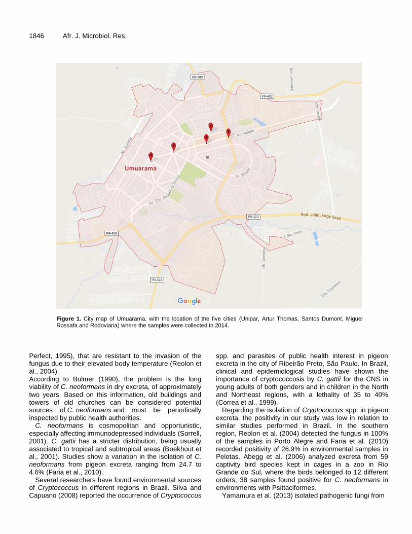
The city has a total of 10 squares, between central and peripheral ones. For the sampling, five squares were selected (Unipar, Artur Thomas, Santos Dumont, Miguel Rossafa and Rodoviaria), as shown in Figure 1, where 10 samples were collected from each square, totaling 50 samples. For the inclusion of the square, the criteria of people flow and large concentration of pigeons were used, these places are located in the central area of the city, with a concentration of shopping streets, which increases the flow of personnel and there is visual observation of the presence of pigeons in these squares.
The collections took place from March to September 2014, and random samples of pigeon feces were chosen in these squares.
Rosa et al. 1845 With the aid of spatulas and gloves, approximately five grams of old and dry excreta were collected, since they offer substrate that is favorable to the growth of yeast and present lower amount of bacteria, since there is no nutritional competition among these microorganisms (Reolon et al., 2004). The samples were collected on alternate days, between 8 and 9 a.m., in different temperature and humidity conditions, with a few samples being exposed to sun radiation, while others were in the shadows, the minimum temperature was 24.7°C and maximum was 38.7°C.
After collection, the excreta were stored in sterile containers, duly identified and forwarded to the Laboratory of Culture and Microbiology at Universidade Paranaense - UNIPAR, where they were stored at room temperature for 48 h. The samples were homogenized with 2 mL sterile saline solution, the supernatant was aspired and sown onto Petri dishes; each sample was sown on a plate containing Agar Niger culture medium, as indicated in Santos et al. (2009). The plates were incubated for seven days, at 29 to 30°C (Machado et al., 1993). In order to confirm colonial growth in culture medium with Cryptococcus spp., coloration was observed, which ranges from light beige to brown. These colonies were applied to blades microscopic analysis of the fungus and stained with China dye, which is the standard dye for visualization of the fungal capsule, and later analyzed under an optical microscope with 1000x magnification. Colonies with yeast characteristics were sent to urease test, an enzyme that aids the diagnosis (Fisher and Cook, 2001).
RESULTS AND DISCUSSION Fifty stool samples from pigeons were analyzed, the samples showed no positive results for the isolation and diagnosis of Cryptococcus spp. and Cryptococcus neoformans is absent in all samples. Environmental sources of C. neoformans are very diversified and related to a few organic substrates, among them bird excreta and vegetables. This species is the main etiological agent of cryptococcosis, a disease considered by a few authors as a fatal infection, which can be transmittable to humans in locations with high number of pigeons or by the contact with the corresponding niche through inhalation of a large amount of propagules (Lazera et al., 1993).
The use of the urease test for diagnosing Cryptococcus is due to the capacity of the fungus in hydrolyzing urea through a metalloenzyme, resulting in ammonia and carbamate, with a change in the pH of the medium, changing its color from yellow to pink. This ability for hydrolyzing urea is important to differentiate other yeast species, such as Candida spp. (Sidrim and Rocha, 2010). The use of the Niger medium provides better visualization and characterization of the colony than the traditional methods for fungal culture. The capacity for producing melanin with the formation of brown colonies in Niger seed extract media is a characteristic of C. neoformans and C. gattii, not happening in other species from the Cryptococcus genus, nor other yeasts of medical interest (Santos et al., 2009).
There are several studies identifying the presence of the C. neoformans fungus in bird excreta. However, there are few reports of cryptococcosis in pigeons (Mitchell and
1846 Afr. J. Microbiol. Res.
Figure 1. City map of Umuarama, with the location of the five cities (Unipar, Artur Thomas, Santos Dumont, Miguel Rossafa and Rodoviaria) where the samples were collected in 2014.
Perfect, 1995), that are resistant to the invasion of the fungus due to their elevated body temperature (Reolon et al., 2004). According to Bulmer (1990), the problem is the long viability of C. neoformans in dry excreta, of approximately two years. Based on this information, old buildings and towers of old churches can be considered potential sources of C. neoformans and must be periodically inspected by public health authorities.
C. neoformans is cosmopolitan and opportunistic, especially affecting immunodepressed individuals (Sorrell, 2001). C. gattii has a stricter distribution, being usually associated to tropical and subtropical areas (Boekhout et al., 2001). Studies show a variation in the isolation of C. neoformans from pigeon excreta ranging from 24.7 to 4.6% (Faria et al., 2010).
Several researchers have found environmental sources of Cryptococcus in different regions in Brazil. Silva and Capuano (2008) reported the occurrence of Cryptococcus
spp. and parasites of public health interest in pigeon excreta in the city of Ribeirão Preto, São Paulo. In Brazil, clinical and epidemiological studies have shown the importance of cryptococcosis by C. gattii for the CNS in young adults of both genders and in children in the North and Northeast regions, with a lethality of 35 to 40% (Correa et al., 1999).
Regarding the isolation of Cryptococcus spp. in pigeon excreta, the positivity in our study was low in relation to similar studies performed in Brazil. In the southern region, Reolon et al. (2004) detected the fungus in 100% of the samples in Porto Alegre and Faria et al. (2010) recorded positivity of 26.9% in environmental samples in Pelotas. Abegg et al. (2006) analyzed excreta from 59 captivity bird species kept in cages in a zoo in Rio Grande do Sul, where the birds belonged to 12 different orders, 38 samples found positive for C. neoformans in environments with Psittaciformes.
Yamamura et al. (2013) isolated pathogenic fungi from

the C. neoformans and C. gattii species in the environment represented by the soil where there is a large movement of passers-by in the central region of Londrina, such as parks, squares and streets. It was isolated in eight (2.2%) of 360 samples. These fungi were isolated from the soil with pigeon excreta and vegetable matter near trees in unsheltered areas, and exposed to constant rain and sunlight.
In Lages/SC, Menezes et al. (2014) analyzed 195 samples of pigeon excreta from six squares located in the central region in the city of Lages, SC. From the analyzed samples, 7.69% (15) were positive for the capsulated yeast. In Cuiabá- MT, Takahara (2013) reported the occurrence of 6.6% C. neoformans in 122 samples of dry pigeon excreta collected in 49 locations in the city. Correa et al. (2011) found 100% positive samples for C. neoformans in excreta from Columba sp. in the city of Cacoal, Rondônia, Brazil.
It is of pivotal importance to guide the population not to feed the pigeons so that there is no proliferation of these animals near homes, schools, squares, churches, sheds, silos and others; as well as closing locations where they are likely to shelter and build their nests (Lima and Lima, 2013).
Cryptococcosis occurs as the first opportunistic manifestation in approximately 4.4% of AIDS cases in Brazil. The prevalence of cryptococcosis associated with AIDS is estimated to be between 8 and 12% in reference centers in the southeastern region (Brasil, 2012). Delgado et al. (2005) and Lindenberg et al. (2008) reported that infection by C. gattii in humans and animals has a broader geographical occurrence than what has been habitually described, and its clinical and epidemiological aspect is not well known, therefore, it is necessary to differentiate it from opportunistic cryptococcosis by C. neoformans. In regions with large endemism, it is significantly associated with AIDS. However, the authors stated that, on the other hand, C. neoformans is capable of causing fatal infection in apparently normal individuals.
Environmental factors might influence n the isolation and diagnosis of the yeast, since the weather in a certain region, allied to temperature, humidity and exposure to sun radiation can influence the recovery and isolation of fungal strains from environmental origin (Quintero et al., 2005).
One of the main strategies is the control of pigeons, and a preventive measure is to damp the locations where there are large accumulation of pigeon feces to avoid the fungus to be dispersed by aerosol. There is no need for notification or isolation of the sick individuals (Hill et al., 1995).
The Ministry of Health does not consider Cryptococcosis as a disease of mandatory notification; therefore, there are no statistical data on its incidence in the city of Umuarama, Paraná. The multiplication of the fungus in
Rosa et al. 1847 feces and environments with lack of cleaning, together with an increase in temperature, provides the fungus basidiospores, by aerogenous route from the excreta, the contamination of Passeriformes and Psittaciformes (Lugarini et al., 2008), yeast reservoirs, as well as contamination of other environments near pigeon niche (Rosario et al., 2008). Conclusion The low occurrence in the isolation of C. neoformans in this study does not exclude the risk for human health, since pigeons are part of the landscape in urban centers, such as in the city of Umuarama, Paraná; there is no need to carry out survey in all the squares of the municipalities in the future. Conflict of interests
The authors have not declared any conflict of interest.
ACKNOWLEDGEMENTS
The authors thank Universidade Paranaense (UNIPAR) for the financial support and for the granting of the PIBIC scholarship. REFERENCES Abegg MA, Fagnello J, Valente P (2006). Cryptococcus neoformans
and Cryptococcus gattiii solated from the excreta of Psittaciformes in a Southern Brazilian Zoological Garden. Mycopathologia. 161(2):83-91.
Boekhout T, Theelen B, Diaz M, Fell JW, Hop WC, Abeln EC, Dromer F, Meyer W (2001). Hybrid genotypes in the pathogenic yeast Cryptococcus neoformans. Microbiology. 147(4):891-907.
Bulmer GS ( 1990). Twenty-five years with Cryptococcus neoformans. Mycopathologia. 22( 1):109-111
Correa EA, F, Casagrande LP (2011). Presença de Cryptococcus neoformans em excretas de Columbasp. na cidade de Cacoal, Rondônia, Brasil. Igapó (CEFET-AM). 5(3):49-55.
Correa MPSC, Oliveira EC, Duarte RRBS, Pardal PPO, Oliveira FM, Severo LC (1999). Criptococose em crianças no Estado do Pará, Brasil. Rev Soc Bras Med Trop S. Paulo. 32(6):505-508.
Del Poeta M, Casadevall A (2011). Ten challenges on Cryptococcus and Cryptococcosis. Mycopathologia, 173:303-310
Delgado ACN, Taguchi H, Mikami Y, Myiajy M, Villares MCB, Moretti ML (2005). Human cryptococcosis: relationship of environmental and clinical strains of Cryptococcus neoformans var. neoformans from urban and rural areas. Mycopathologia. 159(11):7-11.
Faria RO, Nascente PS, Meinerz ARM, Cleff MB, Antunes TA, Silveira ES, Nobre, MO, Meireles V, Mello JRB ( 2010). Ocorrência de Cryptococcus neoformans em excretas de pombos na Cidade de Pelotas, Estado do Rio Grande do Sul. Rev Soc Bras Med Trop S. Paulo. 43(2):198-200.
Filiú WF, Wanke B, Aguena SM (2002). Cativeiro de aves como fonte de Cryptococcus neoformans na cidade de Campo Grande, Mato Grosso do Sul, Brasil. Rev Soc Bras Med Trop S Paulo. 35(6):591-595.

1848 Afr. J. Microbiol. Res. Fisher F, Cook NB (2001). Micologia: fundamentos e diagnóstico.
Revinter, Rio de Janeiro. p.337. Franzot SP, Salkin IF, Casadevall A (1999). Cryptococcus
neoformansvar. grubii: separate varietal status for Cryptococcus neoformans serotype A isolates. J. Clin. Microbiol. 37(3):838-840.
Huffnagle GBA, Casadevall A, Perfect J R (1999). Cryptococcus neoformans. Mycophatologia 147(1):59-60.
Hill FI, Woodgyer AJ, Lintott MA (1995). Cryptococcosis in a North Island Brown kiwi (Apteryx AustralisMantelli) in New Zealand. J. Med. Vet. Mycol. 11(2):305-309.
IBGE - Instituto Brasileiro de Geografia e Estatística. (2012) Cidades, January 10. Brasília.
Kon AS (2008). Consenso em criptococose. Rev. Soc. Bras. Med. Trop. S. Paulo 41(5):524-544.
Lacaz CS (2002). Tratado de Micologia Médica Lacaz. Sarvier, São Paulo. 1(1):1120-1125.
Lazera MS, Wanke B, Nishikawa MM (1993). Isolation of both varieties of Cryptococcus neoformans from saprophytic sources in the city of Rio de Janeiro, Brazil. J. Med. Vet. Mycol. 31(2):449-454.
Lima A, Lima LA (2013). Criptococose: Revisão Cryptococcosis. Rev. Sci. Elect. Arch. 4(1):41-47.
Lindenberg ASC, Chang MR, Paniago AMM, Lazéra MS, Moncada PMF, Bonfim GF, Nogueira SA, Wanke B (2008). Clinical and epidemiological features of 123 cases of cryptococcosis in Mato Grosso do Sul. Brazil. Rev Inst Med trop S Paulo. 50(5):75-78.
Lugarini C, Condas LAZ, Soresini GCG, Santos RCF, Muro MD, Ono M, Farias MR, Montiani-Ferreira F (2008). Screening of antigenemia and isolation of Cryptococcus neoformans and C. gattii from cloaca and crop of birds in the state of Paraná, Brazil. Pesq. Vet. Bras. 28(7):83-91.
Machado CC, Amaral AA, Severo LC (1993). Cryptococcus neoformans var.neoformans isolado do solo. Rev. Inst. Med. Trop. S Paulo. 35(1):77-79.
Menezes T, Scain G, Quadros RM, Miletti LC, Souza AL, Miguel RL, Marques SMT (2014). Cryptococcus spp em excretas de pombos (Columba livia) de áreas públicas de Lages, SC. Sci. Anim. Health 2(2):102-114.
Mitchell TG, Perfect JR (1995). Cryptococcosis in the era of AIDS–100
years after the discovery of Cryptococcus neoformans. Clin. Microbiol. Rev. 8(1):515-548.
Pappalardo MCSM, Melhem MSC (2003). Cryptococcosis: a review of the Brazilian experience for the disease. Rev. Inst. Med. Trop. S Paulo. 45(6):299-305.
Reolon A, Perez LRR, Mezzari A (2004). Prevalência de Cryptococcus neoformans nos pombos urbanos da cidade de Porto Alegre, Rio Grande do Sul. J. Br. Patol Med. Lab. 40(5):288-293.
Rosario I, Acosta B, Colom F (2008). La Paloma y otras aves como reservorio de Cryptococcus spp. Rev. Iberoam de Micol. 25(2):13-18.
Santos LL, Ferreira FM, Lopes SF, Condas LA, Muro MD, Lugarini C (2009). Pesquisa de Cryptococcus neoformans e Candida spp. em excretas de Psitacídeos e passeriformes cativos. Arq. Cienc. Vet. Zool. 12(1):5-9.
Sidrim JJC, Rocha MFG (2010). Micologia Médica à Luz de Autores Contemporâneos. Rio de Janeiro, RJ. Editora Guanabara Koogan LTDA. p226.
Silva JO, Capuano DM (2008). Ocorrência de Cryptococcus spp e de parasitas de interesse em saúde pública, nos excretas de pombos na cidade de Ribeirão Preto, São Paulo, Brasil. Rev. Inst. A Lutz. 67(5):137-141.
Sorrell TC (2001).Cryptococcus neoformans variety gattii. Med. Micol. 39(3):148-155.
Takahara DT, Takahara DT, Lazéra MS, Wanke B, Trilles L, Dutra V, Paula DA, Nakazato L, Anzai MC, Leite Júnior DP, Paula CR, Hahn RC (2013). First report on Cryptococcus neoformans in pigeon excreta from public and residential locations in the metropolitan area of Cuiaba, state of Mato Grosso, Brazil. Rev. Inst. Med. Trop. S Paulo. 55(6):371-376.
Yamamura AAM, Freire RL, Yamamura MH, Taroda A, Felix A (2013). Study of ecological niches from pathogenic yeasts of the species Cryptococcus neoformans and Cryptococcus gattii in Londrina City, PR. Semina: Ci Agrárias, Londrina 34(2):793-804.

Vol. 10(44), pp. 1849-1859, 28 November, 2016
DOI: 10.5897/AJMR2016.8106
Article Number: EA4C60D61836
ISSN 1996-0808
Copyright © 2016
Author(s) retain the copyright of this article
http://www.academicjournals.org/AJMR
African Journal of Microbiology Research
Full Length Research Paper
Induction of defense mechanisms from filtrates of saprophytic fungi against early blight disease in tomato
Antônio Jussiê da Silva Solino*, Kátia Regina Freitas Schwan-Estrada, Juliana Santos Batista Oliveira, Marianna dos Santos Rodrigues Alencar and Lilianne Martins Ribeiro
University State of Maringa, Brazil.
Received 12 May, 2016; Accepted 31 August, 2016
The induction of defense enzymes presents efficient plant disease management when triggered by metabolic products of microorganisms. The aim of the present study is to select filtered saprobes fungi used for the management of early blight of tomato by inducing pathogenesis related to tomato plant. Filtrates of the fungi Curvularia eragrostidis, Curvularia inaequalis, Memnoniella echinata and Pseudobotrytis terrestris were cultured in potato and dextrose (PD) media and maintained in a growth chamber at 25 ± 2°C, with a photoperiod of 12 h light. After 20 days, the mycelial mass was removed through filtration. Concentrations (0, 5, 10, 15 and 20%) of filtrates and Acibenzolar-S-Methyl were applied in the 3rd
tomato leaf 3 days before inoculation with Alternaria solani. Then the disease severity
was analyzed calculating the area under the disease progress curve. The activity of the enzymes, catalase, guaiacol peroxidase and phenylalanine ammonia-lyase (locally and systemically) at plant control (72 h before inoculation) and 0, 24, 48, 72 and 96 h after inoculation with A. solani was analyzed. C. eragrostidis, C. inaequalis, M. echinata and P. terrestris filtrates reduced the AUDPC, both locally and systemically. Greater activity of phenylalanine ammonia-lyase was observed in plants treated with C. eragrostidis (local and systemic) and C. inaequalis filtrates (local). The filtrate of P. terrestris promoted greater catalase activity, either locally or systemically. The filtrate of M. echinata increased peroxidase activity locally and systemically. The filtrates tested are resistance inducers used for the management of tomato early blight, although further testing is necessary to identify elicitors present in filtrates. Key words: Alternaria solani, alternative control, proteins related to pathogenesis, Lycopersicon esculentum.
INTRODUCTION Lycopersicon esculentum is one of the most important crops in the world agricultural scenario. It constitutes an important product for "in natura" trade and extract industry. It generates direct and indirect employment. However, tomato production is considered a risky activity
because the culture is affected by many diseases. Among these, tomato early blight, caused by the fungi Alternaria solani, is a major disease which causes great losses for producers, as it is a highly destructive disease and of rapid proliferation. It focuses on leaves, stems,
*Corresponding author. E-mail: [email protected].
Author(s) agree that this article remains permanently open access under the terms of the Creative Commons Attribution
License 4.0 International License

1850 Afr. J. Microbiol. Res. petioles and fruits of tomato. Studies conducted by Tofoli et al. (2003) on conditions of Brazil observed that early blight, without control, can cause 57% loss in yield of commercial fruits and cause qualitative losses (55%) through burning of fruits caused by defoliation.
Crops that are highly susceptible to pathogens require the adoption of constant pesticide applications during cultivation. The awareness of the problems generated by the adoption of isolated methods for disease management by chemical methods has increased research technologies such as inducing plant resistance to pathogens.
Induction of resistance is a method that promotes the reduction of disease severity. This method consists of sensitizing the plant to activate its defense mechanisms by an elicitor agent, and preparing the plant for the pathogen arrival, its infection and colonization (Conrath et al., 2002). The activation of plant defense system involves an increase in the enzymes activity related to pathogenesis, such as peroxidase, phenylalanine ammonia-lyase and catalase (Garcia-Cristobal et al., 2015; Maschietto et al., 2016).
Among the reported elicitors, there are biotic agents such as microorganisms, which can activate plant defense mechanisms (Chowdappa et al., 2013). Resende et al. (2015), exploring the biodiversity of saprobe fungi present in semi-arid North-east of Brazil, observed that conidial suspension of Curvularia inaequalis was able to potentiate sorghum resistance against Colletotrichum sublineolum infection. Filtrates of pathogenic fungi, non-pathogenic and saprobes are able to activate the defense system because they contain molecules: proteins, oligosaccharides, oligopeptides, toxins and others. They function as microorganism recognition signature, which is perceived by protein receptors present in the cell membrane of the plant cell (Dubery et al., 2012).
In proposing research to identify inducers of plant resistance to pathogens, it is important to note the potential for inhibiting or reducing the symptoms of pathogen attack and to study the biochemical changes occurring in the host. Further, persistence of defense response(s) involved, as well as systemic signaling should also be considered (Choudhary and Johri, 2007).
This current work aimed to study the effect of different concentrations of saprophytic fungal filtrates in induction of defense related enzymes against early blight disease of tomato. MATERIALS AND METHODS The experiments were conducted in the Laboratory of Alternative Control and Induction of Resistance at the State University of Maringa, from January 2012 to December 2013, in the City of Maringá, State of Paraná.
Preparation of A. solani inoculum The pathogen A. solani was obtained from the State University of
Western Paraná, and plated on Potato, Dextrose, and Agar (PDA) at 25 ± 2°C for 2 to 3 days to obtain pure colonies. The pure phytopathogen was kept in a growth chamber at 25 ± 2°C in PDA, with a photoperiod of 12 h light. Preparation of filtrates from fungal saprobes Isolates of saprobes fungi from Brazil’s semi-arid Northeast (Memnoniella echinata, Curvularia eragrostidis, C. inaequalis and Pseudobotrytis terrestris) ceded by the State University of Feira of Santana, and deposited in CCMB (Bahia Microorganisms Collection) were grown on PDA. They were kept in a growth chamber at 25 ± 2°C, with a photoperiod of 12 h light.
The filtrates were obtained by subculturing fungal mycelia disc grown on PDA to 100ml of Potato dextrose broth sterilized at 120°C and 1 atm for 20 min. They were incubated in a growth chamber at a temperature of 25 ± 2°C with a photoperiod of 12 h of light for 20 days. Later, the broth containing fungal liquid metabolites was filtered through Whatman paper No. 1. Effect of fungal filtrate on early blight incidence and defense related enzymes To study the influence of filtrates’ concentrations and induced biochemical aspects, tomato seeds from the cultivar Santa Cruz Kada were used in a 4×4 split-plot design with a negative control (sterile distilled water) and a positive control (ASM) at 5 g 100 L-1. The first factor consisted of filtrates of the fungi P. terrestris, C. inaequalis, C. eragrostidis and M. echinata, and the second factor consisted of 0, 5, 10, 15 and 20% concentrations; there was a total of 18 treatments with five replicates, and plots constituted of a plant. Treatments were applied in the 2nd and 3rd true leaves, three days before inoculation with A. solani pathogen, when the plants had five leaves. The inoculation was performed in 1 × 10⁴ conidia ml-1 (Balbi-Penã et al., 2006), and then the plants were kept in a humid chamber in a greenhouse for 12 h.
The severity evaluation of tomato early blight started from the observation of the first disease symptoms in plants. It was severe in leaflets that were treated and inoculated (3rd leaf) and also in those untreated and inoculated (4th leaf), using the diagrammatic scale of Boff (1988). The evaluations were conducted every 5 days for 25 days; there were a total of 5 ratings. For severity evaluation, the area under the disease progress curve (AUDPC) was calculated according to Campbell and Madden (1990). The experiment was conducted in a completely randomized design with four replications. The AUDPC data were submitted to regression analysis (p<0.05), with one control and additional treatment. Biochemical analysis To evaluate the occurrence of biochemical changes on tomato plants treated with filtrates from saprobes fungi, the specific activity of guaiacol peroxidase, phenylalanine ammonia-lyase and catalase was analyzed.
For biochemical analysis, leaflets were collected from the 3rd leaf (treated and inoculated) to determine the local effect, and also from the 4th leaf (untreated) to observe the systemic effect. The leaves were harvested at 0, 24, 48, 72 and 96 h after inoculation (hpi) with A. solani for all treatments, including non-inoculated plants. The leaflets were weighed and stored at -20°C to perform the subsequent biochemical analysis. The enzyme extract was obtained and stored at -20°C to determine the total content of protein and activity of guaiacol peroxidase, phenylalanine ammonia-lyase and catalase.
Total proteins were quantified (Bradford, 1976) and the activity of

guaiacol peroxidase (GPOX) was determined by the method of Lusso and Pascholati (1999); the specific activity was expressed in absorbance units min-1 mg-1 protein. Catalase activity was determined by Góth method (1991) as adapted by Tomanková et al. (2006) and specific activity was expressed in absorbance unit min-1 mg-1 protein. The activity of phenylalanine ammonia-lyase enzyme was determined by colorimetric quantification of trans-cinnamic acid released from the substrate of phenylalanine, according to Umesha (2006) and the results were expressed as µmg trans-cinnamic acid min-1 mg-1 protein.
The results were submitted to statistical method of surface response (p<0.05) using SAS statistical software, to determine the mathematical model that best fits the data using the formula
. This mathematical model of equation was used in the Statistica Six Sigma program for building graphics.
RESULTS AND DISCUSSION
In analyzing the influence of different concentrations of saprobe fungi filtrates on the management of early blight in the 3rd leaf, there was a cubic regression model, for all treatments. There was reduction of 89, 83, 92 and 91% of disease at 8, 20, 8, and 7% of the filtrates of C. eragrostidis, C. inaequalis, M. echinata and P. terrestris, respectively. Comparing the estimated concentrations of filtrates which promote greater reduction of AUDPC on tomato early blight with ASM, there was a reduction of 79, 63, 81 and 83% in the 3rd leaf with the application of filtrates of C. eragrostidis, C. inaequalis, M. echinata and P. terrestris, respectively (Figure 1A).
In the 4th untreated leaf, AUDPC of tomato early blight was adjusted to the cubic regression model when filtrates of C. eragrostidis and C. inaequalis, M. echinata and P. terrestris were applied. The lowest values of AUDPC were observed at 8, 7, 7 and 7% with a reduction of 81, 70, 82 and 81%. When compared with the ASM inducer, there was a decrease of 44, 10, 13 and 10% in the 4th leaf with filtrates of C. eragrostidis, C. inaequalis, M. echinata and P. terrestris, respectively (Figure 1B).
The application of C. inaequalis filtrate on the 3rd leaf (treated) originated a quadratic response in a surface response to the guaiacol peroxidase activity (GPOX activity), catalase (CAT) and phenylalanine ammonia-lyase (PAL) (Figure 2 IA, IIA and IIIA). The influence of the concentrations of filtrates and/or collection time after inoculation with the pathogen is dependent on the enzyme studied.
In the analysis of the activity of GPOX, there was an interaction between the filtrate concentration and time after inoculation with the pathogen. There was a minimum critical point calculated at 48 h with the application of 10% of filtrate; there was reduction in enzymatic activity when compared with untreated plants. Enzyme activity increased at 72 h, with maximum observed at 96 hpi with A. solani at all concentrations; although greater one was also observed in untreated plants (Figure 2 IA).
For CAT activity, it was only the effect of time that was observed after inoculation with the minimum and maximum
Solino et al. 1851 critical points calculated at 28 and 96 hpi, respectively, independent of the filtrate concentration in the treated leaf (Figure 2 IIA). Interaction between time and concentration was observed for PAL activity. There was an increase at 0 h after inoculation and maximum increase was observed at 72 h at 20% concentration; there was 2-fold increase in the enzyme activity compared to untreated plants control (Figure 2 IIIA).
From the analysis of systemic effect on the 4th leaf treated with filtrate of C. inaequalis, it can be observed that the GPOX response was adjusted to a linear model and the CAT and PAL enzymes to a quadratic by the surface response method (Figure 2 IB, IIB and IIIB).
Considering the GPOX response, there was an increase in the activity of enzyme after inoculation; it was maximum at 96 h independent of the filtrate concentration from C. inaequalis (Figure 2 IB). For CAT, there was concentrations influence, with two maximal activities, one observed at 0 h of inoculation with the pathogen and another 96 hpi, independent of C. inaequalis filtrate concentration (Figure 2 IIB). In untreated leaves, the PAL activity increased at 0 h with 2-increase 80 hpi, at estimated concentration of 16% of C. inaequalis filtrate (Figure 2 IIIB).
Increase in the activity of both GPOX and CAT was observed (local and systemic) from 72 h in tomato plants treated with filtrate of C. inaequalis, after inoculation with A. solani. This may be related to the recognition of phytopathogens by the receptors present in the host. These activate a cascade of signaling and transduction of signals which culminate in increasing the activity of these defense enzymes. Such response occurs because the plants have a defense system able to recognize molecular patterns of the pathogen or the one the plant itself originates during the attack (Dubery et al., 2012).
The increase of enzymes such as CAT and GPOX has been reported in many studies about phytopathogen-host interaction, including A. solani and S. lycopersicum. Here, the defense mechanisms activation in plants can involve the increase of enzymes related to detoxification of reactive oxygen species generated during the plant attack by pathogens, such as GPOX and CAT (Apel and Hirt, 2004). Similar results were found in this study, allowing one to infer that the increase in guaiacol peroxidase does not have a direct relation in the reduction of AUDPC on tomato early blight observed both locally and systemically when the filtrate of C. inaequalis was applied. This is because there was an increased pattern of activity of GPOX and CAT in negative control (Figure 1A and B).
The increase in the activity of phenylalanine ammonia-lyase observed locally and systemically (Figure 2 IIIA and IIIB) explains the decrease in tomato early blight (Figure 1A). This increase may be related to the recognition of elicitors present in the filtrate of C. inaequalis. According to Henry et al. (2012), in the recognition of elicitors related to pathogen, the plants are also able to notice

1852 Afr. J. Microbiol. Res.
Figure 1. Area under the disease progress curve (AUDPC) of tomato early blight in 3rd leaf-treated (A) and 4th leaf-untreated (B) with concentrations of 5, 10, 15 and 20% of the filtrates from saprobes fungi, Maringa, 2013. *Columns followed by the same letter do not differ by T test at 5% of probability.
those that originate from non-pathogenic microorganisms; they activate resistance mechanisms, such as PAL (Figure 2 IIIA and B).
Akram and Anjum (2011) also found that Bacillus strains activate defense responses, by increasing PAL activity and consequently the accumulation of phenolic compounds in tomato plants, which were resistant to the phytopathogen Fusarium oxysporum. According to Cavalcanti et al. (2007), the early activation of the defense mechanisms, such as the PAL enzyme, are critical to plant's resistance to pathogen, because it acts
by converting the phenylalanine amino acid to trans-cinnamic acid. This conversion starts with carbon channeling from the primary metabolism in phenylpropanoid to the secondary metabolism of plants. This includes salicylic acid formed by the benzoic acid which is also a precursor in the synthesis of several phenylpropanoids such as lignin, coumarins and flavonoids (Lister and Lancaster, 1996). In this study, there was a reduction of early blight of about 63 and 10%, indicating the efficiency of C. inaequalis filtrate when tested locally and systemically, respectively.

Solino et al. 1853
Figure 2. Activity of guaiacol peroxidase (I), catalase (II) and phenylalanine ammonia-lyase in leaves (III) treated - local (A) and untreated - systemic (B) with filtrate concentration of Curvularia inaequalis, 72 h before inoculation of Alternaria solani and harvested at 0, 24, 48, 72 and 96 hpi, Maringá, 2013.

1854 Afr. J. Microbiol. Res.
Studying the effect of the concentrations of C. eragrostidis filtrates on treated tomato plants, three days before inoculation on the activity of GPOX, CAT and PAL, there was surface response in a quadratic model, linear and quadratic, respectively, when the samples were collected from the 3rd leaf (treated) (Figure 3 I, II, and II). For the GPOX activity, there was an interaction between the time of harvest after inoculation with A. solani and the concentrations of C. eragrostidis filtrates applied to the 3rd leaf. There was maximum activity of 96 hpi (concentration of 20%) with two fold increase of enzyme activity compared to control plants (Figure 3 IA).
CAT activity increased proportionally to time after pathogen inoculation; the highest peak of the dependent variable was observed at 96 hpi (Figure 3 IIA). For PAL activity, there was an interaction between the application of C. eragrostidis filtrates and the time of collecting the tomato leaves after inoculation with the pathogen. There was maximum enzyme activity of 35 hpi at a concentration of 10%. There was three times increase in the enzyme activity compared to the estimated value of the control plants in the same period (Figure 3 IIIA).
When analyzing untreated plants with filtrate of C. eragrostidis, but inoculated with A. solani, there was an interaction between the filtrate concentration and collection time in GPOX and PAL activity. However, there was no difference between the filtrate concentration in the CAT activity, where the best fit of regression models was linear, linear and quadratic, respectively (Figure 3 I, II and III B). A dose-dependence was observed with GPOX activity systemically, with three times increase at 20% concentration of C. eragrostidis filtrate and 72 hpi with A. solani (Figure 3 IB).
For CAT, there was no effect of the concentration on enzyme activity, but time influenced their response, with greater activity from 24 hpi in treated leaves (Figure 3 IIA). For PAL, it was observed an increase in enzyme activity systemically and in proportion to time and filtrate concentration of C. eragrostidis; there was duplication of enzyme activity at 96 hpi when 20% of the filtrate was applied (Figure 3 IIIB).
The increase of GPOX and PAL promoted by the filtrate of C. eragrostidis can justify the reduction of tomato early blight observed in treated plants (Figure 1A). It may be inferred that the increase of these enzymes can be related to the recognition of elicitor molecules existing in the filtrate by a receptor on the host, both locally and systemically. This is because in both, there was an increase of enzyme activity when the filtrates were applied compared to plants that were untreated but inoculated with the pathogen (Figure 3 IA and IB). Both enzymes, GPOX and PAL have been reported in defense of plants against pathogen attack and may be activated simultaneously when the plant is exposed to an elicitor agent. Chmielowska et al. (2008) verified that seedlings of bean and lupine when treated with copper, but not inoculated with pathogens had a higher activity of
peroxidase and phenylalanine ammonia-lyase.
The activation of peroxidase is associated with the primary events of the resistance mechanisms induced by elicitors. This is because the enzyme acts on hydrogen peroxide detoxification formed during the oxidative explosion caused after recognition of elicitors during the defense system activation (Van Loon et al., 2006). This generation of hydrogen peroxide in elicited cells increases the peroxidase activity involved in the cross oxidative connection of structural proteins in the cell wall rich in the repetition of proline. It also increases tyrosine that strengthens the plant's cell walls, slowing the phytopathogen colonization process (Kishor et al., 2005).
An increase in the PAL activity in plants treated with filtered C. eragrostidis may have an essential role in reducing the AUDPC of tomato early blight. According to Umesha (2006), phenylalanine ammonia-lyase is crucial in the protection of tomato plants against the attack of Clavibacter michiganensis subsp. michiganensis. The enzyme activity is related to the plant resistance to pathogens, essentially, for being involved in the first step of the phenylpropanoids synthesis and its conversion to trans-cinnamic acid, resulting in compounds with antimicrobial activity. This is besides lignin increment, which gives greater resistance to the plants’ cell wall (Nakazawa et al., 2001).
In Figures 4 IA, IIA and IIIA, there was a significant effect (P<0.05) in the interaction between filtrate concentration of P. terrestris and harvest time for determining guaiacol peroxidase, catalase and phenylalanine ammonia-lyase, with best fit in the quadratic model for the three enzymes when analyzed locally (3rd leaf).
GPOX activity was reduced in the gap between 5 and 15% of the filtrate and opposite (Figure 4 IA). However, there was an increase of CAT within the same interval of concentrations; 96 hpi increase in 10 % concentration, where there was 3-fold increase in enzyme activity. PAL was not influenced by the application of P. terrestris filtrate concentrations; however enzyme activity increased with time after inoculation with A. solani (Figure 4 IA).
For the GPOX activity in 4th leaf (untreated), there was
maximum increment of the enzyme in plants that were not treated with P. terrestris filtrate, 96 h after inoculation (Figure 4 IB). CAT activity increased at 72 hpi when 15% was applied, with 2-fold increase in GPOX activity (Figure 4 IIB). For PAL quantification in the untreated leaves, the greatest activity was observed at 96 hpi independent of the filtrate concentration (Figure 4 IIIB).
The reduction of AUDPC in tomato early blight (Figures 1A and B) might be correlated to an increase of CAT and PAL activity in plants treated with filtrates of P. terrestris. Cavalcanti et al. (2006) observed that tomato plants, from the group Santa Cruz Kada, previously induced with an elicitor made of necrotic tissue extract with Xanthomonas vesicatoria, presented less bacterial spot severity caused by X. vesicatoria. This protection was related to the

Solino et al. 1855
Figure 3. Activity of guaiacol peroxidase (I), catalase (II) and phenylalanine ammonia-lyase in leaves (III) treated - local (A) and untreated - systemic (B) with filtrates concentration of Curvularia eragrostidis 72 h before inoculation of Alternaria solani and harvested at 0, 24, 48, 72 and 96 hpi, Maringa, 2013.

1856 Afr. J. Microbiol. Res.
Figure 4. Activity of guaiacol peroxidase (I), catalase (II) and phenylalanine ammonia-lyase in leaves (III) treated - local (A) and untreated - systemic (B) with filtrates concentration of Pseudobotrytis terrestris 72 h before inoculation of Alternaria solani and harvested at 0, 24, 48, 72 and 96 hpi, Maringa, 2013.

Solino et al. 1857
Figure 5. Activity of guaiacol peroxidase (I), catalase (II) and phenylalanine ammonia-lyase in leaves (III) treated - local (A) and untreated - systemic (B) with filtrates concentration of Memnoniella echinata 72 h before inoculation of Alternaria solani and harvested at 0, 24, 48, 72 and 96 hpi, Maringa, 2013.
increase in catalase activity from 24 h with peak at72 hours after the treatments. This similarly occurred with the application of P. terrestris filtrate in the interaction of A. solani and S. lycopersicum (Figure 4 IA).
The enzyme catalase acts in the dismutation of two H2O2 molecules into water and oxygen (Sharma et al., 2012); thus, the hydrogen peroxide accumulation can be estimated indirectly by the action of this enzyme. H2O2 has been applied in various stress conditions, including induction through biotic and abiotic inductors where CAT rapidly degrades H2O2 in an efficient manner (Mallick and
Mohn, 2000). This detoxification is dependent on the intensity, duration and type of elicitation (Han et al., 2009). The equation model of quadratic regression had the best fit for the interaction between the filtrate concentration of M. echinata and time of harvest after inoculation with A. solani for GPOX activity, using the surface response method and the linear method for CAT and PAL, when determined locally (Figure 5 IA, IIA and IIIA).
GPOX increased according to the filtrate concentration and the time after inoculation, with 3-fold increase in

1858 Afr. J. Microbiol. Res. enzyme activity after 96 hpi at a 20% concentration in relation to treatment with control plants (Figure 5 IA). CAT activity increased linearly with time of harvest. However, it was not influenced by concentration (Figure 5 IIA). While the application of M. echinata filtrate reduced PAL activity in 3rd leaf by increasing the filtrate concentration and the harvesting time after pathogen inoculation (Figure 5 IIIA).
Systemically, the enzymes guaiacol peroxidase, catalase and phenylalanine ammonia-lyase were adjusted to the quadratic model (Figure 5 IB, IIB and IIIB). GPOX activity increased with concentration and harvesting time after inoculation with 2-fold increase by applying the filtrate at 20% and 96 hpi, compared to activity of leaves collected from untreated plants at the same period (Figure 5 IB). The CAT was affected with the time of harvest and there was reduction in the activity with A. solani infection, independent of the filtrate concentration of M. echinata (Figure 5 IIB).
The application of concentrations of M. echinata filtrate promoted greater GPOX activity; this indicates a possible reduction of tomato early blight leads to increase in the activity of enzymes. The increase in peroxidase activity may also be associated with an increase of structural proteins synthesis, which strengthens the cell wall, acting as a barrier to pathogen (Van Loon et al. 2006).
By applying filtrates of C. inaequalis, C. eragrostidis, P. terrestris and M. echinata, it can be observed that they are capable of reducing AUDPC of tomato early blight through the induction of distinct biochemical mechanisms. The filtrates of these fungi acted as elicitors agents, being recognized by receptors present in tomato plants, activating its defensive enzymes with increased GPOX, CAT and PAL. The plants' multicomponent defense response is directly related to interactions between the elicitor and receiver, with amplitude, duration and quality dependent on the specific signaling generated (Brencic and Winans, 2005). The knowledge of this variation may explain the distinct behavior observed in each filtrate from the species of saprobes fungi presented in this work.
The peaks of activity from GPOX and CAT simultaneously are noticed when the filtrates of saprobes are applied on the tomato plants. Interestingly, GPOX and CAT showed opposing activities to each other, that is, when there was an increase of GPOX activity the CAT activity was reduced and vice versa. Such response can be observed clearly in plants treated with P. terrestris, in which there was a reduction in the peroxidase activity between the concentrations of 5 and 15%; the catalase increased between the same concentrations. Because the enzymes compete for the same substrate, the H2O2
generated during oxidative explosion is caused by stress (Sen et al., 2003).
When considering the enzymes activation related to pathogenesis, it may be observed that the filtrate of M. echinata induced GPOX activity in untreated leaves and
C. eragrostidis increased the activity of PAL and GPOX, systemically. The potential to activate the defense responses in parts of the plants that were untreated is a characteristic of great importance for resistance inducer, because it prepares the plant tissue that has not received the treatment for a possible pathogen attack. Systemic activation is normally regulated by a signaling cascade mediated by H2O2, salicylic acid, MAPKs and/or other molecules after recognition of elicitor molecule by receptors present in the cell membrane of the plant cell (Conrath et al., 2006).
In conclusion, the filtrates from C. eragrostidis, C. inaequalis, M. echinata and P. terrestris promoted reduction of AUDPC in tomato early blight in greenhouse conditions, when applied three days before inoculation locally and systemically. New studies may be performed to identify elicitor molecule(s) and can be used in the future in the management of plant diseases.
Conflict of Interests
The authors have not declared any conflict of interests. REFERENCES Akram W, Anjum T (2011). Quantitative changes in defense system of
tomato induced by two strains of Bacillus against Fusarium Wilt. Ind. J. Fund. Appl. Life Sci. 1(3):7-13.
Apel K, Hirt H (2004). Reactive oxygen species: metabolism, oxidative stress, and signal transduction. Annu. Rev. Plant Biol. 55:373-399.
Balbi-Peña MI, Becker A, Stangarlin JR, Franzener G, Lopes MC, Schwan-Estrada KRF (2006). Controle de Alternaria solani em Tomateiro por Extratos de Curcuma longa e Curcumina - II. Avaliação in vivo. Fitopatol. Bras. 31(4):401-404.
Boff P (1988). Epidemiologia e controle químico da mancha de estenfílio (Stemphylium solani Weber) e da pinta preta (Alternaria solani (Ellis, Martin) Jones, Grout em dois sistemas de condução do tomateiro (Lycopersicon esculentum Mill). Viçosa, Brasil: Universidade Federal de Viçosa, PhD dissertation.
Brencic A, Winans SC (2005). Detection of and Response to Signals Involved in Host-Microbe Interactions by Plant-Associated Bacteria. Microbiol. Mol. Biol. Rev. 69(1):155-194.
Campbell CL, Madden LV (1990). Introduction to Plant Disease Epidemiology. New York, NY: Wiley & Sons.
Cavalcanti FR, Resende MLV, Lima JPM, Silveira JAF, Oliveira JTA (2006). Activities of antioxidant enzymes and photosynthetic responses in tomato pre-treated by plant activators and inoculated by Xanthomonas vesicatoria. Physiol. Mol. Plant Pathol. 68:198-208.
Cavalcanti FR, Resende MLV, Oliveira JT A (2007). Peroxidases ativadas por frações protéicas de extrato biológico eficaz na proteção do tomateiro contra a mancha bacteriana. Fitop. Brasileira, 32(6):507-511.
Chmielowska J, Deckert J, Díaz J (2008). Activity of peroxidases and phenylalanine ammonia-lyase in lupine and soybean seedlings treated with copper and an ethylene inhibitor. Biol. Lett. 45:59-67.
Choudhary DK, Johri APJ (2007). Induced systemic resistance (ISR) in plants: mechanism of action. Ind. J. Microbiol. 47:289-297.
Chowdappa P, Mohan Kumar SP, Jyothi Lakshmi M, Upreti KK (2013). Growth stimulation and induction of systemic resistance in tomato against early and late blight by Bacillus subtilis OTPB1 or Trichoderma harzianumm OTPB2. Biol. Control 65(1):109-111.
Conrath U, Beckers GJM, Flors V, García-Agustín P, Jakab G, Mauch F, Newman M, Pieterse CMJ, Poinssot B, Pozo MJ, Pugin A, Schaffrath U, Ton J, Wendehenne D, Zimmerli Laurent, Mauch-Mani

B (2006). Priming:Getting Ready for Battle. Mol. Plant Microbe Interact. 19(10):1062-1071.
Conrath U, Pieterse CMJ, Mauch-Mani B (2002). Priming in plant-pathogen interactions. Trends Plant Sci. 7(1): 210-216.
Dubery IA, Sanabria NM, Huang JC (2012). Nonself perception in plant innate immunity. In. Lopes-Larrea C, eds. Self and Nonself. Landes Bioscience and Springer Science+Business Media. 738:79-107.
Garcia-Cristobal J, Garcia-Villaraco A, Ramos B, Gutirrez-Mañero J, Lucas JA (2015). Priming of pathogenesis related proteins and enzymes relate to oxidative stress by plant growth promoting Rhizobacteria on rice plants upon abiotic and biotic stress challenge. J. Plant Physiol. 1(188):72-79.
Góth L (1991). A simple method for determination of serum catalase activity and revision of reference range. Clin. Chim. Acta 196(2-3):143-151.
Han C, Liu Q, Yang DY (2009). Short-term effects of experimental warming and enhanced ultraviolet-B radiation on photosynthesis and antioxidant defense of Picea asperata seedlings. Plant Growth Regul. 58(2):153-162.
Henry G, Thonart P, Ongena M (2012). PAMPs, MAMPs, DAMPs and others: an update on the diversity of plant immunity elicitors. Biotechnol. Agron. Soc. Environ. 16(2):257-268.
Kishor PB, Sangam S, Amrutha RN, Laxmi PS, Naidu KR, Rao KRSS, Rao S, Reddy KJ, Theriappan P, Sreenviasulu N (2005). Regulation of proline biosynthesis, degradation, uptake and transport in higher plants: Its implications in plant growth and abiotic stress tolerance. Curr. Sci. 88(3):424-438.
Lister CE, Lancaster JE (1996). Phenylalanine ammonia-lyase (PAL) activity and its relationship to anthocyanin and flavonoid levels in New Zealand-grown apple cultivars. J. Am. Soc. Hortic. Sci. 121(2):281-285.
Mallick N, Mohn FH (2000). Reactive oxygen species: response of algal cells. J. Plant Physiol. 157:183-193.
Maschietto V, Lanubile A, Leonardes S, Marocco A, Paciolla C (2016). Constituitive expression of pathogenesis-related proteins and antioxydant enzyme activities triggers maize resistance towards Fusarium verticillioides. J. Plant Physiol. 200:53-61.
Nakazawa A, Nozue M, Yasuda H, Takeda G, Kubo H (2001). Expression pattern and gene structure of phenylalanine ammonia-lyase in Pharbitis nil. J. Plant Res. 114:323-328.
Solino et al. 1859 Resende RS, Milagres CA, Rezende D, Aucique-Perez CE, Rodrigues
FA (2015). Bioprospecting of Saprobe Fungi from the Semi-Arid North-East of Brazil for the Control of Anthracnose on Sorghum. J. Phytopathol. 163(10):787-794.
Sen P, Mukherjee S, Bhhaumik G, Dasb P, Ganguly S, Choudhury N, Raha S (2003). Enhancement of catalase activity by repetitive low-grade H2O2 exposures protects fibroblasts from subsequente stress-induced apoptosis. Mutat. Res. 529:87-94.
Sharma P, Jha AB, Dubey RS, Pessarakli M (2012). Reactive oxygen species, oxidative damage, and antioxidative defense mechanism in plants under stressful conditions. J. Bot. 1:1-26.
Tofoli JG, Domingues RJ, Garcia Junior O, Kurozawa C (2003). Controle da pinta preta do tomateiro por fungicidas e seus impactos na produção. Summa Phytopol. 29(3):225-233.
Umesha S (2006). Phenylalanine ammonia liase activity in tomato seedings and its relationship to bacterial canker disease resistance. Phytoparasitica 34:68-71.
Van Loon LC, Rep M, Pieterse CMJ (2006). Significance of inducible defense-related proteins in infected plants. Ann. Rev. Phytopathol. 44:155-162.

Vol. 10(44), pp. 1860-1872, 28 November, 2016
DOI: 10.5897/AJMR2016.8283
Article Number: 6B9F4ED61839
ISSN 1996-0808
Copyright © 2016
Author(s) retain the copyright of this article
http://www.academicjournals.org/AJMR
African Journal of Microbiology Research
Full Length Research Paper
Eugenol and linalool: Comparison of their antibacterial and antifungal activities
Rehab Mahmoud Abd El-Baky and Zeinab Shawky Hashem*
Department of Microbiology and Immunology, Faculty of Pharmacy, Minia University, 61519-Minia, Egypt.
Received 28 August 2016 Accepted 25 October, 2016
Antimicrobial resistance is one of the greatest threats to human health. Alternatives to antimicrobials are needed to combat the rise of bacterial resistance. Essential oils (EOs) and their components are potential sources of new antimicrobials. The present study was conducted to evaluate the antibacterial and antifungal activities of two components of EOs. The antimicrobial mechanisms of eugenol and linalool were investigated against five bacterial strains and four Candida strains. Broth macrodilution method was used to compare the antibacterial and anticandidal activities of the two compounds. They exhibited antimicrobial activity against all tested strains. Germ tube formation by Candida albicans was investigated and it was found that it was completely inhibited at sub-MICs of eugenol while linalool showed minor activity compared to eugenol. Time kill kinetic studies indicated that eugenol was highly toxic to all bacterial and fungal strains within 2.5 h of exposure. Absorbance of intracellular constituents was measured at 260 nm. Only eugenol was highly effective toward lysis and cellular content leakage compared to control drugs. In addition, scanning electron microscopy (SEM) was used to charactize the effect of the two components on cell morphology and showed that both compounds induced cellular deformity of nearly all tested cells. Also, it was found that only eugenol inhibited the Beta-lactamase production and urease activity and it diminished bacterial motility of all tested bacterial strains. These results indicate that eugenol and linalool are effective antimicrobial agents and both antibacterial and antifungal activities of linalool were much weaker than that of eugenol. Key words: Antimicrobial resistance, antimicrobials, germ tube formation, time kill kinetic, cellular deformity.
INTRODUCTION During the past few decades, the incidence of both community-acquired and nosocomial bacterial and fungal infections has significantly raised, increasing the number of patients who are at risk especially those with impaired immunity. There has been a worldwide rapid increase in resistance to antimicrobial agents in almost all bacterial
and fungal genera and to all drug classes. The most important factor influencing the spread of antimicrobial resistance is the excessive microbial exposure to antimicrobials that results in selection pressure in microbial population, allowing only the fittest genotype to thrive (Canton and Morosini, 2011; WHO, 2012).
*Corresponding author. E-mail: [email protected], [email protected]. Tel: 01003780855.
Author(s) agree that this article remains permanently open access under the terms of the Creative Commons Attribution
License 4.0 International License

Currently, high rates of antimicrobial agents use in the community, hospital and agriculture have contributed to fuel this crisis. The cross resistance to two or more antimicrobial agents is often mediated by a single resistance mechanism (Leclercq, 2002). This might be arisen due to plasmids (Tenover, 2006) or emerging mutation of chromosomal DNA (Sanders and Sanders, 1992). The cost of treatment is increased due to antimicrobial resistance. It may result in prolonged hospital stays, higher mortality rates, and creates broader infection control problems (Neu, 1992).
Natural products, either as pure compounds or as plant extracts provide unlimited opportunities for new drugs due to an increasing demand for chemical diversity (Cos et al., 2006). Essential oils (EOs) extracted from plants have been used primarily for flavoring and perfumery (Ben Arfa et al., 2006). It has long been recognized that EOs have antimicrobial properties, and recent studies have demonstrated that these activities are mainly due to the presence of numerous substituted aromatic molecules. Examples of these molecules include eugenol, cinnamaldehyde, and carvacrol (Moleyar and Narasimham, 1992). Currently, there is a trend in food processing to avoid the application of chemical preservatives such as sodium chloride and nitrates. Thus, the use of bioactive compounds derived from EOs as alternative antimicrobial agents is garnering great interest (Gill and Holley, 2004; Tsukiyama et al., 2002). Although, EOs have been empirically used as antimicrobial agents, their spectrum of activity and mechanisms of action remain unknown for most of them.
Linalool (3,7-dimethylocta-1,6-dien-3-ol) is a terpene alcohol that has broad spectrum antimicrobial activity (Alviano et al., 2005). Eugenol (4-allyl-2-methoxyphenol) is the main component of clove oil (phenolic compounds). It is used primarily as a flavoring agent in food and cosmetic products. Eugenol and linalool possesses various biological abilities, including antimicrobial, antioxidant, anti-inflammatory, anticarminative, anti-spasmodic, and antiparasitic activities. They are also effective as antiseptic agents in dentistry. Many studies most focused on the possibility of using clove oil as a replacement for some chemical additives in the preservation of main food categories such as meat and fish (Burt, 2004; Oussalah et al., 2007).
Combinational therapy is essential in the treatment of serious infection and to reduce the risk of resistant microbes (Kamatou et al., 2012). When linalool or menthol is combined with eugenol it showed the highest synergy, indicating that a monoterpenoid phenol combined with a monoterpenoid alcohol is an effective combination (Bassolé et al., 2010). Eugenol shows excellent synergistic activities and decreases MICs of conventional antibiotics as vancomycin, gentamicin and Beta-lactams (Moon et al., 2011). This synergistic effect can be linked to eugenol's ability to damage the membrane of Gram-negative bacteria. Combination between eugenol and
Abd El-Baky and Hashem 1861 cinnamate, cinnamaldehyde, thymol or carvacrol leads to greater antimicrobial effect (Pei et al., 2009; Rico-Molina et al., 2012). Certain combinations of eugenol and thymol show a synergistic effect thus potentiate their inhibition of C. albicans colonization and infectivity (Braga et al., 2007).
The present study evaluates the antibacterial and antifungal activity of eugenol and linalool and investigates the antimicrobial mechanisms of action against some Gram positive, Gram negative and Candida species. MATERIALS AND METHODS
Test organisms
In the current study, five standard strains; Staphylococcus aureus (ATCC 6538), Escherichia coli (ATCC 7839), Klebsiella pneumoniae (ATCC 10031), Pseudomonas aeruginosa (ATCC 10145) and Candida albicans (ATCC 10231) were used. They were obtained from MIRCIN culture collection of the Faculty of Agriculture, Ain Shams University. Clinical strains of Proteus mirabilis and Candida (Candida albicans, Candida glabrata and Candida krusei) were obtained from the Department of Microbiology, Faculty of Pharmacy, Minia University. All cultures were maintained in their appropriate agar slants at 4°C and used as stock cultures.
Antimicrobial agents
Two compounds; Eugenol and linalool (Sigma-Aldrich, Germany) with 98% purity were used in this study.
Determination of minimum inhibitory concentration (MIC) and minimum bactericidal (MBC) or fungicidal (MFC) concentrations
A broth macrodilution method was used to determine the MIC and MBC or MFC according to the Clinical and Laboratory Standards Institute (CLSI) for bacteria and yeasts (2012, 2002). The Mueller Hinton broth (MHB) (Merck) or RPMI-1640 [Roswell Park Memorial Institute medium (with glutamine, without bicarbonate, and with phenol red as a pH indicator) was 1640, buffered to pH 7.0 with MOPS (morpholine propane sulfonic acid) at 0.165 M] (Sigma-Aldrich) was supplemented with dimethylsulfoxyde (DMSO) (Merck) at a 2% concentration in order to enhance sample solubility. Two fold serial dilutions of eugenol and linalool were prepared. The inocula of the microbial strains were prepared from overnight broth cultures and suspensions then adjusted to the turbidity of a 0.5 McFarland standard. Standardized suspension of the test organism was transferred into each tube. Controls without the test compound were prepared.
To determine MBC or MFC, 100 μL of bacterial inoculum was taken aseptically from tubes that had not presented visible turbidity and inoculated in Mueller Hinton agar (MHA) for 20 h at 35°C for bacteria or in MHA supplemented with 2% glucose and 0.5 mg/mL methylene blue for 24 h at 35°C for fungal strains. The MBC/MFC is defined as the lowest concentration of the essential oil at which 99.9% of the initial inoculum was killed. The experiments were repeated three times. To determine the nature of antibacterial or antifungal effect of these compounds, the MBC/MIC or MFC/MIC ratio was used; when the ratio was lower than 4, the compound was

1862 Afr. J. Microbiol. Res. considered as a bactericidal or fungicidal and when the ratio was higher than 4, it was considered as a bacteriostatic or fungistatic (Levison, 2004).
Germ tube inhibition assay
C. albicans (ATCC 10231) and clinical C. albicans strain were cultivated overnight on SDA at 37ᵒC. Cell suspensions were prepared in NYP medium [N-acetylglucosamine (Sigma; 10-3 mol/L), yeast nitrogen base (Difco; 3.35 g/L), proline (Fluka; 10-3 mol/L) and NaCl (4.5 g/L), pH 6.7±0.1] and adjusted to 0.2×10⁶
c.f.u. ml-1. Eugenol and linalool were diluted in DMSO and 10 l
volumes were added to 990 l of the Candida suspensions (final DMSO concentration of 1%, v/v) to obtain 1/2, 1/4 and 1/8 of the MIC values. Untreated eugenol and linalool control suspensions were used. After 3 h incubation with gentle shaking at 37ᵒC, cell suspensions were examined for the presence of germ tubes using a light microscope. Germ tubes were considered positive when they were at least as long as the blastospore (Pinto et al., 2009).
Time kill assay
Four concentrations (0.5, 1, 2 and 4 MIC) of each compound were tested against each microbial strain. The time-kill assay was performed with a final inoculum of approximately 5 × 10⁵ CFU/ml in a final volume of 30 ml. The final inoculum was adjusted to match the 0.5 McFarland standards. The tubes were continuously shaken on an orbital shaker and incubated at 35°C. 10 µl samples were withdrawn from each tube at 30, 60, 90, 150 and 24 h and streaked on MHA plate. Ten fold dilutions were prepared when necessary and plated onto each MHA plate. Plates were kept in triplicates for each compound and they were incubated for 24 h at 35ᵒC, and colony counts were determined. Time-kill curves were plotted as log10 CFU/ml versus time functions. The compound concentration and the 3 log10-fold reductions in the bacterial count compared with the growth control were determined (Devi et al., 2010).
Estimation of the yeast cells or bacterial cytosol release
Bacteria and Candida cells were treated with eugenol or linalool then the release of cytosolic material absorbing at 260 nm from these cells was detected (Bennis et al., 2004b). Aliquots of 1.5 ml of cells suspension for 1 h in phosphate buffer saline (PBS) containing various concentrations of test compound ranging from 1.5 to 12 mM were used. Eugenol, linalool, levofloxacin (Sedico, Egypt) against bacterial cells and ketoconazole (Amriya, Egypt) with fungal cells were tested. Microbial cells suspended in PBS were used as control. Correction was made for the absorption of the suspending liquids containing the same concentration of compound after two minutes contact with bacteria or Candida cells at 260 nm in Beckman UV spectrophotometer.
Scanning electron microscopy (SEM)
Microbial suspensions treated with the MBC or MFC concentrations of eugenol or linalool for 1 h. Treated and untreated (negative control) cells were prefixed in 2.5% glutaraldehyde for 1 h at 4°C. After each fixation, the cells were rinsed twice with PBS. The cells were then dried through an ethanol series. The samples were gold coated by cathodic spraying (Edwards S 150 B). Finally, scanning electron microscopy examinations were done on a JSM-840 SEM (JEOL Ltd., Tokyo, Japan) (Benyahya et al., 1992).
Detection of β-lactamases production All the bacterial strains were screened for β-lactamase production using two methods: 1. Iodometric method. 2. Acidometric method. Iodometric method Beta-lactamase production was tested by an iodometric method (Catlin, 1975) as follows: Sterile potassium or sodium penicillin G powder (6 lac units) was dissolved in potassium phosphate buffer pH 6.0, 0.05 M at a concentration of 6000 μg/ml (freshly prepared). Starch solution was prepared by adding 1 g of soluble starch to 100 ml distilled water and slowly heat to boiling to obtain a clear solution. Iodine solution was prepared by dissolving 2.03 g of iodine and 53.2 g of potassium iodide in 100 ml distilled water. One hundred microliters of the penicillin solution was dispensed. Several colonies of the microorganism were suspended in 0.9% saline solution to get dense suspension. Two drops of starch were added and then the mixture was kept at room temperature for 30 to 60 min. One drop of iodine was added which turn the solution blue. Disappearance of the blue colour in 10 minutes indicates that the organism is β-lactamase producer. Penicillin alone without any culture suspension was used as negative control. Acidometric method Acidimetric method used to assay enzyme β-lactamase using benzylpenicillin as substrate (Koneman, 2006). A single colony was resuspended and mixed with the indicator solution. One hundred μl of 1% phenol red solution was added to 1 ml of sterile distilled water for preparation of indicator solution. After mixing, this solution was added to a vial of one million units of sodium benzylpenicillin (Crystapen, Glaxo). Since this solution was at an acidic pH due to citrate buffer in the penicillin, a solution of 1 N sodium hydroxide was added drop-wise until the development of violet colour (pH 8.5). To get a dense suspension, several colonies were suspended 0.9% Sodium Chloride solution then added to 150 μl of penicillin phenol red solution. The color development observed within 1 h. The solution turned yellow if β-lactamase enzyme was produced. Effect of sub-MIC and MIC levels of eugenol and lialool on bacterial urease activity Urease activity was determined according to the method described by Derakhshan et al. (2008). Urea broth containing sub-MIC and MIC levels of eugenol and linalool was used for cultivation of bacterial strains overnight. Untreated cultures were used as negative control. All tubes were incubated at 37°C for 24 h. Suspensions were centrifuged at 5000×g for 3 min and the supernatants were recovered. Color intensity was measured at 560 nm using a universal microplate reader (Biotek, China). The samples were performed in triplicate. Motility test The motilities of the bacterial cells were determined according to Wojnicz and Tichaczek-Goska (2013). Bacterial strains were incubated for 24, 48 and 72 h at 37°C with and without eugenol and linalool. Suspensions were centrifuged at 2500 rpm for 2 min. Separated bacterial cells were washed three times with PBS then inoculated into motility agar tubes. Controls without the test

Abd El-Baky and Hashem 1863 Table 1. Minimum inhibitory concentration (MIC), minimum bactericidal concentration (MBC), minimum fungicidal concentration (MFC) values against tested strains*.
Strain
Eugenol Linalool
MIC (mM) MBC or MFC MBC/MIC
or MFC/MIC MIC (mM)
MBC or
MFC
MBC/MIC or
MFC/MIC
S. aureus (ATCC 6538) 6.25 25 4 100 100 1
E. coli (ATCC 7839) 25 25 1 50 50 1
K. pneumoniae (ATCC 10031) 12.5 50 4 12.5 50 4
P. mirabilis 12.5 25 2 25 50 2
P. aureginosa (ATCC 10145) 50 100 2 50 100 2
C. albicans (ATCC 10231) 25 50 2 25 25 1
C. albicans 25 25 1 25 50 2
C. glabrata 12.5 12.5 1 25 50 2
C. krusei 12.5 12.5 1 25 25 1
*Tests were performed in triplicate and modal values were represented.
compounds were used.
RESULTS AND DISCUSSION Minimum inhibitory concentration (MIC) and minimum bactericidal/fungicidal concentration (MBC/MFC) of eugenol and linalool In order to achieve precisely the antimicrobial properties of eugenol and linalool compounds, determination of MICs and MBCs were necessarily performed. The assayed compounds showed effectiveness in inhibiting all tested strains. Eugenol showed the highest effect against S. aureus (the lowest MIC) compared to the other Gram-negative bacterial strains. Generally, Gram-negative bacteria are more resistant to EOs and other natural extracts with antimicrobial activity than Gram-positive bacteria (Trombetta et al., 2005). This resistance could be attributed to the structure of the cell walls of Gram-negative bacteria, mainly with regard to the presence of lipoproteins and lipopolysaccharides that form a barrier to restrict entry of hydrophobic compounds (Russell, 1995).
Eugenol and linalool had nearly the same effect against all other tested microorganisms. The MICs of eugenol ranged from 6.25 to 100 mM while for linalool, MICs ranged from 12.5 to 100 mM. For all Candida strains and most bacterial strains, MBCs and MFCs were equal to or two-fold greater than the MICs. However, the MBCs of eugenol against S. aureus and K. pneumoniae and MBCs of linalool against K. pneumoniae were four times greater than that of the MICs (Table 1). The ratios of MBC to MIC and MFC to MIC were ranging from 1 to 4. In most cases, MBC and MFC were close to the MIC, indicating a good bactericidal or fungical activity against the tested strains. Other authors revealed the antibacterial activity of eugenol against various pathogens such as E. coli,
Bacillus cereus, Helicobacter pylori, S. aureus, S. epidermidis, Streptococcus pneumoniae and S. pyogenes (van Zyl et al., 2006; Leite et al., 2007). Previous studies confirmed the antifungal activity for clove oil and eugenol against yeasts and filamentous fungi, such as food-borne fungal species (López et al., 2005) and human pathogenic fungi (Chaieb et al., 2007). Antifungal and antibiofilm activity of linalool against C. tropicalis was previously reported (Souza et al., 2016). Researchers found that linalool compound and basil oil which contains high amounts of the monoterpene linalool had antimicrobial activity against S. aureus, B. subtilis, E. coli and Aspergillus niger (Hussain et al., 2008). Consistent with our results, Hsu et al. (2013) demonstrated the fungicidal effects of linalool against C. albicans clinical isolates and non- C. albicans Candida spp.
The antimicrobial activity of EOs depends on the composition and percentage content of active constituents in EOs, which have been found to have an important role in slowing down or stopping the bacterial growth or killing the bacteria (Bozin et al., 2006). Eugenol belongs to a class of phenylpropenes. Free hydroxyl groups confer the antimicrobial activity of this class (Laekeman et al., 1990). The antimicrobial activity of eugenol can be attributed to the presence of a double bond in α, β positions of the side chain and to a methyl group located in the γ position (Jung and Fahey, 1983). Linalool is one of the most common terpenoids, which are terpenes with added oxygen molecules or that have had their methyl groups moved or removed by specific enzymes (Caballero et al., 2003). Effect of eugenol and linalool on germ tube formation by C. albicans
Germ tube formation is one of the important mechanisms

1864 Afr. J. Microbiol. Res.
(a)
(b)
(c)
(d)
Figure 1. Effect of eugenol and linalool on germ tube formation by Candida albicans strains. a. Untreated C. albicans (ATCC 10231) (Control); b. C. albicans (ATCC 10231) treated with eugenol; c. C. albicans pathogenic strain treated with eugenol; d. C. albicans (ATCC 10231) treated with linalool.
of pathogenicity of C. albicans (Pinto et al., 2008). Most antifungal drugs are germ tube formation inhibitors (Ellepola and Samaranayake, 1998). By testing the effect of eugenol and linalool at sub-inhibitory concentrations on the germ tube formation of Candida albicans strains, it was found that eugenol completely inhibited serum induced hyphae in C. albicans (ATCC 10231) and C. albicans clinical strain at Sub-MICs. The inhibitory effect was dose-dependent. Linalool reduced the germ tube production significantly, rendering the cells less pathogenic as compared to control but to lesser extent compared to eugenol (Figure 1). Consistant with this, Pinto et al. (2009) found that eugenol's sub-inhibitory concentrations completely inhibited germ tube formation by C. albicans. Chami et al. (2004 and b), revealed the antifungal activity of eugenol in the treatment and prophylaxis of animal models of oral and vaginal candidiasis. Also, Hsu et al. (2013) demonstrated that linalool exhibited dose-dependent inhibitory activity against germ tube formation. Time-kill assay The results of time kill kinetic studies are illustrated in
Figures 2 and 3. Eugenol was effective against all bacterial and fungal strains. It inhibited most isolates at half MICs either within 1.5 h or 2.5 h exposure, while standard C. albicans and C. krusei required MICs for inhibition. Linalool showed less activity. It had slower kill rate at 24 h of incubation either at 0.5 MIC in case of K. pneumoniae or at MIC as shown by P. aeruginosa and C. albicans. C. glabrata and P. mirabilis required two fold higher concentration and four fold MICs respectively. Also, linalool showed no activity against S.aureus and E.coli. Only MIC was fungicidal to C.krusei at 2.5 h of incubation. Kill curve assays of C. albicans standard strain was performed by Hsu et al. (2013) using the broth macrodilution method. Linalool showed its fungicidal effect at MIC, and the fungicidal endpoint was achieved within 1 h. Zore et al. (2011) demonstrated that MFC of linalool and eugenol killed 99.9% of C. albicans inoculum within 7.5 min of exposure. Recent investigations about the anti-candidal action of terpenoids showed that they modulate mevalonate pathway (MP), alter cellular levels of intermediate molecules and associated functions in eukaryotic cells (Mo and Elson, 2004). Other researchers reported that terpenoids destabilize membrane and modulate its associated functions like permeability, cell signaling, etc., leading to cell death and ascribed the

Abd El-Baky and Hashem 1865

1866 Afr. J. Microbiol. Res.
Figure 2. Time-kill curves with different concentrations of eugenol (Left panels) and linalool (Right panels) for bacterial species: (a and b) S. aureus (c and d) E. coli (e and f) K. pneumoniae (g and h) P. aeruginosa and (i and j) P. mirabilis.
antibacterial activity of linalool to its membrane destabilizing activity, sensitization of bacteria and enhancement of drug sensitivity (Trombetta et al., 2005). Regarding eugenol, hydroxyl group contributes to its inhibitory effect at sub-lethal concentrations as it binds to and affect the properties of proteins, thus inhibits the activity of some enzymes such as ATPase which may be important for cell killing at high eugenol concentrations because energy generation needed for cell recovery is impaired (Gill and Holley, 2006b). Effect of eugenol and linalool on release of cytosolic material To confirm the results of kill kinetics and for further understanding the mechanism of antimicrobial action of eugenol and linalool we evaluated the lysis of microbial cells, by treatment with different concentrations of the two compounds compared with levofloxacin and ketoconazole and measure the release of substances absorbing at 260 nm. The evaluation of the loss of cell contents contributes to demonstrate the severity of the cell membrane damage.
Our study shows that no significant absorbance was
obtained on treatment with linalool. Concerning C. glabrata, slight increase obtained but still much lesser compared to eugenol and ketoconazole. Figures 4 and 5 show that the absorbane and accordingly the intracellular constituents release was increased in linear pattern with the increase of eugenol concentration. With all bacterial strains and at all tested concentrations, eugenol showed higher absorbance than levofloxacin. On the other hand, release of cellular constituents obtained with ketoconazole was higher than eugenol after 6 mM with standard C.albicans strain and at 12 mM with C. glabrata strain. Also, sudden increase was obtained with S. aureus after 1.5 mM. These results can be attributed to the eugenol's ability to disrupt the membrane and allowing the leakage of cellular content.
A study performed by Oyedemi et al. (2009) on some Gram-negative and Gram-positive bacteria revealed that eugenol induced cell lysis by damaging the cell wall and membrane caused leakage of protein and lipid contents after 120 min of exposure.
Also, it was reported that eugenol’s action on membranes occurs mainly by a non-specific permeabilization which increase the transport of potassium and ATP out of the cells (Gill and Holley, 2006a).

Abd El-Baky and Hashem 1867
Figure 3. Time-kill curves with different concentrations of eugenol (Left panels) and linalool (Right panels) for Candida species: (a and b) standard C. albicans strain (c and d) C. glabrata and (e and f) C. Krusei.
Effect of the tested compounds on the morphology of microorganisms using scanning electron microscopy (SEM) We studied the action of eugenol and linalool on the cell morphology using SEM. Linalool treated S. aureus cells showed disrupted membranes and appeared as a mass of cells (Figure 6b). While in case of P. aeruginosa treated cells no significant changes were obtained compared to the non treated control (Figure 6f). Concerning E. coli cells treated with eugenol visualized
by SEM, significant deformities, irrigularities and holes in the envelope were observed (Figure 6d).
Regarding C. albicans (ATCC 10231), Figure 7a demonstrated that the action of linalool on cells appeared as cell wall deformity and holes. While in case of eugenol treated cells, the cell wall is different compared to that of the control cells as treated ones shows many pores on the membranes (Figure 7b). SEM observations revealed that both compounds affect not only the membrane but all the envelope of bacterial and fungal cells. Bennis et al. (2004a) studied the effect of eugenol on Saccharomyces

1868 Afr. J. Microbiol. Res.
0
0.05
0.1
0.15
0.2
0.25
control 1.5 mM 3 mM 6 mM 12 mM
Ab
so
rban
ce a
t 260 n
m
Eugenol Linalool levofloxacin
a b
c d
e
0
0.05
0.1
0.15
0.2
0.25
control 1.5 mM 3 mM 6 mM 12 mM
Ab
so
rban
ce a
t 260 n
m
Eugenol Linalool levofloxacin
a b
c d
e
0
0.05
0.1
0.15
0.2
0.25
control 1.5 mM 3 mM 6 mM 12 mM
Ab
so
rban
ce a
t 260 n
m
Eugenol Linalool levofloxacin
a b
c d
e
Figure 4. The effect of eugenol, linalool and levofloxacin on the release of 260 nm absorbing material from bacterial strains: (a) S. aureus (b) E.coli (c) K. pneumoniae (d) P. aeruginosa (e) P. mirabilis.
cerevisiae, B. subtilis and E. coli cells. Eugenol induced important morphological damages in S. cerevisiae. Also, the difference in the eugenol's action on the B. subtilis and E. coli was suggested to be the result of the difference in structure between Gram-positive and Gram-negative bacteria. Nazzaro et al. (2013) reported that rod shaped bacterial cells are more sensitive to EOs than coccoid cells. Results obtained by Di Pasqua et al. (2007) revealed alterations in the composition of the fatty acids and the morphology of the cells when treated with eugenol. They suggest that eugenol may disrupt the E. coli membrane and allow the leakage of intracellular contents.
Screening of β-lactamase production, urease production and motility Other possible antibacterial mechanisms of eugenol and linalool were investigated. Two methods were used for detection of β-lactamase production in all strains; iodometric and acidometric methods. Both methods showed that linalool had no significant activity on β-lactamase producing strains, while β-lastamase production was inhibited on treatment with eugenol (Table 2). Previous report of Dhara and Tripathi (2013) demonstrated that eugenol and cinnamaldehyde possess strong antibacterial activity against extended-spectrum β-

Abd El-Baky and Hashem 1869
a b
c d
a b
c d
Figure 5. The effect of eugenol, linalool and ketoconazole on the release of 260 nm absorbing material from Candida strains:
(a) C. albicans (ATCC 10231) (b) C. albicans clinical strain (c) C. glabrata (d) C. krusi. lactamase (ESBL) positive strains. These compounds are hydrogen bonded with catalytic and crucial amino acid residues of ESBL proteins.
Bacteria need urease in order to obtain nitrogen for their growth. Urease inhibitors are potential alternatives for the prevention of K. pneumoniae from colonizing the gastrointestinal tract (Maroncle et al., 2006). Our data showed that eugenol inhibited urease production and arrest motility of all treated cells. On the other hand, linalool failed to inhibit bacterial urease enzyme and motility as summarized in Table 2.
The effect of terpenes on Proteus spp. swarming behavior was indirectly reported by Mansouri et al. (2005). Echeverrigaray et al. (2008) evaluated the effect of 17 monoterpenes on P. mirabilis swarming and they found that 8 compounds inhibited swarming significantly. They revealed that linalool had no activity on the P. mirabilis swarming. The monoterpenes antimicrobial activity increases with the presence of an oxygen containing functional group (Naigre et al., 1996). Effect of other EOs on bacterial motility was studied. Gabel and Berg (2003) reported that carvacrol inhibited flagellin.
Synthesis and decreased the proton motive force
required for flagellar movement of flagellated cells. Conclusion In conclusion, our results indicate that eugenol and linalool exhibit promising antimicrobial activities against Gram-positive, Gram-negative and Candida pathogenic strains and could be used as natural alternatives for application in medical field. It was concluded that the antibacterial activity of linalool was much weaker than the antifungal activity. Also, the antimicrobial activity of eugenol is more powerful than linalool. These results suggest that a number of the components of the EOs such as eugenol and linalool may have potential clinical applications in treating microbial infections alone or in combination with other EO constituents or with conventional antibiotics. Conflict of Interests The authors have not declared any conflict of interests.

1870 Afr. J. Microbiol. Res.
b
c
ab
d
Figure 6. Scanning electron microscopy images showing: (a) S. aureus untreated cells (Control), (b) S. aureus cells treated by linalool, (c) E. coli untreated cells (Control), (d) E. coli cells treated by eugenol, (e) P. aeruginosa untreated cells (Control) and (f) P. aeruginosa treated by linalool.
c b a
Figure 7. Scanning electron microscopy images of C. albicans (ATCC 10231) cells treated by (a) linalool and (b and c) eugenol.

Abd El-Baky and Hashem 1871
Table 2. Effect of eugenol and linalool on β- lactamase production, urease production and motility of the tested strains.
Strains β- lactamase production Urease production Motility
Eugenol Linalool Eugenol Linalool Eugenol Linalool
S. aureus - + - + NT NT
E. coli - + NT NT - +
K. pneumoniae - + - + NT NT
P. aeruginosa - + - + - +
P. mirabilis - + - + - +
-, Negative; +, positive; NT, not tested.
REFERENCES Alviano WS, Mendonca-Filho RR, Alviano DS, Bizzo HR, Souto-Padron
T, Rodrigues ML, Bolognese AM, Alviano CS, Souza MMG (2005).Antimicrobial activity of Croton cajucara Benth linalool-rich essential oil on artificial biofilms and planktonic microorganisms. Oral Microbiol. Immunol. 20:101-105.
Bassolé IHN, Lamien-Meda A, Bay-ala B, Tirogo S, Franz C, Novak J, Nebié RC, and Dicko MH (2010). Composition and antimi-crobial activities of Lippiamulti-flora Moldenke, Mentha x piperita L. and Ocimum basilicum L. essential oils and their major monoterpene alcohols alone and in combination. Molecules 15:7825-7839.
Ben Arfa A, Combes S, Preziosi-Belloy L, Gontard N, Chalier P (2006). Antimicrobial activity of carvacrol related to its chemical structure. Lett. Appl. Microbiol. 43:149-154
Bennis S, Chami F, Chami N, Bouchikhi T, Remmal A (2004a). Surface alteration of Saccharomyces cerevisiae induced by thymol and eugenol. Lett. Appl. Microbiol. 38:454-458.
Bennis S, Chami F, Chami N, Rhayour K, Tantaoui-Elaraki A, Remmal A (2004b). Eugenol induces damage of bacterial and fungal envelope. Moroccan J. Biol. 1
Benyahya M, Senaud J, Bohatier J (1992). Etude en microscopie électronique. Annales des Sciences Naturelles, Paris 13:103-119.
Bozin B, Mimica-Dukic N, Simin N, Anackov G (2006). Characterization of the volatile composition of essential oils of some Lamiaceae spices and the antimicrobial and antioxidant activities of the entire oils. J. Agric. Food Chem. 54:1822-1828.
Braga PC, Sasso MD, Culici M, Alfieri M (2007). Eugenol and thymol, alone or in combination, induce morphological alterations in the envelope of Candida albicans. Fitoterapia 78: 396-400.
Burt S (2004). Essential oils: their antibacterial properties and potential applications in foods – a review. Int. J. Food Microbiol. 94:223-253.
Caballero B, Trugo LC, Finglas PM (2003). Encyclopedia of Food Sciences and Nutrition, 2nd ed.; Elsevier Academic Press: Amsterdam, The Netherlands.
Canton R, Morosini MI (2011). Emergence and spread of antibiotic resistance following exposure to antibiotics. FEMS Microbiol Rev. 35(5):977-91.
Catlin BW (1975). Iodometric detection of Haemophilus beta lactamase; rapid presumptive test for ampicillin resistance. Antimicrob. Agents Chemother. 7:265-270.
Chaieb K, Zmantar T, Ksouri R, Hajlaoui H, Mahdouani K, Abdelly C., Bakhrouf A (2007). Antioxidant properties of the essential oil of Eugenia caryophyllata and its antifungal activity against a large number of clinical Candida species. Mycoses 50:403-406.
Chami F, Chami N, Bennis S, Trouillas J, Remmal A (2004a). Evaluation of carvacrol and eugenol as prophylaxis and treatment of vaginal candidiasis in an immunosuppressed rat model. J. Antimicrob. Chemother. 54:909-914.
Chami N, Chami F, Bennis S, Trouillas J, Remmal A (2004b). Antifungal treatment with carvacrol and eugenol of oral candidiasis in immunosuppressed rats. Braz J. Infect. Dis. 8:217-226.
CLSI (2012). Methods for Dilution Antimicrobial Susceptibility Tests for Bacteria that Grow Aerobically, Approved Standard, 9th ed.,
CLSI document M07-A9. Clinical and Laboratory Standards Institute, CLSI (2002). 950 West
Valley Road, Suite 2500, Wayne, Pennsylvania 19087, USA, CLSI, Reference Method for Broth Dilution Antifungal Suscept- ibility Testing of Yeasts, Approved Standard, 2nd ed., NCCLS document M27- A2. CLSI, 940 West Valley Road, Suite 1400, Wayne, Pennsylvania 19087- 1898, USA,
Cos P, Vlietinck AJ, Berghe DV, Maes L (2006). Anti-infective potential of natural products: How to develop a stronger in vitro ‘proof-of-concept’. J. Ethnopharmacol. 106:290-302.
Derakhshan S, Sattari M, Bigdeli M (2008). Effect of subinhibitory concentrations of cumin (Cuminum cyminum L.) seed essential oil and alcoholic extract on the morphology, capsule expression and urease activity of Klebsiella pneumoniae. International Journal of Antimicrob. Agents 32:432-436.
Devi KP, Nisha SA, Sakthivel R, Pandain SK (2010). Eugenol (an essential oil of clove) acts as antibacterial agent against Salmonella typhi by disrupting the cellular membrane. Journal of Ethnopharmacol.130:107-115.
Dhara L, Tripathi A (2013). Antimicrobial activity of eugenol and cinnamaldehyde against extended spectrum beta lactamase producing enterobacteriaceae by in vitro and molecular docking analysis. Europ. J. Integr. Med. 5(6):527-536
Di Pasqua R, Betts G, Hoskins N, Edwards M, Ercolini D, Mauriello G (2007). Membrane toxicity of antimicrobial compounds from essential oils. J. Agric. Food Chem. 55:4863-4870.
Echeverrigaray S, Michelim L, Delamare AL, Andrade C P, da Costa SOP, Zacaria J (2008). The Effect of monoterpenes on swarming differentiation and haemolysin activity in Proteus mirabilis. Molecules 13:3107-3116
Ellepola AN, Samaranayake LP (1998). The effect of limited exposure to antifungal agents on the germ tube formation of oral Candida albicans. J. Oral Pathol. Med. 27:213-219.
Gabel CV, Berg HC (2003). The speed of the flagellar rotary motor of Escherichia coli varies linearly with proton motive force. Proc. Natl. Acad. Sci. USA; 100:8748-8751.
Gill AO, Holley RA (2004). Mechanisms of bactericidal action of cinnamaldehyde against Listeria monocytogenes and of eugenol against L. monocytogenes and Lactobacillus sakei. Appl. Environ. Microbiol. 70:5750-5755.
Gill AO, Holley RA (2006a). Disruption of Escherichia coli, Listeria monocytogenes and Lactobacillus sakei cellular membranes by plant oil aromatics. Int. J. Food Microbiol. 108:1-9.
Gill AO, Holley RA (2006b). Inhibition of membrane bound ATPases of Escherichia coli and Listeria monocytogenes by plant oil aromatics. Int. J. Food Microbiol. 111:170-174.
Hsu C, Lai W, Chuang K, Lee M, Tsai Y (2013). The inhibitory activity of linalool against the filamentous growth and biofilm formation in Candida albicans. Med. Mycol. 51:473-482
Hussain AI, Anwar F, Sherazi STH, Przybylskmi R. (2008). Chemical composition, antioxidant and antimicrobial activities of basil (Ocimum basilicum) essential oils depends on seasonal variations. Food Chemistry 108(3):986-995.
Jung HG, Fahey GC (1983). Nutritional implications of phenolic

1872 Afr. J. Microbiol. Res.
monomers and lignin: A review. J. Anim. Sci. 57:206-219. Kamatou GP, Vermaak I, Viljoen AM (2012). Eugenol—From the
Remote Maluku Islands to the International Market Place: A Review of a Remarkable and Versatile Molecule. Molecules 17:6953-6981.
Koneman EW (2006). Test for determining inhibitory. In: Koneman’s color atlas and textbook of diagnostic microbiology. 5th ed. Lippincott Williams and Wilkins, editors. Philadelphia. p.1001.
Laekeman GM, van Hoof L, Haemers A, Berghe DAV, Herman AG, Vlietinck AJ (1990). Eugenol a valuable compound for in vitro experimental research and worthwhile for further in vivo investigation. Phytother. Res. 4:90-96.
Leclercq R. (2002). Mechanisms of resistance to macrolides and lincosamides: nature of the resistance elements and their clinical implications. Clin. Infect. Dis. 34(4):482-492.
Leite AM, Lima EDO, de Souza EL, Diniz MDFFM, Trajano VN, de Medeiros IA (2007). Inhibitory effect of β-pinene, α-pinene and eugenol on the growth of potential infectious endocarditis causing Gram-positive bacteria. Braz. J. Pharm. Sci. 43:121-126.
Levison ME (2004). Pharmacodynamics of antimicrobial drugs. Infect. Dis. Clin. North Am. 18(3):451-465.
López P, Sa´nchez C, Batlle R, Nerı´n C (2005). Solid- and vapour-phase antimicrobial activities of six essential oils: susceptibility of selected foodborne bacterial and fungal strains. J. Agric. Food Chem. 53:6939-6946.
Mansouri S, Amari A, Asad AG (2005): Inhibitory effect of some medicinal plants from Iran on swarming motility of Proteus rods. J. Med. Sci. 5:216-221.
Maroncle N, Rich C, Forestier C (2006). The role of Klebsiella pneumoniae urease in intestinal colonization and resistance to gastrointestinal stress. Rev. Microbiol. 157:184-193.
Mo H, Elson CC (2004). Studies on the isoprenoid-mediated inhibition of mevalonate synthesis applied to cancer chemotherapy and chemoprevention. Exp. Biol. Med. 229:567-585.
Moleyar V, Narasimham P (1992). Antibacterial activity of essential oil components. Int. J. Food Microbiol. 16:337-342.
Moon SE, Kim HY, Cha JD (2011). Synergistic effect between clove oil and its major compounds and antibiotics against oral bacteria. Arch. Oral Biol. 56:907-916.
Naigre R, Kalck P, Roques C, Rocux I, Michel G (1996). Comparison of antimicrobial properties of monoterpenes and their carbonylated products. Planta Med. 62:275-277.
Nazzaro F, Fratianni F, De Martino L, Coppola R, De Feo V (2013). Effect of Essential Oils on Pathogenic Bacteria. Pharmaceuticals 6:1451-1474.
Neu HC (1992). The crisis in antibiotic resistance. Science 257(5073):1064-1073.
Oussalah M, Caillet S, Saucier L, Lacroix M (2007). Inhibitory effects of selected plant essential oils on the growth of four pathogenic bacteria: E. coli O157:H7, Salmonella Typhimurium, Staphylococcus aureus and Listeria monocytogenes. Food Control 18(5):414-420.
Oyedemi SO, Okoh AI, Mabinya LV, Pirochenva G, Afolayan AJ (2009). The proposed mechanism of bactericidal action of eugenol, α-terpineol and γ -terpinene against Listeria monocytogenes, Streptococcus pyogenes, Proteus vulgaris and Escherichia coli. Afr. J. Biotechnol. 8:1280–1286.
Pei RS, Zhou F, Ji BP, Xu J (2009). Evaluation of combined
antibacterial effects of eugenol, cinnamaldehyde, thymol, and carvacrol against E. coli with an improved method. J. Food Sci. 74:M379-383.
Pinto E, Ribeiro IC, Ferreira NJ, Fortes CE, Fonseca PA, Figueiral MH (2008). Correlation between enzyme production, germ tube formation and susceptibility to fluconazole in Candida species isolated from patients with denture-related stomatitis and control individuals. J Oral Pathol Med 37:587-592.
Pinto E, Vale-Silva L, Cavaleiro C, Salgueiro L (2009). Antifungal activity of the clove essential oil from Syzygium aromaticum on Candida, Aspergillus and dermatophyte species Journal of Medical Microbiology 58:1454-1462.
Rico-Molina D, Aparicio-Ozores G, Dorantes-Alvarez L, Hernández-Sanchez H (2012). Antimicrobial activity of cinnamate-eugenol: Synergistic potential, evidence of efflux pumps and amino acid effects. Am. J. Food Technol. 7:289-300.
Russell AD (1995). Mechanisms of bacterial resistance to biocides. Int. Biodeter. Biodegr. 36(3):247-265.
Sanders CC, Sanders WE. (1992). beta-Lactam resistance in gram-negative bacteria: global trends and clinical impact. Clin. Infect. Dis. 15:824-839.
Souza CM, Pereira Junior SA, Moraes T da S, Damasceno JL, Amorim Mendes S, Dias HJ, Stefani R, Tavares DC, Martins CH, Crotti AE, Mendes-Giannini MJ, Pires RH (2016). Antifungal activity of plant-derived essential oils on Candida tropicalis planktonic and biofilms cells. Med. Mycol. 54(5):515-23
Tenover FC (2006). Mechanisms of antimicrobial resistance in bacteria. Am. J. Infect. Control 34: S3-S10 and S64-S73.
Trombetta D, Castelli F, Sarpietro MG, Venuti V, Cristani M, Daniele C, et al., (2005). Mechanisms of antibacterial activity of three monoterpenes. Antimicrob. Agents Chemother. 49:2474-2478.
Tsukiyama R, Katsura H, Tokuriki N, Kobayashi M (2002). Antibacterial activity of licochalcone A against spore-forming bacteria. Antimicrob. Agents Chemother. 46:1226-1230.
van Zyl RL, Seatlholo ST, van Vuuren SF, Viljoen AM (2006). The biological activities of 20 nature identical essential oil constituents. J. Essent. Oil Res. 18:129-133.
WHO-World Health Organization (2012). Antimicrobial resistance WHO media centre [updated March 2012; cited 2012 May 5].
Wojnicz D, Tichaczek-Goska D (2013). Effect of sub-minimum inhibitory concentrations of ciprofloxacin, amikacin and colistin on biofilm formation and virulence factors of Escherichia coli planktonic and biofilm forms isolated from human urine. Braz. J. Microbiol. 44(1):259-265.
Zore GB, Thakre AD, Jadhav S, Karuppayil SM (2011). Terpenoids inhibit Candida albicans growth by affecting membrane integrity and arrest cell cycle. Phytomedicine 18:1181-1190.

African Journal of
Microbiology Research
Volume 10 Number 44 28 November, 2016
ISSN 1996-0808


