15
African Journal of Pharmacy and Pharmacology Volume 11 Number 13, 8 April, 2017 ISSN 1996-0816
African Journal
of Pharmacy and
Pharmacology
Volume 11 Number 13, 8 April, 2017 ISSN 1996-0816

ABOUT AJPP The African Journal of Pharmacy and Pharmacology (AJPP) is published weekly (one volume per year) by Academic Journals.
African Journal of Pharmacy and Pharmacology (AJPP) is an open access journal that provides rapid publication (weekly) of articles in all areas of Pharmaceutical Science such as Pharmaceutical Microbiology, Pharmaceutical Raw Material Science, Formulations, Molecular modeling, Health sector Reforms, Drug Delivery, Pharmacokinetics and Pharmacodynamics, Pharmacognosy, Social and Administrative Pharmacy, Pharmaceutics and Pharmaceutical Microbiology, Herbal Medicines research, Pharmaceutical Raw Materials development/utilization, Novel drug delivery systems, Polymer/Cosmetic Science, Food/Drug Interaction, Herbal drugs evaluation, Physical Pharmaceutics, Medication management, Cosmetic Science, pharmaceuticals, pharmacology, pharmaceutical research etc. The Journal welcomes the submission of manuscripts that meet the general criteria of significance and scientific excellence. Papers will be published shortly after acceptance. All articles published in AJPP are peer-reviewed.
Contact Us
Editorial Office: [email protected]
Help Desk: [email protected]
Website: http://www.academicjournals.org/journal/AJPP
Submit manuscript online http://ms.academicjournals.me/

Editors
Himanshu Gupta Department of Pharmacy Practice University of Toledo Toledo, OH USA. Prof. Zhe-Sheng Chen College of Pharmacy and Health Sciences St. John's University New York, USA. Dr. Huma Ikram Neurochemistry and Biochemical Neuropharmacology Research Unit, Department of Biochemistry, University of Karachi Karachi-75270 Pakistan Dr. Shreesh Kumar Ojha Molecular Cardiovascular Research Program College of Medicine Arizona Health Sciences Center University of Arizona Arizona, USA. Dr. Vitor Engracia Valenti Departamento de Fonoaudiologia Faculdade de Filosofia e Ciências, UNESP Brazil. Dr. Caroline Wagner Universidade Federal do Pampa Avenida Pedro Anunciação Brazil. Dr. Ravi Shankar Shukla Macromolecule and Vaccine Stabilization Center Department of Pharmaceutical Chemistry University of Kansas USA.
Associate Editors Dr. B. Ravishankar SDM Centre for Ayurveda and Allied Sciences, SDM College of Ayurveda Campus, Karnataka India. Dr. Natchimuthu Karmegam Department of Botany, Government Arts College, Tamil Nadu, India. Dr. Manal Moustafa Zaki Department of Veterinary Hygiene and Management Faculty of Veterinary Medicine, Cairo University Giza, Egypt. Prof. George G. Nomikos Takeda Global Research & Development Center USA. Prof. Mahmoud Mohamed El-Mas Department of Pharmacology, Faculty of Pharmacy University of Alexandria, Alexandria, Egypt. Dr. Kiran K. Akula Electrophysiology & Neuropharmacology Research Unit Department of Biology & Biochemistry University of Houston Houston, TX USA.
Editorial Board Prof. Fen Jicai Dr. Sirajunnisa Razack
School of life science, Xinjiang University, Department of Chemical Engineering, Annamalai
China. University, Annamalai Nagar, Tamilnadu,
Dr. Ana Laura Nicoletti Carvalho India.
Av. Dr. Arnaldo, 455, São Paulo, SP. Brazil. Prof. Ehab S. EL Desoky
Professor of pharmacology, Faculty of Medicine
Dr. Ming-hui Zhao Assiut University, Assiut,
Professor of Medicine Egypt.
Director of Renal Division, Department of Medicine Peking University First Hospital Dr. Yakisich, J. Sebastian
Beijing 100034 Assistant Professor, Department of Clinical Neuroscience
PR. China. R54 Karolinska University Hospital, Huddinge
Prof. Ji Junjun 141 86 Stockholm ,
Guangdong Cardiovascular Institute, Guangdong General Sweden.
Hospital, Guangdong Academy of Medical Sciences, China. Prof. Dr. Andrei N. Tchernitchin
Head, Laboratory of Experimental Endocrinology and
Prof. Yan Zhang Environmental Pathology LEEPA
Faculty of Engineering and Applied Science, University of Chile Medical School,
Memorial University of Newfoundland, Chile.
Canada. Dr. Sirajunnisa Razack
Dr. Naoufel Madani Department of Chemical Engineering,
Medical Intensive Care Unit Annamalai University, Annamalai Nagar, Tamilnadu,
University hospital Ibn Sina, Univesity Mohamed V India.
Souissi, Rabat, Morocco. Dr. Yasar Tatar
Marmara Unıversıty,
Dr. Dong Hui Turkey.
Department of Gynaecology and Obstetrics, the 1st hospital, NanFang University, Dr Nafisa Hassan Ali
China. Assistant Professor, Dow institude of medical technology Dow University of Health Sciences,Chand bbi Road, Karachi,
Prof. Ma Hui Pakistan.
School of Medicine, Lanzhou University, China. Dr. Krishnan Namboori P. K.
Computational Chemistry Group, Computational
Prof. Gu HuiJun Engineering and Networking,
School of Medicine, Taizhou university, Amrita Vishwa Vidyapeetham, Amritanagar, Coimbatore-
China. 641 112 India.
Dr. Chan Kim Wei Research Officer Prof. Osman Ghani
Laboratory of Molecular Biomedicine, University of Sargodha,
Institute of Bioscience, Universiti Putra, Pakistan.
Malaysia. Dr. Liu Xiaoji
Dr. Fen Cun School of Medicine, Shihezi University,
Professor, Department of Pharmacology, Xinjiang China.
University, China.
African Journal of Pharmacy and Pharmacology
ARTICLE
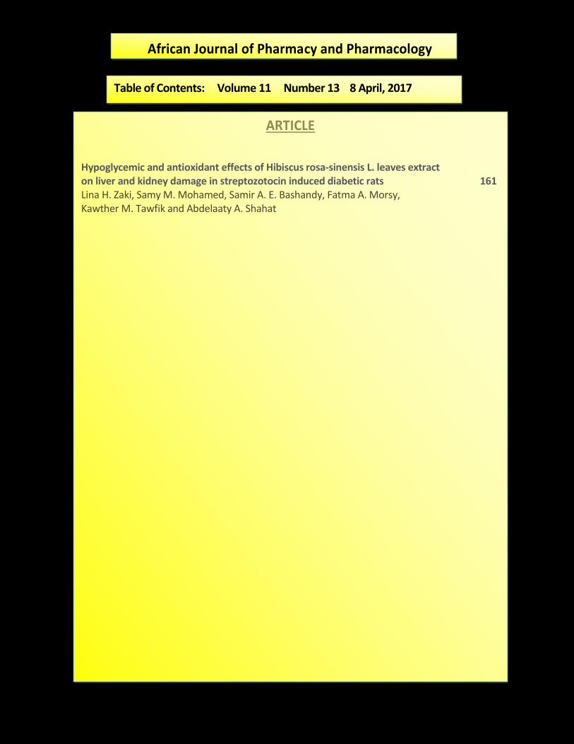
Hypoglycemic and antioxidant effects of Hibiscus rosa-sinensis L. leaves extract on liver and kidney damage in streptozotocin induced diabetic rats 161 Lina H. Zaki, Samy M. Mohamed, Samir A. E. Bashandy, Fatma A. Morsy, Kawther M. Tawfik and Abdelaaty A. Shahat
Table of Contents: Volume 11 Number 13 8 April, 2017

Vol. 11(13), pp. 161-169, 8 April, 2017
DOI: 10.5897/AJPP2017.4764
Article Number: 830D3AF63750
ISSN 1996-0816
Copyright © 2017
Author(s) retain the copyright of this article
http://www.academicjournals.org/AJPP
African Journal of Pharmacy and Pharmacology
Full Length Research Paper
Hypoglycemic and antioxidant effects of Hibiscus rosa-sinensis L. leaves extract on liver and kidney damage in
streptozotocin induced diabetic rats
Lina H. Zaki1, Samy M. Mohamed2, Samir A. E. Bashandy3, Fatma A. Morsy4, Kawther M. Tawfik1 and Abdelaaty A. Shahat5,6*
1Botany Department, Woman’s College, Ain Shams University, Cairo, Egypt.
2Medicinal and Aromatic Plants Research Department, National Research Centre, 33 El Bohouth P.O. Box 12622,
Dokki, Cairo, Egypt. 3Pharmacology Department, National Research Centre, 33 El Bohouth P.O. Box 12622, Dokki, Cairo, Egypt.
4Pathology Department, National Research Centre, 33 El Bohouth. P.O. Box 12622, Dokki, Cairo, Egypt.
5Phytochemistry Department, National Research Centre, 33 El Bohouth P.O. Box 12622, Dokki, Cairo, Egypt.
6Pharmacognosy Department, College of Pharmacy, King Saud University, Riyadh 11451, Saudi Arabia.
Received 6 March, 2017: Accepted 31 March, 2017
Hibiscus is commonly used traditionally for the treatment of some diseases such as hypertension and as antidiabetic herbal medicine. The objective of the present study is to investigate the effect of the oral administration of aqueous methanolic extract of Hibiscus rosa-sinensis leaves (400 mg/Kg) on streptozotocin (STZ) induced diabetic rats and alteration in liver and kidney functions. The treatment of diabetic rats with hibiscus leaves extract reduced levels of plasma glucose, cholesterol, aspartate aminotransferase (AST), alanine aminotransferase (ALT), uric acid and creatinine and hepatic malondialdehyde that was elevated in diabetic rats. Moreover, the Hibiscus leaves extract mitigates the decrease in hepatic superoxide dismutase and plasma protein levels due to STZ injection. The treatment of rats with STZ only results in some pathological effects in liver and kidneys as degeneration in most of hepatocyte and glomeruli. The extract of H. rosa-sinensis leaves reduced the pathological changes. The treatment of diabetic rats with Hibiscus extract was shown to have hepatic and renal protective effects in diabetic rats induced experimentally. Here, two compounds, that is, orientin (Luteolin-8-C-glucoside) and verbascoside (phenylpropanoids glycoside) were isolated from H. rosa-sinsensis. The two compounds were identified by spectral analysis (UV,
1H and
13C-NMR). These results
clearly indicate that aqueous leaves extract of H. rosa-sinensis possess antidiabetic and hypolipidemic effects in diabetic rats which may be due to antioxidant properties of the hibiscus extract. Key words: Hibiscus rosa-sinensis, hypoglycemic, antioxidant, orientin, verbascoside.
INTRODUCTION The plant kingdom represents a large reservoir of biologically active compounds not only as drugs, but also as unique templates that could serve as a starting point for synthetic analogs. Numerous biologically active plants
are discovered by evaluation of ethnopharmacological data, and these plants may offer accessible therapeutic products (Aquino et al., 1995). Numerous medicinal plants are used traditionally for treatment and
162 Afr. J. Pharm. Pharmacol. management of diabetes (Verspohl. 2002).Various active principles of plants with hypoglycemic activity have been identified, including alkaloids, flavonoids, glycosides and polysaccharides (Day 1990). Among the herbal remedies, hibiscus is used in folk medicine. It has antihypertensive (Ojeda et al., 2010), antiatherosclerosis (Chen et al., 2003), anti-inflammatory (Tomar et al., 2010) and analgesic (Sawarkar et al., 2009) activities. Moreover, the extract of Hibiscus can be used effectively in the treatment of peptic ulcer (Mandade et al., 2012) and Leukaemia (Arullappan et al., 2013).
The leaves of Hibiscus rosa-sinensis, a well-known member of the family Malvaceae, were found to contain large amounts of phenolic and flavonoid compounds. Methanolic extract of H. rosa-sinensis possessed significant antioxidant activity as compared to aqueous extract (Garg et al., 2012). It is concluded that Hibiscus cannabinus extract has significant antidiabetic activity, which lowered blood glucose level in diabetic rats (Rajkumar et al., 2011). Diabetes mellitus is one of metabolic syndrome that alter carbohydrate, lipid and protein metabolism and additionally increased risk of complications of various vascular diseases. Hyperlipidemia associated atherosclerosis is the most common cause of death in diabetes (Chait and Bornfeld, 2009). Insulin-dependent diabetes mellitus or type 1 diabetes is an autoimmune disorder characterized by destruction of insulin producing β-cells because auto-aggressive T-lymphocytes infiltrate the pancreas that leads to hypoinsulinemia and thus hyperglycemia (Bach, 1995). Hyperglycemic condition results in an increased glycosylation and biochemical and morphological abnormalities which over a period of time develops diabetic complications such as nephropathy, retinopathy, neuropathy and cardiomyopathy (Arky, 1982). Moreover, diabetes altered liver and kidney functions (Elgazar et al., 2013). The numbers of people with diabetes will be more than double over the next 25 years, to reach a total of 366 million by 2030. Most of this increase will occur in developing countries (WHO, 2015). Preliminary phytochemical screening of H. rosa sinensis stem and leaves revealed the presence of several classes of compounds including flavonoids, flavonoids, glycosides and tannins (Ajay et al., 2007).
In this study, the efficacy of H. rosa -sinensis leaf extract in relieving the hepatic oxidative stress, hypercholesterolemia and kidney damage associated with STZ-induced diabetic rat model were investigated. Here, two compounds of H. rosa-sinsensis, that is, orientin (Luteolin-8-C-glucoside) were isolated from EtOAc fraction and (phenylpropanoids glycoside) verbascoside from BuOH fraction, these two compounds
were isolated for the first time from this plants.
MATERIALS AND METHODS Leaves of H. rosa-sinensis were collected during the flowering period from the garden of the National Research Centre, Cairo, Egypt in April 2010. The plant was kindly identified by, Mrs. Tersea Labib, a taxonomist at Orman Botanical garden, Giza, Egypt and Dr. Mona Marzok, Researcher at the Herbarium of National Research Centre, Giza, Egypt. The voucher specimens were deposited at the herbarium of the National Research Centre, Cairo, Egypt.
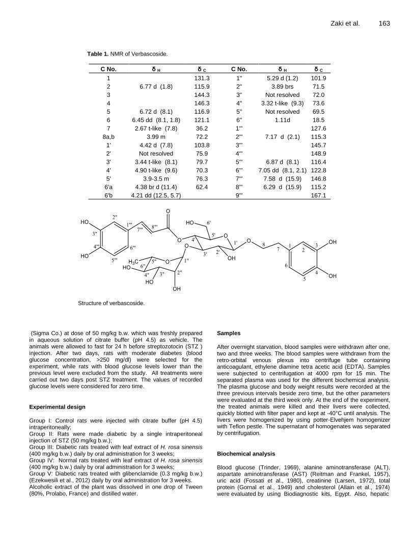
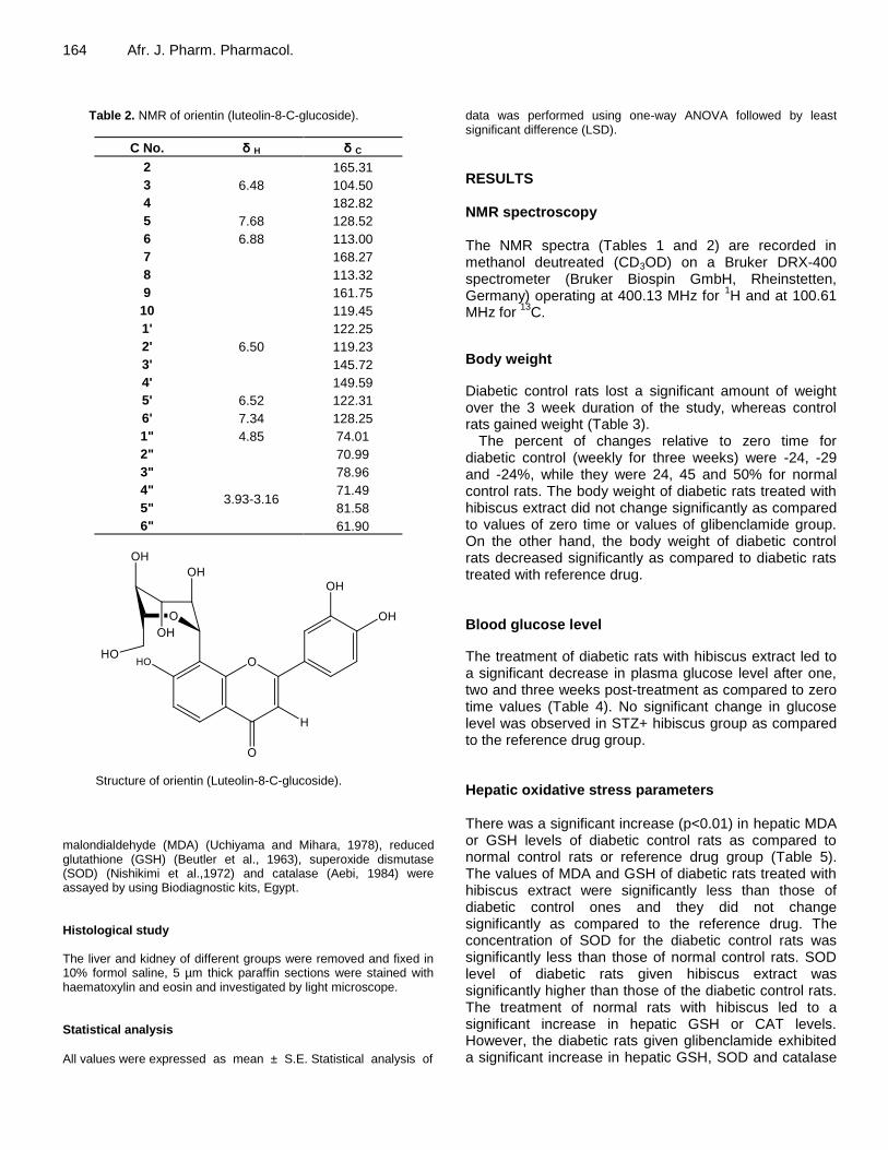
Preparation of the extract and isolation of compounds 1200 g of H. rosa-sinensis (HRS) leaves were cleaned and dried completely under the mild sun and then ground with an electric grinder. The powdered material was extracted with 80% methanol at room temperature for 7 days with shaking and stirring. The extract was filtered and then concentrated to dryness in the rotary evaporator to obtain crude extract. The yield was 9.42% (w/w, in terms of dried starting material). 20 g from the dry extract were dissolved in distill water and partitioned successfully in chloroform (CHCl3), ethyl acetate (EtOAc) and n-butanol (BuOH). The EtOAc and BuOH fractions were subjected to different column chromatography on Silica gel and Sephedex L-H-20 column led to the isolation of orientin from the EtOAc fraction and verbascoside from the BuOH fraction. The two compounds were identified by spectral analysis 1H and 13C-NMR (Tables 1 and 2) and compared with published data (Ismail et al., 1995; kim et al., 2010).
Animals
Adult male albino rats (Sprague Dawley rats) weighing 140 to 190 g were obtained from animal house, National Research Centre, Dokki, Giza. The rats had free access to standard rodent chow and water ad libitum. All animals received human care in compliance with guidelines of Ethical Committee of National Research Center and followed the recommendations of The National Institute of Health Guide for care and use of Laboratory animals (Eighth edition). The animals were housed throughout the experiment in polypropylene cages (each cage housing six animals) and allowed to acclimatize to the laboratory environment for seven days before the beginning of the experiment. Animals were maintained under controlled conditions of temperature (25 ± 1°C), humidity (50±15%) and normal photoperiod (12 to 12 h light-dark cycles).
Acute toxicity studies (LD50) Acute toxicity studies of the plants extracts were conducted for doses of the plant extracts administered to animals in this study. Oral LD50
determination was done by the method of Lorke (1983)
using rats. No mortality in the rats was observed up to 3000 mg/kg.
Induction of diabetes
Diabetes was induced by intraperitoneal injections of streptozotocin
*Corresponding author. E-mail: [email protected]. Tel: +966 537507320. Fax: +966 1 4676220.
Author(s) agree that this article remain permanently open access under the terms of the Creative Commons Attribution
License 4.0 International License
Zaki et al. 163
Table 1. NMR of Verbascoside.
C No. δ H δ C C No. δ H δ C
1
131.3 1" 5.29 d (1.2) 101.9
2 6.77 d (1.8) 115.9 2" 3.89 brs 71.5
3
144.3 3" Not resolved 72.0
4
146.3 4" 3.32 t-like (9.3) 73.6
5 6.72 d (8.1) 116.9 5" Not resolved 69.5
6 6.45 dd (8.1, 1.8) 121.1 6" 1.11d 18.5
7 2.67 t-like (7.8) 36.2 1''' 127.6
8a,b 3.99 m 72.2 2''' 7.17 d (2.1) 115.3
1' 4.42 d (7.8) 103.8 3''' 145.7
2' Not resolved 75.9 4''' 148.9
3' 3.44 t-like (8.1) 79.7 5''' 6.87 d (8.1) 116.4
4' 4.90 t-like (9.6) 70.3 6''' 7.05 dd (8.1, 2.1) 122.8
5' 3.9-3.5 m 76.3 7''' 7.58 d (15.9) 146.8
6'a 4.38 br d (11.4) 62.4 8''' 6.29 d (15.9) 115.2
6'b 4.21 dd (12.5, 5.7) 9''' 167.1
Structure of verbascoside.
(Sigma Co.) at dose of 50 mg/kg b.w. which was freshly prepared in aqueous solution of citrate buffer (pH 4.5) as vehicle. The animals were allowed to fast for 24 h before streptozotocin (STZ ) injection. After two days, rats with moderate diabetes (blood glucose concentration, >250 mg/dl) were selected for the experiment, while rats with blood glucose levels lower than the previous level were excluded from the study. All treatments were carried out two days post STZ treatment. The values of recorded glucose levels were considered for zero time.
Experimental design
Group I: Control rats were injected with citrate buffer (pH 4.5) intraperitoneally; Group II: Rats were made diabetic by a single intraperitoneal injection of STZ (50 mg/kg b.w.); Group III: Diabetic rats treated with leaf extract of H. rosa sinensis (400 mg/kg b.w.) daily by oral administration for 3 weeks; Group IV: Normal rats treated with leaf extract of H. rosa sinensis (400 mg/kg b.w.) daily by oral administration for 3 weeks; Group V: Diabetic rats treated with glibenclamide (0.3 mg/kg b.w.) (Ezekwesili et al., 2012) daily by oral administration for 3 weeks. Alcoholic extract of the plant was dissolved in one drop of Tween (80%, Prolabo, France) and distilled water.
Samples
After overnight starvation, blood samples were withdrawn after one, two and three weeks. The blood samples were withdrawn from the retro-orbital venous plexus into centrifuge tube containing anticoagulant, ethylene diamine tetra acetic acid (EDTA). Samples were subjected to centrifugation at 4000 rpm for 15 min. The separated plasma was used for the different biochemical analysis. The plasma glucose and body weight results were recorded at the three previous intervals beside zero time, but the other parameters were evaluated at the third week only. At the end of the experiment, the treated animals were killed and their livers were collected, quickly blotted with filter paper and kept at -40°C until analysis. The livers were homogenized by using potter-Elvehjem homogenizer with Teflon pestle. The supernatant of homogenates was separated by centrifugation.
Biochemical analysis
Blood glucose (Trinder, 1969), alanine aminotransferase (ALT), aspartate aminotransferase (AST) (Reitman and Frankel, 1957), uric acid (Fossati et al., 1980), creatinine (Larsen, 1972), total protein (Gornal et al., 1949) and cholesterol (Allain et al., 1974) were evaluated by using Biodiagnostic kits, Egypt. Also, hepatic
164 Afr. J. Pharm. Pharmacol.
Table 2. NMR of orientin (luteolin-8-C-glucoside).
C No. δ H δ C
2
165.31
3 6.48 104.50
4
182.82
5 7.68 128.52
6 6.88 113.00
7
168.27
8
113.32
9
161.75
10
119.45
1'
122.25
2' 6.50 119.23
3'
145.72
4'
149.59
5' 6.52 122.31
6' 7.34 128.25
1" 4.85 74.01
2"
3.93-3.16
70.99
3" 78.96
4" 71.49
5" 81.58
6" 61.90
Structure of orientin (Luteolin-8-C-glucoside).
malondialdehyde (MDA) (Uchiyama and Mihara, 1978), reduced glutathione (GSH) (Beutler et al., 1963), superoxide dismutase (SOD) (Nishikimi et al.,1972) and catalase (Aebi, 1984) were assayed by using Biodiagnostic kits, Egypt. Histological study The liver and kidney of different groups were removed and fixed in 10% formol saline, 5 µm thick paraffin sections were stained with haematoxylin and eosin and investigated by light microscope. Statistical analysis
All values were expressed as mean ± S.E. Statistical analysis of
data was performed using one-way ANOVA followed by least significant difference (LSD).
RESULTS NMR spectroscopy The NMR spectra (Tables 1 and 2) are recorded in methanol deutreated (CD3OD) on a Bruker DRX-400 spectrometer (Bruker Biospin GmbH, Rheinstetten, Germany) operating at 400.13 MHz for
1H and at 100.61
MHz for 13
C.
Body weight
Diabetic control rats lost a significant amount of weight over the 3 week duration of the study, whereas control rats gained weight (Table 3).
The percent of changes relative to zero time for diabetic control (weekly for three weeks) were -24, -29 and -24%, while they were 24, 45 and 50% for normal control rats. The body weight of diabetic rats treated with hibiscus extract did not change significantly as compared to values of zero time or values of glibenclamide group. On the other hand, the body weight of diabetic control rats decreased significantly as compared to diabetic rats treated with reference drug. Blood glucose level
The treatment of diabetic rats with hibiscus extract led to a significant decrease in plasma glucose level after one, two and three weeks post-treatment as compared to zero time values (Table 4). No significant change in glucose level was observed in STZ+ hibiscus group as compared to the reference drug group. Hepatic oxidative stress parameters There was a significant increase (p<0.01) in hepatic MDA or GSH levels of diabetic control rats as compared to normal control rats or reference drug group (Table 5). The values of MDA and GSH of diabetic rats treated with hibiscus extract were significantly less than those of diabetic control ones and they did not change significantly as compared to the reference drug. The concentration of SOD for the diabetic control rats was significantly less than those of normal control rats. SOD level of diabetic rats given hibiscus extract was significantly higher than those of the diabetic control rats. The treatment of normal rats with hibiscus led to a significant increase in hepatic GSH or CAT levels. However, the diabetic rats given glibenclamide exhibited a significant increase in hepatic GSH, SOD and catalase

Zaki et al. 165
Table 3. Body weight (g) of diabetic rats treated with H. rosa-sinensis leaves extract.
Treatment Time in week
0 1 2 3
Normal control 144.5±1.52 178.83±9.80@
210.00±8.56@
216.66±8.03@
Diabetic control 140.2±2.30 106.67±6.67@g
100.00±8.94@g
106.67±6.67@g
Diabetic rats+HRS (400 mg/kg) 156.7±2.14 138.33±13.76 135.00±12.04 151.67±11.67
HRS (400 mg/kg) 129±1.73 153.33±8.43 140.00±5.16 143.33±8.03
Diabetic rats+Glib. (0.3 mg/kg) 175±6.71 152.50±5.74 161.67±4.77 186.66±12.82
Each value is the mean±SE, n=6. The values of body weight of all groups before streptozotocin treatment are considered zero time.Values marked with @, differ significantly from zero value; P
@<0.01. Values marked with letter g,
differ significantly from glibenclamide group; Pg<0.01 Statistical analysis of data was performed using ANOVA followed
by least significant difference (LSD). Glib: Glibenclamide
Table 4. Blood glucose level (mg/dl) of diabetic rats treated with H. rosa-sinensis leaves extract.
Treatment Time in week
0 1 2 3
Normal control 84.47±3.16 95.97±3.39
91.9±5.47
93.27±4.76
Diabetic control 389.83±18.72 389.4±27.80g
354.25±33.40g
292.3±24.37g
Diabetic rats + hibiscus (400 mg/kg) 326.67±25.76 205.73±24.06*A
210.05±20.22 *A
154.11±17.91 *A
Hibiscus (400 mg/kg) 91.5±4.56 95.5±5.69 85.06±5.30 61.94±5.98
Diabetic rats+Glib. (0.3 mg/kg) 291.8±19.80*A 201.92±25.92
*A 173.72±9.78
*A 131.67±13.52
*A
Each value is the mean±SE, n=6; Values marked with asterisks differ significantly from control value; P*<0.01; Values marked with letter A, differ significantly from diabetic control group; P
A<0.01; Values marked with letter g, differ significantly from glibenclamide
group; Pg<0.01; Statistical analysis of data was performed using Anova followed by least significant difference (LSD). Glib:
Glibenclamide
Table 5. Hepatic oxidative stress parameters of diabetic rats treated with H. rosa-sinensis leaves extract.
Parameter
Treatment MDA (nmol/mg) GSH (mg/g tissue) SOD (U/g tissue) Catalase (U/g tissue)
Normal control 4.98 ± 0.62 5.12 ± 0.53 223.67 ± 13.38 0.53 ± 0.03
Diabetic control 10.05 ± 0.39*g
17.47 ±2.53*g 177.9 ± 10.78*
g 0.56 ± 0.02
Diabetic rats+HRS
(400 mg/kg) 7.48 ± 0.45*
A 8.18 ± 0.85*
A 246.00 ± 8.63
Ag 0.63 ± 0.02*
HRS (400 mg/kg) 4.77 ± 0.87 9.81 ± 1.59* 211.97 ± 11.82 0.60 ± 0.02*
Diabetic rats+Glib.
(0.3 mg/kg) 7.57 ± 0.19*
A 11.08 ± 1.51*
A 313.20 ± 7.66
*A 0.66 ± 0.02*
A
Each value is the mean±SE, n=6; Values marked with asterisks differ significantly from the control value; P*<0.01. Values marked with letter A, differ significantly from diabetic control group; P
A<0.01. Values marked with letter g, differ
significantly from glibenclamide group; Pg<0.01. Statistical analysis of data was performed using ANOVA followed by
least significant difference (LSD). Glib: Glibenclamide
as compared to the normal control. Hepatic and renal biochemical parameters The concentrations of ALT, AST, uric acid and creatinine of diabetic rats administered hibiscus extract exhibited a
significant decrease as compared to the diabetic control, while they did not change significantly as compared to the reference drug (Table 6). The treatment of diabetic rats with hibiscus extract preserved the protein level in normal range. The cholesterol concentration showed a significant increase in diabetic control rats and it did not change significantly in diabetic rats administered hibiscus

166 Afr. J. Pharm. Pharmacol.
Table 6. Effect of H. rosa-sinensis leaves extract on liver and kidney function parameters.
Parameter
Treatment ALT (U/l) AST (U/l)
Uric acid
(mg/dl)
Creatinine
(mg/dl)
Total protein
(g/dl)
Cholesterol
(mg/dl)
Normal control 24.83±1.96 38.33±4.07 3.68±0.26 1±0.17 7.27±0.24 88.28±2.20
Diabetic control 60.33±1.05*
67.33±1.65*
6.47±0.65*
2.29±0.17*
4.5±0.31*
110.67±7.18*
Diabetic rats+HRS (400 mg/kg) 47.00±4.63*A
55.67±6.59 5.22±0.48*Ag
1.11±0.33A
7.65±0.29Ag
99.67±11.07
HRS (400 mg/kg) 47.17±4.28*A
57.62±3.86*
4.57±0.17*
0.55±0.14*
10.03±0.24*
92.43±3.96
Diabetic rats+Glib. (0.3 mg/kg) 32.5±4.02*A
40±1.29A
3.85±0.16A
1.00±0.06A
6.17±0.35*A
93.17±4.85
Each value is the mean±SE, n=6. Values marked with asterisks, differ significantly from control value; P*<0.01. Values marked with A, differ
significantly from STZ group; PA<0.01. Values marked with g differ significantly from glibenclamide group ,P
g <0.01. Statistical analysis of data
was performed using ANOVA followed by least significant difference (LSD).
extract as compared to the normal control. Histological study Liver sections of the rats treated with streptozotocin only (Figure 1B and C) demonstrated signs of degeneration in most of the hepatocyte in the form of karyolysis and karyorrhexis. Foci of necrosis, an area of hemorrhage in blood sinusoid, dilated congested portal vein and thickening in its wall, cellular infiltration around it, dilated and congested blood sinusoid were seen. Most hepatocyte of liver tissue from rats treated with STZ along with hibiscus extract (Figure 1 D) appeared normal but some signs of degeneration appeared in the form of karyolysis, pyknosis dilated and congested blood sinusoid. Concerning rats treated with streptozotocin in combination with glibenclamide showing improvement in pathological changes in the form of normal hepatocyte, but dilated, congested portal vein and thickening in its wall were seen (Figure 1E). The liver tissue of a rat subjected to hibiscus extract only showed normal histological structure (Figure 1F).
Kidney sections of rats treated with STZ only (Figure 2B) manifested degeneration in most of glomeruli with wide urinary space. On the other hand, decreased pathological changes were recorded in diabetic rats treated with hibiscus extract (Figure 2C). Kidney of diabetic rats treated with glibenclamide had some pathological alterations such as the necrosis in tubular epithelial cells and vacuolar degeneration (Figure 2D). DISCUSSION Medicinal plants have always been an important source for the treatment of many diseases including diabetes owing to their minimal adverse effects. The present experiment indicated the decreased blood glucose level significantly in diabetic rats treated with hibiscus leaves extract compared with diabetic control rats. No significant difference was observed between diabetic rats treated
with hibiscus extract and diabetic rats treated with glibenclamide, a reference drug. The hypoglycemic effect of hibiscus leaves extract is likely due to the enhancement of insulin secretion and the increase of the β-cell number of pancreas islets (Moqbel et al., 2011). The study of Garg et al. (2012) indicated the presence of high content of phenolics, flavonoids and tannins in aqueous and methanolic extracts of H. rosa-sinensis leaves. Jadhav and Puchchakayala (2012) revealed that the blood glucose lowering activity of flavonoid compounds may be by stimulating β-cells to release more insulin or by enhancement peripheral glucose utilization through the skeletal muscle.
Since phenolics and flavonoids are responsible for the antioxidant activity, their presence in the leaf extract of hibiscus indicates good antioxidant activity (Garg et al., 2012). The present results indicated that hibiscus leaves extract reduced oxidative stress in diabetic rats as manifested by decreased hepatic malondialdehyde and enhancement of SOD, an antioxidant enzyme. Antioxidant property of hibiscus leaves extract has been reported (Moqbel and Naik, 2011; Saravanan et al., 2011). This potent antioxidation is thought to form the basis of many of the other healing activities of hibiscus leaves extract including its hepatoprotective activities. This investigation showed increased hepatic glutathione level of diabetic control rats significantly, suggesting a compensatory defense mechanism.
The extract of hibiscus leaves significantly inhibited the increase in the activities of AST and ALT in diabetic rats and it reduced the pathology of the liver. The results of this study showed a significant increase in lipid peroxidation in the liver of STZ diabetic rats. The ability of lipid peroxidation to generate free radicals was confirmed (Szkudelski, 2001) which may lead to the pathological changes observed in liver and kidney of the present work. The activities of AST and ALT are cytosolic marker enzymes, indicating hepatocellular necrosis as they are released into the blood after cell membrane damage. The current study revealed that diabetic control group has significantly higher level of serum AST and ALT as compared to that of the normal control group. Liver

Zaki et al. 167
Figure 1. Liver sections of diabetic or normal rats treated with hibiscus leaves extract. A: Normal control. B: Liver of a rat treated with STZ only showing signs of degeneration in most of hepatocyte in the form of karyolysis (yellow arrow), karyorhexis (black arrow), foci of necrosis and area of hemorrhage in blood sinusoid were seen (star), vacuolar degeneration could be observed at right of figure. C: Another field of previous group (STZ only)showing dilated, congested, edematous, vacuolated portal vein, thickening in the wall (yellow curved arrow). Dilated and congested blood sinusoid (light blue arrow) and ceullar infiltration around (black arrow). D: Liver of diabetic rat treated with hibiscus extract. Most of hepatocyte appeared normal but some exhibited signs of degeneration in the form of karyolysis (green arrow), pyknosis (yellow arrow). Dilated congested portal vein (star) and cellular infiltration (red arrow) were observed. E: Section in the liver of diabetic rat treated with glibenclamide showing minimal pathological changes. F: Liver of normal rat given hibiscus extract showing normal structure. All the sections are Hx&E x400 except C is Hx&E x200.
sections of diabetic control rats showed congestion of hepatic sinusoid, vacuolization of hepatocytes and necrosis of hepatocytes. These results agreed with Hamadi et al. (2012) who reported that elevated activities of serum AST and ALT is a common sign of liver diseases among diabetic rats. Hyperglycemia increases the generation of free radicals by glucose auto-oxidation and the increment of free radicals may lead to cell damage. The present study indicated that diabetic control rats had significant increase in plasma levels of uric acid and creatinine and had significant decrease in concentration of protein as compared to that of the normal control rats.
These results assured, by Parvizi et al., (2014) who
revealed that hyperglycemia is associated with kidney dysfunctions in the diabetic rats which may be related to
the generation of reactive oxygen species and lipid peroxidation that led to tissue injury. In addition, Shah et al. (2007) reported that increased oxidative stress and reduced antioxidative ability in diabetes results in renal tubular injury, proteinuria and leads to gradual loss of renal function.
Hypercholesterolemia has been reported to occur in diabetes (Samarghandian et al., 2014). In this study, TC level increased significantly in diabetic control rats when compared with normal rats. Treatment of diabetic rats with hibiscus extract significantly reduced TC level. The hypolipidemic effect of hibiscus was due to the action of its different constituents, including sitosterol-β- D-galactoside (Mironova and Kalashnikova, 1982) and flavonoids (Liu et al., 2010). Moreover, Yang et al. (2010) reported that polyphenols of hibiscus exhibited more

168 Afr. J. Pharm. Pharmacol.
Figure 2. Kidney sections of diabetic or normal rats treated with hibiscus leaves extract. A: Normal control. B: Kidney of a rat treated with STZ only showing degeneration in most of glomeruli with wide urinary space (red star), while others showed lobulation (olive green). Vacuolar degeneration in some tubular epithelial cells (yellow arrow) was observed. C: Diabetic rat treated with hibiscus extract, most tubule appeared normal. D: Diabetic rat treated with glibenclamide showing wide urinary space (star), necrosis in tubular epithelial cells and vacuolar degeneration (black curved arrow). E: Normal rats treated with hibiscus extract, most renal tubules and glomerulus appeared normal (Hx&E x400).
potency to decrease plasma cholesterol and LDL cholesterol than the crude extract, and increased HDL cholesterol.
In the LD50 study, no mortality was recorded in rats except 3000 mg/kg of the methanolic extract of H. rosa-sinensis. It has been established that any substance with LD50
estimate greater than 2000 mg/kg body weight by
oral route may be considered to have low toxicity and safe (Bruce, 1987).
Conclusion
H. rosa-sinensis could have great importance as a safe therapeutic agent in diabetes mellitus. Hibiscus leaves
extract has a significant hypoglycemic and hypocholestrolemic effects in diabetic rats which may led to a decrease in oxidative stress and improvement of liver and kidney functions. H. rosa-sinensis could have great importance as safe therapeutic agent. CONFLICT OF INTERESTS The authors declare that there is no conflict of interest.
ACKNOWLEDGEMENTS The authors are grateful for the sponsorship of the
Research Centre, College of Pharmacy, the Deanship of the Scientific Research, King Saud University, Riyadh, Saudi Arabia. REFERENCES Aebi H (1984). Catalase in vitro. Methods Enzymol. 105:121-126. Ajay M, Chai HJ, Mustafa AM, Gilani AH, Mustafa MR (2007).
Mechanisms of the anti-hypertensive effect of Hibiscus sabdariffa L. calyces. J. Ethnopharmacol. 109(3):15-388.
Allain CC, Poon LS, Chan CS, Richmond WF, Fu PC (1974). Enzymatic Determination of Total Serum Cholesterol. Clin. Chem. 20:470-475.
Aquino R, De Simone F, De Tommasi N, Piacente S, Pizza C (1995). Structure and biological activity of sesquiterpene and diterpene derivatives from medicinal plants, in Hostettmann K, Maillard M, Hamburger M (eds.), Phytochemistry of Plants Used in Traditional Medicine, Oxford University Press, New York, USA. pp. 249-278,
Arky RA (1982). Clinical correlates of metabolic derangements of Diabetes Mellitus. In: Kozak, G.P. (Ed.), Complications of diabetes mellitus. W.B. Saunders, Philadelphia. pp. 16-20.
Arullappan S, Muhamad S, Zakaria Z (2013). Cytotoxic Activity of the Leaf and Stem Extracts of Hibiscus rosa-sinensis (Malvaceae) against Leukaemic Cell Line (K-562). Trop. J. Pharm. Res. 12(5):743-746.
Bach JF (1995). Insulin-dependent diabetes mellitus as a β-cell targeted disease of immunoregulation. J. Autoimmunol. 8:439-463.
Beutler E, Durgun O, Kelly BM (1963). Improved method for the determination of blood glutathione. J. Lab. Clin. Med. 51:882-888.
Bruce RD (1987). A confirmatory study of the up-and-down method for acute oral toxicity testing. Fundam. Appl. Toxicol. 8(1):97-100.
Chait A, Bornfeldt KE (2009). Diabetes and atherosclerosis: is there Hyperglycemia?. J. Lipid Res. Suppl. 50:S335-S339.
Chen CC, Hsu JD, Wang SF (2003). Hibiscus sabdariffa Extract Inhibits the Development of Atherosclerosis in Cholesterol-Fed Rabbits. J. Agric. Food Chem. 51(18):5472-5477
Day C (1990). Hypoglycaemic compounds from plants, in Bailey CJ, Flatt PR (eds.), New Antidiabetic.,Drugs, Smith-Gordon and Nishimura Company Limited, London, Japan. P 26.
Elgazar AF, Rezq AA, Bukhari HM (2013). Anti-Hyperglycemic Effect of Saffron Extract in Alloxan-Induced Diabetic Rats. Eur. J. Biol. Sci, 5(1):14-22.
Ezekwesili CN, Ogbunugafor HA, Ezekwesili–Ofili JO (2012). Anti-diabetic Activity of Aqueous Extracts of Vitex doniana Leaves and Cinchona calisaya Bark in Alloxan–Induced Diabetic Rats. Int. J. Trop. Health 2(4):290-300.
Fossati P, Prencipe L, Berti G (1980). Use of 3,5-dichloro-2-hydroxybenzenesulfonic acid/4-aminophenazone chromogenic system in direct enzymic assay of uric acid in serum and urine. Clin. Chem. 26(2):227-231.
Garg D, Shaikh A, Muley A, Marar T (2012). In-vitro antioxidant activity and phytochemical analysis in extracts of Hibiscus rosa-sinensis stem and leaves. Free Radic. Antioxid. 2(3):41-46.
Gornal AC, Bardawill CJ, David MM (1949). Determination of serum protein by means of biuret reaction. J. Biol. Chem. 177(2):751-756.
Hamadi N, Mansour A, Hassan MH, Khalifi‐Touhami F, Badary O (2012). Ameliorative Effects of Resveratrol on Liver Injury in Streptozotocin-Induced diabetic Rats. J. Biochem. Mol. Toxicol. 26(10):384-392.
Ismail LD, El-Azizi MM, Khalifa TI, Stermitz FR (1995). Verbascoside derivatives and iridoid glycosides from Penstemon Carndallii. Phytochemistry 39(6):1391-1393.
Jadhav R, Puchchakayala G (2012). Hypoglycemic and antidiabetic activity of flavonoids: Boswellic acid,Ellagic acid,Quercetin,Rutin on streptozotocin-Nicotinamide induced type 2 diabetic rats. Int. J. Pharm. Pharm. Sci. 4(2):251-256.
Kim J, Lee I, Seo J, Jung M, Kim Y, Yim N, Bae K (2010). Vitexin, orientin and other flavonoids from Spirodela polyrhiza inhibit adipogenesis in 3T3-L1 cells. Phytother. Res. 24(10):1543-1548.
Larsen K (1972). Creatinine assay by a reaction-kinetic principle. Clin. Chem. Acta. 41:209-217
Zaki et al. 169 Liu YT, Lu BN, Xu LN, Yin LH, Wang XN, Peng JY, Liu KX (2010). The
antioxidant activity and hypolipidemic activity of the total flavonoids from the fruit of Rosa laevigata Michx. Nat. Sci. 2(3):175-183.
Lorke D (1983). A new approach to acute toxicity testing. Arch. Toxicol. 54(4):275-287.
Mandade RJ, Sreenivas SA, Sakarkar DM, Choudhury A (2011) .Pharmacological effects of aqueous-ethanolic extract of Hibiscus rosasinensis on volume and acidity of stimulated gastric secretion. Asian Pac. J. Trop. Biomed. 4(11):883-888.
Mironova VN, Kalashnikova LA (1982). Hypolipidemic action of beta-D-glycoside beta-sitosterol in the rat. Farmakol. Toksikol. 45(6):45-47.
Moqbel FS, Naik PR (2011). Effect of different fractions of Hibiscus rosa sinensis leaf extract on islets of Langerhans and antioxidant activity in non-obese diabetic (NOD) mouse. J. Appl. Nat. Sci. 3(2):206-210
Moqbel FS, Naik PR, Najma HM, Selvaraj S (2011). Antidiabetic properties of Hibiscus rosa sinensis L. leaf extract fraction on non-obese diabetic (NOD) mouse. Ind. J. Exp. Biol. 49(1):24-29.
Nishikimi M, Appaji N, Yagi K (1972).The occurrence of superoxide anion in the reaction of reduced phenazine methosulfate and
molecular oxygen. Biochem. Biophys. Res. Commun. 46(2):849‐854. Ojeda D, Jiménez-Ferrer E, Zamilpa A, Herrera-Arellano A, Tortoriello
J, Alvarez L (2010). Inhibition of angiotensin convertin enzyme (ACE) activity by the anthocyanins delphinidin- and cyanidin-3-O-sambubiosides from Hibiscus sabdariffa. J. Ethnopharmacol. 127(1):7-10.
Parvizi MR, Parviz M, Tavangar SM, Soltani N, Kadkhodaee M, Seifi B, Azizi Y, Keshavarz M (2014). Protective effect of magnesium on renal functionin STZ-induced diabetic rats. J. Diabetes Metab. Disord. 13(84):1-9.
Rajkumar E, Uhayakumar E, Sekar M, Senthil MK (2011). Antidiabetic activity of methanolic extract of Hibiscus Cannabinus in streptozotocin induced diabetic rats. Int. J. Pharm. BioSci. 2(1):125-130.
Reitman S, Frankel S (1957). A colorimetric method for the determination of serum glutamic oxalacetic and glutamic pyruvic transaminases. Am. J. Clin. Pathol. 28(1):56-63.
Samarghandian S, Azimi-Nezhad M, Samini F (2014). Ameliorative Effect of Saffron Aqueous Extract on Hyperglycemia, Hyperlipidemia, and Oxidative Stress on Diabetic Encephalopathy in Streptozotocin Induced Experimental Diabetes Mellitus. BioMed. Res. Int. pp. 1-12.
Saravanan D, lakshmi A, Gobinath M, Giriesh kumar B, Priya S, Syamala E, Rahamathbee K (2011). Potential antioxidant, hypoglycemic and hypolipidemic effect of leaves of Hibiscus platanifolius Linn Int. J. Pharm. Sci. Drug Res. 3(3):236-240.
Sawarkar A, Jangde CR, Thakre PD, Kadoo R, Shelu S (2009). Analgesic activity of Hibiscus rosa sinensis Linn in rat. Vet. World 2(9):353-354.
Shah SV, Baliga R, Rajapurkar M, Fonseca VA (2007). Oxidants in chronic kidney disease. J. Am. Soc. Nephrol. 18(1):16-28.
Szkudelski T (2001).The Mechanism of Alloxan and Streptozotocin Action in B cells of the Rat Pancreas. Physiol. Res. 50(1):536-546.
Tomar V, Kannojia P, Jain KN, Dubey KS (2010). Anti-noceptive and anti-inflammatory activity of leaves of Hibiscus Rosa Sinesis. Int. J. Res. Ayurveda Pharm. 1(1): 201-205.
Trinder P (1969). Determination of glucose in blood using glucose oxidase with an alternative oxygen acceptor. Ann. Clin. Biochem. 6:24-27.
Uchiyama M, Mihara M (1978). Determination of malonaldehyde precursor in tissues by thiobarbituric acid test. Anal. Biochem. 86(1):271- 278.
Verspohl EJ (2002). Recommended testing in diabetes research. Planta Med. 68(7):581-590.
World Organization of Health (WHO) (2015). Diabetes Fact sheet N°312 Updated January 2015.
Yang MY, Peng CH, Chan KC, Yang YS, Huang CN, Wang CJ (2010). The Hypolipidemic Effect of Hibiscus sabdariffa Polyphenols via Inhibiting Lipogenesis and Promoting Hepatic Lipid Clearance. J. Agric. Food Chem. 58(2):850-859.

African Journal
of Pharmacy and
Pharmacology
Related Journals Published by Academic Journals
■ Journal of Medicinal Plant Research
■ African Journal of Pharmacy and Pharmacology
■ Journal of Dentistry and Oral Hygiene
■ International Journal of Nursing and Midwifery
■ Journal of Parasitology and Vector Biology
■ Journal of Pharmacognosy and Phytotherapy
■ Journal of Toxicology and Environmental Health
Sciences


