ASPECTS ON ADVANCED PROCEDURES DURING ENDOSCOPIC RETROGRADE CHOLANGIOPANCREATOGRAPHY FOR COMPLEX HEPATOBILIARY DISORDERS Faculty of Medicine and Health Science Department of Surgical Sciences, Division of Surgery Stellenbosch University, Tygerberg, South Africa Department of Clinical Science, Intervention and Technology (CLINTEC), Division of Surgery Karolinska Institutet, Stockholm, Sweden Jeanne Adéle Lübbe Stockholm 2021 Stellenbosch University 2021
Transcript
ASPECTS ON ADVANCED PROCEDURES
DURING ENDOSCOPIC RETROGRADE CHOLANGIOPANCREATOGRAPHY FOR COMPLEX
HEPATOBILIARY DISORDERS
Faculty of Medicine and Health ScienceDepartment of Surgical Sciences, Division of Surgery
Stellenbosch University, Tygerberg, South Africa
Department of Clinical Science, Intervention and Technology (CLINTEC), Division of Surgery
Aspects on Advanced Procedures during Endoscopic Retrograde Cholangiopancreatography (ERCP) for Complex Hepatobiliary Disorders
Department of Clinical Science Intervention and Technology (CLINTEC)
THESIS FOR DOCTORAL DEGREE (Ph.D.)
By
Jeanne Adéle Lübbe
The thesis will be defended in public at Birkeaulan 2, Level 5, Karolinska University Hospital Huddinge, Stockholm on Friday the 26th of February 2021 at 09:00
Principal Supervisor: Professor Lars Enochsson Umeå University Department of Surgical and Perioperative Sciences Karolinska Institutet CLINTEC Division of Surgery Co-supervisors: Professor Eduard Jonas University of Cape Town Department of Surgery Karolinska Institutet CLINTEC Division of Surgery Associate Professor Urban Arnelo Karolinska Institutet CLINTEC Division of Surgery Umeå University Department of Surgical and Perioperative Sciences Professor Samuel Moore University of Stellenbosch Department of Surgical Sciences Division of Surgery
Opponent: Professor Ajith Siriwardena University of Manchester Department of Hepatobiliary Surgery Examination Board: Associate Professor Per Stål Karolinska Institutet Department of Medicine Associate Professor Stefan Linder Karolinska Institutet CLINTEC Division of Surgery Professor Marie-Lois Ivarsson University of Gothenburg Department of Surgery Professor Brian Warren University of Stellenbosch Department of Surgical Sciences Division of Surgery Professor Martin Brand University of Pretoria Department of Surgery
Stellenbosch University https://scholar.sun.ac.za
Stellenbosch University https://scholar.sun.ac.za
To Isabelle and Willem, Dané and Ivan
Stellenbosch University https://scholar.sun.ac.za
“In a world deluged by irrelevant information, clarity is power.”
Yuval Noah Harari
Stellenbosch University https://scholar.sun.ac.za
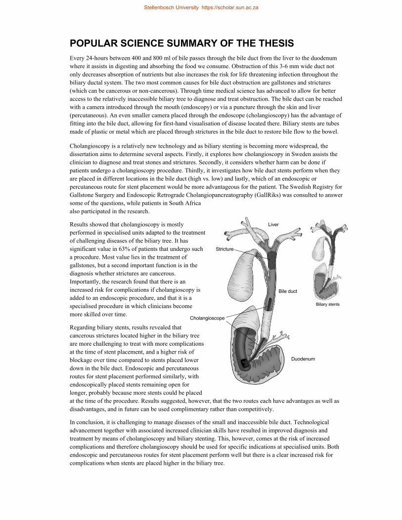
POPULAR SCIENCE SUMMARY OF THE THESIS Every 24-hours between 400 and 800 ml of bile passes through the bile duct from the liver to the duodenum where it assists in digesting and absorbing the food we consume. Obstruction of this 3-6 mm wide duct not only decreases absorption of nutrients but also increases the risk for life threatening infection throughout the biliary ductal system. The two most common causes for bile duct obstruction are gallstones and strictures (which can be cancerous or non-cancerous). Through time medical science has advanced to allow for better access to the relatively inaccessible biliary tree to diagnose and treat obstruction. The bile duct can be reached with a camera introduced through the mouth (endoscopy) or via a puncture through the skin and liver (percutaneous). An even smaller camera placed through the endoscope (cholangioscopy) has the advantage of fitting into the bile duct, allowing for first-hand visualisation of disease located there. Biliary stents are tubes made of plastic or metal which are placed through strictures in the bile duct to restore bile flow to the bowel.
Cholangioscopy is a relatively new technology and as biliary stenting is becoming more widespread, the dissertation aims to determine several aspects. Firstly, it explores how cholangioscopy in Sweden assists the clinician to diagnose and treat stones and strictures. Secondly, it considers whether harm can be done if patients undergo a cholangioscopy procedure. Thirdly, it investigates how bile duct stents perform when they are placed in different locations in the bile duct (high vs. low) and lastly, which of an endoscopic or percutaneous route for stent placement would be more advantageous for the patient. The Swedish Registry for Gallstone Surgery and Endoscopic Retrograde Cholangiopancreatography (GallRiks) was consulted to answer some of the questions, while patients in South Africa also participated in the research.
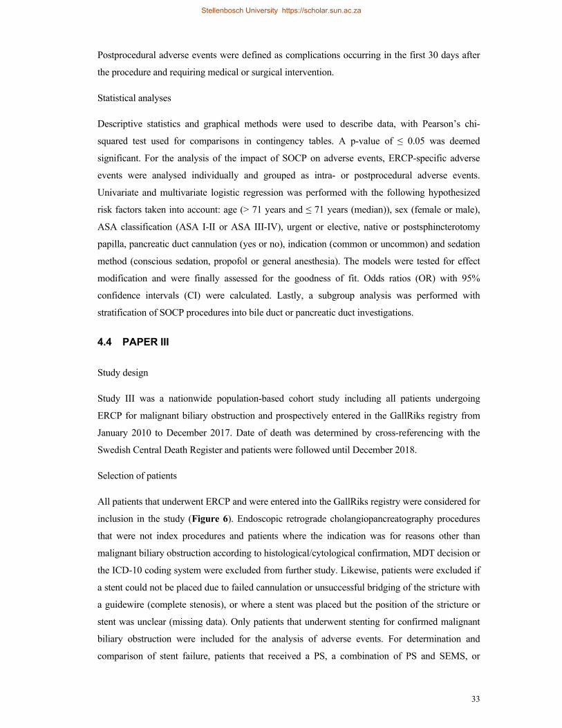
Results showed that cholangioscopy is mostly performed in specialised units adapted to the treatment of challenging diseases of the biliary tree. It has significant value in 63% of patients that undergo such a procedure. Most value lies in the treatment of gallstones, but a second important function is in the diagnosis whether strictures are cancerous. Importantly, the research found that there is an increased risk for complications if cholangioscopy is added to an endoscopic procedure, and that it is a specialised procedure in which clinicians become more skilled over time.
Regarding biliary stents, results revealed that cancerous strictures located higher in the biliary tree are more challenging to treat with more complications at the time of stent placement, and a higher risk of blockage over time compared to stents placed lower down in the bile duct. Endoscopic and percutaneous routes for stent placement performed similarly, with endoscopically placed stents remaining open for longer, probably because more stents could be placed at the time of the procedure. Results suggested, however, that the two routes each have advantages as well as disadvantages, and in future can be used complimentary rather than competitively.
In conclusion, it is challenging to manage diseases of the small and inaccessible bile duct. Technological advancement together with associated increased clinician skills have resulted in improved diagnosis and treatment by means of cholangioscopy and biliary stenting. This, however, comes at the risk of increased complications and therefore cholangioscopy should be used for specific indications at specialised units. Both endoscopic and percutaneous routes for stent placement perform well but there is a clear increased risk for complications when stents are placed higher in the biliary tree.
Liver
Duodenum
Cholangioscope
Stricture
Biliary stents
Bile duct
Stellenbosch University https://scholar.sun.ac.za
ABSTRACT Background: The rapid development in endoscopic technology and associated skills has led to an increase in
more advanced procedures being performed during endoscopic retrograde cholangiopancreatography (ERCP).
Knowledge is limited regarding clinical value, integration, and outcomes for single operator
cholangiopancreatoscopy (SOCP) and endoscopic intervention in the different Bismuth-Corlette (B-C) locations
in the hepatic hilum.
Objectives: To determine the clinical value of SOCP in the diagnosis and treatment of complex hepatobiliary
and pancreatic disease. To describe the nationwide integration of SOCP and the extent to which adverse events
are influenced when SOCP is added to ERCP. To compare adverse events and reintervention rates after
endoscopic stenting for malignant obstruction in the distal and hilar locations of the biliary tree. To compare
outcomes after endoscopic transpapillary (ETP) and percutaneous transhepatic (PTH) stenting in the palliation of
malignant hilar obstruction (MHO).
Methods: In study I all SOCP procedures performed between March 2007-December 2014 at a tertiary high-
volume endoscopy unit were separately graded according to a predefined 4-graded scale estimating therapeutic
value and diagnostic yield. Study II was a nationwide case-control study nested within the cohort of ERCP
procedures, with- or without SOCP, and registered in the Swedish Registry for Gallstone Surgery and ERCP
(GallRiks) between 2007-2012. To assess risk factors for adverse events, multivariate logistic regression was
performed, and odds ratios (OR) calculated. The GallRiks registry was also utilised in study III where all patients
undergoing endoscopic stenting for malignant biliary obstruction between 2010-2017 (based on International
Classification of Diseases (ICD) coding), were included. Kaplan-Meier analysis was employed to calculate stent
patency and Cox proportional hazard models to calculate the risk for recurrent biliary obstruction after single
metal stent placement. To compare ETP and PTH drainage approaches, a retrospective deconstructed analysis of
palliative stenting procedures for MHO at two specialised referral centres over a 5-year period was performed.
Within-group analyses were performed to explore outcomes for different B-C types and Kaplan-Meier and
restricted mean survival time analyses were performed to calculate and compare duration of therapeutic success.
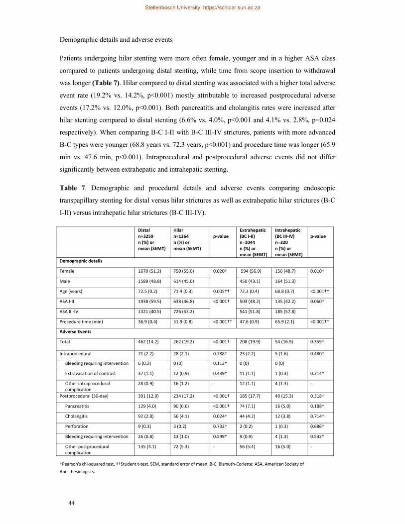
Results: In 365 SOCP procedures, SOCP was found be of pivotal importance in 19% of patients, of great
clinical significance in 44%, and did not affect clinical decision-making or alter clinical course in 37% of
patients. In study II a learning curve was observed after first introduction of 408 SOCP procedures, and
postprocedural adverse events (19.1% vs. 14.0%), pancreatitis (7.4% vs. 3.9%) and cholangitis (4.4% vs. 2.7%)
were more prevalent when SOCP was added to ERCP. After multivariate analysis, the risk for postprocedural
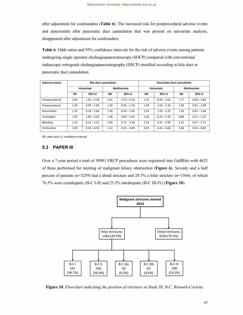
adverse events remained (OR 1.35, 95% CI [1.04 - 1.74]). In 4623 ERCP procedures performed for stenting of
malignant strictures (1364 hilar), adverse events and 6-month reintervention rates were increased after hilar
stenting compared to distal stenting (17.2% vs. 12.0%, 73.4% vs. 55.9%). On multivariate analysis the risk for
reintervention was three times higher after single metal stent placement in the hilum compared to the distal
biliary tree (HR 3.47, 95% CI [2.01-6.00], p<0.001). In 293 patients undergoing palliative stenting for MHO
(52.2% ETP, 47.8% PTH), access and bridging success in the ETP and PTH groups were 83.5% vs. 97.2% and
90.2% vs. 84.5%, respectively. Technical and therapeutic success were equivalent between the two groups, but
duration of therapeutic success was longer after ETP drainage, with a 3-month gain in duration of therapeutic
success after adjustment for B-C type (95% CI [26-160], p=0.006). Cholangitis rates were equivalent (21.4% vs.
24.7%), while pancreatitis was more common in the ETP group and deaths more common in the PTH group.
Conclusions: When added to ERCP, SOCP contributes significant clinical value in 64% of cases. However,
there is an increased risk of intra- and postprocedural adverse events which, together with a learning curve,
suggests that it should likely be performed in specialised high-volume centres. Regarding endoscopic
intervention for MHO, stenting in the hepatic hilum compared to the distal biliary tree is associated with more
adverse events and decreased stent patency. When comparing palliative ETP with PTH stenting for MHO, both
approaches have similar technical and therapeutic success, with ETP drainage being more durable. Future studies
should explore the complimentary role of both approaches in specific B-C types.
Stellenbosch University https://scholar.sun.ac.za
LIST OF SCIENTIFIC PAPERS
I. Marcus Reuterwall, Jeanne Lubbe, Lars Enochsson, Lars Lundell, Magnus Konradsson, Frederik Swahn, Marco Del Chiaro, Matthias Löhr and Urban Arnelo The clinical value of ERCP-guided cholangiopancreatoscopy using a single-operator system BMC Gastroenterology, 2019, 26;19(1):35
II. Jeanne Lubbe, Urban Arnelo, Lars Lundell, Fredrik Swahn, Björn Törnqvist, Eduard Jonas, Matthias Löhr, and Lars Enochsson ERCP-guided cholangioscopy using a single-use system: nationwide register-based study of its use in clinical practice Endoscopy, 2015, 47(9):802–7
III. Jeanne Lubbe, Gabriel Sandblom, Urban Arnelo, Eduard Jonas, and Lars Enochsson Endoscopic stenting for malignant biliary obstruction – results of a nationwide experience Submitted manuscript
IV. Jeanne Lubbe, Jessica Lindemann, Washington Ghondo, Nina Kolev, Peter Aclavio, Stefan Hofmeyr, and Eduard Jonas Endoscopic versus percutaneous drainage of malignant hilar bile duct obstruction – a comparative cohort study Submitted manuscript
RELATED PUBLICATIONS
(Not included in the thesis)
Greger Olsson, Jeanne Lubbe, Urban Arnelo, Eduard Jonas, Björn Törnqvist, Lars Lundell
and Lars Enochsson
The impact of prophylactic pancreatic stenting on post-ERCP pancreatitis: A nationwide,
register-based study
United European Gastroenterology Journal, 2017, 5(1):111–8
Marcus Reuterwall, Alexander Waldthaler, Jeanne Lubbe, Nils Kadesjö, Raffealla Pozzi
Mucelli, Marco Del Chiaro, Matthias Löhr and Urban Arnelo
ESGE European Society of Gastrointestinal Endoscopy
ASGE American Society for Gastrointestinal Endoscopy
CI Confidence interval
OR Odds ratio
HR Hazard ratio
PIEC Percutaneous internal-external catheter
SIS Stent-in-stent
SBS Side-by-side
MDT Multidisciplinary team
ASA American Society of Anesthesiologist
ICD International Classification of Diseases
TB Total bilirubin
ECOG Eastern Cooperative Oncology Group
Stellenbosch University https://scholar.sun.ac.za
Stellenbosch University https://scholar.sun.ac.za
1
1 INTRODUCTION
In healthy individuals between 400-800 ml of bile pass via the bile duct into the duodenum every
24 hours. The two most common ailments affecting this 3-6 mm inaccessible ductal system are
gallstones and biliary strictures. Obstruction to the flow of bile leads to upstream dilation,
secondary bacterial infection (cholangitis), and in time, secondary biliary cirrhosis. Diagnosis as
to the cause and ways in which to relieve biliary obstruction have posed a challenge to physicians
for many years.
The most common benign cause of biliary obstruction is gallstone disease. Gallstones can be
cholesterol or bilirubinate stones that form primarily in the gallbladder and then migrate into the
bile duct, or primary intraductal stones that are formed due to stasis and chronic low-grade
infection. Benign stricture formation (30% of all strictures) can be due to primary sclerosing
cholangitis (PSC), iatrogenic injury, Mirizzi syndrome, anastomotic fibrosis or associated with
chronic pancreatitis.1 Choledochal cysts, haemobilia (blood in the biliary system) and
radiotherapy are rarer causes of benign biliary obstruction. Infections and parasitic infestations
are predominantly seen in developing countries. Malignant biliary strictures are mostly due to
pancreatic / periampullary carcinoma or intra- or extrahepatic cholangiocarcinoma (CC).
Malignant hilar obstruction (MHO) is less frequently caused by gallbladder cancer or centrally
located hepatocellular cancer.2 Lymphoma and malignancy arising anatomically distant from the
biliary system can lead to MHO by means of metastasis to periportal lymph nodes or the liver
parenchyma surrounding the perihilar area. The incidence of both pancreatic adenocarcinoma and
CC has increased in recent years and, as most patients present at an advanced stage of disease,
treatment is mostly aimed at palliation of symptoms.3,4
Imaging of the biliary tree in the 1920s consisted of the oral cholecystogram whereby orally
ingested iodinated phenolphthalein (selectively secreted into bile) provided radiographic images
of the gallbladder and bile ducts.5 As the bile duct was not accessed directly, therapeutic
intervention was not an option. It was not until 1955 when Doubilet and Mulholland injected
contrast into the ampulla of Vater (transpapillary) during open surgery, that direct access to the
biliary tract became feasible.6 Their initial images were static and two-dimensional but were soon
followed by dynamic fluoroscopic imaging and eventual percutaneous biliary access that
followed 30 years later.7
Direct fiberoptic visualization of the bowel lumen was first described in 1957 by Basil
Hirschowitz, and in 1968 McCune was the first to publish a report on endoscopic wire
cannulation of the bile duct in a living patient.8,9 Rapid advancement in endoscopic technology
led to the development of the side-viewing duodenoscope.10
Stellenbosch University https://scholar.sun.ac.za
2
Figure 1. Endoscopic retrograde cholangiopancreatography (ERCP) combined with single
operator cholangiopancreatoscopy (SOCP). Adapted and printed with permission from
Frederik Swahn.
Stellenbosch University https://scholar.sun.ac.za
3
Endoscopic retrograde cholangiopancreatography (ERCP) is the process by which a side-viewing
duodenoscope is used to access the bile duct via the ampulla of Vater in order to obtain
fluoroscopic images (Figure 1).
Currently, the most common means of access to the biliary tree is either via an endoscopic
transpapillary (ETP) approach or a percutaneous transhepatic (PTH) approach. The drive to be
able to perform therapeutic maneuvers during ERCP led to reports of the division of the sphincter
of Oddi (sphincterotomy) both in Germany and Japan in the 1970s, allowing wider access for
insertion of devices into the biliary tree.11,12 In the following years, basic therapeutic mechanisms
were developed. These were aimed at the removal of stones with balloons or baskets and stenting
of strictures with plastic or metal stents.
In 1961, a cholangioscope was introduced directly into the bile duct during open surgery.13 The
advancement from fiberoptic to video-endoscopes allowed for the development of progressively
smaller caliber scopes with sustained good image quality. Currently, less invasive peroral
cholangioscopy can be performed in one of three ways: by directly introducing a cholangioscope
via the mouth into the ampulla of Vater (direct peroral cholangioscopy), by utilizing a specially
designed duodenoscope and custom made cholangioscope (mother-baby system), or by means of
the single operator cholangiopancreatoscopy (SOCP) system. The most common SOCP system is
the SpyGlassTM Direct Visualisation System (Boston Scientific, USA) that passes through a
standard duodenoscope and houses three ports: an optical port that allows passage of optical
fibers for visualisation, an irrigation port that ensures continued optimisation of the visual field,
and a working port through which instruments can be introduced (biopsy forceps or lithotripsy
apparatus). Its single operator status has ensured that it is the most widely adopted means of
performing cholangioscopy in current endoscopic practice. The second-generation digital SOCP
system was introduced in 2015 and allows for improved resolution and a 110° field of vision.
Application in the pancreatic duct is increasingly being reported.
Both the improved visualization of the biliary tree and increased ease of access to the biliary tree
led to ERCP changing from a previously diagnostic modality to mostly a therapeutic intervention
in current practice. Nasobiliary drainage was first reported in 1980, with the placement of a
plastic biliary stent described soon thereafter.14,15 Currently, plastic and metal stents are used.
Stellenbosch University https://scholar.sun.ac.za
4
Plastic stents (PS) have a low cost and
small diameter (maximum of 12F). A
tendency to migration and biofilm
formation results in a limited patency of 3-
6 months (Figure 2). Plastic stents are
mostly used for temporary or short-term
stenting of the bile duct as they can easily
be removed and/or replaced, although
repeated exchanges decrease quality of life
and escalate costs.
Figure 2. Bilateral plastic stents.
Self-expanding metal stents (SEMS) are 10-30
times more expensive than PS but have a larger
diameter (10mm/30F on an 8.5F delivery system)
and thus a patency of 6-12 months (Figure 3).
SEMS occlusion is mostly due to ingrowth in
uncovered SEMS (uSEMS) and overgrowth or
migration in covered SEMS (cSEMS). The
ingrowth occurring in uSEMS makes removal
difficult and precludes its use in scenarios where
temporary stent placement is planned. Similar to
PS, cSEMS can be removed and are thus
considered for short-term stenting in benign
disease. Partially covered SEMS (pcSEMS), where
the flanges are left uncovered, hope to combine the
benefits afforded by both uSEMS and cSEMS.
Regarding ease of placement, the pointed tip and
thin delivery system on which SEMS are preloaded
facilitate passage through tight strictures, whilst PS
passage might have to be preceded by balloon
dilation of very tight strictures. Figure 3. Unisectoral self-expanding metal stent.
Stellenbosch University https://scholar.sun.ac.za
5
Due to the location of the papilla in the duodenal lumen, distal (periampullary) pathology can be
accessed under direct duodenoscopic vision, making therapeutic techniques at this site relatively
straightforward. In contrast, the hepatic hilum can only be indirectly represented on two-
dimensional fluoroscopic imaging or via cholangioscopy. Due to its relative ‘further’ placement
from the duodenal lumen and endoscopist, therapeutic procedures in the hilar biliary system are
technically much more challenging.
Adverse events associated with ERCP are well defined and graded as per consensus agreement.16
Adverse events include pancreatitis, cholangitis, bleeding, perforation, cholecystitis and
cardiopulmonary events. Of these, pancreatitis is the most common. Based on large prospective
series, accepted adverse event rate after ERCP varies between 3%-10% depending on diagnostic
or therapeutic intent.17 When more advanced procedures such as cholangioscopy and/or stenting
are added to ERCP, or when intervention is located in the hepatic hilum as opposed to the
periampullary area, intervention and location-specific complications can arise.
Stellenbosch University https://scholar.sun.ac.za
Stellenbosch University https://scholar.sun.ac.za
7
2 BACKGROUND
2.1 THE HEPATOBILIARY SYSTEM
2.1.1 Biliary anatomy and physiology
In the most commonly encountered anatomy of the biliary tree (56%, type 1), the right anterior
sectoral duct (draining segments 5 and 8) combines with the right posterior sectoral duct (draining
segments 6 and 7) to form the short (1 cm) vertically orientated right hepatic duct which is prone
to tumour involvement (Figure 1).18,19 Confluence of the segmental ducts draining segments 2
and 3 form the left lateral sectoral duct at the umbilical fissure. This duct receives variable
drainage from segment 4 to end in the left hepatic duct. The longer (3 cm) and horizontally
orientated left hepatic duct runs in the peritoneal sheath of the hilar plate. The extrahepatic biliary
tree is formed by the confluence of the left and right hepatic ducts to form the common hepatic
duct, giving origin to the common bile duct after receiving drainage from the cystic duct. The
most common variant (14%, type 2) involves a confluence of the right anterior and posterior
sectoral ducts with the left hepatic duct, and with an absent right hepatic duct. Variable drainage
of the two right-sided sectoral ducts into the left hepatic duct and common hepatic duct have been
described as types 3 (20%) and 4 (10%) respectively.
It is known that the liver is drained by the right hepatic duct (55-60%), the left hepatic duct (30-
35%) and tributaries from the caudate lobe (10%). Computed tomography liver volumetry in
patients undergoing imaging for unrelated disease has established that, in general, the right liver
contributes two thirds to total liver volume and the left liver one third.20 In 75% of patients
segments 2 and 3 together contributed less than 20% of total liver volume. Although
proportionally the right liver usually contributes more to total liver volume, it is important to note
that considerable variation is found between individuals. The right liver contribution ranges
between 49%-82% and the left liver between 17%-49%.
2.1.2 Definitions and classification
There has been more than one attempt at defining the distal extrahepatic bile duct. Some authors
refer to the distal third (intrapancreatic portion only), while the Japanese Society of Hepato-
Biliary-Pancreatic Surgery refers to the distal half.21,22 The recent international Asia-Pacific
Consensus Meeting defined a distal stricture as “an abnormal narrowing of the distal half, which
includes the distal third, of the extrahepatic bile duct”.23
Stellenbosch University https://scholar.sun.ac.za
8
Regarding anatomical classification of
the proximal extrahepatic bile duct, the
Bismuth-Corlette (B-C) classification
system was originally developed in
1975 to assist the operating surgeon in
deciding on the degree of biliary tree
involvement in order to plan the
location for anastomosis after resection
for malignant disease in the hepatic
hilum (Figure 4).24 The classification
system, in its original and modified
format, refers to the most distal extent
of normal biliary mucosa available for
anastomosis.25 With reference to
malignant stricture classification, the
loss of communication between the left
and right liver (hilar block) is classified
as a B-C II subtype. Bismuth-Corlette
IIIa and IIIb indicate extension of the
tumour into the right and left hepatic
ducts respectively but without clarity on
whether sectoral ducts are
communicating.
Figure 4. The Bismuth-Corlette classification system.
Since the introduction of the B-C classification system, it has been applied as the starting point
for anatomical reference to the hepatic hilum in many clinical scenarios, most notably strictures
encountered after iatrogenic bile duct injury (Table 1).26 In this setting, and in contrast to its use
in malignant stricture classification, B-C I-III strictures allow for communication between the left
and right liver, with separation indicating a B-C IV stricture. Several authors (eg, Strassberg,
McMahon, and Way) have developed adaptations to include accompanying injuries, to stratify
severity and to describe mechanism of injury. Costamagna et al., in a modification of the B-C
classification system, attempted to marry B-C types to drainage strategy by indicating the
theoretical number of stents required for drainage of 100% of liver volume.27 Their approach
assumes a hilar block for B-C II types and a right- and left-sided sectoral duct block for B-C IIIa
and IIIb types, respectively, but does not account for the 40% of patients with an aberrant right-
sided sectoral duct that drains into the left hepatic duct or common bile duct.
Stellenbosch University https://scholar.sun.ac.za
9
Table 1. The evolution of the Bismuth-Corlette classification system and its application in hilar
pathology.
Bismuth-Corlette Classification I II IIIa IIIb IV V
Bismuth-Corlette24 1975
Non obstructed primary confluence
Obstruction limited to primary confluence
Primary confluence obstructed with extension to right or left secondary confluence
- -
Modified Bismuth-Corlette25 1992
Lesion confined to bile duct confluence but not involving the superior aspect
Lesion involving superior aspect of confluence, no communication between right and left
Lesion involves superior aspect of biliary confluence and extends into the right hepatic duct
Lesion involves superior aspect of biliary confluence and extends into the left hepatic duct
Lesion involves secondary bile ducts or hepatic parenchyma bilaterally, or main trunk of the hepatic artery or portal vein
-
Bismuth-Corlette26 bile duct injury 2001
Common hepatic or main bile duct stump ≥ 2 cm
Common hepatic duct stump > 2 cm
Ceiling of the biliary confluence is intact; right and left ductal systems communicate
Ceiling of the confluence is destroyed; bile ducts are separated
Stricture of an isolated right duct is present
Costamagna27 modification and application 2004 Number of stents for complete drainage
Stricture does not interrupt the main hepatic confluence 1
Stricture interrupts the main hepatic confluence 2
Stricture interrupts the main and the right secondary hepatic confluence 3
Stricture interrupts the main and the left secondary hepatic confluence 3
Primary and both, right and left, secondary hepatic confluences are interrupted 4
-
2.2 DISEASES OF THE BILIARY SYSTEM
2.2.1 Gallstones
Most gallstones are cholesterol (90%) or pigmented stones (10%). Pigmented stones can be either
‘black’ bilirubinate stones or ‘brown’ infected stones. Cholesterol and bilirubinate stones form
primarily in the gallbladder (cholecystolithiasis) but can find their way to the bile duct (secondary
choledocholithiasis), while brown pigmented stones form in infected bile ducts (primary
choledocholithiasis).28 Gallstones that migrate from the gallbladder into the bile duct can pass
through the ampulla of Vater spontaneously. This process may be asymptomatic, or gallstones
may become lodged in the bile duct leading to obstruction. Most bile duct stones (90%-95%) are
successfully removed by means of ERCP with biliary sphincterotomy (endoscopic division of the
sphincter of Oddi) and balloon extraction, with the term ‘difficult’ bile duct stone assigned to the
5%-10% of stones resistant to removal by ‘conventional’ ERCP techniques.29 There are several
techniques that can be utilised for the endoscopic removal of “difficult” stones. Endoscopic
papillary large balloon dilation (EPLBD) entails the insufflation of a 12-20 mm balloon inside the
Stellenbosch University https://scholar.sun.ac.za
10
ampulla of Vater to facilitate the passage of larger stones. Mechanical lithotripsy allows for the
crushing of large stones by closing a wire basket over a captured stone.
There are currently more than 15 available society/consensus guidelines regarding the treatment
of cholelithiasis, all with varying definitions of a ‘difficult’ bile duct stone and ‘conventional’
methods for stone removal. Stone attributes that render simple balloon extraction difficult include
size (> 1.5 cm), number (multiple), shape (barrel-shaped), anatomical location (intrahepatic or in
the cystic duct) or stones that have become impacted due to narrowing or angulation of the bile
duct. Some guidelines include EPLBD and mechanical lithotripsy as conventional methods for
stone removal, but most agree that intraductal treatment by employing cholangioscopy constitutes
advanced therapy.30 Either hydraulic or laser lithotripsy can be performed at the time of
cholangioscopy, and although no comparable studies are available, success rates are similar in
reported observational studies.31
2.2.2 Primary sclerosing cholangitis
Primary sclerosing cholangitis (PSC) is a chronic autoimmune disease resulting in progressive
intra- and/or extrahepatic bile duct fibrosis and widespread structuring, with a lifetime risk of
developing CC of up to 30%.32 A dominant stricture is cholangiographically defined as a stricture
of ≤ 1.5 mm in the common bile duct and ≤ 1 cm in the right or left hepatic duct.33 The presence
of a dominant stricture carries a high risk for subsequent development of CC (particularly
perihilar). An indeterminate stricture is generally defined as one where radiological imaging and
conventional ERCP fail to definitively determine the benign or malignant nature of the stricture.
2.2.3 Cholangiocarcinoma
The Asia-Pacific region has the highest prevalence of CC.34 However, the incidence in Europe
and the United States has increased over the past few years.3,35,36 After hepatocellular cancer, CC
is the most common hepatic malignancy, and after pancreatic adenocarcinoma, it is the most
common cause of malignant distal biliary obstruction.
Many classification systems exist, with the most universal being the anatomical classification of
intrahepatic, perihilar (most common) and distal.37 Perihilar CC is defined as originating from
cholangiocytes above the cystic duct and below second-order bile ducts (sectoral ducts) and can
be morphologically subclassified according to growth pattern into mass forming, periductal
infiltrating (most common) or intraductal types.38 Pathological classification recognises three
distinct subtypes: sclerosing (70%), nodular (20%) and papillary (5%–10%). Risk factors for the
development of CC are chronic biliary inflammation associated with Hepatitis B and C infection,
PSC, Caroli’s disease, liver fluke infestation, hepatolithiasis and bilio-enetric anastomotic
Stellenbosch University https://scholar.sun.ac.za
11
reconstruction.39 Patients usually present with painless obstructive jaundice (90%) and rarely
cholangitis (10%). Computed tomographic and magnetic resonance imaging provide confirmation
of an underlying biliary stricture and assist in staging. The diagnostic challenge lies in confirming
whether the underlying stricture is malignant. Tumour markers (CA19-9 and CEA) can be falsely
elevated or negative (10%), brush cytology at the time of ERCP is confirmatory in less than 40%
of cases, and the addition of fluoroscopically guided biopsies or fluorescent in situ hybridization
(FISH) increases sensitivity to no higher than 60%.37,40 Up to 40% of patients remain with
diagnostic uncertainty, risking unnecessary hepatectomy. Based on observational studies, current
guidelines support the use of SOCP for intrinsic strictures, while endoscopic ultrasound-guided
fine-needle aspiration is suggested for lesions where external compression or a mass lesion is
present.41
There are currently two staging system in use for perihilar CC. The Memorial Sloan Kettering
Cancer Centre (MSKCC) system considers B-C classification (tumour extent), portal vein
involvement and the presence of lobar atrophy.42 The American Joint Committee on Cancer
(AJCC) staging system takes the size and extent of the tumour as well as lymph node and distant
metastasis into consideration.43 This assists with determination of resectability. Surgical resection
is the only option for cure, however, most patients (60%-80%) presenting to specialised centres
are diagnosed at an advanced stage and qualify for palliative biliary drainage rather than resection
or transplantation.42 Treatment goals are specific to the preoperative or palliative setting, with
palliation consisting largely of biliary drainage.44
2.3 ERCP ASSOCIATED ADVERSE EVENTS
2.3.1 Pancreatitis
In a 2007 meta-analysis of 21 studies, Andriulli et al. determined an overall adverse event rate
after ERCP of 6.9%, with pancreatitis the most common at a rate of 3.8%.17 Seventy-six percent
of pancreatitis cases were graded as mild to moderate and 24% as severe. The associated
mortality rate was 3%. The origin of the current definition of pancreatitis dates back to a 2007
consensus meeting and has been widely accepted in clinical practice (Suppl. Table 1).45
Pancreatitis is defined as a serum amylase or lipase of at least 3 times above the upper limit of
normal, 24 hours post-procedure, accompanied by new onset abdominal pain consistent with
pancreatitis, symptoms severe enough to require a hospital stay (or extend stay if already
hospitalised) and/or abdominal computerised tomography scan consistent with the diagnosis of
acute pancreatitis. It is postulated that pancreatitis is triggered by an ERCP induced event leading
to pancreatic ductal hypertension by means of direct mechanical, chemical, thermal, hydrostatic,
enzymatic or microbial injury to the pancreatic ductal epithelium. Although results from
Stellenbosch University https://scholar.sun.ac.za
12
observational studies differ somewhat, patient and procedure-related risk factors for the
development of pancreatitis have been identified in two recent systematic reviews evaluating
seven and eighteen risk factors, respectively.46,47 A large multicentre randomised controlled trial
(RCT) investigated the administration of rectal non-steroidal anti-inflammatories (NSAIDs) post-
ERCP in high-risk cases.48 The authors found that in 602 patients, rectal indomethacin reduced
the incidence of pancreatitis from 16.9%-9.2%. These results have been confirmed in subsequent
RCTs and 27 meta-analyses and currently form part of most guidelines as part of
chemoprevention (also for average-risk patients in whom no contra-indication exist).16,49
Aggressive hydration and sublingual nitrates are options in patients with a contraindication to
NSAIDs, while prophylactic pancreatic stenting is reserved for high-risk patients (pancreatic wire
passage or contrast injection).16
Updated European Society of Gastrointestinal Endoscopy (ESGE) guidelines suggest defining
additional ERCP-related adverse events according to the 2010 American Society for
Gastrointestinal Endoscopy (ASGE) lexicon and cholecystitis according to the revised 2018
Tokyo guidelines (Suppl. Table 1).16,45,50
2.3.2 Cholangitis
Cholangitis is defined as a temperature above 38°C for more than 24 hours in the presence of
cholestasis. The incidence of post ERCP cholangitis is low (1%).16 As a result of a landmark
publication in 2008, there has been a move away from routine administration of antibiotic
prophylaxis prior to ERCP.51 For some time, incomplete drainage has been known to be a risk
factor for the development of cholangitis.52 Patients with PSC are at increased risk for cholangitis
with reported rates ranging from 2.4%-4.0%.53,54 One recent retrospective, single centre report on
4324 patients aimed to identify independent risk factors for the development of cholangitis.55
Hilar obstruction, age ≥ 60 years and a history of previous ERCP were listed as significant risk
factors. The investigators did not include patients in whom cholangioscopy was added to the
ERCP procedure. Evidence regarding the relationship between cholangioscopy and cholangitis
risk will be explored later in this chapter. Incomplete biliary drainage (hilar obstruction and PSC)
and the addition of cholangioscopy is currently recognised as risk factors for post ERCP
cholangitis.16 Prophylactic antibiotics are advocated in cases where incomplete drainage is
anticipated, in immunocompromised patients or in cases where cholangioscopy is added to the
ERCP procedure.
Stellenbosch University https://scholar.sun.ac.za
13
2.3.3 Grading
Universal grading of adverse events assists with comparison of research results and is constantly
evolving. A grading system for the common ERCP associated adverse events has been suggested
where: grading of pancreatitis is according to the Atlanta classification for pancreatitis, grading of
cholangitis and cholecystitis according to the 2018 Tokyo guidelines, and grading of other ERCP-
related adverse events remain according to the 2007 ASGE lexicon.16,45,50,56,57
2.4 SINGLE OPERATOR CHOLANGIOPANCREATOSCOPY
Since the first bench simulations and feasibility studies reported by Chen et al. in 2007, SOCP in
its second-generation digital format has evolved to become the most widespread system currently
in use.58,59 Although many applications for SOCP have been described, established indications
include the treatment of ‘difficult’ bile duct stones and the diagnosis of indeterminate strictures.60
2.4.1 Therapeutic single operator cholangioscopy
For treatment of the 5%-10% ‘difficult’ bile duct stones not removed by means of conventional
ERCP, SOCP has been combined with electrohydraulic and laser lithotripsy since the late
1980’s.31 Ongoing reports on SOCP use in the primary treatment of large stones (> 1 cm), and its
ability to diagnose ‘hidden’ stones escaping detection at ERCP and non-invasive imaging,
promise continuous possible benefit in both simple and complex cases.61–63
A 2015 meta-analysis, reviewing 49 studies (33 addressing difficult bile duct stones and 29
addressing indeterminate strictures), reported an estimated overall stone clearance rate of 88% for
all types of cholangioscopy assisted stone removal.64 More recent pooled and multicentre studies
report SOCP single-session stone clearance rates between 70%-80%, with 94% eventual stone
clearance and the pooled number of sessions to stone clearance as 1.26.65,66 Temporary
endoscopic stenting between SOCP treatment sessions allows friction to be generated between the
stent and stones, and can assist with stone fragmentation.67,68 The requirement for additional
treatment sessions, however, remains a limitation.
Guidelines regarding an approach to ‘difficult’ bile duct stones currently suggest the use of
endoscopic sphincterotomy and EPLBD as first-line treatment for stones not removed with initial
simple balloon sweep, and mechanical lithotripsy or SOCP assisted lithotripsy if EPLBD fails or
is contraindicated.29 Guidelines are based on randomised comparisons of SOCP with both EPLBL
and mechanical lithotripsy.69–71 These studies report superior or similar stone clearance rates, with
significantly shorter procedural times favouring conventional ERCP techniques. Surgery is
reserved for when endoscopic intervention fails or is not available.
Stellenbosch University https://scholar.sun.ac.za
14
2.4.2 Diagnostic single operator cholangioscopy
Specificity and sensitivity of ERCP brush cytology and fluoroscopic-guided biopsy can reach up
to 60%, however, SOCP has received considerable attention as a means to avoid the 7%-25%
unnecessary resections performed for undiagnosed benign pathology.72,73 It can assist in definitive
diagnosis of indeterminate biliary strictures in one of two ways: firstly, by means of visual
inspection, and secondly, by means of tissue acquisition with directed biopsies.
No standardised classification system exists to guide image interpretation when visually assessing
indeterminate strictures and reports on its accuracy vary. In observational studies, the accuracy of
visual inspection ranges from 83%-91%, while the accuracy for SOCP targeted biopsies range
from 79%-96%.64,74 Despite the expectation that these statistical measures would improve over
time due to evolving technology, a recent large Japanese multicentre study reported lower overall
accuracy for SOCP biopsies (70.7%).75 Attempts at on-site vs. off-site processing of these small
tissue samples and the use of cell-block technology has little impact on accuracy.76,77 Two recent
meta-analyses reporting on the accuracy of SOCP biopsies and including 539 and 356 patients,
respectively, found the pooled sensitivity and specificity to be 72%-74% and 98%-99%.78,79
Single operator cholangiopancreatoscopy application in patients with PSC not only aids in
confirmation of the nature of the stricture but also assists in traversing otherwise inaccessible
strictures, enabling dilation and/or establishment of adequate drainage.80
2.4.3 Single operator pancreatoscopy
Compared to the bile duct, maneuvering of the cholangioscope into the pancreatic duct is more
challenging. Nonetheless, reports on the role of SOCP in pancreatic stone treatment and the
management of intraductal papillary mucinous neoplasms (IPMN) are promising. Limited small
prospective studies (none randomised) report successful pancreatic stone clearance rates of
between 37%-100%, with an adverse event rate of 0%-30%.81,82 Single operator
cholangiopancreatoscopy has special application in patients with suspected IPMN and has
recently been incorporated into consensus guidelines.83 It assists with differentiation of IPMN
from chronic pancreatitis and determines the extent of main duct involvement pre- or
intraoperatively. Evaluation of 44 patients with IPMN undergoing pre-operative SOCP found the
diagnostic accuracy to vary from 76% (main duct type) to 78% (branch duct type).84 The promise
of pre-operative diagnostic confirmation was, however, offset by a post-ERCP pancreatitis
incidence of 17%. A more recent retrospective series spanning reported combined visual
impression and SOCP-guided tissue sampling accuracy between 90%-95%, with an adverse event
rate of 12%.85 Reports on the use of SOCP in the pancreatic duct are, however, from high-volume
centres and experienced operators, and therefore efficacy figures might be overestimated.
Stellenbosch University https://scholar.sun.ac.za
15
2.4.4 Clinical value of single operator cholangiopancreatoscopy
Whereas clinical utility of a novel procedure can be measured by effectiveness such as calculation
of a stone clearance rate or diagnostic accuracy estimation, these measures speak to procedure
performance. Quantifying the clinical value or impact that a new procedure contributes to patient
care is more complex. Reports on the clinical value of SOCP are limited. An early retrospective
multicentre series reported successful provision of treatment in 87% of patients with stone disease
and modification of treatment in 69% of patients with indeterminate strictures.86 The authors
failed to provide methodological details of how the diagnostic impact of SOCP was assessed. In
the first multicentre report on the use of SOCP, Chen at al. reported a change in patient
management in 64% of patients, as assessed by the attending investigator.87
Three recent studies comprising relatively small sample sizes, assessed the impact of SOCP on
patient management in cases of indeterminate strictures.88–90 The first report evaluated 13 SOCP
procedures with a change in patient management after the procedure as a secondary outcome.88
The authors indicated that SOCP permitted exclusion of malignancy and, as such, avoided
surgery in 9 patients (69%). Prat et al., in their multicentre study on the impact of SOCP use on
patient management, included 61 patients and calculated the percentage of patients in whom
SOCP changed outcomes favourably.89 For each patient the investigators established: (a) planned
management before SOCP vs. management after definitive diagnosis, and (b) planned
management after SOCP vs. management after definitive diagnosis. Based on predefined criteria
for adequacy between diagnosis and management, two teams (investigators and independent
experts) rated all patients. They found that the addition of SOCP changed management in 60% of
patients. De Vries et al. estimated the impact on patient management in 77 patients undergoing
SOCP for evaluation of an indeterminate stricture.90 The author reviewed records and classified
management of patients into one of three categories: changed (17%), confirmation of planned
management (51%) or no influence on management approach (32%). Forty percent of the patients
had PSC, where the pretest probability for malignancy is known to be < 5%, likely explaining the
lower impact in this study.91
Regarding the clinical value of SOCP application in the pancreatic duct, reports are from small
patient samples. Single operator cholangiopancreatoscopy used to guide intra-operative extent of
surgical resection in patients with IPMN has been reported in 21 patients.92 Occult disease was
diagnosed in eight and operative strategy was altered in five (23.8%). The value of SOCP in
planning the extent of surgical resection was evaluated in 18 patients with IPMN.93 Four patients
(31%) had more extensive surgery, and 4 patients (31%) had less extensive surgery after SOCP
examination than was initially planned.
Stellenbosch University https://scholar.sun.ac.za
16
2.4.5 Adverse events after single operator cholangiopancreatoscopy
Most publications on SOCP outcomes reflect efficacy studies from single, high-volume tertiary
centres, complicating interpretation of reported adverse events. A multicentre study from the
United States (available only in abstract format) included 224 SOCP procedures.94 The primary
outcome was adverse events. Pancreatitis, cholangitis, bleeding and perforation were reported in
3.9%, 1.4%, 3.1% and 3.9% of patients, respectively. A more recent retrospective review of
multicentre data revealed adverse events in 13.2% of patients after SOCP, with cholangitis rates
decreasing from 12.8% to 1% when prophylactic antibiotics were administered.95 In the raw data
from the meta-analysis by Korrapati et al. (including 49 observational studies) adverse events
after cholangioscopy applied in the treatment of stone disease ranged between 0% and 25%, with
cholangitis being the most common.64 Pancreatitis was relatively rare likely due to the presence of
a previous sphincterotomy in many cases. A more recent aggregate review and meta-analyses
reported adverse event rates after cholangioscopy of between 6.1%-9.4%, and adverse event rates
after pancreatoscopy between 0%-35%.65,74,81,96
Only two previous studies report on adverse events associated with the addition of SOCP at the
time of ERCP.97,98 Sethi et al. found an increase in the rate of adverse events from 2.9% to 7%
when cholangioscopy was added to ERCP (OR 2.50; 95% CI [1.56-3.89]).97 A significant
difference in rate, particularly in terms of cholangitis, was determined (0.2% to 1%), however,
adverse event rates remained comparable for pancreatitis (1.3% vs. 2.2%). Their report was a
single centre study including 3475 ERCP procedures and 402 SOCP and ‘mother-baby’
procedures and was based on data from a prospectively maintained database. Limitations
included an inability to establish pre-determined definitions of adverse events and that data was
dependent on endoscopist self-reporting (without subsequent validation). Hammerle et al., in a
single centre comparison of 1918 ERCP procedures and 169 SOCP procedures, found an overall
adverse event rate of 7.7% and rates for pancreatitis, cholangitis, bleeding and perforation of
2.2%, 1.1%, 2.1% and 0.8%, respectively.98 After multivariate analysis, they found no increase in
adverse events if SOCP was added to ERCP (OR 1.43, 95% CI [0.77–2.65]). The authors relied
on data from patient charts and laboratory reports, and referral centre reporting of delayed adverse
events.
When considering the introduction of a new technology, associated cost, learning curve, clinical
gain and adverse events need to be considered by the clinician. The main aim of study I was to
define the clinical gain of SOCP when added to ERCP in a large patient sample. Study II was
designed to describe the nationwide integration of SOCP and the extent to which adverse events
are influenced when SOCP is added to ERCP.
Stellenbosch University https://scholar.sun.ac.za
17
2.5 ENDOSCOPIC STENTING
The motivation for stenting in patients with malignant biliary strictures is twofold; on the one
hand, it provides preoperative drainage as a ‘bridge-to-surgery’ in tumours deemed resectable but
where there is a specific indication for drainage; on the other hand, it provides palliative drainage
when tumours are irresectable or metastatic, or when patient functional reserve precludes curative
surgery. Most patients presenting with malignant biliary obstruction are candidates for palliative
stenting, with curative treatment being the exception.
The treatment goal differs for patients undergoing preoperative vs. palliative drainage although
minimising adverse events and the number of interventions is universal. The main aim of
palliative drainage is relief of jaundice to improve quality of life (appetite, pruritis and general
well-being) as part of end-of-life care. The treatment goal in palliative drainage is thus to achieve
and maintain drainage of enough liver volume to allow for symptomatic control and to facilitate
administration of oncological therapy. Preoperative drainage is a contentious issue and not
universally advocated as infective perioperative complications are increased in such patients,
demanding a risk vs. benefit calculation. The goal in the preoperative setting is to primarily drain
the future liver remnant in selected patients only, mainly to limit perioperative complications.
2.5.1 Approach to distal drainage
Options for palliative distal drainage include surgical bypass (hepatico-, choledocho- and
cholecystojejenostomy), ETP drainage, PTH drainage and more recently, endoscopic ultrasound-
guided drainage (EUS-GD). Due to easy endoscopic access to the distal biliary tract and success
and morbidity rates of > 95% and < 5%, respectively, ETP stenting is the most common approach
for palliative drainage in patients with obstructing distal cancer.23 Surgery is reserved for tumours
deemed irresectable at the time of operative exploration and PTH drainage for cases of failed
ERCP. Two meta-analyses of five available RCTs comparing surgery to ETP drainage
consistently found surgery to provide longer-lasting relief of jaundice.99,100 Notably, findings in
terms of rates of morbidity and mortality were contradictory between the aforementioned meta-
analyses. The analysis performed by Lima et al. highlighted the many biases involved and found
lower procedure-related morbidity and 30-day mortality in patients treated with endoscopy.100 All
five above-mentioned RCTs reported 30-day mortality in absolute numbers with a low risk of
bias demonstrated.
PTH drainage is often performed as a two-stage procedure, with initial percutaneous internal-
external catheter (PIEC) or external catheter (pigtail) placement, followed by stenting. Technical
challenges to a transhepatic approach are encountered in patients with minimal biliary dilation,
Stellenbosch University https://scholar.sun.ac.za
18
ascites or multiple liver metastases. In practice, the decision between an ETP and PTH approach
is often dependent on local expertise and accessibility. Combination approaches, either
simultaneous or sequential, can also be employed. The first EUS-guided
cholangiopancreatography was performed by Wiersema in 1996, soon followed by the first EUS-
GD procedure by creation of a choledochoduodenostomy.101,102 The role of EUS-GD is rapidly
expanding to include creation of a hepaticogastrostomy as well as combination and antegrade
biliary stenting techniques.
The choice between PTH drainage and EUS-GD for salvage after failed ERCP depends on local
expertise, and while PTH drainage is more widely available, there has been a recent rise in reports
on the use of EUS-GD. The first published meta-analyses favour EUS-GD above a PTH approach
as first choice after failed endoscopic stenting due to less complications and reinterventions, and
guidelines advise accordingly.103–107 The use of EUS-GD as primary drainage approach ‘in lieu
of’ ETP stenting in patients with distal malignant obstruction has been evaluated in 3 RCTs and a
single meta-analysis.108–111 Similar findings in terms of technical- and therapeutic success rates
and risk of stent occlusion were reported, but with a decreased risk of post-procedure pancreatitis
after EUS-GD (RR 0.22, 95% CI [0.05-1.02]). In recent meta-analyses comparing EUS-GD with
both ETP and PTH drainage, EUS-GD had equivalent technical and therapeutic success and total
adverse events rates.112–114 Reports on EUS-GD are, however, mostly from selected highly skilled
EUS operators and published technical success rates (44%-100%) and adverse event rates (3%-
34%) are difficult to replicate in wide clinical practice.
2.5.2 Distal stenting - stent type
When comparing SEMS to PS performance in the distal bile duct for palliative distal drainage,
four previous meta-analyses and a recent fifth (including 1713 patients) found SEMS to have
improved patency and decreased re-intervention rates, most notably 3-4 months after first stent
placement.115–119 Cost appears to be similar (even in patients with a life expectancy of < 3
months), while quality of life is better after SEMS placement.120,121 The first comparisons of
cSEMS vs. uSEMS did not uniformly support the anticipated increased patency of cSEMS, likely
as decreased ingrowth is balanced out by increased migration.122–124 More recent meta-analyses,
with contrasting inclusion criteria and outcomes, have all failed to show clear superiority for
cSEMS vs. uSEMS.125–128 Plastic stent use is considered in cases where imaging is yet to be
completed and a management plan (curative vs. palliative) yet to be finalised, and in patients
scheduled to undergo biliary radiofrequency ablation.129
Stellenbosch University https://scholar.sun.ac.za
19
2.5.3 Preoperative distal drainage
Multiple meta-analyses have confirmed worse operative outcomes if preoperative drainage is
employed in patients with distal malignant obstruction.130–132 Indications for preoperative
drainage are limited to patients presenting with cholangitis or intense severe pruritis, to those that
will undergo neo-adjuvant therapy and to those in which surgery is delayed.23,133 As with
palliative distal stenting, evidence, consensus and guidelines suggest an endoscopic approach
(lower seeding and recurrence risk) with SEMS placement (longer patency) as preferred
options.16,23,134 The performance of uSEMS and cSEMS is similar.135 Plastic stent use might be
prudent to limit artefact on cross sectional imaging (compromising future diagnostic certainty)
when stricture nature has not yet been confirmed.131
In summary, evidence is almost unanimously in support of endoscopic SEMS placement for
drainage in distal malignant obstruction. The situation for patients with MHO is less clear.
2.5.4 Hilar stenting
Endoscopic retrograde cholangiopancreatography in patients with MHO is technically more
challenging as it is pathophysiologically and anatomically more complex. Patients with MHO
often require drainage of more than one obstructed area, and a lack of adequate drainage (from an
isolated segment) at the same setting predisposes to cholangitis.136 The anatomical angles formed
by the confluence of the left and right hepatic ducts begs for controlled and directed actions.
However, the area of intervention is anatomically further away from the endoscopist, affording
less maneuverability/pushability when compared to distal stenting. There is debate as to how
much of the liver volume needs to be drained to achieve sufficient decompression after palliative
drainage. There is evidence for 25%-30% representing adequate treatment, whereas, particularly
in B-C types III and IV, drainage of > 50% (requiring bilateral or bisectoral stents) is associated
with improved survival.137,138 As lobar atrophy can be found in patients with MHO, computerised
tomography or magnetic resonance imaging targeted drainage of functional volume may be more
important than absolute liver volume drained.139,140
2.5.5 Approach to hilar drainage
The best surgical option for drainage of MHO is the segment III cholangiojejenostomy due to the
high rates of morbidity (51%) and mortality (27%) associated with intra-operative transtumoural
tube/stent placement, right sectoral duct bypass and palliative resection.141 Retrospective series
report superior patency, a better quality of life and increased survival when surgical drainage is
compared to both ETP and PTH drainage, but inclusion is marred by selection bias, with frail
patients never reaching the operating room.142,143 Even in the absence of RCTs comparing surgery
Stellenbosch University https://scholar.sun.ac.za
20
with ETP/PTH approaches, non-operative treatment is considered the treatment of choice in most
centres. Moreover, surgical drainage is often not possible in patients with extensive left-sided
hepatic metastasis or atrophy, or when direct tumour spread or portal hypertension precludes safe
open access to the left hepatic duct.
Non-surgical options for hilar drainage include ETP drainage, PTH drainage and EUS-GD. An
endoscopic approach is not always the approach of choice for MHO, unlike for distal malignant
obstruction. There are five RCTsthat compare an ETP with a PTH approach in patients with
MHO (Table 2).144–148 Three older studies are difficult to interpret due to inclusion of distal and
hilar tumours, failure to control for stent type or evaluation in patients with gallbladder cancer
only.144–146 The two more recent RCTs were both closed prematurely due to higher-than-expected
mortality in the PTH group in one study, and slow accrual due to referring clinician bias in the
other.147,148 Of 17 available retrospective studies, eight evaluated preoperative drainage and nine
evaluated palliative drainage, making comparative analyses challenging.149–165
The reviewed meta-analyses are somewhat heterogenous, with some restricted to patients
undergoing preoperative drainage and others including patients undergoing preoperative and/or
palliative drainage for both distal and hilar cancers.166–168 The two most recent meta-analyses that
compared an ETP with a PTH approach in malignant biliary obstruction (both distal/hilar and
preoperative/palliative) reported similar findings and comparable technical and therapeutic
success, overall complications and mortality.167,168 Cholangitis and pancreatitis rates were higher
after an ETP approach, while bleeding rate was increased after a PTH approach. The pooled
analysis by Moole et al. in 2016 is the only publication that exclusively addresses palliative
drainage of MHO, and included 546 patients.103 The author found higher odds for successful
drainage after a PTH approach (pooled OR 2.53, 95% [CI 1.57–4.08]), with similar complications
reported for both approaches.
Stellenbosch University https://scholar.sun.ac.za
21
Table 2. Randomised control trials comparing endoscopic transpapillary (ETP) and percutaneous transhepatic (PTH) approaches for drainage of malignant hilar
obstruction.
No. of patients Type of cancer Setting
Stricture location (%)
Technical success Therapeutic success
Salvage (%) Crossover (%) No. of procedures (mean or %)
Extent of intended drainage in cases of unilateral stenting for Bismuth‐Corlette IIIa and IIIb types
Ipsilateral NA ‐ NA ‐ 2/7 (28.6) 0.049 0/1 (0) 0.018 NA ‐ NA ‐
Contralateral NA NA 3/4 (75.0) 6/6 (100) NA NA
Bilateral NA NA 10/12 (83.3) 1/1 (100) NA NA
SEMS, self‐expanding metal stent; PS, plastic stent; PIEC, percutaneous internal‐external catheter; NA, not applicable.
Stellenbosch University https://scholar.sun.ac.za
52
Table 11. The association of the total number of segments and estimated percentage of liver volume drained with the achievement of therapeutic success per
Bismuth-Corlette type regardless of approach.
Bismuth‐Corlette stricture type ‐ therapeutic success achieved
Duration of therapeutic success was significantly longer after an ETP approach compared to a
PTH approach (p=0.009) (Figure 15a). After an ETP approach there was a 3-month gain in
duration of therapeutic success over the first 400 days of follow-up adjusted for B-C type I vs. II-
IV (95% CI [26-160], p=0.006) (Figure 15b).
Figure 15. a) Duration of therapeutic success following achievement of therapeutic success after
an endoscopic transpapillary and percutaneous transhepatic approach. b) Duration of
therapeutic success following achievement of therapeutic success after an endoscopic
transpapillary and percutaneous transhepatic approach per Bismuth-Corlette type (B-C I vs. B-C
II-IV).
Duration of therapeutic success decreased progressively for B-C types I through to III, with
duration shortest in B-C IIIa types (p=0.096) (Figure 16a). There were 7 patients with B-C IV
types that were followed up, with only 1 failure of therapeutic success. After grouping into B-C I
and B-C II-IV, a significant difference in duration of therapeutic success was observed between
B-C I vs. B-C II-IV types (p=0.023) (Figure 16b).
Figure 16. a) Duration of therapeutic success following achievement of therapeutic success per
Bismuth-Corlette type. b) Duration of therapeutic success following achievement of therapeutic
success per Bismuth-Corlette type (B-C I vs. B-C II-IV).
a b
a b
Stellenbosch University https://scholar.sun.ac.za
54
Complications
There were more intraprocedural penetrations (unintended penetration beyond the mucosa or
duct) in the PTH group (4.8% vs. 0%, p=0.018) but similar severity grading between the two
groups. Within 14 days from drainage, pancreatitis occurred in 9.4% of patients after an ETP
approach while cholangitis rates were similar between the two groups (21.4% vs. 24.7%,
p=0.530). There were more postprocedural deaths (MAGS grade 6) in the PTH group (15.8% vs.
7%, p<0.001) of which most were sudden deaths where the exact cause of death could not be
established definitively. Postprocedural complications > 14 days after drainage were similar
between the two groups.
Stellenbosch University https://scholar.sun.ac.za
55
Supplementary Table 1. Definition and grading of endoscopic retrograde cholangiopancreatography (ERCP) and percutaneous transhepatic cholangiography related adverse events.
Endoscopic Retrograde Cholangiopancreatography (ERCP) related adverse events
European Society of Gastrointestinal Endoscopy (ESGE) Guidelines16 2020
Complication Reference, year Definition
Pancreatitis ESGE Guidelines16 2020 New or worsened abdominal pain combined with > 3 times the normal value of amylase or lipase at more than 24 hr after the procedure
and requirement of admission or prolongation of a planned admission.
Bleeding ASGE Lexicon45 2010 Hematemesis and/or melena or hemoglobin drop > 2 g/dL
Cholangitis ASGE Lexicon45 2010 Temperature of > 38°C for > 24 hr with cholestasis
Cholecystitis Tokyo Guidelines50 2018 A) Local signs of inflammation etc. (1) Murphy’s sign, (2) Right upper quadrant mass/pain/tenderness
B) Systemic signs of inflammation etc. (1) Fever, (2) elevated C‐reactive protein, (3) elevated white cell count
C) Imaging findings characteristic of acute cholecystitis
Suspected diagnosis: One item in A and one item in B
Definite diagnosis: One item in A and one item in B and C
Perforation ASGE Lexicon45 2010 Evidence of air or intraluminal content outside of the gastrointestinal tract.
Penetration ASGE Lexicon45 2010 Visual or radiographic evidence of unintended penetration beyond the mucosa or duct, without perforation
Sepsis of unknown origin ASGE Lexicon45 2010 Temperature of > 38°C for > 24 hr without an obvious cause
Cardiovascular ASGE Lexicon45 2010 Hypotension ‐ < 90/50 mm/Hg or down 20%, Hypertension > 190/130 mm/Hg or up 20%, Dysrhythmia – must specify
Thromboembolic ASGE Lexicon45 2010 Deep vein thrombosis, Pulmonary embolism
Instrumental ASGE Lexicon45 2010 Impaction – Unable to remove instrument or device, Malfunction
Adverse event ASGE Lexicon45 2010 An adverse event is an event that prevents completion of the planned procedure and/or results in admission to hospital, prolongation of
existing hospital stay, another procedure (needing sedation/anesthesia), or subsequent medical consultation.
Incidents ASGE Lexicon45 2010 Incidents are unplanned events that do not interfere with completion of the planned procedure or change the plan of care, (ie, do not
fulfil the stated criteria for AEs). Examples include bleeding that stops spontaneously or with endoscopic therapy and transient hypoxia
that resolves with or without reversal agents, supplemental oxygen, or bagging.
Timing of adverse events ASGE Lexicon45 2010 Events can occur pre‐procedure, intra‐procedure (from entering the preparation area through leaving the endoscopy room), post‐
procedure (up to 14 days), and late (any time after 14 days).
Reporting of adverse events ASGE Lexicon45 2010 When reporting complication rates, only definite and probably attributable events occurring within 14 days should be included. Rare
adverse events that occur after 14 days and are clearly attributable can be recorded as a separate category. Examples include a proven
nosocomial infection or stent migration causing a new clinical problem, not just failure of the original treatment goal.
Percutaneous transhepatic cholangiopancreatography related adverse events
Cardiovascular and Interventional Radiological Society of Europe (CIRSE) Quality Assurance Document and Standards for Classification of Complications: The CIRSE Classification System218
Complication Reference, year Definition
Complication or adverse event CIRSE Classification System218 2017 Defined as any unfavorable and unintended sign (including an abnormal laboratory finding), symptom, or disease temporally associated
with the use of a medical treatment or procedure that may or may not be considered related to the medical treatment or procedure.
Stellenbosch University https://scholar.sun.ac.za
56
Percutaneous transhepatic cholangiopancreatography related adverse events
Quality Improvement Guidelines for Percutaneous Transhepatic Cholangiography, Biliary Drainage, and Percutaneous Cholecystostomy217
Intraprocedural death Quality Improvement Guidelines217 2010 ‐
Postprocedural catheter
discontinuation
Quality Improvement Guidelines217 2010 Requiring de novo procedure, death and/or surgery
Grading of complications ‐ The Accordion Severity Grading System of Surgical Complications219
Complication Reference, year Definition
Grade 1
Accordion Classification219 2009 Mild complication. Requires minor invasive procedure that can be done at the bedside such as insertion of intravenous lines, urinary
catheters, and nasogastric tubes, and drainage of wound infections. Physiotherapy and antiemetics, antipyretics, analgesics, diuretics,
electrolytes, and physiotherapy are allowed.
Grade 2
Accordion Classification219 2009 Moderate complication. Requires pharmacologic treatment with drugs other than such allowed for minor complications, for instance
antibiotics. Blood transfusions and total parenteral nutrition are also included.
Grade 3 Accordion Classification219 2009 Severe: invasive procedure without general anesthesia (endoscopic or interventional procedure / re‐opera on† without GA).
Grade 4 Accordion Classification219 2009 Severe: requires management by an operation under GA.
Grade 5 Accordion Classification219 2009 Severe: organ system failure††.
Defini ons of organ failure for the Accordion classifica on system†††
Cardiac
Accordion Classification219 2009 Need for any of the following medications in the following doses: Norepinephrine > 0.1 µg/kg‐1 ∙ min‐1, Epinephrine > 0.1 µg/kg‐1 ∙ min‐1,
Dopamine > 15 µg/kg‐1 ∙ min‐1
Central nervous system Accordion Classification219 2009 Glasgow coma scale equal to or less than 6.
Hematologic Accordion Classification219 2009 Platelet count less than 20 x 109/L.
Liver
Accordion Classification219 2009 Liver: Need for FFP to correct INR in patient with serum bilirubin > 12 mg/dL (205 mMol/L) OR INR > 2.5 in patient with serum bilirubin >
12 mg/dL (205 mMol/L).
Renal Accordion Classification219 2009 Need for dialysis in patient not on dialysis preoperatively.
Respiratory
Accordion Classification219 2009 Need for mechanical ventilation for greater than 24 hr in a patient who requires reintubation after surgery OR need for mechanical
ventilation of greater than 72 hr in a patient who is not extubated on the day of surgery. Does not include patients already on a
mechanical ventilator for respiratory failure.
†An example would be a wound re‐explora on under conscious seda on and/or local anaesthe c. ††Such complica ons would normally be managed in an increased acuity setting but in some cases patients with
complications of lower severity might also be admitted to an intensive care unit. †††The defini ons used here for failure in cardiac, central nervous system, and hematologic systems are derived from definitions of “score 4”
in the sequential organ failure assessment (SOFA) scale.220 The definition for liver failure is derived in part from the SOFA scale, which uses bilirubin > 12 mg/dL as the sole criterion. The definitions for renal and respiratory
failure rely on the need for dialysis and mechanical ventilation in keeping with the basic concept of T92 that the severity is reflected by the treatment. ESGE, European Society of Gastrointestinal Endoscopy; ASGE, American
Society for Gastrointestinal Endoscopy; CIRSE, Cardiovascular and Interventional Radiological Society of Europe (CIRSE); GA, general anesthesia; INR, international normalised ratio.
Continuation of Supplementary Table 1.
Stellenbosch University https://scholar.sun.ac.za
57
Supplementary Table 2. Demographic and clinical characteristics, diagnoses and Bismuth-Corlette
classification of the endoscopic transpapillary and percutaneous transhepatic intended versus ‘crossover to’
†Including lymphoma and neuroendocrine tumour. BMI, body mass index; COAD, chronic obstructive airway disease; IHD, ischaemic heart
disease; HIV, human immunodeficiency virus; ECOG, Eastern Cooperative Oncology Group; CA, cancer antigen
Stellenbosch University https://scholar.sun.ac.za
Stellenbosch University https://scholar.sun.ac.za
61
6 DISCUSSION
6.1 FINDINGS AND IMPLICATIONS
6.1.1 Single operator cholangiopancreatoscopy
Study I reported on 365 SOCP procedures from a tertiary centre and is, to our knowledge, the
largest to date to assess the clinical value of adding SOCP to ERCP. Single operator
cholangiopancreatoscopy had significant clinical value in 63% of patients. Reviewing its
application in the biliopancreatic ductal system revealed that 71% were cholangioscopy
procedures, 24% were pancreatoscopy procedures, while in 5% of cases both the bile and
pancreatic ducts were targeted. In 21.4% of cases the SOCP procedure was enlisted in the
treatment of benign (mostly stone) disease (biliary 15.9% vs. pancreatic 5.5%), while in 72.6% of
cases it was utilised to assist in distinguishing benign from malignant disease (biliary 55.1% vs.
pancreatic 17.5%). This utilisation of SOCP is in line with reports from most tertiary endoscopy
units and likely reflects the spread of complex diseases challenging clinicians in practice.221
Eighty percent of procedures were successfully performed in an outpatient setting, emphasising
the minimally invasive nature of the procedure.
Study II was an investigation of the nationwide integration of SOCP. Procedural adverse events in
patients undergoing SOCP compared to conventional ERCP were higher. Similarly, overall, the
incidence of postprocedural adverse events was 19.1% vs. 14.0% for the two procedures (SOCP
vs. ERCP): pancreatitis (7.4% vs. 3.9%) and cholangitis (4.4% vs. 2.7%). These are important
findings as previous studies from single centres and smaller patient samples reported lower
adverse event rates when SOCP is added to ERCP (7.0%-7.7%).97,98 The overall increase in
SOCP procedures and accompanying decrease in mother-baby procedures observed in this report
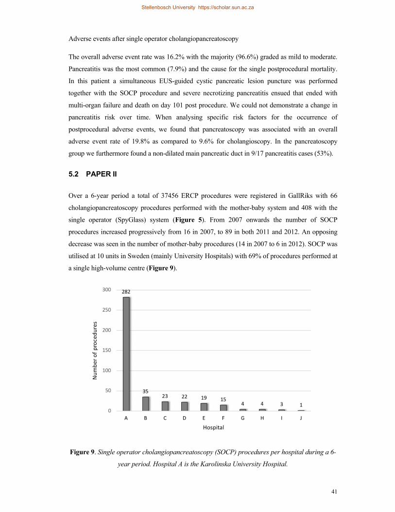
reflects the ease of use of the single operator system. Its predominant use at a single University
Hospital is in keeping with its application in complex hepatobiliary diseases, underscored by the
fact that 27% of SOCP procedures were performed in patients with PSC and 17% were performed
for stones not removed at previous ERCP.221
Clinical utility of single operator cholangiopancreatoscopy
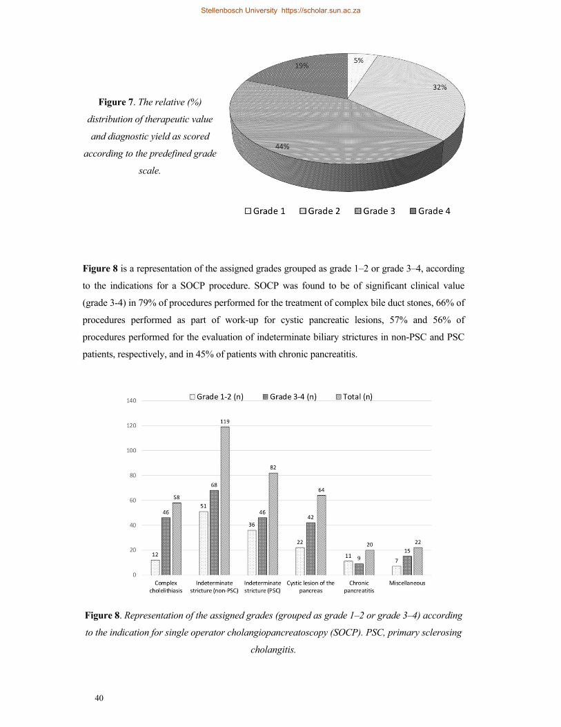
In scrutinising the impact of SOCP for specific indications, it was found to have most value in the
treatment of complex bile duct stones (79% grade 3-4). This is in keeping with previous reports of
its effectiveness regarding eventual complete stone clearance (70%-94%) and becomes especially
relevant to patients who are poor operative candidates.64,65 The long procedure time (mean of 99
min) restricts is use and demonstrates a limitation of the procedure.
Stellenbosch University https://scholar.sun.ac.za
62
The second largest clinical value of adding SOCP to an ERCP procedure was found to be in the
evaluation of cystic pancreatic lesions (66% grade 3-4). This correlates with recent reports that
note that SOCP findings altered the extent of surgical resection in 62% of patients with IPMN.93
Although dependent on frozen sections, intra-operative pancreatoscopy avoids ‘cannulation’
associated complications and may be a safer approach when employing SOCP to guide the extent
of resection.92
The clinical value of SOCP in the evaluation of indeterminate strictures graded as grades 3-4
ranged from 56%-57% in patients without and with PSC, respectively. This illustrates the
difficulty encountered when trying to differentiate malignant from benign strictures in the bile
duct. Cytological confirmation of malignancy is possible in only 40%-60% of cases.37,40 The
challenge in obtaining tissue for confirmatory diagnosis together with the increasing role of
multimodality treatment, will likely drive clinicians to utilise SOCP in the evaluation of
indeterminate strictures even in cases where the clinical yield is lower than for other indications.
In study I, over half of patients (55%) underwent SOCP for indeterminate biliary stricture
evaluation, demonstrating the aforementioned.
In contrast to its value in the treatment of biliary stone disease, SOCP appeared to have the least
impact in the small subgroup of patients who underwent SOCP for chronic pancreatitis with or
without pancreatic stone lithotripsy. This subgroup was the only group where more patients were
graded as grades 1-2 (55%) rather than grades 3-4 (45%). However, given the small sample size,
caution must be exercised when interpreting this finding. In light of an increased adverse event
rate associated with pancreatoscopy determined in both studies I and III, together with previously
reported stone clearance rates from as low as 37% in the pancreatic duct, larger prospective
studies are needed to assess the value of SOCP in patients with chronic pancreatitis.81
Adverse events after single operator cholangiopancreatoscopy
As SOCP is mainly utilised in patients with complex disorders (difficult stones and indeterminate
strictures), multiple confounding factors can influence adverse event rates and multivariate
analysis is of paramount importance to provide context. Possible risk factors for pancreatitis (e.g.,
female sex, younger age, sphincterotomy and EPLBD) and cholangitis (PSC and older age) could
be statistically adjusted for in study II. On multivariate analysis, the risk for pancreatitis and
cholangitis became less relevant, an increased risk for intraprocedural adverse events became
evident and statistically significant, and the risk for postprocedural adverse events remained.
Previous multicentre observational studies reported similar adverse event rates after SOCP and
ERCP, while smaller comparative studies did not report an increased risk with the addition of
SOCP (after adjusting for confounders).94,98 Results from study II assist in placing previous
Stellenbosch University https://scholar.sun.ac.za
63
reports of increased risk into perspective.97 Our results from a large and well-validated registry
emphasise the caution that should be exercised when adding SOCP to an ERCP procedure and
lend support to its use in high-volume centres. Comparing adverse events in patients undergoing
SOCP in the first 3 years of study to the last 3 years of study suggested a definite learning curve,
as the postprocedural adverse event rate after SOCP decreased from 25% to 16%.
Adverse events after pancreatoscopy are less well defined due mainly to the retrospective nature
of reports on small patient numbers.81,82,84,85 As wire passage into the pancreatic duct increases the
risk for pancreatitis at the time of ERCP, the main concern would be the increased risk for
pancreatitis particularly in patients with no previous history of pancreatitis or sphincterotomy. In
our analysis of SOCP use in study I, pancreatoscopy was associated with an adverse event rate of
20% (vs. cholangioscopy 9.6%). Although numbers were too small to analyse comprehensively,
9/17 patients (53%) who developed pancreatitis after pancreatoscopy had a non-dilated pancreatic
duct. In our exploration of adverse events after SOCP in study II, most confounders could be
adjusted for, leading to the added risk for pancreatitis and cholangitis to diminish. After
stratification into pancreatic and bile duct cannulation respectively, when the pancreatic duct was
cannulated, the risks of postprocedural adverse events and pancreatitis were increased in the
unadjusted analysis but this difference disappeared in the multivariate analysis.
6.1.2 Hilar stenting
Study III is, to our knowledge, the largest study to compare adverse events and reintervention
rates after stenting of malignant biliary obstruction in different locations in the biliary tree.
Adverse event rates when stenting takes place in the hilum compared to the distal biliary tree
have not been well-defined in previous studies. The large sample size in study III allowed for
increased power to detect statistically significant findings. Hilar stenting compared to distal
stenting was associated with an increased total adverse event rate (19% vs. 14%), pancreatitis rate
(6.6% vs. 4%) and cholangitis rate (4% vs. 3%).
Study IV was a comparative cohort study of ETP and PTH palliative drainage procedures
performed for MHO and is, to our knowledge, the largest comprehensive comparison of the two
approaches to date. The strengths and weaknesses of the two approaches were highlighted for B-
C subgroups in terms of access, bridging and technical success, and achievement and duration of
therapeutic success.
Adverse events after hilar stenting
Prior studies have not reported an increased risk of pancreatitis after hilar stenting and the high
rates of pancreatitis observed in both studies III and IV (6.6% and 9.4%, respectively) may be
Stellenbosch University https://scholar.sun.ac.za
64
explained by the increased procedure time associated with hilar stenting (mean of 51.9 min). In
addition, the non-dilated distal biliary tree, a well-known risk factor for pancreatitis, might
contribute to increased rates. Our findings support current guidelines suggesting pancreatitis
prophylaxis (rectal NSAIDs) for average-risk patients undergoing ERCP and emphasise this
practice in patients undergoing hilar stenting.16,49
Cholangitis rates after hilar stenting observed in study III (4.1%) reflect rates reported in RCTs
(5%-17%) and confirm the increased risk when compared to distal stenting (2.8%).181,184
Cholangitis rates reported in study IV after ETP and PTH drainage approaches (21.4% and
24.7%, respectively) are more in line with reports from observational studies (30%-45%).203
Interestingly, neither postprocedural adverse events nor cholangitis rates differed significantly
between intrahepatic (B-C I-II) and extrahepatic (B-C III-IV) stenting in study III, contrasting
with previous reports where more advanced B-C types predisposed to the development of
cholangitis.201,204 The inability to control for mitigating and risk factors such as periprocedural
antibiotics and undrained segments, may have confounded results in this smaller subgroup
analysis.
Hilar stent patency
The definition and classification of recurrent biliary obstruction and the reasons for
reintervention/stent failure are not well defined in current literature, making direct comparison
difficult.23,205,206 In study III, 6-month patency rates for distal strictures compared to B-C I
through to IV types were 44%, 30%, 24%, 10%, 12% and 14%, respectively. Reported 6-month
patency rates for stenting of distal and hilar strictures in RCTs are 68%-78% and 25%-35%,
respectively.118,177,178,207 Study III is the first to describe triple the risk for requiring reintervention
in patients receiving a single SEMS for MHO compared to those receiving a SEMS for distal
malignant obstruction. The innate anatomy of the biliary tree allows for more proximal stent
overlap when distal stenting is performed. High reintervention rates in hilar compared to distal
strictures might be partly explained by tumour progression and resultant non-occlusive stent
failure (with- or without cholangitis), where reintervention is performed for undrained segments
even though an adequate decrease in bilirubin has been established. It is important to note that our
results reflect the need for reintervention in the population at large.
Our finding of worsening patency in more advanced B-C types is in contrast to previous studies
where B-C type was not associated with decreased patency.184 Patency was lowest for B-C IIIa
types in both studies III and IV, questioning previous reports where left-sided SEMS placement
predicted lower patency.202 This finding is most likely to be the effect of proximal disease
progression around the short right hepatic duct, resulting in occlusion of segments that may be
colonized, and requiring additional intervention.
Stellenbosch University https://scholar.sun.ac.za
65
Stent type and extent of drainage in the hilum
Results from study IV support the superiority of uSEMS for drainage in MHO.116 The finding that
B-C III types achieved therapeutic success more readily after bilateral stent placement
substantiates bilateral drainage for more advanced B-C types. This topic is still under debate in
current literature.186,187,191 Our finding that B-C III types achieved therapeutic success more
readily after bilateral or contralateral vs. ipsilateral stent placement merits further discussion.
Based on functional volumes of liver segments, drainage of > 50% of liver volume requires right-
sided or bilateral stenting in B-C II and IIIb types, and bilateral stenting in B-C IIIa and IV
types.20 The short right hepatic duct is predisposed to sectoral duct involvement (B-C IIIa and IV)
and a single right-sided stent in this scenario will unlikely achieve and maintain drainage of >
50% of liver volume.
Approach when stenting the hilum
Study IV is the first to deconstruct access, bridging and technical success for ETP and PTH
approaches, highlighting the strengths of each. An ETP compared to a PTH approach had inferior
access success but superior bridging success, while technical success was similar. Most patients
who crossed over from an ETP to a PTH approach did so after unsuccessful access (failed
cannulation) and more procedures were required for eventual technical success via a PTH
approach due to repeated attempts at bridging. Previous studies have not reported on reason for
crossover or number of procedures required to reach technical success.
Study IV was also the first to compare stent characteristics and extent of drainage between ETP
and PTH approaches at the time of technical success. One of the advantages of an ETP approach
is the potential to place multiple stents from one access point. Results from study IV revealed
more two stent and bilateral stent placement after an ETP approach in advanced B-C types, which
resulted in more segments and a larger percentage of liver drained. This did not have an impact
on achievement of therapeutic success or time to therapeutic success, which was similar between
the two approaches, but likely contributed to the superior duration of therapeutic success that was
found after an ETP compared to a PTH approach. The 2016 meta-analysis by Moole et al. (the
only to specifically compare palliative ETP with PTH drainage of MHO) included 2 RCTs and 7
retrospective studies (546 patients).103 Our results, with equivalent therapeutic success for ETP
and PTH approaches, challenge their finding of superior successful drainage after a PTH
approach in advanced B-C types. Results from study IV question the dogma that a PTH approach
should be used in advanced B-C types.133,201
In study IV, B-C IIIa patients were more likely to receive bilateral stents and B-C IIIb patients
were more likely to receive contralateral stents after an ETP approach. Liu et al., in a
Stellenbosch University https://scholar.sun.ac.za
66
retrospective analysis of 446 patients undergoing unilateral PS placement via a PTH approach
found entry via the left lobe (compared to the right) associated with higher technical and
therapeutic success, and decreased adverse events compared to a right-sided puncture.222 It may
be that, after MDT discussion, if a decision is made to prioritise right-sided drainage, an ETP
approach should be pursued first. If left-sided drainage is prioritised, a PTH approach should be
pursued first. Future studies should continue to explore outcome differences for ETP and PTH
approaches for advanced B-C types.
Two recent meta-analyses reported similar overall complication and mortality rates, with
pancreatitis and cholangitis rates higher after an ETP approach and bleeding rate increased after a
PTH approach.167,168 Theoretically, unsuccessful access (failed cannulation) after an ETP
approach predisposes to pancreatitis, whereas unsuccessful bridging after a PTH approach allows
for placement of an external catheter (pigtail) and so protecting against cholangitis. Study IV
confirmed more patients developed pancreatitis after an ETP approach, mostly of MAGS grade 1-
3, with a single pancreatitis-associated death (MAGS grade 6). Cholangitis rates between the two
approaches were similar, likely due to more double/bilateral stent placement after ETP drainage.
A recent RCT was prematurely closed after accrual of 54 patients due to a higher-than-expected
mortality in the PTH approach group compared to the ETP group (41% vs. 11%) and, although
not designed to compare survival, our study found more sudden deaths after a PTH approach
(15.8% vs. 6.0%).147
Comparative patency duration has not been extensively explored for ETP and PTH approaches. In
prospective studies a PTH approach appeared superior, although not statistically significant.144,146
In retrospective reports earlier studies favoured a PTH approach, while more recent comparisons
found equivalence or superiority for an ETP approach.150,151,154,161 The longer duration of
therapeutic success resulting in a gain of 3 months after an ETP approach seen in study IV is
likely secondary to more bilateral stent placements and a larger volume of liver drained in
especially advanced B-C types, as such safeguarding against future symptomatic recurrent
obstruction.
As the goals for preoperative and palliative drainage differ, the first aiming to drain the future
liver remnant to limit perioperative complications and the second to drain enough functional liver
volume to afford symptomatic relief or administer oncological therapy, comparative analyses are
challenging. Two recent meta-analyses, including distal and hilar malignant obstruction in
patients undergoing both preoperative and palliative drainage, reported similar technical and
therapeutic success rates, overall complications and 30-day mortality for ETP and PTH
approaches.167,168 The equivalence in technical success, therapeutic success and time to
therapeutic success after ETP and PTH approaches found in study IV are in accordance with
Stellenbosch University https://scholar.sun.ac.za
67
these reports and again corroborate the philosophy that the two approaches are complementary
rather than competitive.
6.2 METHODOLOGICAL ASPECTS
6.2.1 Paper I
In the absence of an available system to assess the impact on patient management when SOCP is
added to ERCP, the grading scale used in study I had to be developed anew. To ensure content
validity, the grading scale had to be:
1. Complex enough to measure all aspects of SOCP clinical value to allow differentiation
between therapeutic and diagnostic intent.
2. Practical and simple enough to be applied to a large number of procedures (n=365).
It should be noted that the grading scale could not be correlated with previous scales, due to a
lack of previously published scales. Criterion validity could not be calculated, and final validation
will depend on future studies.
The single reviewer used in study I was not involved in the clinical care of included patients, but
the absence of more than one reviewer meant that reproducibility (interobserver variation) could
not be calculated. Repeatability (intra-observer reliability) was maximized by defining examples
in each grade (therapeutic vs. diagnostic). In cases where it was difficult to ascertain the relative
contribution that other treatment or diagnostic efforts lent to the final management plan, cases
were graded as grade 2. To further minimise bias, the reviewer decision as to final grading of
diagnostic value was measured utilising the final MDT meeting decision, with the benefit of
extensive expert input.
The observer bias introduced when the endoscopists had access to all relevant clinical information
at the time of visual assessment of biliary strictures or IPMN lesions, risks overestimation of the
clinical value of SOCP. It is difficult from an ethical point of view to argue for withholding
relevant information from the endoscopist. More importantly, incorporating clinical information
(history, examination, serum markers and imaging) as the diagnostic process progresses (as new
information becomes available), reflects everyday clinical practice.
Inherent in studies attempting to assess diagnostic accuracy or impact on management is the
absence of the gold standard of diagnosis (histology of the resected specimen) in strictures
diagnosed as benign. Under these circumstances the clinical course can be used as a substitute,
and due to the retrospective nature of the current study, this benefit could be utilised.
Stellenbosch University https://scholar.sun.ac.za
68
6.2.2 The GallRiks Registry
Study II and III utilised prospective data from the nationwide Swedish Registry for Gallstone
Surgery and ERCP (GallRiks). The main strength of both studies lies in the large sample sizes
available for inclusion and comparison, minimising random errors and the effect of chance, and
increasing statistical power of the studies.
An advantage of the registry, when reporting on both SOCP and ERCP associated adverse events,
is that since its conception adverse events have been defined according to internationally accepted
criteria.45 This ensures reporting conformity and allows for comparison with previous reports. The
population-based registry has nationwide participation (> 90% of hospitals in Sweden) thus
minimising selection bias and increasing applicability to the population at large (high external
validity). Selection bias is, however, inherent in self-reported registries where clinicians might be
reluctant to declare undesirable outcomes. In the Gallriks registry this risk is minimised by the
capturing and reviewing of 30-day adverse events by an independent non-physician coordinator.
The 30-day follow-up frequency for GallRiks after cholecystectomy and ERCP is 96% and 95%
respectively.210
Population-based registry studies reflect what takes place at the bedside (high external validity)
but internal validity becomes vulnerable due to the presence of unaccounted confounders.223 All
possible variables that increase pancreatitis risk could not be accounted for (such as sphincter of
Oddi dysfunction, a non-dilated bile duct and normal preprocedural liver function tests).
Likewise, known factors that increase cholangitis risk (incomplete drainage in hilar obstruction)
and bleeding risk (anticoagulant and antiplatelet use) could not be adjusted for. In both studies I
and II, data on the use of known mitigating factors for pancreatitis (NSAIDs) and cholangitis
(prophylactic antibiotics) was not available from the start of participant inclusion. In study III,
important possible confounders regarding hilar stenting such as stricture number and length, the
number of segments obstructed and subsequently drained, the volume of liver drained and the
reasons for reintervention were not known, making definitive conclusions and comprehensive
multivariate analysis problematic.
Coordinator review and regular external auditing of adverse event documentation allowed for
minimal missing data in study II (2.7%). There might be a concern regarding missing data in
study III (34.3%) regarding stricture location and stent position. This was, however, a result of an
inability to definitively classify stricture/stent position from the images captured in GallRiks, in
which case patients were excluded.
Stellenbosch University https://scholar.sun.ac.za
69
6.2.3 Paper II
Study II was a representation of the nationwide integration of SOCP, including data from 10
Swedish centres, but there might be a concern that 69% of procedures were performed at a single
high-volume University Hospital. On subgroup analysis and comparison there was, however, no
difference in adverse events between this centre and the other units included in the study.
National coverage of the registry between 2007 and 2009 saw an increase in participation from
73% to 90%. The increase in participation was mostly due to the addition of regions and not due
to increased participation in units already making use of the registry. With reference to analysis of
adverse events after ERCP it should be noted that completeness for GallRiks participating units
during this time period varied between 97.2% and 98.2%.211
Both studies I and II included SOCP procedures performed with the first-generation SOCP
system in use from 2007 to 2015. The digitalized second-generation system offers improved
image quality and thus clinical value might be underestimated and adverse events overestimated
when results from the current data is extrapolated to its future use.
6.2.4 Paper III
There might be concern for the different pathological processes and prognoses that are involved
in distal and MHO. However, the focus in both studies III and IV was on anatomical and
technical aspects of transpapillary stenting for malignant biliary disease such as approach,
stricture and stent location, stent characteristics and patency. Because of differences in tumour
biology regarding progression (ingrowth/overgrowth) and response to oncological therapy,
survival was not addressed in either of the reports. Survival in patients with especially MHO is,
however, limited.44 Once a diagnosis of irresectable hilar malignancy is made, the main treatment
goal is symptomatic relief with/without the administration of oncological therapy, with survival
following thereafter.
The definition of a distal stricture used in study III (originating in the common bile duct below the
cystic duct junction) does not consider the variability in cystic duct anatomy and can lead to
misclassification of distal strictures as B-C I strictures and vice versa. The Asia Pacific consensus
meeting in 2020 defined a distal stricture as an abnormal narrowing of the distal half of the
extrahepatic bile duct and this definition will allow for consistency in future studies.23
To enable comparison of the need for reintervention in study III and to limit confounders, we
included only patients in whom a single metal stent was placed. The superiority of metal over
plastic stents regarding patency for both distal and hilar malignant strictures has already been
Stellenbosch University https://scholar.sun.ac.za
70
proven and was not the objective of this study.116,119 We did not distinguish between the use of
uSEMS or cSEMS in the current study as there is a lack of collaborating evidence showing a
significant difference in outcomes between these two stent types for distal strictures.128 Both stent
types are predisposed to recurrent biliary obstruction albeit via different pathways (cSEMS
migration and overgrowth, uSEMS ingrowth). The use of metal stents in the hepatic hilum has
been restricted to uSEMS to prevent blockage of secondary intrahepatic biliary radicles.133,179 A
single stent establishes 100% drainage in distal and B-C I malignant strictures, while two stents is
required in the hilum to drain more than 50% of the liver in more advanced B-C types (B-C IIIa
and IV). There is ongoing robust debate as to the number of stents to be placed to achieve the
treatment goal (safe resection, adequate symptom relief or administration of oncological
therapy).186,187
Matching of the GallRiks registry with the Swedish Central Death Register allowed for complete
mortality data in patients stented for malignant biliary obstruction. The registry does not,
however, account for indications for reintervention (stent factors, tumour factors or non-occlusion
stent failure/cholangitis) and thus the reason for the much higher reintervention rate after hilar
stenting could not be explored further. The registry furthermore only account for endoscopic
interventions and operative, percutaneous, EUS-guided and oncological interventions are not
represented. This underestimates the number of stent failures, especially for advanced B-C types.
An important statistical limitation of the study also warrant consideration. Reintervention rates
were calculated from retrospective data in contrast to patency duration calculated in a prospective
manner. Death without re-intervention was treated as a censored event, but actual stent patency at
the time of death could not be confirmed, and random censoring and an occluded stent at time of
death could not be excluded (underestimating stent failure).
The study has simplified and does not cover the whole spectrum of hilar stenting, having
excluded patients treated with plastic or multiple stents and percutaneous or EUS-guided stents.
Registries such as GallRiks, designed for ERCP and not specifically for hilar strictures, lack
granular information on key variables, precluding in-depth analyses of important unanswered
questions. However, the RCTs that have generated strong evidence regarding numerous issues
concerning distal stenting, cannot be replicated in MHO.147,148 Despite the inherent drawbacks
encountered when a registry is used retrospectively, and due the challenges posed by selecting a
prospective randomised study design in many instances, combining these two methodologies in
future can draw on the strengths of each (as evidenced in previous publications).224,225 Analyses of
large population-based cohorts can be a platform for studies creating more robust data and the
challenges encountered in study III could inspire the design of study IV.
Stellenbosch University https://scholar.sun.ac.za
71
6.2.5 Paper IV
The use of classification systems, definitions and grading scales allows for standardization and
easy comparison between reports. The B-C classification, definition of therapeutic success and
MAGS grading system used in study IV posed specific challenges but were chosen for specific
reasons.
The B-C classification system was developed to guide the operating surgeon but has limitations
when applied by the endoscopist or interventional radiologist.
1. It indicates hepatic duct (common, left and right) involvement but disregards strictures
stretching beyond sectoral ducts and does not take into account the 40% of anatomical variants
where aberrant intrahepatic drainage is present.
2. It does not account for functional liver volume proximal to strictures, whether healthy or
atrophied, which is essential when planning drainage in more advanced stricture types.
3. It fails to standardise the definition of a hilar or sectoral block which can be dependent on the
force of contrast injection at the time of intervention.
An ideal classification system to describe and guide hilar drainage is still lacking but should aim
to reflect the functional state of the proximal liver parenchyma in addition to ductal topography.
The definition of therapeutic success (TB value of ≤ 40 µmol/L) is based on previous publications
where populations are undergoing drainage mostly as a bridge-to-surgery.152,155 It is less well
suited as surrogate for the treatment goal in patients with irresectable or metastatic MHO where
two groups are identified: those where TB values need to normalize before commencement of
palliative oncological therapy and those with advanced disease where purely symptomatic relief
is pursued. In accordance with end-of-life treatment goals, patients in study IV were not
biochemically followed up beyond symptom resolution if symptomatic relief was the only
intended outcome.44
As additional access to the biliary tract is allowed endoscopically, percutaneously, and recently
via EUS, definition and grading systems for complications applicable across all access
approaches are lacking. Internationally recognised definitions were chosen for endoscopic and
radiological complications respectively (Suppl. Table 1).16,217,218 An ideal grading system should
be practical, easily clinically applied, general enough to reach across specialties and yet allow for
a relatively detailed and accurate description of most eventualities. The Clavien classification
system for post-surgical complications introduced the principle of grading according to the
amount of effort required to reverse or manage a complication and was later modified by Dindo
Stellenbosch University https://scholar.sun.ac.za
72
(Clavien-Dindo) and Martin et al. (Memorial Sloan Kettering grading system).226–228 The
Accordion grading system was developed as an adaptation of previous classifications to be more
universally applicable to large and complex studies and was the system chosen for this study,
allowing for applicability across different biliary approaches.219
The retrospective nature of study IV meant that there was no control for random and institutional
bias, e.g., selection of patients, biliary approach and stent type and number. Inclusion of patients
from two centres with high-volume endoscopy and interventional radiology units (with
independent functioning) mitigates this to some extent. Volumetric assessment was not performed
on all patients and patients with no further interventions could not be accounted for. However, as
the early closure of two recent RCTs demonstrates, comparison of ETP and PTH approaches is
not easily performed prospectively.147,148 This is mostly due to divergent institutional expertise
and availability and/or clinician bias towards a specific approach. Many confounders that need to
be considered complicate the construction of practical inclusion and exclusion criteria. Future
studies will have to depend on large multi-institutional registries designed specifically to include
the number of possible confounders that need to be considered in MHO.
For comparison of ETP and PTH approaches per B-C type in this study we focused on within-
group comparisons, with the main drawback being that numbers become smaller after
subgrouping, especially when controlling for multiple confounders. For comparison of time to
therapeutic success between ETP and PTH approaches per B-C type, the Mann-Whitney U test
was utilised due to a very skewed dependent variable. Comparisons were not likely to show
significance due to wide ranges (and even interquartile ranges) with large overlap. When
attempting to compare time to therapeutic success per B-C type and as a function of stent and
drainage characteristics, numbers became too small for meaningful analyses.
Stellenbosch University https://scholar.sun.ac.za
73
7 CONCLUSIONS
This thesis provides results to support the following conclusions:
Single operator cholangiopancreatoscopy, when added to ERCP, has significant
clinical value in 63% of cases. In current endoscopic practice it is applied in the
biliary tract (70%), pancreatic duct (25%) and both ducts (5%). Its clinical value lies
firstly in the treatment of complex bile duct stones, and secondly as aid in the
diagnosis of cystic pancreatic lesions and indeterminate biliary strictures.
The SOCP system has replaced the performance of cholangiopancreatoscopy by the
mother-baby system and there is clear learning curve when first introduced.
Intraprocedural and postprocedural adverse events are increased when SOCP is added
to an ERCP procedure.
In patients with malignant biliary obstruction the risk for adverse events, including
pancreatitis and cholangitis, is increased after hilar compared to distal stenting.
Stent patency rates are lower for a single SEMS placed for MHO compared to distal
malignant obstruction, with stenting for B-C IIIa types having the worst patency.
In patients with MHO undergoing palliative biliary drainage, ETP and PTH
approaches have distinct strengths and weaknesses. Technical and therapeutic success
rates are similar, while therapeutic success durability is better after an ETP approach.
The two approaches have similar overall adverse event and cholangitis rates, while
pancreatitis is more common after an ETP approach and deaths more common after a
PTH approach.
Stellenbosch University https://scholar.sun.ac.za
Stellenbosch University https://scholar.sun.ac.za
75
8 POINTS OF PERSPECTIVE
Although many reports on SOCP performance regarding stone clearance rates and the accuracy of
visual inspection/biopsy is available, quantification of the clinical value/impact of SOCP is dated
and limited.86,87 Measurement of its clinical value was attempted in three small recent reports,
restricted to the management of indeterminate biliary strictures.88–90 The grading scale used in
study I is the only of its kind that allows for simultaneous assessment of SOCP therapeutic and
diagnostic value but requires validation. Inclusion of more than one blinded reviewer will allow
for calculation of interobserver variation (reproducibility) and its use in future studies will allow
for measurement of criterion validity.
Results from this thesis, highlighting the clinical value but also the increased risk for adverse
events when SOCP is added to ERCP, corroborate the placement of SOCP in treatment
algorithms in current guidelines for the treatment of ‘difficult’ bile duct stones and in the
investigation of indeterminate strictures.29,174 A demonstratable learning curve cautions for its
continued use in specialised high-volume centres where staff are adequately trained to prevent,
recognise and treat associated adverse events. Future meticulous documentation of investigations
to generate more evidence is a prerequisite for the firm establishment of this important procedure
in pancreaticobiliary endoscopy algorithms.
Although the robust data collected in the GallRiks registry (accounting for most confounders)
allowed for comprehensive multivariate analysis of the risk of adverse events when SOCP is
added to ERCP in study II, study III underscored the difficulties encountered in investigating
stenting for MHO when making use of registry data. The GallRiks dataset is reviewed annually
and adjustment of variables is performed accordingly, ensuring its continued excellence as data
source in answering research questions pertaining to endoscopy in the hepatobiliary systems.
Analysis of large population-based cohorts remains a platform for studies creating more robust
data, as exemplified by the challenges encountered in study III leading to the creation of a
multicentre registry dedicated to MHO in study IV. This registry currently offers analysis of
outcomes after simultaneous or sequential combined ETP and PTH approaches, as well as
mechanisms of recurrent biliary obstruction in the hepatic hilum. The combination of established
national registries with a prospective randomised study design will increasingly in future make
economic sense.
The B-C classification system was designed for the operating surgeon to indicate the most distal
extent of normal biliary mucosa available for anastomosis after curative resection in patients with
CC.25 It is not optimal as a guide to the endoscopist or interventional radiologist as it does not
give sufficiently detailed information on the extent of obstruction of sectoral ducts and does not
Stellenbosch University https://scholar.sun.ac.za
76
account for upstream liver functionality. A dedicated classification where not only the actual
ductal topography is described, but in which functional parenchymal volumes beyond strictures
are also quantified, would be of more value.
The definition of therapeutic success after MHO drainage in published literature is based on
decreased TB values which is used as a surrogate for the goal of treatment. In patients with MHO
the treatment goal differs for patients undergoing preoperative stenting, patients planned for
palliative oncological therapy and those with advanced disease where purely symptomatic relief
is pursued. These differences need to be considered in future study designs and when comparing
results between different patient populations.
The definition and classification of recurrent biliary obstruction is not well defined in current
literature.23,205,206 Mechanisms of recurrent obstruction, reasons for reintervention and treatment
options at the time of reintervention differ somewhat for distal and hilar obstruction. Future
studies and classification systems should explore and consider these differences.
In study IV, patients with B-C IIIa types had 100% technical success after a single ETP
procedure. They also had significantly more bilateral stents placed, a higher percentage of liver
volume drained and trended towards a faster time to therapeutic success. The general dictum that
B-C III-IV types should be drained via a percutaneous approach is challenged by our results
showing ETP outperforming PTH in BC IIIa patients and being equal to PTH in BC IIIb and IV
patients.103,133,174 Although small numbers limited statistical power in some analyses (especially
B-C IIIb and IV), our data suggest that ETP drainage might be more successful in selected
advanced B-C types. Future studies should endeavor to increase patient numbers and explore
outcome differences for advanced B-C types.
Stellenbosch University https://scholar.sun.ac.za
77
9 ACKNOWLEDGEMENTS
For ongoing assistance and encouragement to complete these 4 Papers, sincere thanks to all
who played a part. And for shaping my life forever as part of this journey, my most heartfelt
thanks specifically to…
Lars Enochsson, Swedish main supervisor, for teaching me how to do proper research. With
a Mac on a balcony. For the hours of energy put into making this happen. And for Sara
providing a welcome supper every single time I arrived in Stockholm.
Eduard Jonas, Swedish / South-African co-supervisor, without you this would not have
been possible, not one step. Thank you for teaching me that everything can be done at once.
And that research should just flow out of everything we do in the hospital every day.
You sir, are a true mentor.
Urban Arnelo, Swedish co-supervisor, for teaching me that anything can be cannulated, that
the endoscopist always has a role to play, and that hard work allows for hard play.
Samuel Moore, South African co-supervisor, for orchestrating the South African side of this
endeavor and ‘pearls of wisdom’ every time we met.
Karin Baatjes, my official mentor, for showing that it is possible, listening for hours and
encouragement when silence fell.
Magnus Nilsson, Head of the Division of Surgery in Huddinge, who had the courage to
embark on an international PhD road with me, and the patience to persist even when a
pandemic got in the way. For teaching me that for every clinical question that comes up, a
scientific answer is not far away if you know where to look - it is in a Swedish registry!
Also, current and previous colleagues at ÖAK and in particular…
Mari Hult, for showing me how to be Swedish. And never-ending advice. And sharing your
home and family.
Frederik Swahn, whose Kappa inspired me to embark on this journey and whose artistic
ability knows no bounds. Lars Lundell emeritus professor of surgery and Matthias Löhr,
professor of gastroenterology and hepatology at Clintec, whose scientific standards set the
bar. I am so thankful to have experienced how you do things.
Marcus Reuterwall, for the smoothest co-authorship / partnership I could have hoped for,
and unofficial assistance in getting metabolic surgery started in Africa. Björn Törnqvist, for
co-authorship, wishing me a high cannulation rate and welcoming me the second time to
Danderyd Hospital. Christoph Ansorge, for inviting this stranger to her first and best-ever
graduation party. I think of it often. Stefan Gilg and Ioannis Rouvelas for being solid friends
and great humans, and paying my bill if I just get up and leave…
Stellenbosch University https://scholar.sun.ac.za
78
Marco del Chiaro, Ernesto Sparrelid, Melroy D’Souza, Elena Rangelova, and so many
more. Thank you for welcoming me in.
Hélène Jansson, for ‘administrating’ this thesis all the way to the end. And for being me in
Stockholm all the times that I was busy being me in South Africa.
To Nina Gustafsson, and Christel Nilsson before her, guardians to all at the Department of
Upper Abdominal Surgery in Huddinge. Thank you for the extra effort to adopt a more
distant child. And for becoming my friends.
To the endoscopy staff at the ERCP unit, and especially to Inger, who had to put up with me
twice. Thank you for opening the door to me, and for patience and friendship.
Johanna Ackemar in Solna and Agneta Wittlock in Flemingsberg, for endless e-mails and
keeping things going forward across the waters.
To beautiful Corinne, Julia and Claudia Jonas, who became my family in Sweden, showed
me the beauty of the Swedish forests, the Baltic sea and Gotland, brought music into
everyday life, and will forever be a part of me going forward…
Brian Warren, previous Head of Surgery at Stellenbosch, for years ago cultivating a special
love for the hepatobiliary system, and for opening doors that I will forever be thankful for.
And to Elmin Steyn, who took over half-way and made it her own.
Also, current and previous colleagues at Stellenbosch, and in particular…
Anton Lambrecht, for unflinchingly taking over Upper Gastrointestinal Surgery at
Tygerberg Hospital every time that I disappeared. And Stefan Hofmeyr, for holding the fort
in all other ways, and for advice that is always worth taking.
Jenny Edge and Casey Wallace, for turning out to be a multitalented duo, and playing a
huge role in the aesthetics of what has been produced.
My brother Johann, my sister Inge, and Dané and Ivan. For bringing normality into my life
and teaching me what really matters.
Ricky, for owning half of this thesis. Because that is the reality.
My mom, Isabelle, and my dad, Willem. How do you thank parents if not for everything?
Stellenbosch University https://scholar.sun.ac.za
79
10 REFERENCES
1. Tummala P, Munigala S, Eloubeidi MA, et al. Patients with obstructive jaundice and biliary
stricture ± mass lesion on imaging: prevalence of malignancy and potential role of EUS-FNA. J Clin Gastroenterol. 2013;47(6):532-537.
2. Boulay BR, Birg A. Malignant biliary obstruction: From palliation to treatment. World J Gastrointest Oncol. 2016;8(6):498-508.
3. Tyson GL, Ilyas JA, Duan Z, et al. Secular trends in the incidence of cholangiocarcinoma in the USA and the impact of misclassification. Dig Dis Sci. 2014;59(12):3103-3110.
4. Yadav D, Lowenfels AB. The epidemiology of pancreatitis and pancreatic cancer. Gastroenterology. 2013;144(6):1252-1261.
5. Kreel L. Radiology of the biliary system. Clin Gastroenterol. 1973;2(1):185-212. 6. Doubilet H, Poppel MH, Mulholland JH. Pancreatography; technics, principles, and
observations. Radiology. 1955;64(3):325-339. 7. Pinchuk L, Magnanini F, Nardi G, et al. [Percutaneous transhepatic biliary drainage in
obstructive jaundice]. Acta Gastroenterol Latinoam. 1981;11(2):279-284. 8. Hirschowitz BI, Peters CW, Curtiss LE. Preliminary report on a long fiberscope for
examination of stomach and duodenum. Med Bull Ann Arbor Mich. 1957;23(5):178-180. 9. McCune WS, Shorb PE, Moscovitz H. Endoscopic cannulation of the ampulla of vater: a
preliminary report. Ann Surg. 1968;167(5):752-756. 10. Oi I, Takemoto T, Nakayama K. “Fiberduodenoscopy”-early diagnosis of cancer of the
papilla of Vater. Surgery. 1970;67(4):561-565. 11. Demling L. Operative Endoskopie. Schattauer; 1976. 12. Kawai K, Akasaka Y. Hashimoto. Y. et al. Preliminary report on endoscopical papillotomy.
J Kyoto Pref Univ Med. 1973;82:353. 13. Allegaert W. Report concerning cholangioscopy, using closed-circuit television, during
surgical operations on the biliary tract. Acta Gastro-Enterol Belg. 1961;24:599. 14. Laurence BH, Cotton PB. Decompression of malignant biliary obstruction by duodenoscopic
intubation of bile duct. Br Med J. 1980;280(6213):522-523. 15. Huibregtse K, Tytgat GN. Palliative treatment of obstructive jaundice by transpapillary
introduction of large bore bile duct endoprosthesis. Gut. 1982;23(5):371-375. 16. Dumonceau J-M, Kapral C, Aabakken L, et al. ERCP-related adverse events: European
Society of Gastrointestinal Endoscopy (ESGE) Guideline. Endoscopy. 2020;52(2):127-149. 17. Andriulli A, Loperfido S, Napolitano G, et al. Incidence rates of post-ERCP complications: a
systematic survey of prospective studies. Am J Gastroenterol. 2007;102(8):1781-1788. 18. Nakamura T, Tanaka K, Kiuchi T, et al. Anatomical variations and surgical strategies in
right lobe living donor liver transplantation: lessons from 120 cases. Transplantation. 2002;73(12):1896-1903.
19. Varotti G, Gondolesi GE, Goldman J, et al. Anatomic variations in right liver living donors. J Am Coll Surg. 2004;198(4):577-582.
20. Abdalla EK, Denys A, Chevalier P, et al. Total and segmental liver volume variations: Implications for liver surgery. Surgery. 2004;135(4):404-410.
21. Domagk D, Poremba C, Dietl K-H, et al. Endoscopic transpapillary biopsies and intraductal ultrasonography in the diagnostics of bile duct strictures: a prospective study. Gut. 2002;51(2):240-244.
22. Miyazaki M, Ohtsuka M, Miyakawa S, et al. Classification of biliary tract cancers established by the Japanese Society of Hepato-Biliary-Pancreatic Surgery: 3(rd) English edition. J Hepato-Biliary-Pancreat Sci. 2015;22(3):181-196.
Stellenbosch University https://scholar.sun.ac.za
80
23. Nakai Y, Isayama H, Wang H-P, et al. International consensus statements for endoscopic management of distal biliary stricture. J Gastroenterol Hepatol. Published online December 4, 2019.
24. Bismuth H, Corlette MB. Intrahepatic cholangioenteric anastomosis in carcinoma of the hilus of the liver. Surg Gynecol Obstet. 1975;140(2):170-178.
25. Bismuth H, Nakache R, Diamond T. Management strategies in resection for hilar cholangiocarcinoma. Ann Surg. 1992;215(1):31-38.
26. Bismuth H, Majno PE. Biliary strictures: classification based on the principles of surgical treatment. World J Surg. 2001;25(10):1241-1244.
27. Costamagna G, Tringali A, Petruzziello L, et al. Hilar tumours. Can J Gastroenterol J Can Gastroenterol. 2004;18(7):451-454.
28. Schafmayer C, Hartleb J, Tepel J, et al. Predictors of gallstone composition in 1025 symptomatic gallstones from Northern Germany. BMC Gastroenterol. 2006;6:36.
29. Manes G, Paspatis G, Aabakken L, et al. Endoscopic management of common bile duct stones: European Society of Gastrointestinal Endoscopy (ESGE) guideline. Endoscopy. 2019;51(5):472-491.
30. ASGE Standards of Practice Committee, Buxbaum JL, Abbas Fehmi SM, et al. ASGE guideline on the role of endoscopy in the evaluation and management of choledocholithiasis. Gastrointest Endosc. 2019;89(6):1075-1105.e15.
31. Lux G, Ell C, Hochberger J, Müller D, et al. The first successful endoscopic retrograde laser lithotripsy of common bile duct stones in man using a pulsed neodymium-YAG laser. Endoscopy. 1986;18(4):144-145.
32. Dyson JK, Beuers U, Jones DEJ, et al. Primary sclerosing cholangitis. Lancet Lond Engl. 2018;391(10139):2547-2559.
33. Hilscher MB, Tabibian JH, Carey EJ, et al. Dominant strictures in primary sclerosing cholangitis: A multicenter survey of clinical definitions and practices. Hepatol Commun. 2018;2(7):836-844.
34. Shaib Y, El-Serag HB. The epidemiology of cholangiocarcinoma. Semin Liver Dis. 2004;24(2):115-125.
35. Khan SA, Emadossadaty S, Ladep NG, et al. Rising trends in cholangiocarcinoma: is the ICD classification system misleading us? J Hepatol. 2012;56(4):848-854.
36. Bertuccio P, Bosetti C, Levi F, et al. A comparison of trends in mortality from primary liver cancer and intrahepatic cholangiocarcinoma in Europe. Ann Oncol Off J Eur Soc Med Oncol. 2013;24(6):1667-1674.
37. Blechacz B. Cholangiocarcinoma: Current Knowledge and New Developments. Gut Liver. 2017;11(1):13-26.
38. Banales JM, Cardinale V, Carpino G, et al. Expert consensus document: Cholangiocarcinoma: current knowledge and future perspectives consensus statement from the European Network for the Study of Cholangiocarcinoma (ENS-CCA). Nat Rev Gastroenterol Hepatol. 2016;13(5):261-280.
39. Razumilava N, Gores GJ. Cholangiocarcinoma. Lancet Lond Engl. 2014;383(9935):2168-2179.
40. Navaneethan U, Njei B, Lourdusamy V, et al. Comparative effectiveness of biliary brush cytology and intraductal biopsy for detection of malignant biliary strictures: a systematic review and meta-analysis. Gastrointest Endosc. 2015;81(1):168-176.
41. Sun B, Moon JH, Cai Q, et al. Review article: Asia-Pacific consensus recommendations on endoscopic tissue acquisition for biliary strictures. Aliment Pharmacol Ther. 2018;48(2):138-151.
Stellenbosch University https://scholar.sun.ac.za
81
42. Jarnagin WR, Fong Y, DeMatteo RP, et al. Staging, resectability, and outcome in 225 patients with hilar cholangiocarcinoma. Ann Surg. 2001;234(4):507-517; discussion 517-519.
43. Edge SB, Compton CC. The American Joint Committee on Cancer: the 7th Edition of the AJCC Cancer Staging Manual and the Future of TNM. Ann Surg Oncol. 2010;17(6):1471-1474.
44. Shariff MIF, Khan SA, Westaby D. The palliation of cholangiocarcinoma. Curr Opin Support Palliat Care. 2013;7(2):168-174.
45. Cotton PB, Eisen GM, Aabakken L, et al. A lexicon for endoscopic adverse events: report of an ASGE workshop. Gastrointest Endosc. 2010;71(3):446-454.
46. Chen J-J, Wang X-M, Liu X-Q, et al. Risk factors for post-ERCP pancreatitis: a systematic review of clinical trials with a large sample size in the past 10 years. Eur J Med Res. 2014;19:26.
47. Ding X, Zhang F, Wang Y. Risk factors for post-ERCP pancreatitis: A systematic review and meta-analysis. Surg J R Coll Surg Edinb Irel. 2015;13(4):218-229.
48. Elmunzer BJ, Scheiman JM, Lehman GA, et al. A randomized trial of rectal indomethacin to prevent post-ERCP pancreatitis. N Engl J Med. 2012;366(15):1414-1422.
49. ASGE Standards of Practice Committee, Chandrasekhara V, Khashab MA, et al. Adverse events associated with ERCP. Gastrointest Endosc. 2017;85(1):32-47.
50. Yokoe M, Hata J, Takada T, et al. Tokyo Guidelines 2018: diagnostic criteria and severity grading of acute cholecystitis (with videos). J Hepato-Biliary-Pancreat Sci. 2018;25(1):41-54.
51. Cotton PB, Connor P, Rawls E, et al. Infection after ERCP, and antibiotic prophylaxis: a sequential quality-improvement approach over 11 years. Gastrointest Endosc. 2008;67(3):471-475.
52. Freeman ML, Nelson DB, Sherman S, et al. Complications of endoscopic biliary sphincterotomy. N Engl J Med. 1996;335(13):909-918.
53. Bangarulingam SY, Gossard AA, Petersen BT, et al. Complications of endoscopic retrograde cholangiopancreatography in primary sclerosing cholangitis. Am J Gastroenterol. 2009;104(4):855-860.
54. Navaneethan U, Jegadeesan R, Nayak S, et al. ERCP-related adverse events in patients with primary sclerosing cholangitis. Gastrointest Endosc. 2015;81(2):410-419.
55. Chen M, Wang L, Wang Y, et al. Risk factor analysis of post-ERCP cholangitis: A single-center experience. Hepatobiliary Pancreat Dis Int HBPD INT. 2018;17(1):55-58.
56. Banks PA, Bollen TL, Dervenis C, et al. Classification of acute pancreatitis--2012: revision of the Atlanta classification and definitions by international consensus. Gut. 2013;62(1):102-111.
57. Kiriyama S, Kozaka K, Takada T, et al. Tokyo Guidelines 2018: diagnostic criteria and severity grading of acute cholangitis (with videos). J Hepato-Biliary-Pancreat Sci. 2018;25(1):17-30.
58. Chen YK. Preclinical characterization of the Spyglass peroral cholangiopancreatoscopy system for direct access, visualization, and biopsy. Gastrointest Endosc. 2007;65(2):303-311.
59. Chen YK, Pleskow DK. SpyGlass single-operator peroral cholangiopancreatoscopy system for the diagnosis and therapy of bile-duct disorders: a clinical feasibility study (with video). Gastrointest Endosc. 2007;65(6):832-841.
60. Ramchandani M, Reddy DN, Lakhtakia S, et al. Per oral cholangiopancreatoscopy in pancreatico biliary diseases--expert consensus statements. World J Gastroenterol. 2015;21(15):4722-4734.
Stellenbosch University https://scholar.sun.ac.za
82
61. Buxbaum J, Sahakian A, Ko C, et al. Randomized trial of cholangioscopy-guided laser lithotripsy versus conventional therapy for large bile duct stones (with videos). Gastrointest Endosc. 2018;87(4):1050-1060.
62. Sejpal DV, Trindade AJ, Lee C, et al. Digital cholangioscopy can detect residual biliary stones missed by occlusion cholangiogram in ERCP: a prospective tandem study. Endosc Int Open. 2019;7(4):E608-E614.
63. Averbukh LD, Miller D, Birk JW, et al. The utility of single operator cholangioscope (Spyglass) to diagnose and treat radiographically negative biliary stones: A case series and review. J Dig Dis. 2019;20(5):262-266.
64. Korrapati P, Ciolino J, Wani S, et al. The efficacy of peroral cholangioscopy for difficult bile duct stones and indeterminate strictures: a systematic review and meta-analysis. Endosc Int Open. 2016;4(3):E263-275.
65. Jin Z, Wei Y, Tang X, et al. Single-operator peroral cholangioscope in treating difficult biliary stones: A systematic review and meta-analysis. Dig Endosc Off J Jpn Gastroenterol Endosc Soc. 2019;31(3):256-269.
66. Maydeo AP, Rerknimitr R, Lau JY, et al. Cholangioscopy-guided lithotripsy for difficult bile duct stone clearance in a single session of ERCP: results from a large multinational registry demonstrate high success rates. Endoscopy. 2019;51(10):922-929.
67. Ye X, Huai J, Sun X. Effectiveness and safety of biliary stenting in the management of difficult common bile duct stones in elderly patients. Turk J Gastroenterol Off J Turk Soc Gastroenterol. 2016;27(1):30-36.
68. Hartery K, Lee CS, Doherty GA, et al. Covered self-expanding metal stents for the management of common bile duct stones. Gastrointest Endosc. 2017;85(1):181-186.
69. Franzini T, Moura RN, Bonifácio P, et al. Complex biliary stones management: cholangioscopy versus papillary large balloon dilation - a randomized controlled trial. Endosc Int Open. 2018;6(2):E131-E138.
70. Angsuwatcharakon P, Kulpatcharapong S, Ridtitid W, et al. Digital cholangioscopy-guided laser versus mechanical lithotripsy for large bile duct stone removal after failed papillary large-balloon dilation: a randomized study. Endoscopy. 2019;51(11):1066-1073.
71. Galetti F, Moura DTH de, Ribeiro IB, et al. Cholangioscopy-guided lithotripsy vs. conventional therapy for complex bile duct stones: a systematic review and meta-analysis. Arq Bras Cir Dig ABCD Braz Arch Dig Surg. 2020;33(1):e1491.
72. Clayton R a. E, Clarke DL, Currie EJ, et al. Incidence of benign pathology in patients undergoing hepatic resection for suspected malignancy. Surg J R Coll Surg Edinb Irel. 2003;1(1):32-38.
73. Wakai T, Shirai Y, Sakata J, et al. Clinicopathological features of benign biliary strictures masquerading as biliary malignancy. Am Surg. 2012;78(12):1388-1391.
74. Laleman W, Verraes K, Van Steenbergen W, et al. Usefulness of the single-operator cholangioscopy system SpyGlass in biliary disease: a single-center prospective cohort study and aggregated review. Surg Endosc. 2017;31(5):2223-2232.
75. Kurihara T, Yasuda I, Isayama H, et al. Diagnostic and therapeutic single-operator cholangiopancreatoscopy in biliopancreatic diseases: Prospective multicenter study in Japan. World J Gastroenterol. 2016;22(5):1891-1901.
76. Baars JE, Keegan M, Bonnichsen MH, et al. The ideal technique for processing SpyBite tissue specimens: a prospective, single-blinded, pilot-study of histology and cytology techniques. Endosc Int Open. 2019;7(10):E1241-E1247.
77. Bang JY, Navaneethan U, Hasan M, et al. Optimizing Outcomes of Single-Operator Cholangioscopy-Guided Biopsies Based on a Randomized Trial. Clin Gastroenterol Hepatol Off Clin Pract J Am Gastroenterol Assoc. 2020;18(2):441-448.e1.
Stellenbosch University https://scholar.sun.ac.za
83
78. Badshah MB, Vanar V, Kandula M, et al. Peroral cholangioscopy with cholangioscopy-directed biopsies in the diagnosis of biliary malignancies: a systemic review and meta-analysis. Eur J Gastroenterol Hepatol. 2019;31(8):935-940.
79. Wen L-J, Chen J-H, Xu H-J, et al. Efficacy and Safety of Digital Single-Operator Cholangioscopy in the Diagnosis of Indeterminate Biliary Strictures by Targeted Biopsies: A Systematic Review and Meta-Analysis. Diagn Basel Switz. 2020;10(9).
80. Arnelo U, von Seth E, Bergquist A. Prospective evaluation of the clinical utility of single-operator peroral cholangioscopy in patients with primary sclerosing cholangitis. Endoscopy. 2015;47(8):696-702.
81. Gerges C, Pullmann D, Schneider M, et al. Pancreatoscopy in endoscopic treatment of pancreatic duct stones: a systematic review. Minerva Chir. 2019;74(4):334-347.
82. Saghir SM, Mashiana HS, Mohan BP, et al. Efficacy of pancreatoscopy for pancreatic duct stones: A systematic review and meta-analysis. World J Gastroenterol. 2020;26(34):5207-5219. 07
83. European Study Group on Cystic Tumours of the Pancreas. European evidence-based guidelines on pancreatic cystic neoplasms. Gut. 2018;67(5):789-804.
84. Arnelo U, Siiki A, Swahn F, et al. Single-operator pancreatoscopy is helpful in the evaluation of suspected intraductal papillary mucinous neoplasms (IPMN). Pancreatol Off J Int Assoc Pancreatol IAP Al. 2014;14(6):510-514.
85. El Hajj II, Brauer BC, Wani S, et al. Role of per-oral pancreatoscopy in the evaluation of suspected pancreatic duct neoplasia: a 13-year U.S. single-center experience. Gastrointest Endosc. 2017;85(4):737-745.
86. Fishman DS, Tarnasky PR, Patel SN, et al. Management of pancreaticobiliary disease using a new intra-ductal endoscope: the Texas experience. World J Gastroenterol. 2009;15(11):1353-1358.
87. Chen YK, Parsi MA, Binmoeller KF, et al. Single-operator cholangioscopy in patients requiring evaluation of bile duct disease or therapy of biliary stones (with videos). Gastrointest Endosc. 2011;74(4):805-814.
88. Pereira P, Vilas-Boas F, Peixoto A, et al. How SpyGlassTM May Impact Endoscopic Retrograde Cholangiopancreatography Practice and Patient Management. GE Port J Gastroenterol. 2018;25(3):132-137.
89. Prat F, Leblanc S, Foissac F, et al. Impact of peroral cholangioscopy on the management of indeterminate biliary conditions: a multicentre prospective trial. Frontline Gastroenterol. 2019;10(3):236-243.
90. de Vries AB, van der Heide F, Ter Steege RWF, et al. Limited diagnostic accuracy and clinical impact of single-operator peroral cholangioscopy for indeterminate biliary strictures. Endoscopy. 2020;52(2):107-114.
91. Razumilava N, Gores GJ, Lindor KD. Cancer surveillance in patients with primary sclerosing cholangitis. Hepatol Baltim Md. 2011;54(5):1842-1852.
92. Navez J, Hubert C, Gigot J-F, et al. Impact of Intraoperative Pancreatoscopy with Intraductal Biopsies on Surgical Management of Intraductal Papillary Mucinous Neoplasm of the Pancreas. J Am Coll Surg. 2015;221(5):982-987.
93. Tyberg A, Raijman I, Siddiqui A, et al. Digital Pancreaticocholangioscopy for Mapping of Pancreaticobiliary Neoplasia: Can We Alter the Surgical Resection Margin? J Clin Gastroenterol. 2019;53(1):71-75.
94. Adler DG, Cox K, Milliken M, et al. A large multicenter study analysis of adverse events associated with single operator cholangiopancreatoscopy. Minerva Gastroenterol Dietol. 2015;61(4):179-184.
Stellenbosch University https://scholar.sun.ac.za
84
95. Turowski F, Hügle U, Dormann A, et al. Diagnostic and therapeutic single-operator cholangiopancreatoscopy with SpyGlassDSTM: results of a multicenter retrospective cohort study. Surg Endosc. 2018;32(9):3981-3988.
96. Kaura T, Willingham FF, Chawla S. Role of pancreatoscopy in management of pancreatic disease: A systematic review. World J Gastrointest Endosc. 2019;11(2):155-167.
97. Sethi A, Chen YK, Austin GL, et al. ERCP with cholangiopancreatoscopy may be associated with higher rates of complications than ERCP alone: a single-center experience. Gastrointest Endosc. 2011;73(2):251-256.
98. Hammerle CW, Haider S, Chung M, et al. Endoscopic retrograde cholangiopancreatography complications in the era of cholangioscopy: is there an increased risk? Dig Liver Dis Off J Ital Soc Gastroenterol Ital Assoc Study Liver. 2012;44(9):754-758.
99. Glazer ES, Hornbrook MC, Krouse RS. A meta-analysis of randomized trials: immediate stent placement vs. surgical bypass in the palliative management of malignant biliary obstruction. J Pain Symptom Manage. 2014;47(2):307-314.
100. Lima SLA de, Bustamante FAC, Moura EGH de, et al. Endoscopic palliative treatment versus surgical bypass in malignant low bile duct obstruction: A systematic review and meta-analysis. Int J Hepatobiliary Pancreat Dis. 2015;5:35.
102. Giovannini M, Moutardier V, Pesenti C, et al. Endoscopic ultrasound-guided bilioduodenal anastomosis: a new technique for biliary drainage. Endoscopy. 2001;33(10):898-900.
103. Moole H, Dharmapuri S, Duvvuri A, et al. Endoscopic versus Percutaneous Biliary Drainage in Palliation of Advanced Malignant Hilar Obstruction: A Meta-Analysis and Systematic Review. Can J Gastroenterol Hepatol. 2016;2016:4726078.
104. Sharaiha RZ, Khan MA, Kamal F, et al. Efficacy and safety of EUS-guided biliary drainage in comparison with percutaneous biliary drainage when ERCP fails: a systematic review and meta-analysis. Gastrointest Endosc. 2017;85(5):904-914.
105. Baniya R, Upadhaya S, Madala S, et al. Endoscopic ultrasound-guided biliary drainage versus percutaneous transhepatic biliary drainage after failed endoscopic retrograde cholangiopancreatography: a meta-analysis. Clin Exp Gastroenterol. 2017;10:67-74.
106. Guo J, Giovannini M, Sahai AV, et al. A multi-institution consensus on how to perform EUS-guided biliary drainage for malignant biliary obstruction. Endosc Ultrasound. 2018;7(6):356-365.
107. Teoh AYB, Dhir V, Kida M, et al. Consensus guidelines on the optimal management in interventional EUS procedures: results from the Asian EUS group RAND/UCLA expert panel. Gut. 2018;67(7):1209-1228.
108. Paik WH, Lee TH, Park DH, et al. EUS-Guided Biliary Drainage Versus ERCP for the Primary Palliation of Malignant Biliary Obstruction: A Multicenter Randomized Clinical Trial. Am J Gastroenterol. 2018;113(7):987-997.
109. Park JK, Woo YS, Noh DH, et al. Efficacy of EUS-guided and ERCP-guided biliary drainage for malignant biliary obstruction: prospective randomized controlled study. Gastrointest Endosc. 2018;88(2):277-282.
110. Bang JY, Navaneethan U, Hasan M, et al. Stent placement by EUS or ERCP for primary biliary decompression in pancreatic cancer: a randomized trial (with videos). Gastrointest Endosc. 2018;88(1):9-17.
111. Bishay K, Boyne D, Yaghoobi M, et al. Endoscopic ultrasound-guided transmural approach versus ERCP-guided transpapillary approach for primary decompression of malignant biliary obstruction: a meta-analysis. Endoscopy. 2019;51(10):950-960.
Stellenbosch University https://scholar.sun.ac.za
85
112. Miller CS, Barkun AN, Martel M, et al. Endoscopic ultrasound-guided biliary drainage for distal malignant obstruction: a systematic review and meta-analysis of randomized trials. Endosc Int Open. 2019;7(11):E1563-E1573.
113. Lou X, Yu D, Li J, et al. Efficacy of endoscopic ultrasound-guided and endoscopic retrograde cholangiopancreatography-guided biliary drainage for malignant biliary obstruction: a systematic review and meta-analysis. Minerva Med. 2019;110(6):564-574.
114. Jin Z, Wei Y, Lin H, et al. Endoscopic ultrasound-guided versus endoscopic retrograde cholangiopancreatography-guided biliary drainage for primary treatment of distal malignant biliary obstruction: A systematic review and meta-analysis. Dig Endosc Off J Jpn Gastroenterol Endosc Soc. 2020;32(1):16-26.
115. Moss AC, Morris E, Mac Mathuna P. Palliative biliary stents for obstructing pancreatic carcinoma. Cochrane Database Syst Rev. 2006;(1):CD004200.
116. Hong W, Chen X, Wu W-Z, et al. Metal versus plastic stents for malignant biliary obstruction: an update meta-analysis. Clin Res Hepatol Gastroenterol. 2013;37(5):496-500.
117. Sawas T, Al Halabi S, Parsi MA, et al. Self-expandable metal stents versus plastic stents for malignant biliary obstruction: a meta-analysis. Gastrointest Endosc. 2015;82(2):256-267.e7.
118. Zorrón Pu L, de Moura EGH, Bernardo WM, et al. Endoscopic stenting for inoperable malignant biliary obstruction: A systematic review and meta-analysis. World J Gastroenterol. 2015;21(47):13374-13385.
119. Almadi MA, Barkun A, Martel M. Plastic vs. Self-Expandable Metal Stents for Palliation in Malignant Biliary Obstruction: A Series of Meta-Analyses. Am J Gastroenterol. 2017;112(2):260-273.
120. Walter D, van Boeckel PGA, Groenen MJ, et al. Cost Efficacy of Metal Stents for Palliation of Extrahepatic Bile Duct Obstruction in a Randomized Controlled Trial. Gastroenterology. 2015;149(1):130-138.
121. Walter D, van Boeckel PGA, Groenen MJM, et al. Higher quality of life after metal stent placement compared with plastic stent placement for malignant extrahepatic bile duct obstruction: a randomized controlled trial. Eur J Gastroenterol Hepatol. 2017;29(2):231-237.
122. Saleem A, Leggett CL, Murad MH, et al. Meta-analysis of randomized trials comparing the patency of covered and uncovered self-expandable metal stents for palliation of distal malignant bile duct obstruction. Gastrointest Endosc. 2011;74(2):321-327.e1-3.
123. Almadi MA, Barkun AN, Martel M. No benefit of covered vs uncovered self-expandable metal stents in patients with malignant distal biliary obstruction: a meta-analysis. Clin Gastroenterol Hepatol Off Clin Pract J Am Gastroenterol Assoc. 2013;11(1):27-37.e1.
124. Yang Z, Wu Q, Wang F, et al. A Systematic Review and Meta-Analysis of Randomized Trials and Prospective Studies Comparing Covered and Bare Self-Expandable Metal Stents for the Treatment of Malignant Obstruction in the Digestive Tract. Int J Med Sci. 2013;10(7):825-835.
125. Li J, Li T, Sun P, et al. Covered versus Uncovered Self-Expandable Metal Stents for Managing Malignant Distal Biliary Obstruction: A Meta-Analysis. PloS One. 2016;11(2):e0149066.
126. Moole H, Bechtold ML, Cashman M, et al. Covered versus uncovered self-expandable metal stents for malignant biliary strictures: A meta-analysis and systematic review. Indian J Gastroenterol Off J Indian Soc Gastroenterol. 2016;35(5):323-330.
127. Chen M-Y, Lin J-W, Zhu H-P, et al. Covered Stents versus Uncovered Stents for Unresectable Malignant Biliary Strictures: A Meta-Analysis. BioMed Res Int. 2016;2016:6408067.
Stellenbosch University https://scholar.sun.ac.za
86
128. Tringali A, Hassan C, Rota M, et al. Covered vs. uncovered self-expandable metal stents for malignant distal biliary strictures: a systematic review and meta-analysis. Endoscopy. 2018;50(6):631-641.
129. Larghi A, Rimbaș M, Tringali A, et al. Endoscopic radiofrequency biliary ablation treatment: A comprehensive review. Dig Endosc Off J Jpn Gastroenterol Endosc Soc. 2019;31(3):245-255.
130. Sewnath ME, Karsten TM, Prins MH, et al. A meta-analysis on the efficacy of preoperative biliary drainage for tumors causing obstructive jaundice. Ann Surg. 2002;236(1):17-27.
131. Lee PJ, Podugu A, Wu D, Lee AC, Stevens T, et al. Preoperative biliary drainage in resectable pancreatic cancer: a systematic review and network meta-analysis. HPB. 2018;20(6):477-486.
132. Scheufele F, Schorn S, Demir IE, et al. Preoperative biliary stenting versus operation first in jaundiced patients due to malignant lesions in the pancreatic head: A meta-analysis of current literature. Surgery. 2017;161(4):939-950.
133. Dumonceau J-M, Tringali A, Papanikolaou IS, et al. Endoscopic biliary stenting: indications, choice of stents, and results: European Society of Gastrointestinal Endoscopy (ESGE) Clinical Guideline - Updated October 2017. Endoscopy. 2018;50(9):910-930.
134. Tol J a. MG, van Hooft JE, Timmer R, et al. Metal or plastic stents for preoperative biliary drainage in resectable pancreatic cancer. Gut. 2016;65(12):1981-1987.
135. Seo DW, Sherman S, Dua KS, et al. Covered and uncovered biliary metal stents provide similar relief of biliary obstruction during neoadjuvant therapy in pancreatic cancer: a randomized trial. Gastrointest Endosc. 2019;90(4):602-612.e4.
136. Chang WH, Kortan P, Haber GB. Outcome in patients with bifurcation tumors who undergo unilateral versus bilateral hepatic duct drainage. Gastrointest Endosc. 1998;47(5):354-362.
137. Baer HU, Rhyner M, Stain SC, et al. The effect of communication between the right and left liver on the outcome of surgical drainage for jaundice due to malignant obstruction at the hilus of the liver. HPB Surg World J Hepatic Pancreat Biliary Surg. 1994;8(1):27-31.
138. Vienne A, Hobeika E, Gouya H, et al. Prediction of drainage effectiveness during endoscopic stenting of malignant hilar strictures: the role of liver volume assessment. Gastrointest Endosc. 2010;72(4):728-735.
139. Freeman ML, Overby C. Selective MRCP and CT-targeted drainage of malignant hilar biliary obstruction with self-expanding metallic stents. Gastrointest Endosc. 2003;58(1):41-49.
140. Takahashi E, Fukasawa M, Sato T, et al. Biliary drainage strategy of unresectable malignant hilar strictures by computed tomography volumetry. World J Gastroenterol. 2015;21(16):4946-4953.
141. Jarnagin WR, Burke E, Powers C, et al. Intrahepatic biliary enteric bypass provides effective palliation in selected patients with malignant obstruction at the hepatic duct confluence. Am J Surg. 1998;175(6):453-460.
142. Li H-M, Dou K-F, Sun K, et al. Palliative surgery for hilar cholangiocarcinoma. Hepatobiliary Pancreat Dis Int HBPD INT. 2003;2(1):110-113.
143. Wongkonkitsin N, Phugkhem A, Jenwitheesuk K, et al. Palliative surgical bypass versus percutaneous transhepatic biliary drainage on unresectable hilar cholangiocarcinoma. J Med Assoc Thail Chotmaihet Thangphaet. 2006;89(11):1890-1895.
144. Speer AG, Cotton PB, Russell RC, et al. Randomised trial of endoscopic versus percutaneous stent insertion in malignant obstructive jaundice. Lancet Lond Engl. 1987;2(8550):57-62.
145. Piñol V, Castells A, Bordas JM, et al. Percutaneous self-expanding metal stents versus endoscopic polyethylene endoprostheses for treating malignant biliary obstruction: randomized clinical trial. Radiology. 2002;225(1):27-34.
Stellenbosch University https://scholar.sun.ac.za
87
146. Saluja SS, Gulati M, Garg PK, et al. Endoscopic or percutaneous biliary drainage for gallbladder cancer: a randomized trial and quality of life assessment. Clin Gastroenterol Hepatol Off Clin Pract J Am Gastroenterol Assoc. 2008;6(8):944-950.e3.
147. Coelen RJS, Roos E, Wiggers JK, et al. Endoscopic versus percutaneous biliary drainage in patients with resectable perihilar cholangiocarcinoma: a multicentre, randomised controlled trial. Lancet Gastroenterol Hepatol. 2018;3(10):681-690.
148. Elmunzer BJ, Smith ZL, Tarnasky P, et al. An Unsuccessful Randomized Trial of Percutaneous vs Endoscopic Drainage of Suspected Malignant Hilar Obstruction. Clin Gastroenterol Hepatol Off Clin Pract J Am Gastroenterol Assoc. Published online May 23, 2020.
149. Born P, Rösch T, Brühl K, et al. Long-term outcome in patients with advanced hilar bile duct tumors undergoing palliative endoscopic or percutaneous drainage. Z Gastroenterol. 2000;38(6):483-489.
150. Lee SH, Park JK, Yoon WJ, et al. Optimal biliary drainage for inoperable Klatskin’s tumor based on Bismuth type. World J Gastroenterol. 2007;13(29):3948-3955.
151. Paik WH, Park YS, Hwang J-H, et al. Palliative treatment with self-expandable metallic stents in patients with advanced type III or IV hilar cholangiocarcinoma: a percutaneous versus endoscopic approach. Gastrointest Endosc. 2009;69(1):55-62.
152. Kloek JJ, van der Gaag NA, Aziz Y, et al. Endoscopic and percutaneous preoperative biliary drainage in patients with suspected hilar cholangiocarcinoma. J Gastrointest Surg Off J Soc Surg Aliment Tract. 2010;14(1):119-125.
153. Kawakami H, Kuwatani M, Onodera M, et al. Endoscopic nasobiliary drainage is the most suitable preoperative biliary drainage method in the management of patients with hilar cholangiocarcinoma. J Gastroenterol. 2011;46(2):242-248.
154. Choi J, Ryu JK, Lee SH, et al. Biliary drainage for obstructive jaundice caused by unresectable hepatocellular carcinoma: the endoscopic versus percutaneous approach. Hepatobiliary Pancreat Dis Int HBPD INT. 2012;11(6):636-642.
155. Walter T, Ho CS, Horgan AM, et al. Endoscopic or percutaneous biliary drainage for Klatskin tumors? J Vasc Interv Radiol JVIR. 2013;24(1):113-121.
156. Hirano S, Tanaka E, Tsuchikawa T, et al. Oncological benefit of preoperative endoscopic biliary drainage in patients with hilar cholangiocarcinoma. J Hepato-Biliary-Pancreat Sci. 2014;21(8):533-540.
157. Kim KM, Park JW, Lee JK, et al. A Comparison of Preoperative Biliary Drainage Methods for Perihilar Cholangiocarcinoma: Endoscopic versus Percutaneous Transhepatic Biliary Drainage. Gut Liver. 2015;9(6):791-799.
158. Yang L-C, Shi H-Y, Huang J-W, et al. Biliary stenting for unresectable cholangiocarcinoma: A population-based study of long-term outcomes and hospital costs in Taiwan. Kaohsiung J Med Sci. 2015;31(7):370-376.
159. Inamdar S, Slattery E, Bhalla R, et al. Comparison of Adverse Events for Endoscopic vs Percutaneous Biliary Drainage in the Treatment of Malignant Biliary Tract Obstruction in an Inpatient National Cohort. JAMA Oncol. 2016;2(1):112-117.
160. Kishi Y, Shimada K, Nara S, et al. The type of preoperative biliary drainage predicts short-term outcome after major hepatectomy. Langenbecks Arch Surg. 2016;401(4):503-511.
161. Jang SI, Hwang J-H, Lee K-H, et al. Percutaneous biliary approach as a successful rescue procedure after failed endoscopic therapy for drainage in advanced hilar tumors. J Gastroenterol Hepatol. 2017;32(4):932-938.
162. Jo JH, Chung MJ, Han DH, et al. Best options for preoperative biliary drainage in patients with Klatskin tumors. Surg Endosc. 2017;31(1):422-429.
Stellenbosch University https://scholar.sun.ac.za
88
163. Komaya K, Ebata T, Yokoyama Y, et al. Verification of the oncologic inferiority of percutaneous biliary drainage to endoscopic drainage: A propensity score matching analysis of resectable perihilar cholangiocarcinoma. Surgery. 2017;161(2):394-404.
164. Ba Y, Yue P, Leung JW, et al. Percutaneous transhepatic biliary drainage may be the preferred preoperative drainage method in hilar cholangiocarcinoma. Endosc Int Open. 2020;8(2):E203-E210.
165. O’Brien S, Bhutiani N, Egger ME, et al. Comparing the efficacy of initial percutaneous transhepatic biliary drainage and endoscopic retrograde cholangiopancreatography with stenting for relief of biliary obstruction in unresectable cholangiocarcinoma. Surg Endosc. 2020;34(3):1186-1190.
166. Zhao X, Dong J, Jiang K, et al. Comparison of percutaneous transhepatic biliary drainage and endoscopic biliary drainage in the management of malignant biliary tract obstruction: a meta-analysis. Dig Endosc Off J Jpn Gastroenterol Endosc Soc. 2015;27(1):137-145.
167. Duan F, Cui L, Bai Y, et al. Comparison of efficacy and complications of endoscopic and percutaneous biliary drainage in malignant obstructive jaundice: a systematic review and meta-analysis. Cancer Imaging Off Publ Int Cancer Imaging Soc. 2017;17(1):27.
168. Rizzo A, Ricci AD, Frega G, et al. How to Choose Between Percutaneous Transhepatic and Endoscopic Biliary Drainage in Malignant Obstructive Jaundice: An Updated Systematic Review and Meta-analysis. Vivo Athens Greece. 2020;34(4):1701-1714.
169. Hameed A, Pang T, Chiou J, et al. Percutaneous vs. endoscopic pre-operative biliary drainage in hilar cholangiocarcinoma - a systematic review and meta-analysis. HPB. 2016;18(5):400-410.
170. Al Mahjoub A, Menahem B, Fohlen A, et al. Preoperative Biliary Drainage in Patients with Resectable Perihilar Cholangiocarcinoma: Is Percutaneous Transhepatic Biliary Drainage Safer and More Effective than Endoscopic Biliary Drainage? A Meta-Analysis. J Vasc Interv Radiol JVIR. 2017;28(4):576-582.
171. Tang Z, Yang Y, Meng W, et al. Best option for preoperative biliary drainage in Klatskin tumor: A systematic review and meta-analysis. Medicine (Baltimore). 2017;96(43):e8372.
172. Liu J-G, Wu J, Wang J, et al. Endoscopic Biliary Drainage Versus Percutaneous Transhepatic Biliary Drainage in Patients with Resectable Hilar Cholangiocarcinoma: A Systematic Review and Meta-Analysis. J Laparoendosc Adv Surg Tech A. 2018;28(9):1053-1060.
173. Wang L, Lin N, Xin F, et al. A systematic review of the comparison of the incidence of seeding metastasis between endoscopic biliary drainage and percutaneous transhepatic biliary drainage for resectable malignant biliary obstruction. World J Surg Oncol. 2019;17(1):116.
174. Rerknimitr R, Angsuwatcharakon P, Ratanachu-ek T, et al. Asia-Pacific consensus recommendations for endoscopic and interventional management of hilar cholangiocarcinoma. J Gastroenterol Hepatol. 2013;28(4):593-607.
175. Nakai Y, Kogure H, Isayama H, et al. Endoscopic Ultrasound-Guided Biliary Drainage for Unresectable Hilar Malignant Biliary Obstruction. Clin Endosc. 2019;52(3):220-225.
176. Wagner HJ, Knyrim K, Vakil N, et al. Plastic endoprostheses versus metal stents in the palliative treatment of malignant hilar biliary obstruction. A prospective and randomized trial. Endoscopy. 1993;25(3):213-218.
177. Sangchan A, Kongkasame W, Pugkhem A, et al. Efficacy of metal and plastic stents in unresectable complex hilar cholangiocarcinoma: a randomized controlled trial. Gastrointest Endosc. 2012;76(1):93-99.
178. Mukai T, Yasuda I, Nakashima M, et al. Metallic stents are more efficacious than plastic stents in unresectable malignant hilar biliary strictures: a randomized controlled trial. J Hepato-Biliary-Pancreat Sci. 2013;20(2):214-222.
Stellenbosch University https://scholar.sun.ac.za
89
179. Inoue T, Naitoh I, Okumura F, et al. Reintervention for stent occlusion after bilateral self-expandable metallic stent placement for malignant hilar biliary obstruction. Dig Endosc Off J Jpn Gastroenterol Endosc Soc. 2016;28(7):731-737.
180. Xia M-X, Cai X-B, Pan Y-L, et al. Optimal stent placement strategy for malignant hilar biliary obstruction: a large multicenter parallel study. Gastrointest Endosc. 2020;91(5):1117-1128.e9.
181. De Palma GD, Galloro G, Siciliano S, et al. Unilateral versus bilateral endoscopic hepatic duct drainage in patients with malignant hilar biliary obstruction: results of a prospective, randomized, and controlled study. Gastrointest Endosc. 2001;53(6):547-553.
182. Iwano H, Ryozawa S, Ishigaki N, et al. Unilateral versus bilateral drainage using self-expandable metallic stent for unresectable hilar biliary obstruction. Dig Endosc Off J Jpn Gastroenterol Endosc Soc. 2011;23(1):43-48.
183. Liberato MJA, Canena JMT. Endoscopic stenting for hilar cholangiocarcinoma: efficacy of unilateral and bilateral placement of plastic and metal stents in a retrospective review of 480 patients. BMC Gastroenterol. 2012;12(1):103.
184. Lee TH, Kim TH, Moon JH, et al. Bilateral versus unilateral placement of metal stents for inoperable high-grade malignant hilar biliary strictures: a multicenter, prospective, randomized study (with video). Gastrointest Endosc. 2017;86(5):817-827.
185. Puli SR, Kalva N, Pamulaparthy SR, et al. Bilateral and unilateral stenting for malignant hilar obstruction: a systematic review and meta-analysis. Indian J Gastroenterol Off J Indian Soc Gastroenterol. 2013;32(6):355-362.
186. Ashat M, Arora S, Klair JS, et al. Bilateral vs unilateral placement of metal stents for inoperable high-grade hilar biliary strictures: A systemic review and meta-analysis. World J Gastroenterol. 2019;25(34):5210-5219.
187. Aghaie Meybodi M, Shakoor D, Nanavati J, et al. Unilateral versus bilateral endoscopic stenting in patients with unresectable malignant hilar obstruction: a systematic review and meta-analysis. Endosc Int Open. 2020;8(3):E281-E290.
188. Fu Y-F, Xu Y-S, Shi Y-B, et al. Percutaneous metal stenting for malignant hilar biliary obstruction: a systematic review and meta-analysis of unilateral versus bilateral stenting. Abdom Radiol N Y. Published online July 15, 2020.
189. Moon JH, Rerknimitr R, Kogure H, et al. Topic controversies in the endoscopic management of malignant hilar strictures using metal stent: side-by-side versus stent-in-stent techniques. J Hepato-Biliary-Pancreat Sci. 2015;22(9):650-656.
190. Lee TH, Moon JH, Choi J-H, et al. Prospective comparison of endoscopic bilateral stent-in-stent versus stent-by-stent deployment for inoperable advanced malignant hilar biliary stricture. Gastrointest Endosc. 2019;90(2):222-230.
191. Hong W, Chen S, Zhu Q, et al. Bilateral stenting methods for hilar biliary obstructions. Clin Sao Paulo Braz. 2014;69(9):647-652.
192. Katanuma A, Irisawa A, Itoi T. Otaru consensus on biliary stenting for unresectable malignant hilar biliary obstruction. Dig Endosc Off J Jpn Gastroenterol Endosc Soc. 2013;25 Suppl 2:58-62.
193. Kaneko T, Sugimori K, Shimizu Y, et al. Efficacy of plastic stent placement inside bile ducts for the treatment of unresectable malignant hilar obstruction (with videos). J Hepato-Biliary-Pancreat Sci. 2014;21(5):349-355.
194. Kanno Y, Koshita S, Ogawa T, et al. Inside Plastic Stents versus Metal Stents for Treating Unresectable Malignant Perihilar Biliary Obstructions: A Retrospective Comparative Study. Clin Endosc. 2020;53(6):735-742.
195. Cosgrove N, Siddiqui AA, Adler DG, et al. A Comparison of Bilateral Side-by-Side Metal Stents Deployed Above and Across the Sphincter of Oddi in the Management of Malignant Hilar Biliary Obstruction. J Clin Gastroenterol. 2017;51(6):528-533.
Stellenbosch University https://scholar.sun.ac.za
90
196. Shin J, Park J-S, Jeong S, et al. Comparison of the Clinical Outcomes of Suprapapillary and Transpapillary Stent Insertion in Unresectable Cholangiocarcinoma with Biliary Obstruction. Dig Dis Sci. 2020;65(4):1231-1238.
197. Teng F, Tang Y-Y, Dai J-L, et al. The effect and safety of preoperative biliary drainage in patients with hilar cholangiocarcinoma: an updated meta-analysis. World J Surg Oncol. 2020;18(1):174.
198. Cillo U, Fondevila C, Donadon M, et al. Surgery for cholangiocarcinoma. Liver Int Off J Int Assoc Study Liver. 2019;39 Suppl 1:143-155.
199. Nakai Y, Yamamoto R, Matsuyama M, et al. Multicenter study of endoscopic preoperative biliary drainage for malignant hilar biliary obstruction: E-POD hilar study. J Gastroenterol Hepatol. 2018;33(5):1146-1153.
200. Mansour JC, Aloia TA, Crane CH, et al. Hilar cholangiocarcinoma: expert consensus statement. HPB. 2015;17(8):691-699.
201. Rerknimitr R, Kladcharoen N, Mahachai V, et al. Result of endoscopic biliary drainage in hilar cholangiocarcinoma. J Clin Gastroenterol. 2004;38(6):518-523.
202. Miura S, Kanno A, Masamune A, et al. Risk factors for recurrent biliary obstruction following placement of self-expandable metallic stents in patients with malignant perihilar biliary stricture. Endoscopy. 2016;48(6):536-545.
203. Caillol F, Bories E, Zemmour C, et al. Palliative endoscopic drainage of malignant stenosis of biliary confluence: Efficiency of multiple drainage approach to drain a maximum of liver segments. United Eur Gastroenterol J. 2019;7(1):52-59.
204. Xia M-X, Wang S-P, Wu J, et al. The risk of acute cholangitis after endoscopic stenting for malignant hilar strictures: A large comprehensive study. J Gastroenterol Hepatol. Published online December 4, 2019.
205. Isayama H, Hamada T, Yasuda I, et al. TOKYO criteria 2014 for transpapillary biliary stenting. Dig Endosc Off J Jpn Gastroenterol Endosc Soc. 2015;27(2):259-264.
206. Boškoski I, Tringali A, Familiari P, et al. A 17 years retrospective study on multiple metal stents for complex malignant hilar biliary strictures: Survival, stents patency and outcomes of re-interventions for occluded metal stents. Dig Liver Dis Off J Ital Soc Gastroenterol Ital Assoc Study Liver. 2019;51(9):1287-1293.
207. Conio M, Mangiavillano B, Caruso A, et al. Covered versus uncovered self-expandable metal stent for palliation of primary malignant extrahepatic biliary strictures: a randomized multicenter study. Gastrointest Endosc. 2018;88(2):283-291.e3.
208. Sofi AA, Khan MA, Das A, et al. Radiofrequency ablation combined with biliary stent placement versus stent placement alone for malignant biliary strictures: a systematic review and meta-analysis. Gastrointest Endosc. 2018;87(4):944-951.e1.
209. Del Chiaro M, Verbeke C, Salvia R, et al. European experts consensus statement on cystic tumours of the pancreas. Dig Liver Dis Off J Ital Soc Gastroenterol Ital Assoc Study Liver. 2013;45(9):703-711.
210. Enochsson L, Blohm M, Sandblom G, et al. Inversed relationship between completeness of follow-up and coverage of postoperative complications in gallstone surgery and ERCP: a potential source of bias in patient registers. BMJ Open. 2018;8(1):e019551.
211. Rystedt J, Montgomery A, Persson G. Completeness and correctness of cholecystectomy data in a national register--GallRiks. Scand J Surg SJS Off Organ Finn Surg Soc Scand Surg Soc. 2014;103(4):237-244.
212. Olsson G, Arnelo U, Lundell L, et al. The role of antibiotic prophylaxis in routine endoscopic retrograde cholangiopancreatography investigations as assessed prospectively in a nationwide study cohort. Scand J Gastroenterol. 2015;50(7):924-931.
213. Strömberg J, Sandblom G. Impact of Comorbidity and Prescription Drugs on Haemorrhage in Cholecystectomy. World J Surg. 2017;41(8):1985-1992.
Stellenbosch University https://scholar.sun.ac.za
91
214. Törnqvist B, Waage A, Zheng Z, et al. Severity of Acute Cholecystitis and Risk of Iatrogenic Bile Duct Injury During Cholecystectomy, a Population-Based Case-Control Study. World J Surg. 2016;40(5):1060-1067.
215. Swahn F, Nilsson M, Arnelo U, et al. Rendezvous cannulation technique reduces post-ERCP pancreatitis: a prospective nationwide study of 12,718 ERCP procedures. Am J Gastroenterol. 2013;108(4):552-559.
216. Porembka MR, Hall BL, Hirbe M, et al. Quantitative weighting of postoperative complications based on the accordion severity grading system: demonstration of potential impact using the american college of surgeons national surgical quality improvement program. J Am Coll Surg. 2010;210(3):286-298.
217. Saad WEA, Wallace MJ, Wojak JC, et al. Quality improvement guidelines for percutaneous transhepatic cholangiography, biliary drainage, and percutaneous cholecystostomy. J Vasc Interv Radiol JVIR. 2010;21(6):789-795.
218. Filippiadis DK, Binkert C, Pellerin O, et al. Cirse Quality Assurance Document and Standards for Classification of Complications: The Cirse Classification System. Cardiovasc Intervent Radiol. 2017;40(8):1141-1146.
219. Strasberg SM, Linehan DC, Hawkins WG. The accordion severity grading system of surgical complications. Ann Surg. 2009;250(2):177-186.
220. Afessa B, Gajic O, Keegan MT. Severity of illness and organ failure assessment in adult intensive care units. Crit Care Clin. 2007;23(3):639-658.
221. Pérez-Cuadrado-Robles E, Deprez PH. Indications for Single-Operator Cholangioscopy and Pancreatoscopy: an Expert Review. Curr Treat Options Gastroenterol. 2019;17(3):408-419.
222. Liu Y-S, Lin C-Y, Chuang M-T, et al. Success and complications of percutaneous transhepatic biliary drainage are influenced by liver entry segment and level of catheter placement. Abdom Radiol N Y. 2018;43(3):713-722.
223. Sørensen HT. Regional administrative health registries as a resource in clinical epidemiologyA study of options, strengths, limitations and data quality provided with examples of use. Int J Risk Saf Med. 1997;10(1):1-22.
224. Stenberg E, Szabo E, Ågren G, et al. Closure of mesenteric defects in laparoscopic gastric bypass: a multicentre, randomised, parallel, open-label trial. Lancet Lond Engl. 2016;387(10026):1397-1404.
225. Nørgaard M, Wogelius P, Pedersen L, et al. Maternal use of oral contraceptives during early pregnancy and risk of hypospadias in male offspring. Urology. 2009;74(3):583-587.
226. Clavien PA, Sanabria JR, Strasberg SM. Proposed classification of complications of surgery with examples of utility in cholecystectomy. Surgery. 1992;111(5):518-526.
227. Dindo D, Demartines N, Clavien P-A. Classification of surgical complications: a new proposal with evaluation in a cohort of 6336 patients and results of a survey. Ann Surg. 2004;240(2):205-213.
228. Martin RCG, Brennan MF, Jaques DP. Quality of complication reporting in the surgical literature. Ann Surg. 2002;235(6):803-813.