Real-time quantitative PCR for analysis of candidate fungal biopesticides against malaria: Technique validation and first applications Andrew S. Bell a,b, * , Simon Blanford a,b , Nina Jenkins b,c , Matthew B. Thomas b,c , Andrew F. Read a,b a School of Biological Sciences, University of Edinburgh, West Mains Road, Edinburgh EH9 3JT, UK b Centre for Infectious Disease Dynamics, Departments of Biology and Entomology, Pennsylvania State University, University Park, PA 16802, USA c CSIRO Entomology, GPO Box 1700, Canberra ACT 2601, Australia article info Article history: Received 12 August 2008 Accepted 30 January 2009 Available online 7 February 2009 Keywords: Real-time quantitative PCR assays Fungal biopesticides Malaria Plasmodium chabaudi Beauveria bassiana Metarhizium anisopliae Anopheles stephensi Growth kinetics Vector control abstract Recent research has indicated that fungal biopesticides could augment existing malaria vector control tools. Here we present a set of methodologies to monitor the in vivo kinetics of entomopathogenic fungi in Anopheles in the presence or absence of malaria parasites using quantitative real-time PCR. Three qPCR assays were successfully developed for counting fungal genomes: ‘‘specific” assays capable of distinguish- ing two well characterized fungal entomopathogens Beauveria bassiana isolate IMI391510 and Metarhiz- ium anisopliae var. acridum isolate IMI330189, both of which have previously been shown to be virulent to Anopheles mosquitoes, and a ‘‘generic” fungal assay for determining any fungal burden. A fourth assay to Plasmodium chabaudi enabled quantification of co-infecting malarial parasites. All qPCR assays provide sensitive, target-specific, and robust quantification over a linear range of greater than five orders of mag- nitude (seven orders of magnitude for the fungal assays). B. bassiana growth within mosquitoes exposed to three different conidial challenge doses was monitored using the B. bassiana-specific assay and repre- sents the first description of entomopathogenic fungal replication within an insect host. This revealed that, irrespective of challenge dose, after several days of relatively little replication, a sudden on-set of substantial nuclear division occurs, accompanied by physical fungal growth (hyphae) within the mos- quito haemocoel shortly before death. Exposure to higher densities of conidia resulted in significantly greater pick-up by mosquitoes and to elevated fungal burdens at each time point sampled. High fungal burdens, comparable to those identified in cadavers, were attained more rapidly and mortalities occurred earlier post-exposure with increasing challenge dose. The lines of research made possible by the qPCR assays described here will contribute to optimization of fungal biopesticides against malaria and other vector-borne diseases. Ó 2009 Elsevier Inc. Open access under CC BY license. 1. Introduction The evolution of insecticide resistance and possible environ- mental and human health risks increasingly challenge the otherwise successful use of chemical insecticides to control vector-borne diseases like malaria and dengue (Shiff, 2002). Recent research has raised the prospect that fungal biopesticides could augment existing vector control tools. These insecticides are based on oil-formulated spores of entomopathogenic fungi applied to surfaces on which adult mosquitoes will rest after blood feeding. Biopesticides for malaria control are still at an early research stage, but they very effectively block malaria transmission in the laboratory and can be delivered in African houses (Blanford et al., 2005; Scholte et al., 2005; Thomas and Read, 2007). Research on insect fungal pathogens such as Beauveria bassiana and Metarhizium anisopliae has a longer history in the context of agricultural pests. Modern molecular techniques have enabled the characterization, detection and tracking of fungal isolates in the environment (e.g. Hegedus and Khachatourians, 1995; Castrillo et al., 2003; Hynes et al., 2006; Takatsuka, 2007), and the elucida- tion of mechanisms involved in host recognition and penetration, toxin production, and immune stimulation and evasion (e.g. St. Le- ger et al., 1996; Gillespie et al., 1998; Charnley, 2003; Ouedraogo et al., 2003; Dean et al., 2004, Wang and St. Leger, 2006). However, several determinants of fungal biopesticide efficacy remain inac- cessible, particularly the factors influencing spore loads contacted by target insects and the subsequent kinetics of fungal growth within an insect. Progress on these issues requires determination of fungal load. Standard approaches for assessing fungal load in vivo centre on visual quantification of blastospores and mycelial fragments in hemolymph, or of numbers of colony forming units (CFUs) in cultures of hemolymph or other body parts (Goettel and Inglis, 0022-2011/Ó 2009 Elsevier Inc. Open access under CC BY license. doi:10.1016/j.jip.2009.01.006 * Corresponding author. Address: Centre for Infectious Disease Dynamics, Departments of Biology and Entomology, Pennsylvania State University, University Park, PA 16802, USA. Fax: +1 11 814 865 9131. E-mail address: [email protected](A.S. Bell). Journal of Invertebrate Pathology 100 (2009) 160–168 Contents lists available at ScienceDirect Journal of Invertebrate Pathology journal homepage: www.elsevier.com/locate/yjipa
Transcript
Journal of Invertebrate Pathology 100 (2009) 160–168
Real-time quantitative PCR for analysis of candidate fungal biopesticidesagainst malaria: Technique validation and first applications
Andrew S. Bell a,b,*, Simon Blanford a,b, Nina Jenkins b,c, Matthew B. Thomas b,c, Andrew F. Read a,b
a School of Biological Sciences, University of Edinburgh, West Mains Road, Edinburgh EH9 3JT, UKb Centre for Infectious Disease Dynamics, Departments of Biology and Entomology, Pennsylvania State University, University Park, PA 16802, USAc CSIRO Entomology, GPO Box 1700, Canberra ACT 2601, Australia
a r t i c l e i n f o a b s t r a c t
Article history:Received 12 August 2008Accepted 30 January 2009Available online 7 February 2009
Recent research has indicated that fungal biopesticides could augment existing malaria vector controltools. Here we present a set of methodologies to monitor the in vivo kinetics of entomopathogenic fungiin Anopheles in the presence or absence of malaria parasites using quantitative real-time PCR. Three qPCRassays were successfully developed for counting fungal genomes: ‘‘specific” assays capable of distinguish-ing two well characterized fungal entomopathogens Beauveria bassiana isolate IMI391510 and Metarhiz-ium anisopliae var. acridum isolate IMI330189, both of which have previously been shown to be virulentto Anopheles mosquitoes, and a ‘‘generic” fungal assay for determining any fungal burden. A fourth assayto Plasmodium chabaudi enabled quantification of co-infecting malarial parasites. All qPCR assays providesensitive, target-specific, and robust quantification over a linear range of greater than five orders of mag-nitude (seven orders of magnitude for the fungal assays). B. bassiana growth within mosquitoes exposedto three different conidial challenge doses was monitored using the B. bassiana-specific assay and repre-sents the first description of entomopathogenic fungal replication within an insect host. This revealedthat, irrespective of challenge dose, after several days of relatively little replication, a sudden on-set ofsubstantial nuclear division occurs, accompanied by physical fungal growth (hyphae) within the mos-quito haemocoel shortly before death. Exposure to higher densities of conidia resulted in significantlygreater pick-up by mosquitoes and to elevated fungal burdens at each time point sampled. High fungalburdens, comparable to those identified in cadavers, were attained more rapidly and mortalities occurredearlier post-exposure with increasing challenge dose. The lines of research made possible by the qPCRassays described here will contribute to optimization of fungal biopesticides against malaria and othervector-borne diseases.
� 2009 Elsevier Inc. Open access under CC BY license.
1. Introduction
The evolution of insecticide resistance and possible environ-mental and human health risks increasingly challenge theotherwise successful use of chemical insecticides to controlvector-borne diseases like malaria and dengue (Shiff, 2002). Recentresearch has raised the prospect that fungal biopesticides couldaugment existing vector control tools. These insecticides are basedon oil-formulated spores of entomopathogenic fungi applied tosurfaces on which adult mosquitoes will rest after blood feeding.Biopesticides for malaria control are still at an early researchstage, but they very effectively block malaria transmission in thelaboratory and can be delivered in African houses (Blanford et al.,2005; Scholte et al., 2005; Thomas and Read, 2007).
Y license.
fectious Disease Dynamics,a State University, University
Research on insect fungal pathogens such as Beauveria bassianaand Metarhizium anisopliae has a longer history in the context ofagricultural pests. Modern molecular techniques have enabledthe characterization, detection and tracking of fungal isolates inthe environment (e.g. Hegedus and Khachatourians, 1995; Castrilloet al., 2003; Hynes et al., 2006; Takatsuka, 2007), and the elucida-tion of mechanisms involved in host recognition and penetration,toxin production, and immune stimulation and evasion (e.g. St. Le-ger et al., 1996; Gillespie et al., 1998; Charnley, 2003; Ouedraogoet al., 2003; Dean et al., 2004, Wang and St. Leger, 2006). However,several determinants of fungal biopesticide efficacy remain inac-cessible, particularly the factors influencing spore loads contactedby target insects and the subsequent kinetics of fungal growthwithin an insect. Progress on these issues requires determinationof fungal load.
Standard approaches for assessing fungal load in vivo centre onvisual quantification of blastospores and mycelial fragments inhemolymph, or of numbers of colony forming units (CFUs) incultures of hemolymph or other body parts (Goettel and Inglis,
A.S. Bell et al. / Journal of Invertebrate Pathology 100 (2009) 160–168 161
1997). These techniques have their limitations. For example, Oue-draogo et al. (2003) used measures of hyphal body concentrationand CFUs from hemolymph samples to investigate the effects oftemperature on the growth of M. anisopliae var. acridum in locusts.Hyphal bodies could not be detected microscopically until 3 daysafter inoculation, suggesting insensitivity at low fungal concentra-tions, but CFUs, only detectable from day 2, rapidly became toonumerous to be counted on subsequent days, suggesting insensi-tivity at high fungal concentrations. More generally, the CFU tech-nique requires a selective media (not necessarily available for allspecies), is unable to differentiate between colonies developingfrom a single cell vs. clumps of cells or mycelial fragments, and nei-ther technique is able to differentiate between co-infecting strainsof fungi.
Here we present a set of methodologies to monitor the in vivokinetics of entomopathogenic fungi in Anopheles in the presenceor absence of malaria parasites using quantitative real-time PCR.qPCR has been utilized to quantify, among others, Aspergillus, Can-dida and Pneumocystis fungi, with authors extolling its enhancedsensitivity, objectivity and speed (see Espy et al., 2006 for review).Indeed, Castrillo et al. (2008) recently used qPCR for determiningthe persistence of B. bassiana (strain GHA) sprayed on ash treesand leached onto soil. We sought to exploit this technology inthe context of malaria control because there are a large range ofquestions relating to the lethal and sub-lethal effects of differentfungal isolates on different mosquitoes, the interaction betweenco-infecting isolates and between fungi and malaria parasites,the effect of mosquito condition and environment on the outcomeof infection, and influence of behavior and delivery systems on fun-gal infection. Here we illustrate how, by providing quantitativemeasures of fungal load/growth over time, the qPCR approach willenable thorough investigation of such questions in the future. Weexpect the techniques presented here will also be useful for biopes-ticides against other public health and agricultural pest problems.
2. Materials and methods
2.1. Overview
Fungal assays were designed for the quantification of the num-ber of fungal genomes on and/or within mosquitoes which mayalso be infected with malaria parasites. Fungal kinetics withinmosquitoes was defined in terms of nuclear division: the increasein fungal genome number with respect to time post-challenge.Counts were based on ‘‘conidial units”: a single unit being thenumber of copies of the target gene within a single conidium.We focused our work on the three fungal isolates which are cur-rently the leading candidates for biopesticide control of malaria.For two of these, we developed assays which can distinguish twoof these from any of the other three (hereafter call ‘‘specific” as-says), and a single assay that could quantify any of the three (here-after called the ‘‘generic” assay). ‘‘Specific” assays were designed todiscriminate the target isolate from the other isolates utilized inthe current study, so that in future studies, each isolate of interestcan be quantitated in mixed infections. The general specificity ofthese assays was not tested because such global isolate specificitywas not the aim: we are developing tools to work experimentallywith the particular isolates that are current candidates for malariacontrol. This means that the utility of the B. bassiana isolateIMI391510 assay is likely to extend to other B. bassiana isolatesthat share sequence identity. Quantitation of malaria parasite loadin fungal-infected mosquitoes will also be of frequent interest, andso we included a Plasmodium assay in our work, and checked forcross-reactivity. We primarily used P. chabaudi from laboratorymice, a rodent model of human malaria, but checked it would also
work with other Plasmodium species likely be involved in subse-quent developments of fungal biopesticides. Each fungal or Plasmo-dium assay was tested to ensure repeatability, linearity across thedynamic range and specificity to their targeted DNAs. We thenused the B. bassiana-specific qPCR protocol to determine its growthkinetics in mosquitoes that had been exposed to three differentchallenge doses of conidia.
2.2. Mosquitoes and fungi
Anopheles stephensi larvae were reared under standard insectaryconditions at 26 �C, 75% humidity and a 12L:12D photo-period.Eggs were placed in plastic trays (25 cm � 25 cm � 7 cm) filledwith 1.5 l of distilled water. To reduce variation in adult size atemergence, larvae were reared at a fixed density of 400 per tray.Larvae were fed on Liquifry for 5 days and then on TetraFin fishflakes. From approximately two weeks after egg hatch pupae werecollected daily and placed in emergence cages. The adults thatemerged were fed ad libitum on a 10% glucose solution supple-mented with 0.05% paraaminobenzoic acid (PABA). Adult femalemosquitoes between 4 and 6 days old were equally distributedacross all experimental cages.
Three mitosporic Ascomycete entomopathogenic fungi wereused in this study; B. bassiana isolate IMI391510, M. anisopliaevar. anisopliae isolate ICIPE30 and M. anisopliae var. acridum isolateIMI330189. Two of these isolates (IMI391510 and ICIPE30) havepreviously been shown to successfully infect Anopheles mosquitoesand to have malaria control potential (Blanford et al., 2005; Scholteet al., 2005). Isolate IMI330189 (hereafter called ‘189’) is a wellcharacterised fungal entomopathogen (Driver et al., 2000) thathas been the subject of intensive development as a biopesticidefor locusts and grasshoppers (Lomer et al., 2001). Specific assayswere developed for isolate IMI391510 and IMI330189 but not forICIPE30. This latter isolate was used to test the accuracy of thetwo isolate-specific assays, and the utility of the ‘‘generic” (iso-late-independent) fungal assay.
Application of fungal spores to the challenge pots was carriedout according to the following protocol. Fungal spores were formu-lated in a mix of mineral oils (80% Isopar M:20% Ondina 22) similarto that described previously (Blanford et al., 2005) and the sporeconcentration adjusted to give 5 � 109 spores/ml�1 (high dose),1 � 109 spores/ml�1 (medium dose) or 5 � 108 spores/ml�1 (lowdose). Spray applications employed a hand-held artist’s air brushwhich produced an aerosol of the spore formulation from a 25 mlglass jar attached to the spray nozzle. Each waxed cardboard chal-lenge pot was opened and attached flat to the centre of the 1 m2
vertical spray zone within a laminar-flow hood. 20 ml of suspen-sion was sprayed evenly from a distance of 25 cm across the entirespray zone providing the following theoretical conidial densitiesper dose: high, 1 � 107 conidia/cm2; medium, 2 � 106 conidia/cm2; and low, 1 � 106 conidia/cm2. Pots were reassembled andmosquitoes then left in them for 6 h before being removed to un-treated net cages where they were again provided with an ad libi-tum supply of glucose, kept at 25 �C and 80% RH and where theyremained for the rest of the experiments’ duration.
2.3. DNA extraction
Quantification standards were obtained for all three fungal iso-lates by extracting DNA from 108 of their respective conidia. Conid-ial suspensions in 0.05% Tween solution were counted using ahemocytometer and their numbers adjusted to 108 ml�1. Aliquots(1 ml) were taken, the conidia pelleted by centrifugation, thetween solution removed and the pellets stored at �80 �C untilrequired.
162 A.S. Bell et al. / Journal of Invertebrate Pathology 100 (2009) 160–168
Mechanical disruption of conidia was achieved with a TissueLy-ser (Qiagen) under the following conditions. Altogether, 0.25 g ofsterile 0.2 mm zirconium beads (OPS Diagnostics, LLC) and 0.25 gof sterile 0.8 mm silica beads (OPS Diagnostics, LLC) were addedto each collection microtube bearing a conidial pellet and the sam-ple dry ground for 1 min at 30 Hz. Microtubes were repositionedwithin the TissueLyser every 15 s to ensure uniformity of disrup-tion for all samples. Four hundred microlitres of lysis solution fromthe DNeasy 96 Plant KitTM (Qiagen) was then added to each tubeand the samples ground for a further 1 min at 30 Hz, with the tubeorientations changed every 15 s.
Extraction protocols utilizing different volumes and sizes ofbeads, different oscillation frequencies and time periods in the Tis-sueLyser, wet or dry disruption and in the presence/absence ofmosquitoes were all tested empirically (data not shown). The re-gime described was found to be optimal and linear for yield acrossseven orders of magnitude of conidia (108–102) and recovery wasequivalent across the dynamic range in the presence or absenceof a mosquito (Pearson correlation: r2 = 0.99, p < 0.001; inter-cept – 0: T = 0.58, p = 0.57). Yields were also found to be equiva-lent or greater to those obtained by our previous ‘‘gold standard”methodology of grinding conidia with a pestle under liquid Nitro-gen (data not shown) prior to DNA extraction. Under these condi-tions mosquitoes were thoroughly disrupted thereby exposinginternal fungal burdens to the grinding action of the TissueLyserand the lysis solution.
Directly extracted conidial samples (from 108 to 102) yieldedthe same DNA concentrations as those obtained by the serial dilu-tion of DNA from 108 conidial extractions (Pearson correlation:r2 = 0.99, p < 0.001; intercept – 0: T = 1.1, p = 0.3).
DNA was subsequently isolated from the disrupted and lysedsamples using the DNeasy 96 Plant KitTM (Qiagen) according tothe manufacturers instructions, resuspended in 200 ll of elutionbuffer and stored at �80 �C. Challenged mosquitoes and samplesof challenge pots were mechanically disrupted and the DNA col-lected using the same methodologies.
Quantitative standards for P. chabaudi were obtained byextracting DNA from a known number of infected murine redblood cells utilising the BloodPrep� kit (Applied Biosystems) onthe ABI Prism� 6100 Nucleic Acid Prep Station according to manu-facturer’s instructions, as described by Bell et al. (2006). DNA waseluted in a total volume of 200 ll, aliquoted, and stored at �80 �C.
2.4. Real-time quantitative PCR assays
Specific PCR primers and minor grove-binder (MGB) probeswere designed using Primer Express� (Applied Biosystems) soft-ware to develop four real-time quantitative PCR assays: a ‘‘generic”fungal assay for quantifying any of the fungi of interest; an assay‘‘specific” for B. bassiana GHA-strain; an assay ‘‘specific” for M. ani-sopliae var. acridium isolate 189; and an assay for counting Plasmo-dium parasites.
Real-time quantitative PCRs were performed on an Applied Bio-systems 7500 Fast Real-Time PCR System with an initial denatur-ation of 95 �C for 20 s followed by 40 cycles of denaturation at95 �C for 3 s and annealing/extension at 60 �C for 30 s. Two micro-litre of DNA was included in a 25 ll volume PCR reaction with thefollowing components: 1.5 ll each of forward and reverse primer,both at a final concentration of 300 nM; 12.5 ll of 2 � PerfeCTaTM
qPCR FastMixTM, Low Rox; 1 ll of MGB probe at a final concentra-tion of 200 nM and 6.5 ll of sterile water.
Absolute quantification of experimental samples was deter-mined by comparing threshold cycle numbers against a standardcurve. A series of quantification standards were generated from se-rial dilutions of a thawed B. bassiana DNA aliquot obtained from108 conidia. Three replicates of each DNA standard (covering six
orders of magnitude from 107 conidia to 102 conidia) were in-cluded in each quantitative PCR run. 102 conidia was consideredthe detection threshold of the fungal assays as only 1/100th (2 llof 200 ll) of the total volume of DNA obtained was utilized in eachqPCR: equivalent to the DNA extracted from a single conidium.Quantification is possible at levels below this due to the multiplecopy number of the rRNA gene, but such counts are excessivelyinfluenced by pipetting variation.
2.5. Application of the B. bassiana-specific assay: the effect of challengedose on B. bassiana replication within mosquitoes
Mosquitoes were placed into pots previously sprayed with asuspension of B. bassiana conidia at three different doses: high,1 � 107 conidia/cm2; medium, 2 � 106 conidia/cm2; or low,1 � 106 conidia/cm2 as detailed above. Sub-samples of 20 live mos-quitoes were removed from each challenge environment after 6 h(immediately post-exposure = conidial pick-up), and the remainingmosquitoes transferred to rearing cages (two cages per treatmentwith approximately 200 mosquitoes per cage). Sub-samples ofthese mosquitoes were then removed daily until day 6 post-chal-lenge. Mosquitoes, killed by an overdose of chloroform, wereplaced in a bijoux (five individuals per container) containing adamp plug of cotton wool at its base. Bijouxs were kept horizontalso that the mosquitoes did not come into contact with the cottonwool plug and placed immediately into a �20 �C freezer untilDNA extraction. Such storage has been previously shown to be sta-ble for mosquito-borne DNAs (Bell and Ranford-Cartwright, 2004).A cohort of 20 mosquitoes were sub-sampled prior to the fungalchallenge and quantified by both the ‘‘generic” fungal assay (toindicate background levels of fungi, such as Aspergillus sp., presentin the rearing environment) and the B. bassiana-specific assay (toensure no prior exposure to the challenge fungus). Fresh cadavers(less than 24 h since death) were collected on day 5 post-challenge.A further sub-sample of 30 live mosquitoes was also taken fromthe medium dose rearing cage on day 5 post-exposure, the mosqui-toes dissected and identified as either visually-infected or visually-uninfected prior to the determination of their respective fungalburdens. Conidial densities actually present on the walls of chal-lenge chambers (pots) were determined by counting the numberof conidia (extraction and quantification methodologies as formosquito material) present on eight 0.5 cm2 samples taken ran-domly across each pot after mosquitoes had been transferred torearing cages.
3. Results
3.1. Real-time quantitative PCR assay development
Three qPCR assays were successfully developed for countingfungal genomes, thereby enabling a measure of the replication –based on nuclear division – of the target fungi. The assays were de-signed for the quantification of fungal burdens on/within mosquitohosts, but could equally be utilized for determining numbers inother hosts. Here quantification was based on conidial units (eachunit being the equivalent of the DNA from a single conidia), but itcould be performed in terms of ng DNA. It should be noted thatstandards derived from conidia require the use of conidia of thetarget fungi – the ploidy of conidia varying among different fungi.
All fungal assays were found to be linear over 7+ orders of mag-nitude and that developed for P. chabaudi was linear over 5+ ordersof magnitude (upper testable limits restricted by parasite numbersattainable from infected murine blood and blood volumes tolerableto DNA extraction methodologies). Specificities of particular assaysare provided below and in Table 1.
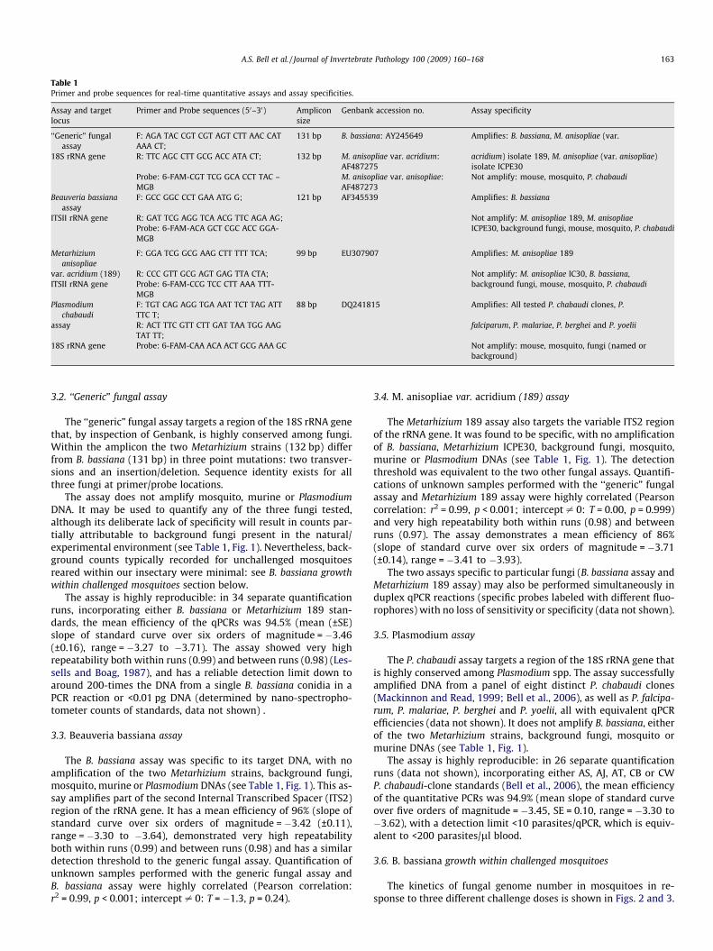
Table 1Primer and probe sequences for real-time quantitative assays and assay specificities.
Assay and targetlocus
Primer and Probe sequences (50–30) Ampliconsize
Genbank accession no. Assay specificity
‘‘Generic” fungalassay
F: AGA TAC CGT CGT AGT CTT AAC CATAAA CT;
131 bp B. bassiana: AY245649 Amplifies: B. bassiana, M. anisopliae (var.
18S rRNA gene R: TTC AGC CTT GCG ACC ATA CT; 132 bp M. anisopliae var. acridium:AF487275
acridium) isolate 189, M. anisopliae (var. anisopliae)isolate ICPE30
Probe: 6-FAM-CGT TCG GCA CCT TAC –MGB
M. anisopliae var. anisopliae:AF487273
Not amplify: mouse, mosquito, P. chabaudi
Beauveria bassianaassay
F: GCC GGC CCT GAA ATG G; 121 bp AF345539 Amplifies: B. bassiana
ITSII rRNA gene R: GAT TCG AGG TCA ACG TTC AGA AG; Not amplify: M. anisopliae 189, M. anisopliaeProbe: 6-FAM-ACA GCT CGC ACC GGA-MGB
ICPE30, background fungi, mouse, mosquito, P. chabaudi
Metarhiziumanisopliae
F: GGA TCG GCG AAG CTT TTT TCA; 99 bp EU307907 Amplifies: M. anisopliae 189
var. acridium (189) R: CCC GTT GCG AGT GAG TTA CTA; Not amplify: M. anisopliae IC30, B. bassiana,ITSII rRNA gene Probe: 6-FAM-CCG TCC CTT AAA TTT-
MGBbackground fungi, mouse, mosquito, P. chabaudi
Plasmodiumchabaudi
F: TGT CAG AGG TGA AAT TCT TAG ATTTTC T;
88 bp DQ241815 Amplifies: All tested P. chabaudi clones, P.
A.S. Bell et al. / Journal of Invertebrate Pathology 100 (2009) 160–168 163
3.2. ‘‘Generic” fungal assay
The ‘‘generic” fungal assay targets a region of the 18S rRNA genethat, by inspection of Genbank, is highly conserved among fungi.Within the amplicon the two Metarhizium strains (132 bp) differfrom B. bassiana (131 bp) in three point mutations: two transver-sions and an insertion/deletion. Sequence identity exists for allthree fungi at primer/probe locations.
The assay does not amplify mosquito, murine or PlasmodiumDNA. It may be used to quantify any of the three fungi tested,although its deliberate lack of specificity will result in counts par-tially attributable to background fungi present in the natural/experimental environment (see Table 1, Fig. 1). Nevertheless, back-ground counts typically recorded for unchallenged mosquitoesreared within our insectary were minimal: see B. bassiana growthwithin challenged mosquitoes section below.
The assay is highly reproducible: in 34 separate quantificationruns, incorporating either B. bassiana or Metarhizium 189 stan-dards, the mean efficiency of the qPCRs was 94.5% (mean (±SE)slope of standard curve over six orders of magnitude = �3.46(±0.16), range = �3.27 to �3.71). The assay showed very highrepeatability both within runs (0.99) and between runs (0.98) (Les-sells and Boag, 1987), and has a reliable detection limit down toaround 200-times the DNA from a single B. bassiana conidia in aPCR reaction or <0.01 pg DNA (determined by nano-spectropho-tometer counts of standards, data not shown) .
3.3. Beauveria bassiana assay
The B. bassiana assay was specific to its target DNA, with noamplification of the two Metarhizium strains, background fungi,mosquito, murine or Plasmodium DNAs (see Table 1, Fig. 1). This as-say amplifies part of the second Internal Transcribed Spacer (ITS2)region of the rRNA gene. It has a mean efficiency of 96% (slope ofstandard curve over six orders of magnitude = �3.42 (±0.11),range = �3.30 to �3.64), demonstrated very high repeatabilityboth within runs (0.99) and between runs (0.98) and has a similardetection threshold to the generic fungal assay. Quantification ofunknown samples performed with the generic fungal assay andB. bassiana assay were highly correlated (Pearson correlation:r2 = 0.99, p < 0.001; intercept – 0: T = �1.3, p = 0.24).
3.4. M. anisopliae var. acridium (189) assay
The Metarhizium 189 assay also targets the variable ITS2 regionof the rRNA gene. It was found to be specific, with no amplificationof B. bassiana, Metarhizium ICPE30, background fungi, mosquito,murine or Plasmodium DNAs (see Table 1, Fig. 1). The detectionthreshold was equivalent to the two other fungal assays. Quantifi-cations of unknown samples performed with the ‘‘generic” fungalassay and Metarhizium 189 assay were highly correlated (Pearsoncorrelation: r2 = 0.99, p < 0.001; intercept – 0: T = 0.00, p = 0.999)and very high repeatability both within runs (0.98) and betweenruns (0.97). The assay demonstrates a mean efficiency of 86%(slope of standard curve over six orders of magnitude = �3.71(±0.14), range = �3.41 to �3.93).
The two assays specific to particular fungi (B. bassiana assay andMetarhizium 189 assay) may also be performed simultaneously induplex qPCR reactions (specific probes labeled with different fluo-rophores) with no loss of sensitivity or specificity (data not shown).
3.5. Plasmodium assay
The P. chabaudi assay targets a region of the 18S rRNA gene thatis highly conserved among Plasmodium spp. The assay successfullyamplified DNA from a panel of eight distinct P. chabaudi clones(Mackinnon and Read, 1999; Bell et al., 2006), as well as P. falcipa-rum, P. malariae, P. berghei and P. yoelii, all with equivalent qPCRefficiencies (data not shown). It does not amplify B. bassiana, eitherof the two Metarhizium strains, background fungi, mosquito ormurine DNAs (see Table 1, Fig. 1).
The assay is highly reproducible: in 26 separate quantificationruns (data not shown), incorporating either AS, AJ, AT, CB or CWP. chabaudi-clone standards (Bell et al., 2006), the mean efficiencyof the quantitative PCRs was 94.9% (mean slope of standard curveover five orders of magnitude = �3.45, SE = 0.10, range = �3.30 to�3.62), with a detection limit <10 parasites/qPCR, which is equiv-alent to <200 parasites/ll blood.
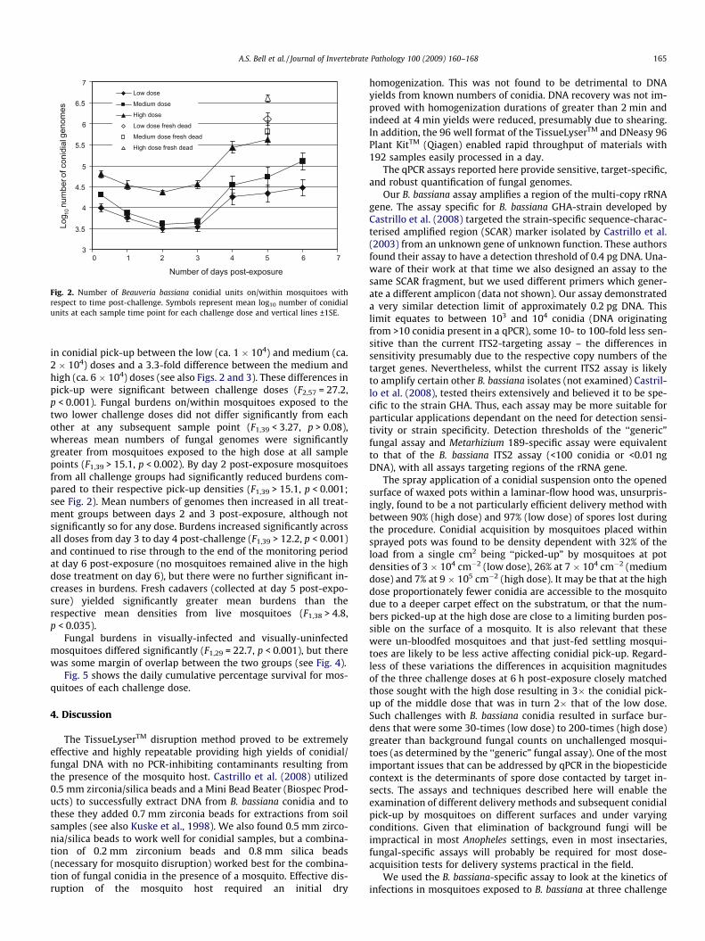
3.6. B. bassiana growth within challenged mosquitoes
The kinetics of fungal genome number in mosquitoes in re-sponse to three different challenge doses is shown in Figs. 2 and 3.
Fig. 1. Assay amplification plots and standard curves. Left hand panels show real-time qPCR amplification plots: each set of parallel lines indicate a 10-fold dilution in DNAsample (known standards – for fungal assays from 108 to 102 conidia) from which the standard curves are derived (right hand panels) by plotting the cycle at which eachstandard enters into log-linear amplification (crosses the machine-determined threshold: solid horizontal line) against the known number of conidia present in that standard.Conidial numbers in unknown samples are determined from where their amplification plot crosses the threshold and is read from the standard curve. Non-amplified samplesfail to generate an amplification plot and cross the threshold. A standard curve with a slope of �3.32 represents an assay with a hypothetical PCR efficiency of 100%.
164 A.S. Bell et al. / Journal of Invertebrate Pathology 100 (2009) 160–168
The ‘‘generic” fungal assay revealed unchallenged mosquitoesto have mean (±SE) background fungal counts of 347 (±37) conid-ial units (B. bassiana standards), whilst the B. bassiana-specific as-say showed 12 of these 20 mosquitoes to be negative for B.bassiana genomes and 8 to bear only trace numbers (data notshown).
Actual conidial densities present on challenge pots are shown inTable 2. These differed 2.2-fold between the low and mediumdoses (in theory should be 2-fold) and 13.2-fold between the med-ium and high doses (in theory 5-fold). Nevertheless, conidial acqui-sition by mosquitoes exposed to the high dose was proportionatelyless than at the other two doses and there was a 1.9-fold difference
Number of days post-exposure
Log 1
0nu
mbe
r of c
onid
ial g
enom
es
3
3.5
4
4.5
5
5.5
6
6.5
7
0 1 2 3 4 5 6 7
Low dose
Medium dose
High dose
Low dose fresh dead
Medium dose fresh dead
High dose fresh dead
Fig. 2. Number of Beauveria bassiana conidial units on/within mosquitoes withrespect to time post-challenge. Symbols represent mean log10 number of conidialunits at each sample time point for each challenge dose and vertical lines ±1SE.
A.S. Bell et al. / Journal of Invertebrate Pathology 100 (2009) 160–168 165
in conidial pick-up between the low (ca. 1 � 104) and medium (ca.2 � 104) doses and a 3.3-fold difference between the medium andhigh (ca. 6 � 104) doses (see also Figs. 2 and 3). These differences inpick-up were significant between challenge doses (F2,57 = 27.2,p < 0.001). Fungal burdens on/within mosquitoes exposed to thetwo lower challenge doses did not differ significantly from eachother at any subsequent sample point (F1,39 < 3.27, p > 0.08),whereas mean numbers of fungal genomes were significantlygreater from mosquitoes exposed to the high dose at all samplepoints (F1,39 > 15.1, p < 0.002). By day 2 post-exposure mosquitoesfrom all challenge groups had significantly reduced burdens com-pared to their respective pick-up densities (F1,39 > 15.1, p < 0.001;see Fig. 2). Mean numbers of genomes then increased in all treat-ment groups between days 2 and 3 post-exposure, although notsignificantly so for any dose. Burdens increased significantly acrossall doses from day 3 to day 4 post-challenge (F1,39 > 12.2, p < 0.001)and continued to rise through to the end of the monitoring periodat day 6 post-exposure (no mosquitoes remained alive in the highdose treatment on day 6), but there were no further significant in-creases in burdens. Fresh cadavers (collected at day 5 post-expo-sure) yielded significantly greater mean burdens than therespective mean densities from live mosquitoes (F1,38 > 4.8,p < 0.035).
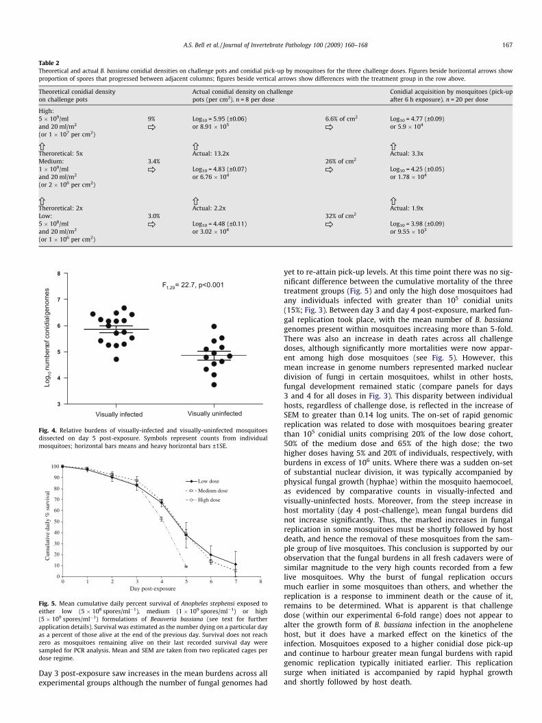
Fungal burdens in visually-infected and visually-uninfectedmosquitoes differed significantly (F1,29 = 22.7, p < 0.001), but therewas some margin of overlap between the two groups (see Fig. 4).
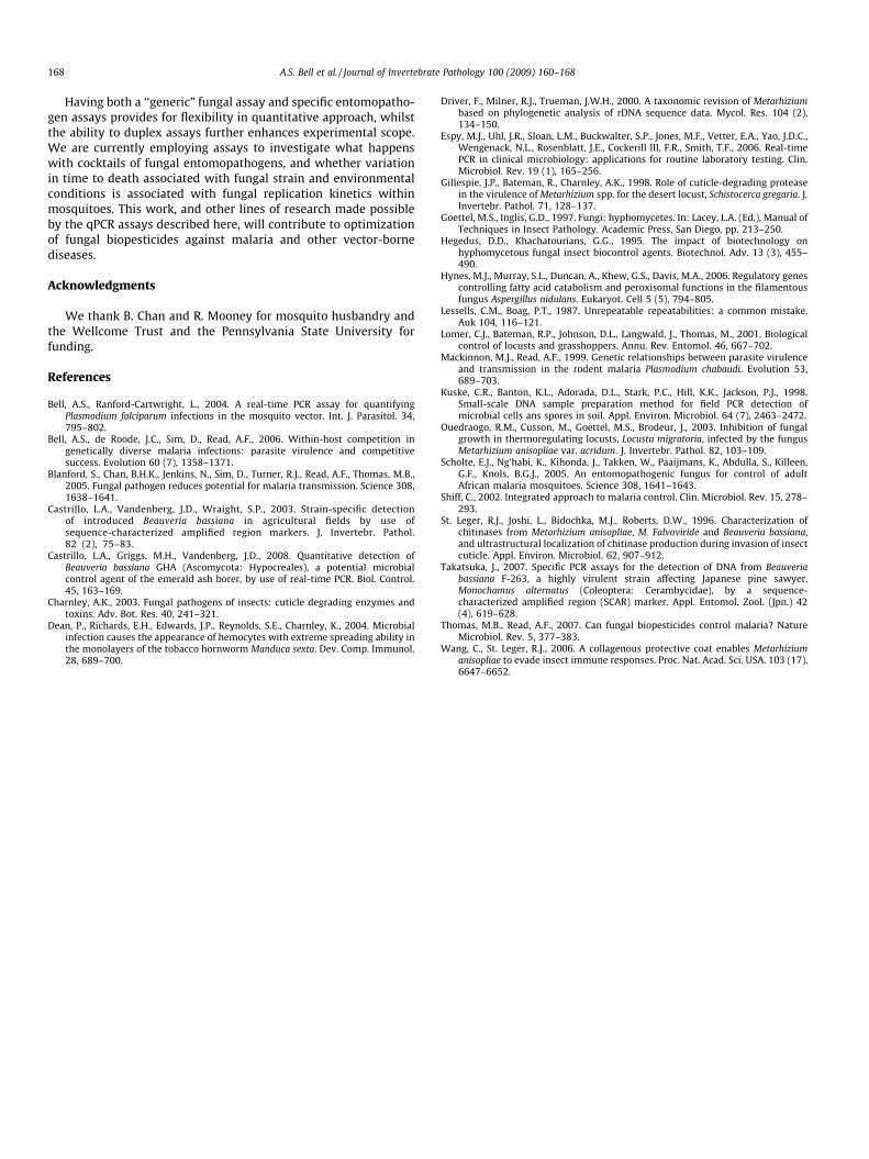
Fig. 5 shows the daily cumulative percentage survival for mos-quitoes of each challenge dose.
4. Discussion
The TissueLyserTM disruption method proved to be extremelyeffective and highly repeatable providing high yields of conidial/fungal DNA with no PCR-inhibiting contaminants resulting fromthe presence of the mosquito host. Castrillo et al. (2008) utilized0.5 mm zirconia/silica beads and a Mini Bead Beater (Biospec Prod-ucts) to successfully extract DNA from B. bassiana conidia and tothese they added 0.7 mm zirconia beads for extractions from soilsamples (see also Kuske et al., 1998). We also found 0.5 mm zirco-nia/silica beads to work well for conidial samples, but a combina-tion of 0.2 mm zirconium beads and 0.8 mm silica beads(necessary for mosquito disruption) worked best for the combina-tion of fungal conidia in the presence of a mosquito. Effective dis-ruption of the mosquito host required an initial dry
homogenization. This was not found to be detrimental to DNAyields from known numbers of conidia. DNA recovery was not im-proved with homogenization durations of greater than 2 min andindeed at 4 min yields were reduced, presumably due to shearing.In addition, the 96 well format of the TissueLyserTM and DNeasy 96Plant KitTM (Qiagen) enabled rapid throughput of materials with192 samples easily processed in a day.
The qPCR assays reported here provide sensitive, target-specific,and robust quantification of fungal genomes.
Our B. bassiana assay amplifies a region of the multi-copy rRNAgene. The assay specific for B. bassiana GHA-strain developed byCastrillo et al. (2008) targeted the strain-specific sequence-charac-terised amplified region (SCAR) marker isolated by Castrillo et al.(2003) from an unknown gene of unknown function. These authorsfound their assay to have a detection threshold of 0.4 pg DNA. Una-ware of their work at that time we also designed an assay to thesame SCAR fragment, but we used different primers which gener-ate a different amplicon (data not shown). Our assay demonstrateda very similar detection limit of approximately 0.2 pg DNA. Thislimit equates to between 103 and 104 conidia (DNA originatingfrom >10 conidia present in a qPCR), some 10- to 100-fold less sen-sitive than the current ITS2-targeting assay – the differences insensitivity presumably due to the respective copy numbers of thetarget genes. Nevertheless, whilst the current ITS2 assay is likelyto amplify certain other B. bassiana isolates (not examined) Castril-lo et al. (2008), tested theirs extensively and believed it to be spe-cific to the strain GHA. Thus, each assay may be more suitable forparticular applications dependant on the need for detection sensi-tivity or strain specificity. Detection thresholds of the ‘‘generic”fungal assay and Metarhizium 189-specific assay were equivalentto that of the B. bassiana ITS2 assay (<100 conidia or <0.01 ngDNA), with all assays targeting regions of the rRNA gene.
The spray application of a conidial suspension onto the openedsurface of waxed pots within a laminar-flow hood was, unsurpris-ingly, found to be a not particularly efficient delivery method withbetween 90% (high dose) and 97% (low dose) of spores lost duringthe procedure. Conidial acquisition by mosquitoes placed withinsprayed pots was found to be density dependent with 32% of theload from a single cm2 being ‘‘picked-up” by mosquitoes at potdensities of 3 � 104 cm�2 (low dose), 26% at 7 � 104 cm�2 (mediumdose) and 7% at 9 � 105 cm�2 (high dose). It may be that at the highdose proportionately fewer conidia are accessible to the mosquitodue to a deeper carpet effect on the substratum, or that the num-bers picked-up at the high dose are close to a limiting burden pos-sible on the surface of a mosquito. It is also relevant that thesewere un-bloodfed mosquitoes and that just-fed settling mosqui-toes are likely to be less active affecting conidial pick-up. Regard-less of these variations the differences in acquisition magnitudesof the three challenge doses at 6 h post-exposure closely matchedthose sought with the high dose resulting in 3� the conidial pick-up of the middle dose that was in turn 2� that of the low dose.Such challenges with B. bassiana conidia resulted in surface bur-dens that were some 30-times (low dose) to 200-times (high dose)greater than background fungal counts on unchallenged mosqui-toes (as determined by the ‘‘generic” fungal assay). One of the mostimportant issues that can be addressed by qPCR in the biopesticidecontext is the determinants of spore dose contacted by target in-sects. The assays and techniques described here will enable theexamination of different delivery methods and subsequent conidialpick-up by mosquitoes on different surfaces and under varyingconditions. Given that elimination of background fungi will beimpractical in most Anopheles settings, even in most insectaries,fungal-specific assays will probably be required for most dose-acquisition tests for delivery systems practical in the field.
We used the B. bassiana-specific assay to look at the kinetics ofinfections in mosquitoes exposed to B. bassiana at three challenge
0123456789
0123456789
0123456789
0123456789
0123456789
0123456789
0123456789
0123456789
<33.2
-3.43.6
-3.8 4-4.2
4.4-4.
64.8
-55.2
-5.45.6
-5.8 6-6.2
6.4-6.6
6.8-7.
07.2
-7.4
01234567890123456789
0123456789
0123456789
0123456789
0123456789
0123456789
0123456789
<33.2
-3.4
3.6-3.8 4-4
.24.4
-4.6 4.8-5
5.2-5.4
5.6-5.
86-6
.26.4
-6.6
6.8-7.
07.2
-7.4
0123456789
0123456789
0123456789
0123456789
0123456789
0123456789
0123456789
<33.2
-3.4
3.6-3.
84-4
.24.4
-4.6
4.8-5
5.2-5.
4
5.6-5.
86-6
.26.4
-6.6
6.8-7.
0
7.2-7.
4
Challenge doseLow Medium High
6 hours post-
exposure
(pick-up)
Day 1
Day 2
Day 3
Day 4
Day 5
Day 6 All dead
Fresh cadavers from day 5 post-exposure
Log10 number of conidial genomes
x = 3.99 (0.09)
x = 3.75 (0.09)
x = 3.49 (0.09)
x = 3.53 (0.1)
x = 4.25 (0.18)
x = 4.33 (0.2)
x = 4.47 (0.19)
x = 6.1 (0.16)
x = 4.3 (0.05)
x = 3.86 (0.06)
x = 3.60 (0.07)
x = 3.65 (0.09)
x = 4.74 (0.21)
x = 4.73 (0.23)
x = 5.1 (0.2)
x = 5.81 (0.26)
x = 4.8 (0.09)
x = 4.53 (0.1)
x = 4.36 (0.05)
x = 4.55 (0.09)
x = 5.43 (0.14)
x = 5.63 (0.14)
x = 6.61 (0.09)
Num
ber o
f mos
quito
es
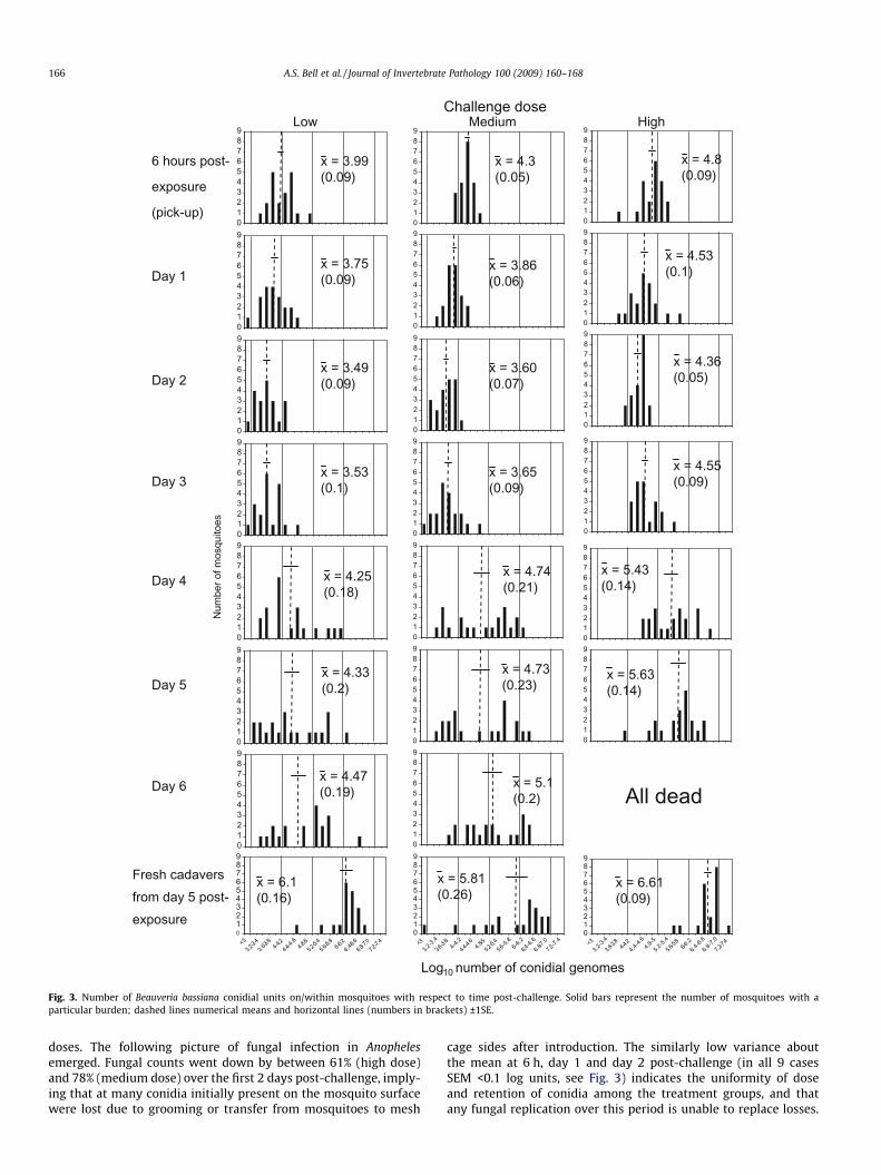
Fig. 3. Number of Beauveria bassiana conidial units on/within mosquitoes with respect to time post-challenge. Solid bars represent the number of mosquitoes with aparticular burden; dashed lines numerical means and horizontal lines (numbers in brackets) ±1SE.
166 A.S. Bell et al. / Journal of Invertebrate Pathology 100 (2009) 160–168
doses. The following picture of fungal infection in Anophelesemerged. Fungal counts went down by between 61% (high dose)and 78% (medium dose) over the first 2 days post-challenge, imply-ing that at many conidia initially present on the mosquito surfacewere lost due to grooming or transfer from mosquitoes to mesh
cage sides after introduction. The similarly low variance aboutthe mean at 6 h, day 1 and day 2 post-challenge (in all 9 casesSEM <0.1 log units, see Fig. 3) indicates the uniformity of doseand retention of conidia among the treatment groups, and thatany fungal replication over this period is unable to replace losses.
Table 2Theoretical and actual B. bassiana conidial densities on challenge pots and conidial pick-up by mosquitoes for the three challenge doses. Figures beside horizontal arrows showproportion of spores that progressed between adjacent columns; figures beside vertical arrows show differences with the treatment group in the row above.
Theoretical conidial densityon challenge pots
Actual conidial density on challengepots (per cm2). n = 8 per dose
Conidial acquisition by mosquitoes (pick-upafter 6 h exposure). n = 20 per dose
High:5 � 109/ml 9% Log10 = 5.95 (±0.06) 6.6% of cm2 Log10 = 4.77 (±0.09)and 20 ml/m2 or 8.91 � 105 or 5.9 � 104
(or 1 � 107 per cm2)
Theroretical: 5x Actual: 13.2x Actual: 3.3xMedium: 3.4% 26% of cm2
Fig. 4. Relative burdens of visually-infected and visually-uninfected mosquitoesdissected on day 5 post-exposure. Symbols represent counts from individualmosquitoes; horizontal bars means and heavy horizontal bars ±1SE.
0
10
20
30
40
50
60
70
80
90
100
0 1 2 3 4 5 6 7 8
Low dose
Medium dose
High dose
Cum
ulat
ive
daily
% s
urvi
val
Day post-exposure
Fig. 5. Mean cumulative daily percent survival of Anopheles stephensi exposed toeither low (5 � 108 spores/ml�1), medium (1 � 109 spores/ml�1) or high(5 � 109 spores/ml�1) formulations of Beauveria bassiana (see text for furtherapplication details). Survival was estimated as the number dying on a particular dayas a percent of those alive at the end of the previous day. Survival does not reachzero as mosquitoes remaining alive on their last recorded survival day weresampled for PCR analysis. Mean and SEM are taken from two replicated cages perdose regime.
A.S. Bell et al. / Journal of Invertebrate Pathology 100 (2009) 160–168 167
Day 3 post-exposure saw increases in the mean burdens across allexperimental groups although the number of fungal genomes had
yet to re-attain pick-up levels. At this time point there was no sig-nificant difference between the cumulative mortality of the threetreatment groups (Fig. 5) and only the high dose mosquitoes hadany individuals infected with greater than 105 conidial units(15%; Fig. 3). Between day 3 and day 4 post-exposure, marked fun-gal replication took place, with the mean number of B. bassianagenomes present within mosquitoes increasing more than 5-fold.There was also an increase in death rates across all challengedoses, although significantly more mortalities were now appar-ent among high dose mosquitoes (see Fig. 5). However, thismean increase in genome numbers represented marked nucleardivision of fungi in certain mosquitoes, whilst in other hosts,fungal development remained static (compare panels for days3 and 4 for all doses in Fig. 3). This disparity between individualhosts, regardless of challenge dose, is reflected in the increase ofSEM to greater than 0.14 log units. The on-set of rapid genomicreplication was related to dose with mosquitoes bearing greaterthan 105 conidial units comprising 20% of the low dose cohort,50% of the medium dose and 65% of the high dose; the twohigher doses having 5% and 20% of individuals, respectively, withburdens in excess of 106 units. Where there was a sudden on-setof substantial nuclear division, it was typically accompanied byphysical fungal growth (hyphae) within the mosquito haemocoel,as evidenced by comparative counts in visually-infected andvisually-uninfected hosts. Moreover, from the steep increase inhost mortality (day 4 post-challenge), mean fungal burdens didnot increase significantly. Thus, the marked increases in fungalreplication in some mosquitoes must be shortly followed by hostdeath, and hence the removal of these mosquitoes from the sam-ple group of live mosquitoes. This conclusion is supported by ourobservation that the fungal burdens in all fresh cadavers were ofsimilar magnitude to the very high counts recorded from a fewlive mosquitoes. Why the burst of fungal replication occursmuch earlier in some mosquitoes than others, and whether thereplication is a response to imminent death or the cause of it,remains to be determined. What is apparent is that challengedose (within our experimental 6-fold range) does not appear toalter the growth form of B. bassiana infection in the anophelenehost, but it does have a marked effect on the kinetics of theinfection. Mosquitoes exposed to a higher conidial dose pick-upand continue to harbour greater mean fungal burdens with rapidgenomic replication typically initiated earlier. This replicationsurge when initiated is accompanied by rapid hyphal growthand shortly followed by host death.
168 A.S. Bell et al. / Journal of Invertebrate Pathology 100 (2009) 160–168
Having both a ‘‘generic” fungal assay and specific entomopatho-gen assays provides for flexibility in quantitative approach, whilstthe ability to duplex assays further enhances experimental scope.We are currently employing assays to investigate what happenswith cocktails of fungal entomopathogens, and whether variationin time to death associated with fungal strain and environmentalconditions is associated with fungal replication kinetics withinmosquitoes. This work, and other lines of research made possibleby the qPCR assays described here, will contribute to optimizationof fungal biopesticides against malaria and other vector-bornediseases.
Acknowledgments
We thank B. Chan and R. Mooney for mosquito husbandry andthe Wellcome Trust and the Pennsylvania State University forfunding.
References
Bell, A.S., Ranford-Cartwright, L., 2004. A real-time PCR assay for quantifyingPlasmodium falciparum infections in the mosquito vector. Int. J. Parasitol. 34,795–802.
Bell, A.S., de Roode, J.C., Sim, D., Read, A.F., 2006. Within-host competition ingenetically diverse malaria infections: parasite virulence and competitivesuccess. Evolution 60 (7), 1358–1371.
Gillespie, J.P., Bateman, R., Charnley, A.K., 1998. Role of cuticle-degrading proteasein the virulence of Metarhizium spp. for the desert locust, Schistocerca gregaria. J.Invertebr. Pathol. 71, 128–137.
Goettel, M.S., Inglis, G.D., 1997. Fungi: hyphomycetes. In: Lacey, L.A. (Ed.), Manual ofTechniques in Insect Pathology. Academic Press, San Diego, pp. 213–250.
Hegedus, D.D., Khachatourians, G.G., 1995. The impact of biotechnology onhyphomycetous fungal insect biocontrol agents. Biotechnol. Adv. 13 (3), 455–490.
Hynes, M.J., Murray, S.L., Duncan, A., Khew, G.S., Davis, M.A., 2006. Regulatory genescontrolling fatty acid catabolism and peroxisomal functions in the filamentousfungus Aspergillus nidulans. Eukaryot. Cell 5 (5), 794–805.
Lessells, C.M., Boag, P.T., 1987. Unrepeatable repeatabilities: a common mistake.Auk 104, 116–121.
Lomer, C.J., Bateman, R.P., Johnson, D.L., Langwald, J., Thomas, M., 2001. Biologicalcontrol of locusts and grasshoppers. Annu. Rev. Entomol. 46, 667–702.
Mackinnon, M.J., Read, A.F., 1999. Genetic relationships between parasite virulenceand transmission in the rodent malaria Plasmodium chabaudi. Evolution 53,689–703.
Kuske, C.R., Banton, K.L., Adorada, D.L., Stark, P.C., Hill, K.K., Jackson, P.J., 1998.Small-scale DNA sample preparation method for field PCR detection ofmicrobial cells ans spores in soil. Appl. Environ. Microbiol. 64 (7), 2463–2472.
Ouedraogo, R.M., Cusson, M., Goettel, M.S., Brodeur, J., 2003. Inhibition of fungalgrowth in thermoregulating locusts, Locusta migratoria, infected by the fungusMetarhizium anisopliae var. acridum. J. Invertebr. Pathol. 82, 103–109.
Scholte, E.J., Ng’habi, K., Kihonda, J., Takken, W., Paaijmans, K., Abdulla, S., Killeen,G.F., Knols, B.G.J., 2005. An entomopathogenic fungus for control of adultAfrican malaria mosquitoes. Science 308, 1641–1643.
St. Leger, R.J., Joshi, L., Bidochka, M.J., Roberts, D.W., 1996. Characterization ofchitinases from Metarhizium anisopliae, M. Falvoviride and Beauveria bassiana,and ultrastructural localization of chitinase production during invasion of insectcuticle. Appl. Environ. Microbiol. 62, 907–912.
Takatsuka, J., 2007. Specific PCR assays for the detection of DNA from Beauveriabassiana F-263, a highly virulent strain affecting Japanese pine sawyer,Monochamus alternatus (Coleoptera: Cerambycidae), by a sequence-characterized amplified region (SCAR) marker. Appl. Entomol. Zool. (Jpn.) 42(4), 619–628.
Thomas, M.B., Read, A.F., 2007. Can fungal biopesticides control malaria? NatureMicrobiol. Rev. 5, 377–383.
Wang, C., St. Leger, R.J., 2006. A collagenous protective coat enables Metarhiziumanisopliae to evade insect immune responses. Proc. Nat. Acad. Sci. USA. 103 (17),6647–6652.