Purificación López-Garcíaa,⁎, Laura Emeb, David Moreiraa
a Ecologie Systématique Evolution, CNRS, Université Paris-Sud, Université Paris-Saclay, AgroParisTech, 91400 Orsay, Franceb Centre for Comparative Genomics and Evolutionary Bioinformatics, Department of Biochemistry and Molecular Biology, Dalhousie University, Halifax,Canada NS B3H 4R2
Fifty years ago, Lynn Margulis, inspiring in early twentieth-century ideas that put forward a symbiotic origin forsome eukaryotic organelles, proposed a unified theory for the origin of the eukaryotic cell based on symbiosis asevolutionary mechanism. Margulis was profoundly aware of the importance of symbiosis in the naturalmicrobial world and anticipated the evolutionary significance that integrated cooperative interactions mighthave as mechanism to increase cellular complexity. Today, we have started fully appreciating the vast extent ofmicrobial diversity and the importance of syntrophic metabolic cooperation in natural ecosystems, especially insediments and microbial mats. Also, not only the symbiogenetic origin of mitochondria and chloroplasts hasbeen clearly demonstrated, but improvement in phylogenomic methods combined with recent discoveries ofarchaeal lineages more closely related to eukaryotes further support the symbiogenetic origin of the eukaryoticcell. Margulis left us in legacy the idea of ‘eukaryogenesis by symbiogenesis’. Although this has been largelyverified, when, where, and specifically how eukaryotic cells evolved are yet unclear. Here, we shortly reviewcurrent knowledge about symbiotic interactions in the microbial world and their evolutionary impact, the statusof eukaryogenetic models and the current challenges and perspectives ahead to reconstruct the evolutionarypath to eukaryotes.
1. Introduction
In 1967, Lynn Margulis (Sagan) published her famous On theorigin of mitosing cells (Sagan, 1967), where she set up the basis of herSerial Endosymbiotic Theory for the origin of the eukaryotic cell(Margulis, 1981, 1996). In her manuscript, Margulis revived previousideas proposing the endosymbiotic origin of chloroplasts from 'blue-green algae' (cyanobacteria) (Mereschkowsky, 1905, 1910) and mito-chondria from purple bacteria (alphaproteobacteria) (Wallin, 1927);and further put forward the idea that the eukaryotic flagellum derivedfrom symbiotic bacteria (spirochete-like). Although KonstantinMereschkowsky had also advocated for the endosymbiotic origin ofthe eukaryotic nucleus (Mereschkowsky, 1910), Margulis discardedthis possibility without nonetheless proposing a clear mechanism forthe origin of the nucleus in an original heterotrophic amoeboid host(Sagan, 1967). Later, after the recognition of archaea as a thirdphylogenetic domain of life (Woese and Fox, 1977; Woese et al.,1990) showing significant similarities in terms of informationalprocesses (replication, transcription, translation) with eukaryotes(Rivera et al., 1998; Thiergart et al., 2012), she postulated that thehost of the future mitochondrion derived from a thermoacidophilic
wall-less archaeon similar to contemporary Thermoplasma spp.(Euryarchaeota) (Margulis, 1981, 1996), an idea that she adoptedfrom Dennis Searcy (Searcy, 1992). The nucleus would have evolvedautogenously (not by symbiosis) in a chimeric archaeal-bacterial cell(Margulis et al., 2000).
The strength and innovative character of Margulis' ideas lay in theproposal of a unified theory based on symbiosis as evolutionarymechanism for the origin of the eukaryotic cell: eukaryogenesis bysymbiogenesis. Strongly criticized at the time, at least part of theseideas became credible with the advent of molecular phylogeny that,among its earliest achievements, allowed demonstrating that conservedgenes in chloroplast and mitochondrial genomes clustered with,respectively, cyanobacterial and alphaproteobacterial genes in phylo-genetic trees (Schwartz and Dayhoff, 1978). The endosymbiotic originof these membrane-bound organelles became mainstream science andit is now well established how their bacterial ancestors evolved,undergoing a process of genome reduction, impacting their host andleading to a variety of organelle derivatives (Dyall et al., 2004; Gray,2012; Muller et al., 2012; Schwartz and Dayhoff, 1978). However, theorigin of her eukaryotic 'nucleocytoplasm' was much controversial;neither the Thermoplasma-like nature of the host nor (far less) the
http://dx.doi.org/10.1016/j.jtbi.2017.02.031Received 2 January 2017; Received in revised form 19 February 2017; Accepted 25 February 2017
symbiotic origin of the flagellum were ever considered seriously. In herview, a metabolic symbiosis based on interspecific sulfur transfer wasestablished between a Thermoplasma-like archaeon, which generatedhydrogen sulfide, and a spirochete, which oxidized sulfide to sulfur and,at the same time, provided motility to the symbiotic consortium(Margulis et al., 2000), being at the origin of the eukaryotic cytoske-leton and the mitotic apparatus (Margulis et al., 2006; Sagan, 1967).Margulis argued that, similarly to mitochondria and chloroplasts,which retained reduced genomes, a remnant genome should be foundin connection with the (9+2) microtubular basal bodies associated toeukaryotic flagella (Sagan, 1967). But such remnant genomes werenever found and phylogenomic analyses in the genomics era did notshow any particular similarity between eukaryote and spirochetegenomes. Thus, while the endosymbiotic origin of chloroplasts andmitochondria in a eukaryotic host became widely accepted, thesymbiotic origin of the eukaryotic cell itself was not and Margulis'ideas stood alone for almost three decades before other symbiogeneticmodels made it through to the scientific arena.
Lynn Margulis had a classical, exceptional knowledge on themorphology, biology and ecology of microorganisms, which led her toactively participate in attempts to establish global classification systemsfor microorganisms. She sustained that life should be classified in twosuperkingdoms or domains (prokaryotes and eukaryotes) and fivekingdoms (Margulis, 1992; Margulis and Schwartz, 1998; Whittakerand Margulis, 1978), and co-edited the famous Handbook ofProtoctista (Margulis et al., 1990) widely used by eukaryotic micro-biologists. Paradoxically, although she recognized the importance ofDNA comparisons in establishing a natural (evolutionary) classificationsystem for all organisms (Sagan, 1967), she was reluctant to adopt(Margulis and Guerrero, 1991) the Carl R. Woese's three-domainclassification system that resulted from molecular phylogenetic ana-lyses of universally conserved genes (Woese and Fox, 1977; Woeseet al., 1990). The molecular revolution initiated by Woese using 16S/18S rRNA genes had three major outcomes that have subsequentlyimpacted many areas in biology, from taxonomy and systematics tomicrobial ecology and comparative genomics. He showed that it waspossible to build universal phylogenetic trees for all cellular life andhence establish a natural biological classification system. In doing so,he incidentally discovered that a group of prokaryotic cells, thearchaea, defined a distinct phylogenetic group (a third domain of life)(Woese and Fox, 1977; Woese et al., 1990). And finally, as corollary ofhis work, it was in principle possible to explore microbial diversity innatural environments by amplifying and sequencing conserved markergenes, thus sidestepping the well-known problem of culture bias (only atiny fraction of microorganisms are amenable to culture in thelaboratory). The amplification, cloning and sequencing of 16S and18S rRNA genes from many different environments led in the 1990sand early 2000s to the realization that the diversity of both prokaryoticand eukaryotic microorganisms was much more important than everthought (Moreira and López-García, 2002; Pace, 1997). Today, larger-scale metabarcoding analyses based on amplicon high-throughputsequencing, together with single cell genomics and metagenome-basedgenome reconstruction not only reinforce the view of an extraordinarilyimportant microbial diversity but at the same time validate the threedomains of life (Archaea, Bacteria, Eucarya) that Woese identified fortyyears ago (Eme and Doolittle, 2015; Hug et al., 2016; Rinke et al.,2013; Yarza et al., 2014).
This possibility to study novel, uncultured organisms in the wild hasrevealed crucial to advance in the resolution of the long-term query ofeukaryotic origins. Notwithstanding Margulis symbiogenetic ideas, theeukaryogenetic model that prevailed in the last part of the twentiethcentury and until recently was that of an autogenous origin of all typicaleukaryotic features but mitochondria and chloroplasts (of demon-strated endosymbiotic origin) in a proto-eukaryotic lineage sister toarchaea (de Duve, 2007; Jekely, 2003; Poole and Penny, 2007). Thediscovery that amitochondriate protists lost (or modified) mitochon-
dria secondarily, implying that the last eukaryotic common ancestor(LECA) possessed mitochondria (Embley, 2006; Embley and Hirt,1998), left some room for the proposal of new symbiogenetic modelsfor the origin of eukaryotes (for review, see (Embley and Martin, 2006;Keeling, 2014; Lopez-Garcia and Moreira, 2015; McInerney et al.,2014)). These models denied the existence of a third, proto-eukaryoticlineage different from classical bacteria and archaea and explained themixed eukaryotic heritage (archaeal informational genes, bacterialoperational genes) by archaea-bacteria merging. Nonetheless, thesemodels remained little accepted, in part by the failure of phylogenomicanalyses to identify clear prokaryotic ancestors (other than alphapro-teobacteria) of eukaryotes (Gribaldo et al., 2010). However, the recentdiscovery of Lokiarchaeota (Spang et al., 2015) and other relatedlineages (collectively, Asgard archaea) (Zaremba-Niedzwiedzka et al.,2017), grouping anaerobic archaea that share more and more similargenes with eukaryotes than the rest of known archaea strongly suggestthat the archaeal component of eukaryotes did indeed derive fromwithin archaea, giving fresh credit to symbiogenetic models (Koonin,2015; Lopez-Garcia and Moreira, 2015; Williams and Embley, 2015).
The seminal On the origin of mitosing cells (Sagan, 1967) did notonly stirred evolutionary thinking at the time but today, fifty yearslater, it is still striking in its modernity and, in many ways, visionarycontent about the role of symbiosis in eukaryotic evolution. Here, werevisit some of the ideas put forward by Margulis that have made itthrough time and briefly discuss current knowledge and challengesabout eukaryogenesis.
2. Symbiosis in natural ecosystems
One of the most important, yet little appreciated, strengths ofMargulis' ideas was her awareness, at a time when molecular biologyand reductionist approaches were on the rise, of the intricate, oftenmutualistic interactions that prevail among microorganisms in naturalenvironments, which she understood as key in fostering evolutionaryprocesses. This allowed her to conceive holistic scenarios about theevolution of life on Earth based in the distribution of energy and carbonmetabolism in prokaryotes, the fossil record and her recurrentobservation of protist-prokaryote symbioses under the microscope(Margulis et al., 1986; Sagan, 1967). Several decades later, moleculartools for the study of natural microbial communities have nothing butproved her intuition about the importance of mutualistic interactionsin ecology and evolution right.
2.1. Symbiosis, cooperation, evolution
The role of cooperation in evolution is being increasingly recog-nized, particularly in the microbial world. Several recent reviewsanalyze cooperation in microbes and point to the factors that lead tostable cooperation over time (e.g. (Celiker and Gore, 2013; Mitri andFoster, 2013; Nadell et al., 2016)). Some of these principles have beentested experimentally in biofilm populations (Steenackers et al., 2016).Part of this interest has grown exponentially due to studies on human(and other animal) microbiomes and the effect that, more specifically,gut microbial communities have in human metabolism and thedevelopment of the immune system (Sonnenburg and Backhed,2016; Thaiss et al., 2016). Likewise, although the importance of themicroorganisms from the rhizosphere has been recognized for a longtime (Philippot et al., 2013), interest in microbial endophytes in plantsis expanding (Hardoim et al., 2015). This new interest has even led tothe qualification, by some authors, of these collective microbialcommunities (microbiomes) as 'symbionts' and humans, other animals(e.g. corals, insects) or plants as 'holobionts'. Strictly speaking, theseterms are stretched inappropriately because the specific interactionsamongst all the components of those communities and their multi-cellular hosts do not necessarily correspond to co-evolutionary mutua-listic symbioses but are likely to be largely governed by classical
P. López-García et al. Journal of Theoretical Biology xxx (xxxx) xxx–xxx
2
microbial ecology determinants (Moran and Sloan, 2015; Mushegianand Ebert, 2016).
But what is exactly symbiosis? 'Living together' by etymology,symbiosis has two major definitions in biology. One is looser andrefers to the co-existence and dependency of at least one organismupon a partner. Symbiosis would then accommodate three types ofinteractions depending on the effect on the host's fitness: mutualism(fitness increase), commensalism (no fitness change), and parasitism(fitness decrease) (Mushegian and Ebert, 2016). In reality, the linebetween mutualism and parasitism is thin and these kinds of interac-tions are often seen as a continuum, sometimes depending on theenvironmental conditions. Examples of parasite-mutualist transitionsare well-documented in plant mycorrhiza (Paszkowski, 2006),Wolbachia-insect interactions (Hosokawa et al., 2010) and even inintrinsic genetic parasites such as plasmids or viruses (Bao andRoossinck, 2013). The other type of definition equals symbiosis andmutualism, so that symbiosis implies a mutually beneficial interaction.This is the type of definition that Margulis used when she referred tosymbiosis. In her view, the holobiont properly meant an integratedsymbiotic consortium (obligate mutualism) that behaves as a unit ofevolution (Guerrero et al., 2013). Eukaryogenesis could only beunderstood in this context.
We are progressively discovering that mutualistic symbioses (here-after, symbioses) are plentiful in nature and, in particular environ-ments such as sediments or microbial mats with steep redox gradients,they may be the rule. Some examples follow.
2.2. Eukaryote – prokaryote symbioses
This type of symbioses is often easy to identify since, in many cases,it implies endosymbiosis within a larger host and, in the case ofmulticellular organisms, the frequent evolution of dedicated structures,such as bacteriocytes in aphids, trophosomes in deep-sea polychaetesor root nodules in plants. Many of these symbioses are the result oflong co-evolutionary processes that have shaped the genotypes andphenotypes of the different partners involved.
Prokaryotic symbioses with animals have been widely documented(Moya et al., 2008). They have been particularly well studied in insects,where they can have astounding impacts not only in nutritional but alsoin reproductive aspects of their biology. Thus, vertically-transmittedalphaproteobacterial endosymbionts of the genus Wolbachia impactsex determination and can induce feminization, parthenogenesis, malekilling and sperm-egg incompatibility (Cordaux et al., 2011; Werrenet al., 2008). Comparative genomic analyses are starting to reveal theunderlying basis for these effects, which is sometimes linked tohorizontal gene transfer from the bacterium to the host genome. Forinstance, a 3-Mb insert of a feminizingWolbachia genome was recentlytransferred into the pillbug nuclear genome and its occurrencecorrelates with the female sex (Leclercq et al., 2016). Also well-studiedsymbioses are those involving aphids and other phloem-sap feedinginsects with different bacteria that complement their imbalanced,sugar-rich diet by supplying amino acids. A famous symbiotic coupleis that of aphids with Buchnera aphidicola, an endosymbiotic gamma-proteobacterium living in specialized aphid cells (bacteriocytes). Thesebacterial endosymbionts, being vertically inherited and having lowpopulation sizes, are prone to genetic drift. Their genes are fast-evolving and their genomes, AT-biased and progressively reduced(McCutcheon and Moran, 2012; Moran and Bennett, 2014). Genomereduction can lead to the loss of essential genes for amino-acidbiosynthesis. When this happens, a second, less reduced endosymbiontthat complements the lost biosynthetic pathways can be acquired andmaintained in tripartite symbiosis (Perez-Brocal et al., 2006) or it canbe simply lost and replaced by a new symbiont (Koga and Moran,2014). The replacement of a reduced-genome symbiont is not infre-quent and can be achieved experimentally (Moran and Yun, 2015).
Less well-known, but equally important are symbioses of bacteria
and marine animals. The microbial communities that inhabit sponges,making up to one third of their biomass, are specific to their hosts andmany of their bacterial and archaeal members may be true symbionts(Hentschel et al., 2012). Members of the phylum Poribacteria arespecifically associated with sponges and seem to be able to fix carbonand contain putative symbiotic factors including adhesins and otherproteins potentially mediating host-microbe interactions (Siegl et al.,2011). Similarly, microbial communities in corals are highly diverse and,although several members may be true mutualists (Hernandez-Agredaet al., 2016), the clear established case is that of euphotic-zone coralsand dinoflagellate algae of the genus Symbiodinium (Roth, 2014), whichalso occur in some bivalves and anemones. Climate-change inducedocean acidification and warming appear to correlate with symbiont loss,leading to coral bleaching and death, and seriously affecting coral-reefecosystems. The counterpart of these phototrophic symbioses, wherebyfixed carbon is handed over to the host in exchange for nutrientcollection and the availability of a stable environment, is that ofchemotrophic symbioses in deep-sea or sediment fauna. Soon after thediscovery of deep-sea vents and their cohort of exotic fauna in the late1970s, symbiotic bacteria filling out completely the modified gut of thegiant worm Riftia pachyptila led to propose a chemoautotrophicsymbiosis (Cavanaugh et al., 1981). Today the basis for this symbiosisis well known; the gammaproteobacterial symbiont, acquired anew ateach generation, fixes carbon gaining energy by the oxidation of H2S,while the polychaete worm transports O2, NO3
− and H2S for the needs ofits otherwise metabolically versatile symbiont (Robidart et al., 2008).Similar chemotrophic symbioses also abound in the gills of deep-seabivalves and other deep-sea metazoans and, more generally, ecto- andendosymbionts are frequently associated to animals living in oxygen-deprived marine settings, from the deep-sea to coastal sedimentssettings (Dubilier et al., 2008). These most often correspond to sulfide-or sulfur-oxidizing Gamma- or Epsilonproteobacteria, methanotrophicGammaproteobacteria or sulfate-reducing Deltaproteobacteria (Dubilieret al., 2008). In many cases, these symbioses are multiple, involvingdiverse endosymbionts with distinct metabolic capabilities. For instance,clams of the genus Bathymodiolus harbor dual symbioses with metha-notrophic and thiotrophic bacteria (Duperron et al., 2007). The case ofthe oligochaete Olavius algarvensis is most remarkable; this worm,lacking mouth, gut and nephridia, hosts co-existing sulfide-oxidizing(Gammaproteobacteria) and sulfate-reducing (Deltaproteobacteria) bac-teria and benefits from the versatile metabolism of its hosted, sulfur-cycling consortium (Woyke et al., 2006).
Microbial symbioses with plants are also widespread and possiblyexplain their evolutionary and ecological success. Over 80% plantsestablish symbioses with specific fungi that provide humidity, nitrogenand phosphorous to the roots in exchange for fixed carbon. Themycorrhizal symbiosis is thought to be at the origin of terrestrialcolonization by plants (Field et al., 2015), the algal ancestor of landplants likely being pre-adapted for symbiosis (Delaux et al., 2015).Almost as important are nitrogen-fixing symbioses established withbacteria from various phylogenetic groups, the most successful andbest-known of which are the alphaproteobacterial Rhizobiales.Rhizobium-legume symbioses have co-evolved leading to specificsignaling processes that involve, among others, immunity suppressionduring the establishment and maintenance of the symbiosis (Geurtset al., 2016; Gourion et al., 2015).
Prokaryotic symbioses established with protists are more difficult toidentify because many protists being grazers, it is difficult to distin-guish endosymbionts from prey. Also, although many classical worksdescribe the recurrent presence of endobionts in protists, they often failto inquire further about their role and many of these potentialendosymbioses remain cryptic. Known endosymbioses in microbialeukaryotes often involve photosynthetic cyanobacteria, nitrogen-fix-ing/recycling bacteria, or methanogenic archaea (in some hydrogeno-some-bearing anaerobic protists) (Nowack and Melkonian, 2010).Symbioses with cyanobacteria occur frequently, and one of them,
P. López-García et al. Journal of Theoretical Biology xxx (xxxx) xxx–xxx
3
particularly successful, gave rise to the chloroplast more than 1 billionyears ago (see below). An intriguing symbiosis occurs in oligotrophicoceans between marine haptophyte algae and the derived, nitrogen-fixing U-CYNA cyanobacteria, which have secondarily lost the ability tomake photosynthesis (Zehr et al., 2016). Like in the case of animals,and although not well-known, protist-prokaryote symbioses are parti-cularly abundant in suboxic environments (Bernhard et al., 2000).Hydrogen-transfer appears important, as in methanogenic symbiosesof ciliates and other anaerobic protists or the ectosymbiosis establishedby the breviate Lenisia limosa with Epsilonproteobacteria of the genusArcobacter (Hamann et al., 2016), also frequent epibionts in deep-seavent fauna, including alvinellids and shrimps. Similarly, sulfide-oxidiz-ing epsilonproteobacterial epibionts are associated to 'Symbiontida'euglenozoans in anoxic sediments (Edgcomb et al., 2011b). Multiplesymbioses, including sometimes ecto- and endosymbionts, such as inthe case of oxymonads with spirochetes and Bacteroidales (Noda et al.,2009), have also been described. Some multiple symbioses includethree or more partners. For instance, a ciliate from deep, anoxic,sulfide-rich sediments contains at least three different endosymbiontsin specialized membrane-bound sub-cellular regions, including twosulfate-reducing Deltaproteobacteria (related to the familiesDesulfobulbaceae and Desulfobacteraceae) and a methanogenic ar-chaeon; it possibly also includes one endosymbiotic Bacteroidetes anda Type I methanotroph (Edgcomb et al., 2011a). This further supportsthe idea that multiple symbioses offer to the host the benefit ofsynergistic metabolisms in this type of anoxic environments.
2.3. Prokaryote-prokaryote syntrophy
Most symbioses imply metabolic interactions. This is particularlytrue in the case of prokaryote-prokaryote symbioses. Microbial syn-trophy implies obligatory mutualistic metabolic cooperation. This typeof symbiosis is especially important, sometimes mandatory, in low-energy environments lacking strong electron acceptors and wheremany endergonic reactions can become exergonic only when onepartner acts as an electron sink for the other (Morris et al., 2013).Syntrophic interactions mediated by interspecies hydrogen or formatetransfer are possibly the most conspicuous and profuse in the planet,being essential in the anaerobic conversion of organic matter down tomethane in anoxic sediments worldwide (McInerney et al., 2009). Inaddition of playing key roles in the intermediate steps of the carboncycle, syntrophic interactions can also involve the exchange of organic,sulfur- or nitrogen-containing molecules (Morris et al., 2013).
Symbiotic partners can be so well integrated that it is sometimespossible to isolate and study such consortia (though not the individualpartners). Classical examples are 'Methanobacillus omelianski', formedby an ethanol fermenter producing acetate and hydrogen and amethanogen using that hydrogen to reduce CO2 to CH4 (Bryant et al.,1967), or phototrophic consortia of the type 'Chlorochromatiumaggregatum', formed by a central motile heterotrophic betaproteobac-terium and several peripheral anoxygenic photosynthesizing greensulfur bacteria (Chlorobi) (Overmann and Van Gemerden, 2000).Genomic analysis show that the non-motile chlorobi can fix nitrogenand carbon that it can transfer to the central bacterium which, in turn,can sense and move the consortium towards light and provide sulfidethat is used as electron donor for photosynthesis (Cerqueda-Garciaet al., 2014; Liu et al., 2013). Nonetheless, identifying syntrophicpartners in natural ecosystems is difficult due to the high diversity andcomplexity of microbial communities in this kind of settings, and to thefact that a large part of this diversity corresponds to divergent bacteriaand archaeal taxa (including the recently discovered Asgard clades) forwhich the metabolic potential is only beginning to be explored viametagenomics and single-cell genomics (Baker et al., 2016; Biddleet al., 2011, 2006; Rinke et al., 2013; Sousa et al., 2016). However,unveiling cryptic microbial symbioses in complex environments likesediments, soils or mats, will require ultimately the visualization of
metabolic exchange, which can be achieved by a variety of sophisticatedtechniques including fluorescent in situ hybridization coupled tosecondary ion mass spectrometry or bioorthogonal noncanonicalamino acid tagging coupled to fluorescence-activated cell sorting(Hatzenpichler et al., 2016; Orphan, 2009).
Deltaproteobacteria and Gram positive bacteria together withmethanogen-related archaea are the better-known and most prominentgroups of prokaryotes involved in syntrophic interactions in sedimentsand similar environments worldwide (McInerney et al., 2008).Deltaproteobacteria are metabolically versatile; most are sulfate-redu-cing, but some have secondarily lost this ability in favor of anaerobicfermentation or aerobic heterotrophy, while some are predatory, likethe bdellovibrios (Madigan et al., 2014). Deltaproteobacterial fermen-ters are usually engaged in syntrophic partnership with methanogenicarchaea, some genera being obligatory syntrophic (Syntrophus,Syntrophobacter), as some Firmicutes are (Syntrophomonas).Interestingly, this type of mutualism can evolve extremely fast, as hasbeen shown by experimental evolution of co-cultures of the sulfate-reducing Desulfovibrio vulgaris and the archaeon Methanococcusmaripaludis. When these organisms, with no known history ofprevious interaction, were grown in co-culture, the deltaproteobacter-ium stopped reducing sulfate (which consumes hydrogen) and startedto ferment, liberating hydrogen that was used by the methanogen forits metabolism. This syntrophy was efficient (evolved co-cultures grewup to 80% faster and were up to 30% more productive in terms ofbiomass per mole of substrate) and evolved in less than 300 genera-tions (Hillesland and Stahl, 2010).
In addition, sulfate-reducing deltaproteobacteria also establishsyntrophic interactions with methanotrophic archaea (methanogen-related ANME lineages that most likely perform partly reversedmethanogenesis). Anaerobic oxidation of methane (AOM) was thoughtto be impossible because energetically unfavorable, until it wasdiscovered that this could be achieved by syntrophic consortia inmarine sediments worldwide (Boetius et al., 2000; Orphan et al., 2001;Thauer and Shima, 2008). AOM is also crucial for the global carboncycle, being responsible for the transformation of a large part ofmethane in sediments (Wegener et al., 2016). Remarkably, directcell-cell electron transfer between by nanowire-like structures has beendemonstrated in this type of consortia (Wegener et al., 2015). Directinterspecies electron transfer has raised a lot of attention, becausesome organisms (cable bacteria) can exchange electrons over longdistances; it typically occurs in anaerobic methane-producing andmethane-consuming communities (Lovley, 2016).
Metagenomic and metatranscriptomic analyses are starting toprovide clues into syntrophy for anaerobic metabolic cooperation(Sieber et al., 2012). This kind of studies often reveals the involvementof more than two syntrophic partners in a consortium, as is the case inAOM (Pernthaler et al., 2008). Indeed, it appears that multi-membersyntrophic communities are generally favored in nature even inenvironments not as challenging as anoxic ecosystems, since theyreduce the individual metabolic burden in favor of cross-feeding andsynergistic growth. This is suggested by experimental evolution of co-existing strains that tend to reduce the biosynthetic cost of amino acidsynthesis promoting cooperative interactions, which are stronger forthe more costly amino acids (Mee et al., 2014). Although not wellunderstood, multidimensional interactions are also important innatural anaerobic methanogenic communities involved in carboncycling (Embree et al., 2015).
Most known prokaryote-prokaryote symbioses involve cell-cellcontact but not direct endosymbiosis. However, rare cases of endo-symbiosis of prokaryotes within prokaryotes are known. Intracellularbacterial endosymbionts exist within endosymbiotic bacteria in mealy-bugs (von Dohlen et al., 2001), and the symbiont can be replaced inthis nested symbiosis (Husnik and McCutcheon, 2016). A similarnested symbiosis involves intracellular alphaproteobacteria residingwithin tick mitochondria (Sassera et al., 2006).
P. López-García et al. Journal of Theoretical Biology xxx (xxxx) xxx–xxx
4
3. Symbiosis in eukaryotic evolution
If Margulis was well aware of the importance of symbiosis innature, her major contribution was to propose a symbiogenetic originof chloroplasts, mitochondria and the eukaryotic cell itself. Except fordetails regarding the partners and the specific mechanisms, which stillremain to be elucidated, she was essentially right.
3.1. Symbiosis and the origin of plastids
Photosynthesis originated very early during the evolution ofbacteria, as attested by the phototactic behavior observed in the oldestfossil stromatolites, more than 3,4 Gya (Awramik, 1992) and, espe-cially, by the oxygenation of Earth's atmosphere that started at least 2,4Gya (Lyons et al., 2014). Whereas the organisms that built the oldeststromatolites were most likely anoxygenic photosynthesizers, theenormous amount of oxygen that was necessary to oxidize the atmo-sphere was undoubtedly produced by cyanobacteria, the only bacteriallineage able to carry out oxygenic photosynthesis. Thus, for more than2 billion years, photosynthetic primary production was exclusivelyassured by bacteria, as photosynthetic eukaryotes only evolved ~1 Gya(Eme et al., 2014) (see below). Interestingly, eukaryotes did not evolvea brand new oxygenic photosynthetic metabolism but, asMereschkowsky (Mereschkowsky, 1905, 1910) and, later on,Margulis (Sagan, 1967) posited, they just adopted it from bacteria bythe endosymbiosis of a cyanobacterium that became the first photo-synthetic plastid. The number of endosymbiotic events and the identityof the partners involved have been the matter of debate for decades(Moreira and Philippe, 2001). Nevertheless, phylogenetic analysis ofplastid-encoded genes points to a single origin of all eukaryoticplastids, namely, a single initial cyanobacterial endosymbiosis withina heterotrophic eukaryotic host (an evolutionary event that is known asprimary endosymbiosis) (Archibald, 2009; Keeling, 2013; Moreiraet al., 2000; Rodriguez-Ezpeleta et al., 2005). Recently, it has beenshown that the cyanobacterial endosymbiont most likely belonged tothe Gloeomargaritales, a group of unicellular cyanobacteria widelydistributed in freshwater systems (Ponce-Toledo et al., 2017). Althoughvirtually all eukaryotic plastids derive from this endosymbiont, asecond case of primary endosymbiosis has been identified, involvinga cyanobacterium from a different group (the Synechococcus-Prochlorococcus clade) that established a primary symbiosis withtestate amoebae of the genus Paulinella (Nakayama and Ishida,2009). In all these symbioses, a recurrent observation is that genesfrom the cyanobacterial endosymbiont are massively transferred intothe nucleus of the eukaryotic host, a phenomenon called endosymbioticgene transfer –EGT- (Martin et al., 1998). Since several proteinsencoded by those transferred genes remain necessary for plastidfunction, this implies that a system to address them into the plastidhas to evolve.
The initial primary endosymbiosis gave rise to three groups ofphotosynthetic eukaryotes: red algae, green algae and plants, andglaucophyte algae. They form a monophyletic supergroup calledArchaeplastida (Adl et al., 2005) or Plantae (Cavalier-Smith, 1982).However, photosynthetic plastids can be noticed in many othereukaryotic lineages. In contrast with the initial cyanobacterial symbio-sis, these plastids originated by the endosymbiosis of red or green algaein other eukaryotic hosts. These eukaryote-within-eukaryote secondaryendosymbioses are at the origin of a vast diversity of algae, includingthe euglenids and chlorarachniophytes (with green plastids) and thecryptophytes, haptophytes, dinoflagellates, stramenopiles (diatoms,brown and golden algae, etc.) and several other groups containingred plastids. In some cases, plastids have lost their photosyntheticactivity, but they remain in the cell to carry out other functions, anotorious case being that of the apicoplast, a non-photosyntheticorganelle found in the Apicomplexa, a major group of parasites(Keeling, 2013). Several lineages have even more complex histories
as they may have acquired secondary plastids by tertiary endosym-bioses (e.g., haptophyte and diatom plastids found in some dinofla-gellates) or may have replaced a secondary plastid by another one bysecondary plastid replacement (e.g., green algal plastids in the dino-flagellate Lepidodinium, which replaced the original red algal one).
Whereas the monophyly of the Archaeplastida is widely accepted,the evolutionary relationships among the lineages containing red algalsecondary plastids remain controversial (in the case of euglenids andchlorarachniophytes it is clear that they derive from two independentendosymbioses with two different green algae). As they account for asubstantial fraction of the whole eukaryotic diversity, the phylogeneticuncertainty around the secondary red lineages represents a major openquestion in eukaryotic evolution. It was initially thought that alleukaryotes with red algal plastids were monophyletic (the'Chromalveolate hypothesis' (Cavalier-Smith, 1999)), as supported bythe phylogeny of plastid-encoded genes where all red algal-derivedsecondary plastids form a monophyletic group. However, the phylo-geny of nucleus-encoded genes does not support the expected mono-phyly of the different hosts. To reconcile both observations, it has beenproposed that red secondary plastids were acquired by one eukaryoticlineage and this foundational event was followed by an undeterminednumber of tertiary endosymbioses that spread the red plastids across avariety of lineages (Baurain et al., 2010). As in the case of the primaryendosymbiosis, the secondary endosymbioses have been accompaniedby massive EGT from the nucleus of the red or green algal secondaryplastid to the nuclei of the hosts, which are therefore highly chimericsince they contain genes with very different evolutionary origins. Thiscomplicates the study of these organisms as the phylogenies of thedifferent genes may be discordant and difficult to interpret (Moreiraand Deschamps, 2014). At any rate, the contemporary diversity andecological significance of photosynthetic eukaryotes demonstrates thepower of symbiosis to generate evolutionary novelty in eukaryotes.Moreover, this is an ongoing phenomenon as many eukaryotes mayacquire facultative photosynthetic abilities through the endosymbiosisof a variety of algae. This is very common in animals (e.g., zooxanthel-lae in corals and other invertebrates) but also in single-celled eukar-yotes (e.g., endosymbiotic green algae in some ciliates). Some of theseendosymbioses may become obligatory, especially by EGT if essentialgenes are transferred from the endosymbiont to the host, and enlargethe list of eukaryotic photosynthetic lineages.
3.2. Symbiosis and the origin of mitochondria
In aerobic eukaryotes, mitochondria are the energy factories of thecell, the organelles were oxygen respiration is used to convertbiochemical energy from nutrients into ATP. The presence of a smallgenome within these organelles prompted biologists to speculate thatthey derive from endosymbiotic organisms, though the diversity oftheir genome size and physical organization and some other peculia-rities (e.g., variations in the genetic code in mitochondria of someeukaryotic lineages) casted doubts on their monophyly and the natureof the endosymbiotic organisms at their origin (Gray and Doolittle,1982). However, sequence similarities between mitochondrial andbacterial rRNAs (Bonen et al., 1977) and, especially, phylogeneticanalysis of c-type cytochrome sequences (Schwartz and Dayhoff, 1978)unequivocally demonstrated the bacterial origin of mitochondria,closely related to the nonsulfur purple bacteria, known today asProteobacteria. Subsequent analyses showed that all eukaryotic mito-chondria originated from a single ancestor. Much more debated wasthe timing of the endosymbiosis that gave rise to mitochondria duringeukaryotic evolution. In fact, a variety of anaerobic eukaryotes lackconventional mitochondria and, in some cases, no trace of a mitochon-drial genome was detectable. Moreover, some of these eukaryotes(microsporidia, trichomonads, diplomonads) branched at the base ofthe first eukaryotic phylogenies reconstructed using 18S rRNA se-quences (Sogin, 1991), which led to propose that these organisms were
P. López-García et al. Journal of Theoretical Biology xxx (xxxx) xxx–xxx
5
some kind of living fossils that predated the mitochondrial endosym-biosis: the 'Archezoa hypothesis' (Cavalier-Smith, 1989).
This hypothesis did not survive the passing of time when genes ofclear mitochondrial origin were discovered in the nuclear genomes of'amitochondriate' eukaryotes and when alternative phylogenetic mar-kers replaced these lineages far from the base of the eukaryotic tree(Roger, 1999). In fact, as in the case of plastids described above, themitochondrial endosymbiosis has been followed by massive EGT fromthe mitochondrial ancestor to the nuclear host genome and several ofthe proteins encoded by the transferred genes are targeted back intothe mitochondria by a specialized translocation mechanism. Thus, evenin the case of complete loss of the mitochondrial genome (Dyall et al.,2004; Gray, 2012; Timmis et al., 2004) (Dyall et al., 2004; Gray, 2012;Muller et al., 2012; Schwartz and Dayhoff, 1978), some mitochondrialactivities can be maintained thanks to the genes transferred into thenucleus. This is the case for many anaerobic eukaryotes, which do notcarry out aerobic respiration any more, but retain mitochondria-derived organelles that may have energy-related activities (such asATP synthesis in hydrogenosomes (Müller, 1975)) or other functions(such as the synthesis of Fe-S clusters (Gill et al., 2007)). The presenceof conventional mitochondria or of mitochondria-related organelles(MROs) in all major eukaryotic lineages strongly advocates for amitochondrial endosymbiosis that predated the diversification of allcontemporary eukaryotes. Thus, not only photosynthetic eukaryotespossess chimeric genomes because of the EGT of cyanobacterial genes,but all eukaryotes do as they contain genes of mitochondrial origin intheir nuclear genomes. A possible outstanding exception has come withthe characterization of the anaerobic oxymonad Monocercomonoidessp., which appears to have completely lost all typical mitochondrialgenes (Karnkowska et al., 2016). This whole absence of mitochondrialgenes reflects a secondary loss, as the phylogenetic position of thisspecies confirms that it derives from mitochondriate ancestors.Nonetheless, this discovery has interesting evolutionary implicationsas it demonstrates that mitochondria (or any form of MRO) are not arequirement for eukaryotic cell existence despite the fact that, histori-cally, the mitochondrial symbiosis was indissolubly linked to eukar-yotic origins.
3.3. Eukaryogenesis by symbiogenesis
As explained above, there is overwhelming evidence supporting thatthe mitochondrial endosymbiosis occurred before the diversification ofall contemporary eukaryotic lineages. Thus, if we define eukaryogenesisas the whole evolutionary process leading from prokaryotic ancestorsto the last common ancestor of all eukaryotes, this process isnecessarily symbiogenetic at the very least because of the ubiquity ofmitochondria in eukaryotes. Mitochondria did not only provide anefficient energy metabolism, oxygen respiration, but genes of mito-chondrial origin seem to be involved in a variety of cell activities ineukaryotes, such as DNA repair (Lin et al., 2007) and diverse metabolicfunctions (Thiergart et al., 2012). Therefore, mitochondrial acquisitionnot only represents an ancient event in eukaryotic evolution, but itprobably entailed important changes at various functional levels of thehost cell. Some authors have suggested that the mitochondrial en-dosymbiosis was the key event that triggered the diversification ofcontemporary eukaryotes (Philippe et al., 2000) and others have evenproposed that it triggered the very origin of the eukaryotic cell itself(see below). At any rate, there is no doubt that contemporaryeukaryotes are chimeras as they are formed by the integration of atleast two types of cells: an ancient bacterium that transformed intomitochondria plus the host that acquired it. This fact is significant as itallowed to discard a series of classical hypotheses for the origin ofeukaryotes that posited that all eukaryotic hallmark features, includingmitochondria and the other organelles, evolved by transformation ofpre-existing structures of a prokaryotic cell. These hypotheses, collec-tively called 'autogenous' (Gray and Doolittle, 1982), were instrumental
in the early opposition to Margulis’ symbiogenetic ideas but wereabandoned when molecular biology and phylogenetics demonstratedthe endosymbiotic origin of mitochondria and chloroplasts.Nevertheless, the term autogenous is still used by several authors torefer to the origin of the most idiosyncratic eukaryotic cellularstructure: the nucleus (see below).
Whereas the nature of the mitochondrial ancestor was easy todetermine by using phylogenetic analysis, which placed mitochondriawithin Proteobacteria more specifically, within Alphaproteobacteria(Andersson et al., 1998), the nature of the host and its nucleus haveremained much more elusive. Analysis of the macromolecular structureof ribosomes of a variety of organisms revealed interesting similaritiesbetween eukaryotes and a group of 'sulfur dependent bacteria', whichwere called 'eocytes' (Lake et al., 1984). These organisms were laterrecognized to actually be a major subgroup of Archaea, theCrenarchaeota (Woese et al., 1990). Subsequent phylogenetic analysesof diverse proteins, especially when the first complete genomesequences became available, led to recognize that eukaryotic genomesappeared to be composed of two fractions with different functions andevolutionary origins: eukaryotic genes which function in translation,transcription, and replication (called 'informational genes') are closelyrelated to archaeal homologs, whereas genes involved in energy andintermediary metabolism and in the synthesis of cell components,including amino acids, cofactors, the cell envelope, fatty acids, phos-pholipids, and nucleotides (referred to as 'operational genes') are moreclosely related to bacterial homologs (Rivera et al., 1998). The presenceof bacterial-like genes in eukaryotic genomes was easy to explain asgenes acquired by EGT from the mitochondrial endosymbiont.However, the presence of archaeal-like genes was more intriguingand has been the focus of most debates on eukaryogenesis in recentdecades. The hypothesis initially favored was that a third lineagedifferent from the two prokaryotic ones (Archaea and Bacteria) butphylogenetically sister to the Archaea was at the origin of theeukaryotic nucleocytoplasm and, therefore, the host of the mitochon-drial endosymbiont. This hypothetical lineage would have evolved'autogenously' many of the hallmark eukaryotic features (notably acomplex cytoskeleton and endomembrane system, including the nu-cleus). This idea fitted well the rRNA-based Woesian tree of life thatsupports the division of living beings into three major lineages (orDomains): Archaea, Bacteria and Eucarya (Woese et al., 1990) and bythis reason it was called the '3D' hypothesis (Gribaldo et al., 2010).Despite some contradictory results (Lake, 1987), phylogenetic analysisof protein markers also appeared to support this view, as the archaeal-like component of eukaryotes tended to branch deep, before thediversification of contemporary archaeal lineages (Yutin et al., 2008).
This situation has radically changed in recent years thanks tosignificant improvements in phylogenetic reconstruction methods andin the taxon sampling available. The first ones are mostly related to theimplementation of site-heterogeneous mixture models of sequenceevolution, which better account for the substitution patterns observedin sequence datasets (Lartillot and Philippe, 2004). The application ofthose methods to the reconstruction of universal trees has yieldedphylogenies where eukaryotes do not form an independent branch butemerge within the Archaea (Williams et al., 2013, 2012). On the otherhand, taxon sampling improvement has been made possible by theexploration of a variety of environments using new tools, which haveled to the discovery of a vast diversity of archaeal lineages (Pester et al.,2011), some of which are phylogenetically related to the Crenarchaeota(the 'eocytes’) and form a major archaeal supergroup, the TACK orProteoarchaeota (Guy and Ettema, 2011). Among them, a recentlydiscovered lineage has had a major impact on our view of eukaryogen-esis: the Asgard archaeal clades (Thor-, Odin-, Heimdall andLokiarchaeota) (Spang et al., 2015; Zaremba-Niedzwiedzka et al.,2017). Although not yet cultured in the laboratory, composite genomesequences reconstructed from sediment metagenomes coming from avariety of sources, from deep sea to hot springs, have shown that
P. López-García et al. Journal of Theoretical Biology xxx (xxxx) xxx–xxx
6
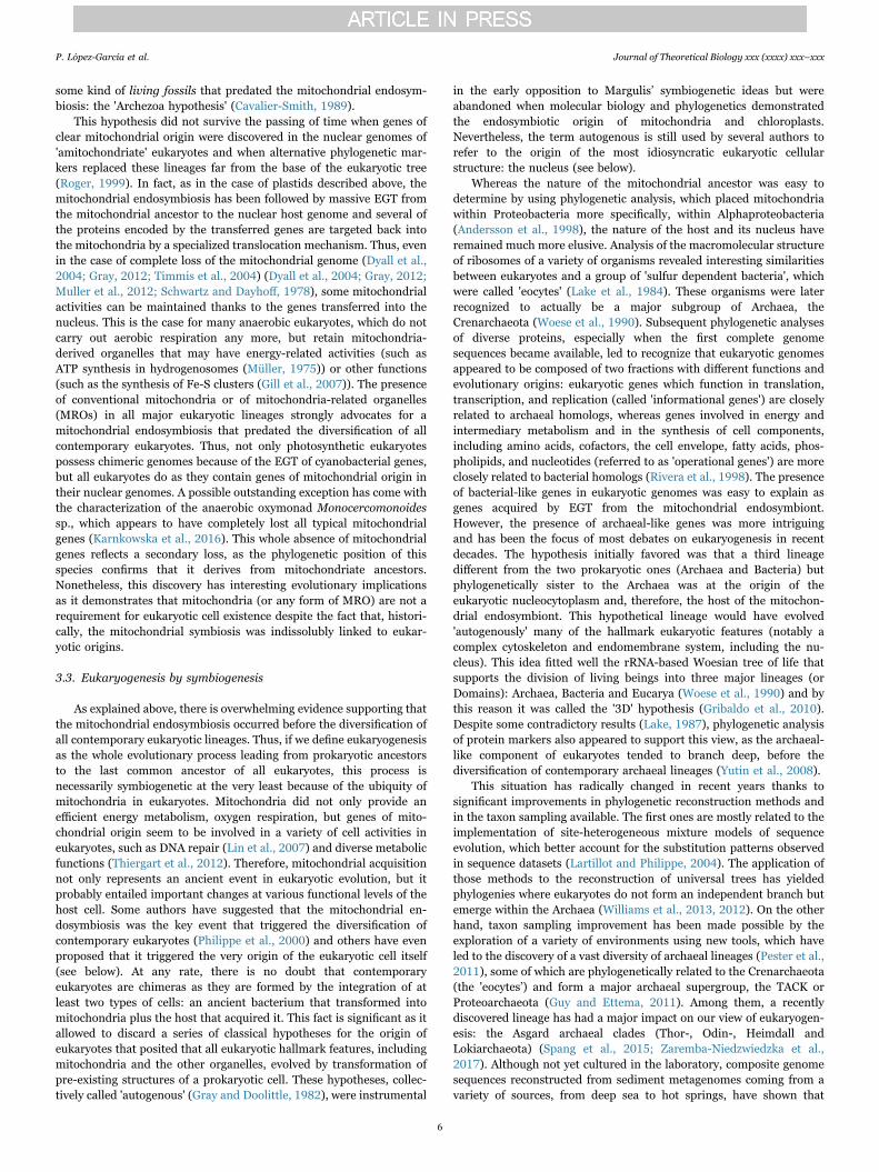
Asgard lineages appear to contain more genes shared with eukaryotesthan any other archaea and, even more importantly, phylogeneticanalysis of conserved markers retrieves the monophyly of eukaryotesand Asgard archaea (Spang et al., 2015; Zaremba-Niedzwiedzka et al.,2017). These results provide strong support to the so-called '2Dmodels', which posit that there are only two primary domains(Bacteria and Archaea) and that eukaryotes constitute a derivedsecondary domain, originated from some sort of mix of lineages ofthe two prokaryotic domains (Fig. 1). Thus, the archaeal-like compo-nent of eukaryotic genomes, namely the informational genes, seems tohave originated from within the archaea. These discoveries have led notonly to discard the 3D models mentioned above, but also to furtherdiscredit other models that proposed that these informational genes,and even the nucleus itself, were acquired from giant viruses, amiscellaneous group of viruses belonging to the nucleocytoplasmiclarge DNA virus (NCLDV) superfamily (Bell, 2009; Boyer et al., 2010;Forterre and Gaia, 2016; Raoult et al., 2004). Indeed, even before thediscovery of Lokiarchaeota and other Asgard archaea, it had alreadybeen shown that eukaryotic-like genes found in NCLDV had beenacquired by the viruses from their hosts via horizontal gene transfer(HGT) and not the other way round. This can be masked by the highevolutionary rate of viruses that can distort phylogenetic reconstruc-tion, but when accurate phylogenetic methods are applied to retrace theevolutionary history of these genes, their eukaryotic origin becomesclear (Lopez-Garcia and Moreira, 2015; Moreira and Lopez-Garcia,2005; Williams et al., 2011). Therefore, a NCLDV virus was not at theorigin of the informational genes and/or the eukaryotic nucleus, but anarchaeon, most likely an ancient member of the Asgard archaea orrelated lineages. Nonetheless, how this archaeon established a symbio-tic relationship with bacteria and the number of its bacterial partnersremain open questions that will occupy research in the field ofeukaryogenesis in the next years (Lopez-Garcia and Moreira, 2015).
4. Eukaryogenesis: open questions
It has become clear now that eukaryogenesis occurred by symbio-genesis of archaea and bacteria (Fig. 1), supporting Margulis' visionaryidea, also adopted by later symbiogenetic models, that symbiosis wascrucial in eukaryotic evolution, leading to an increase in average cell
complexity. Symbiogenesis was an implicit conclusion from recent genecontent and phylogenomic analyses suggesting that archaea of theTACK superphylum were more similar to eukaryotes (Martijn andEttema, 2013; McInerney et al., 2014; Williams and Embley, 2014),which eliminated the possibility that a third, proto-eukaryotic lineagedifferent from archaea ever existed, and became much more explicitwhen Lokiarchaeota and other Asgard archaea were discovered(Koonin, 2015; Lopez-Garcia and Moreira, 2015; Spang et al., 2015;Zaremba-Niedzwiedzka et al., 2017). However, despite the confirma-tion of the symbiogenetic origin of eukaryotes, many crucial questionsremain open (reviewed in (Lopez-Garcia and Moreira, 2015)).
4.1. When did eukaryotes evolve?
The geological record preserves part of life history, includingevidence for early (chemical and morphological) traces of the existenceof eukaryotes. When considering this evidence, it may be important todistinguish between 'stem' and 'crown' lineages. Briefly, all lineagesthat descend from LECA are often referred to as crown eukaryotes(Fig. 1). But before a full-fledged LECA evolved, a more or less longperiod of evolution occurred from the time when, depending on themodel, the archaeal ancestor of eukaryotes started to acquire eukar-yotic features (Fig. 2A) or when the first eukaryogenetic archaea-bacteria consortium started to co-evolve together (Fig. 2B-D). Lineagesdiverging during that period are considered stem lineages and are nowall extinct (but the one that successfully lead to 'crown' eukaryotes).Any characteristic feature of eukaryotes evident in ancient microfossilscan, in principle, be a property of either stem or crown organisms.Therefore, unless there are specific features that definitively associatethe fossils/biomarkers with particular crown eukaryotic group, it is notpossible to distinguish whether they represent stem or crown eukar-yotes. Additionally, identifying with confidence microfossils as eukar-yotic rather than bacterial is not a trivial task and only fossilscombining large size, ornamented walls, complex ultrastructure, anda preservable composition are regarded as probable eukaryotes (Knoll,2015). As such, while the oldest of microfossils of possible eukaryoticorigin were described from 3200 Ma shales, their simple ultrastructureprevents from any definitive conclusion (Javaux et al., 2010). Theoldest fossil showing robust structural evidence of eukaryotic affiliation
Fig. 1. Schematic representation of the tree of life and the origin of eukaryotes in an approximate historical framework.
P. López-García et al. Journal of Theoretical Biology xxx (xxxx) xxx–xxx
7
appears in rocks as old as ~1700 Ma (Javaux, 2007; Knoll et al., 2006;Yan and Liu, 1993). However, most of the ancient Proterozoicassemblages (i.e., 1800–1000 Ma) include fossils that are difficult, ifnot impossible, to associate with crown eukaryotic groups and theycould in fact, represent stem eukaryote lineages. A notable exception isBangiomorpha pubescens from the 1.2-Ga Hunting Formation(Butterfield, 2000) that represents the oldest fossil that some paleon-tologists confidently assign to a crown eukaryotic lineage, the bangio-phyte red algae, setting a lower boundary for eukaryotic diversification.
A complementary approach to determining the timing of eukaryoticevolution is through molecular dating, which allows divergence timesto be estimated from genetic distances, based on the simple idea thatdifferences between homologous proteins of different species areproportional to their divergence time (Zuckerkandl and Pauling,1965). However, variation in substitution rates has been widelydocumented; to cope with it, 'relaxed molecular clock' (RMC) methodswere developed to allow the rate of sequence evolution to vary acrossdifferent branches (for reviews, see (Ho et al., 2015; Kumar andHedges, 2016)). To estimate divergence times by using RMC ap-proaches, the phylogenetic tree is calibrated with several known datesassociated with the available paleobiological data. As our understand-ing of eukaryote phylogeny has improved, fossil-calibrated molecular-clock-based methods have been applied to date important diversifica-tion events, but have yielded vastly different estimates (Berney andPawlowski, 2006; Douzery et al., 2004; Eme et al., 2014; Hedges andKumar, 2004; Hedges et al., 2004; Parfrey et al., 2011). These
discrepancies can be explained by a myriad of sources of variabilityand error (Eme et al., 2014; Kumar and Hedges, 2016; Roger and Hug,2006) such as the relaxed molecular clock models and methods used,and the nature and treatment of fossil calibrations. The most recentanalyses provide estimates for the age of LECA in the range of 1000–1600 Ma (Eme et al., 2014).
From these results, estimating the age of the mitochondrialendosymbiosis intrinsically depends on the model favored for eukar-yogenesis. In the case of 'mitochondria-early' scenarios, which considerthe mitochondrial endosymbiosis to be the initial triggering event ofeukaryogenesis (Fig. 2B), the timing of the endosymbiosis can beinferred to be older than the oldest evidence for the occurrence ofeukaryotes (i.e., 1.7 Ga). It is worth noting that the first organismsbelonging to the eukaryotic lineage might have been morphologicallyindistinguishable from their prokaryotic ancestors. Consequently, anymicrofossil clearly distinguishable as eukaryotic (i.e., combining all themorphological features listed above) would be considerably morerecent than the origin of the eukaryotic lineage itself, and thus, thanthe mitochondrial endosymbiosis. By contrast, 'mitochondria-late'hypotheses postulate that a significant number of specific eukaryotic-like features predated the acquisition of the mitochondrion (Fig. 2A, C-D). In this case, the timing of the mitochondrial endosymbiosis wouldbe considerably closer to the age of LECA.
The timing of the primary plastid endosymbiosis can be more easilybracketed since it had to occur after LECA and before the last commonancestor of Archaeplastida. Most recent analyses estimate the latter tobe 900–1300 Ma (Eme et al., 2014). It is worth noting that theBangiomorpha fossils seem to be much older than any of the datesestimated from molecular clock data (Berney and Pawlowski, 2006;Eme et al., 2014; Parfrey et al., 2011; Sharpe et al., 2015) and at oddswith all other microfossil calibrations for crown eukaryotes. Thisdiscrepancy can stem from a number of reasons. For example, eitherthe taxonomic identification of Bangiomorpha fossils, or the estimatedage of the rocks in which they were found, could be erroneous, althoughthe latter is thought to be unlikely (see discussion in (Knoll, 2014;Parfrey et al., 2011)). Alternatively, it is possible that currentlyavailable molecular clock models do not capture properly the evolu-tionary process occurring in some lineages, leading to incorrect dateestimates. The second case of primary stable endosymbiosis identifiedin Paulinella is considerably younger and has been estimated to haveoccurred ~60 Mya (Delaye et al., 2016). As to the time when thelineages containing secondary plastids evolved, their confused evolu-tionary relationships among their heterotrophic hosts currently ham-pers any serious attempt to date secondary and tertiary endosymbioses.Future phylogenomics progress on the reconstruction of the eukaryotictree should help constraining the evolution of plastids derived fromgreen and red algae.
4.2. Where did eukaryotes evolve?
One key question concerns the environmental setting whereeukaryotes evolved and the metabolic nature of the symbiosis thatgave rise to eukaryotes. Metabolic endosymbioses can drive evolutionand have explanatory power in evolution (Lopez-Garcia and Moreira,2015; O'Malley, 2015). Because prokaryotic symbioses essentiallyinvolve syntrophy, the eukaryogenetic symbiogenesis was most likelybased on metabolic cooperation. Some insight can be obtained from thekind of ecosystems and metabolic networks established in ecosystemswhere Asgard archaeal lineages occur. Lokiarchaeota, previouslyknown as Deep Sea Archaeal Group (DSAG) or marine benthic groupB, have been classically detected in sediments and microbial mats(Biddle et al., 2006; Jorgensen et al., 2013, 2012; Spang et al., 2015).Because their native habitats are anoxic, they must be anaerobic, whichwould be consistent with a putative dependency on hydrogen deducedfrom their gene content (Sousa et al., 2016); they have indeed beensuggested to be involved in syntrophic interactions leading to dissim-
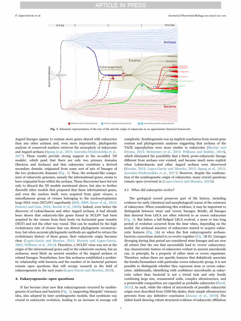
Fig. 2. Schematic representation of current eukaryogenetic models based on symbiosis.A, one archaeon develops endomembranes and the nucleus, acquiring the capacity ofphagocytosis and the possibility to engulf the mitochondrial ancestor. B, the mitochon-drial ancestor becomes an early endosymbiont in an archaeon, triggering eukaryogenesis.C, endosymbiotic origin of the nucleus (archaeon) within a bacterium; the mitochondrionresults from a second endosymbiosis. D, multiple successive symbioses forge theeukaryotic cell.
P. López-García et al. Journal of Theoretical Biology xxx (xxxx) xxx–xxx
8
ilatory CH4 oxidation (Biddle et al., 2006) or iron or manganesereduction (Jorgensen et al., 2012). Likewise, the rest of recentlyidentified Asgard archaea are associated to sediments from variousenvironmental settings (Zaremba-Niedzwiedzka et al., 2017).Obviously, these Asgard archaea constitute a collection of derivedlineages that may be quite different from the archaeal ancestor thatestablished the eukaryogenetic symbiosis with bacteria. Also, it mightwell be that there are other archaeal lineages that are even closer toeukaryotes, since archaeal diversity is far from being completelyexplored. Actually, a large diversity of deeply divergent archaeal cladesoccurs precisely in anoxic settings, including the deep subsurface,sediments and different kinds of microbial mats (Baker et al., 2016;Biddle et al., 2006; Harris et al., 2013; Hedlund et al., 2013; Jorgensenet al., 2013, 2012; Kozubal et al., 2013; Lloyd et al., 2013). However,the fact that Asgard and many other deep-branching archaea aresystematically found in anoxic settings where syntrophic interactionsare the rule supports the idea that eukaryotes emerged from metabo-lically cooperative consortia in anoxic settings likely close to redoxboundaries.
Indeed, symbiotic models that have advanced detailed mechanisms,including metabolic interactions, for the origin of eukaryotes, such asthe hydrogen (Martin and Muller, 1998) and the syntrophy (López-García and Moreira, 2006; Moreira and López-García, 1998) hypoth-eses implied that eukaryotes evolved in anoxic settings where redoxgradients occurred and that the mitochondrial ancestor was a faculta-tive anaerobe (López-García and Moreira, 1999). This would offer thepossibility, among others, to establish a symbiosis with a facultativeaerobic alphaproteobacterium endowed with oxygen respiration butable to thrive in oxygen-depleted environments. A facultative anaerobicalphaproteobacterium, possibly microaerophilic (adapted to low-oxy-gen concentrations) as mitochondrial ancestor would make sense at atime when oxygen started to accumulate in the atmosphere and most ofthe oceanic water column was still anoxic (Scott et al., 2008). Both,hydrogen and syntrophy hypotheses proposed hydrogen transferbetween symbiotic partners, in the former between a fermentativealphaproteobacterium that established as endosymbiont within amethanogenic archaeon (Martin and Muller, 1998), in the secondbetween an ancestral sulfate-reducing fermentative myxobacterium(Deltaproteobacteria) and an endosymbiotic methanogenic archaeon(future nucleus) to which a versatile alphaproteobacterial methano-troph would have joined in a tripartite symbiosis (López-García andMoreira, 2006; Moreira and López-García, 1998). Thought to beexclusive of the Euryarchaeota branch, methanogenesis has recentlybeen inferred in the Bathyarchaeota, within the TACK superphylum,making methanogenesis likely ancestral in archaea (Evans et al., 2015;Lever, 2016). Nonetheless, as mentioned in Section 2.3, the numberand type of syntrophic interactions mediated by hydrogen amongarchaea and bacteria in sediments and other oxygen-depleted environ-ments are far from fully understood. Multiple syntrophic interactionsappear to be the rule in natural conditions, so that multiple symbiosesat the origin of eukaryotes cannot be excluded on the basis ofparsimony criteria because microbial ecology contradicts those argu-ments. At any rate, better understanding metabolic interactionsbetween archaea and bacteria in this kind of ecosystems will greatlyhelp proposing plausible metabolic interactions at the origin of theeukaryotic cell.
4.3. How did eukaryotes evolve?
Many models proposing symbiotic merging between archaea andbacteria at the origin of eukaryotes avoid providing detailed mechan-istic scenarios explaining the origin of the most characteristic featuresof the eukaryotic cell. However, the most open and contentiousquestions in eukaryogenesis precisely relate to the mechanisms bywhich the different eukaryotic features appeared (reviewed in (Lopez-Garcia and Moreira, 2015)), such that exposing the details is important
to discriminate between existing models, refine them or propose morerealistic ones. The different types of models that can currently beconsidered are schematically shown in Fig. 2. The most popular (Fig. 2.A) would correspond to the transposition of old '3D' models to a '2D'situation. Here, an archaeon would develop an endomembrane systemby invagination of the plasma membrane and the nucleus in an'autogenous' way together with the capacity of phagocytosis, whichwould allow it to engulf the mitochondrial ancestor (phagocytosingarchaeon model, PhAT (Martijn and Ettema, 2013)). Many of thesemodels are mechanistically naive but could be nurtured by much moredeveloped character evolution scenarios from previous 3D phagocyto-sis-based models, including particular variants such as the Neomurahypothesis (Cavalier-Smith, 2014). A second model would correspondto the initial endosymbiosis of the alphaproteobacterial ancestor ofmitochondria within an archaeon, the rest of archaeal features wouldevolve after the symbiosis established (Fig. 2. B; e.g. hydrogenhypothesis (Martin and Muller, 1998)). A third type of models wouldcorrespond to the endosymbiotic origin of the nucleus within onebacterium, the mitochondrion deriving for a second endosymbiosiswith an alphaproteobacterium (Fig. 2. C; e.g. syntrophy hypothesis(López-García and Moreira, 2006; Moreira and López-García, 1998)).Finally, a fourth type of models would imply multiple serial endosym-bioses (either within a bacterium or within an archaeon) with theparticipation of several bacteria that would have left an imprint in theeukaryotic nucleus by successive waves of EGT (Fig. 2. D) (Pittis andGabaldon, 2016).
Although there is some debate about the specific identity of themitochondrial ancestor within the Alphaproteobacteria and as towhether it was or not a facultative anaerobe, the most controversialpoint refers to whether mitochondria arrived early or not (Fig. 2). Inthe hydrogen hypothesis, mitochondria arrive early because themitochondrial endosymbiosis is proposed to be the cause of eukar-yogenesis (Martin and Muller, 1998). By contrast, in the phagocytosingarchaeon model, the mitochondria arrive late because they are, in away, the consequence of an autogenous eukaryogenetic process that ledto phagocytosis and the possibility to engulf exogenous bacteria(Martijn and Ettema, 2013). In the syntrophy hypothesis, the mito-chondrial symbiosis is the last, highly successful, endosymbiotic eventthat determines the eukaryogenetic closure, providing a clear selectivemetabolic advantage related to the oxygenic respiration (López-Garcíaand Moreira, 2006). Recent phylogenomic analyses exploring therelative timing of prokaryotic gene acquisition in eukaryotes indeedsuggest that mitochondria arrived late and might additionally supportsuccessive, multiple symbioses (Pittis and Gabaldon, 2016).
Another crucial problem relates to the origin of the eukaryoticnucleus, which many eukaryogenetic models leave unexplained. Inaddition to the selective forces that led to the evolution of thisidiosyncratic structure, which remain elusive (Lopez-Garcia andMoreira, 2015), the process by which the nuclear membrane evolved,like the timing of mitochondrial acquisition, also discriminates thehydrogen hypothesis from the rest of symbiogenetic models. The latterpropose an autogenous origin of the endoplasmic reticulum and thenuclear membrane (López-García and Moreira, 2006; Martijn andEttema, 2013), which is consistent with cell biology knowledge andwith the presence of many proteins involved in membrane remodelingand vesicle trafficking in archaea including, notably, Asgard archaea,but also other prokaryotes (Devos et al., 2004; Klinger et al., 2016;Spang et al., 2015; Surkont and Pereira-Leal, 2016). By contrast, thehydrogen hypothesis, speculates that the nuclear membrane and theendomembrane system would derive from the formation and progres-sive fusion of vesicles budding off the alphaproteobacterial ancestor ofmitochondria around what would be the future nucleus (Gould et al.,2016). The production of lipid vesicles from that alphaproteobacterialendosymbiont would also explain, according to these authors, anotherfundamental problem in eukaryogenesis: the bacterial nature ofeukaryotic membrane phospholipids.
P. López-García et al. Journal of Theoretical Biology xxx (xxxx) xxx–xxx
9
Indeed, if the host that acquired mitochondria was an archaeon,then the archaeal membranes should have undergone a profoundtransformation, substituting their membrane phospholipids, very dif-ferent from bacterial ones, by bacterial phospholipids (Lombard et al.,2012), but also adapting all the integral membrane proteins to a verydifferent physicochemical environment (Lopez-Garcia and Moreira,2015). In Gould's et al.'s view, the archaeal lipids of the plasmamembrane would have been substituted by mitochondria-derived lipidsvia vesicles protruding from the mitochondria that would fuse with thearchaeal membrane and progressively replace archaeal lipids. This stillleaves unexplained the selective forces that drove that putativemembrane transition, as membrane proteins would still be adaptedto archaeal lipids. Likewise, bacteria-to-archaea horizontal gene trans-fer of genes involved in, e.g. fatty acid biosynthesis, has been evoked toexplain a putative archaea-to-bacteria phospholipid transition, butsome archaea containing those genes (e.g. haloarchaea) have typicalarchaeal membranes (López-García et al., 2015; Nelson-Sathi et al.,2012), indicating that fatty acids are used for other purpose in the cell.Likewise, the absence of the glycerol-1-phosphate dehydrogenase(G1PDH) gene, which is responsible for the G1P isomer characteris-tically used to synthesize archaeal phospholipids, seems to be absentfrom Lokiarchaeota and marine Euryarchaeota groups II/III, leading topropose that these archaea might have chimeric archaea-bacteria oreven bacterial-like membrane phospholipids (Villanueva et al., 2016).Failure to identify G1PDH is intriguing. However, this observationcannot be taken for proof in the absence of direct access to themembrane lipids of these organisms. First, the genomes of theseorganisms have been reconstructed from metagenomes and are notnecessarily complete (Deschamps et al., 2014; Iverson et al., 2012;Spang et al., 2015). Second, the gene might have significantly divergedin these organisms and, in any case, the absence of a classical G1PDHdoes not necessarily imply that G1P is not synthesized in an alternativeway. It is indeed worth noting that archaeal lipids were dominant insediments with abundant Lokiarchaeota (DSAG) (Biddle et al., 2006).Finally, unusual butane- and pentanetriol-based tetraether lipidsreplacing glycerol-based backbones have been recently detected in agroup of methanogens sister to Group II/III archaea andThermoplasmatales, and these special lipids seem also abundant insediments (Becker et al., 2016), suggesting a modified, but notbacterial, nature for the lipids of this clade. Clearly, having access tothe real biochemistry of the membrane lipids for these novel archaealgroups is required.
5. Conclusion
Fifty years after the publication of On the origin of mitosing cells,and after having endured harsh criticism and important doses ofindifference, the legacy of Lynn Margulis seems as solid as ever.Certainly, some of her original ideas, notably the symbiotic origin offlagella, have not withstood the test of time. However, not only thesymbiotic origin of mitochondria and chloroplasts but, importantly,that of the eukaryotic cell itself are now beyond any doubt. Eukaryotesare symbiotic mergers forged via cooperative metabolic interactions byprogressive physical integration, endosymbiotic gene transfer and thevast evolutionary possibilities that duplicated genes offered to createinnovations. A more or less long co-evolutionary path, especially in thecase of symbiogenetic models that imply early syntrophy betweenbacteria and archaea (Fig. 2), offered a 'stem' period during which deeptransformations occurred in the prokaryotic ancestors of eukaryotes.By becoming individual units of evolution of increased average com-plexity, eukaryotes accessed a wealth of new ecological niches anddiversified.
Many questions remain still unanswered in terms of the numberand nature of the specific prokaryotic partners that took part in theeukaryogenetic process, their metabolic interactions and the historicalprocess that led to LECA. Hopefully, we have left behind the 'phylo-
genomic impasse'. Combining metagenomic and single-cell genomicanalyses of natural microbial communities where Asgard and otherdeep-branching archaea typically thrive with metabolic modeling andwith the use of better phylogenomic tools, we should be able toreconstruct a plausible evolutionary model for the origin of eukaryotes.
Acknowledgements
This work was supported by the European Research CouncilAdvanced Grant ProtistWorld [grant number agreement 322669].
References
Adl, S.M., Simpson, A.G., Farmer, M.A., Andersen, R.A., Anderson, O.R., Barta, J.R.,Bowser, S.S., Brugerolle, G., Fensome, R.A., Fredericq, S., James, T.Y., Karpov, S.,Kugrens, P., Krug, J., Lane, C.E., Lewis, L.A., Lodge, J., Lynn, D.H., Mann, D.G.,McCourt, R.M., Mendoza, L., Moestrup, O., Mozley-Standridge, S.E., Nerad, T.A.,Shearer, C.A., Smirnov, A.V., Spiegel, F.W., Taylor, M.F., 2005. The new higher levelclassification of eukaryotes with emphasis on the taxonomy of protists. J. Eukaryot.Microbiol 52, 399–451.
Andersson, S.G., Zomorodipour, A., Andersson, J.O., Sicheritz-Ponten, T., Alsmark, U.C.,Podowski, R.M., Naslund, A.K., Eriksson, A.S., Winkler, H.H., Kurland, C.G., 1998.The genome sequence of Rickettsia prowazekii and the origin of mitochondria.Nature 396, 133–140.
Archibald, J.M., 2009. The puzzle of plastid evolution. Curr. Biol. 19, R81–R88.Awramik, S.M., 1992. The oldest records of photosynthesis. Photosynth Res 33, 75–89.Baker, B.J., Saw, J.H., Lind, A.E., Lazar, C.S., Hinrichs, K.U., Teske, A.P., Ettema, T.J.,
2016. Genomic inference of the metabolism of cosmopolitan subsurface Archaea,Hadesarchaea. Nat. Microbiol 1, 16002.
Bao, X., Roossinck, M.J., 2013. A life history view of mutualistic viral symbioses: quantityor quality for cooperation? Curr. Opin. Microbiol 16, 514–518.
Baurain, D., Brinkmann, H., Petersen, J., Rodriguez-Ezpeleta, N., Stechmann, A.,Demoulin, V., Roger, A.J., Burger, G., Lang, B.F., Philippe, H., 2010. Phylogenomicevidence for separate acquisition of plastids in cryptophytes, haptophytes, andstramenopiles. Mol. Biol. Evol. 27, 1698–1709.
Becker, K.W., Elling, F.J., Yoshinaga, M.Y., Sollinger, A., Urich, T., Hinrichs, K.U., 2016.Unusual butane- and pentanetriol-based tetraether lipids in Methanomassiliicoccusluminyensis, a representative of the seventh order of methanogens. Appl. Environ.Microbiol. 82, 4505–4516.
Bell, P.J., 2009. The viral eukaryogenesis hypothesis: a key role for viruses in theemergence of eukaryotes from a prokaryotic world environment. Ann. N.Y. Acad. Sci.1178, 91–105.
Berney, C., Pawlowski, J., 2006. A molecular time-scale for eukaryote evolutionrecalibrated with the continuous microfossil record. Proc. Biol. Sci. 273, 1867–1872.
Bernhard, J.M., Buck, K.R., Farmer, M.A., Bowser, S.S., 2000. The Santa Barbara Basin isa symbiosis oasis. Nature 403, 77–80.
Biddle, J.F., White, J.R., Teske, A.P., House, C.H., 2011. Metagenomics of the subsurfaceBrazos-Trinity Basin (IODP site 1320): comparison with other sediment andpyrosequenced metagenomes. ISME J. 5, 1038–1047.
Biddle, J.F., Lipp, J.S., Lever, M.A., Lloyd, K.G., Sorensen, K.B., Anderson, R., Fredricks,H.F., Elvert, M., Kelly, T.J., Schrag, D.P., Sogin, M.L., Brenchley, J.E., Teske, A.,House, C.H., Hinrichs, K.U., 2006. Heterotrophic Archaea dominate sedimentarysubsurface ecosystems off Peru. Proc. Natl. Acad. Sci. USA 103, (3846-3451).
Boetius, A., Ravenschlag, K., Schubert, C.J., Rickert, D., Widdel, F., Gieseke, A., Amann,R., Jorgensen, B.B., Witte, U., Pfannkuche, O., 2000. A marine microbial consortiumapparently mediating anaerobic oxidation of methane. Nature 407, 623–626.
Boyer, M., Madoui, M.A., Gimenez, G., La Scola, B., Raoult, D., 2010. Phylogenetic andphyletic studies of informational genes in genomes highlight existence of a 4 domainof life including giant viruses. PLoS One 5, e15530.
Bryant, M.P., Wolin, E.A., Wolin, M.J., Wolfe, R.S., 1967.Methanobacillus omelianskii, asymbiotic association of two species of bacteria. Arch. Microbiol. 59, 20–31.
Butterfield, N.J., 2000. Bangiomorpha pubescens n. gen., n. sp.: implications for theevolution of sex, multicellularity, and the Mesoproterozoic/Neoproteorozoicradiation of eukaryotes. Paleobiology 26, 386–404.
Cavalier-Smith, T., 1982. The origin of plastids. Bull. J. Linn. Soc. 17, 289–306.Cavalier-Smith, T., 1989. Molecular phylogeny. Archaebacteria Archezoa. Nat. 339,
l00–l01.Cavalier-Smith, T., 1999. Principles of protein and lipid targeting in secondary
symbiogenesis: euglenoid, dinoflagellate, and sporozoan plastid origin and theeukaryote family tree. J. Euk. Microbiol 46, 347–366.
Cavalier-Smith, T., 2014. The neomuran revolution and phagotrophic origin ofeukaryotes and cilia in the light of intracellular coevolution and a revised tree of life.Cold Spring Harb. Perspect. Biol. 6, a016006.
analysis of Chlorobium chlorochromatii CaD3 reveals clues of the symbiosis in'Chlorochromatium aggregatum'. ISME J. 8, 991–998.
Cordaux, R., Bouchon, D., Greve, P., 2011. The impact of endosymbionts on the evolutionof host sex-determination mechanisms. Trends Genet. 27, 332–341.
de Duve, C., 2007. The origin of eukaryotes: a reappraisal. Nat. Rev. Genet. 8, 395–403.Delaux, P.M., Radhakrishnan, G.V., Jayaraman, D., Cheema, J., Malbreil, M., Volkening,
J.D., Sekimoto, H., Nishiyama, T., Melkonian, M., Pokorny, L., Rothfels, C.J.,Sederoff, H.W., Stevenson, D.W., Surek, B., Zhang, Y., Sussman, M.R., Dunand, C.,Morris, R.J., Roux, C., Wong, G.K., Oldroyd, G.E., Ane, J.M., 2015. Algal ancestor ofland plants was preadapted for symbiosis. Proc. Natl. Acad. Sci. USA 112,13390–13395.
Delaye, L., Valadez-Cano, C., Pérez-Zamorano, B., 2016. How really ancient is Paulinellachromatophora? PLoS Curr. Tree Life. http://dx.doi.org/10.1371/currents.tol.e68a099364bb1a1e129a17b4e06b0c6b.
Deschamps, P., Zivanovic, Y., Moreira, D., Rodriguez-Valera, F., Lopez-Garcia, P., 2014.Pangenome evidence for extensive interdomain horizontal transfer affecting lineagecore and shell genes in uncultured planktonic thaumarchaeota and euryarchaeota.Genome Biol. Evol. 6, 1549–1563.
Devos, D., Dokudovskaya, S., Alber, F., Williams, R., Chait, B.T., Sali, A., Rout, M.P.,2004. Components of coated vesicles and nuclear pore complexes share a commonmolecular architecture. PLoS Biol. 2, e380.
Douzery, E.J., Snell, E.A., Bapteste, E., Delsuc, F., Philippe, H., 2004. The timing ofeukaryotic evolution: does a relaxed molecular clock reconcile proteins and fossils?Proc. Natl. Acad. Sci. USA 19, 19.
Dubilier, N., Bergin, C., Lott, C., 2008. Symbiotic diversity in marine animals: the art ofharnessing chemosynthesis. Nat. Rev. Microbiol. 6, 725–740.
Duperron, S., Sibuet, M., MacGregor, B.J., Kuypers, M.M., Fisher, C.R., Dubilier, N.,2007. Diversity, relative abundance and metabolic potential of bacterialendosymbionts in three Bathymodiolus mussel species from cold seeps in the Gulf ofMexico. Environ. Microbiol. 9, 1423–1438.
Dyall, S.D., Brown, M.T., Johnson, P.J., 2004. Ancient invasions: from endosymbionts toorganelles. Science 304, 253–257.
Edgcomb, V.P., Leadbetter, E.R., Bourland, W., Beaudoin, D., Bernhard, J.M., 2011a.Structured multiple endosymbiosis of bacteria and archaea in a ciliate from marinesulfidic sediments: a survival mechanism in low oxygen, sulfidic sediments? Front.Microbiol. 2, 55.
Edgcomb, V.P., Breglia, S.A., Yubuki, N., Beaudoin, D., Patterson, D.J., Leander, B.S.,Bernhard, J.M., 2011b. Identity of epibiotic bacteria on symbiontid euglenozoans inO2-depleted marine sediments: evidence for symbiont and host co-evolution. ISMEJ. 5, 231–243.
Embley, T.M., 2006. Multiple secondary origins of the anaerobic lifestyle in eukaryotes.Philos Trans. R. Soc. Lond. B Biol. Sci. 361, 1055–1067.
Embley, T.M., Martin, W., 2006. Eukaryotic evolution, changes and challenges. Nature440, 623–630.
Embree, M., Liu, J.K., Al-Bassam, M.M., Zengler, K., 2015. Networks of energetic andmetabolic interactions define dynamics in microbial communities. Proc. Natl. Acad.Sci. USA 112, 15450–15455.
Eme, L., Doolittle, W.F., 2015. Microbial diversity: a bonanza of phyla. Curr. Biol. 25,R227–R230.
Eme, L., Sharpe, S.C., Brown, M.W., Roger, A.J., 2014. On the age of eukaryotes:evaluating evidence from fossils and molecular clocks. Cold Spring Harb. Perspect.Biol. 6, a016139.
Evans, P.N., Parks, D.H., Chadwick, G.L., Robbins, S.J., Orphan, V.J., Golding, S.D.,Tyson, G.W., 2015. Methane metabolism in the archaeal phylum Bathyarchaeotarevealed by genome-centric metagenomics. Science 350, 434–438.
Field, K.J., Pressel, S., Duckett, J.G., Rimington, W.R., Bidartondo, M.I., 2015. Symbioticoptions for the conquest of land. Trends Ecol. Evol. 30, 477–486.
Forterre, P., Gaia, M., 2016. Giant viruses and the origin of modern eukaryotes. Curr.Opin. Microbiol. 31, 44–49.
Geurts, R., Xiao, T.T., Reinhold-Hurek, B., 2016. What does it take to evolve a nitrogen-fixing endosymbiosis? Trends Plant Sci. 21, 199–208.
Gill, E.E., Diaz-Trivino, S., Barbera, M.J., Silberman, J.D., Stechmann, A., Gaston, D.,Tamas, I., Roger, A.J., 2007. Novel mitochondrion-related organelles in theanaerobic amoeba Mastigamoeba balamuthi. Mol. Microbiol. 66, 1306–1320.
Gould, S.B., Garg, S.G., Martin, W.F., 2016. Bacterial vesicle Secretion and theevolutionary origin of the eukaryotic endomembrane system. Trends Microbiol. 24,525–534.
Gray, M.W., 2012. Mitochondrial evolution. Cold Spring Harb. Perspect. Biol. 4, a011403.Gray, M.W., Doolittle, W.F., 1982. Has the endosymbiont hypothesis been proven?
Microbiol. Rev. 46, 1–42.Gribaldo, S., Poole, A.M., Daubin, V., Forterre, P., Brochier-Armanet, C., 2010. The
origin of eukaryotes and their relationship with the Archaea: are we at aphylogenomic impasse? Nat. Rev. Microbiol. 8, 743–752.
Guerrero, R., Margulis, L., Berlanga, M., 2013. Symbiogenesis: the holobiont as a unit ofevolution. Int. Microbiol. 16, 133–143.
Guy, L., Ettema, T.J., 2011. The archaeal 'TACK' superphylum and the origin ofeukaryotes. Trends Microbiol. 19, 580–587.
Hamann, E., Gruber-Vodicka, H., Kleiner, M., Tegetmeyer, H.E., Riedel, D., Littmann, S.,Chen, J., Milucka, J., Viehweger, B., Becker, K.W., Dong, X., Stairs, C.W., Hinrichs,K.U., Brown, M.W., Roger, A.J., Strous, M., 2016. Environmental Breviatea harbourmutualistic Arcobacter epibionts. Nature 534, 254–258.
Hardoim, P.R., van Overbeek, L.S., Berg, G., Pirttila, A.M., Compant, S., Campisano, A.,
Doring, M., Sessitsch, A., 2015. The hidden world within plants: ecological andevolutionary considerations for defining functioning of microbial endophytes.Microbiol. Mol. Biol. Rev. 79, 293–320.
Harris, J.K., Caporaso, J.G., Walker, J.J., Spear, J.R., Gold, N.J., Robertson, C.E.,Hugenholtz, P., Goodrich, J., McDonald, D., Knights, D., Marshall, P., Tufo, H.,Knight, R., Pace, N.R., 2013. Phylogenetic stratigraphy in the Guerrero Negrohypersaline microbial mat. ISME J. 7, 50–60.
Hatzenpichler, R., Connon, S.A., Goudeau, D., Malmstrom, R.R., Woyke, T., Orphan,V.J., 2016. Visualizing in situ translational activity for identifying and sorting slow-growing archaeal-bacterial consortia. Proc. Natl. Acad. Sci. USA 113, E4069–E4078.
Hedges, S.B., Kumar, S., 2004. Precision of molecular time estimates. Trends Genet. 20,242–247.
Hedges, S.B., Blair, J.E., Venturi, M., Shoe, J.L., 2004. A molecular timescale ofeukaryote evolution and the rise of complex multicellular life. BMC Evol. Biol. 4, 2.
Hedlund, B.P., Dodsworth, J.A., Cole, J.K., Panosyan, H.H., 2013. An integrated studyreveals diverse methanogens, Thaumarchaeota, and yet-uncultivated archaeallineages in Armenian hot springs. Antonie Van. Leeuwenhoek 104, 71–82.
Hernandez-Agreda, A., Gates, R.D., Ainsworth, T.D., 2016. Defining the core microbiomein corals' microbial soup. Trends Microbiol..
Hillesland, K.L., Stahl, D.A., 2010. Rapid evolution of stability and productivity at theorigin of a microbial mutualism. Proc. Natl. Acad. Sci. USA 107, 2124–2129.
Hosokawa, T., Koga, R., Kikuchi, Y., Meng, X.Y., Fukatsu, T., 2010. Wolbachia as abacteriocyte-associated nutritional mutualist. Proc. Natl. Acad. Sci. USA 107,769–774.
Hug, L.A., Baker, B.J., Anantharaman, K., Brown, C.T., Probst, A.J., Castelle, C.J.,Butterfield, C.N., Hernsdorf, A.W., Amano, Y., Ise, K., Suzuki, Y., Dudek, N., Relman,D.A., Finstad, K.M., Amundson, R., Thomas, B.C., Banfield, J.F., 2016. A new view ofthe tree of life. Nat. Microbiol. 1, 16048.
Husnik, F., McCutcheon, J.P., 2016. Repeated replacement of an intrabacterial symbiontin the tripartite nested mealybug symbiosis. Proc. Natl. Acad. Sci. USA 113,E5416–E5424.
Iverson, V., Morris, R.M., Frazar, C.D., Berthiaume, C.T., Morales, R.L., Armbrust, E.V.,2012. Untangling genomes from metagenomes: revealing an uncultured class ofmarine Euryarchaeota. Science 335, 587–590.
Javaux, E.J., 2007. The early eukaryotic fossil record. Adv. Exp. Med. Biol. 607, 1–19.Javaux, E.J., Marshall, C.P., Bekker, A., 2010. Organic-walled microfossils in 3.2-billion-
year-old shallow-marine siliciclastic deposits. Nature 463, 934–938.Jekely, G., 2003. Small GTPases and the evolution of the eukaryotic cell. Bioessays 25,
Quantitative and phylogenetic study of the Deep Sea Archaeal Group in sediments ofthe Arctic mid-ocean spreading ridge. Front. Microbiol. 4, 299.
Jorgensen, S.L., Hannisdal, B., Lanzen, A., Baumberger, T., Flesland, K., Fonseca, R.,Ovreas, L., Steen, I.H., Thorseth, I.H., Pedersen, R.B., Schleper, C., 2012. Correlatingmicrobial community profiles with geochemical data in highly stratified sedimentsfrom the Arctic Mid-Ocean Ridge. Proc. Natl. Acad. Sci. USA 109, E2846–E2855.
Karnkowska, A., Vacek, V., Zubacova, Z., Treitli, S.C., Petrzelkova, R., Eme, L., Novak, L.,Zarsky, V., Barlow, L.D., Herman, E.K., Soukal, P., Hroudova, M., Dolezal, P., Stairs,C.W., Roger, A.J., Elias, M., Dacks, J.B., Vlcek, C., Hampl, V., 2016. A eukaryotewithout a mitochondrial organelle. Curr. Biol. 26, 1274–1284.
Keeling, P.J., 2013. The number, speed, and impact of plastid endosymbioses ineukaryotic evolution. Annu. Rev. Plant Biol. 64, 583–607.
Keeling, P.J., 2014. The impact of history on our perception of evolutionary events:endosymbiosis and the origin of eukaryotic complexity. Cold Spring Harb. Perspect.Biol. 6, a016196.
Klinger, C.M., Spang, A., Dacks, J.B., Ettema, T.J., 2016. Tracing the archaeal origins ofeukaryotic membrane-trafficking system building blocks. Mol. Biol. Evol. 33,1528–1541.
Knoll, A.H., 2014. Paleobiological perspectives on early eukaryotic evolution. Cold SpringHarb. Perspect. Biol., 6.
Knoll, A.H., 2015. Paleobiological perspectives on early microbial evolution. Cold SpringHarb. Perspect. Biol. 7, a018093.
Knoll, A.H., Javaux, E.J., Hewitt, D., Cohen, P., 2006. Eukaryotic organisms inProterozoic oceans. Philos Trans. R. Soc. Lond. B Biol. Sci. 361, 1023–1038.
Koga, R., Moran, N.A., 2014. Swapping symbionts in spittlebugs: evolutionaryreplacement of a reduced genome symbiont. ISME J. 8, 1237–1246.
Koonin, E.V., 2015. Archaeal ancestors of eukaryotes: not so elusive any more. BMC Biol.13, 84.
Kozubal, M.A., Romine, M., Jennings, R., Jay, Z.J., Tringe, S.G., Rusch, D.B., Beam, J.P.,McCue, L.A., Inskeep, W.P., 2013. Geoarchaeota: a new candidate phylum in theArchaea from high-temperature acidic iron mats in Yellowstone National Park. ISMEJ. 7, 622–634.
Kumar, S., Hedges, S.B., 2016. Advances in time estimation methods for molecular data.Mol. Biol. Evol. 33, 863–869.
Lake, J.A., 1987. Prokaryotes and archaebacteria are not monophyletic: rate invariantanalysis of rRNA genes indicates that eukaryotes and eocytes form a monophyletictaxon. Cold Spring Harb. Symp. Quant. Biol. 52, 839–846.
Lake, J.A., Henderson, E., Oakes, M., Clark, M.W., 1984. Eocytes: a new ribosomestructure indicates a kingdom with a close relationship to eukaryotes. Proc. Natl.Acad. Sci. USA 81, 3786–3790.
Lartillot, N., Philippe, H., 2004. A Bayesian mixture model for across-site heterogeneitiesin the amino-acid replacement process. Mol. Biol. Evol. 21, 1095–1109.
P. López-García et al. Journal of Theoretical Biology xxx (xxxx) xxx–xxx
Leclercq, S., Theze, J., Chebbi, M.A., Giraud, I., Moumen, B., Ernenwein, L., Greve, P.,Gilbert, C., Cordaux, R., 2016. Birth of a W sex chromosome by horizontal transfer ofWolbachia bacterial symbiont genome. Proc. Natl. Acad. Sci. USA, (pii: 201608979. )(Epub ahead of print).
Lever, M.A., 2016. A new era of methanogenesis research. Trends Microbiol. 24, 84–86.Lin, Z., Nei, M., Ma, H., 2007. The origins and early evolution of DNA mismatch repair
genes–multiple horizontal gene transfers and co-evolution. Nucleic Acids Res. 35,7591–7603.
Lloyd, K.G., Schreiber, L., Petersen, D.G., Kjeldsen, K.U., Lever, M.A., Steen, A.D.,Stepanauskas, R., Richter, M., Kleindienst, S., Lenk, S., Schramm, A., Jorgensen,B.B., 2013. Predominant archaea in marine sediments degrade detrital proteins.Nature 496, 215–218.
Lombard, J., López-García, P., Moreira, D., 2012. The early evolution of lipid membranesand the three domains of life. Nat. Rev. Microbiol. 10, 507–515.
Lopez-Garcia, P., Moreira, D., 2015. Open questions on the origin of eukaryotes. TrendsEcol. Evol. 30, 697–708.
López-García, P., Moreira, D., 1999. Metabolic symbiosis at the origin of eukaryotes.Trends Biochem Sci. 24, 88–93.
López-García, P., Moreira, D., 2006. Selective forces for the origin of the eukaryoticnucleus. Bioessays 28, 525–533.
Lovley, D.R., 2016. Happy together: microbial communities that hook up to swapelectrons. ISME J..
Lyons, T.W., Reinhard, C.T., Planavsky, N.J., 2014. The rise of oxygen in Earth's earlyocean and atmosphere. Nature 506, 307–315.
Brock biology of microorganism. In: Madigan, M.T., Martinko, J.M., Bender, K.S.,Buckley, D.H., Stahl, D.A., Brock, T. (Eds.), s14th edition. Benjamin Cummings, SanFrancisco, California, U.S.A.
Margulis, L., 1981. Symbiosis in cell evolution. Freeman, W. H., San Francisco, CA.Margulis, L., 1992. Biodiversity: molecular biological domains, symbiosis and kingdom
origins. Biosystems 27, 39–51.Margulis, L., 1996. Archaeal-eubacterial mergers in the origin of Eukarya: phylogenetic
classification of life. Proc. Natl. Acad. Sci. USA 93, 1071–1076.Margulis, L., Guerrero, R., 1991. Kingdoms in turmoil. New Sci. 1761, 46–50.Margulis, L., Schwartz, K.V., 1998. Five Kingdoms: an Illustrated Guide to the Phyla of
Life on Earth. W. H. Freeman and Co., New York.Margulis, L., Dolan, M.F., Guerrero, R., 2000. The chimeric eukaryote: origin of the
nucleus from the karyomastigont in amitochondriate protists. Proc. Natl. Acad. Sci.USA 97, 6954–6959.
Margulis, L., Lopez Baluja, L., Awramik, S.M., Sagan, D., 1986. Community living longbefore man: fossil and living microbial mats and early life. Sci. Total Environ. 56,379–397.
Margulis, L., Corliss, J.O., Melkonian, M., Chapman, D.J. (Eds.), 1990. Handbook ofProtoctista. Jones & Bartlett, Boston.
Margulis, L., Chapman, M., Guerrero, R., Hall, J., 2006. The last eukaryotic commonancestor (LECA): acquisition of cytoskeletal motility from aerotolerant spirochetes inthe Proterozoic Eon. Proc. Natl. Acad. Sci. USA 103, 13080–13085.
Martijn, J., Ettema, T.J., 2013. From archaeon to eukaryote: the evolutionary dark agesof the eukaryotic cell. Biochem. Soc. Trans. 41, 451–457.
Martin, W., Muller, M., 1998. The hydrogen hypothesis for the first eukaryote. Nature392, 37–41.
Martin, W., Stoebe, B., Goremykin, V., Hapsmann, S., Hasegawa, M., Kowallik, K.V.,1998. Gene transfer to the nucleus and the evolution of chloroplasts. Nature 393,162–165.
McInerney, J.O., O'Connell, M.J., Pisani, D., 2014. The hybrid nature of the Eukaryotaand a consilient view of life on Earth. Nat. Rev. Microbiol. 12, 449–455.
Mereschkowsky, C., 1905. Über natur und usprung der chromatophoren impflanzenreiche. Biol. Cent. 25, 593–604.
Mereschkowsky, K., 1910. Theorie der zwei Plasmaarten als Grundlage derSymbiogenesis, einer neuen Lehre von der Ent-stehung der Organismen. Biol. Cent.30, 353–367.
Mitri, S., Foster, K.R., 2013. The genotypic view of social interactions in microbialcommunities. Annu Rev. Genet. 47, 247–273.
Moran, N.A., Sloan, D.B., 2015. The hologenome concept: helpful or hollow? PLoS Biol.13, e1002311.
Moran, N.A., Yun, Y., 2015. Experimental replacement of an obligate insect symbiont.Proc. Natl. Acad. Sci. USA 112, 2093–2096.
Moreira, D., López-García, P., 1998. Symbiosis between methanogenic archaea and
delta-Proteobacteria as the origin of eukaryotes: the syntrophic hypothesis. J. Mol.Evol. 47, 517–530.
Moreira, D., Philippe, H., 2001. Sure facts and open questions about the origin andevolution of photosynthetic plastids. Res. Microbiol. 152, 771–780.
Moreira, D., López-García, P., 2002. Molecular ecology of microbial eukaryotes unveils ahidden world. Trends Microbiol. 10, 31–38.
Moreira, D., Lopez-Garcia, P., 2005. Comment on "The 1.2-megabase genome sequenceof Mimivirus". Science 308, 1114.
Moreira, D., Deschamps, P., 2014. What was the real contribution of endosymbionts tothe eukaryotic nucleus? Insights from photosynthetic eukaryotes. Cold Spring Harb.Perspect. Biol. 6, a016014.
Moreira, D., Le Guyader, H., Philippe, H., 2000. The origin of red algae and the evolutionof chloroplasts. Nature 405, 69–72.
Morris, B.E., Henneberger, R., Huber, H., Moissl-Eichinger, C., 2013. Microbialsyntrophy: interaction for the common good. FEMS Microbiol. Rev. 37, 384–406.
Moya, A., Pereto, J., Gil, R., Latorre, A., 2008. Learning how to live together: genomicinsights into prokaryote-animal symbioses. Nat. Rev. Genet. 9, 218–229.
Muller, M., Mentel, M., van Hellemond, J.J., Henze, K., Woehle, C., Gould, S.B., Yu, R.Y.,van der Giezen, M., Tielens, A.G., Martin, W.F., 2012. Biochemistry and evolution ofanaerobic energy metabolism in eukaryotes. Microbiol. Mol. Biol. Rev. 76, 444–495.
Müller, M., 1975. Biochemistry of protozoan microbodies: peroxisomes, alpha-glycerophosphate oxidase bodies, hydrogenosomes. Annu. Rev. Microbiol. 29,467–483.
Mushegian, A.A., Ebert, D., 2016. Rethinking "mutualism" in diverse host-symbiontcommunities. Bioessays 38, 100–108.
Nadell, C.D., Drescher, K., Foster, K.R., 2016. Spatial structure, cooperation andcompetition in biofilms. Nat. Rev. Microbiol. 14, 589–600.
Nakayama, T., Ishida, K., 2009. Another acquisition of a primary photosyntheticorganelle is underway in Paulinella chromatophora. Curr. Biol. 19, R284–R285.
Nelson-Sathi, S., Dagan, T., Landan, G., Janssen, A., Steel, M., McInerney, J.O.,Deppenmeier, U., Martin, W.F., 2012. Acquisition of 1000 eubacterial genesphysiologically transformed a methanogen at the origin of Haloarchaea. Proc. Natl.Acad. Sci. USA 109, 20537–20542.
Noda, S., Hongoh, Y., Sato, T., Ohkuma, M., 2009. Complex coevolutionary history ofsymbiotic Bacteroidales bacteria of various protists in the gut of termites. BMC Evol.Biol. 9, 158.
Nowack, E.C., Melkonian, M., 2010. Endosymbiotic associations within protists. PhilosTrans. R. Soc. Lond. B Biol. Sci. 365, 699–712.
O'Malley, M.A., 2015. Endosymbiosis and its implications for evolutionary theory. Proc.Natl. Acad. Sci. USA 112, 10270–10277.
Orphan, V.J., 2009. Methods for unveiling cryptic microbial partnerships in nature. Curr.Opin. Microbiol. 12, 231–237.
Overmann, J., Van Gemerden, H., 2000. Microbial interactions involving sulfur bacteria:implications for the ecology and evolution of bacterial communities. FEMSMicrobiol. Ecol. 24, 591–599.
Pace, N.R., 1997. A molecular view of microbial diversity and the biosphere. Science 276,734–740.
Parfrey, L.W., Lahr, D.J., Knoll, A.H., Katz, L.A., 2011. Estimating the timing of earlyeukaryotic diversification with multigene molecular clocks. Proc. Natl. Acad. Sci.USA 108, 13624–13629.
Paszkowski, U., 2006. Mutualism and parasitism: the yin and yang of plant symbioses.Curr. Opin. Plant Biol. 9, 364–370.
Perez-Brocal, V., Gil, R., Ramos, S., Lamelas, A., Postigo, M., Michelena, J.M., Silva, F.J.,Moya, A., Latorre, A., 2006. A small microbial genome: the end of a long symbioticrelationship? Science 314, 312–313.
Pernthaler, A., Dekas, A.E., Brown, C.T., Goffredi, S.K., Embaye, T., Orphan, V.J., 2008.Diverse syntrophic partnerships from deep-sea methane vents revealed by direct cellcapture and metagenomics. Proc. Natl. Acad. Sci. USA 105, 7052–7057.
Pester, M., Schleper, C., Wagner, M., 2011. The Thaumarchaeota: an emerging view oftheir phylogeny and ecophysiology. Curr. Opin. Microbiol. 14, 300–306.
Philippe, H., Lopez, P., Brinkmann, H., Budin, K., Germot, A., Laurent, J., Moreira, D.,Muller, M., Le Guyader, H., 2000. Early-branching or fast-evolving eukaryotes? Ananswer based on slowly evolving positions. Proc. R. Soc. Lond. B Biol. Sci. 267,1213–1221.
Philippot, L., Raaijmakers, J.M., Lemanceau, P., van der Putten, W.H., 2013. Going backto the roots: the microbial ecology of the rhizosphere. Nat. Rev. Microbiol. 11,789–799.
Pittis, A.A., Gabaldon, T., 2016. Late acquisition of mitochondria by a host withchimaeric prokaryotic ancestry. Nature 531, 101–104.
Ponce-Toledo, R.I., Deschamps, P., López-García, P., Zivanovic, Y., Benzerara, K.,Moreira, D., 2017. An early-branching freshwater cyanobacterium at the origin ofchloroplasts. Curr. Biol. 27, 386–391.
Poole, A.M., Penny, D., 2007. Evaluating hypotheses for the origin of eukaryotes.Bioessays 29, 74–84.
Raoult, D., Audic, S., Robert, C., Abergel, C., Renesto, P., Ogata, H., La Scola, B., Suzan,M., Claverie, J.M., 2004. The 1.2-megabase genome sequence of Mimivirus. Science306, 1344–1350.
functionally distinct gene classes. Proc. Natl. Acad. Sci. USA 95, 6239–6244.Robidart, J.C., Bench, S.R., Feldman, R.A., Novoradovsky, A., Podell, S.B., Gaasterland,
T., Allen, E.E., Felbeck, H., 2008. Metabolic versatility of the Riftia pachyptilaendosymbiont revealed through metagenomics. Environ. Microbiol. 10, 727–737.
Rodriguez-Ezpeleta, N., Brinkmann, H., Burey, S.C., Roure, B., Burger, G., Loffelhardt,W., Bohnert, H.J., Philippe, H., Lang, B.F., 2005. Monophyly of primaryphotosynthetic eukaryotes: green plants, red algae, and glaucophytes. Curr. Biol. 15,(1325-1230).
Roger, A.J., 1999. Reconstructing early events in eukaryotic evolution. Am. Nat. 154,S146–S163.
Roger, A.J., Hug, L.A., 2006. The origin and diversification of eukaryotes: problems withmolecular phylogenetics and molecular clock estimation. Philos Trans. R. Soc. Lond.B Biol. Sci. 361, 1039–1054.
Roth, M.S., 2014. The engine of the reef: photobiology of the coral-algal symbiosis. Front.Microbiol. 5, 422.
Sagan, L., 1967. On the origin of mitosing cells. J. Theor. Biol. 14, 255–274.Sassera, D., Beninati, T., Bandi, C., Bouman, E.A., Sacchi, L., Fabbi, M., Lo, N., 2006.
'Candidatus Midichloria mitochondrii', an endosymbiont of the tick Ixodes ricinuswith a unique intramitochondrial lifestyle. Int. J. Syst. Evol. Microbiol. 56,2535–2540.
Scott, C., Lyons, T.W., Bekker, A., Shen, Y., Poulton, S.W., Chu, X., Anbar, A.D., 2008.Tracing the stepwise oxygenation of the Proterozoic ocean. Nature 452, 456–459.
Searcy, D.G., 1992. Origins of mitochondria and chloroplasts from sulfur-basedsymbioses. In: Hartman, H., Matsuno, K. (Eds.), The origin and Evolution of the Cell.World Scientific, Singapore, 47–78.
Sharpe, S.C., Eme, L., Brown, M.W., Roger, A.J., 2015. Timing the origins ofmulticellular eukaryotes through phylogenomics and relaxed molecular clockanalyses. In: Ruiz-Trillo, I., Nedelcu, A.M. (Eds.), Evolutionary Transitions toMulticellular Life. Springer, Dordrecht, 3–29.
Sieber, J.R., McInerney, M.J., Gunsalus, R.P., 2012. Genomic insights into syntrophy:the paradigm for anaerobic metabolic cooperation. Annu. Rev. Microbiol. 66,429–452.
Siegl, A., Kamke, J., Hochmuth, T., Piel, J., Richter, M., Liang, C., Dandekar, T.,Hentschel, U., 2011. Single-cell genomics reveals the lifestyle of Poribacteria, acandidate phylum symbiotically associated with marine sponges. ISME J. 5, 61–70.
Sogin, M.L., 1991. Early evolution and the origin of eukaryotes. Curr. Opin. Genet. Dev.1, 457–463.
Spang, A., Saw, J.H., Jorgensen, S.L., Zaremba-Niedzwiedzka, K., Martijn, J., Lind, A.E.,van Eijk, R., Schleper, C., Guy, L., Ettema, T.J., 2015. Complex archaea that bridgethe gap between prokaryotes and eukaryotes. Nature.
Surkont, J., Pereira-Leal, J.B., 2016. Are there Rab GTPases in archaea? Mol. Biol. Evol.33, 1833–1842.
Thaiss, C.A., Zmora, N., Levy, M., Elinav, E., 2016. The microbiome and innateimmunity. Nature 535, 65–74.
Thauer, R.K., Shima, S., 2008. Methane as fuel for anaerobic microorganisms. Ann. N.Y.Acad. Sci. 1125, 158–170.
Thiergart, T., Landan, G., Schenk, M., Dagan, T., Martin, W.F., 2012. An evolutionarynetwork of genes present in the eukaryote common ancestor polls genomes oneukaryotic and mitochondrial origin. Genome Biol. Evol. 4, 466–485.
Wallin, I.E., 1927. Symbionticism and the Origin of Species. Willliams & Wilkins,Baltimore.
Wegener, G., Krukenberg, V., Riedel, D., Tegetmeyer, H.E., Boetius, A., 2015.Intercellular wiring enables electron transfer between methanotrophic archaea andbacteria. Nature 526, 587–590.
Wegener, G., Krukenberg, V., Ruff, S.E., Kellermann, M.Y., Knittel, K., 2016. Metaboliccapabilities of microorganisms involved in and associated with the anaerobicoxidation of methane. Front. Microbiol. 7, 46.
Whittaker, R.H., Margulis, L., 1978. Protist classification and the kingdoms of organisms.Biosystems 10, 3–18.
Williams, T.A., Embley, T.M., 2014. Archaeal "dark matter" and the origin of eukaryotes.Genome Biol. Evol. 6, 474–481.
Williams, T.A., Embley, T.M., 2015. Changing ideas about eukaryotic origins. Philos.Trans. R. Soc. Lond. B Biol. Sci. 370, 20140318.
Williams, T.A., Embley, T.M., Heinz, E., 2011. Informational gene phylogenies do notsupport a fourth domain of life for nucleocytoplasmic large DNA viruses. PLoS One6, e21080.
Williams, T.A., Foster, P.G., Cox, C.J., Embley, T.M., 2013. An archaeal origin ofeukaryotes supports only two primary domains of life. Nature 504, 231–236.
Williams, T.A., Foster, P.G., Nye, T.M., Cox, C.J., Embley, T.M., 2012. A congruentphylogenomic signal places eukaryotes within the Archaea. Proc. Biol. Sci. 279,4870–4879.
Woese, C.R., Fox, G.E., 1977. Phylogenetic structure of the prokaryotic domain: theprimary kingdoms. Proc. Natl. Acad. Sci. USA 74, 5088–5090.
Woese, C.R., Kandler, O., Wheelis, M.L., 1990. Towards a natural system of organisms:proposal for the domains Archaea, Bacteria, and Eucarya. Proc. Natl. Acad. Sci. USA87, 4576–4579.
Woyke, T., Teeling, H., Ivanova, N.N., Huntemann, M., Richter, M., Gloeckner, F.O.,Boffelli, D., Anderson, I.J., Barry, K.W., Shapiro, H.J., Szeto, E., Kyrpides, N.C.,Mussmann, M., Amann, R., Bergin, C., Ruehland, C., Rubin, E.M., Dubilier, N., 2006.Symbiosis insights through metagenomic analysis of a microbial consortium. Nature443, 950–955.
Yan, Y., Liu, Z.L., 1993. Significance of eukaryotic organisms in the microfossil flora ofChangcheng system. Acta Micropalaeontol Sin. 10, 167–180.
Yarza, P., Yilmaz, P., Pruesse, E., Glockner, F.O., Ludwig, W., Schleifer, K.H., Whitman,W.B., Euzeby, J., Amann, R., Rossello-Mora, R., 2014. Uniting the classification ofcultured and uncultured bacteria and archaea using 16S rRNA gene sequences. Nat.Rev. Microbiol. 12, 635–645.