62
JOURNAL REVIEW DR. PIYUSH OJHA DM RESIDENT DEPARTMENT OF NEUROLOGY GOVT MEDICAL COLLEGE, KOTA
| Date post: | 12-Apr-2017 |
| Category: |
Health & Medicine |
| Upload: | neurologykota |
| View: | 38 times |
| Download: | 2 times |

JOURNAL REVIEW
DR. PIYUSH OJHADM RESIDENT
DEPARTMENT OF NEUROLOGYGOVT MEDICAL COLLEGE, KOTA
Contrasting disease patterns in Seropositive and Seronegative Neuromyelitis optica: A multicentre study of 175 patients
- Sven Jarius, Klemens Ruprecht, Brigitte Wildemann et al- Published in Journal of Neuroinflammation 2012, 9:14

• The diagnostic and pathophysiological relevance of antibodies to aquaporin-4 (AQP4-Ab) in patients with Neuromyelitis optica spectrum disorders (NMOSD) has been intensively studied.
• However, little is known so far about the clinical impact of AQP4-Ab seropositivity.
• OBJECTIVE :- – To analyse systematically the clinical and paraclinical
features associated with NMO spectrum disorders in Caucasians in a stratified fashion according to the patients’ AQP4-Ab serostatus.

BACKGROUND• Neuromyelitis optica (NMO) is a severely disabling
inflammatory disorder of the CNS.
• Autoimmune aetiology that predominantly affects the optic nerves and spinal cord.
• Associated with serum antibodies to Aquaporin-4 (NMO-IgG or AQP4-Ab), the most abundant water channel in the CNS in ~ 80% cases - thought to be directly involved in the disease pathogenesis.

• The clinical spectrum of NMO as defined by Wingerchuk et al. (2007) comprises cases of : – simultaneous optic neuritis (ON) and myelitis– cases of myelitis and ON, in which the two index events do
not develop simultaneously but successively, and – limited or inaugural forms such as single or recurrent
events of longitudinally extensive myelitis (LETM) or recurrent ON.
• Rarely presentations with brain stem encephalitis

• Similar to other autoimmune neurological diseases - a subset of patients exists who are seronegative.
• AQP4-Ab discovered only a few years ago – so many previous studies investigating the clinical and paraclinical features associated with myelitis and optic neuritis in patients with NMOSD – either did not determine AQP4-Ab at all or – did not stratify patients according to their AQP4-Ab
serostatus or – Were based on relatively small patient numbers– many previous studies were monocentre investigations
and thus prone to potential selection bias.

REVISED DIAGNOSTIC CRITERIA FOR NEUROMYELITIS OPTICA ( WINGERCHUK 2006)

METHODS• Clinical, MRI, and CSF features from all Caucasian patients
with NMOSD as defined by Wingerchuk et al. (2007) and known AQP4-Ab serostatus documented in the database of the German Neuromyelitis optica Study Group were analysed retrospectively.
• All patients were seen at one of the 29 participating NEMOS centres, which included neurological departments at 17 university hospitals and at 12 academic teaching hospitals with adjacent specialised outpatient clinics for neuroinflammatory disorders.

• Case ascertainment was performed between August 2009 and August 2011 by an expert panel of NEMOS members.
• Ethical approval taken.
• The Mann Whitney U test - used to test for significant differences between continuous variables
• Fisher’s exact test – to compare proportions.

• At the time of analysis the database contained retrospective data from 175 Caucasian patients with NMOSD as defined by Wingerchuk et al. (2007) and known AQP4-Ab serostatus.
• 119 patients had a history of both ON and Myelitis and met Wingerchuk ’ s 2006 revised criteria (seropositive in 77.3%)
• 49 had a history of isolated LETM as defined by clinical Myelitis and MRI lesions extending over three or more vertebral segments (seropositive in 81.6%)
• 7 had a history of recurrent ON (seropositive in 71.4%).
• The median disease duration at last follow-up was 57.5 months (range, 0-390) and did not differ significantly between seropositives (60 months; range, 0-390) and seronegatives (51 months; range, 0-290).
• 89% had been treated with immunosuppressive or immunomodulatory agents at least once, with no significant difference between seropositive and seronegative patients (p = 0.133;);

• Treatments included – Interferon beta (20.6%)– Azathioprine (45.1%)– Rituximab (32.6%)– Mitoxantrone (21.1%)– Oral steroids (18.9%) – Cyclophosphamide (12.6%)– Intravenous immunoglobulins (6.9%)– Mycophenolate mofetil (4%)– Methotrexate (4%) and – Natalizumab (2.9%).
• If only immunosuppressive drugs are considered, the proportion of treated patients was higher in the seropositive group (83% vs 65%; p = 0.015).

RESULTS
• DEMOGRAPHIC DATA :– Female to male ratio = 6:1 (N = 175) – Significantly higher among seropositive patients
compared to seronegative.– 83.3% female but only 48% male patients were
seropositive.– Median age at onset = 39 years (range 10-81)


• DISEASE ONSET :• Among patients with a history of both ON and myelitis,
disease started with :– Isolated ON in 68/117 cases (58.1%)– Isolated myelitis in 29 (24.8%)– With simultaneous myelitis and ON in 15 (12.8%; bilateral
ON in 6) and– Brain stem encephalitis without concomitant myelitis or
ON in 5 (4.3%).
• Simultaneous myelitis and ON at onset - common among seronegative patients .
• Bliateral ON at onset more frequent in seronegative patients.


• DISEASE COURSE :
• The disease was relapsing at last follow-up in 156/175 (89.1%) patients and monophasic in 19 (ratio of monophasic to relapsing = 1:8.21).
• A monophasic course – More common among seronegative patients – More frequent in patients in whom the disease started
with simultaneous myelitis and ON (ratio 1:2) compared to those in whom the disease started with either myelitis or ON (1:10.43) (p < 0.014).

• TIME TO DIGNOSIS AND RELAPSE :• Among patients with a history of both ON and myelitis, the
correct diagnosis of NMO was made by the treating physicians after a median of 37.5 months (range 0-390 ).
• In 31/73 cases (42.5%), patients were misdiagnosed with
multiple sclerosis (MS) by the initially treating physicians, mostly prior to the availability of NMO-IgG/AQP4-Ab testing (83.9%).
• A wrong initial diagnosis of MS became less common once NMO-IgG/AQP4-Ab testing became commercially available in 2005 (20% vs 54.2% before 2005) (p < 0.007).

• The correct diagnosis was made earlier if the disease started with myelitis than if it started with ON.
• Partly explained by a longer median interval between first ON and first myelitis than between first myelitis and first ON.
• Since NMO started more frequently with simultaneous myelitis and ON in the seronegative group, the median time to correct diagnosis was shorter among seronegative patients.
• The median number of ON and myelitis attacks until the diagnosis of NMO was made was 1.0 (range, 1-5) and 1.0 (range, 1-8) attacks, respectively.


• Overall , the median time between first and second event (irrespective of whether it was myelitis, ON, a combination of both, or brain stem encephalitis) was 8.5 months.
• This interval did not differ significantly between seropositive and seronegative patients, but was significantly longer if the first event was ON than if it was myelitis.
• Similarly, the median latency between the first ON and the second ON was longer than the median latency between the first and the second myelitis.

• The difference was even more significant (p < 0.0002), if patients with isolated ON at onset were compared to all patients with symptoms other than isolated ON at onset (i.e. myelitis, or simultaneous myelitis and ON, or isolated brain stem symptoms)


• RELAPSE FREQUENCY :• The median number of documented relapses per patient was
5 (range, 1-29).
• The ON to myelitis ratio was 0.9.
• Among all patients with a history of myelitis and disease duration of > 12 months, the median annual rate of myelitis attacks was 0.53 (range, 0.03-3.21).
• Among all patients with a history of ON and disease duration of > 12 months, a median of 0.38 (range, 0.04-3) ON attacks per year had occurred until the time of last follow-up.

• No significant difference regarding the annual myelitis relapse, the annual ON relapse rate, or the ON to myelitis ratio was found between seropositive and seronegative patients.
• In 44/104 (42.3%) patients with NMO, myelitis and ON occurred simultaneously at least once; in another 32/60 (53.3%) the latency between myelitis and ON (or vice versa) was as short as 1-3 months at least once.

• ACCRUAL OF DISABILITY OVER TIME :
• In patients with a disease duration of ≥ 12 months, the median annualized expanded disability status scale (EDSS) increase was 0.65 (range, 0-4.29) based on the presumption that the EDSS was 0 prior to disease onset.
• The annualized EDSS progression index did not differ significantly between seropositive and seronegative patients.

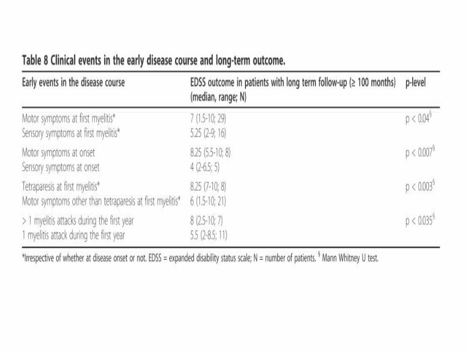
• PREDICTIVE VALUE OF EARLY CLINICAL EVENTS :• In patients with myelitis (with or without concomitant ON)
and long term follow-up data ( ≥ 100 months), pure sensory symptoms at onset or at first myelitis were associated with a better EDSS outcome at last follow-up compared to patients with motor symptoms at onset.
• Also, tetraparesis at first myelitis and more than one myelitis attack in the first year - worse EDSS long term outcome.
• By contrast, neither bilateral optic nerve involvement at first ON nor brain stem involvement (at any point in time) did predict the EDSS long term outcome.

• MYELITIS CLINICAL FINDINGS :• Motor symptoms occurred during 347/503 (69%) attacks (197
- paraparesis, 81 - tetraparesis, 31 - hemiparesis, 34 - monoparesis, 4 - Brown Sequard syndrome).
• The median MRC grade of all documented myelitis attacks with motor symptoms was 3 (range 0-5).
• Severe paresis (MRC grade ≤ 2) recorded during 49.3% of all relapses with motor symptoms (including MRC grade 0 in one or more limbs in 19.7%), paresis was mild (MRC grade 4 or 5-) in 31%.

• Severe paresis - more frequent in the seropositive group than in the seronegative group.
• Pure sensory attacks - more common in the seronegative group.
• Motor symptoms - more common in the seropositive group.

• MYELITIS – MRI FINDINGS :• At first myelitis, MRI showed at least one spinal cord lesion
extending over 3 or more vertebral segments in 127/137 patients (92.7%).
• The median extension was 6 segments (range, 1-21; N = 137) with a non-significant trend towards longer lesions in seropositives .
• In 21 patients (18 seropositive), a second lesion was detected (median extension 2 segments; range, 1-8), and in 8 patients (all seropositive) an additional third lesion was present.
• In those patients with several lesions, the median total number of segments involved was 8 (range 2-21).

• The total lesion load at first MRI correlated with the EDSS at last follow up in patients with a disease duration > = 100 months.
• 37 patients - lesions in the cervical portion of the spinal cord • 27 - thoracic segments• 44 - both the cervical and the thoracic segments. • Lesion involving Conus – in 4 patients
• If all 326 spinal MRIs are considered, the median length of the longest lesion in each MRI was 5 segments (range, 1-21) and was slightly longer in seropositive patients than in seronegative patients.

• The median total spinal cord lesion load of all MRIs was higher in the seropositive group .
• Very long lesions extending over 6 or more segments were more frequent among seropositives.
• Entire spinal cord involvement as defined by contiguous spinal cord lesions extending ≥17 vertebral segments occurred only in seropositive patients.


• MYELTIS – OUTCOME :• Myelitis attacks mainly treated with I/V MPS (378 attacks)
and/or plasma exchange (78 attacks). • Other treatments included oral prednisolone, IVIG, and
Rituximab.• Complete remission reported in 62/359 (17.3%), partial
remission in 248 (69.1%), and no remission in 49 (14%). • The proportion of attacks with only partial or no recovery was
lowest at the time of the first attack and increased with the number of subsequent attacks
• No significant difference regarding attack outcome (complete, partial, or no recovery) found between seropositive and seronegative patients.

• OPTIC NEURITIS – CLINICAL FINDINGS :
• During the first documented ON attack, visual acuity was ≤ 0.1 in either eye in 30/39 (76.9%) patients.
• In total, results from 158 eye examinations during acute ON were available for analysis.
• 96 examinations (60.8%) in 62 patients showed a visual acuity of ≤ 0.1 in the affected eye(s) - more frequent among seropositive than among seronegative patients.

• At the end of the observation period, both the right and the left eye had been affected at least once (either simultaneously or successively) over the course of disease in 64% (78/121) of patients with a history ON and in 76.1% (35/46) of those with long term follow-up ( ≥100 months).
• If all documented ON attacks are considered, ON was bilateral in 19.8% or 62/251 attacks.
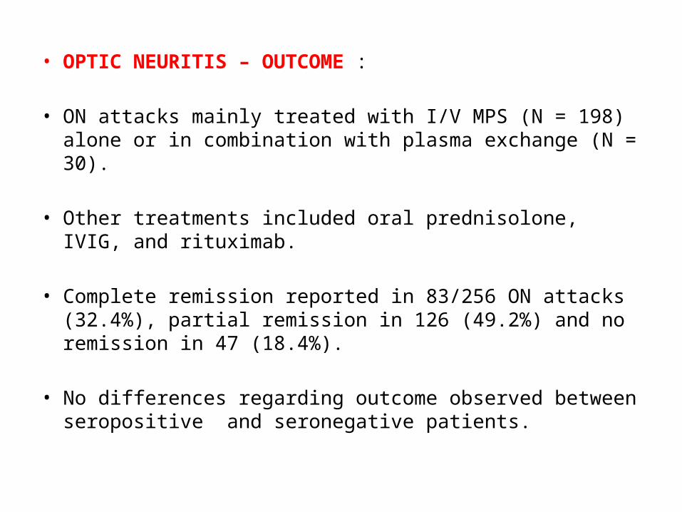
• OPTIC NEURITIS – OUTCOME :
• ON attacks mainly treated with I/V MPS (N = 198) alone or in combination with plasma exchange (N = 30).
• Other treatments included oral prednisolone, IVIG, and rituximab.
• Complete remission reported in 83/256 ON attacks (32.4%), partial remission in 126 (49.2%) and no remission in 47 (18.4%).
• No differences regarding outcome observed between seropositive and seronegative patients.

• OTHERS :
• In 46/175 patients (26.3%), clinical and/or radiological signs of brain stem involvement were recorded at least once over the course of disease, with no significant difference between seropositive and seronegative cases.
• The first available brain MRI showed supratentorial brain lesions in 81/168 (48.2%) cases.

• At first lumbar puncture (LP), cerebrospinal fluid (CSF) restricted oligoclonal bands (OCBs) were present in 42/144 patients (29.2%) with no significant difference between seropositives and seronegatives.
• Follow-up results were available from 83 patients (65 from initially OCB-negative patients and 18 from initially OCB-positive patients).
• 10/65 patients that were negative for OCBs at first LP (15.4%) developed OCBs later, and 10/18 patients positive for OCBs at first LP (55.6%), turned negative at follow-up.
• Consequently, 52/144 (36.1%) patients were positive for OCBs at least once.

• CSF WBC count at first lumbar puncture were rather low (median 7/ μ l; range, 0-750; N = 136).
• CSF pleocytosis (defined by a WBC > 5/μl) was present at first LP in 95/146 patients .
• The frequency of pleocytosis as well as the median WBC count did not differ between seropositive and seronegative patients .

CONCLUSIONS• Seropositive and seronegative patients - found to differ with
regard to attack severity and clinical presentation.
• Following were the observations in Seropositive > Seronegatives :– Visual acuity of ≤ 0.1 during acute ON attacks.– Motor symptoms– The median MRC grade during acute myelitis attacks worse– MRC grades ≤ 2 more frequent

• On the other hand, simultaneous myelitis and ON as well as bilateral ON at disease onset, and sensory symptoms were more frequent in the seronegative group.
• By contrast, the two groups did not differ significantly with regard to :– The median annualized total relapse rate– Median annualized myelitis specific relapse rate– Median annualized ON specific relapse rate– Relapse outcome (no remission,partial or complete
remission) and – Frequency of brainstem involvement.

• Given both the low remission rate (as compared to MS) found in this study already at disease onset and the fact that the first event was followed by a relapse after a median latency of just 9 months (and only 5 months in the seronegative group), early treatment and, therefore, an early diagnosis of NMO is crucial.
• Study revealed a marked delay in the diagnosis of NMO (16 months from onset if the disease started with myelitis, and even 55 months if the disease started with ON;p < 0.013).

• A substantial number of patients (42.5%) were initially wrongly diagnosed with MS.
• The median time to diagnosis was shorter among seronegative patients (11 months) than among seropositive patients (45 months) - partly explained by the fact that NMO started more frequently with simultaneous myelitis and ON in the seronegative group.

• NMO started in most cases with unilateral ON and, more rarely, with isolated myelitis.
• Importantly, the first ON attack was followed by myelitis after a median of just 14 months in the seropositive group, and the first myelitis was followed by ON after a median of only 3 months.
• This corroborates findings from previous, smaller studies, which found that AQP4-Ab seropositivity in patients with isolated ON or myelitis confers a high risk of conversion to NMO within one year.
• Strongly underlines the need for early prophylactic treatment in patients presenting with seropositive isolated ON or myelitis.

• LIMITATIONS OF THE STUDY :• Retrospective study• Similar to previous studies, analysis of MRI results was based
upon patient records.• As in previous studies, most patients were treated at least
once with immunomodulatory or immunosuppressive agents. Although Longitudinal studies without treatment not possible due to aggressive course of disease.
• Sensitivity of the assays for antibodies could be a potential limitation.
• Patients with a benign long-term course are less likely to be admitted to hospital and might thus be under-represented.

THANK YOU

The Frequency of Anti-Aquaporin-4 Ig GAntibody in Neuromyelitis Optica and Its Spectrum Disorders at a Single Tertiary Referral Center in Malaysia
- Shanthi Viswanathan, Masita Arip,Norhazlin Mustafa et al- Published in Multiple Sclerosis International in November 2014

OBJECTIVE :-
• To evaluate the frequency of anti-aquaporin-4 Ig G antibody (Anti-AQP4 antibody) amongst patients with Neuromyelitis optica (NMO) and its spectrum disorders (NMOSD) and the differences between the seropositive and seronegative groups.

• METHODS :
• Hospital based retrospective study with longitudinal follow-up of patients who had presented to the Department of Neurology, Kuala Lumpur Hospital, between January 2009 and January 2014 with Idiopathic inflammatory demyelinating disease.
• Data was obtained from case notes retrospectively and clinic follow-ups.

Inclusion Criteria (1) All patients with Idiopathic inflammatory demyelinating
disease fulfilling Wingerchuk criteria of 2006 for NMO excluding anti-AQP4 testing
(2) Patients at high risk for NMOSD, that is, those with single episode, monophasic or recurrent relapsing optic neuritis (RON/MON) and single (monophasic) episode or recurrent myelitis (MTM/RTM), as outlined by Wingerchuk et al.
(3) Patients with brain symptoms and signs at onset of disease with MRI Brain showing demyelinating lesions atypical for multiple sclerosis but typical for NMOSD as described by Pittock et al. [10],
(4) Monophasic or multiphasic, monofocal or multifocal demyelinating disease involving the brain but did not fulfil the criteria for brain involvement in NMOSD, acute demyelinating encephalomyelitis, or MS.

Exclusion Criteria
(1) Patients who refused or were unable to consent for inclusion of their data in the study and/or for the purpose of testing for anti-AQP4 antibody.
(2) Patients with Nondemyelinating idiopathic inflammatory diseases involving the brain.
(3) Patients diagnosed with symptoms and signs suggestive of Multiple sclerosis based on McDonalds’s 2005 and 2010 diagnostic criteria were excluded from the study.
(4) Patients diagnosed with ADEM.

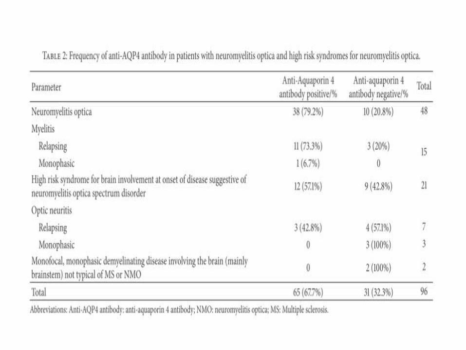
RESULTS• Female proponderance (88.5% vs 11.5%)
• Testing for anti-aquaporin-4 antibody was done on :– 48 patients with NMO– 21 with NMOSD (brain involvement at onset)– 15 with transverse myelitis– 7 with relapsing optic neuritis– 3 with monophasic optic neuritis, and – 2 in patients with monophasic, monofocal brain disease at
onset (whose brain MRI lesions did not look like those described by Pittock et al. but still were possibly at high risk for NMOSD)


• Blindness in one or both eyes and poor visual acuity were significantly seen in seropositive patients rather than seronegative patients.
• NMO/NMOSD patients with longitudinally extensive cord lesions of contiguous or linear nature with or without fragmentation/interrupted lesions were significantly associated with being anti-AQP4 antibody positive rather than being negative, < 0.001. 𝑃
• Seropositive patients had significantly more lesions in the cervical, thoracic, and cervicothoracic cord regions, < 0.001. 𝑃
• Holocord or central gray matter lesions were significantly associated with seropositivity( < 0.001).𝑃
• On the other hand, seropositivity and seronegativity did not discriminate between the different ethnic races or the type of clinical presentation at onset


• While MS primarily causes demyelination, NMO attacks are often associated with severe necrosis.


