1
A Novel Function of RNAs Arising From the LTR of Human Endogenous 1
Retrovirus-9 in Cell Cycle Arrest 2
Lai Xua, Abdel G Elkahlounb, Fabio Candottic, Andrzej Grajkowskia, 3
Serge L. Beaucagea, Emanuel F. Petricoind, Valerie Calvertd, Hartmut Juhle, 4
Frederick Millsa, Karen Masona, Neal Shastria, Josh Chika, Cynthia Xua and 5
Amy S. Rosenberga# 6
Division of Therapeutic Proteins, Center for Drug Evaluation and Research, U. S. 7
Food and Drug Administrationa, Cancer Genetics Branch, National Human Genome 8
Research Instituteb, Genetics and Molecular Biology Branch, National Human 9
Genome Research Institutec, National Institutes of Health ,Center for Applied 10
Proteomics and Molecular Medicine, George Mason Universityd, Indivumed, Gmbhe. 11
#Correspondence should be addressed to A.R. ([email protected]) 12
The human genome contains approximately 50 copies of the replication defective 13
Human Endogenous Retrovirus 9 (ERV-9) and thousands of copies of its solitary 14
Long Term Repeat (sLTRs) element. While some sLTRs are located upstream of 15
critical genes and have enhancer activity, other sLTRs are located within introns 16
and may be transcribed as RNAs. We found that intronic RNAs arising from U3 17
sLTRs of ERV-9 were expressed as both sense (S) and antisense (AS) transcripts in 18
all human cells tested but that expression levels differed in malignant vs non-19
malignant cells. In non-malignant cells, AS was expressed at higher levels than S 20
and at higher levels compared to AS expression in malignant cells; in malignant 21
Copyright © 2012, American Society for Microbiology. All Rights Reserved.J. Virol. doi:10.1128/JVI.01648-12 JVI Accepts, published online ahead of print on 24 October 2012
on June 7, 2018 by guesthttp://jvi.asm
.org/D
ownloaded from
2
cells, AS was expressed at equivalent amounts to S RNA. Critically, U3 AS RNA was 22
found to physically bind to key transcription factors for cellular proliferation 23
including NF-Y, p53 and sp1, indicating that such RNA transcripts may function as 24
decoy targets or traps for NF-Y and thus inhibit the growth of human cancer cells. 25
Indeed, short U3 oligodeoxynucleotides (ODN) based on these RNA sequences ably 26
inhibited proliferation of cancer cell lines driven by cyclins B1/B2, the gene targets 27
of NF-Y. 28
Human endogenous retroviruses (HERVs) comprise approximately 8% of the human 29
genome (39), and have physiological functions as well as a potential role in some human 30
diseases (38). During primate evolution, thousands of copies of sLTRs were generated 31
due to duplications of the founder provirus genome followed by recombinational deletion 32
of most of the proviral genome, leaving intact sLTRs (5). Different from other known 33
retroviral LTRs, ERV-9 LTRs have a variable number of tandem repeats with multiple 34
enhancer binding sites for NF-Y (CCAAT), MZF1 (GTGGGGA) and GATA-2 (GATA) 35
in their U3 region (30, 31). Substantial progress has been made in uncovering the 36
promoter activities of the ERV9 LTR U3 DNA (pertaining to its binding of transcription 37
factors) in regulating several important genes including p63 isoforms which protect the 38
male germ-line via tumor suppressor activity (8, 22, 37), and the globin gene (30, 31). 39
However, little information has been published regarding the activities and functions of 40
RNAs originating from the U3 region. Since some ERV-9 LTRs locate within introns of 41
coding sequences of genes, such as the axin1 gene (in the reverse orientation) (21, 23) 42
and the HLA-DRB gene (in the forward orientation) (41), both U3 S and AS RNAs 43
on June 7, 2018 by guesthttp://jvi.asm
.org/D
ownloaded from
3
should be transcribed in cells as the result of spliced by-products from mRNAs of these 44
genes. 45
In this work we evaluated the function, not of the ERV-9 LTR U3 DNA, but rather of the 46
U3 RNA transcripts originating from the U3 repeat region. 47
48
Materials and Methods 49
50
Cells. Cell lines were maintained at 37°C in a humidified atmosphere of 5% CO2. Human 51
cancer cells; MDA231, MCF-7, K562, LNcap, HepG2, HT1080, HTB77 and HTB78 52
cells were cultured in either in DMEM or RPMI medium. All of the media were 53
purchased from Life Technologies, Inc 54
55
ODN decoy treatment. 2.5X10⁴ cells were treated with ERV-9 U3 S or AS ODNs at 56
different concentrations for 72 hours. GFP ODN was used as a control to subtract the ~3-57
6% non-specific cytotoxicity of phosphorothioate based ODN. G3139, GRN163 and 58
MDM2AS were three positive controls. Cells were counted by FACS Calibur (BD 59
Company, USA). Relative cell proliferation levels were expressed as percentage of 60
control ODN treatment. ODN and biotinylated ODNs: ERV-9LTR U3 S: 61
CTCAAGGTTTGTAAACACACCAATCAG ERV-9LTR U3 AS: 62
CTGATTGGTGTGTTTACAAACCTTGAG (AF064190: 3329-3355 bp), U3 S mut: 63
CTCAAGGTTTGTAAA CACAtgtgaCAG, U3 AS mut: 64
CTGacactTGTGTTTACAAACCTTGAG, GFP: CATTATCAACA 65
on June 7, 2018 by guesthttp://jvi.asm
.org/D
ownloaded from
4
AAATACTCCAATTGGC.G3139:TCTCCCAGCGTGCGCCAT. GRN163: 66
TAGGGTTAGACAA. MDM2 AS: TGACACCTGTTCTCACTCAC. 67
68
Cell Cycle Analysis. ODN treated cells were suspended in phosphate buffer solution 69
(PBS), centrifuged (1500 r/min, 8 minutes) and the supernatant was discarded. The 70
suspension was fixed with precooled 75% ethanol overnight at -20℃, treated with 71
ribonuclease (RNAase) 0.5mg/ml at room temperature for 30 minutes then 10ug/ml 72
propidium iodide. After 30 minutes at room temperature, the percentage of the cells at 73
different phases in the cell cycle and cell apoptosis was determined by FACS Calibur 74
(BD Company, USA). 75
76
77
PCR primers: supplementary information. 78
79
RNA extraction, reverse transcription and Real-Time PCR quantification. Total 80
RNA was extracted from cells with a cell density of 75% confluence using Trizol® total 81
RNA isolation reagent (Gibco BRL, Life Technologies, Gaithersburg, MD, USA) as per 82
the manufacturer's protocol and RNeasy Mini Kit (Qiagen Cat# 74104). Total RNAs 83
were treated by DNase I (Invitrogen Cat#18068015). There was no PCR amplification of 84
ERV-9 LTR U3 products from DNase I treated RNA samples (Supplemental figure 1a). 85
cDNA was synthesized from total RNA using gene-specific primers or random primers 86
by using High Capacity cDNA Reverse transcription Kit (Applied Biosystems, ABI) 87
Part#4368813). Real-time PCR was performed using an (ABI) 7300 Sequence Detection 88
on June 7, 2018 by guesthttp://jvi.asm
.org/D
ownloaded from
5
system. Real–time PCR was performed by using Integrated DNA Technologies (IDT) 89
designed or ABI probe/primers to detect gene of interest with 18S rRNA or GAPDH as 90
internal controls. Cellular and viral PCR primers for real-time evaluations are included in 91
supporting information. 92
Primary ductal carcinomas. RNAs of 4 matched pairs of human primary ductal 93
carcinomas (BT1-4) (TNM Classification, 6th Edition. T2) and their respective adjacent 94
normal mammary tissues (BN1-4) designated as K135 (BT1, BN1), K165 (BT2, BN2), 95
K172 (BT3, BN3) and K191 (BT4, BN4) were purchased from Indivumed GmbH (RIN > 96
7). K135 and 163 were estrogen receptor positive. K172 and 191 were estrogen receptor 97
negative. K135 and 172 were HER2 positive. K165 and 191 were HER2 negative. All 98
four were progesterone receptor negative. 99
Northern Blot and Western blot 100
RNAs were separated on 12% denaturing urea polyacryamide gels and transferred to a 101
nylon membrane. P32 end labeled S and AS ODN probes were used to detect AS and S 102
endogenous U3 RNAs separately. RNAs of four matched pairs of human breast cancer 103
and adjacent normal mammary tissues were obtained from Indivumed (www.indivumed). 104
The RNA of EL4, a mouse lymphoma cell line was used as the negative control. 105
NuPAGE 4-12% Bis-Tris Gel and LAS4000 biomolecular imager were used for Western 106
blot analysis. 107
108
RNA-immunoprecipitation (RIP). RNA-IP was carried out using an RIP-Assay Kit 109
(MBL Cat# RN1001). Precleared cell lysates were incubated with 2 µg of antibodies to 110
one of the following: NF-Y, p53, p21 p300, Est-1, sp1 HDAC1, PTBP1 and mouse IgG 111
on June 7, 2018 by guesthttp://jvi.asm
.org/D
ownloaded from
6
at 4°C for overnight. The RNA-protein immunocomplexes were brought down by protein 112
A/G beads, and RNA was then purified from the complexes for RT–PCR as described 113
previously. For in vitro competition, 100 ug of cellular lysate was incubated with 5 ug of 114
different AS, S and control ODNs and rocked at 4°C overnight. The same concept was 115
used to design U3 SL, ASL U3SmL U3ASmL and KL. OND sequences used in RNA-IP are 116
included in supporting information. Antibodies of NFY-A/sc-10779, NFY-C/sc7715-R, 117
NFY-B/sc-13045, NFKB-p65/sc372, HDAC1/sc-7872, Est-1/sc5558, sp1/sc-14027, 118
p53/sc-6243, p21/DCS60, p300/sc-584, PTBP1/sc-133677 and Biotin/sc-57636 were 119
used for RIP. 120
121
Xenograft models. BALB/c scid male mice (JAX® Mice), 4- to 6-week-old, were used 122
to make HT1080 and MDA231 xenografts. Cultured cells were washed with and 123
resuspended in serum-free media. The suspension (2 × 10⁶ cells in 0.2 ml per mouse) was 124
then injected into the chest area of the mice. The mice were monitored by measuring 125
tumor growth and body weight and by general clinical observation. Tumor-bearing mice 126
were randomly divided into multiple treatment and control groups (8 mice per group) on 127
day 7. All ODNs, dissolved in 0.2 ml physiological saline (0.9% NaCl), were injected 128
daily into adjacent area of tumor at doses of 50ug/mouse for 7 days. The control group 129
received saline only. Animals were sacrificed and tumor were isolated and weighed. 130
ODN treatment was blinded and data was decoded. 131
PCR cloning and sequence. [email protected] and pcDNA3.1/NT-GFP-TOPO vectors 132
were used for cloning and sequence. 133
Statistics. A 2-tailed unpaired t test 134
on June 7, 2018 by guesthttp://jvi.asm
.org/D
ownloaded from
7
(http://www.graphpad.com/quickcalccs/ttest1.cfm?Format+SD) and Sigma Plot for 135
ANOVA test. A P value than 0.05 was considered statistically significant. All real-time 136
PCR and ODN growth inhibition data shown in this work are the mean s.d. of 3 137
replicates. Each assay was repeated three times with comparable results. 138
RESULTS 139
140
Expression of both S and AS RNAs of the ERV-9 LTR U3 repeat region in human 141
tumor cell lines and in primary ductal carcinomas 142
143
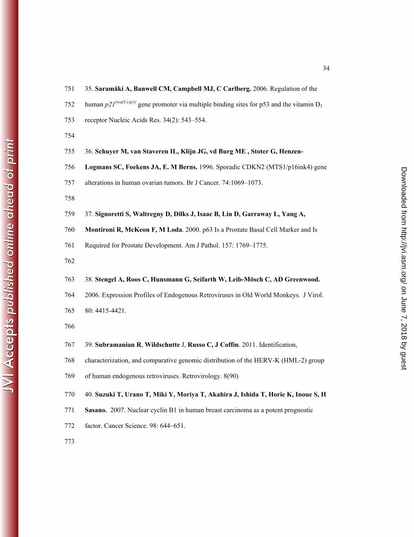
In initial experiments, we investigated the expression of ERV-9 LTR U3 RNAs in human 144
cells by directional RT-PCR assays. A pair of primers was designed to specifically detect 145
the first four repeat unit (0-1-2-3-4)1 sequences of the ERV-9 U3 LTR based on the 146
sequence of the ERV-9 LTR located upstream of the human β-globin gene locus (21, 23, 147
30, 31) (Fig. 1a). Both U3 S and AS RNAs of the ERV-9 LTR (U3 S and U3 AS RNA, 148
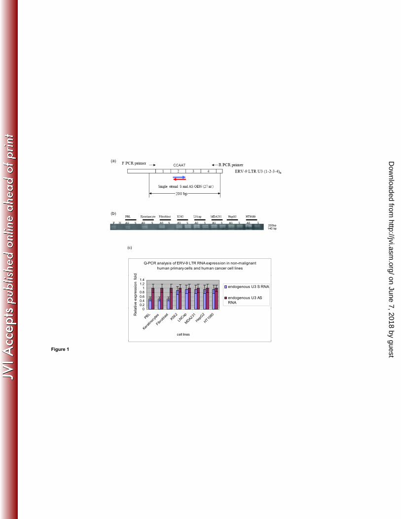
respectively) were detected as ~140 and ~200 nt fragments in diverse human tumor cell 149
lines (Fig. 1b). Cloning and sequencing of RT PCR products from MDA231 revealed that 150
the ~140 and ~ 200 nt amplicons are actually 155 nt and 196 nt, respectively. The 155 nt 151
amplicon has repeat elements of sequence (0-1-3-4) (Fig. 3 1c, Supplementary fig. 2a) 152
while the 196 nt amplicon has repeat elements of sequence (0-1-2-3-4) (Fig. 3 1d, 153
Supplementary fig. 2b). The 155 nt amplicon has 2 and the 196 nt amplicon has 3 154
CCAAU motifs (AUUGG in AS), respectively. Real-time PCR primers were then used to 155
compare the relative levels of AS and S within malignant and non-malignant cells. The 156
U3 AS RNA was found to be expressed at a 2 fold higher level than the U3 S RNA in 157
on June 7, 2018 by guesthttp://jvi.asm
.org/D
ownloaded from
8
primary, non-malignant cells while U3 AS and S RNAs were expressed at equal levels in 158
cancer cell lines (Fig. 1c). 159
160
Out of concern that cancer cell lines might not reflect the true expression of U3 RNAs in 161
primary human cancers, we assessed expression of U3 RNAs in primary mammary ductal 162
carcinomas (TNM Classification, 6th Edition: T2) and in their donor matched adjacent 163
normal mammary tissues. Normal mammary glands expressed 2-3 fold more U3 AS 164
RNA and 2 fold less U3 S RNA than did their adjacent primary breast tumor counterparts 165
(Fig. 2a). High resolution Northern Blot confirmed this differential expression of U3 166
RNAs: normal mammary tissues expressed higher levels of a ~500 nt U3 AS RNA 167
species and lower levels of a ~200 nt U3 S RNA species than did primary ductal 168
carcinomas (Fig. 2b). Thus both PCR and Northern Blot analyses indicate that U3 RNAs 169
arising from the ERV-9 LTR are differentially expressed in non-malignant vs malignant 170
cells, with a higher level of expression of AS in non-malignant primary cells vs 171
malignant tumor cells. The ~200 nt S and ~500 nt AS appear to be novel U3 RNA 172
transcripts since they have not hitherto been reported (20, 42) (Fig. 2b). PCR cloning and 173
sequencing with nested U3 primers revealed that the ~500 nt AS RNA is 549 nt (Fig. 3 174
1e) and that this 549 nt AS RNA originates in intron 10 of the BRCAA1-012 gene 175
(AL732292.12) and contains six AUUGG motifs (Supplementary fig. 2d). BRCAA1-012 176
is a transcriptional variant of the BRCAA1 gene, sharing the last 6 exons with BRCAA1 177
(Fig. 3 2a, 2b). Moreover, the ~200 S nt was cloned and its actual size was found to be 178
240 nt (Fig. 3f). The transcriptional origin of the 240 nt U3 S RNA is not known as yet 179
on June 7, 2018 by guesthttp://jvi.asm
.org/D
ownloaded from
9
since its sequence is found in regions of chromosome 5 (AC008885.5) and chromosome 180
12 (AC126564.7) from which no other transcripts have originated. 181
182
ERV-9 LTR U3 AS RNA binds to NF-Y, p53 and sp1 in vivo 183
184
U3 RNAs of the ERV-9 LTR contain multiple CCAAT motifs (Supplementary fig. 1b) 185
which are known binding sites for NF-Y, a transcription factor associated with cellular 186
proliferation, principally through its binding to the CCAAT motifs of cyclin B1/B2 genes 187
(7). Moreover, NF-Y itself binds to additional factors important in cellular proliferation 188
including p53, sp1, p300, Ets-1 and HDAC1 (28), Thus, it was plausible that modulating 189
levels of free NF-Y via binding to U3 RNAs of ERV-9 LTR could potentially modulate 190
cellular proliferation by serving as a “decoy” target or a “trap” in competing with 191
binding of free NF-Y and its associated factors, to genes mediating proliferation such as 192
cyclins B1/B2, CDC25C, CDC2 and topoisomerase (7). Therefore, we investigated 193
whether endogenous U3 RNAs bound to NF-Y and its associated factors in vivo by using 194
antibodies to specifically immunoprecipitate NF-Y and other transcription factors in the 195
cytoplasmic fraction of cells followed by PCR to detect NF-Y bound U3 RNAs. In 196
malignant cell lines (HT1080, MDA231) and in quiescent and proliferating T cells, the 197
endogenous ~140 and ~200 bp U3 AS RNAs bound to NF-Y, p53 and sp1, but not to 198
p300, Ets-1, and HDAC1 (Fig. 4). Cloning and sequencing of AS ~140 and ~200 nt U3 199
amplicons from such immunoprecipitations (Fig. 4) confirmed that they are the same 155 200
and 196 nt U3 amplicons identified previously (Fig. 3 1c, 1d). The binding of NF-Y to 201
U3 AS RNAs of ERV-9 LTR was relatively specific, since NF-Y did not bind to the AS 202
on June 7, 2018 by guesthttp://jvi.asm
.org/D
ownloaded from
10
RNA of the ERV-K111 LTR (Supplementary fig. 2a). As the U3 AS 196 nt transcript 203
originates from the MAPK-10 and/or IL-15 gene loci, and the transcription factor CREB 204
regulates the expression of MAPK-10 (15) and transcription factors GR, ISGF-205
3, STAT3, GR-alpha, STAT5A and AP-2 gamma regulate the expression of IL-15 (33), a 206
higher expression level of MAPK-10 and IL-15, as has been reported previously in 207
normal mammary glands (15, 33), may correlate with the higher expression level of the 208
AS 196 nt “decoy” in normal cells. However, the transcriptional origin of the AS 155 nt 209
amplicon is uncertain since its sequence was not found within other identified transcripts 210
and therefore will require additional study. To our knowledge, this is the first report of 211
an endogenous RNA molecule specifically binding to an important transcription factor in 212
eukaryotic cells. Thus, the low expression level of U3 AS RNAs in breast cancer cells 213
may facilitate the high level binding of NF-Y to the cyclin B1/B2 genes. Indeed, we 214
found that all primary breast tumors had heightened expression of cyclins B1/B2 RNAs 215
without increased NF-Y mRNA levels (Supplemental fig. 3a). Although the expression of 216
cyclinB1/B2 mRNAs can be upregulated by NF-Y protein (7) and downregulated by p53 217
as well as by p21/WAF1 (2, 16, 17, 18), the factors driving the maintenance of high 218
expression levels of cyclins B1/B2 in primary breast cancers (40) have not been fully 219
elucidated. Indeed we found reduced levels of message for cell cycle inhibitors 220
p21/WAF1 and p15INK4b in primary breast tumors (Supplemental fig. 3a). 221
222
In vitro and in vivo inhibitory effects of U3 S and AS ODNs in growth of human 223
cancer cell lines 224
225
on June 7, 2018 by guesthttp://jvi.asm
.org/D
ownloaded from
11
To further study the biological functions of ERV-9 LTR U3 S and AS RNAs in cell 226
growth, we administered short U3S and U3AS ODN sequences, based on sequences in 227
these LTR elements, to cancer cells. Phosphorothioate ODNs (27 nt) corresponding to 228
subunit 2 of the (1-2-3-4)1 tandem repeat in S and AS orientation were constructed (S 229
ODN, AS ODN) (Fig. 1a). These two ODNs contain one NF-Y binding motif (CCAAT 230
in sense, ATTGG in antisense) and, critically, lack CpG sequences which are known to 231
induce non-specific inflammatory or IFN responses that could diminish cell proliferation. 232
Both U3 S and AS ODNs reduced the growth of HT1080 and MDA231 by 2-3 fold, but 233
did not inhibit the growth of normal primary keratinocytes stimulated by EGF, quiescent 234
or proliferating PBL, or monocytes stimulated with pooled human serum (Supplemental 235
figure 3). Control U3 S and AS ODNs (U3Sm, U3ASm) which were mutated so that they 236
lacked the NF-Y binding motif showed significant restoration of cellular proliferation, 237
indicating the criticality of the NF-Y binding motifs (Supplemental fig. 4). We then tested 238
these ODNs in six diverse human cancer cell lines and found that both ODNs reduced 239
cell growth 1.5-2 fold greater than did G3139 (42), GRN163 (4) and MDM2 AS (47) 240
ODNs (Fig. 5a). G3139, GRN163 and MDM2 AS are known to inhibit growth of 241
malignant cells by targeting Bcl2, telomerase and MDM2, respectively. In a 242
chemosensitivity assay, S and AS ODNs sensitized all six cell lines to etoposide 243
phosphate (VP16) 1.5-2 fold greater than did the three positive controls (Supplemental 244
fig. 5). To assess whether such effects on in vitro tumor cell growth would be realized in 245
vivo, we investigated the effects of ERV-9 LTR U3 S and AS ODNs on tumor growth in 246
a xenotransplant model, using HT1080 and MDA231 tumors in SCID mice. We found 247
that U3 S and AS ODNs potently inhibited in-vivo tumor growth (Fig. 5b). 248
on June 7, 2018 by guesthttp://jvi.asm
.org/D
ownloaded from
12
249
250
ERV-9 LTR U3 ODNs regulate the cell cycle by acting as NF-Y traps 251
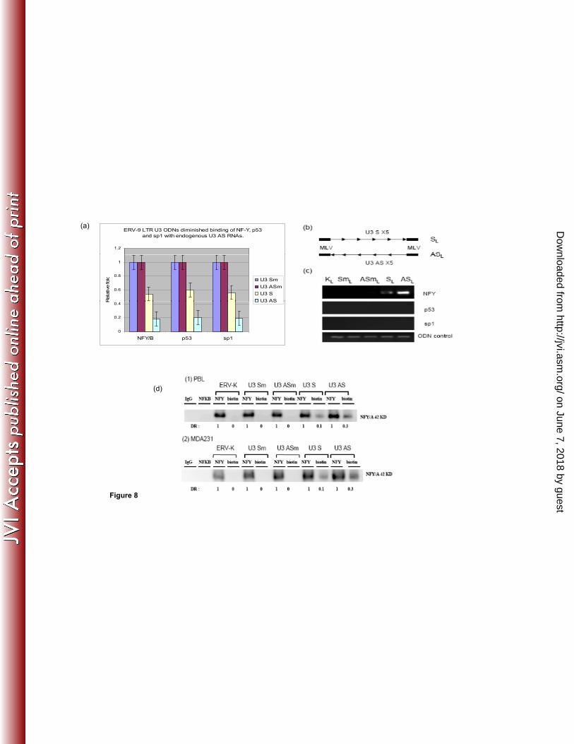
Since U3 S and AS ODNs contain NF-Y binding motifs and NF-Y has been shown to 252
regulate cell cycle progression (7), we performed cell cycle analyses following treatment 253
of cell lines with S and AS ODNs. Compared to the GFP ODN negative control, U3 S 254
and AS ODNs increased the percentage of cells in G0/G1 phases from 80% to 90% in 255
HT1080 and from 64% to 77% in MDA231, while the three positive controls increased 256
the percentage of cells in the G0/G1 phases from 80% to 82% (HT1080) and from 64% to 257
71% (MDA231) (Fig. 6). These results indicate that one of the mechanisms by which U3 258
S and AS ODNs of ERV-9 LTR inhibit human cancer cell growth is by causing arrest in 259
G0/G1. To uncover the molecular basis by which ERV-9 LTR ODNs caused G0/G1 260
phase arrest, we assessed cell cycle genes related to the NF-Y pathway by real-time PCR 261
in ODN treated cells. Both U3 S and AS ODN down-regulated mRNAs of cyclins B1/B2, 262
CDC2, and CDC25C 2-3 fold and upregulated mRNA of the cell cycle inhibitor 263
p21/WAF1 three- fold in HT1080 and MDA231cells (Fig. 7c). Additionally, U3 AS 264
ODN upregulated mRNA for IL24, a cytokine known to inhibit cell proliferation, by 7 265
fold in MDA231 cells (Fig. 7c). These ODNs failed to down-regulate mRNAs of cyclins 266
D1/D3, which are not strongly regulated by NF-Y (5) (Fig. 7a, b), and, critically, neither 267
U3 S nor AS ODNs altered the mRNA levels of critical transcription factors themselves 268
including NF-Y, sp1 and p53 (Supplement fig. 6). Because some ODNs induce type I 269
interferons which are known to cause G0/G1 phase arrest (34), we evaluated whether 270
type I interferon was involved in the U3 ODN mediated G1 arrest. Thus, we co-treated 271
on June 7, 2018 by guesthttp://jvi.asm
.org/D
ownloaded from
13
HT1080 and MDA231 cells with U3 ODNs and a soluble interferon receptor, B18R, 272
which has high affinity for type I interferons and blocks both autocrine and paracrine 273
functions of type I interferons. We found that B18R did not significantly reduce the anti-274
proliferative activity of U3 ODNs, but ably blocked the anti-proliferative activity of the 275
IFN-β control, indicating that type I interferons did not play a major role in G1 phase 276
arrest by U3 ODNs (Supplemental fig. 7). We also failed to find a role for enhanced 277
apoptosis as an explanation for diminished cell growth in ODN treated cells. Moreover, 278
the non-specific cytotoxicity of the ODNs themselves was less than 6% in all tested 279
samples. 280
Since NF-Y has been reported to have a minimal impact on the p16/cyclin 281
D1/CDK4/pRB proliferation pathway (7), U3 ODNs theoretically should have less 282
inhibitory effect in cancer cell lines whose proliferation is largely mediated by activation 283
of the p16/cyclin D pathway. To test whether this was indeed the case, we evaluated 284
ovarian cancer cell lines that differed with respect to dependence on cyclins D vs B. In 285
comparing the expression of 15 cell cycle related genes between the two cell lines, we 286
found that HTB77 had lost p16 expression but had no significant differences in the 287
remaining 14 genes, compared to HTB78 (Supplemental fig. 8a, b) (36). Since p16 288
inhibits the cyclinD/CDK4/6 pathway, the loss of p16 allows constitutive activation of 289
the cyclinD/CDK4/pRB pathway. In treating these two cell lines with U3 ODNs, we 290
found that while U3 AS ODN inhibited the growth of HTB78 cells, it did not 291
significantly inhibit the growth of the cyclin D driven HTB77 cells (Supplemental fig. 292
8c). The HTB77 cell line was, however, inhibited by NFKB AS ODN which targets the 293
on June 7, 2018 by guesthttp://jvi.asm
.org/D
ownloaded from
14
cyclin D pathway (Supplemental fig. 8c). This result further supports the specificity of 294
U3 AS ODN for the cyclin B1/B2 proliferation pathway. 295
296
Finally, to evaluate whether such exogenous U3 ODNs functioned as “traps” to bind NF-297
Y, we performed a competition assay in which U3 ODNs competed for NF-Y binding to 298
its endogenous ligand, U3 AS RNA. AS ODN diminished binding of NF-Y, p53 and sp1 299
to U3 AS RNA by 5 fold compared to its U3 mut AS ODN counterpart lacking an NF-Y 300
binding site, while U3 S ODN diminished binding of NF-Y, p53 and sp1 to U3 AS RNA 301
by 2 fold compared to its mutant control counterpart (Fig. 8a). To more directly assess 302
the physical binding of NF-Y to the ODNs, we designed a pair of long (SL and ASL) 303
ODNs (158 nt) with 5 tandem repeats of the original 27 nt S and AS sequence containing 304
5 NF-Y binding motifs (Fig. 8 b) flanked by a 12 nt murine leukemia virus (MLV) PCR 305
linker on both ends to distinguish the exogenous ODN from the endogenous ERV-9 LTR 306
U3 DNA. Our data demonstrated that ASL ODNs strongly bound to NF-Y but not 307
directly to p53 and sp1 while SL ODNs more weakly bound to NF-Y, but not directly to 308
p53 and sp1 (Fig. 8c) compared to the U3 mutant and ERV-K controls lacking such NF-309
Y binding sites. 310
311
Although we could not directly address what fraction of NFY was bound to endogenous 312
U3 RNAs, because endogenous U3 RNAs cannot specifically be isolated by current 313
immunoprecipitation methods, we used biotinylated U3 ODNs as a surrogate. We treated 314
unstimulated PBL and MDA231 with biotinylated ODNs and then used an antibody 315
against biotin to specifically pull down NF-Y protein complexed with the U3 ODN. Our 316
on June 7, 2018 by guesthttp://jvi.asm
.org/D
ownloaded from
15
data demonstrated that U3 S ODN bound to ~ 10% total NF-Y and U3 AS ODN bound to 317
~30% total NF-Y protein (Fig 8d) compared to ERV-K and both U3 mutant ODNs 318
(lacking NF-Y binding sites). Taken together, these results indicate that the CCAAT 319
motif plays an essential role in NF-Y binding to U3 ODNs and suggest that the 320
mechanism by which exogenous U3 ODNs inhibit cell cycle progression is by out 321
competing the binding of NF-Y to both endogenous U3 AS RNA and to the CCAAT 322
motifs of cyclins B1/B2, CDC2 and CDC25C genes. These findings also indicate that the 323
U3S RNA binds to NF-Y, explaining its anti-proliferative activity. However, its relatively 324
weak binding likely explains why we could not detect binding of U3S RNA to NF-Y 325
previously. 326
327
As regards the lack of inhibitory effect of U3 ODNs on the growth of normal cell 328
populations such as PBL, two factors may be important. First, PBL and many other cell 329
types proliferate predominantly through a cyclin D mediated pathway (10, 44). Second, 330
non-malignant cells such as PBL express 2 fold more AS U3 RNA and 1.5-3 fold less 331
NF-Y mRNA than HT1080 and MDA231 (Supplemental fig. 9), suggesting that higher, 332
perhaps plateau, levels of endogenous anti-proliferative U3 AS RNA and lower levels of 333
NF-Y provide relative resistance to effects of exogenous U3 ODNs. That stimulated PBL 334
maintain high levels of U3 AS RNA also indicates that loss of high levels of expression 335
of U3 AS RNA may be unique to the malignant state. Studies are underway to examine 336
the basis for loss of high level expression of U3 AS RNA in tumor cells. 337
338
DISCUSSION 339
on June 7, 2018 by guesthttp://jvi.asm
.org/D
ownloaded from
16
340
Our data regarding the pattern of expression of ERV9-LTR U3 RNA transcripts in 341
primary human cells and in their malignant primary cancer and cancer cell line 342
counterparts suggest that U3AS RNA with AUUGG motifs functions as a decoy receptor 343
or “trap” for NF-Y that diverts it from binding to cyclin B genes, and thus keeps cell 344
proliferation in check. This hypothesis was supported by the finding that U3 AS RNAs of 345
ERV-9 LTR do indeed form RNA-protein complexes with NF-Y, p53 and sp1. That U3 S 346
RNAs were not found to bind such factors, despite containing CCAAU motifs, further 347
supports the known complexity regarding binding of transcription factors to their ligands 348
(24) as well as to the limit of sensitivity of our assay, as our long U3S ODN construct, 349
containing multiple NF-Y binding sites, did show binding. The difference in NF-Y 350
binding to U3 S vs AS RNA may thus be significantly influenced by tertiary structure, 351
abundance, and stability of U3 S and AS RNAs (24). Our decoy hypothesis is further 352
supported by a recent study in which it was proposed that the 4000 DNA copies of the 353
sLTR in the human genome may competitively bind NY-F, present in limiting amounts, 354
and selectively transfer NF-Y to the promoters of cis-linked genes (31). Indeed, our 355
findings, provide evidence that the non-coding AS RNA of ERV-9 LTR U3 may actually 356
serve a novel function as a natural regulatory reservoir (Fig. 15a) for cell cycle 357
transcription factors such as NF-Y, p53 and sp1 in human cells, as distinct from 358
previously elucidated functions of non-coding RNAs including the direct targeting of 359
RNA polymerases, X-inactivating-specific RNA transcripts and histone-modifying 360
enzymes (6). 361
362
on June 7, 2018 by guesthttp://jvi.asm
.org/D
ownloaded from
17
That the evolutionary origin of the AS RNA decoys described in these studies is from 363
ERV9 LTR sequences is best supported by the homologies of such RNA transcripts to 364
the sequences and order of the repeat elements in the ERV9 LTR associated with the β-365
globin gene. Thus, the 196 nt amplicon contains four identical subunits (1, 2, 3, 4), the 366
identical repeat alignment (0-1-2-3-4) (Fig. 3 1a, 1d) and is ~99% homologous to the β-367
globin’s LTR sequence, while the 155 nt amplicon contains three identical subunits (1, 3, 368
4), a similar, but not identical, repeat alignment (0-1-3-4) (Fig. 3 1a, 1c) and is ~75% 369
homologous to the β-globin LTR sequence (due to missing subunit 2) (Fig. 3 1a, 1c). The 370
155 and 196 nt do not appear to be processed from the 240 or 549 nt species since the 371
alignments of the repeat elements differ among these species (Fig. 3 1c, d, e, f). 372
Therefore, it is likely that they arose independently from different chromosomal loci (Fig. 373
3 1c, d, e, f). Additional efforts will be made to evaluate the evolutionary origins of these 374
sLTR elements. Also more ~140 and 200 amplicons will be sequenced to look for 375
transcriptional variants other than the identified 155 and 196 nt RNAs. 376
377
Our studies further identified a 549 nt AS RNA transcript expressed at a higher level in 378
primary non-malignant cells than in malignant cells and, reciprocally, a 240 nt S RNA 379
transcript expressed at a higher level in malignant breast tumors than in primary cells. 380
These U3 AS (549 nt) and U3 S (240 nt) RNAs appear to be novel species, since they are 381
much smaller than the previously reported 1350 nt RNA of the ERV-9 LTR (20, 41), and 382
are different in the alignment of their repeat elements compared to both ERV-9 LTRs at 383
the globin and axin 1 gene loci (Fig. 3 1a, b, e, f). Moreover, as the 549 nt AS originates 384
from sequences in intron 10 of the BRCAA1-012 gene (AL732292.12), a transcriptional 385
on June 7, 2018 by guesthttp://jvi.asm
.org/D
ownloaded from
18
variant of the BRCAA1 gene, transcription factors such as HOXA9B, HOXA9, CULT1, 386
LCR-F1, YY1, FOX04, Sox9, IRF-7A, Meis-1a and Meis have the potential to regulate 387
the expression of this transcript via their known regulation of BRCAA1 (3). The 388
relationship between these 10 transcription factors and the expression of the 549 nt RNA 389
will be studied by using SiRNAs to individually knockdown these transcription factors. 390
BRCAA1 is a known tumor suppressor gene since its deficiency in mice leads to 391
leukemia (45) and mutations in this gene contribute to development of human diseases 392
and cancer through epigenetic mechanisms (45). However, given that the 549 nt AS RNA 393
is embedded in BRCAA1 intronic elements, it is tempting to speculate that one potential 394
mechanism of the tumor suppression mediated by BRCAA1 gene pertains to potential 395
anti-proliferative activity of this AS species, which contains NF-Y binding sites. Thus, 396
higher expression of the tumor suppressor gene BRCAA1 RNA in normal mammary 397
glands compared to breast cancers (3) may correlate with the higher expression of the 398
~500 (549) nt AS RNA in normal vs malignant tissues in our northern blot. But why 399
levels of BRCAA1, and the 549nt AS RNA are diminished in tumor cells is not yet clear. 400
The expression pattern of this 549 nt AS, whether it binds NF-Y protein, and diminishes 401
cell proliferation will be examined in multiple cancer cell lines. The 240 nt sequence is 402
not found embedded in other identified transcripts and thus, its origin and the molecular 403
basis of the differential expression of U3 S RNA between normal and tumors require 404
further study. Further characterization of both U3 RNA species as regards their origin, 405
function and splicing fates are underway. There is, thus far, no evidence to support the 406
possibility that intronic ERV-9 LTRs can also act as promoter or enhancer elements to 407
regulate their own, or cis-linked genes (21, 23, 30, 31). Genomic knockout studies are 408
on June 7, 2018 by guesthttp://jvi.asm
.org/D
ownloaded from
19
being considered to further address this point.A similar small non-coding U3 RNA (104 409
nt) derived from the Feline leukemia retrovirus was also recently documented (13). 410
Additional contributions of ERV9 U3 RNAs to tumor cell proliferation may arise from 411
miRNAs associated with such transcripts. Thus, given the equal levels of expression of S 412
and AS RNA transcripts in cancer cells, we evaluated whether such S and AS hybridized 413
with each other to form stem structures (Fig. 15b). In fact, three ERV-9 U3 RNAs cloned 414
from a primary breast tumor (BT1 in Fig. 2) appear to be microRNAs in preliminary 415
studies. These U3 RNAs have CCAAU motifs in the stem structure and may also have a 416
role in NF-Y regulation. 417
418
ERV9 U3 ODNs may have potential as therapeutic agents. Indeed, they induced G1 419
phase arrest in the cell cycle, decreased the expression of pro-proliferative cyclins B1/B, 420
CDC2, CDC25C, increased the expression of anti-proliferative p21/WAF1 and IL24, but 421
did not affect the RNA expression levels of NF-Y, p53 and sp1. Their induction of 422
G0/G1 phase arrest is highly similar to the cell cycle arrest mediated by other anti-tumor 423
ODNs which induce cell cycle arrest in ~7-10% of cells (1, 25). Important differences in 424
the “decoy” mechanism of the U3 ODNs as opposed to the endogenous U3 AS, include 425
the fact that p53 and sp1 did not form protein-protein complexes with NF-Y bound to 426
exogenous ODN, perhaps due to structural constraints. This failure to bind p53 and sp1 427
may well be critical to the potency of AS ODN activity, as increased free levels of p53 428
and sp1 have been reported to enhance expression of p21/WAF1 (35, 11, 46, 16, 14) and 429
IL-24 (28), both of which mediate anti-proliferative activity via G0/G1 phase arrest (19, 430
32). Indeed the high level of induction of IL24 mRNA in AS ODN treated MDA1 cells 431
on June 7, 2018 by guesthttp://jvi.asm
.org/D
ownloaded from
20
may pertain, at least in part, to the activity of free sp1 (Fig. 8 a, c) which was reported to 432
enhance the expression of IL-24 (32). This hypothesis will be evaluated in future studies. 433
The complexity of IL-24 mRNA induction is further highlighted by the fact that its 434
induction was only observed in U3 AS ODN treated MDA231, but not in HT1080 cells, 435
indicating that there may also be tissue specific factors involved. That the 436
antiproliferative activity of U3 AS ODN depended on its binding to NF-Y, and thus 437
affected the cyclin B1/B2 proliferative pathway, was shown not only by specific controls 438
lacking NF-Y binding sites, but also by the lack of effect of U3AS ODN on cells whose 439
proliferation is predominantly driven by the cyclin D pathway (Fig. 12). Finally remains 440
the question of the mechanism by which S RNA and ODNs mediated anti-proliferative 441
activity, as we failed to document binding of NF-Y to endogenous S RNA. Nonetheless, 442
we did find that our extended S ODN bound weakly to NF-Y and therefore, it is possible 443
that both U3 S and AS ODNs have similar mechanisms of action in competing for 444
binding of NF-Y with cellular CCAAT motifs in normal cells, suggesting that the failure 445
to observe binding of endogenous U3 S to NF-Y, using our current RT-PCR protocol, 446
may be related to the lack of sensitivity of the assay. Based on our data, we postulate that 447
dysregulation of expression of ERV9 LTR AS RNA and NF-Y, may be important in 448
inducing and/or maintaining a malignant phenotype. Since ODNs were used at a 449
relatively high dosage in vitro as well as being delivered directly into the tumor bed in 450
vivo, it is uncertain whether such U3 ODN would have inhibitory function at a lower 451
level or delivered by a less direct route. Therefore, methods to increase bioavailability 452
and cell delivery of ODN are challenges (49). 453
454
on June 7, 2018 by guesthttp://jvi.asm
.org/D
ownloaded from
21
455
In conclusion, this is the first study that describes the differential expression of RNAs of 456
the ERV-9 LTR U3 region in normal human primary and cancer cells, the capacity of 457
ERV-9 LTR U3 AS RNAs to assemble a protein-RNA complex with transcription 458
factors, and the ability of U3 ODNs to diminish proliferation of some tumor cells. Since 459
U3 ODNs are equally or more potent than some known anti-tumor ODN agents, we 460
believe that targeting the NF-Y pathway with ERV-9 LTR U3 ODN decoys could be 461
considered as a potential therapeutic approach with appropriate development, but one 462
unlikely to mediate significant and sustained tumor regression in the absence of agents 463
that also target the cyclin D proliferation pathway. Since thousands of copies of sLTRs of 464
ERV-9 have been inherited by successive generations and are widely expressed in 465
multiple tissues, it is possible that there may be additional beneficial activities not yet 466
uncovered (39). Thus, sLTRs of ERV-9 in both DNA and RNA forms may have 467
important epigenetic activity in multiple critical cellular functions, potentially explaining 468
their evolutionary preservation and offering the possibility of therapeutics based on their 469
sequences. Overall, the studies of the interaction between hosts and endogenous retroviruses as 470
well as retrotransposons may lead to novel therapeutic molecular arsenals in the future (12). 471
472
473
474
475
476
477
478
on June 7, 2018 by guesthttp://jvi.asm
.org/D
ownloaded from
22
Figure 1. Detection of sense (S) and antisense (AS) RNAs of the U3 ERV-9 LTR (1-2-3-4)1 479
repeat region by directional RT-PCR. (a) The PCR primers used to detect the 200 bp S and AS 480
transcripts from the U3 ERV-9 LTR. (b) Directional RT-PCR analysis of ERV-9 LTR U3 RNAs 481
in non-malignant primary human cells and in human cancer cell lines. P (positive control) is the 482
U3 ERV-9 LTR directly amplified from K562 genomic DNA, and N (negative control) is the U3 483
ERV-9 LTR directly amplified from K562 RNA treated with DNaseI. ERV-9 LTR U3 products 484
could not be detected from DNase I treated RNA samples of all cell lines (Supplemental 485
figure 1a). (c) Real-time PCR analysis of U3 AS and S RNAs in non-malignant and cancer cell 486
lines. U3 AS RNA was expressed at a significantly higher level than U3 S RNA in normal cells 487
(P<0.05) but not in cancer cells (P>0.05). The relative ratio of U3 RNAs between two 488
samples was measured with GAPDH as a cDNA loading control but did not assess the 489
absolute copy numbers of U3 RNAs. Relative fold was calculated by setting AS RNA 490
expression level in each cell line as 1 fold. T-Test was used to evaluate significance. 491
492
493
494
495
496
497
498
499
500
501
502
on June 7, 2018 by guesthttp://jvi.asm
.org/D
ownloaded from
23
503
504
505
506
507
508
Figure 2. (a) Real-time PCR analysis of U3 AS and S RNAs in matched human primary ductal 509
carcinomas (BT) and adjacent normal mammary tissue pairs (BN). U3 AS RNA was expressed at 510
a significantly higher level than U3 S RNA in normal tissue (P<0.05) but not in cancer tissue 511
(P>0.05) within each sample pair. Relative fold was calculated by setting AS RNA expression 512
level in each corresponding normal tissue as 1 fold. ANOVA was used to evaluate significance. 513
(b) High resolution northern blot analysis of U3 AS and S RNAs in matched pairs of human 514
ductal carcinomas and adjacent normal mammary tissue. Ductal carcinomas expressed less U3 515
AS and more U3 S RNAs than adjacent normal tissues. The ~500 nt AS RNA was detected by 516
nested U3 (1-2-3-4)1 S ODN probes and the ~200 nt S RNA was detected by nested U3 (1-2-3-4)1 517
AS ODN probes. The nested probes were then used as PCR primers to clone these two novel 518
RNAs (Fig. 3). 519
520
521
522
Figure 3 (1) ERV-9 LTR variants: a and b are LTRs at globin and axin1 genes, c and d 523
are 196 and 155 nt LTRs cloned by a pair of PCR primers shown in fig.1, e and f are 549 524
and 240 nt LTRs cloned by nested PCR primers (Supplementary methods). Sequences 525
were analyzed by NCBI/BLAST. The cDNA sequences of 155, 196, 240 and 549 nt 526
on June 7, 2018 by guesthttp://jvi.asm
.org/D
ownloaded from
24
amplicons are shown in supplementary fig 1. (2) The locations ERV-9 LTRs in introns of 527
different coding genes: a is BRCAA1 transcript, b is 549 nt LTR embedded in BRCAA1-528
012 transcript, c is 196 nt LTR embedded in MAPK10-018 transcript and d is 196 nt LTR 529
embedded in IL-15-008 transcript. Sequences were analyzed by ensemble/blast/blat. 530
531
Figure 4 Analysis of transcription factors assembled with ERV-9 LTR U3 RNAs by 532
RNA–IP. 5 X10⁷ tumor cell lines and stimulated or unstimulated primary cells were used 533
for RNA-IP analysis. The cell lysates were treated with antibodies to each of the 534
following: NF-Y(A), p53, p300, Ets-1, Sp1, HDAC1, IgG (non-specific negative control 535
shown in supplemental fig. 2b), p21/WAF1 (transcription factor specific negative 536
control), as well as antibody to Polypyrimidine tract-binding protein 1 (PTBP1) as a 537
loading control. The RT-PCR detection of ERV-9 LTR U3 region was described 538
previously in (Fig.1b). NF-Y, p53 and sp1 assemble with U3 AS RNAs in HT1080, 539
MDA231 and in quiescent and stimulated human primary T cells. 540
(a) 541
542
Figure 5 (a) Treatment of human cancer cell lines with ERV-9 LTR U3 S and AS ODNs in 543
vitro. 2.5X10⁴ cells were treated with ODNs at 10, 20 or 30ug/ml for 72 hours. The inhibition of 544
proliferation mediated by both U3 S and AS ODNs exceeded those of G3139, GRN163 and 545
MDM2 AS at all three concentration levels (P<0.05) in all six cell lines. ANOVA was used to 546
evaluate significance. Relative fold was calculated by setting each cell number in GFP 547
treated sample as 1. (b) Treatment of human tumor xenografts with ERV-9 LTR U3 S and AS 548
RT-PCR
on June 7, 2018 by guesthttp://jvi.asm
.org/D
ownloaded from
25
ODNs in vivo. Animals were inoculated with 2x10⁶ tumor cells on day 0. Tumor bearing mice 549
were randomly sorted into 5 groups (n=8) on day 7. Injection of the tumor site with ODNs was 550
initiated on day 7 and continued daily for 7 days. U3 S and AS ODNs diminished HT1080 and 551
MDA 231 tumor growth to a greater extent than did PBS and GFP controls (P<0.05). ANOVA 552
was used to evaluate significance. G3139 diminished HT1080 and MDA 231 tumor growth but it 553
was not statistically significantly different from negative control by ANOVA. 554
555
Figure 6 Cell cycle analysis of ODN treated HT1080 and MDA231 cells. 2.5X10⁴ cells 556
were treated with ODNs at 30ug/ml for 72 hours. The G3139, GRN163 and MDM2 AS were 557
positive controls and GFP ODN was universal phosphorothioate ODN control. ODN treated cells 558
were then subjected to flow cytometric cycle analysis. Both U3S and AS ODNs increased 559
the percentage of cells in G0/G1 and reduced the percentage of cells in S/G2 phase. 560
561
562
563
Figure 7 Real-time PCR analysis of cell cycle genes in U3 ODN treated HT1080 and 564
MDA231 cells. (a) and (b) U3 ODNs downregulated the mRNA of cyclins B1/B2, 565
CDC2, CDC25C (P<0.05) but not of cyclins D1/D3 in both cell lines. (c) U3 ODNs 566
upregulated mRNA of p21/WAF1 (P<0.05). U3 AS ODN also upregulated mRNA of 567
IL24 (P<0.05) in MDA231 cells. ANOVA was used to evaluate significance. Relative 568
fold was calculated by setting gene expression levels in GFP ODN treated cells as 1 fold 569
and defining significance by ANOVA. GFP, U3 Sm and ASm ODN treatments were used 570
as controls. 571
(a) 572
on June 7, 2018 by guesthttp://jvi.asm
.org/D
ownloaded from
26
Figure 8 (a) RNA–IP and real-time PCR analysis of transcription factors assembled with ERV-9 573
LTR U3 RNAs in MDA231 cell lysates from cells treated with U3 ODNs. U3 ODNs diminished 574
binding of NF-Y, p53 and sp1 with endogenous U3 AS RNAs (P<0.05). PTBP1 primers were 575
added with ERV-9 LTR primers to amplify PTBP1 RNA as a loading control. Relative fold was 576
calculated by setting the gene expression level in control GFP ODN as 1 fold. ANOVA was used 577
to evaluate significance. (b) Design of a pair of longer U 3 SL and ASL ODNs (158 nt) with 5 578
tandem repeats of the original 27 nt S or AS in the middle and flanked by a12 nt murine leukemia 579
virus (MLV) PCR linker on both ends. SmL, ASmL and KL (ERV-K111) are control ODNs which 580
also have 5 tandem repeats of their corresponding sequences (lacking NF-Y binding motifs) in the 581
middle and MLV PCR linker on both ends. (c) Both SL and ASL ODNs bound to NF-Y but not to 582
p53 and sp1, with ASL ODN demonstrating more avid binding to NF-Y. MLV PCR primers 583
detected all constructs with MLV linkers. (d) 2.5X10⁴ cells were treated with biotinylated ODNs 584
30ug/ml for 12 hours, washed and lysed. The lysates from each treatment were 585
immunoprecipitated separately with antibodies to NFY and biotin. The precipitated 586
pellets were analyzed by Western blot. IgG and NFKB-Abs were used as negative 587
controls for immunoprecipitation. ERV-K, U3 Sm and ASm ODNs were used as negative 588
controls for ODN treatment. The ratio of U3ODN-biotinAb/NFYAb is the percentage of 589
cellular NFY bound by U3 ODNs. The U3 S and AS ODNs bound approximately 10% 590
and 30 % total NFY respectively. 591
592
593
594
595
596
on June 7, 2018 by guesthttp://jvi.asm
.org/D
ownloaded from
27
597
598
599
600
601
602
Figure 9 (a) A model depicting the interactions of the U3 AS RNA of ERV-9 LTR with 603
NF-Y, p53 and sp1 as they bear on inhibition of NF-Y binding to promoters of 604
cyclinB1/B2 in normal cells. (b) Cancer cells express less U3 AS RNA and more NF-Y 605
transcripts favoring binding to and upregulation of cyclins B1/B2. Novel U3 miRNA may 606
be present in tumor cells. (c) Exogenous U3 S and AS ODN compete for binding to NF-607
Y with both endogenous U3 AS RNA of ERV-9 LTR and the promoters of cyclinB1/B2 608
thereby inhibiting progression through the cell cycle. 609
610
611
612
ACKNOWLEDGMENTS Yaqin Zhang (Division of Therapeutic Proteins, FDA) for 613
MDA231 and MCF-7 cells, Division of Monoclonal Antibodies (FDA) for human PBL 614
and monocytes, and Carole Yee (Laboratory of Tumor Virus Biology and the Laboratory 615
of Pathology, NCI) for human keratinocytes. This work was also supported by NHGRI 616
intramural program funds (Fabio Candotti). 617
618
619
on June 7, 2018 by guesthttp://jvi.asm
.org/D
ownloaded from
28
620
621
622
623
REFERENCES 624
1. Akiyama M, Hideshima T, Shammas MA, Hayashi T, Hamasaki M, Tai YT, 625
Richardson P, Gryaznov S, Munshi NC, KC Anderson. 2003. Effects of 626
oligonucleotide N3'-->P5' thio-phosphoramidate (GRN163) targeting telomerase RNA in 627
human multiple myeloma cells. Cancer Res. 63:6187-94. 628
629 2. Archer SY, Johnson J, Kim HJ, Ma Q, Mou H, Daesety V, Meng S, RA Hodin. 630
2005. The histone deacetylase inhibitor butyrate downregulates cyclin B1 gene 631
expression via a p21/WAF-1-dependent mechanism in human colon cancer cells. Am J 632
Physiol Gastrointest. Liver Physiol. 289:G696-703 633
634
3. ARIB4Dgenecard/http://www.genecards.org/cgi-bin/carddisp.pl?gene=ARID4B 635
636
4. Asai A, Oshima Y, Yamamoto Y, Uochi TA, Kusaka H, Akinaga S, Yamashita Y, 637
Pongracz K, Pruzan R, Wunder E, Piatyszek M, Li S, Chin AC, Harley CB, S 638
Gryaznov. 2003. A novel telomerase template antagonist (GRN163) as a potential 639
anticancer agent. Cancer Res. 63: 3931-9. 640
on June 7, 2018 by guesthttp://jvi.asm
.org/D
ownloaded from
29
5. Bannert N, R Kurth. 2006. The evolutionary dynamics of human endogenous 641
retroviral families.Annu Rev Genomics Hum Genet. 2006;7:149-73. 642
643
6. Barrandon C, Spiluttini B, O Barrandon. 2008. Non-coding RNAs regulating the 644
transcriptional machinery. Biol. Cell. (100):83–95. 645
646
7. Benatti P, Basile V, Merico D, Fantoni LI, Taqliafico E, C Imbriano. 2008. A 647
balance between NF-Y and p53 governs the pro- and anti-apoptotic transcriptional 648
response. Nucleic Acids Res. 36: 1415-1428. 649
8. Beyer U, Moll-Rocek J, Moll UM, M Dobbelstein. 2011. Endogenous retrovirus 650
drives hitherto unknown proapoptotic p63 isoforms in the male germ line of humans and 651
great apes. PNAS. 108:9 3624-3629 652
653
9. Bowen NJ, Jordan K, Epstein J, Wood V, H Levin. 2003. Retrotransposons and 654
Their Recognition of pol II Promoters: A Comprehensive Survey of the Transposable 655
Elements From the Complete Genome Sequence of Schizosaccharomyces pombe. 656
Genome Res. 2003. 13: 1984-1997. 657
658
10. Caetano MS, Vieira-de-Abreu A, Teixeira LK, Werneck MB, Barcinski MA, JO 659
JP Viola. 2002. NFATC2 transcription factor regulates cell cycle progression during 660
lymphocyte activation: evidence of its involvement in the control of cyclin gene 661
expression. The FASEB Journal. 16:1940-1942. 662
663
on June 7, 2018 by guesthttp://jvi.asm
.org/D
ownloaded from
30
11. Elbendary AA, Cirisano FD, Evans AC Jr, Davis PL, Iglehart JD, Marks JR, A 664
Berchuck.1996. Relationship between p21 expression and mutation of the p53 tumor 665
suppressor gene in normal and malignant ovarian epithelial cells. 666
Clin Cancer Res. 2:1571-5. 667
668
12. Feschotte C, C Gilbert. 2012. Endogenous viruses: insights into viral evolution and 669
impact on host biology Nature Reviews Genetics. 13: 283-296. 670
13. Forman LW, Pal-Ghosh R, Spanjaard RA, Faller DV, SK Ghosh. 2009. 671
Identification of LTR-specific small non-coding RNA in FeLV infected cells. FEBS Lett. 672
583:1386-1390. 673
14. Gartel AL, Goufman E, Najmabadi F, AL Tyner. 2000. Sp1 and Sp3 activate p21 674
(WAF1/CIP1) gene transcription in the Caco-2 colon adenocarcinoma cell line. 675
Oncogene. 19(45):5182-8. 676
15. Gene Expression for MAPK10 (Array Express) 677
/http://www.itb.cnr.it/breastcancer/php/array.php?id=5602 678
16. Han JW, Ahn SH, Kim YK, Bae GU, Yoon JW, Hong S, Lee HY, Lee YW, Lee 679
HW. 2001. Activation of p21(WAF1/Cip1) transcription through Sp1 sites by histone 680
deacetylase inhibitor apicidin: involvement of protein kinase C. J Biol Chem. 276:42084-681
42090. 682
17. Ho J, S Benchimol. 2003. Transcriptional repression mediated by the p53 tumour 683
suppressor Cell Death and Differentiation. 2003 10:404–408. 684
on June 7, 2018 by guesthttp://jvi.asm
.org/D
ownloaded from
31
18. Innocente S, Abrahamson J, Cogswell J, M Jonathan. 1997.p53 regulates a G2 685
checkpoint through cyclin B1. PNAS. 96:2147–2152. 686
19. Lepley DM, JC Pelling. 1997. Induction of p21/WAF1 and G1 cell-cycle arrest by 687
the chemopreventive agent apigenin. Mol Carcinog. 19:74-82. 688
20. Lania L, Di Cristofano A, Strazzullo M, Pengue G, Majello B, G La Mantia. 689
1992. Structural and functional organization of the human endogenous retroviral ERV9 690
sequences. Virol. 191: 464-468. 691
21. Ling J, Pi W, Bollag R, Zeng S, Keskintepe M, Saliman H, Krantz S, Whitney B, 692
D Tuan. 2002.The solitary long terminal repeats of ERV-9 endogenous retrovirus are 693
conserved during primate evolution and possess enhancer activities in embryonic and 694
hematopoietic cells. J Virol. 76: 2410–2423. 695
696
22. Liu M, MV Eiden. 2011. Role of Human Endogenous Retroviral Long Terminal 697
Repeats (LTRs) in Maintaining the Integrity of the Human Germ Line. Viruses. 3: 901–698
905 699
700
23. Long Q, Bengra C, Li C, Kutlar F, D Tuan. 1998. A long terminal repeat of the 701
human endogenous retrovirus ERV-9 is located in the 5′ boundary area of the human β-702
globin locus control region. Genomics. 54: 542–555. 703
24. Methods for Detecting Protein-RNA 704
Interactionshttp://www.piercenet.com/browse.cfm?fldID=B7A23C70-5056-8A76-4E39-705
B7DB24C3742C 706
on June 7, 2018 by guesthttp://jvi.asm
.org/D
ownloaded from
32
707
25. Meye A, Würl P, Bache M, Bartel F, Grünbaum U, Mansa-ard J, Schmidt H, H 708
Taubert . 2000. Colony formation of soft tissue sarcoma cells is inhibited by lipid-709
mediated antisense oligodeoxynucleotides targeting the human mdm2 oncogene. Cancer 710
Lett. 28:181-8. 711
712
26. Mills RE, Bennett EA, Iskow RC, Luttig CT, Tsui C, Pittard WS, SE Devine. 713
2006. Recently Mobilized Transposons in the Human and Chimpanzee Genomes Am J 714
Hum Genet.78: 671–679. 715
716
27. Mori N et al Mori N, Fujii M, Hinz M, Nakayama K, Yamada Y, Ikeda S, 717
Yamasaki Y, Kashanchi F, Tanaka Y, Tomonaga M, N Yamamoto. 2002. Activation 718
of cyclin D1 and D2 promoters by human T-cell leukemia virus type I tax protein is 719
associated with IL-2-independent growth of T cells. Int J Cancer. 99:378-85. 720
721
28. Pan L, Pan H, Jiang H, Du J, Wang X, Huang B, J Lu. 2010. HDAC4 inhibits the 722
transcriptional activation of mda-7/IL-24 induced by Sp1. Cell Mol Immunol. 7: 221-226. 723
724
29. Peart MJ, C Prives. 2006. Mutant p53 gain of function: The NF-Y connection. 725
Cancer Cell. 10: 173-174. 726
727
30. Pi W, Yang Z, Wang J, Ruan L, Yu X, Ling J, Krantz S, Isales C, Conway SJ, 728
Lin S, D Tuan. 2004. The LTR enhancer of ERV-9 human endogenous retrovirus is 729
on June 7, 2018 by guesthttp://jvi.asm
.org/D
ownloaded from
33
active in oocytes and progenitor cells in transgenic zebrafish and humans. PNAS.101: 730
805-810. 731
732
31. Pi W, Yang Z, Wu M, Wang Y, Fulzele S, Eroglu A Ling J, D Tuan. 2010. 733
Long-range function of an intergenic retrotransposon. PNAS. 2010: 107:12992-12997. 734
735
32. Sainz-Perez A, Gary-Gouy H, Gaudin F, Maarof G, Marfaing-Koka A, de Revel 736
T, A Dalloul. 2008. IL-24 induces apoptosis of chronic lymphocytic leukemia B cells 737
engaged into the cell cycle through dephosphorylation of STAT3 and stabilization of p53 738
expression. J Immunol. 181:6051-60. 739
740
33. Sanders AJ, Ye L, XQ Wei XQ, Mansel RE, WG Jiang. 2011. Expression of 741
Interleukin-15 (IL-15) and the IL-15 Receptor in Human Breast Cancer 742
http://cancerres.aacrjournals.org/cgi/content/meeting_abstract/71/24_MeetingAbstracts/P743
1-01-08. and IL-15 genecard/ http://www.genecards.org/cgi-bin/carddisp.pl?gene=IL15 744
745
34. Sangfelt O, Erickson S, Castro J, Heiden T, Gustafsson A, Einhorn S, D 746
Grandér . 1999. Molecular mechanisms underlying interferon-alpha-induced G0/G1 747
arrest: CKI-mediated regulation of G1 Cdk-complexes and activation of pocket proteins. 748
Oncogene. 18:2798-810. 749
750
on June 7, 2018 by guesthttp://jvi.asm
.org/D
ownloaded from
34
35. Saramäki A, Banwell CM, Campbell MJ, C Carlberg. 2006. Regulation of the 751
human p21(waf1/cip1) gene promoter via multiple binding sites for p53 and the vitamin D3 752
receptor Nucleic Acids Res. 34(2): 543–554. 753
754
36. Schuyer M, van Staveren IL, Klijn JG, vd Burg ME , Stoter G, Henzen-755
Logmans SC, Foekens JA, E. M Berns. 1996. Sporadic CDKN2 (MTS1/p16ink4) gene 756
alterations in human ovarian tumors. Br J Cancer. 74:1069–1073. 757
758
37. Signoretti S, Waltregny D, Dilks J, Isaac B, Lin D, Garraway L, Yang A, 759
Montironi R, McKeon F, M Loda. 2000. p63 Is a Prostate Basal Cell Marker and Is 760
Required for Prostate Development. Am J Pathol. 157: 1769–1775. 761
762
38. Stengel A, Roos C, Hunsmann G, Seifarth W, Leib-Mösch C, AD Greenwood. 763
2006. Expression Profiles of Endogenous Retroviruses in Old World Monkeys. J Virol. 764
80: 4415-4421. 765
766
39. Subramanian R, Wildschutte J, Russo C, J Coffin. 2011. Identification, 767
characterization, and comparative genomic distribution of the HERV-K (HML-2) group 768
of human endogenous retroviruses. Retrovirology. 8(90) 769
40. Suzuki T, Urano T, Miki Y, Moriya T, Akahira J, Ishida T, Horie K, Inoue S, H 770
Sasano. 2007. Nuclear cyclin B1 in human breast carcinoma as a potent prognostic 771
factor. Cancer Science. 98: 644–651. 772
773
on June 7, 2018 by guesthttp://jvi.asm
.org/D
ownloaded from
35
41. Svensson AC, Setterblad N, Sigurdardottir S, Rask L, G Andersson. 1995. 774
Primate DRB genes from the DR3 and DR8 haplotypes contain ERV9 LTR elements at 775
identical positions. Immunogenetics. 92:74–82. 776
42. Svensson AC, Raudsepp T, Larsson C, Di Cristofano A, Chowdhary B, La 777
Mantia G, Rask L, G Andersson. 2001. Chromosomal distribution, localization and 778
expression of the human endogenous retrovirus ERV9. Cytogenet Cell Genet. 92: 89-96. 779
43. Wacheck V Krepler C, Strommer S, Heere-Ress E, Klem R, Pehamberger H, 780
Eichler HG, B Jansen. 2002. Antitumor Effect of G3139 Bcl-2 Antisense 781
Oligonucleotide Is Independent of Its Immune Stimulation by CpG. Antisense and 782
Nucleic Acid Drug Dev. 12:359-367. 783
44. White PC, Shore AM, Clement M, Mclaren J, Soeiro I Lam EW, P Brennan. 784
2006. Regulation of cyclin D2 and the cyclin D2 promoter by protein kinase A and 785
CREB in lymphocytes. Oncogene. 25:2170- 786
45. Wu MY, Eldin KW, AL Beaudet. 2008. Identification of chromatin remodeling 787
genes Arid4a and Arid4b as leukemia suppressor genes. J Natl Cancer Inst. 3:1247-59. 788
46. Xiao H, Hasegawa T, K Isobe. 2000. p300 collaborates with Sp1 and Sp3 in 789
p21(waf1/cip1) promoter activation induced by histone deacetylase inhibitor. J Biol 790
Chem. 275:1371-6. 791
47. Yu X, Zhu X, Pi W, Ling J, Ko L, Takeda Y, D Tuan. 2005. The Long Terminal 792
Repeat (LTR) of ERV-9 Human Endogenous Retrovirus Binds to NF-Y in the Assembly 793
on June 7, 2018 by guesthttp://jvi.asm
.org/D
ownloaded from
36
of an Active LTR Enhancer Complex NF-Y/MZF1/GATA-2. J. Biol. Chem. 280: 35184-794
35194. 795
48. Zhang Z, Li M, Wang H, Agrawal S, RW Zhang. 2003. Antisense therapy 796
targeting MDM2 oncogene in prostate cancer: Effects on proliferation, apoptosis, 797
multiple gene expression, and chemotherapy. PNAS 100(20): 11636-11641. 798
49. Zhao Q, Matson S, Herrera CJ, Fisher, E Yu H, AM Krieg. Comparison of cellular 799
binding and uptake of antisense phosphodiester, phosphorothioate, and mixed 800
phosphorothioate and methylphosphonate oligonucleotides. Antisense Res Dev, 3: 53−66. 801
on June 7, 2018 by guesthttp://jvi.asm
.org/D
ownloaded from
Q-PCR analysis of ERV-9 LTR RNA expression in non-malignant human primary cells and human cancer cell lines
d
(c)
00.20.40.60.8
11.21.4
ativ
e ex
pres
sion
fol
endogenous U3 S RNA
endogenous U3 AS RNA
0
PBL
Keratin
ocyte
s
Fibrob
last
K562
LNCap
MDA231
HepG2
HT1080
cell lines
Rel
a
Figure 1
on June 7, 2018 by guesthttp://jvi.asm
.org/D
ownloaded from
Q-PCR analysis of ERV-9 LTR RNA expression in 4 matched human primary breast cancer and normal mammary tissue pairs
1.4
(a)
0.6
0.8
1
1.2
ve e
xpre
ssio
n fo
ld
endogenous U3 S RNA
endogenous AS RNA
0
0.2
0.4
BN1 BT1 BN2 BT2 BN3 BT3 BN4 BT4
Rel
ativ
(b)
Figure 2
on June 7, 2018 by guesthttp://jvi.asm
.org/D
ownloaded from
Figure 3
on June 7, 2018 by guesthttp://jvi.asm
.org/D
ownloaded from
on June 7, 2018 by guesthttp://jvi.asm
.org/D
ownloaded from
(a)
(b)
Figure 5
on June 7, 2018 by guesthttp://jvi.asm
.org/D
ownloaded from
Figure 6
on June 7, 2018 by guesthttp://jvi.asm
.org/D
ownloaded from
Figure 7
on June 7, 2018 by guesthttp://jvi.asm
.org/D
ownloaded from
ERV-9 LTR U3 ODNs diminished binding of NF-Y, p53 and sp1 with endogenous U3 AS RNAs.
1.2
(a)
0.6
0.8
1
Rel
ativ
e fo
ld U3 SmU3 ASmU3 SU3 AS
0
0.2
0.4
NFY/B p53 sp1
R U3 AS
(d)
Figure 8
on June 7, 2018 by guesthttp://jvi.asm
.org/D
ownloaded from
Figure 9
on June 7, 2018 by guesthttp://jvi.asm
.org/D
ownloaded from